Article

Physicochemical Properties of Supramolecular Complexes Formed Between Cyclodextrin and Rice Bran-Derived Komecosanol

1
Laboratory of Nutri-Pharmacotherapeutics Management, Faculty of Pharmacy and Pharmaceutical Sciences, Josai University, 1-1 Keyakidai, Sakado, Saitama 3500295, Japan
2
Instrument Analysis Center, Josai University, 1-1 Keyakidai, Sakado, Saitama 3500295, Japan
3
CycloChem Bio Co., Ltd., Chuo-ku, Kobe 6500047, Japan
*
Author to whom correspondence should be addressed.
Physchem 2025, 5(3), 34; https://doi.org/10.3390/physchem5030034
Submission received: 4 July 2025 / Revised: 26 July 2025 / Accepted: 30 July 2025 / Published: 13 August 2025
(This article belongs to the Section Biophysical Chemistry)

Abstract

In this study, supramolecular inclusion complexes composed of komecosanol (Ko), a lipophilic compound derived from rice bran, and α-cyclodextrin (αCD) were prepared using a solvent-free three-dimensional (3D) ball milling method. Their physicochemical properties were examined using various techniques. Powder X-ray diffraction analysis of the ground mixture at a Ko/αCD ratio of 1/8 revealed the disappearance of diffraction peaks characteristic of Ko and the emergence of new peaks, indicating the formation of a distinct crystalline phase. Moreover, differential scanning calorimetry analysis showed the disappearance of the endothermic peaks corresponding to Ko, indicating molecular-level interactions with αCD. Near-infrared spectroscopy results suggested the formation of hydrogen bonds between the C–H groups of Ko and the O–H groups of αCD. Solid-state 13C CP/MAS NMR and T1 relaxation time measurements indicated the formation of a pseudopolyrotaxane structure, while scanning electron microscopy images confirmed distinct morphological changes consistent with complex formation. These findings demonstrate that 3D ball milling facilitates the formation of Ko/αCD inclusion complexes with a supramolecular architecture, providing a novel approach to improve the formulation and bioavailability of poorly water-soluble lipophilic compounds.

Graphical Abstract

1. Introduction

In the context of an aging population, the effective utilization of biomass resources such as rice bran is gaining increasing attention owing to its potential contributions to both sustainable resource management and the promotion of human health. Rice bran is a byproduct generated during the milling of brown rice into white rice [1]. However, rice bran remains underutilized and is often discarded as waste [2], despite being rich in bioactive compounds such as γ-oryzanol, ferulic acid, and tocotrienols. Among its constituents, policosanol (also referred to as komecosanol (Ko) when derived from rice bran) is a compound of particular interest. Policosanol is a mixture of long-chain aliphatic alcohols, typically containing 20 or more carbon atoms, and has been reported to exert various physiological effects, including enhanced exercise capacity [3], alleviation of psychological stress [4], increased basal metabolic rate [4], improved liver function [5], and reduced levels of small dense low-density lipoprotein cholesterol [6]. Notably, rice-derived Ko has demonstrated significant potential in supporting hepatic function and regulating blood pressure. For example, a study involving healthy middle-aged and elderly Japanese individuals showed that Ko supplementation improved liver enzyme profiles and produced a statistically significant reduction in systolic blood pressure. Enhancements in antioxidant status and renal function markers have also been reported [7]. However, Ko, which is a lipophilic long-chain alcohol found in rice bran and sugarcane wax, exhibits poor intestinal absorption owing to its extremely low aqueous solubility, consequently limiting its oral bioavailability.
Cyclodextrins (CDs) are cyclic oligosaccharides consisting of D-glucopyranose units linked by α-1,4-glycosidic bonds. CDs containing six, seven, or eight D-glucopyranose units are referred to as α-cyclodextrin (αCD), β-cyclodextrin (βCD), and γ-cyclodextrin (γCD), respectively. These molecules form inclusion complexes by encapsulating guest compounds within their hydrophobic cavities, thereby enhancing properties such as taste masking, aqueous solubility, and drug delivery across biological membranes [8]. Moreover, CDs are approved pharmaceutical excipients, and their safety has been well established [9]. CD-based inclusion complexes can be prepared using several techniques, including co-precipitation [10], freeze-drying [11], and mechanochemical grinding [12]. Among these, the grinding method offers notable advantages, as it can produce pharmaceutically favorable inclusion complexes without using solvents, relying instead on mechanical energy such as friction and impact. Our previous study demonstrated that piperine/CD inclusion complexes could be efficiently formed using this method, thereby significantly enhancing the solubility of piperine [13].
Notably, our laboratory recently developed an improved mechanochemical method using a three-dimensional (3D) ball mill, offering enhanced manufacturing efficiency and reduced environmental impact [14]. This 3D ball mill features two orthogonal rotational axes, enabling full utilization of the inner surface of the spherical vessel. Moreover, this configuration minimizes issues commonly associated with conventional two-dimensional (2D) ball mills, such as localized heat generation and mixing inhomogeneity, thereby enabling rapid and highly uniform grinding and mixing. CDs have been extensively investigated for enhancing the solubility and absorption of various lipophilic drugs; however, to date, no studies have explored the use of mechanochemical grinding to prepare inclusion complexes between Ko and CDs. If successfully formed, such inclusion complexes may offer a novel and promising strategy for pharmaceutical formulation.
Supramolecular chemistry involves assemblies of molecules that exhibit collective functions surpassing those of the individual components. A representative example is rotaxane, in which a linear axle molecule is threaded through a cyclic component. When the axle is capped at both ends with bulky stopper groups to prevent the ring from slipping off, the structure is referred to as a rotaxane, while without such end groups, it is referred to as a pseudorotaxane [15]. Harada et al. and Okumura et al. previously reported a pseudorotaxane structure known as a “molecular necklace,” in which multiple αCD molecules spontaneously thread onto a polyethylene glycol (PEG) chain in an aqueous solution [16,17]. These supramolecular systems have attracted considerable interest for applications in drug delivery systems (DDSs) and soft robotic materials.
Supramolecular networks have been applied in diverse fields, including smartphone and automotive coatings [18], thermosetting films [19], and DDSs [20,21], with further expansion into other areas anticipated. Accordingly, the successful preparation of an inclusion complex between Ko and αCD could enhance the solubility of the former in simulated intestinal fluids and improve intestinal absorption, thereby offering significant potential for pharmaceutical development.
The present pilot study provides a foundation for developing functional foods and dietary supplements containing Ko. Specifically, inclusion complexes composed of Ko and αCD were prepared by 3D ball milling using a mechanochemical grinding approach. The physicochemical properties of the resulting complexes were characterized, with a focus on the potential formation of rotaxane-like supramolecular structures. This study aims to contribute to the advancement of formulation technologies and facilitate the development of novel Ko-based dosage forms.

2. Materials and Methods

2.1. Materials

Ko and αCD (CAVAMAX®W6 Food) were provided from CycloChem Bio Co., Ltd. (Tokyo, Japan) (Figure 1). All other reagents were purchased from Fujifilm Wako Pure Chemical Corporation (Osaka, Japan) and Kanto Chemical Co., Inc. (Tokyo, Japan) for use in this study.

2.2. Preparation of Physical Mixtures (PMs) and 3D Ground Mixtures (3DGMs)

PMs were prepared by weighing Ko and αCD at weight ratios of 1/2, 1/4, and 1/8 (Ko/αCD), followed by mixing for 3 min using a vortex mixer. Subsequently, 500 mg of each PM (Ko/αCD) was placed into a 100 g milling jar along with 5 mm-diameter milling balls. For 3DGM samples, 200 μL of water was added prior to milling. The mixtures were then ground for 30 min using a 3D ball mill (3D-80, Nagao System Inc. (Yokohama, Japan)) under these conditions. In the grinding process, 40% (w/w) of water was added to the solid mixture to improve reactant contact and facilitate molecular mobility. This corresponds to the concept of liquid-assisted grinding (LAG), which is commonly employed to accelerate solid-state reactions. In contrast, physical mixtures were prepared in the absence of water to serve as dry-state controls. Although water may enhance hydrophobic interactions between αCD and komecosanol, the PXRD and solid-state NMR results strongly indicate that mechanical energy input during milling is essential for inclusion complex formation. No similar structural changes were observed in the physical mixtures. For comparison, the PXRD pattern of the PM(Ko/αCD) sample is provided as Supplementary Figure S1. The resulting products were designated as ground mixtures (GMs, Ko/αCD). To provide additional clarity, we estimated the approximate molar ratios corresponding to the tested Ko/αCD weight ratios, taking into account that Ko is a heterogeneous mixture of long-chain aliphatic alcohols (C24–C32). Assuming an average molecular weight of approximately 420 g/mol for Ko and 972 g/mol for αCD, the following molar ratios were calculated:1:2 (w/w) ≈ 1.16: 1 (mol/mol), 1:4 (w/w) ≈ 0.58:1 (mol/mol), 1:8 (w/w) ≈ 0.29:1 (mol/mol). These results indicate that αCD is in molar excess in all tested formulations, and the 1:2 weight ratio is closest to equimolar.

2.3. Powder X-Ray Diffraction (PXRD) Analysis

PXRD measurements were conducted using a MiniFlex II powder X-ray diffractometer (Rigaku, Tokyo, Japan). Cu Kα radiation was used as the X-ray source, and diffraction intensities were recorded using a NaI scintillation counter. Data were collected at a scan speed of 4°/min over a 2θ range of 5–40°, with an operating voltage of 30 kV and a current of 15 mA.

2.4. Differential Scanning Calorimetry (DSC)

DSC measurements were performed using a Thermo Plus Evo instrument (Rigaku, Tokyo, Japan). Approximately 2 mg of each sample was placed in a sealed aluminum pan and analyzed under a nitrogen gas flow of 60 mL/min. The samples were heated at a rate of 10 °C/min.

2.5. Near-Infrared (NIR) Absorption Spectroscopy

NIR spectra were recorded using a V-770 EX UV-Vis-NIR spectrophotometer (JASCO, Tokyo, Japan) over a wavenumber range of 10,000–4000 cm−1, with an acquisition time of 8 s at 25 °C. Samples were loaded into sample cups, and spectra were collected at 1 nm intervals along the optical path. The resulting spectra were then processed using a second-derivative transformation.

2.6. 13C Solid-State Nuclear Magnetic Resonance (NMR) Spectroscopy

13C cross-polarization magic angle spinning (CP/MAS) NMR spectra were acquired using a Bruker AVANCE NEO 400 spectrometer (Bruker Japan Corporation, Tokyo, Japan) equipped with a 4 mm iProbeCPMAS probe. The resonance frequencies were 400.13 MHz for 1H and 100.62 MHz for 13C nuclei. Experiments were conducted at a spinning speed of 12 kHz and a controlled temperature of 25 °C. Chemical shifts were externally referenced to the carbonyl resonance of solid-state glycine at 176.46 ppm.

2.6.1. 13C CP/MAS NMR Spectra Acquisition

Spectral width was set to 30,120.48 Hz, with an acquisition time of 42.5 ms per scan. A recycle delay (relaxation delay) of 10 s was employed, and 512 scans were accumulated to enhance the signal-to-noise ratio. The contact time for cross-polarization was set to 2 ms.

2.6.2. T1 Relaxation Measurements via Saturation Recovery

T1 relaxation times for 13C nuclei were measured indirectly via 1H observation using the saturation recovery method. Acquisition time per scan was 42.5 ms, with a relaxation delay of 5 s and cross-polarization contact time of 2 ms.

2.7. Scanning Electron Microscopy (SEM)

SEM was conducted using a field-emission scanning electron microscope (JSM-IT800, JEOL, Tokyo, Japan). Each sample was coated with gold for 60 s prior to observation. Imaging was performed under an accelerating voltage of 1 kV.

3. Results and Discussion

3.1. Evaluation of the Crystalline State via PXRD Analysis

PXRD is a valuable technique for evaluating structural changes and crystallinity in CD inclusion complexes. The formation of such complexes typically results in new crystalline structures or amorphous states distinct from those of the individual drug or CD components. These structural changes are reflected in PXRD patterns by the disappearance or shifting of diffraction peaks, emergence of new peaks, or peak broadening [22].
Sharp diffraction peaks are commonly observed in the PXRD patterns of αCD, βCD, and γCD. The incorporation of hydrophobic drugs into the hydrophobic cavities of CDs may alter or disrupt their crystalline structures, resulting in reduced crystallinity or amorphization. Consequently, their PXRD patterns differ from those of the pure drug or simple PMs, indirectly indicating the formation of inclusion complexes [23].
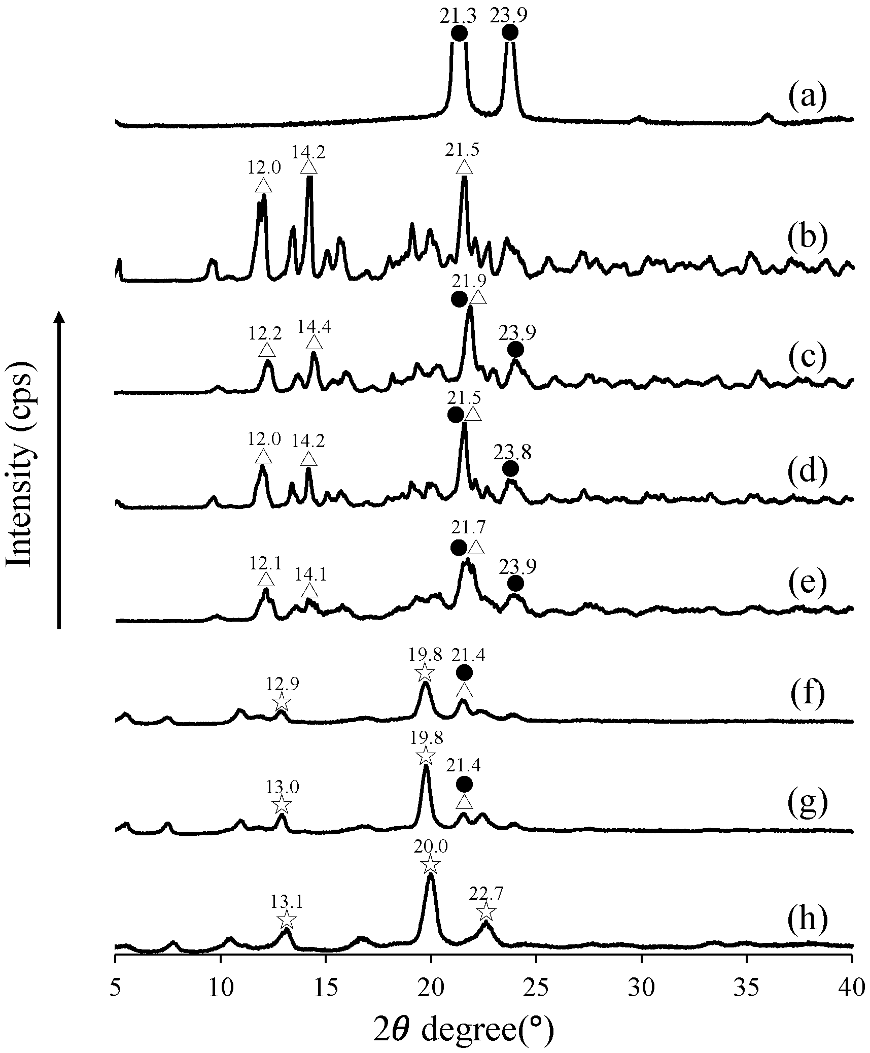
PXRD analysis was conducted (Figure 2) to investigate the crystalline structure changes in the Ko/αCD complexes. As shown in Figure 2a,b, Ko exhibits characteristic diffraction peaks at 2θ values of 21.3° and 23.9°, while αCD shows distinct peaks at 2θ values of 12.0°, 14.2°, and 21.5°. Peaks originating from both Ko and αCD can be observed in the PXRD patterns of the PMs at Ko/αCD ratios of 1/2, 1/4, and 1/8 (Figure 2c–e). In contrast, GMs prepared by 3D ball milling at Ko/αCD ratios of 1/2 and 1/4 exhibit new diffraction peaks at approximate 2θ values of 12.9° and 13.0°, along with residual peaks near 21.4°, corresponding to Ko and αCD (Figure 2f,g). Notably, in the PXRD pattern of the GM sample with a Ko/αCD ratio of 1/8, the characteristic peaks of both Ko and αCD are absent, while new diffraction peaks can be observed at approximate 2θ values of 13.1°, 20.0°, and 22.7° (indicated by ☆) (Figure 2h).
Harada et al. previously reported that the PXRD patterns of PEG/αCD mixtures forming pseudopolyrotaxanes exhibit a characteristic diffraction peak at 2θ = 20°, which is attributed to the channel-type packing of αCD [24].This suggests that the Ko/αCD complexes prepared by 3D ball milling in the present study formed inclusion complexes with a pseudopolyrotaxane structure.

3.2. Evaluation of Thermal Behavior Using DSC

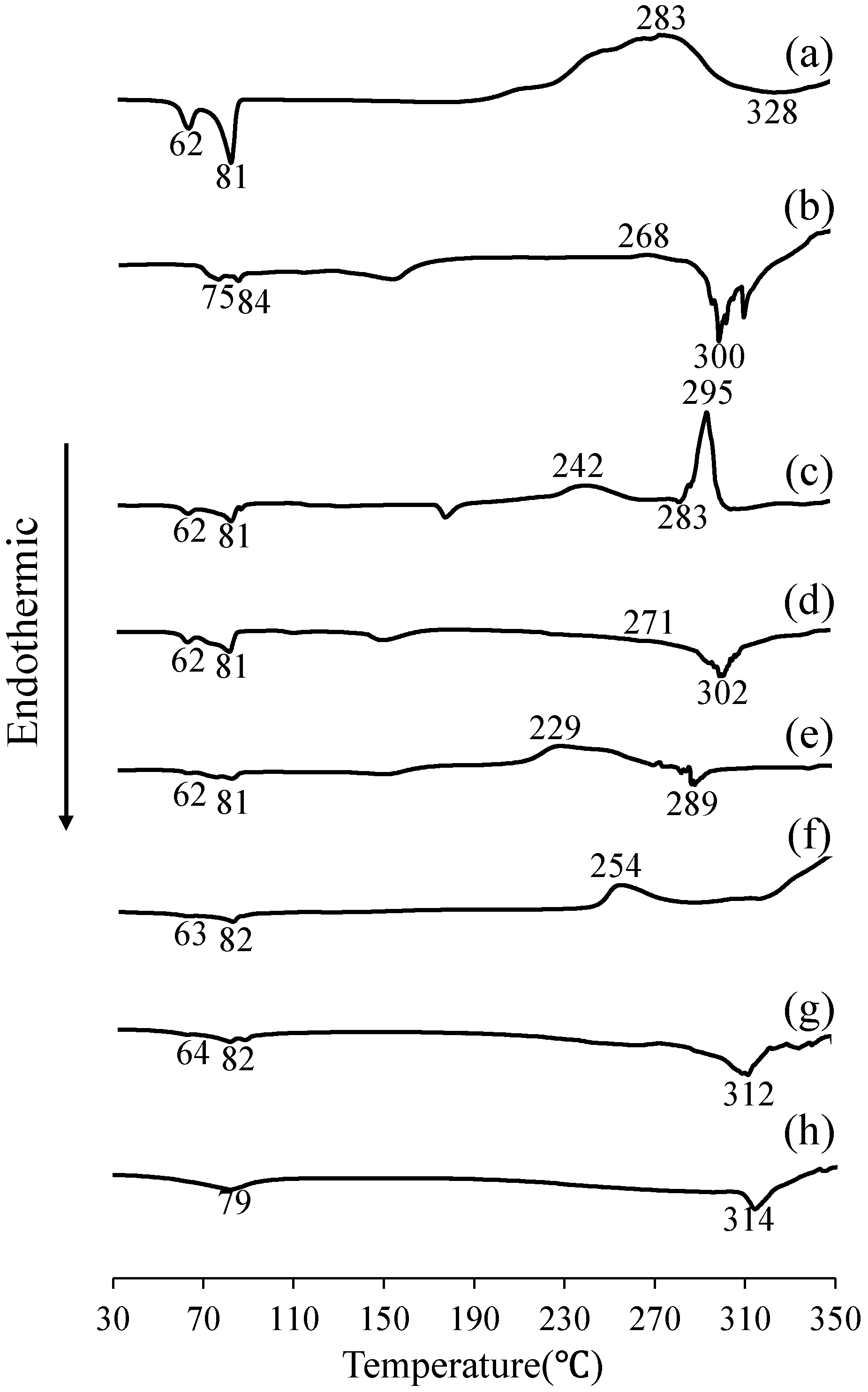
Considering the formation of the Ko/αCD complexes, as indicated by the PXRD patterns, DSC measurements were conducted to assess their thermal behavior. As shown in Figure 3a, Ko exhibits endothermic peaks at approximately 62 and 81 °C, corresponding to its melting points, along with a decomposition peak at approximately 328 °C. αCD shows an endothermic peak near 100 °C, which can be attributed to the dehydration of adsorbed water, and a decomposition peak at approximately 300 °C (Figure 3b). The PMs of Ko/αCD at ratios of 1/2, 1/4, and 1/8 exhibit endothermic peaks characteristic of both Ko and αCD, along with decomposition peaks at approximately 300 °C (Figure 3c–e). Similarly, the GMs of Ko/αCD at ratios of 1:2 and 1:4 exhibit Ko-specific endothermic peaks at 62 and 81 °C (Figure 3f,g). Notably, the GM sample with a Ko/αCD ratio of 1/8 lacks these Ko-specific endothermic peaks and instead shows a decomposition peak near 314 °C (Figure 3f–h).
The disappearance or significant reduction of endothermic peaks upon the formation of inclusion complexes has been widely reported [25]. This phenomenon can be attributed to the encapsulation of drug molecules within the CD cavity, which restricts molecular mobility, leading to the loss of crystalline structure and amorphization of the drug [26]. Specifically, in pseudopolyrotaxane structures formed with polymeric axle chains such as PEG, drug molecules are stably retained within the CD cavities [27]. In this study, an inclusion complex between Ko and αCD is thought to form at a weight ratio of 1/8.

3.3. Evaluation of Intermolecular Interactions Using NIR Spectroscopy

The PXRD and DSC results indicate the formation of inclusion complexes between Ko and αCD at a weight ratio of 1/8. Therefore, NIR spectroscopy, which is highly sensitive to changes in molecular vibrations such as those involving C–H, O–H, and N–H bonds, as well as to alterations in hydrogen bonding, was employed to further investigate molecular interactions in the Ko/αCD systems. In particular, NIR spectroscopy is an effective method for indirectly evaluating intermolecular interactions involved in the inclusion of alkyl chain-containing compounds such as Ko into the hydrophobic cavity of αCD [28].
NIR measurements were conducted to assess intermolecular interactions in the Ko/αCD mixtures. The spectrum of intact Ko shows peaks corresponding to C–H stretching vibrations at 8264 and 5780 cm−1 (Figure 4a,c). For αCD, an O–H stretching band can be observed at 6849 cm−1, and a broad band attributed to hydrated water is detected at approximately 5208 cm−1 (Figure 4b,d). The GM sample with Ko/αCD = 1/2 exhibits C–H peaks of Ko, which are broadened compared to those of intact Ko (Figure 4a,c). The O–H peak of αCD is shifted to 6872 cm−1 with additional broadening, and the water-associated peak is broadened and shifted to 5167 cm−1 (Figure 4b,d). In the GM sample with Ko/αCD = 1/4, the C–H peak at 8264 cm−1 shifted to 8333 cm−1 with broadening (Figure 4a), and the 5780 cm−1 peak also broadened (Figure 4c). The αCD O–H band shifted from 6849 cm−1 to 6872 cm−1, while the hydrated water peak shifted from 5181 cm−1 to 5167 cm−1, both exhibiting significant broadening (Figure 4b,d). In the GM sample with Ko/αCD = 1/8, the 8264 cm−1 C–H peak shifted to 8333 cm−1 and broadened (Figure 4a), while the 5780 cm−1 peak shifted to 5747 cm−1 and also exhibited broadening (Figure 4c). The O–H peak of αCD is shifted to 6920 cm−1, and the hydrated water band is shifted to 5167 cm−1, both exhibiting further broadening (Figure 4b,d).
The observed shifts and broadening of the O–H and water-associated peaks indicate a reduction in free water and corresponding changes in the hydrogen bonding networks within αCD. In addition, the shifts in the C–H bands of Ko, along with those of the O–H vibrations of αCD, suggest enhanced intermolecular interactions. Collectively, these spectral changes confirm the formation of Ko/αCD inclusion complexes mediated by hydrogen bonding. However, we acknowledge that hydrogen bonding between aliphatic C–H groups of komecosanol and hydroxyl groups of αCD is unlikely due to the inherently weak nature of such interactions. Although slight peak shifts were observed in the NIR spectra of the ground samples, these changes are more plausibly attributed to altered molecular environments arising from inclusion complexation, rather than specific hydrogen bonding. Accordingly, the observed spectral changes are better explained by van der Waals forces and hydrophobic interactions, which are well known to drive the formation of cyclodextrin inclusion complexes.

3.4. Evaluation of the Relative Positional Relationship Using 13C CP/MAS NMR Spectroscopy

Solid-state 13C NMR spectroscopy can be used to examine changes in the chemical environment resulting from inclusion complexation. In particular, analyzing chemical shift variations of both the host molecule (CD) and guest molecule provides insight into the inclusion behavior. Thus, solid-state NMR is a highly effective technique for assessing inclusion modes at the molecular level [29,30].
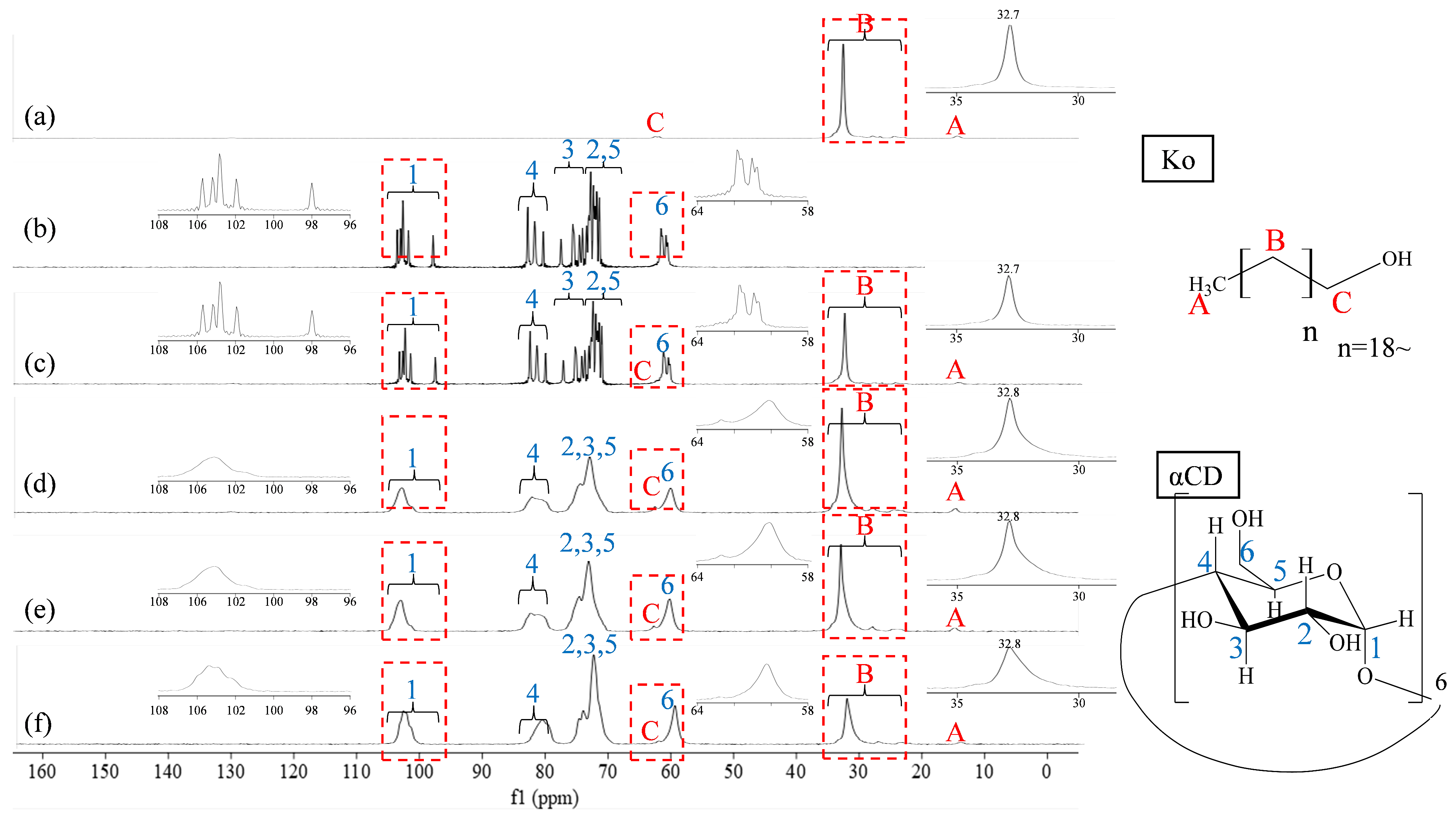
13C CP/MAS NMR measurements were performed to investigate the detailed intermolecular interactions of the GMs (Ko/αCD) in the solid state (Figure 5). As shown in Figure 5a, the spectrum of Ko shows peaks at 14.65 ppm (C-A), 24.69–32.78 ppm (C-B), and 62.11–62.72 ppm (C-C). In the spectrum of αCD, characteristic peaks can be observed at 97.98–103.69 ppm (C-1), 80.47–82.94 ppm (C-4), 74.22–77.64 ppm (C-3), 71.54–73.58 ppm (C-2,5), and 60.67–61.70 ppm (C-6) (Figure 5b). The PM sample (Ko/αCD = 1/8) exhibits peaks corresponding to both Ko and αCD, without significant shifts or broadening (Figure 5c). In contrast, the GM samples (Ko/αCD = 1/2, 1/4, and 1/8) show peak broadening, along with the disappearance of the αCD C-1 peak at 97.98 ppm. Notably, in the GM sample with a Ko/αCD ratio of 1/8, the C-4 peak of αCD is shifted, while the C-B peak of Ko exhibits considerable broadening, indicating a molecular behavior distinct from that observed at other GM ratios (Figure 5e–f).
Harada et al. previously observed that the characteristic αCD peaks at ~98 ppm (C-4) and ~80 ppm (C-1) disappeared in PEG/αCD pseudopolyrotaxane systems, with all αCD carbons exhibiting a single peak in the 13C CP/MAS spectrum [31]. These findings suggest that the GM (Ko/αCD) system in the present study also forms a pseudopolyrotaxane structure. Furthermore, the GM sample with a Ko/αCD ratio of 1/8 exhibits distinct molecular characteristics compared to those of other mixing ratios, further supporting the formation of a structure analogous to the pseudorotaxane configuration described by Harada et al. It is worth noting that the formation of a pseudopolyrotaxane architecture may not solely depend on the molar ratio of Ko to αCD. Given that Ko consists of a mixture of long-chain saturated fatty alcohols (C24–C32), the ability of these chains to thread into the hydrophobic cavity of αCD is likely influenced by their length, flexibility, and hydrophobic surface area. Such structural characteristics may enhance host–guest interactions and promote the formation of stable supramolecular assemblies, even under conditions where the stoichiometric ratio is not strictly equimolar.
Given the limited solubility and heterogeneous composition of Ko, solution-state NMR measurements were not pursued. Instead, solid-state NMR techniques were prioritized as a more appropriate and reliable method for evaluating the supramolecular structure of the Ko/αCD complexes under our experimental conditions.

3.5. T1-NMR Measurement of Ko/αCD Complexes

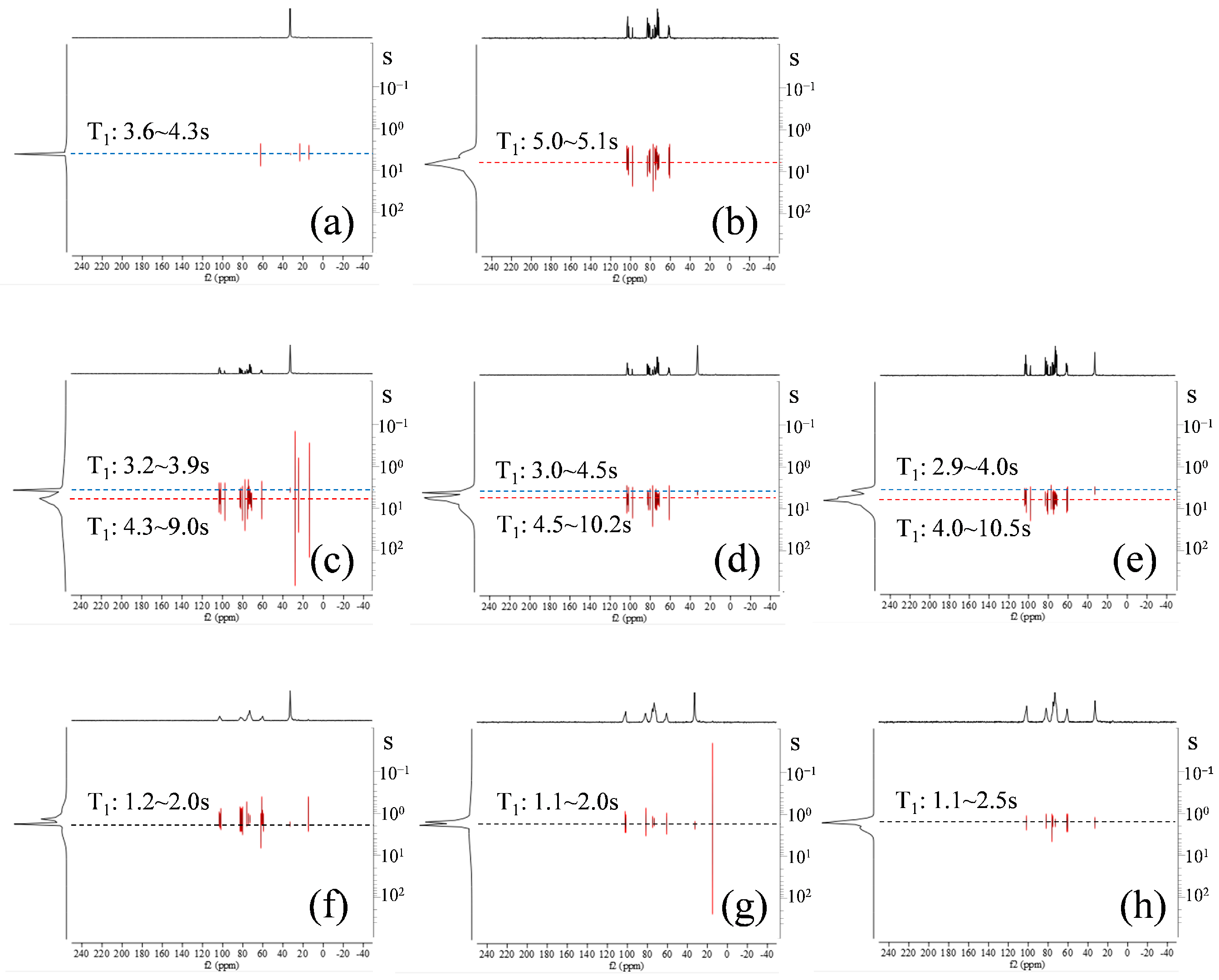
The solid-state NMR results indicate the formation of a pseudorotaxane structure in the Ko/αCD system. T1-NMR measurements were conducted to further evaluate the molecular mobility within this complex. T1-NMR is an analytical technique that leverages differences in the 1H spin–lattice relaxation times (T1) of individual components in a mixture to resolve their corresponding 13C CP/MAS NMR spectra. Unlike that in solution-state NMR spectroscopy, solid-state samples typically exhibit a single T1 value per compound, enabling spectral separation when components show sufficiently distinct relaxation behaviors. This method enables direct analysis of mixtures in the solid state, preserving structural information that would otherwise be lost upon dissolution, thus making it highly effective for examining solid-state systems. The intact Ko sample exhibited T1 values between 3.6 and 4.3 s (Figure 6a), while the αCD sample showed T1 values ranging from 3.2 to 9.5 s (Figure 6b). In the PM sample with Ko/αCD = 1:2, Ko-derived T1 values ranged from 3.2 to 3.9 s, while αCD-derived T1 values ranged from 4.3 to 9.0 s (Figure 6c). In contrast, the GM sample with Ko/αCD = 1/2 exhibited significantly shorter T1 values, ranging from 1.2 to 2.0 s (Figure 6d). Samples with other Ko/αCD ratios showed similar trends: in the PM sample with Ko/αCD = 1/4, Ko-derived T1 values ranged from 3.0 to 4.5 s and αCD-derived values ranged from 4.5 to 10.2 s (Figure 6e), while the GM sample with Ko/αCD = 1:4 exhibited T1 values ranging from 1.1 to 2.0 s (Figure 6f). In the PM sample with Ko/αCD = 1/8, Ko-derived T1 values ranged from 2.9 to 4.0 s and αCD-derived values ranged from 4.0 to 10.5 s (Figure 6g), while the GM sample with Ko/αCD = 1/8 exhibited T1 values between 1.1 and 2.5 s (Figure 6h). Relaxation times in solid-state NMR spectroscopy are influenced by dipolar interactions between protons within a magnetic field. When molecular mobility is high, these interactions weaken owing to dynamic averaging, resulting in longer relaxation times. Conversely, shorter T1 values indicate more restricted molecular motion [32]. Therefore, the markedly shorter and distinct T1 values observed in all GM (Ko/αCD) samples, compared to those of the intact and PM samples, strongly suggest the formation of a pseudorotaxane-type inclusion complex in which molecular motion is constrained by host–guest interactions.

3.6. Evaluation of Morphological Properties via SEM Analysis

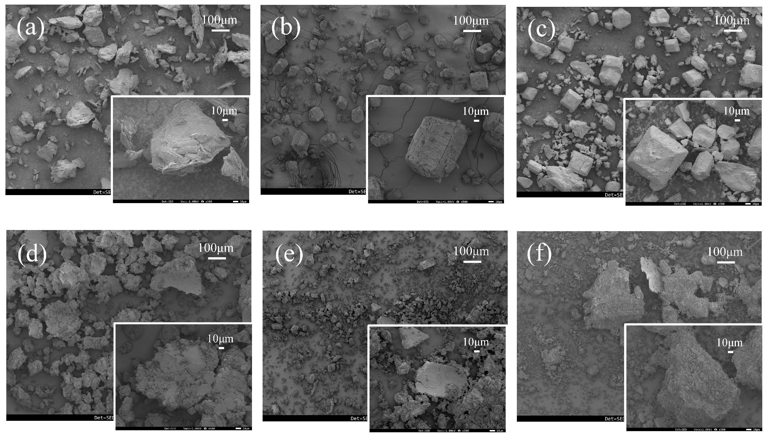
As demonstrated in the previous section, the GM sample with a Ko/αCD ratio of 1/8 formed a pseudorotaxane-type inclusion complex. To further investigate the morphological characteristics associated with this complexation, the particle size and surface structure of the Ko/αCD samples were examined using SEM. As shown in Figure 7a, the αCD particles exhibit a smooth surface and an elongated shape with a diameter of approximately 60 μm. Intact Ko shows irregular, rough particle surfaces with a thick, fragmented morphology (Figure 7a–d). In the GM samples, Ko particles appear smoother and larger compared to intact Ko (Figure 7a–d). In the PM sample (Ko/αCD = 1/8), Ko and αCD particles remain separate, indicating a lack of interaction at the morphological level (Figure 7a–e). In contrast, the GM samples (Ko/αCD = 1/2, 1/4, and 1/8) exhibit thin, flake-like particles with smooth surfaces (Figure 7b–f). Notably, no distinct Ko particles can be observed in the GM sample with Ko/αCD = 1/8, indicating that these morphological changes resulted from the formation of an inclusion complex. These findings suggest that αCD molecules reorient to encapsulate Ko molecules, resulting in structural transformation and the formation of a pseudorotaxane architecture. This structural change alters both the crystalline behavior and the intermolecular interactions of the system. The propensity of α-cyclodextrin to include long-chain aliphatic compounds, such as fatty alcohols, is well-documented in the literature [15,33,34]. This inclusion behavior is attributed to the favorable size and hydrophobicity of the αCD cavity, which allows it to selectively accommodate linear hydrocarbon chains. These precedents support the structural rationale for using αCD as a host for Ko in the present study.
Although the formation of pseudorotaxane structures with αCD in solution systems has been extensively studied [35,36,37], to the best of our knowledge, reports on the formation of such structures through simple co-grinding with lipophilic compounds remain rare. Tuntipopipat et al. investigated the formation of an inclusion complex between policosanol (a mixture of long-chain aliphatic alcohols) and βCD by mixing the two components in solution [38]. They reported that the hydrophobic alkyl chains of policosanol were encapsulated within the hydrophobic cavity of βCD, resulting in improved dispersibility in water and enhanced stability. However, policosanol was first dissolved in ethanol before complex formation in the liquid phase. In contrast, the inclusion complex in the present study was formed in the solid state rather than in solution. Inclusion complexes were also prepared via co-grinding using βCD and γCD in addition to αCD. Notably, the new diffraction peaks observed in the XRD pattern of the GM (Ko/αCD) complex were not detected in the samples prepared with βCD or γCD. This suggests that the formation of a unique crystalline phase is specific to the Ko/αCD system under the applied grinding conditions. Pseudopolyrotaxanes incorporating rice-derived policosanol may offer potential advantages for future development of topical delivery systems, owing to their expected biocompatibility and molecular mobility. Although further experimental studies are needed, such systems could be considered for sustained release formulations or skincare applications such as creams or lip balms. However, additional investigations into skin absorption and functional efficacy are required to validate these possibilities. Although the solubility evaluation of Ko/αCD ground mixtures was initially attempted using UV spectroscopy and liquid chromatography, these methods failed to yield consistent results due to the extremely poor aqueous solubility and chemical heterogeneity of Ko. Preliminary 1H NMR and 2D NOESY measurements also proved inconclusive, owing to poor spectral resolution and the lack of solubility in D2O. Given these analytical limitations, we are currently pursuing the development of a GC-based quantification method. Further investigation into anti-inflammatory and skin barrier effects as biochemical markers is also expected to strengthen the advantages of this material as a sustainable resource. Therefore, this study provides novel insights by demonstrating that Ko, a lipophilic carbon-rich material, can form a pseudorotaxane structure with αCD through mechanochemical grinding. This finding represents a significant contribution to the development of functional material design and solid-state formulation strategies.

4. Conclusions

PXRD and DSC analyses suggested the formation of Ko/αCD inclusion complexes at weight ratios of 1:2, 1:4, and 1:8. NIR spectroscopy revealed intermolecular interactions between the C–H side-chain moieties of Ko and the O–H groups of αCD, confirming that complexation was achieved through mechanochemical grinding using a 3D ball mill. Solid-state 13C CP/MAS NMR spectra of the GM samples indicated the formation of a pseudopolyrotaxane architecture. Collectively, these results confirm that 3D ball milling facilitates the formation of supramolecular Ko/αCD complexes, providing a foundation for the sustainable utilization of Ko. Moreover, the novel finding that a long-chain (C10–C30) aliphatic alcohol such as Ko can form pseudorotaxane inclusion structures opens new avenues for enhancing lipid metabolism, improving exercise performance, and increasing stress tolerance, thereby contributing to increasing healthy lifespan.

Supplementary Materials

The following supporting information can be downloaded at: https://www.mdpi.com/article/10.3390/physchem5030034/s1, Figure S1: PXRD patterns of Ko/αCD systems. (a) Ko intact, (b) αCD, (c) PM (Ko/αCD=1/2), (d) PM (Ko/αCD=1/2) without water ● Ko, △ αCD.

Author Contributions

Conceptualization, M.U., A.O., J.T., Y.I. (Yoshiyuki Ishida), D.N., K.T. and Y.I. (Yutaka Inoue); methodology, M.U., A.O., J.T. and Y.I. (Yutaka Inoue); software, M.U., A.O., J.T., Y.I. (Yoshiyuki Ishida), D.N., K.T. and Y.I. (Yutaka Inoue); validation, M.U., A.O., J.T. and Y.I. (Yutaka Inoue); formal analysis, M.U., A.O., J.T. and Y.I. (Yutaka Inoue); investigation, M.U., A.O., J.T., Y.I. (Yoshiyuki Ishida), D.N., K.T. and Y.I. (Yutaka Inoue); resources, M.U., A.O., J.T., Y.I. (Yoshiyuki Ishida), D.N., K.T. and Y.I. (Yutaka Inoue); data curation, M.U., A.O., J.T., Y.I. (Yoshiyuki Ishida), D.N., K.T. and Y.I. (Yutaka Inoue); writing—original draft preparation, M.U., A.O., J.T. and Y.I. (Yutaka Inoue); writing—review and editing, M.U., A.O., J.T., Y.I. (Yoshiyuki Ishida), D.N., K.T. and Y.I. (Yutaka Inoue); visualization, M.U., A.O., J.T., Y.I. (Yoshiyuki Ishida), D.N., K.T. and Y.I. (Yutaka Inoue); supervision, M.U., A.O., J.T. and Y.I. (Yutaka Inoue); project administration, M.U., A.O., J.T., Y.I. (Yoshiyuki Ishida), D.N., K.T. and Y.I. (Yutaka Inoue); funding acquisition, Y.I. (Yoshiyuki Ishida), D.N., K.T. and Y.I. (Yutaka Inoue). All authors have read and agreed to the published version of the manuscript.

Funding

This research received no external funding.

Institutional Review Board Statement

Not applicable.

Informed Consent Statement

Not applicable.

Data Availability Statement

The data presented in this study are available on request from the corresponding author.

Acknowledgments

The authors sincerely thank CycloChemBio for providing the CDs used in this study and appreciate the valuable advice provided by CycloChemBio researchers during the course of these experiments.

Conflicts of Interest

Author Yoshiyuki Ishida, Daisuke Nakata and Keiji Terao are employed by the company CycloChem Bio Co., Ltd. The authors declare that the present study received γ-cyclodextrin from CycloChem Co., Ltd.; however, the company was not involved in the study design, collection, analysis, interpretation of data, in the writing of this article, or the decision to submit it for publication. This research was supported by Josai University. The authors declare no competing financial interest.

Abbreviations

The following abbreviations are used in this manuscript:
KoKomecosanol
PMPhysical mixture
3DGM3D ground mixture
PXRDPowder X-ray diffraction
DSCDifferential scanning calorimetry
NIRNear-infrared
NMRNuclear magnetic resonance
SEMScanning electron microscopy
DDSDrug delivery system
CDCyclodextrin
PEGPolyethylene glycol
CP/MASCross-polarization magic angle spinning

References

  1. Tufail, T.; Ain, H.B.U.; Chen, J.; Virk, M.S.; Ahmed, Z.; Ashraf, J.; Shahid, N.U.A.; Xu, B. Contemporary views of the extraction, health benefits, and industrial integration of rice bran oil: A prominent ingredient for holistic human health. Foods 2024, 13, 1305. [Google Scholar] [CrossRef] [PubMed] [PubMed Central]
  2. Tanaka, T.; Hoshina, M.; Tanabe, S.; Sakai, K.; Ohtsubo, S.; Taniguchi, M. Production of D-lactic acid from defatted rice bran by simultaneous saccharification and fermentation. Bioresour. Technol. 2006, 97, 211–217. [Google Scholar] [CrossRef] [PubMed]
  3. Lee, S.-H.; Scott, S.D.; Pekas, E.J.; Lee, J.-G.; Park, S.-Y. Improvement of lipids and reduction of oxidative stress with octacosanol after Taekwondo training. Int. J. Sports Physiol. Perform. 2019, 14, 1297–1303. [Google Scholar] [CrossRef]
  4. Kaushik, M.K.; Aritake, K.; Takeuchi, A.; Yanagisawa, M.; Urade, Y. Octacosanol restores stress-affected sleep in mice by alleviating stress. Sci. Rep. 2017, 7, 8892. [Google Scholar] [CrossRef] [PubMed]
  5. Crespo, N.; Illnait, J.; Mas, R.; Fernandez, L.; Fernandez, J.; Castano, G. Comparative study of the efficacy and tolerability of policosanol and lovastatin in patients with hypercholesterolemia and noninsulin dependent diabetes mellitus. Int. J. Clin. Pharmacol. Res. 1999, 19, 117–127. [Google Scholar] [PubMed]
  6. Ohkubo, T.; Ito, T.; Hibino, H. Feasibility study on the use of policosanol to reduce blood acetaldehyde levels in ethanol-administered humans. Alcohol Clin. Exp. Res. 2009, 33, 1407–1412. [Google Scholar] [CrossRef]
  7. Cho, K.H.; Kim, J.E.; Komatsu, T.; Uehara, Y. Protection of liver functions and improvement of kidney functions by twelve weeks consumption of Cuban policosanol (Raydel®) with a decrease of glycated hemoglobin and blood pressure from a randomized, placebo-controlled, and double-blinded study with healthy and middle-aged Japanese participants. Life 2023, 13, 1319. [Google Scholar] [CrossRef]
  8. Saokham, P.; Muankaew, C.; Jansook, P.; Loftsson, T. Cyclodextrins: A versatile tool for drug delivery systems. Molecules 2018, 23, 1161. [Google Scholar] [CrossRef]
  9. Stevens, D.A. Itraconazole in cyclodextrin solution. Pharmacotherapy 1999, 19, 603–611. [Google Scholar] [CrossRef] [PubMed]
  10. Ogata, Y.; Inoue, Y.; Ikeda, N.; Murata, I.; Kanamoto, I. Improvement of stability due to a cyclamen aldehyde/β-cyclodextrin inclusion complex. J. Mol. Struct. 2020, 1215, 128161. [Google Scholar] [CrossRef]
  11. Tachikawa, R.; Saito, H.; Moteki, H.; Kimura, M.; Kitagishi, H.; Arce, F., Jr.; See, G.L.; Tanikawa, T. Preparation, characterization, and in vitro evaluation of inclusion complexes formed between S-allylcysteine and cyclodextrins. ACS Omega 2022, 7, 1064–1073. [Google Scholar] [CrossRef]
  12. Ikeda, N.; Inoue, Y.; Ogata, Y.; Murata, I.; Meiyan, X.; Takayama, J.; Sakamoto, T.; Okazaki, M.; Kanamoto, I. Improvement of the solubility and evaluation of the physical properties of an inclusion complex formed by a new ferulic acid derivative and γ-cyclodextrin. ACS Omega 2020, 5, 12073–12080. [Google Scholar] [CrossRef]
  13. Ezawa, T.; Inoue, Y.; Murata, I.; Takao, K.; Sugita, Y.; Kanamoto, I. Characterization of the dissolution behavior of piperine/cyclodextrins inclusion complexes. AAPS PharmSciTech 2018, 19, 923–933. [Google Scholar] [CrossRef] [PubMed]
  14. Inoue, Y.; Osada, M.; Murata, I.; Kobata, K.; Kanamoto, I. Evaluation of solubility characteristics of a hybrid complex of components of soy. ACS Omega 2019, 4, 8632–8640. [Google Scholar] [CrossRef] [PubMed]
  15. Harada, A.; Kamachi, M. Complex formation between poly(ethylene glycol) and α-cyclodextrin. Macromolecules 1990, 23, 2821–2823. [Google Scholar] [CrossRef]
  16. Harada, A.; Takashima, Y.; Yamaguchi, H. Cyclodextrin-based supramolecular polymers. Chem. Soc. Rev. 2009, 38, 875–882. [Google Scholar] [CrossRef]
  17. Okumura, Y.; Ito, K. The polyrotaxane gel: A topological gel by figure-of-eight cross-links. Adv. Mater. 2001, 13, 485–487. [Google Scholar] [CrossRef]
  18. Molero, G.; Liu, C.; Zhu, Z.; Chen, Q.; Peterson, S.R.; Kolluru, P.V.; Sue, H.J.; Uenuma, S.; Mayumi, K.; Ito, K. Fracture behavior of polyrotaxane-toughened poly(methyl methacrylate). Langmuir 2022, 38, 2335–2345. [Google Scholar] [CrossRef]
  19. Wang, X.-S.; Kim, H.-K.; Fujita, Y.; Sudo, A.; Nishida, H.; Endo, T. Relaxation and Reinforcing Effects of Polyrotaxane in an Epoxy Resin Matrix. Macromolecules 2006, 39, 1046–1052. [Google Scholar] [CrossRef]
  20. Das, S.; Subuddhi, U. Controlled Delivery of Ibuprofen from Poly(vinyl alcohol)-Poly(ethylene glycol) Interpenetrating Polymeric Network Hydrogels. J. Pharm. Anal. 2019, 9, 108–116. [Google Scholar] [CrossRef]
  21. Ooya, T.; Yui, N. Multivalent interactions between biotin–polyrotaxane conjugates and streptavidin as a model of new targeting for transporters. J. Control Release 2002, 80, 219–228. [Google Scholar] [CrossRef]
  22. Loftsson, T.; Duchêne, D. Cyclodextrins and their pharmaceutical applications. Int. J. Pharm. 2007, 329, 1–11. [Google Scholar] [CrossRef]
  23. Mura, P. Analytical techniques for characterization of cyclodextrin complexes in the solid state: A review. J. Pharm. Biomed. Anal. 2015, 113, 226–238. [Google Scholar] [CrossRef]
  24. Harada, A.; Okada, M.; Kawaguchi, Y. Formation of Self-assembled Tubular Structures by Mixing Cyclodextrin and Polymers without Solvents. Chem. Lett. 2005, 34, 542–543. [Google Scholar] [CrossRef]
  25. Nisar, T.; Wang, Z.C.; Yang, X.; Tian, Y.; Iqbal, M.; Guo, Y. Characterization of citrus pectin films integrated with clove bud essential oil: Physical, thermal, barrier, antioxidant and antibacterial properties. Int. J. Biol. Macromol. 2018, 106, 670–680. [Google Scholar] [CrossRef] [PubMed]
  26. Loftsson, T.; Brewster, M.E. Pharmaceutical applications of cyclodextrins. 1. Drug solubilization and stabilization. J. Pharm. Sci. 1996, 85, 1017–1025. [Google Scholar] [CrossRef] [PubMed]
  27. Harada, A.; Li, J.; Kamachi, M. The molecular necklace: A rotaxane containing many threaded α-cyclodextrins. Nature 1992, 356, 325–327. [Google Scholar] [CrossRef]
  28. Liu, F.; He, Y.; Wang, L. Discrimination of β-cyclodextrin inclusion complexes using near-infrared spectroscopy and chemometrics. Anal. Chim. Acta 2011, 689, 75–81. [Google Scholar] [CrossRef]
  29. Chierotti, M.R.; Gobetto, R. Solid-State NMR Studies of Weak Interactions in Supramolecular Systems. Chem. Commun. 2008, 1621–1634. [Google Scholar] [CrossRef]
  30. Fadler, R.E.; Flood, A.H. Rigidity and Flexibility in Rotaxanes and Their Relatives; On Being Stubborn and Easy-Going. Front. Chem. 2022, 10, 856173. [Google Scholar] [CrossRef]
  31. Harada, A.; Li, J.; Kamachi, M. Preparation and properties of inclusion complexes of polyethylene glycol with alpha-cyclodextrin. Macromolecules 1993, 26, 5698–5703. [Google Scholar] [CrossRef]
  32. Nishida, M.; Tanaka, T.; Hayakawa, Y.; Nishida, M. Solid-State Nuclear Magnetic Resonance (NMR) and Nuclear Magnetic Relaxation Time Analyses of Molecular Mobility and Compatibility of Plasticized Polyhydroxyalkanoates (PHA) Copolymers. Polymers 2018, 10, 506. [Google Scholar] [CrossRef] [PubMed]
  33. Rekharsky, M.V.; Inoue, Y. Complexation Thermodynamics of Cyclodextrins. Chem. Rev. 1998, 98, 1875–1918. [Google Scholar] [CrossRef] [PubMed]
  34. Uekama, K. Recent Aspects of Pharmaceutical Application of Cyclodextrins. J. Incl. Phenom. Macrocycl. Chem. 2002, 44, 3–7. [Google Scholar] [CrossRef]
  35. Li, X.; Xiao, J.; Wang, X.; Yi, C.; Su, S. pH-Responsive Pseudorotaxane between Comblike PEO-Grafted Triblock Polymer and α-Cyclodextrin. Colloid Polym. Sci. 2014, 292, 3243–3249. [Google Scholar] [CrossRef]
  36. Lyon, A.P.; Banton, N.J.; Macartney, D.H. Kinetics of the Self-Assembly of α-Cyclodextrin [2]Pseudorotaxanes with Polymethylene Threads Bearing Quaternary Ammonium and Phosphonium End Groups. Can. J. Chem. 1998, 76, 666–674. [Google Scholar] [CrossRef]
  37. Castelletto, V.; Kowalczyk, R.M.; Seitsonen, J.; Hamley, I.W. Tuning the solution self-assembly of a peptide-PEG (polyethylene glycol) conjugate with α-cyclodextrin. ChemBioChem 2023, 24, e202200743. [Google Scholar] [CrossRef]
  38. Tuntipopipat, S.; Tajarati, S.; Nesterenko, P.N.; Paull, B. Inclusion Complexes of Policosanol with β-Cyclodextrin and Its Derivatives. J. Pharm. Sci. 2006, 95, 1825–1834. [Google Scholar] [CrossRef]
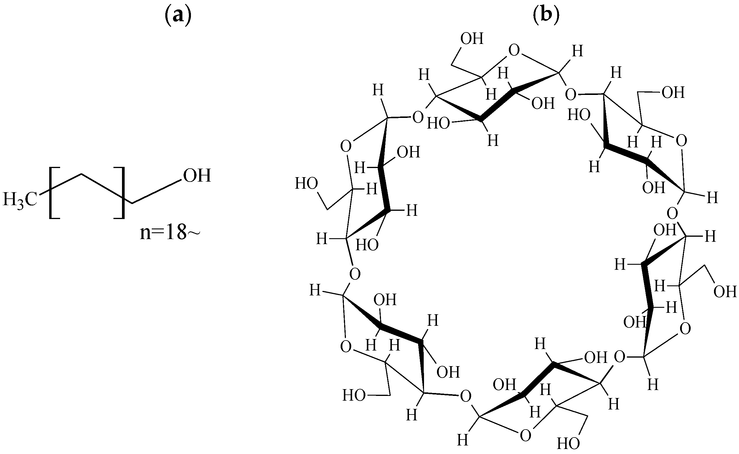
Figure 1. Chemical structures of Ko, αCD. (a) Ko, (b) αCD.
Physchem 05 00034 g001
Figure 2. PXRD patterns of Ko/αCD systems. (a) Ko intact, (b) αCD, (c) PM (Ko/αCD = 1/2), (d) PM (Ko/αCD = 1/4), (e) PM (Ko/αCD = 1/8), (f) GM (Ko/αCD = 1/2), (g) GM (Ko/αCD = 1/4), (h) GM (Ko/αCD = 1/8). ● Ko, Δ αCD, ☆ New Peak.
Physchem 05 00034 g002
Figure 3. DSC curves of Ko/αCD systems. (a) Ko, (b) αCD, (c) PM (Ko/αCD = 1/2), (d) PM (Ko/αCD = 1/4), (e) PM (Ko/αCD = 1/8), (f) GM (Ko/αCD = 1/2), (g) GM (Ko/αCD = 1/4), (h) GM (Ko/αCD = 1/8).
Physchem 05 00034 g003
Figure 4. 2nd differentiation NIR absorption spectra of Ko/αCD systems. (a) 8000–8700 cm−1, (b) 7200–6200 cm−1, (c) 5500–6200 cm−1, (d) 4900–5500 cm−1.
Physchem 05 00034 g004
Figure 5. 13C-CP/MAS NMR spectra of Ko/αCD systems. (a) Ko intact, (b) αCD, (c) PM (Ko/αCD = 1/8), (d) GM (Ko/αCD = 1/2), (e) GM (Ko/αCD = 1/4), (f) GM (Ko/αCD = 1/8).
Physchem 05 00034 g005
Figure 6. T1-NMR measurement of Ko/αCD systems. (a) Ko intact, (b) αCD, (c) PM (Ko/αCD = 1/2), (d) PM (Ko/αCD = 1/4), (e) PM (Ko/αCD = 1/8), (f) GM (Ko/αCD = 1/2), (g) GM (Ko/αCD = 1/4), (h) GM (Ko/αCD = 1/8).
Physchem 05 00034 g006
Figure 7. SEM morphology of Ko/αCD. (a) Ko intact, (b) αCD, (c) PM (Ko/αCD = 1/8), (d) GM (Ko/αCD = 1/2), (e) GM (Ko/αCD = 1/4), (f) GM (Ko/αCD = 1/8).
Physchem 05 00034 g007
Disclaimer/Publisher’s Note: The statements, opinions and data contained in all publications are solely those of the individual author(s) and contributor(s) and not of MDPI and/or the editor(s). MDPI and/or the editor(s) disclaim responsibility for any injury to people or property resulting from any ideas, methods, instructions or products referred to in the content.