Article

Increasing the Probability of Obtaining Intergrown Mixtures of Nanostructured Manganese Oxide Phases Under Solvothermal Conditions by Mixing Additives with Weak and Strong Chelating Natures

by
María Lizbeth Barrios-Reyna
1,2,
Enrique Sánchez-Mora
1 and
Enrique Quiroga-González
1,*
1
Institute of Physics, Benemérita Universidad Autónoma de Puebla, Puebla 72570, Mexico
2
Department of Mechanics and Advanced Materials, School of Engineering and Sciences, Instituto Tecnológico y de Estudios Superiores de Monterrey, Puebla 72453, Mexico
*
Author to whom correspondence should be addressed.
Physchem 2025, 5(3), 35; https://doi.org/10.3390/physchem5030035 (registering DOI)
Submission received: 31 May 2025 / Revised: 10 August 2025 / Accepted: 11 August 2025 / Published: 16 August 2025
(This article belongs to the Section Solid-State Chemistry and Physics)

Abstract

Intergrown mixtures of nanostructured manganese oxide phases have been obtained using a highly complexing agent (ethylenediamine) and a weak complexer (urea) during their solvothermal synthesis. In this work, through a detailed structural analysis, it is evidenced the formation of an intergrown mixture of three distinct manganese oxide phases (β-MnO2, α-Mn2O3, and Mn3O4). Scanning electron microscopy shows that the products have just one morphology, indicating that the different manganese oxide phases may have grown together, organizing themselves in a 3D crystal network. The reaction mechanisms are discussed in this paper. It is of great interest to produce intergrown mixtures of manganese oxide phases to take advantage of the availability of the different oxidation states of Mn in neighboring crystallites for applications like catalysis.

Graphical Abstract

1. Introduction

Manganese oxides (MnO, MnO2, Mn2O3, and Mn3O4) can be attractive materials due to their wide range of applications, such as catalysis [1,2,3], alkaline fuel cells [4], sensors [5], batteries [6], capacitors [7], and other electrochemical applications [8], among others. Manganese oxides have been obtained using different synthesis methods such as electrospinning [9], thermolysis of organometallic precursors [10], microwave-assisted solvothermal [11], electrochemical deposition [12,13], and conventional solvothermal synthesis [14]. In particular, the solvothermal route is well-known for preparing metal oxide nanocatalysts with good crystallinity at relatively low temperatures and costs [15]. Via this technique, the composition, morphology, size, and crystal structure of the synthesized particles may be controlled by the synthesis conditions, such as pressure, reaction time, and temperature [16,17]. Another important parameter is the pH of the mother solution [18,19].
In recent years, different procedures for preparing nano- or microparticles of manganese oxides by the solvothermal method have been reported, making the solvent one of the most important parameters to consider. Among the characteristics that are considered to select the solvent, one finds the melting point, boiling point, acid or basic nature, dielectric constant, and chelating capability.
The chemical nature of solvents plays a key role in the solvothermal process, limiting the solubility and reactivity of the precursors, in addition to directing the structure and morphology of the products [20]. Y.C. Zhang et al. [21] used ethylenediamine (EDA) or ethylene glycol (EG) as a solvent, obtaining different results with one or the other. When the Mn3O4 product was obtained in EDA, well-defined nanocrystallites were formed with a size of 15–35 nm. In comparison, the one obtained in EG consisted of aggregated nanoparticles with a size less than 18 nm. In a different work using urea solutions as a solvent, Mn2O3 nanoparticles with a cubic structure and spherical morphology with a size in the range of 6–8 nm were obtained by K.K. Hazarika et al. [22].
On the other hand, G. Qiu et al. reported a relatively complex procedure with the solvents tetramethylammonium bromide (TAB), tetrathylammonium bromide (TEAB), tetrabutylammonium bromide (TBAB), and tetraoctylammonium bromide (TOAB) to obtain manganese oxide octahedral molecular sieves [23]. Hence, it is clear that the chemical nature of the reaction media is an important factor, allowing for the control of the resulting structural, morphological, and chemical properties of the materials. However, it is not common to mix solvents, especially when they have chelating properties. There are just a few works exploiting that possibility. For example, 2-mercapto-quinazolin-4-one (HL1) and 2-mercapto-3-phenyl-quinazolin-4-one (HL2) have been used as a primary ligand and 1,10-phenanthroline (phen) as a secondary ligand, modifying the electrochemical properties of the products [24].
In the present work, two substances with different chelating natures were mixed and used as a solvent. The first one, ethylenediamine (EDA), is a strong bidentate chelating ligand with the two nitrogen atoms donating their unique pairs of electrons, which is a chemical structure that allows it to act as both a nucleophile and a ligand, enabling it to participate in complexation with manganese ions and in various substitution reactions. The second one, urea, is a weak chelating agent that has been widely used due to its ability to bind metal ions with its nitrogen and oxygen atoms.
On the other hand, intergrown materials alter the specific physicochemical properties and applications of the materials, which are correlated with the chemical nature of these intergrowths [25,26,27,28]. Alessandro Porta et al. compared the catalytic behavior of an intimate mixture (Ru-BaO/Al2O3) with a mechanical mixture (BaO/Al2O3 + Ru/Al2O3). It was found that the intimate mixture showed stronger catalytic activity than the mechanical mixture, indicating the strong synergistic effect between the different phases intimately grown [29]. However, the structures of these intergrown materials are particularly difficult to synthesize, understand, and characterize in detail [30,31]. In this work, solvents with different chelating capacities were mixed to obtain intergrown mixtures of manganese oxide phases. It is of great interest to produce such intergrown mixtures, which present different active sites within a short distance, thus allowing the achievement of oxidation and reduction processes at neighboring sites for applications such as catalysis [32], while their surface area is high (which is one of the most important parameters for catalysis) [33].

2. Materials and Methods

2.1. MnxOy Materials Synthesis

All the precursors used in the experiments were of reagent grade without any further purification. Manganese oxides were prepared by solvothermal synthesis using the following precursors: urea (NH2CONH2, 99%, Sigma-Aldrich, Burlington, MA, USA), ethylenediamine (C2H8N2, 99% Sigma-Aldrich), deionized water (18.3 Ω·cm), manganese (II) chloride tetrahydrate (MnCl2·4H2O, 99%, Sigma-Aldrich), and ethanol (CH3CH2OH, 99%, J.T. Baker).
The synthesis of manganese oxides using urea (a weakly chelating agent) and ethylenediamine (EDA, a highly complexing agent) was used as a reference. This first sample was called MnxOy(urea). Then, a second sample was synthesized using ethylenediamine (EDA, a strongly chelating agent); this sample was called MnxOy(EDA). On the other hand, a modified synthesis approach was conceived by adding urea and ethylenediamine (a mix of the weakly and strongly chelating agents). This third sample was named MnxOy(urea+EDA). The mother solution to synthesize MnxOy(urea) was prepared in the following way: a 9 M aqueous solution of urea and a 0.044 M aqueous solution of MnCl2·4H2O were prepared separately, stirring for 10 min. Afterward, both solutions were mixed and continuously stirred for 30 min at room temperature. The material MnxOy(EDA) was synthesized by mixing a 10.2 M EDA solution and 0.044 M aqueous solution of MnCl2·4H2O. This mother solution was continuously stirred for 30 min at room temperature. The mother solution for the synthesis of MnxOy(urea+EDA) was prepared as follows: First, a 9.0 M urea solution was obtained by mixing urea and deionized water. Then, an equal volume of ethylenediamine was added and stirred for 10 min. In the second stage, a 0.044 M aqueous solution of MnCl2·4H2O was added and stirred for 30 min. For the different samples, 8 mL of the mother solutions was transferred to a 15 mL Teflon-lined stainless-steel autoclave and then heated at 120 °C for 24 h. The autoclaves were left to cool down to room temperature afterward. The products were centrifuged and washed with ethanol and deionized water to eliminate the precursor and solvent residues and dried at room temperature. A portion of each product was thermally treated under residual air in a muffle at 450 °C for 6 h (samples called “with annealing”). This temperature was selected considering that MnCO3 (the main product besides manganese oxides after solvothermal synthesis, as will become clear in the “Results and Discussion” section) completely decomposes above 450 °C, forming manganese oxides.

2.2. Characterization

The morphology of the samples was analyzed by scanning electron microscopy using a Philips XL30E SEM microscope (FEI, Hillsboro, OR, USA). Their crystal structures were identified by means of X-ray diffraction on a RigakuD/max-2100 diffractometer (Rigaku Corporation, Tokyo, Japan) (Cu K_(α_1) radiation, 1.5406 Å). Raman spectroscopy was performed with a Dilor Labram microscope (HORIBA Scientific, Kyoto, Japan), with a He-Ne (632 nm) laser and using a 50× objective. Fourier transform infrared (FT-IR) spectroscopy in the attenuated total reflectance (ATR) mode was performed with a Bruker Vertex 70 spectrometer (Bruker Corporation, Billerica, MA, USA).

3. Results and Discussion

Figure 1 shows SEM micrographs of the MnxOy(urea) (Figure 1a,b), MnxOy(EDA) (Figure 1c,d), and MnxOy(urea+EDA) (Figure 1e,f), without and with annealing treatment, respectively. The materials were morphologically different. It can be seen that there was a significant change in crystal size and morphology when a highly complexing agent (ethylenediamine) and a weak complexer (urea) were used as the reaction medium for the solvothermal synthesis. It will also become interesting to notice that, despite each product having mainly one morphology, the grains were composed of mixtures of different nanocrystalline manganese oxide phases (confirmed by XRD and Raman studies shown below). One could infer that, to exhibit just one morphology, the manganese oxide phases should be intimately grown. This could have important effects on the properties of the mixtures and could impact their possible applications [34].
The SEM micrograph of MnxOy(urea) without annealing treatment exhibited particles with cuboid shape with an average particle size of 11 µm ± 4 µm (based on the measurement of 36 particles). Image J software (version 1.54p) was used for particle analysis. The particle count performed in each case varied, and the length and diameter of each particle were determined according to the morphology observed in the SEM micrographs. Figure 2 shows the particle size distribution histogram of the different products before and after annealing.
This result is interesting considering the composition of the sample, with none of the samples exhibiting a cubic crystal structure, as will become clear in the next paragraphs. This suggests that the particles are agglomerates of smaller particles, probably adhered to by weak interactions promoted by urea, a weak complexer, and surfactant. The SEM micrograph of the MnxOy(urea) sample with annealing treatment at 450 °C/6 h shows that the cuboids decreased in size to 5.1 µm ± 0.55 µm (based on the measurement of 50 particles), as seen in Figure 1b and Figure 2b. The cuboid aggregates may densify due to annealing, which generates a mass loss of organic compounds. This may imply changes in the effective surface area of the manganese oxide particles, affecting their catalytic activity [35]. The material synthesized in ethylenediamine without annealing treatment (Figure 1c) is formed by aggregates of elongated particles with irregular forms, with an average length of 12 µm ± 4 µm (based on the measurement of 25 particles), as seen Figure 2c. The particle size decreases with the annealing treatment at 450 °C/6 h, as seen in Figure 1d. The latter sample is formed by irregular-shaped particle aggregates with rugose morphology, with an average particle size of 6.7 µm ± 2.2 µm (based on the measurement of 25 particles), as shown in Figure 2d.
The material synthesized in ethylenediamine and urea without annealing treatment (Figure 1e) is formed by faceted elongated particles with an average length of 131 nm ± 60 nm (Figure 2e) and an average width of 28 ± 7 nm (based on the measurement of 300 particles). The particle size decreased with the annealing treatment at 450 °C/6 h, as seen in Figure 1f. The latter sample is formed by nearly spherical-shaped aggregates with an average particle size of 24 nm ± 7 nm (Figure 1f).
Comparing the sample prepared at 450 °C for 6 h with the material without thermal treatment (Figure 1e,f), it can be inferred that the annealing treatment promoted the fracture of the faceted elongated particles into nearly spherical-shaped nanoparticles. This effect could be advantageous for using the product as an electrocatalyst since it is expected that the smaller particles possess a larger effective surface area, which enhances the catalytic activity [36].
The smaller size of the MnxOy(urea+EDA) particles compared with the ones of MnxOy(urea) and MnxOy(EDA), respectively, is due to the presence of a mix of weakly and strongly chelating agents in the synthesis, like EDA. This solvent is a bidentate ligand that can successfully control the size and shape of the products. Its strongly complexing capacity allows for the formation of stable N-chelated manganese ions [37]. The ions are released slowly for the formation of manganese oxides. Furthermore, the [Mn(en)3)]2+ lives for long enough that its nucleus could act as a nucleation center for the formation of spherical-like particles of manganese oxide nanoparticles.
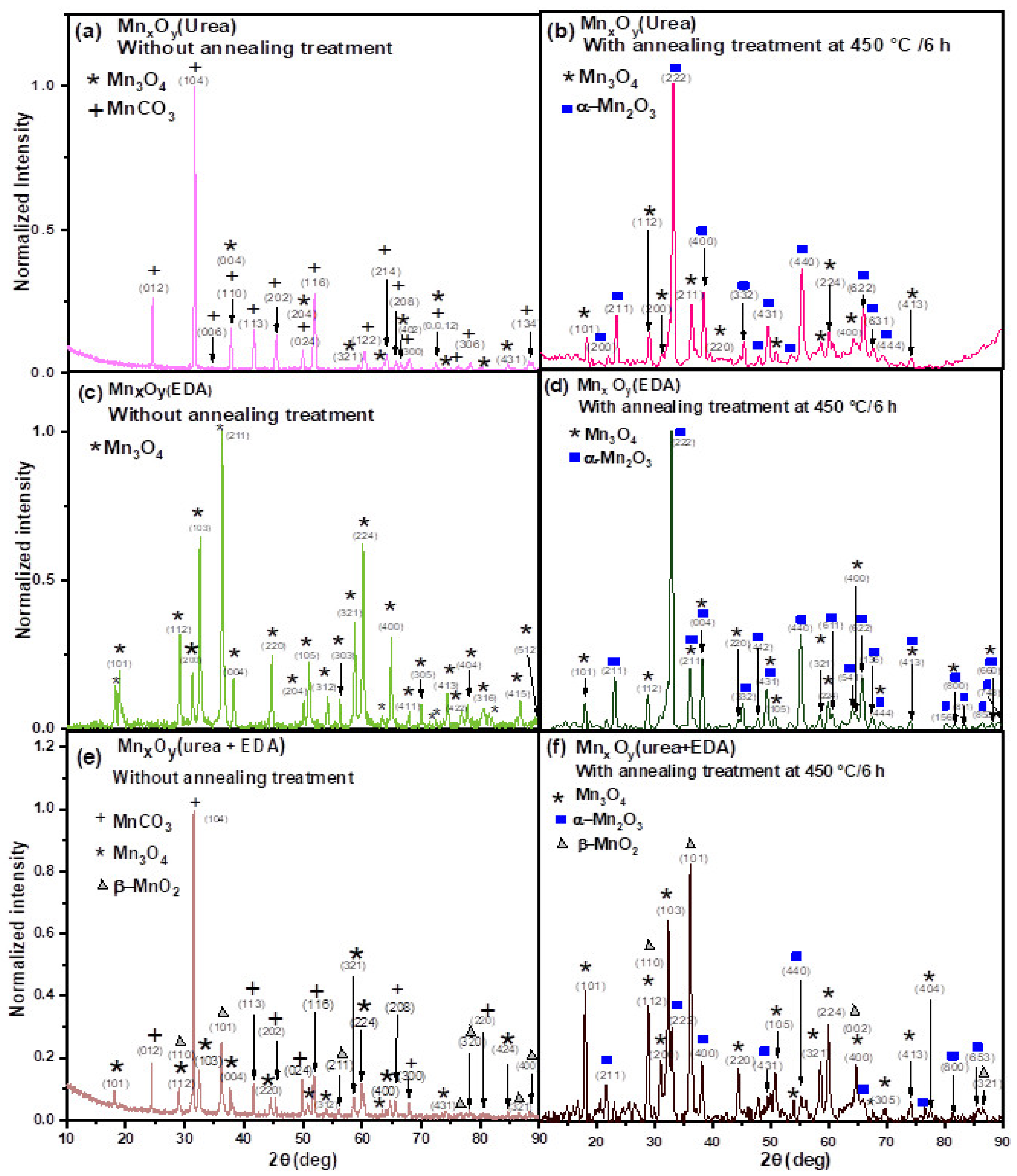
Figure 3 shows the X-ray diffraction patterns of MnxOy materials obtained under different solvothermal conditions: (a) with urea (sample A), (b) using EDA (sample B), and (c) mixing additives with weakly and strongly chelating nature (urea+EDA; sample C), respectively. As can be observed, the manganese oxide phases are different in all the cases. It is also important to notice that the diffraction patterns of all materials indicate the formation of phases with good crystallinity and no phases related to the reaction media or any impurities.
Using urea as the reaction medium and without annealing treatment, MnCO3 with a hexagonal structure (JADE PDF No. 44-1472) was successfully synthesized, as seen in the X-ray diffractogram of Figure 3a. For this to happen, urea must have decomposed during the hydrothermal synthesis, as seen in Equations (12)–(14), where the species are gradually released and then form manganese carbonate (Equations (9)–(11)) [38]. In the diffractogram, one can find additional low-intensity peaks corresponding to Hausmannite Mn3O4 (JADE PDF No. 24-0734). However, the ratio of the intensity of the strongest peak of this phase to the strongest peak of MnCO3 is 0.045:1, indicating that the product is predominantly a single phase (MnCO3). It is important to point out that this result is only an approximation; aspects like instrumental parameters, sample preparation, preferred orientation, and crystallite size can also affect peak intensity [38,39]. The literature suggests that the reference intensity ratio (RIR) method is one of the most effective tools for calculating mass ratios of crystalline phases using X-ray diffraction (XRD) data [40,41,42]. However, the RIR method has some limitations when the diffractogram quality is low and the sample is amorphous or contains unknown phases [43,44,45], even when it has been used by some authors [46]. That is why in such cases a phase composition analysis based on the ratio of the intensities of the XRD peaks (as conducted in this work) is an excellent methodology, accepted as a first approximation for estimating the amount of each phase [47,48,49,50].
The product of the reaction with ethylenediamine without annealing treatment shows only the presence of Mn3O4, as can be seen in Figure 3c. This confirms that the presence of a strongly chelating agent (EDA) produces one phase of manganese oxide.
The product of the reaction with ethylenediamine and urea without annealing treatment contains not only Mn3O4 and MnCO3, but also β-MnO2 (JADE PDF no. 24-0735), as shown in Figure 3e. The ratio of the most intense peaks of the Mn3O4, MnCO3, and β-MnO2 phases is 0.12:0.99:0.25, indicating a mixture of phases (not in the impurity level). This confirms that both chelating agents (urea and EDA) produce mixtures of manganese oxide and carbonate phases.
On the other hand, the diffraction pattern of MnxOy(urea) with annealing treatment at 450 °C for 6 h shows mainly the α-Mn2O3 phase (JADE PDF No. 41-1442) with sharp peaks as seen in Figure 3b. The presence of Hausmannite Mn3O4 (JADE PDF No. 24-0734) is also observed, but with low-intensity diffraction peaks. The intensity ratio of the strongest peak of these two phases is 0.99:0.23, indicating that the thermal treatment leads to the formation of predominantly α-Mn2O3 (bixbyite) phase.
The diffraction pattern of the MnxOy(EDA) sample annealed at 450 °C for 6 h shows the presence of α-Mn2O3 (JADE PDF no. 41-1442) in addition to Mn3O4 (JADE PDF No. 076-0150). The ratio of the most intense peak of the mentioned phases is 0.98:0.23 (Mn3O4:α-Mn2O3), confirming a mixture of phases. No additional phases were detected, indicating a partial transformation of the Hausmannite Mn3O4 to α-Mn2O3 through thermal treatment.
In contrast, the diffraction pattern of the MnxOy(urea+EDA) sample with annealing treatment at 450 °C for 6 h depicts that, in addition to Mn3O4, there is the presence of α-Mn2O3 (JADE PDF no. 41-1442) and β-MnO2 (JADE PDF no. 24-0735). The ratio of the most intense peak of the phases is 0.82:0.43:0.99 (Mn3O4:α-Mn2O3:β-MnO2), confirming a mixture of phases. No additional phases were detected, indicating a complete transformation of the carbonate to oxides through thermal treatment.
For the determination of the cell parameters of the different crystalline phases, the Rietveld refinement method is a powerful tool [51,52]. However, this method has several limitations when dealing with low-quality diffractograms and when the samples are composed of nanocrystalline phases, as is the case in the present work [53,54]. Other important limitations are noisy data or a poor signal-to-noise ratio, peak overlap, preferred orientation, and similar diffraction patterns of the different phases present, which can lead to imprecise structural parameters and unreliable qualitative phase analysis [55,56,57].
Due to all the exposed limitations, all the calculations of crystallite sizes and cell parameters were performed manually based on Bragg’s law. They should be considered as a rough estimation, which is enough for the purposes of the present work.
Starting from Bragg’s law, the interplanar spacing of all samples was determined. This law can be written as follows [58]:
n λ = 2 d sin θ
where λ is the X-ray wavelength, d is the spacing of the diffracting planes, and θ is the angle between the incident rays and the diffracting planes, known as the Bragg angle. Afterward, the lattice parameter “a” for the manganese oxide with a cubic crystal structure was determined using the following equation [59]:
d h k l =   a h 2 + k 2 + l 2
where dhkl is the lattice spacing within (hkl) planes, and “a” is the lattice parameter of the cubic structure. In the case of a tetragonal structure, the “a” and “c” parameters can be calculated using the (400) and (101) peaks [60]. First, “a” is calculated from the “d” of the (400) peak, as:
d =   a 4
On the other hand, “c” is calculated from the “d” value of the (101) peak [61], as:
d 2 =   a 2 +   c 2
The “a” and “c” parameters of the manganese oxide with a hexagonal structure were calculated using the (300) and (006) peaks [62], in a way similar to Equation (3).
Table 1 shows the crystal parameters of the phases present in MnxOy(urea), MnxOy(EDA), and MnxOy(urea+EDA) with and without thermal treatment, calculated from their XRD patterns.
In particular, the lattice parameter “a” of the produced Mn3O4 decreases in the urea-ethylenediamine system without the annealing, while “c” increases, indicating a possible relaxation of the strains when EDA is provided in the solvothermal process. This result confirms that the unit cell is distorted by an elongation along the c-axis. In contrast, the MnxOy(EDA) and MnxOy(urea+EDA) materials show a slight decrease in their value in all cases.
The unit cell volumes were calculated using the following equations:
For the cubic structure [67]:
V = a 3
where “V” is the volume of the cubic structure and “a” is the cell parameter.
For the tetragonal crystal structure, as follows [68]:
V = a 2 c
where “a” and “c” are the lattice parameters.
For the hexagonal structure, it was calculated as follows [69]:
V = 0.866 a 2 c
The unit cell volumes of the produced compounds are summarized in Table 1. It can be observed that, before annealing, the value calculated for MnCO3 (hexagonal) is significantly higher for MnxOy(urea) (308 Å3) than for MnxOy(urea+EDA) (277 Å3). Meanwhile, the unit cell volume of Mn3O4 is higher in MnxOy(urea+EDA) (328 Å3) than in MnxOy(urea) and MnxOy(EDA), with values of 315 Å3 and 294 Å3, respectively. This result is due to the expansion of the crystal lattice when a stronger complexing agent, such as ethylenediamine, is added in the synthesis process. The dependences of a, c, and c/a indicate a slightly anisotropic character of this expansion, corresponding to the deformation along the c-axis.
The annealing process also has an effect. The unit cell volume of β-MnO2 (only appearing in the sample urea+EDA) shows a decrease when annealing at 450 °C, as shown in Table 1. This behavior may be due to densification, given by defect curing. On the other hand, Mn3O4 suffers from an anisotropic growth in the c-axis in all samples, but this effect is stronger in the urea+EDA sample (see the c/a ratio of Table 1). These results confirm that the presence of both urea and ethylenediamine strongly influences the structural properties of the resulting MnxOy.
On the other hand, the crystallite size was determined by two methods, and the results are shown in Table 2. First, it was estimated using the Scherrer formula:
D = k λ β c o s θ
where k is the Scherrer constant, the value of which denotes the particle shape, and its most common value is 0.9 for quasi-spherical particles, λ is the wavelength of the X-ray beam used (1.5406 Å), β is the full width at half maximum (FWHM) of the most intense peak of the studied crystal phase, and θ is the Bragg angle [70]. The (hkl) of the most intense peak for each sample is also shown in the table. However, it is important to mention that the Scherrer equation should be considered just as an approximation, since it based on the idea that the peak width depends only on the crystallite size, and it does not consider that the peak could suffer some broadening caused, for example, by lattice strain, which could originate from crystal defects, grain boundaries, etc. [71].
If the line width broadening could come primarily from micro-strains (the instrumental broadening is minimal), the Williamson–Hall method (also known as the Uniform Deformation Model—UDM) could be more reliable [72]. Table 2 also shows results with this method.
In this method, the total peak width is expressed as:
β t o t a l = β I n s t r u m e n t + β S a m p l e
β s a m p l e = β C r y s t a l l i t e   s i z e + β L a t t i c e   s t r a i n
βCrystallite size and βLattice strain can be expressed by the following equations:
β C r y s t a l l i t e   s i z e = K λ D cos θ
β l a t t i c e   s t r a i n = 4 ε tan θ = 4 ε sin θ cos θ
where K is the Scherrer constant, the value of which denotes the particle shape, and its most common value is 0.9, λ is the wavelength of the X-ray beam used (1.5406 Å), D is the crystallite size, and ε (adimensional) is the lattice strain [73].
The Williamson–Hall equation could be then expressed as:
β s a m p l e = β C r y s t a l l i t e   s i z e + β L a t t i c e   s t r a i n   = K λ D cos θ + 4 ε sin θ cos θ
Multiplying both sides of the equation by cosθ, it acquires a linear form:
β h k l cos θ = K λ D + 4 ε sin θ
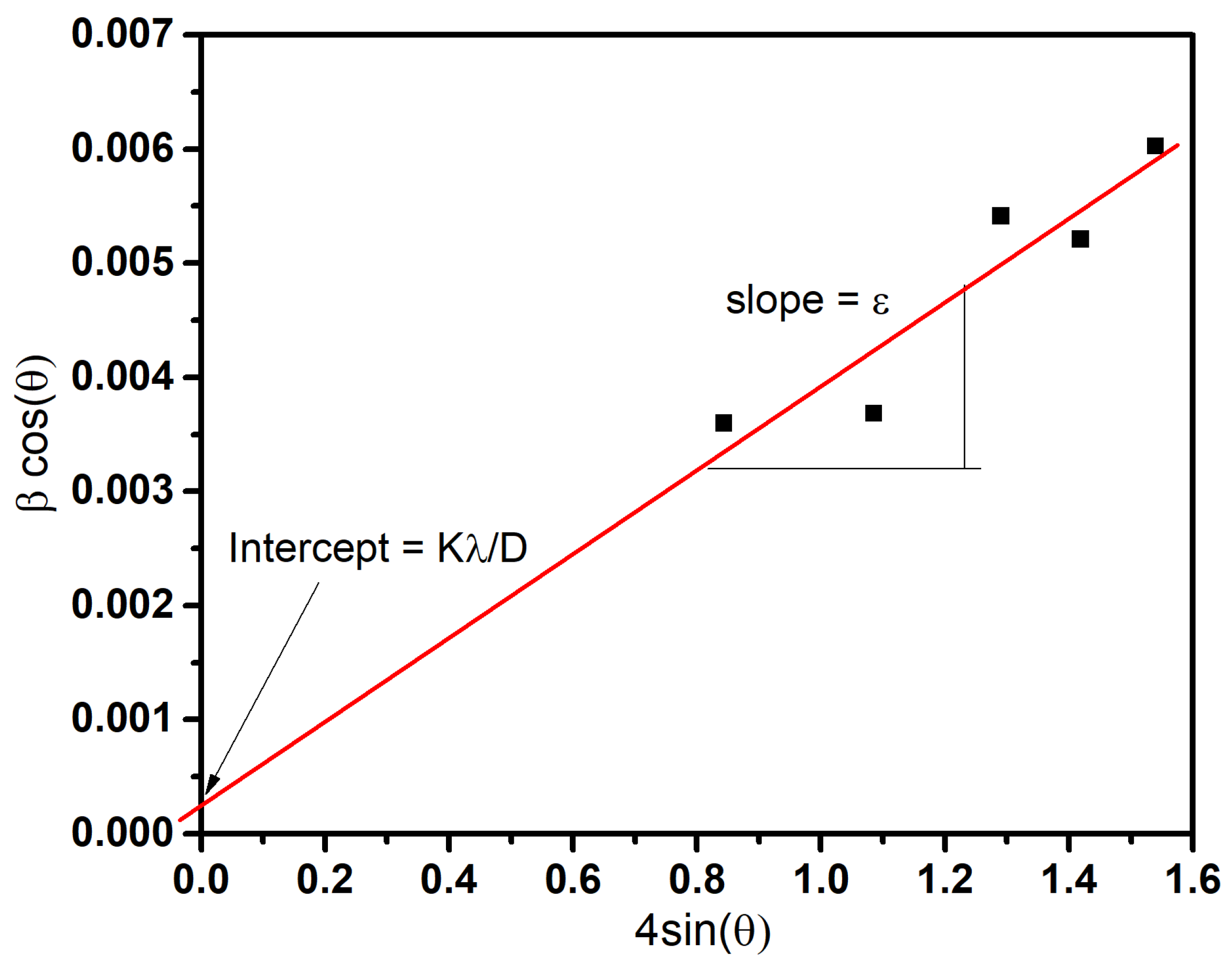
With this equation, it is possible to calculate the crystallite size and lattice strain by plotting β(hkl) cosθ against 4sinθ (where β(hkl) is in radians and θ is in degrees) for all peaks and fitting the best line. From the y-intercept (Kλ/D), it is possible to calculate the crystallite size (D), and the slope is the strain (ε). A negative value for strain means compressive stress in the sample, whereas a positive value for intercept means tensile strain [74]. For a better understanding, as an example, Figure 4 shows the plot used for calculating the crystallite size and strain for the MnxOy(urea) material without annealing treatment. From the y-intercept, it was determined that the average crystallite size is 42.4 nm, and from the slope, that the lattice strain is 0.0037.
Table 2 shows the crystallite size of MnxOy(urea), MnxOy(EDA), and MnxOy(urea+EDA) materials with and without annealing treatment at 450 °C/6 h, calculated from their XRD patterns (Figure 3). As can be observed, similar results are obtained with the two methods used. However, in certain cases, it was not possible to obtain reliable results by the Williamson–Hall method, since it depends on the quality of the data to be able to perform a linear fit. In those cases, the most reliable results are obtained by the Scherrer method, since just one XRD peak is needed for the calculation, where one can reliably extract the peak width (commonly the most intense peak is analyzed). It can be observed that, at low temperatures, the crystallite size of the MnxOy(urea) compounds is slightly higher (Table 2) than that of the MnxOy(urea+EDA) in the predominant hexagonal phase, but at larger temperatures, manganese carbonate transforms into manganese oxide (α-Mn2O3) with a BCC structure, and the crystallite size is significantly lower in both cases.
It is also important to note that the crystallite size values of the manganese compounds do not agree with the particle size values observed in the SEM micrographs of Figure 1. According to XRD, the crystallite sizes are about 1000 times smaller; therefore, the particles observed in the SEM should be aggregates.
Only in the case of MnxOy(urea+EDA), the crystallite sizes observed by SEM coincide with the particle sizes calculated from XRD data. However, it is not possible to distinguish between the particles of the different phases by SEM (there is no contrast by secondary electrons between MnCO3, α-Mn2O3, Mn3O4, and/or β-MnO2, as can be observed in Figure 1).
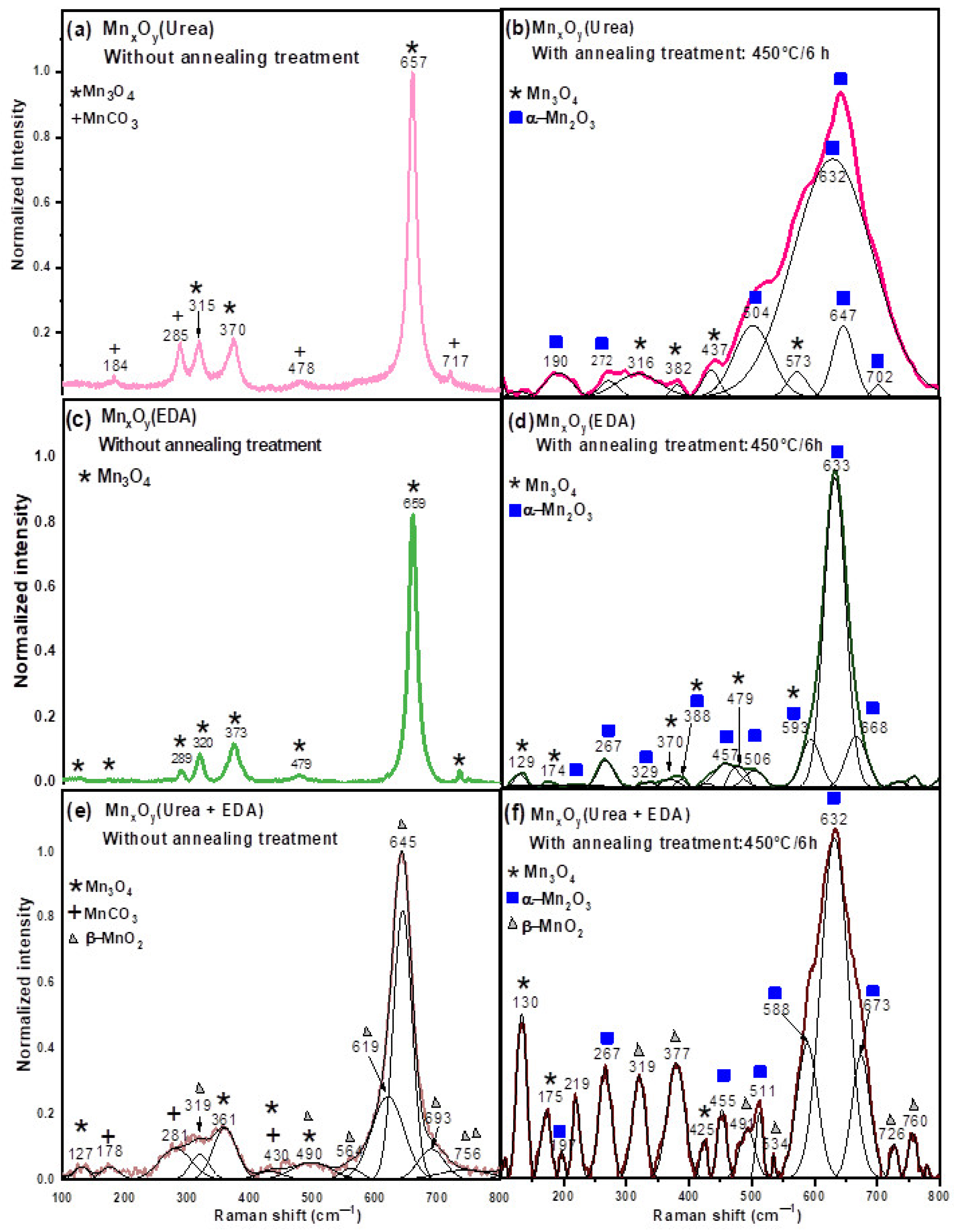
Figure 5 shows the micro-Raman spectra obtained from the MnxOy(urea) (Figure 5a,b), MnxOy(EDA) (Figure 5c,d), and MnxOy(urea+EDA) (Figure 5e,f) materials with and without annealing treatment, respectively. The Raman spectrum of the material synthesized in urea without annealing treatment (Figure 5a) displays a broad and intense band centered at 657 cm−1 and two smaller bands at 315 and 370 cm−1, which can be attributed to Mn3O4 (Hausmannite) stretching vibrations [75]. The bands at 184, 285, 478, and 717 cm−1 can be assigned to rhodochrosite MnCO3 [76]. This Raman result confirms the X-ray diffraction results, which indicate the presence of manganese carbonate (MnCO3). This is possibly due to the urea decomposition in the presence of water, which produces OH, and species (Equations (19)–(21)) [77,78], but is consumed in the formation process of MnCO3 during the hydrothermal reaction (Equations (16)–(18)) [79].
Conversely, the Raman spectrum of the material synthesized in urea with annealing treatment at 450°/6 h (Figure 5b) shows a broad and intense band centered at 632 cm−1 and a shoulder at about 504 cm−1, which are assigned to the vibrational mode of α-Mn2O3 [80]. Five smaller bands are located at 190, 272, 504, 647, and 702 cm−1, which are assigned to the stretching vibrational mode of the Mn-O bond in which the distance is modulated in the Mn2O3 lattice [81]. The bands at 316, 382, 437, and 573 cm−1 can be assigned to Mn3O4 vibrational modes [82]. These Raman results agree with the X-ray diffraction results, showing that the transition from rhodochrosite MnCO3 to bixbyite α-Mn2O3 and Mn3O4 phases occurs at 450 °C.
The Raman spectrum of the material synthesized in ethylenediamine, without annealing treatment (Figure 5c), shows the presence of an intense band at 659 cm−1 and five small bands at 289, 320, 373, and 7479 cm−1, which are attributed to Mn3O4 vibrational modes [83]. No other vibrational modes were observed, confirming the XRD results. Figure 5d shows the Raman spectra of MnxOy(EDA) with annealing treatment at 450 °C/6 h. The presence of an intense band at 633 cm−1 and seven smaller bands at 267, 329, 388, 457, 506, 593, and 688 cm−1 can be observed, which are attributed to the vibrational modes of α-Mn2O3 [84].
On the other hand, the Raman spectrum of the material synthesized in ethylenediamine and urea, without annealing treatment (Figure 5e), consists of different manganese oxides (β-MnO2 and Mn3O4). The presence of an intense band at 645 cm−1 and very small bands at 319, 564, 619, and 756 cm−1 can be observed, which are attributed to β-MnO2 vibrations, corresponding to the Mn–O stretching mode within a rutile-type MnO6 octahedral lattice [85]. The weak bands at 127, 361, and 500 cm−1 correspond to Mn3O4 (Hausmannite) vibrational modes [86,87]. The MnCO3 bands are observed at 178, 281, and 430 cm−1 [88].
The Raman spectrum of the material synthesized in ethylenediamine and urea, with annealing treatment at 450 °C for 6 h in an air atmosphere (Figure 5f), shows the presence of an intense band located at 632 cm−1 and very small bands at 197, 267, 455, 511, 588, and 673 cm−1, which are attributed to α-Mn2O3 vibrations [89]. This behavior is possibly due to the thermal decomposition of the MnCO3 phase (at 450 °C for 6 h in an air atmosphere), leading to the formation of α-Mn2O3 and Mn3O4, as seen in Figure 5f. Furthermore, as the Raman bands are broad, the materials must be nanocrystalline [90]. This observation was confirmed by XRD and SEM, which confirm that the crystallite sizes are in the order of tens of nanometers. The bands at 319, 377, 491, and 726 cm−1 can be assigned to β-MnO2 vibrational modes, which are attributed to the skeletal vibrations of the Mn–O bond in the MnO6 octahedral matrix [91]. The lower-frequency bands correspond to the deformation modes of a metal–oxygen chain of Mn-O-Mn in the MnO6 octahedral lattice [92]. The weak bands at 534 and 760 cm−1 correspond to the Eg and B2g vibrational modes, respectively. The latter involves antisymmetric Mn–O vibrations in the MnO2 octahedral framework [93]. Four smaller bands are located at 130, 175, and 425 cm−1, attributed to vibrational modes of Mn3O4 [94]. These results agree with the X-ray diffraction results, which are consistent with the presence of β-MnO2, α-Mn2O3, and Mn3O4 phases in the material. This mixture, which cannot be identified by SEM, confirms the nanoscale intimacy between each manganese oxide phase obtained. This could have a huge impact on their physico-chemical properties [95].
It can be summarized that, after thermal treatment, the materials synthesized with urea+EDA are mixtures of three manganese oxide phases (β-MnO2, α-Mn2O3, and Mn3O4), whereas materials synthesized solely with urea or EDA consist mainly of manganese oxide (III) and manganese oxide (II, III).
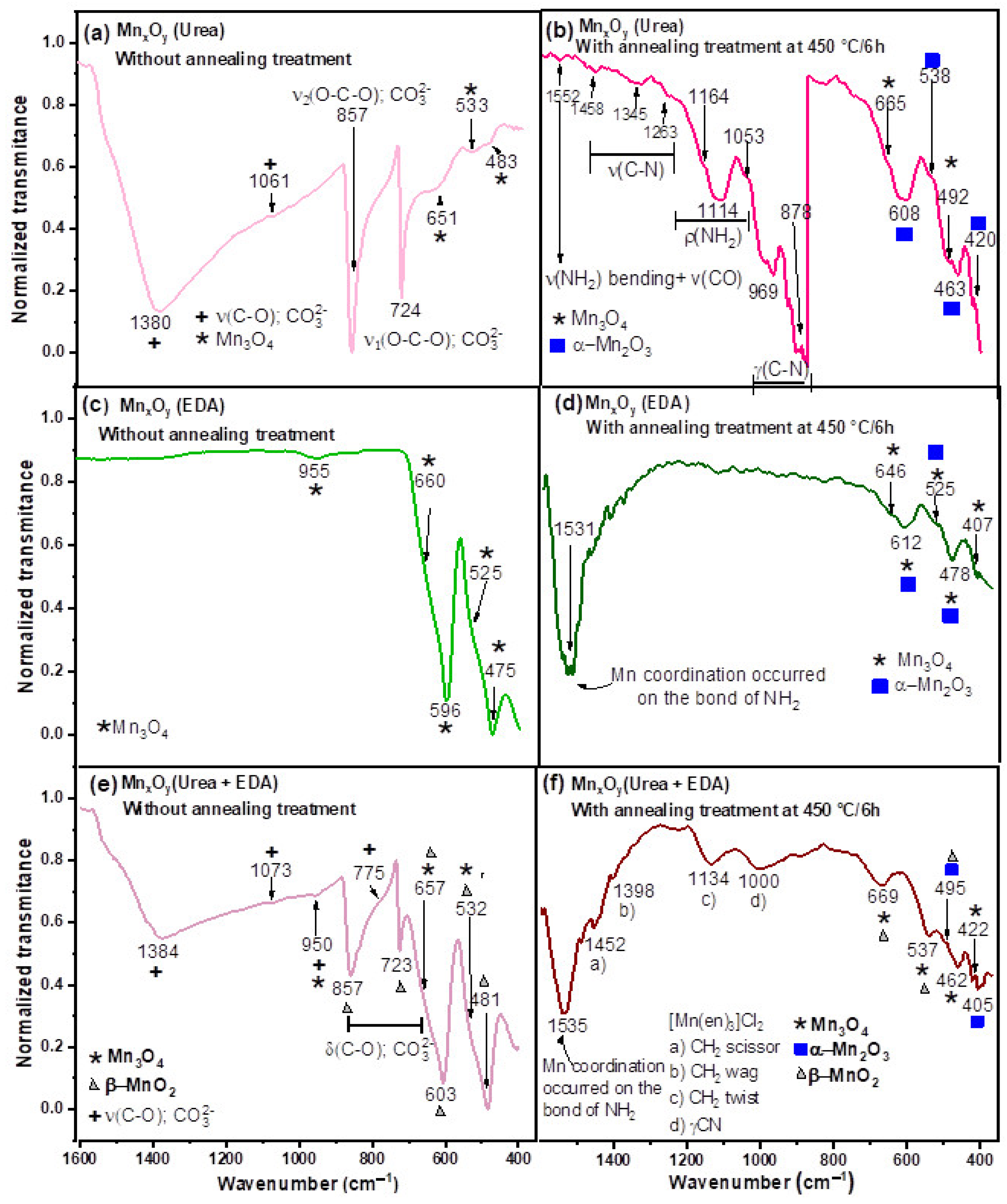
Figure 6 shows the FTIR spectra of MnxOy(urea), MnxOy(EDA), and MnxOy(urea+EDA) materials with and without annealing treatment at 450 °C/6 h, respectively. In the spectrum of MnxOy(urea) without annealing treatment, one can observe the presence of four vibrational modes of C O 3 2 [96]. The two strong and broad bands at 724 and 872 cm−1 correspond to symmetric bending ν1(O-C-O), and asymmetric bending ν2 (O-C-O) of the group. The weak vibrational mode observed at 1061 cm−1 is associated with symmetric stretching, ν3 (C-O). The broad and intense band at 1380 cm−1 corresponds to an asymmetric stretching, ν4 (C-O) [97,98]. It is interesting to note that the same bands are observed in the Raman spectrum, which shows four very weak peaks [99]. This behavior confirms the formation of MnCO3 during solvothermal synthesis, as seen in Equation (18). In Figure 6a, three FTIR bands are observed at 483, 533, and 651 cm−1, corresponding to the vibrational modes of the tetragonal Hausmannite Mn3O4 [100].
Figure 6b shows the FTIR spectrum of MnxOy(urea) with annealing treatment at 450 °C/6 h. The weak absorption peaks at 1552, 1458, 1345, and 1263 cm−1 correspond to the ν(NH2) bending ν(CO) and ν(C-N) vibrational modes, respectively. The bands at 1114 and 969 cm−1 correspond to the bending and stretching frequencies of ρ(NH2) and γ(C-N), respectively [101]. This behavior confirms the coordination of the urea functional groups with the manganese ion, allowing for the formation of a manganese complex, as seen in Equations (16)–(18) [102].
Thermal decomposition of the manganese complex at 450 °C for 6 h leads to the formation of manganese oxide. This can be seen from the following equation [103]:
M a n g a n e s e u r e a   c o m p l e x T h e r m a l   d e c o m p o s i t i o n   α M n 2 O 3 + M n 3 O 4
The absorption of peaks at 492 and 665 cm−1 corresponds to the vibrational modes of Hausmannite Mn3O4 [104], as seen in Figure 6c. Four absorption peaks at 420, 463, 538, and 608 cm−1 correspond to α-Mn2O3 vibrational modes [105]. This FTIR analysis confirms the thermal decomposition of the manganese complex and MnCO3 to allow the formation of both manganese oxide phases, α-Mn2O3 and Mn3O4.
Figure 5c shows the FTIR spectrum of MnxOy(EDA) without annealing treatment. The absorption peaks at 475, 525, 596, 660, and 955 cm−1 correspond to tetragonal Hausmannite Mn3O4 [106,107]. No other vibrational modes were observed, indicating the formation of pure phases. The strong absorption band at 1531 cm−1 corresponds to the Mn coordination that has occurred on the bond of NH2 from the ethylenediamine group [108,109], as seen in Figure 6d. The presence of this absorption band confirms the reaction mechanism shown in Equation (12). Three bands at 478, 525, and 612 cm−1 correspond to the vibrational modes of tetragonal α-Mn2O3 [110]. Absorption peaks at 407, 478, 525, 612, and 646 cm−1 can be associated with the vibrational modes of hausmannite Mn3O4 [111,112]. It is also interesting to note that both reaction media (urea+EDA in the solvothermal synthesis) allow for the formation of β-MnO2, as shown in Figure 5e. The FTIR spectra of β-MnO2 display four strong absorption peaks at 481, 603, 723, and 857 cm-1, which are in good agreement with the well-developed tetragonal-type structure with an interstitial space consisting of narrow one-dimensional (1×1) channels [113]. The strong absorption peak at 603 cm−1 is attributed to the Mn-O-Mn vibrational modes of MnO2 [114]. Two absorption bands at 730 and 857 cm−1 are attributed to the symmetric M-O stretching vibration of the MnO6 octahedral group of the MnO2 [115]. Moreover, the absorption bands at 775, 950, 1073, and 1384 cm−1 are attributed to the MnCO3 vibrational modes [116]. These FTIR results confirm the presence of vibrational modes of coming of the urea decomposition [117,118], as seen in Equations (13) and (14); this decomposition product allows for the formation of manganese carbonate (Equation (11)).
On the other hand, the FTIR spectrum of MnxOy(urea+EDA) with annealing treatment at 450 °C/6 h, shows four weak absorption peaks at 1452, 1398, 1134, and 1000 cm−1, which correspond to the CH2 scissor, CH2 wag, CH2 twist, and γ(C-N) vibrational modes of the [Mn(en)3]Cl2 complex [119]. The strong band at 1535 cm−1 is associated with the Mn coordination occurring on the N-H bond [120,121]. The absorption bands at 405 and 495 cm−1 confirm the thermal transformation of MnCO3 to α-Mn2O3 at 450 °C/6 h, as reported in the literature [122]. Three weak peaks at 495, 537, and 669 cm−1 correspond to the vibrational modes of β-MnO2 [123]. The weak absorption peak at 422 cm−1 can be assigned to the vibrational mode of Mn3+-O from the Mn3O4 bonds [124,125]. The absorption peak at 462 cm−1 is due to the stretching vibrations of Mn4+-O bonds [126], which can be related to cation vacancies in the manganese oxide lattice [127]. The absorption band at 537 cm−1 corresponds to the distortion vibration at octahedral sites and peaks at 669 cm−1 due to the tetrahedral stretching vibration in the Mn3O4 phase [128]. It is also interesting to note that FTIR studies further show the formation of Mn3O4, confirming the partial thermal transformation of MnCO3 to Mn3O4, which agrees with the XRD analysis.
Starting from the structural analysis of the materials synthesized with urea, EDA, and urea + EDA, we confirmed that, although multiple washes with ethanol were performed in both materials, molecules of the reaction products (NH2CONH2 or/and C2H8N2) are observed in the FTIR spectra [128,129,130,131,132], indicating residues of -complex- intermediate products, in all materials with annealing treatment, as seen in Figure 6. These complexes are imperceptible in XRD and Raman spectra due to their amount in the impurity range.
The results can be explained by the following reaction:
(a)
Chemical reactions using urea as a reaction medium
  • An aqueous solution was prepared with manganese (II) chloride and deionized water, which forms manganese ions, according to the reaction [133]:
    MnCl2 + H2OMn2+ + 2Cl
  • Afterward, an aqueous solution of urea was prepared separately, and then both solutions were mixed and transferred to a Teflon container and then to a stainless-steel autoclave. This resulting solution could form manganese carbonate as a decomposition product, according to the chemical reactions [134,135,136]:
    M n 2 + +   4 N H 2 C O N H 2     M n N H 2 C O N H 2 4 2 +
    M n N H 2 C O N H 2 4 2 + + 2 C l + 6 H 2 O 120 ° C / 24   h   M n C O 3 + 6 N H 3 + 3 C O 2 + 4 N H 4   C l + H 2 O
  • During the hydrothermal synthesis, urea also gradually releases species (it hydrolyses) according to the chemical reactions [137,138]:
    N H 2 C O N H 2 +   H 2 O   N H 2 C O O + N H 4 +
    N H 2 C O O + N H 4 +   H 2 O   H C O 3 + N H 3
    H C O 3 H + + C O 3 2
Equation (14) indicates that the solvothermal decomposition of urea produces C O 3 2 . It is simple to deduce that this could cause the formation of manganese carbonate [139], which has been detected by FTIR, Raman, and X-ray diffraction.
The acid/base equilibrium associated with the urea decomposition products produces OH species [140]; the latter provides alkaline conditions in the precursor solution, which forms surface tension and increases the viscosity of the reaction media.
These synthesis conditions provide a lower transport mass and consequently, the Mn2+ ions are co-precipitated into hydroxides, as seen in the following chemical reactions [141,142]:
M n 2 + + 3   O H M n O O H + H 2 O
M n 2 + + 2   O H M n ( O H ) 2
Finally, Mn3O4 is formed by the dehydration of hydroxides [143,144]. Thus, according to the Raman and X-ray diffraction results, we can corroborate the solvothermal decomposition of the materials with the following reaction [145]:
M n ( O H ) 2   +   2   M n O O H   120   ° C / 24   h   M n 3 O 4   +   2   H 2 O
(b)
Chemical reactions using EDA as a reaction medium
Manganese chloride in aqueous solution releases manganese ions, which form a stable complex with ethylenediamine [146]:
M n C l 2 +   3   H 2   N C H 2   C H 2   N H 2     H 2 O   M n ( H 2   N C H 2   C H 2   N H 2   ) 3 2 + + 2 C l
During the hydrothermal synthesis, there exists a chemical decomposition of the manganese–EDA complex, according to the chemical reaction [147]:
M a n g a n e s e E D A   c o m p l e x 120   ° C / 24   h   M n 3 O 4
Finally, annealing treatment allows for the partial thermal decomposition of the Mn3O4 phase (at 450 °C for 6 h in air atmosphere) leading to the formation of bixbyite α-Mn2O3, according to the chemical reaction [148]:
2 M n 3 O 4 +   1 2 O 2 T h e r m a l   d e c o m p o s i t i o n 450   ° C / 6   h   3 α M n 2 O 3
(c)
Chemical reactions using the urea–EDA system as a reaction medium
In the presence of both types of reaction media (urea and urea+EDA), the materials show some differences, i.e., the material synthesized in ethylenediamine and urea consists of different manganese oxides, as seen in the following chemical reaction:
M a n g a n e s e U r e a E D A   c o m p l e x S o l v o t h e r m a l   s y n t h e s i s 120 ° C / 24   h   β M n O 2 + M n C O 3 + M n 3 O 4
In this case, the presence of both types of reaction media leads to the formation of different manganese oxides (β-MnO2 and Mn3O4).
Particularly, EDA is a bidentate ligand with a strongly complexing capacity that allows for the formation of stable N-chelated manganese ions [149], which probably occurred due to the coordination of the amine group from the EDA with the Mn(III) ion according to the following chemical reaction [150,151]:
Physchem 05 00035 i001
The formation of the [Mn(en)3)]2+ complex favors the slowing down of the reaction kinetics and consequently leads to the formation of the thermodynamically stable manganese oxide phases [152]. Furthermore, the intermediate manganese–EDA complexes live long enough so that their nucleus can act as a nucleation center for forming manganese oxide nanoparticles [153]. The latter complex is favorable for reacting with anions (e.g., OH-) or ligands (e.g., NH3) from the urea solution [154]. For this reason, it is possible to obtain MnO2, a stable and low-energy phase, which is difficult to obtain in the absence of O2 [155].
It is also interesting to note the chemical formation of MnCO3 from de urea decomposition, which limits the chemical reaction with the other reactive system (EDA). This behavior is probably due to the chemical competition between reactive systems with different chemical natures (weakly and strongly chelating agents).
The XRD, FT-IR, and Raman results confirm the presence of MnCO3, Mn3O4, β-MnO2, and α-Mn2O3 phases in the material synthesized in urea and EDA.
The results are very interesting since it has been shown that, with the addition of ethylenediamine as a highly complexing agent in the solvothermal synthesis, it is possible to obtain an intimate mixture of phases, and the thermal decomposition of MnCO3 with good crystallinity leads to the formation of α-Mn2O3 and Mn3O4, as the main phases. In addition, it can be seen the presence of β-MnO2 phase, even in the MnxOy(urea+EDA) sample without annealing treatment, in contrast with the synthesis with only urea, where one obtains mainly bixbyite and Mn3O4 phases, where we did not observe the formation of β-MnO2 phase, under the same synthesis conditions.
It can be seen that the basic nature of the amine group of the ethylenediamine leads to the formation of coordinated ligands to prepare complexes, which favors the slowing of the reaction kinetics and possibly forms thermodynamically stable manganese oxide phases [156,157]. On the contrary, with urea as the reaction medium, the kinetics are fast, and mainly MnCO3 is formed.
After thermal treatment (at 450 °C for 6 h; in a low-oxygen atmosphere) of MnxOy(urea), MnCO3 decomposes to form α-Mn2O3 and Mn3O4 (see the XRD and Raman results), as suggested by previous reports [158,159,160], according to the chemical reactions:
4 M n C O 3   + O 2   450 ° C / 6 h   2   α M n 2 O 3 + 4   C O 2
6 M n C O 3 +   O 2   450 ° C / 6 h   2   M n 3 O 4 + 6 C O 2
The same happens with the MnxOy(urea+EDA) sample. However, in this case, there is the presence of the β-MnO2 phase before the thermal treatment. This compound is stable at high temperatures and remains after the treatment, but densifies (the unit cell volume before and after the treatment is 55.73 Å and 49.87 Å, respectively). No additional β-MnO2 can be formed due to the oxygen-poor atmosphere of thermal treatment.
The presence of the Mn with different oxidation states (Mn2+, Mn3+, and Mn4+) in the material prepared with ethylenediamine and urea could be beneficial for electrocatalytic applications. The catalytic activity was generally attributed to the ability of the materials to act as oxygen acceptor/donor mediators [161,162,163].

4. Conclusions

The influence of the reaction media on the structural and morphological properties of the manganese oxide materials was evaluated. An essential advantage of the synthesis process of the MnO nanomaterials is that the use of a highly complexing agent such as ethylenediamine becomes an important factor in obtaining a β-MnO2 phase and an intimate mixture of manganese oxide phases (α-Mn2O3 and Mn3O4), even in the samples without annealing (as can be concluded from the structural analysis), in contrast to the synthesis with only urea, where only mainly α-Mn2O3 and Mn3O4 was obtained, and we did not observe the formation of β-MnO2 phase under the same synthesis conditions.
Under solvothermal conditions, it was found that the use of mixtures of high-chelating (ethylenediamine) and low-chelating (urea) reaction media increased the probability of obtaining intergrowth mixtures constituted of nanocrystals of three different intimately bonded manganese oxide phases (Mn3O4:α-Mn2O3:β-MnO2) after thermal annealing. The mixing of low- and high-chelating solvents is the decisive factor influencing the morphology, crystallite size, crystallite size (~24 nm ± 7 nm), and structure of the manganese oxides (mixture of phases).
Awareness of the chemical nature of the reaction media is important since the competing crystal-growth mechanisms generated under solvothermal conditions can have profound effects on the final structure of intergrowths and thus can have a large effect on their properties and applications.
Another important advantage of the mixed precursor solution is that it yields MnO nanomaterials with different oxidation states, a fact that has positive future implications for catalytic technological applications.
This material is innovative because it could replace platinum group metals and add a non-noble transition metal oxide, thus reducing the cost of using this type of technology.

Author Contributions

M.L.B.-R.: formal analysis, investigation, writing—original draft preparation, data curation; E.S.-M. visualization, writing—review and editing; E.Q.-G.: writing—review and editing, methodology, formal analysis, resources, project administration, funding acquisition, supervision. All authors have read and agreed to the published version of the manuscript.

Funding

The financial support of SECIHTI through the project Frontiers of Science CF-2023-G-28, and VIEP-BUAP through the project 00462-PV/2024 is highly appreciated.

Data Availability Statement

The original contributions presented in this study are included in the article. Further inquiries can be directed to the corresponding author.

Acknowledgments

The authors thank Francisco Severiano Carrillo (CIBA-Tlaxcala), Anabel Romero López (IFUAP), and María Eunice De Anda Reyes (IFUAP) for technical support. M. L. Barrios-Reyna is grateful to SECIHTI for a postdoctoral fellowship.

Conflicts of Interest

The authors declare no conflicts of interest.

References

  1. Calegaro, M.L.; Lima, F.H.B.; Ticianelli, E.A. Oxygen reduction reaction on nanosized manganese oxide particles dispersed on carbon in alkaline solutions. J. Power Sources 2006, 158, 735–739. [Google Scholar] [CrossRef]
  2. Bezdička, P.; Grygar, T.; Klápŝtê, B.; Vondrák, J. MnOx:C composites as electrode materials. I. Synthesis, XRD and cyclic volammetric investigation. Electrochim. Acta 1999, 45, 913–920. [Google Scholar] [CrossRef]
  3. Roche, I.; Scott, K. Carbon-supported manganese oxide nanoparticles as electrocatalysts for oxygen reduction reaction (ORR). J. Appl. Electrochem. 2009, 39, 197–204. [Google Scholar] [CrossRef]
  4. Ghos, S.; Kar, P.; Bhandary, N.; Basu, S.; Maiyalagan, T.; Sardar, S.; Kumar Pal, S. Reduced graphene oxide supported hierarchical flower like manganese oxide as efficient electrocatalysts toward reduction and evolution of oxygen. Int. J. Hydrogen Energy 2017, 42, 4111–4122. [Google Scholar] [CrossRef]
  5. Ryabova, A.S.; Bonnefont, A.; Zagrebin, P.; Poux, T.; Paria Sena, R.; Hadermann, J.; Abakumov, A.M.; Kéranguéven, G.; Istomin, S.Y.; Antipov, E.V.; et al. Study of hydrogen peroxide reactions on manganese oxides as a tool to decode the oxygen reduction reaction mechanism. Chem. ElectroChem 2016, 3, 1667–1677. [Google Scholar] [CrossRef]
  6. Cai, Z.; Xu, L.; Yan, M.; Han, C.; He, L.; Hercule, K.M.; Niu, C.; Yuan, Z.; Xu, W.; Qu, L.; et al. Manganese oxide/carbon yolk-shell nanorod anodes for high-capacity lithium batteries. Nano Lett. 2015, 15, 738–744. [Google Scholar] [CrossRef]
  7. Julien, C.M.; Mauger, A. Nanostructured MnO2 as electrode materials for energy storage. J. Nanomater. 2017, 7, 396. [Google Scholar] [CrossRef]
  8. Hashema, A.M.; Abdel-Latif, A.M.; Abuzeid, H.M.; Abbas, H.M.; Ehrenberg, H.; Farag, R.S.; Mauger, A.; Julien, C.M. Improvement of the electrochemical performance of nanosized α-MnO2 used as cathode material for Li-batteries by Sn-doping. J. Alloys Compd. 2011, 509, 9669–9674. [Google Scholar] [CrossRef]
  9. Delmondo, L.; Salvador, G.P.; Muñoz-Tabares, J.A.; Sacco, A.; Garino, N.; Castellino, M.; Gerosa, M.; Massaglia, G.; Chiodoni, A.; Quaglio, M. Nanostructured MnxOy for oxygen reduction reaction (ORR) catalysts. Appl. Surf. Sci. 2016, 388, 631–639. [Google Scholar] [CrossRef]
  10. Augustin, M.; Fenske, D.; Bardenhagen, I.; Westphal, A.; Knipper, M.; Plaggenborg, T.; Kolny-Olesiak, J.; Parisi, J. Manganese oxide phases and morphologies: A study on calcination temperature and atmospheric dependence. Beilstein J. Nanotechnol. 2015, 6, 47–59. [Google Scholar] [CrossRef]
  11. Ghosh, S.; Kar, P.; Bhandary, N.; Basu, S.; Sardar, S.; Maiyalagan, T.; Majumdar, D.; Bhattacharya, S.K.; Bhaumik, A.; Lemmens, P.; et al. Microwave-assisted synthesis of porous Mn2O3 nanoballs as bifunctional electrocatalyst for oxygen reduction and evolution reaction. Catal. Sci. Technol. 2016, 6, 1417–1429. [Google Scholar] [CrossRef]
  12. Luo, X.F.; Wang, J.; Liang, Z.S.; Chen, S.Z.; Liu, Z.L.; Xu, C.W. Manganese oxide with different morphology as efficient electrocatalyst for oxygen evolution reaction. Int. J. Hydrogen Energy 2017, 42, 7151–7157. [Google Scholar] [CrossRef]
  13. Bhandary, N.; Ingole, P.P.; Basu, S. Electrosynthesis of Mn-Fe oxide nanopetals on carbon paper as bi-functional electrocatalyst for oxygen reduction and oxygen evolution reaction. Int. J. Hydrogen Energy 2018, 43, 3165–3171. [Google Scholar] [CrossRef]
  14. Prasetya, P.; Awaluddin, A.; Muhdarina, M.; Saputra, E.; Linggawati, A.; Nurhayati, N.; Fudholi, A. Solvothermal synthesis of α-MnO2 and Mn2O3 for efficient catalytic dye degradation. Case Stud. Chem. Environ. Eng. 2025, 12, 101242. [Google Scholar] [CrossRef]
  15. Deng, B.; Huang, H. Hydrothermal synthesis and characterization of Mn2O3 nanowires. Adv. Mat. Res. 2014, 1033, 1040–1043. [Google Scholar]
  16. Wang, X.; Li, Y. Selected-control hydrothermal synthesis of α-and β-MnO2 single crystal nanowires. J. Am. Chem. Soc. 2002, 124, 2880–2881. [Google Scholar] [CrossRef]
  17. Pang, S.C.; Chin, S.F.; Ling, C.Y. Controlled synthesis of manganese dioxide nanostructures via a facile hydrothermal route. J. Nanomater. 2012, 2012, 607870. [Google Scholar] [CrossRef]
  18. Taliakos, M.; Katsoulakou, E.; Terzis, A.; Raptopoulou, C.; Cordopatis, P.; Manessi-Zoupa, E. The dipeptide H-Aib-l-Ala-OH ligand in copper(II) chemistry: Variation of product identity as a function of pH. Inorg. Chem. Commun. 2005, 8, 1085–1089. [Google Scholar] [CrossRef]
  19. Qian, C.; Liu, G.; Okamura, T.; Huang, Y.; Sun, W.; Ueyama, N. Structure modulation of metal–organic frameworks via reaction pH: Self-assembly of a new carboxylate containing ligand N-(3-carboxyphenyl)iminodiacetic acid with cadmium(II) and cobalt(II) salts. Polyhedron 2008, 27, 812–820. [Google Scholar]
  20. Deng, Z.; Li, L.; Li, Y. Novel inorganic-organic-layered structures: Crystallographic understanding of both phase and morphology formations of one-dimensional CdE (E=S, Se, Te) nanorods in ethylenediamine. Inorg. Chem. 2003, 42, 2331–2341. [Google Scholar] [CrossRef]
  21. Zhang, Y.C.; Qiao, T.; Hu, X.Y. Preparation of Mn3O4 nanocrystallites by low-temperature solvothermal treatment of γ-MnOOH nanowires. J. Solid. State Chem. 2004, 177, 4093–4097. [Google Scholar] [CrossRef]
  22. Hazarika, K.K.; Goswami, C.; Saikia, H.; Borah, B.J.; Bharali, P. Cubic Mn2O3 nanoparticles on carbon as bifunctional electrocatalyst for oxygen reduction and oxygen evolution reactions. Mol. Catal. 2018, 451, 153–160. [Google Scholar]
  23. Qiu, G.; Huang, H.; Dharmarathna, S.; Benbow, E.; Stafford, L.; Suib, S.L. Hydrothermal synthesis of manganese oxide nanomaterials and their catalytic and electrochemical properties. Chem. Mater. 2011, 23, 3892–3901. [Google Scholar] [CrossRef]
  24. Sawant, V.A.; Gotpagar, S.N.; Yamgar, B.A.; Sawant, S.K.; Kankariya, R.D.; Chavan, S.S. Characterization and electrochemical studies of Mn(II), Co(II), Ni(II) and Cu(II) complexes with 2-mercapto-3-substituted-quinazolin-4-one and 1,10-phenanthroline or ethylenediamine as ligands. Spectrochim. Acta Part. 2009, 72, 663–669. [Google Scholar] [CrossRef] [PubMed]
  25. Dollé, M.; Patoux, S.; Doeff, M.M. Layered manganese oxide intergrowth electrodes for rechargeable lithium batteries. 1. Substitution with Co or Ni. Chem. Mater. 2005, 17, 1036–1043. [Google Scholar] [CrossRef]
  26. Zhu, X.; Li, X.; Zhu, Y.; Jin, S.; Wang, Y.; Qian, Y. LiNi0.5Mn1.5O4 nanostructures with two-phase intergrowth as enhanced cathodes for lithium-ion batteries. Electrochim. Acta 2014, 121, 253–257. [Google Scholar] [CrossRef]
  27. Smith, R.L.; Sławiński, W.A.; Lind, A.; Wragg, D.S.; Cavka, J.H.; Arstad, B.; Fjellvåg, H.; Attfield, M.P.; Akporiaye, D.; Anderson, M.W. Nanoporous intergrowths: How crystal growth dictates phase composition and hierarchical structure in the CHA/AEI system. Chem. Mater. 2015, 27, 4205–4215. [Google Scholar] [CrossRef]
  28. Porta, A.; Visconti, C.G.; Castoldi, L.; Matarrese, R.; Jeong-Potter, C.; Farrauto, R.; Lietti, L. Ru-Ba synergistic effect in dual functioning materials for cyclic CO2 capture and methanation. Appl. Catal. 2021, 283, 119654. [Google Scholar] [CrossRef]
  29. Charenton, J.C.; Strobel, P. Experimental evidence of the ramsdellite-rutile intergrowth model in electrolytic γ-MnO2. J. Solid. State Chem. 1988, 77, 33–39. [Google Scholar] [CrossRef]
  30. Alemayehu, M.B.; Ta, K.; Falmbigl, M.; Johnson, D.C. Structure, stability, and properties of the intergrowth compounds ([SnSe]1+δ)m(NbSe2)n, where m = n = 1–20. J. Am. Chem. Soc. 2015, 137, 4831–4839. [Google Scholar] [CrossRef]
  31. Galliez, K.; Deniard, P.; Petit, P.; Lambertin, D.; Bart, F.; Jobic, S. Modelling and quantification of intergrowth in γ-MnO2 by laboratory pair distribution function analysis. J. Appl. Crystallogr. 2014, 47, 552–560. [Google Scholar] [CrossRef]
  32. Sharma, R.K.; Sharma, P.; Maitra, A. Size-dependent catalytic behavior of platinum nanoparticles on the hexacyanoferrate(III)/thiosulfate redox reaction. J. Colloid. Interface Sci. 2003, 265, 134–140. [Google Scholar] [CrossRef] [PubMed]
  33. Wang, J.; Zhao, H.; Song, J.; Zhu, T.; Xu, W. Structure-activity relationship of manganese oxide catalysts for the catalytic oxidation of (chloro)-VOCs. Catalysts 2019, 9, 726. [Google Scholar] [CrossRef]
  34. Patoux, S.; Dollé, M.; Doeff, M.M. Layered manganese oxide intergrowth electrodes for rechargeable lithium batteries. 2. Substitution with Al. Chem. Mater. 2005, 17, 1044–1054. [Google Scholar] [CrossRef]
  35. Paweł, B.; Paweł, S.; Paweł, B.; Wacław, M.; Andrzej, K. Demonstration of the influence of specific surface area on reaction rate in heterogeneous catalysis. J. Chem. Educ. 2021, 98, 935–940. [Google Scholar] [CrossRef]
  36. Dey, S.; Kumar, V.V.P. The performance of highly active manganese oxide catalysts for ambient conditions carbon monoxide oxidation. Curr. Opin. Green Sustain. Chem. 2020, 3, 100012–100026. [Google Scholar] [CrossRef]
  37. Alan Palmer, R.; Chin-Lan Yang, M.; Hempel, J.C. Electronic Structure of the Tris(ethylenediamine) manganese(II) Ion. Circular and Linear Dichroism and Electron Paramagnetic Resonance Spectra of Mn(en)3(NO3)2. Inorg. Chem. 1978, 17, 1200–1203. [Google Scholar] [CrossRef]
  38. Kaduk, J.A.; Billinge, S.J.L.; Dinnebier, R.E.; Henderson, N.; Madsen, I.; Černý, R.; Leoni, M.; Lutterotti, L.; Thakral, S.; Chateigner, D. Powder diffraction. Nat. Rev. Methods Primers 2021, 1, 77–81. [Google Scholar] [CrossRef]
  39. Suescun, L. International Tables for Crystallography, Volume H, Powder Diffraction. J. Appl. Crystallogr. 2021, 54, 710–713. [Google Scholar] [CrossRef]
  40. Zhou, X.; Liu, D.; Bu, H.; Deng, L.; Liu, H.; Yuan, P.; Du, P.; Song, H. XRD-based quantitative analysis of clay minerals using reference intensity ratios, mineral intensity factors, Rietveld, and full pattern summation methods: A critical review. Solid Earth Sci. 2018, 3, 16–29. [Google Scholar] [CrossRef]
  41. Hupp, B.N.; Donovan, J.J. Quantitative mineralogy for facies definition in the Marcellus Shale (Appalachian Basin, USA) using XRD-XRF integration. Sediment. Geol. 2018, 371, 16–31. [Google Scholar] [CrossRef]
  42. Marques, C.F.; Olhero, S.; Abrantes, J.C.C.; Marote, A.; Ferreira, S.; Vieira, S.I.; Ferreira, J.M.F. Biocompatibility and antimicrobial activity of biphasic calcium phosphate powders doped with metal ions for regenerative medicine. Ceram. Int. 2017, 43, 15719–15728. [Google Scholar] [CrossRef]
  43. Gates-Rector, S.; Blanton, T. The Powder Diffraction File: A quality materials characterization database. Powder Diffr. 2019, 34, 352–360. [Google Scholar] [CrossRef]
  44. Hübschen, G.; Altpeter, I.; Tschuncky, R.; Herrmann, H.-G.J. X-ray diffraction (XRD) techniques for materials characterization. In Materials Characterization Using Nondestructive Evaluation (NDE) Methods; Woodhead Publishing: Sawston, UK, 2016; pp. 81–124. [Google Scholar]
  45. Bratoev, B.; Doykov, I.; Ninov, J.; Lenchev, A. Pozzolanic activity assessment of calcined clays with complex minerals content. Adv. Cem. Res. 2018, 30, 103–112. [Google Scholar] [CrossRef]
  46. Qinyuan, H.; Chunjian, W.; Quan, S. Quantitative Deviation of Nanocrystals Using the RIR Method in X-ray Diffraction (XRD). Nanomaterials 2022, 12, 2320. [Google Scholar] [CrossRef]
  47. Wolfgang, B.; Quiroga-González, E.; Kienle, L.; Duppel, V.; Lee, D.-K.; Janek, J. In-CuInS2 nanocomposite film prepared by pulsed laser deposition using a single source precursor. Solid State Sci. 2010, 12, 1953–1959. [Google Scholar]
  48. Scardi, P. IUCr Commission on Powder Diffraction News; International Union of Crystallography: Chester, UK, 2000; pp. 24–27. [Google Scholar]
  49. Madsen, I.C.; Scarlett, N.V.; Cranswick, L.M.; Lwin, T. Outcomes of the International Union of Crystallography Commission on Powder Diffraction Round Robin on Quantitative Phase Analysis: Samples 1a to 1h. J. Appl. Cryst. 2001, 34, 409–426. [Google Scholar] [CrossRef]
  50. Scarlett, N.V.Y.; Madsen, I.C.; Cranswick, L.M.; Lwin, T.; Groleau, E.; Stephenson, G.; Aylmore, M.; Agron-Olshina, N. Outcomes of the International Union of Crystallography 20. Commission on Powder Diffraction Round Robin on Quantitative Phase Analysis: Samples 2, 3, 4, synthetic bauxite, natural granodiorite and pharmaceuticals. J. Appl. Cryst. 2002, 35, 383–400. [Google Scholar] [CrossRef]
  51. Young, R.A. The Rietveld method, International Union of Crystallography; Oxford University Press: New York, NY, USA, 1993; pp. 298–300. [Google Scholar]
  52. Bish, D.L.; Post, J.E. Modern Powder Diffraction, Reviews in Mineralogy Vol 20; Mineralogical Society of America: Washington, DC, USA, 1989; pp. 1–10. [Google Scholar]
  53. Etter, M.; Müller, M.; Hanfland, M.; Dinnebier, R.E. Possibilities and limitations of parametric Rietveld refinement on high pressure data: The case study of LaFeO. Z. Kristallogr. 2014, 229, 246–258. [Google Scholar]
  54. Rietveld, H.M. The Rietveld method. Phys. Scr. 2014, 89, 098002. [Google Scholar] [CrossRef]
  55. Toby, B.H. A simple solution to the Rietveld refinement recipe problem. J. Appl. Cryst. 2024, 57, 175–180. [Google Scholar] [CrossRef]
  56. León-Reina, L.; García-Maté, M.; Álvarez-Pinazo, G.; Santacruz, I.; Vallcorba, O.; De la Torre Aranda, M.A. Accuracy in Rietveld quantitative phase analysis: A comparative study of strictly monochromatic Mo and Cu radiations. J. Appl. Crystallogr. 2016, 12, 722–735. [Google Scholar] [CrossRef]
  57. McCusker, L.B.; Von Dreele, R.B.; Cox, D.E.; Louërd, D.; Scardie, P. Rietveld refinement guidelines. J. Appl. Crystallogr. 1999, 32, 36–50. [Google Scholar] [CrossRef]
  58. Cullity, B.D.; Stock, S.R. Elements of X-Ray Diffraction, 3rd ed.; Prentice Hall: London, UK, 2001; pp. 359–374. [Google Scholar]
  59. Namigata, H.; Watanabe, K.; Welling, T.A.J.; Suga, K.; Nagao, D. Colloidal photonic crystals with tunable reflection wavelengths or intensities derived from their reconfigurable structures. Colloids Interface Sci. Commun. 2024, 62, 100806. [Google Scholar] [CrossRef]
  60. Ermrich, M.; Opper, D. X-Ray Powder Diffraction: XRD for the Analyst Getting Acquainted with the Principles; PANalytical B.V.: Almelo, The Netherlands, 2011; pp. 58–72. [Google Scholar]
  61. Waseda, Y.; Matsubara, E.; Shinoda, K. X-Ray Diffraction Crystallography Introduction, Examples and Solved Problems; Springer: Berlin/Heidelberg, Germany, 2011; pp. 107–179. [Google Scholar]
  62. Kacher, J.; Landon, C.; Adams, B.L.; Fullwood, D. Bragg’s Law diffraction simulations for electron backscatter diffraction analysis. Ultramicroscopy 2009, 109, 1148–1156. [Google Scholar] [CrossRef] [PubMed]
  63. Shuijin Lei, Zhihong Liang, Lang Zhou, Kaibin Tang, Synthesis and morphological control of MnCO3 and Mn(OH)2 by a complex homogeneous precipitation method. Mater. Chem. Phys. 2009, 113, 445–450. [CrossRef]
  64. Regulski, M.; Przeniosło, R.; Sosnowska, I.; Hohlwein, D.; Schneider, R. Neutron diffraction study of the magnetic structure of α-Mn2O3. J. Alloys Compd. 2004, 362, 236–240. [Google Scholar] [CrossRef]
  65. Guo, L.W.; Peng, D.L.; Makino, H.; Inaba, K.; Ko, H.J.; Sumiyama, K.; Yao, T. Structural and magnetic properties of Mn3O4 films grown on MgO(001) substrates by plasma-assisted MBE. J. Magn. Magn. Mater. 2000, 213, 321–325. [Google Scholar] [CrossRef]
  66. Gangwar, D.; Rath, C. Structural, optical, and magnetic properties of α- and β-MnO2 nanorods. Appl. Surf. Sci. 2021, 557, 149693. [Google Scholar] [CrossRef]
  67. Jenkins, R.; Snyder, R.L. Introduction to X-Ray Powder Diffractometry; John Wiley & Sons, Inc.: New York, NY, USA, 1996; pp. 287–317. [Google Scholar]
  68. Klug, H.P.; Alexander, L.E. Crystallite size and lattice strains from line broadening. In X-Ray Diffraction Procedures for Polycrystalline and Amorphous Materials, 2nd ed.; John Wiley & Sons: New York, NY, USA, 1974; pp. 618–708. [Google Scholar]
  69. Askeland, D.R.; Wendelin, J.W.; Bhattacharya, D.K.; Raj, P.; Peralta Rosales, C.L. Ciencia e Ingeniería de Materiales, 7th ed.; Cengage Learning: Mexico City, Mexico, 2017; pp. 50–58. [Google Scholar]
  70. Nasiri, S.; Rabiei, M.; Palevicius, A.; Janusas, G.; Vilkauskas, A.; Nutalapati, V.; Monshi, A. Modified scherrer equation to calculate crystal size by XRD with high accuracy, examples Fe2O3, TiO2 and V2O5. Nano Trends 2023, 3, 100015. [Google Scholar] [CrossRef]
  71. Nath, D.; Singh, F.; Das, R. X-ray diffraction analysis by Williamson-Hall, Halder-Wagner and size-strain plot methods of CdSe nanoparticles-A comparative study. Mater. Chem. Phys. 2019, 239, 122021. [Google Scholar] [CrossRef]
  72. Jacob, R.; Isac, J. X-ray diffraction line profile analysis of Ba0.6Sr0.4FexTi(1-x)O3-8, (x = 0.4). Int. J. Chem. Stud. 2015, 2, 12–21. [Google Scholar]
  73. Warren, B.E.; Averbach, B.L. The separation of cold-work distortion and particle size broadening in X-ray patterns. J. Appl. Phys. 1952, 23, 497. [Google Scholar] [CrossRef]
  74. Williamson, G.; Hall, W. X-ray line broadening from filed aluminium and wolfram. Acta Metall. 1953, 1, 22–31. [Google Scholar] [CrossRef]
  75. Bernard, M.C.; Hugot-Le Goff, A.; Thi, B.V.; Cordoba de Torresi, S. Electrochromic reactions in manganese oxides: I. Raman analysis. J. Electrochem. Soc. 1993, 140, 3065–3070. [Google Scholar] [CrossRef]
  76. Zhao, C.; Li, H.; Jiang, J.; He, Y.; Liang, W. Phase transition and vibration properties of MnCO3 at high pressure and high temperature by Raman spectroscopy. High-Press. Res. 2018, 38, 212–223. [Google Scholar] [CrossRef]
  77. Barzagli, F.; Mani, F.; Peruzzinib, M. From greenhouse gas to feedstock: Formation of ammonium carbamate from CO2 and NH3 in organic solvents and its catalytic conversion into urea under mild conditions. Green Chem. 2011, 13, 1267–1274. [Google Scholar] [CrossRef]
  78. Mavis, B.; Akinc, M. Kinetics of urea decomposition in the presence of transition metal ions: Ni2+. J. Am. Ceram. Soc. 2006, 89, 471–477. [Google Scholar] [CrossRef]
  79. Al-Hazmi, G.H.; Alibrahim, K.A.; Refat, M.S.; Ibrahim, O.B.; Adam, A.M.A.; Shakya, S. A new simple route for synthesis of cadmium(II), zinc(II), cobalt(II), and manganese(II) carbonates using urea as a cheap precursor and theoretical investigation. Bull. Chem. Soc. Ethiop. 2022, 36, 363–372. [Google Scholar] [CrossRef]
  80. Shim, S.H.; LaBounty, D.; Duffy, T.S. Raman spectra of bixbyite, Mn2O3, up to 40 GPa. Phys. Chem. Miner. 2011, 38, 685–691. [Google Scholar] [CrossRef]
  81. White, W.B.; Keramidas, V.G. Vibrational spectra of oxides with the C-Type rare earth oxide structure. Spectrochim. Acta 1972, 28, 501–509. [Google Scholar] [CrossRef]
  82. Kim, M.; Chen, M.; Wang, X.; Nelson, C.S.; Budakian, R.; Abbamonte, P.; Cooper, S.L. Pressure and field tuning the magnetostructural phases of Mn3O4 Raman scattering and x-ray diffraction studies. Phys. Rev. 2011, 84, 174424. [Google Scholar] [CrossRef]
  83. Xu, H.Y.; Xu, S.H.; Li, X.D.; Wang, H.; Yan, H. Chemical bath deposition of hausmannite Mn3O4 thin films. Appl. Surf. Sci. 2006, 252, 4091–4096. [Google Scholar] [CrossRef]
  84. Han, Y.F.; Chen, L.W.; Ramesh, K.; Widjaja, E.; Chilukoti, S.; Surjami, I.K.; Chen, J. Kinetic and spectroscopic study of methane combustion over α-Mn2O3 nanocrystal catalysts. J. Catal. 2008, 253, 261–268. [Google Scholar] [CrossRef]
  85. Gao, T.; Fjellvåg, H.; Norby, P. A comparison study on Raman scattering properties of α- and β-MnO2. Anal. Chim. Acta 2009, 648, 235–239. [Google Scholar] [CrossRef]
  86. Shah, H.U.; Wang, F.; Toufiq, A.M.; Ali, S.; Haq Khan, Z.U.; Li, Y.; Hu, J.; He, K. Electrochemical properties of controlled size Mn3O4 nanoparticles for supercapacitor applications. J. Nanosci. Nanotechnol. 2018, 18, 719–724. [Google Scholar] [CrossRef]
  87. Nam, K.; Kim, M.G.; Kim, K. In situ Mn K-edge X-ray absorption spectroscopy studies of electrodeposited manganese oxide films for electrochemical capacitors. J. Phys. Chem. C 2007, 111, 749–758. [Google Scholar] [CrossRef]
  88. Lee, H.K.; Sakemi, D.; Selyanchyn, R.; Lee, C.G.; Lee, S.W. Titania nanocoating on MnCO3 microspheres via liquid-phase deposition for fabrication of template-assisted core−shell- and hollow-structured composites. Appl. Mater. Interfaces 2014, 6, 57–64. [Google Scholar] [CrossRef]
  89. Julien, C.M.; Massot, M.; Poinsignon, C. Lattice vibrations of manganese oxides: Part I. Periodic structures. Spectrochim. Acta A 2004, 60, 689–700. [Google Scholar] [CrossRef]
  90. Chen, Z.W.; Lai, J.K.L.; Shek, C.H. Influence of grain size on the vibrational properties in Mn2O3 nanocrystals. J. Non. Cryst. Solids 2006, 352, 3285–3289. [Google Scholar] [CrossRef]
  91. Dong, Y.; Li, K.; Jiang, P.; Wang, G.; Miao, H.; Zhang, J.; Zhang, C. Simple hydrothermal preparation of α-, β-, and γ-MnO2 and phase sensitivity in catalytic ozonation. RSC Adv. 2014, 4, 39167–39173. [Google Scholar] [CrossRef]
  92. Zaki, M.I.; Hasan, M.A.; Pasupulety, L.; Kumari, K. Thermochemistry of manganese oxides in reactive gas atmospheres: Probing redox compositions in the decomposition course MnO2→MnO. Thermochim. Acta 1997, 303, 171–181. [Google Scholar] [CrossRef]
  93. Julien, C.; Massot, M.; Rangan, S.; Lemal, M.; Guyomard, D. Study of structural defects in γ-MnO2 by Raman spectroscopy. J. Raman Spectrosc. 2002, 33, 223–228. [Google Scholar] [CrossRef]
  94. Strohmeier, B.R.; Hercules, D.M. Surface spectroscopic characterization of manganese/aluminum oxide catalysts. J. Phys. Chem. 1984, 88, 4922–4929. [Google Scholar] [CrossRef]
  95. Oenema, J.; Harmel, J.; Pérez Vélez, R.; Meijerink, M.J.; Eijsvogel, W.; Poursaeidesfahani, A.; Vlugt, T.J.H.; Zečević, J.; Jong, K.P. Influence of nanoscale intimacy and zeolite micropore size on the performance of bifunctional catalysts for n-heptane hydroisomerization. ACS Catal. 2020, 10, 14245–14257. [Google Scholar] [CrossRef]
  96. Mu, Y.; Wang, L.; Zhao, Y.; Liu, M.; Zhang, W.; Wu, J.; Lai, X.; Fan, G.; Bi, J.; Gao, D. 3D flower-like MnCO3 microcrystals: Evolution mechanisms of morphology and enhanced electrochemical performances. Electrochim. Acta 2017, 251, 119–128. [Google Scholar] [CrossRef]
  97. Chalmers, J.M.; Griffiths, P.R. Handbook of Vibrational Spectroscopy; Wiley: West Sussex, UK, 2002; pp. 110–112. [Google Scholar]
  98. Kim, Y.; Caumon, M.C.; Barres, O.; Sall, A.; Cauzid, J. Identification and composition of carbonate minerals of the calcite structure by Raman and infrared spectroscopies using portable devices. Spectrochim. Acta Part A Mol. Biomol. Spectrosc. 2021, 261, 119980–119992. [Google Scholar] [CrossRef] [PubMed]
  99. Rutt, H.N.; Nicola, J.H. Raman spectra of carbonates of calcite structure. J. Phys. C Solid State Phys. 1974, 7, 4522–4528. [Google Scholar] [CrossRef]
  100. Todorova, S.; Naydenov, A.; Kolev, H.; Ivanov, G.; Ganguly, A.; Mondal, S.; Saha, S.; Ganguli, A.K. Reaction kinetics and mechanism of complete methane oxidation on Pd/Mn2O3 catalyst. React. Kinet. Mech. Catal. 2018, 123, 585–605. [Google Scholar] [CrossRef]
  101. Penland, R.B.; Mizushima, S.; Curran, C.; Quagliano, J.V. Infrared Absorption Spectra of Inorganic Coordination Complexes. X. Studies of Some Metal-Urea Complexes. J. Am. Chem. Soc. 1957, 79, 1575–1578. [Google Scholar] [CrossRef]
  102. Barbier, J.P.; Hugel, R. Coordination of manganese(II): High spin complexes with urea. Inorg. Chim. Acta 1974, 10, 93–96. [Google Scholar] [CrossRef]
  103. Lupin, M.S.; Peters, G.E. Thermal decomposition of aluminum, iron and manganese complexes of urea. Thermochim. Acta 1984, 73, 79–87. [Google Scholar] [CrossRef]
  104. Chen, Z.W.; Lai, J.K.L.; Shek, C.H. Nucleation site and mechanism leading to growth of bulk-quantity Mn3O4 nanorods. Appl. Phys. Lett. 2005, 86, 181911. [Google Scholar] [CrossRef]
  105. Srivastava, G.; Dalela, S.; Kumar, S.; Choudhary, B.L.; Alvi, P.A. Structural and Raman studies of MnO2 and Mn2O3 nanoparticles. Mater. Today Proc. 2023, 79, 169–171. [Google Scholar] [CrossRef]
  106. Jansi Rani, B.; Ravina, M.; Ravi, G.; Ravichandran, S.; Ganesh, V.; Yuvakkumar, R. Synthesis and characterization of hausmannite (Mn3O4) nanostructures. Surf. Interfaces 2018, 11, 28–36. [Google Scholar] [CrossRef]
  107. Kirillov, S.A.; Aleksandrova, V.S.; Lisnycha, T.V.; Dzanashvili, D.I.; Khainakov, S.A.; García, J.R.; Visloguzova, N.M.; Pendelyuk, O.I. Oxidation of synthetic hausmannite (Mn3O4) to manganite (MnOOH). J. Mol. Struct. 2009, 928, 89–94. [Google Scholar] [CrossRef]
  108. Gabelica, Z. Vibrational studies of metal-ethylenediamine thiosulfates-I. Infrared and Raman spectra of the tris-ethylenediamine thiosulfates M11(en)3S2O2(M11= Zn, Cd, Fe, Ni, Co, Mn) and some of their N-deuterated analogues. Spectrochim. Acta 1976, 32, 327–336. [Google Scholar] [CrossRef]
  109. Yang, W.; Wang, C.; Arrighi, V. An organic silver complex conductive ink using both decomposition and self-reduction mechanisms in film formation. J. Mater. Sci. Mater. Electron. 2018, 29, 2771–2783. [Google Scholar] [CrossRef]
  110. Sharrouf, M.; Awad, R.; Roumié, M.; Marhaba, S. Structural, optical and room temperature magnetic study of Mn2O3 nanoparticles. Mater. Sci. Appl. 2015, 6, 850–859. [Google Scholar] [CrossRef]
  111. Shaik, M.R.; Syed, R.; Adil, S.F.; Kuniyil, M.; Khan, M.; Alqahtani, M.S.; Shaik, J.P.; Siddiqui, M.R.H.; Al-Warthan, A.; Sharaf, M.A.F.; et al. Mn3O4 nanoparticles: Synthesis, characterization and their antimicrobial and anticancer activity against A549 and MCF-7 cell lines. Saudi J. Biol. Sci. 2021, 28, 1196–1202. [Google Scholar] [CrossRef]
  112. Kong, Y.; Jiao, R.; Zeng, S.; Cui, C.; Li, H.; Xu, S.; Wang, L. Study on the synthesis of Mn3O4 nanooctahedrons and their performance for lithium ion batteries. Nanomaterials 2020, 10, 367. [Google Scholar] [CrossRef]
  113. Buciuman, F.; Patcas, F.; Craciun, R.; Zahn, D.R.T. Vibrational spectroscopy of bulk and supported manganese oxides. Phys. Chem. Chem. Phys. 1999, 1, 185–190. [Google Scholar] [CrossRef]
  114. Sannasi, V.; Subbian, K. Influence of moringa oleifera gum on two polymorphs synthesis of MnO2 and evaluation of the pseudo-capacitance activity. J. Mater. Sci. Mater. Electron. 2020, 31, 17120–17132. [Google Scholar] [CrossRef]
  115. Ananth, M.V.; Pethkar, S.; Dakshinamurthi, K. Distortion of MnO6 octahedra and electrochemical activity of Nstutite-based MnO2 polymorphs for alkaline electrolytes—An FTIR study. J. Power Sources 1998, 75, 78–282. [Google Scholar] [CrossRef]
  116. Boulard, E.; Goncharov, A.F.; Blanchard, M.; Mao, W.L. Pressure-induced phase transition in MnCO3 and its implications on the deep carbon cycle. J. Geophys. Res. Solid Earth 2015, 120, 3991–4679. [Google Scholar] [CrossRef]
  117. Stradella, L.; Argentero, M. A study of the thermal decomposition of urea, of related compounds and thiourea using DSC and TG-EGA. Termochimica Acta 1993, 219, 315–323. [Google Scholar] [CrossRef]
  118. Theophanides, T.; Harvey, P.D. Structural and spectroscopic properties of metal-urea complex. Coord. Chem. Rev. 1987, 76, 237–264. [Google Scholar] [CrossRef]
  119. Bennett, A.M.A.; Foulds, G.A.; Thornton, D.A.; Watkins, G.M. The infrared spectra of ethylenediamine complexes--ll. Tris-, his- and mono(ethylenediamine) complexes of metal(ll) halides. Spectrochim. Acta 1990, 46, 13–22. [Google Scholar] [CrossRef]
  120. Srivastava, P.C.; Singh, B.N.; Aravindakshan, C.; Banerji, K.C. Studies on the structure and thermal behaviour of the Mn-urea complex. Thermochim. Acta 1983, 71, 227–236. [Google Scholar] [CrossRef]
  121. Shena, X.; Ji, Z.; Miao, H.; Yang, J.; Chen, K. Hydrothermal synthesis of MnCO3 nanorods and their thermal transformation into Mn2O3 and Mn3O4 nanorods with a single crystalline structure. J. Alloys Compd. 2011, 509, 5672–5676. [Google Scholar] [CrossRef]
  122. Astam, A.; İnanç, C.T. Hydrothermal synthesis of MnCO3 thin film and its conversion to Mn-oxides by annealing in different atmospheres. J. Mater. Sci. Mater. Electron. 2023, 34, 2078. [Google Scholar] [CrossRef]
  123. Julien, C.; Massot, M. Lattice dynamics of manganese oxides and their intercalated compounds. In New Trends in Intercalation Compounds for Energy Storage; Kluwer Academic Publishers: Dordrecht, The Netherlands, 2002; pp. 235–252. [Google Scholar]
  124. Salavati-Niasari, M.; Davar, F.; Mazaheri, M. Synthesis of Mn3O4 nanoparticles by thermal decomposition of a [bis(salicylidiminato)manganese(II)] complex. Polyhedron 2008, 27, 3467–3471. [Google Scholar] [CrossRef]
  125. Kang, L.; Zhang, M.; Liu, Z.H.; Ooi, K. IR spectra of manganese oxides with either layered or tunnel structures. Spectrochim. Acta A 2007, 67, 864–869. [Google Scholar] [CrossRef] [PubMed]
  126. Bose, V.C.; Biju, V. Optical, electrical and magnetic properties of nanostructured Mn3O4 synthesized through a facile chemical route. Physica E Low Dimens. Syst. Nanostruct. 2015, 66, 24–32. [Google Scholar] [CrossRef]
  127. Lan, D.; Qin, M.; Yang, R.; Wu, H.; Jia, Z.; Kou, K.; Wu, G.; Fan, Y.; Fu, Q.; Zhang, F. Synthesis, characterization and microwave transparent properties of Mn3O4 microspheres. J. Mater. Sci. Mater. Electron. 2019, 30, 8771–8776. [Google Scholar] [CrossRef]
  128. Hao, X.; Zhao, J.; Li, Y.; Zhao, Y.; Ma, D.; Li, L. Mild aqueous synthesis of octahedral Mn3O4 nanocrystals with varied oxidation states. Colloids Surf. A Physicochem. Eng. Asp. 2011, 374, 42–47. [Google Scholar] [CrossRef]
  129. Bose, V.C.; Biju, V. Structure, cation valence states and electrochemical properties of nanostructured Mn3O4. Mater. Sci. Semicond. Process 2015, 35, 1–9. [Google Scholar] [CrossRef]
  130. Ozutsumi, K.; Taguchi, Y.; Kawashima, T. Thermodynamics of formation of urea complexes with manganese(II), nickel(II) and zinc(II) ions in N,N-dimethylformamide. Talanta 1995, 42, 535–541. [Google Scholar] [CrossRef]
  131. Stojceva Radovanovic, B.C.; Premovic, P.I. Thermal behaviour of Cu(II)-urea complex. J. Therm. Anal. 1992, 38, 715–719. [Google Scholar] [CrossRef]
  132. Bennett, A.M.A.; Foulds, G.A.; Thornton, D.A. The IR spectra of ethylenediamine complexes-I. The tris (ethylenediamine) complexes of first transition series metal(II) sulphates. Spectrochim. Acta 1989, 45, 219–233. [Google Scholar] [CrossRef]
  133. Gammons, C.H.; Seward, T.M. Stability of manganese (II) chloride complexes from 25 to 300 °C. Geochim. Cosmochim. Acta 1996, 60, 4295–4311. [Google Scholar] [CrossRef]
  134. Beddie, C.; Webster, C.E.; Hall, M.B. Urea decomposition facilitated by a urease model complex: A theoretical investigation. Dalton Trans. 2005, 21, 3542–3551. [Google Scholar] [CrossRef]
  135. Yang, L.X.; Zhu, Y.J.; Tong, H.; Wang, W.W. Submicrocubes and highly oriented assemblies of MnCO3 synthesized by ultrasound agitation method and their thermal transformation to nanoporous Mn2O3. Ultrason. Sonochem. 2007, 14, 259–265. [Google Scholar] [CrossRef] [PubMed]
  136. Refat, M.S.; Al-Qahtani, M.M. Unusual route for preparation of manganese(II), cobalt(II), zinc(II) and cadmium(II) carbonate compounds: Synthesis and spectroscopic characterizations. Bull. Mater. Sci. 2011, 34, 853–857. [Google Scholar] [CrossRef]
  137. Liu, L.; Yang, Z.; Liang, H.; Yang, H.; Yang, Y. Facile synthesis of MnCO3 hollow dumbbells and their conversion to manganese oxide. Mater. Lett. 2010, 64, 2060–2063. [Google Scholar] [CrossRef]
  138. Tischer, S.; Börnhorst, M.; Amsler, J.; Schochb, G.; Deutschmann, O. Thermodynamics and reaction mechanism of urea decomposition. Phys. Chem. Chem. Phys. 2019, 21, 16785–16797. [Google Scholar] [CrossRef]
  139. Shaw, W.H.R.; Bordeaux, J.J. The Decomposition of urea in aqueous media. J. Am. Chem. Soc. 1955, 77, 4729–4733. [Google Scholar] [CrossRef]
  140. Liu, M.; Wang, Q.; Liu, Z.; Zhao, Y.; Lai, X.; Bi, J.; Gao, D. In-situ N-doped MnCO3 anode material via one-step solvothermal synthesis: Doping mechanisms and enhanced electrochemical performances. J. Chem. Eng. 2020, 383, 123161. [Google Scholar] [CrossRef]
  141. Soler-Illia, G.J.d.A.A.; Jobbagy, M.; Candal, R.J.; Regazzoni, A.E.; Blesa, M.A. Synthesis of metal oxide particles from aqueous media: The homogeneous alkalinization method. J. Dispers. Sci. Technol. 1998, 19, 207–228. [Google Scholar] [CrossRef]
  142. Musić, S.; Ristić, M.; Popović, S. Synthesis and microstructure of porous Mn-oxides. J. Mol. Struct. 2009, 924, 243–247. [Google Scholar] [CrossRef]
  143. Zemieche, A.; Chetibi, L.; Hamana, D.; Achour, S.; Pagot, G.; Noto, V.D. Synthesis and phase transformation study of nanostructured manganese oxide polymorphs. J. Cryst. Growth 2024, 633, 127661. [Google Scholar] [CrossRef]
  144. Yang, J.; Ren, S.; Zhou, Y.; Su, Z.; Yao, L.; Cao, J.; Jiang, L.; Hu, G.; Kong, M.; Yang, J.; et al. In situ IR comparative study on N2O formation pathways over different valence states manganese oxides catalysts during NH3–SCR of NO. Chem. Eng. J. 2020, 397, 125446. [Google Scholar] [CrossRef]
  145. Atmane, I.; Sobti, N.; Chetibi, L.; Dimitrova, A.; Zerkout, S.; Achour, S. Defective graphite and its decoration with copper oxide nanoparticles synthesized with olive leaf extract for electrochemical water splitting. J. Inorg. Organometal. Polym. Mater. 2019, 29, 132–143. [Google Scholar] [CrossRef]
  146. Earnshaw, A.; Larkworthy, L.F.; Patel, K.C. Chromium(II) chemistry. Part V. Ethylenediamine complexes. J. Chem. Soc. A Inorg. Phys. Theor. A 1969, 1339–1343. [Google Scholar] [CrossRef]
  147. Leussing, D.L. Complex Formation between Manganese(II), Nickel(II), and Zinc(II) ions and some symmetrically substituted ethylenediamines: The use of E and δH values in assessing inductive and steric effects. Inorg. Chem. 1963, 2, 77–82. [Google Scholar] [CrossRef]
  148. Mellan, T.A.; Maenetja, K.P.; Ngoepe, P.E.; Woodley, S.M.; Catlow, C.R.A.; Grau- Crespo, R. Lithium and oxygen adsorption at the β-MnO2 (110) surface. J. Mater. Chem. 2013, 1, 14879–14887. [Google Scholar] [CrossRef]
  149. Paoletti, P. Formation of metal complexes with ethylenediamine: A critical survey of equilibrium constants, enthalpy and entropy values. Pure Appi. Chem. 1984, 56, 491–522. [Google Scholar] [CrossRef]
  150. Bampidis, V.; Azimonti, G.; Bastos, M.L.; Christensen, H.; Dusemund, B.; Fašmon-Durjava, M.; Kouba, M.; López-Alonso, M.; López Puente, S.; Marcon, F.; et al. Scientific Opinion on the safety and efficacy of a feed additive consisting of manganese chelate of ethylenediamine for all animal species (Zinpro Animal Nutrition (Europe), Inc.). EFSA J. 2021, 19, 6468–6485. [Google Scholar]
  151. Jackson, T.B.; Edwards, J.O. Coordination compounds of labile metals with ethylenimine. J. Am. Chem. Soc. 1961, 83, 355–360. [Google Scholar] [CrossRef]
  152. Sharnin, V.A. Thermochemistry of formation of copper (II) ethylenediamine complexes and solvation of reagents in aqueous organic solvents. J. Therm. Anal. 1995, 45, 721–728. [Google Scholar] [CrossRef]
  153. Segoviano-Garfias, J.J.N.; Zanor, G.A.; Ávila-Ramos, F. Solution equilibria formation of manganese(II) complexes with ethylenediamine, 1,3-propanediamine and 1,4-butanediamine in methanol. Molbank 2022, 2022, M1367. [Google Scholar] [CrossRef]
  154. Sordelet, D.; Akinc, M. Preparation of spherical, monosized yttrium oxide precursor particles. J. Colloid Interface Sci. 1988, 122, 47–59. [Google Scholar] [CrossRef]
  155. Aiken, B.; Hsu, W.P.; Matijevic, E. Preparation and properties of monodispersed colloidal particles of lanthanide compounds: III. Yttrium (III) and mixed yttrium (III)/cerium (III) systems. J. Am. Ceram. Soc. 1988, 71, 845–853. [Google Scholar] [CrossRef]
  156. Mustapha, A.; Shehu, A.; Ahmed, A. Synthesis of Mn(II) and Fe(II) Complexes with ethylenediamine and acetylacetonate ligands. J. Nat. Sci. Res. 2014, 4, 65–66. [Google Scholar]
  157. Douglas, B.E.; Alexander, J.J. Conceptos y Modelos de Química Inorgánica; Reverté: Barcelona, Spain, 1994. [Google Scholar]
  158. Pudukudy, M.; Yaakob, Z.; Rajendran, R. Facile synthesis of mesoporous α-Mn2O3 microspheres via morphology conserved thermal decomposition of MnCO3 microspheres. Mater. Lett. 2014, 136, 85–89. [Google Scholar] [CrossRef]
  159. Lei, S.; Tang, K.; Fang, Z.; Liu, Q.; Zheng, H. Preparation of α-Mn2O3 and MnO from thermal decomposition of MnCO3 and control of morphology. Mater. Lett. 2006, 60, 53–56. [Google Scholar] [CrossRef]
  160. Zhao, J.; Xu, L.; Xie, T. Preparation of high-purity γ-Mn3O4 by decomposing MnCO3 from low-grade rhodochrosite ore. Miner. Process. Extr. Metall. 2015, 124, 132–136. [Google Scholar] [CrossRef]
  161. Roche, I.; Chaînet, E.; Chatenet, M.; Vondrák, J. Carbon-supported manganese oxide nanoparticles as electrocatalysts for the oxygen reduction reaction (ORR) in alkaline medium: Physical characterizations and ORR mechanism. J. Phys. Chem. 2007, 111, 1434–1443. [Google Scholar] [CrossRef]
  162. Vondrák, J.; Klápštˇe, B.; Velická, J.; Sedlăríková, M.; Reitera, J.; Roche, I.; Chainet, E.; Fauvarque, J.F.; Chatenet, M. Electrochemical activity of manganese oxide/carbon-based electrocatalysts. J. New Mat. Electrochem. Syst. 2005, 8, 209–212. [Google Scholar]
  163. Hong, W.T.; Risch, M.; Stoerzinger, K.A.; Grimaud, A.; Suntivich, J.; Shao-Horn, Y. Toward the rational design of non-precious transition metal oxides for oxygen electrocatalysis. Energy Environ. Sci. 2015, 8, 1404–1427. [Google Scholar] [CrossRef]
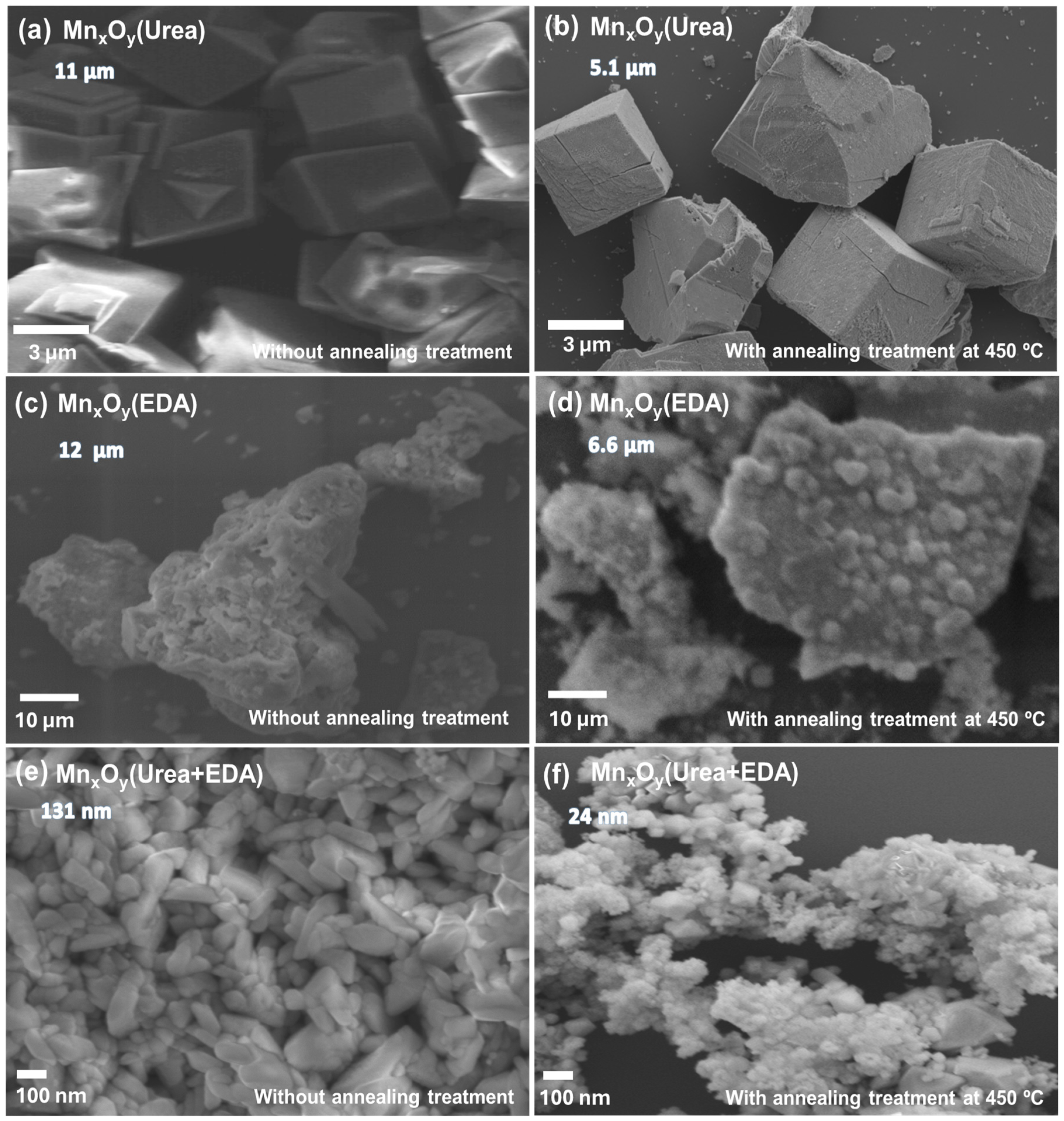
Figure 1. SEM micrographs of MnxOy(urea) (a,b), MnxOy(EDA) (c,d), and MnxOy(urea+EDA) (e,f), without and with annealing at 450 °C/6 h, respectively. The numbers below the names of the samples are the average particle sizes.
Physchem 05 00035 g001
Figure 2. Particle size distribution histogram of MnxOy (urea) (a,b), MnxOy(EDA) (c,d), and MnxOy(urea+EDA) (e,f), without and with annealing at 450 °C/6 h, respectively.
Physchem 05 00035 g002
Figure 3. X-ray diffraction patterns of the studied samples: (a,b) MnxOy(urea), (c,d) MnxOy(EDA), and (e,f) MnxOy(urea+EDA), without and with annealing treatment at 450 °C/6 h, respectively.
Physchem 05 00035 g003
Figure 4. Plot of Williamson–Hall method. The figure is shown as a reference for the calculation process of the crystallite size and lattice strain values of all materials.
Physchem 05 00035 g004
Figure 5. Raman spectra of the studied samples: (a,b) MnxOy(urea), (c,d) MnxOy(EDA), and (e,f) MnxOy(urea+EDA), without and with annealing treatment at 450 °C/6 h, respectively.
Physchem 05 00035 g005
Figure 6. FT-IR spectra of the studied samples: (a,b) MnxOy(urea), (c,d) MnxOy(EDA), and (e,f) MnxOy(urea+EDA), without and with annealing treatment at 450 °C/6 h, respectively.
Physchem 05 00035 g006
Table 1. Structural parameters of MnxOy(urea), MnxOy(EDA), and MnxOy(urea+EDA) materials calculated from their XRD patterns.
SampleCompoundLattice Parameters (Å)c/aVolume (Å3)
abc
Without annealing treatment
Urea MnCO34.774.7715.73.29308.7
Mn3O45.625.629.991.78315.3
EDAMn3O45.585.589.481.7294.7
Urea+EDAMnCO34.784.7814.12.94277.8
Mn3O45.575.5710.51.89328.7
β-MnO24.284.283.040.7155.7
With annealing treatment: 450 °C/6 h
Ureaα-Mn2O39.49.49.41831.1
Mn3O45.695.6910.11.78328.6
EDAα-Mn2O39.399.399.391827.16
Mn3O45.575.5710.21.82315.2
Urea+EDAα-Mn2O39.439.439.431838.3
Mn3O45.585.5810.41.86324.6
β-MnO23.973.973.160.7949.9
Reference work
MnCO3 [63]4.774.7715.63.278308.17
α-Mn2O3 [64]9.419.419.411833.2
Mn3O4 [65]5.755.759.421.64269.71
β-MnO2 [66]4.44.42.880.6555.8
Table 2. Comparison of the crystallite size for the MnxOy(urea) and MnxOy(urea+EDA) materials calculated from their XRD patterns.
SampleCompoundCrystal Symmetry(hkl)Crystal Size Sherrer EquationCrystal Size Williamson–HallLattice Strain
(nm)(nm)
Without annealing treatment
Urea MnCO3Hexagonal (R-3c)1044042.40.0037
Mn3O4Tetragonal (I41/amd)41319.824.80.004
EDAMn3O4Tetragonal (I41/amd)103 26.8--
Urea+EDAMnCO3Hexagonal (R-3c)10439.734.80.0028
Mn3O4Tetragonal (I41/amd)10322.716.50.005
β-MnO2Tetragonal (P42/mnm)10129.629.90.002
With annealing treatment: 450 °C/6 h
Ureaα-Mn2O3Cubic (Ia3)22217.2--
Mn3O4Tetragonal (I41/amd)21116.517.90.0023
EDAα-Mn2O3Cubic (Ia3) 22216.8--
Mn3O4Tetragonal (I41/amd)11218.1--
Urea+EDAα-Mn2O3Cubic (Ia3)22215.5--
Mn3O4Tetragonal (I41/amd)10321.8--
β-MnO2Tetragonal (P42/mnm)10123.8--
Disclaimer/Publisher’s Note: The statements, opinions and data contained in all publications are solely those of the individual author(s) and contributor(s) and not of MDPI and/or the editor(s). MDPI and/or the editor(s) disclaim responsibility for any injury to people or property resulting from any ideas, methods, instructions or products referred to in the content.