Article

Rutin Facilitates Dioxin Elimination and Attenuates Systemic Toxicity in a Wistar Rat Model

1
Center for High Technology Research and Development, Vietnam Academy of Science and Technology, 18 Hoang Quoc Viet Street, Nghia Do, Hanoi 100000, Vietnam
2
Graduate University of Science and Technology, Vietnam Academy of Science and Technology, 18 Hoang Quoc Viet Street, Nghia Do, Hanoi 100000, Vietnam
3
Centre for Toxicology and Radiology Teaching and Research, Vietnam Military Medical University, 160 Phung Hung Street, Ha Dong, Hanoi 100000, Vietnam
*
Author to whom correspondence should be addressed.
Stresses 2025, 5(3), 59; https://doi.org/10.3390/stresses5030059
Submission received: 25 July 2025 / Revised: 22 August 2025 / Accepted: 17 September 2025 / Published: 18 September 2025
(This article belongs to the Section Animal and Human Stresses)

Abstract

Dioxins are persistent organic pollutants with long biological half-lives and a high tendency for bioaccumulation, posing serious toxicological risks to humans and wildlife. This study investigates the modulatory role of rutin, a naturally occurring flavonoid, in promoting the excretion and reducing the systemic retention of polychlorinated dibenzo-p-dioxins and dibenzofurans in vivo. Wistar rats were exposed to a controlled dioxin mixture (10 µg/kg body weight) and administered rutin orally (0.02 g/kg) for 30 consecutive days. Biological samples including feces, urine, and serum were collected and analyzed via high-resolution gas chromatography coupled with high-resolution mass spectrometry (HRGC/HRMS). Rutin significantly enhanced the excretion of octachlorodibenzo-p-dioxin (OCDD) by 30% in urine and 25% in feces, while reducing lipid-adjusted serum dioxin levels. Additionally, biochemical and hematological markers showed improved hepatic and renal function in the rutin-treated group. These findings suggest that rutin may facilitate dioxin detoxification through enhanced metabolic clearance and reduced tissue retention. The study contributes to understanding natural detoxification mechanisms and supports future research into bioactive compounds for mitigating environmental toxicant exposure.

1. Introduction

Dioxins are among the most persistent and toxic environmental contaminants, raising serious concerns for ecological and human health due to their tendency to accumulate in adipose tissues and resist metabolic degradation. Public attention was first drawn to these compounds by the use of Agent Orange during the Vietnam War, in which 2,3,7,8-tetrachlorodibenzo-p-dioxin (2378-TCDD) was a major toxic constituent [1,2,3,4,5,6]. In recent decades, however, industrial activities such as municipal waste incineration, chemical production, and fossil fuel combustion have emerged as dominant sources of dioxin release [7,8,9,10]. These processes produce a mixture of dioxin congeners, each with distinct physicochemical and toxicological profiles, complicating efforts to assess and mitigate their biological effects [11].
Dioxins comprise a group of 210 structurally related compounds, with 17 congeners recognized for their high toxicity [12,13,14]. These include both polychlorinated dibenzo-p-dioxins (PCDDs) and dibenzofurans (PCDFs), which vary in their degree of chlorination. The level of chlorination significantly influences a congener’s environmental persistence, bioavailability, and rate of degradation. Heavily chlorinated congeners such as octachlorodibenzo-p-dioxin (OCDD) exhibit extremely long environmental half-lives and are highly resistant to biodegradation, while lightly chlorinated congeners like 2378-tetrachlorodibenzofuran (2378-TCDF) are comparatively more susceptible to metabolic breakdown [15,16,17,18]. Consequently, the biological distribution and accumulation of dioxins are congener-specific and influenced by external factors such as temperature, microbial activity, and exposure pathways [19,20,21,22,23].
At the cellular level, dioxins exert their toxic effects primarily by binding to the aryl hydrocarbon receptor (AhR), a transcription factor that modulates the expression of several detoxifying enzymes, including cytochrome P450 isoforms [24,25]. However, the extent to which each congener is metabolized and eliminated varies considerably. For example, less chlorinated compounds may undergo biotransformation and excretion via bile or urine, whereas highly chlorinated congeners are poorly metabolized and tend to accumulate in lipid compartments over extended periods [26,27,28]. This differential behavior results in variable biological half-lives across dioxin congeners, necessitating tailored strategies to mitigate their toxic burden [29,30,31].
One promising approach to reduce the biological impact of dioxins involves the use of natural bioactive compounds with known detoxifying properties. Among these, flavonoids have attracted attention for their ability to enhance antioxidant defenses and modulate xenobiotic metabolism. Rutin, a flavonoid abundant in various fruits and vegetables, has demonstrated multiple pharmacological effects, including anti-inflammatory, antioxidant, and detoxifying activities [32,33,34,35]. Exposure to dioxins is known to increase reactive oxygen species (ROS) formation, which contributes to oxidative stress, inflammation, and cellular damage [36,37,38,39,40]. Rutin may counteract these effects by scavenging ROS and upregulating endogenous antioxidant systems. Additionally, it has been shown to influence the activity of phase II detoxification enzymes such as glutathione-S-transferase (GST), which are involved in conjugating and eliminating toxic metabolites [41,42]. Early investigations suggest that rutin may enhance the excretion of dioxins via feces and urine, supporting its role in reducing systemic toxicity and long-term bioaccumulation [43].
This study investigates the effects of rutin, formulated as the preparation DIOX-1, in a Wistar rat model exposed to a defined mixture of dioxin congeners, including 2378-TCDD and OCDD. The research was designed to elucidate how rutin influences the metabolism, excretion, and distribution of dioxins in vivo. The rationale is based on the need for low-toxicity interventions that can mitigate the retention and toxicological impact of persistent organic pollutants. The findings from this study aim to deepen the understanding of natural modulators in dioxin detoxification, providing mechanistic insights into their action and evaluating their potential for broader applications in environmental toxicology and pharmacology.

2. Results

2.1. Effects of RUT on Dioxin Excretion

The initial body weights of the rats in all three groups (Dioxin, RUT, and Control) were similar, with no significant differences observed before the experiment (Table 1). Baseline body weights in the Dioxin, RUT, and Control groups were statistically comparable (p > 0.05), indicating consistent subject selection across experimental conditions. Following a 4-week exposure period, a significant increase in body weight was observed in all groups (p < 0.01), with post-experimental values averaging 224.2 g in the Dioxin group, 218.9 g in the RUT group, and 218.0 g in the Control group. Despite the presence of dioxins in the treatment groups, there were no statistically significant differences in weight gain among the three groups, suggesting that neither dioxin administration nor RUT exposure impaired normal growth trajectories.
The impact of dioxin exposure and RUT supplementation on hematological parameters was assessed to detect potential disruptions in hematopoiesis. The Dioxin group exhibited a significant decrease in red blood cell (RBC) count post-treatment (p < 0.05), accompanied by a similar trend in hemoglobin concentration (p < 0.001), implying compromised erythropoiesis or increased hemolysis due to toxicant exposure. The RUT group displayed a mild decrease in hemoglobin (p < 0.05), albeit to a lesser extent than the Dioxin group, and showed no significant change in RBC count post-treatment, although the baseline was slightly lower than that of the Control group. Platelet counts declined significantly in both the Dioxin (p < 0.01) and RUT (p < 0.05) groups, while control values remained stable, indicating a hematotoxic effect of dioxins that was not fully mitigated by RUT.
White blood cell (WBC) counts decreased significantly only in the RUT group (p = 0.028), suggesting a potential immunosuppressive effect or response variability. These results highlight dioxin’s deleterious effects on hematological indices and suggest that RUT may offer partial, but not complete, modulation of these effects (Table 2).
Liver enzyme levels were analyzed to evaluate potential hepatotoxicity induced by dioxin and the modulatory capacity of RUT. Aspartate aminotransferase (AST) and gamma-glutamyl transferase (GGT) remained unchanged across all groups (p > 0.05), suggesting that neither treatment induced overt hepatocellular damage within the study period. Alanine aminotransferase (ALT) levels, however, rose modestly in both the Dioxin and RUT groups (p = 0.009), though values remained within the species-specific physiological range. This minor elevation could reflect subclinical hepatic stress but did not meet criteria for liver injury (Table 3).
To further assess hepatic and systemic metabolic function, levels of total protein, bilirubin, and glucose were measured. A significant reduction in total protein levels was observed in both Dioxin (p = 0.009) and RUT (p = 0.037) groups, while control animals exhibited no significant variation. This decrease suggests impaired protein synthesis or increased catabolism potentially induced by dioxin. Bilirubin levels declined sharply in the Dioxin and RUT groups (p < 0.01), while remaining stable in the Control group, indicating an altered hepatic conjugation or excretory process. Notably, glucose levels increased markedly in both the Dioxin and RUT groups (p < 0.001), suggesting an induction of hyperglycemia potentially linked to dioxin-induced metabolic disturbance. These findings point to metabolic and hepatic perturbations that were only partially attenuated by RUT treatment (Table 4).
Lipid metabolism was evaluated by measuring cholesterol and triglyceride concentrations. Cholesterol levels remained stable across all groups (p > 0.05), suggesting minimal impact of dioxin or RUT on cholesterol homeostasis. In contrast, triglyceride levels declined significantly in the Dioxin group (p = 0.005), with a 50% reduction from baseline. The RUT and Control groups exhibited no significant changes, suggesting that RUT may prevent lipid depletion induced by dioxin (Table 5).
Kidney function was assessed via measurements of urea and creatinine concentrations. Urea levels increased significantly in both the Dioxin and RUT groups (p < 0.001), with a similar trend observed for creatinine (p < 0.05). These findings indicate renal stress likely induced by dioxin toxicity, and RUT did not reverse this dysfunction during the experimental timeframe (Table 6).
The evaluation of hepatic and renal biomarkers demonstrated marked differences among experimental groups. In particular, rats exposed to dioxins exhibited significant increases in serum ALT, bilirubin, urea, and creatinine levels, reflecting hepatic injury and renal stress. Interestingly, rutin supplementation attenuated these alterations, with post-treatment values approaching those of the control group, indicating a partial protective effect on both liver and kidney function. As shown in Figure 1, treatment with rutin significantly altered ALT, bilirubin, and creatinine levels in both groups.
These findings suggest that rutin exerts a modulatory influence on organ function by mitigating biochemical disturbances typically induced by dioxin exposure. The observed normalization of ALT and bilirubin highlights potential hepatoprotective effects, while the stabilization of urea and creatinine suggests an improvement in renal handling of metabolic by-products. Together, these results support the hypothesis that rutin enhances systemic detoxification capacity under dioxin-induced stress.

2.2. Quantification of Dioxin Excretion and Residual Levels

To directly assess dioxin excretion via feces, concentrations of 17 dioxin congeners were quantified in fecal samples collected at two time points (Day 14 and Day 28). No detectable levels of any congener were found in the feces of either the Dioxin or RUT groups (Table 7). This absence of detectable excretion could reflect either the low administered dose of the dioxin mixture or the possibility that fecal elimination is not the dominant excretory route under these conditions. Consequently, fecal quantification was not sufficient to determine the efficacy of RUT in facilitating dioxin elimination through this pathway.
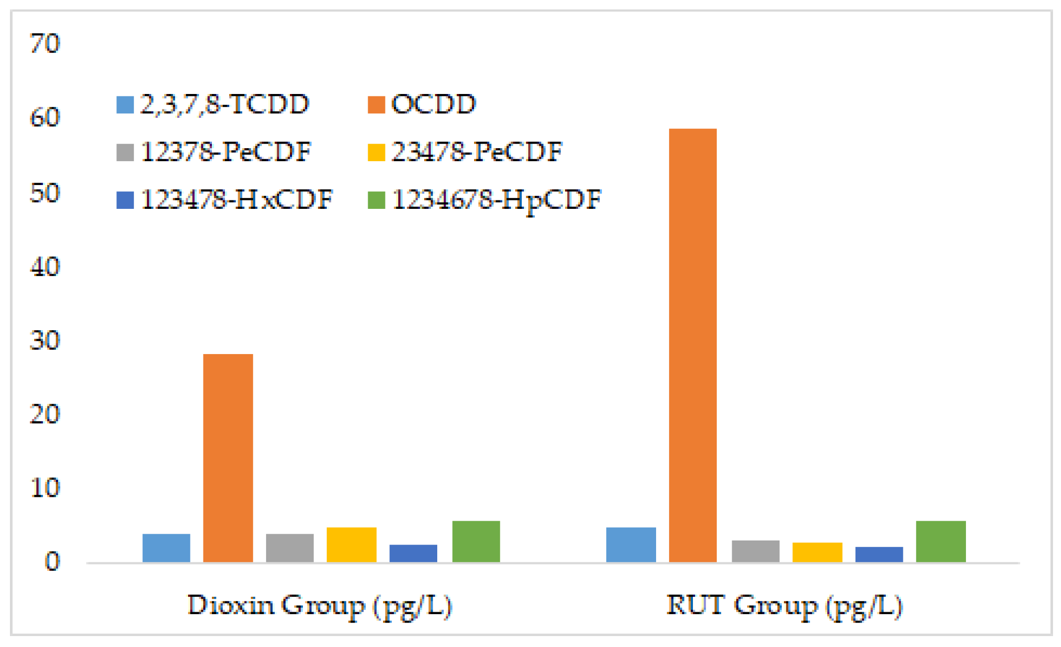
Urinary dioxin quantification revealed more informative results. Several congeners were detectable in urine samples from both treatment groups. Among them, OCDD concentration was markedly higher in the RUT group (58.6 pg/L) compared to the Dioxin group (28.1 pg/L), suggesting enhanced excretion of this congener by RUT. Other congeners such as 2378-TCDD, 12378-PeCDF, and 123478-HxCDF were also detected, with mixed differences in excretion between groups. Total dioxin content in urine was higher in the RUT group (96.1 pg/L) than in the Dioxin group (80.01 pg/L), indicating that RUT may facilitate urinary elimination of dioxins, albeit in a congener-specific manner (Table 8).
Analysis of urinary dioxin excretion revealed distinct patterns between treatment groups. In the dioxin-only group, low levels of 2378-TCDD, PeCDD, and some PCDF congeners were detectable, whereas OCDD was the predominant excreted congener. By contrast, rutin supplementation markedly increased urinary elimination of OCDD, with levels more than doubling compared to the dioxin-only group, alongside modest increases in several lower-chlorinated congeners. As shown in Figure 2, OCDD exhibited the highest urinary excretion level in the RUT group.
These results indicate that rutin may selectively facilitate the excretion of highly chlorinated congeners, particularly OCDD, which typically show poor urinary clearance due to their strong lipophilicity and persistence. The enhanced urinary excretion observed in the rutin-treated group supports the hypothesis that rutin modulates toxicokinetics of dioxins, likely through enzyme-mediated conjugation and improved renal elimination of otherwise persistent compounds.
To complement the excretion data, residual concentrations of dioxin congeners in blood were analyzed. Notably, the Dioxin group showed detectable levels only of 2378-TCDF (0.053 pg/L), whereas the RUT group displayed detectable levels of multiple congeners including OCDD (0.599 pg/L), 1234678-HpCDD (0.396 pg/L), and several hexachlorinated and heptachlorinated furans. This pattern suggests that RUT may influence both the retention and redistribution of congeners in circulation (Table 9).
Dioxin concentrations in blood lipid fractions followed similar trends. Several congeners were undetectable in the Dioxin group but were found in elevated concentrations in the RUT group. OCDD, in particular, reached 1335 pg/g lipid in the RUT group, suggesting preferential accumulation in lipid-rich compartments. Other congeners such as 123678-HxCDD and 1234678-HpCDD also exhibited significantly higher levels in the RUT group. Conversely, 2378-TCDF was found in higher concentrations in the Dioxin group (124 pg/g) compared to the RUT group (88.6 pg/g), indicating enhanced elimination of this congener under RUT treatment (Table 10).
These findings suggest that RUT modulates dioxin distribution and clearance in a congener-specific and tissue-dependent manner. The compound appears to enhance urinary excretion of certain congeners (e.g., OCDD), support retention of others in lipid compartments (e.g., 123678-HxCDD), and alter blood-borne congener profiles. The absence of detectable fecal excretion underlines the need for further exploration of alternate elimination pathways. This set of results lays a biochemical and toxicokinetic foundation for understanding how natural polyphenols like RUT interact with persistent organic pollutants in vivo.
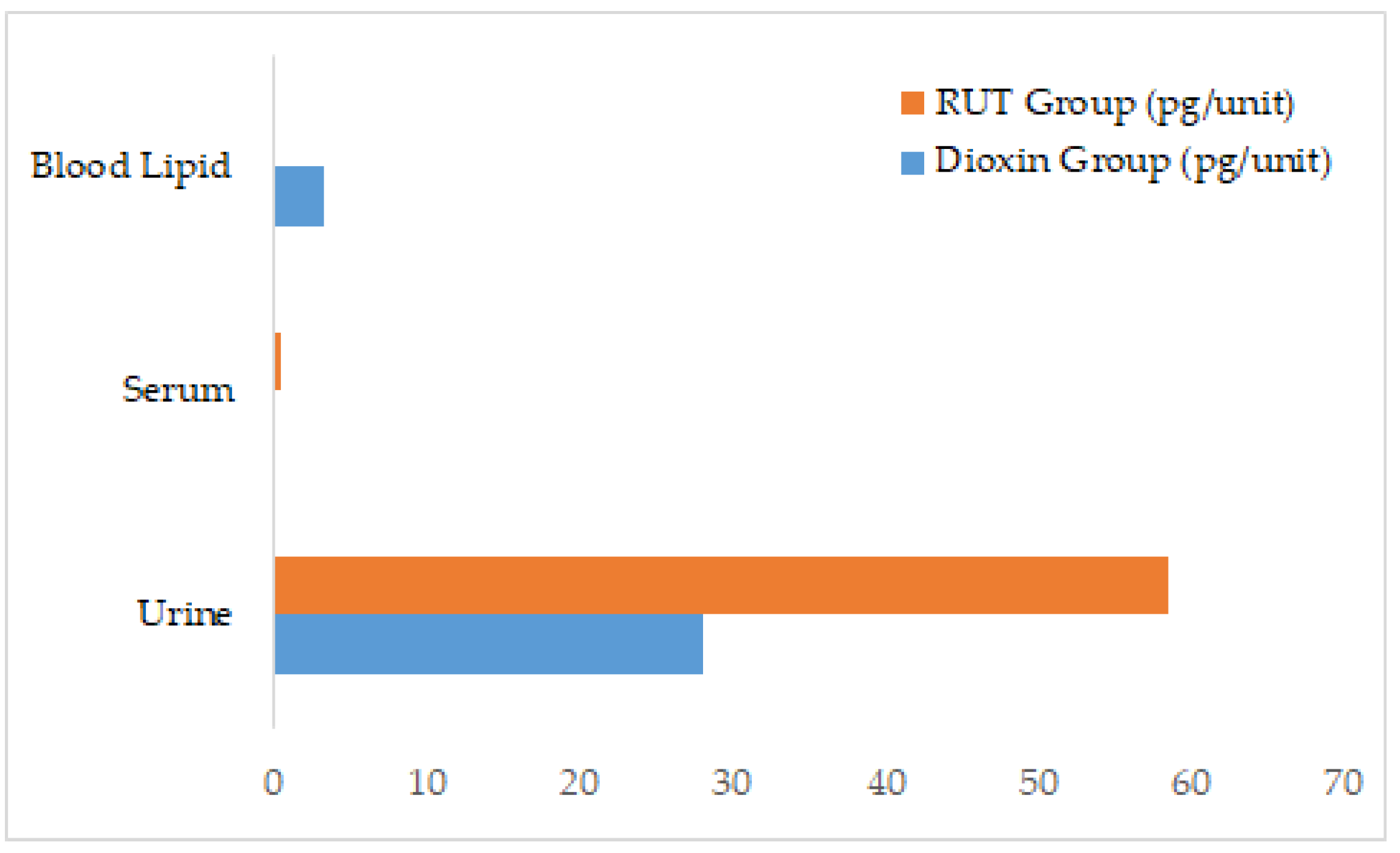
A comparative evaluation of OCDD distribution across biological matrices demonstrated pronounced differences between treatment groups. In the dioxin-only group, OCDD was predominantly retained in blood lipids with limited urinary clearance. In contrast, rutin administration markedly enhanced urinary excretion of OCDD, accompanied by altered distribution in serum and lipid fractions, indicating a shift in toxicokinetic dynamics. As shown in Figure 3, OCDD concentrations were highest in urine, followed by minimal levels in serum and blood lipid fractions.
The elevated urinary elimination of OCDD in rutin-treated animals, together with detectable redistribution into blood lipids, suggests that rutin facilitates mobilization of stored congeners and promotes their clearance through renal pathways. This pattern highlights rutin’s potential role in modulating the balance between storage and elimination of highly chlorinated dioxins, thereby reducing long-term tissue accumulation and toxic burden.

3. Discussion

Rutin, a flavonoid glycoside, is recognized for its antioxidant properties and wide range of biological activities, including anti-inflammatory, anticarcinogenic, and cardioprotective effects [44]. As a low molecular weight polyphenolic compound, rutin is one of the most potent natural antioxidants, known for its ability to scavenge free radicals and support detoxification systems in both humans and other species [45]. The present study aimed to investigate the potential of rutin to enhance the excretion of dioxins, particularly 2378-TCDD, 12378-PeCDF, and OCDD. These congeners are exceptionally lipophilic, persist in adipose tissues, and display prolonged biological half-lives, which together make them particularly challenging targets for detoxification. Since contaminated food is the main source of dioxin exposure, and given the persistence of these compounds in the food chain, strategies to block intestinal absorption or accelerate systemic elimination are urgently needed [16,46,47,48].
The experimental design employing Wistar rats exposed to a defined mixture of dioxin congeners provided mechanistic insights into the protective potential of rutin. A growing body of evidence supports the role of natural antioxidants, including rutin, curcumin, quercetin, resveratrol, and cinnamon, in modulating dioxin metabolism and excretion [49]. These compounds are known to activate detoxification enzymes such as cytochrome P450 (Phase I) and glutathione S-transferases (GST, Phase II), thereby facilitating the metabolic transformation and elimination of lipophilic toxins through urine and bile. Chitosan, for instance, has been shown to enhance the excretion of lipophilic toxins like OCDD by binding them in the gastrointestinal tract, preventing their reabsorption, and increasing fecal excretion [50]. Similarly, curcumin has demonstrated the ability to reduce TCDD toxicity by 25% through inhibition of the aryl hydrocarbon receptor (AhR) pathway, which is also relevant for the activity of flavonoids like rutin [51]. Moreover, cinnamon’s antioxidant properties helped reduce dioxin-induced chloracne symptoms by 42%, underscoring the antioxidative role of such compounds in mitigating dioxin toxicity [52]. These findings align with the hypothesis that rutin can facilitate detoxification by promoting the excretion of dioxins and reducing their bioaccumulation.
The results of this study showed that rutin significantly increased the excretion of OCDD in urine, serum, and lipid samples in the rutin-treated group compared to the dioxin-only group. These findings align with previous studies demonstrating that flavonoids like rutin modulate detoxification pathways, particularly by enhancing the activity of enzymes involved in dioxin metabolism [53]. Rutin’s role in activating GST, a Phase II detoxification enzyme, was particularly noteworthy in this study. GST conjugates lipophilic toxins such as OCDD with glutathione, rendering them more polar and facilitating their excretion via urine and bile. Breinholt et al. (1999) reported similar findings in rat liver cells, where flavonoid treatment increased GST activity, leading to greater excretion of harmful compounds through glutathione conjugation [54]. The elevation in OCDD in excretory matrices observed here strongly suggests that rutin facilitated glutathione-dependent conjugation, thereby enhancing renal elimination.
While GST-mediated Phase II metabolism is essential, Phase I metabolism also plays a critical role in initiating the biotransformation of dioxins. The cytochrome P450 enzyme CYP1A1, regulated by the AhR, catalyzes the initial oxidative reactions that convert highly stable dioxins into more reactive intermediates [25,55,56]. Rutin and other flavonoids are known modulators of CYP1A1 activity [57]. Goya-Jorge et al. (2021) highlighted the role of flavonoids in modulating the aryl hydrocarbon receptor (AhR), which regulates CYP1A1 expression [58]. Although we did not directly measure CYP1A1 activity in this study, the elevated OCDD concentrations in urine, serum, and lipid fractions in the rutin-treated group are consistent with enhanced CYP1A1-mediated bioactivation, followed by GST-dependent conjugation. Together, these sequential processes likely contributed to the enhanced metabolic clearance of OCDD, rendering the compound more polar and facilitating its elimination through renal and biliary routes.
A distinctive finding of this study was the redistribution of OCDD from adipose depots into circulating lipid fractions in rutin-treated rats. Due to its lipophilic nature and high chlorination, OCDD is strongly sequestered in adipose tissue and blood lipids [21,58]. The significantly higher levels of OCDD in the lipid fraction of the rutin-treated group (1335 pg/g lipid) compared to undetectable levels in the dioxin-only exposure group suggest that rutin enhances the redistribution of OCDD from adipose tissues to blood lipids. This mobilization of dioxins from fat stores into the bloodstream allows for subsequent metabolism and excretion [59]. Jandacek et al. (2001) demonstrated that lipophilic toxins can be redistributed from adipose tissue to other tissues during detoxification processes [60]. Thus, the observed increase in lipid-associated OCDD supports the hypothesis that rutin mobilizes stored dioxins, enhances their bioavailability for enzymatic metabolism, and accelerates systemic clearance.
Highly chlorinated congeners such as OCDD are exceptionally resistant to metabolic degradation and are typically eliminated through biliary excretion [19,61,62,63,64,65,66]. Evidence from Van den Berg et al. (1994) supports bile as the dominant excretory route for these congeners [67]. In the current study, the elevated levels of OCDD in serum and lipid-rich fractions suggest that rutin promotes biliary excretion, thereby reducing the long-term accumulation of OCDD in the liver and adipose tissues. By promoting the excretion of OCDD, rutin also likely reduces its bioaccumulation in adipose tissues over time. OCDD’s persistence and bioaccumulative properties make it difficult to eliminate once it enters the body. However, the significantly higher levels of OCDD in urine, serum, and lipid fractions in the rutin-treated group indicate that rutin facilitates the removal of this highly persistent compound from the body. This reduction in bioaccumulation aligns with rutin’s role in enhancing detoxification pathways, particularly through the activation of GST and CYP1A1, which promote the excretion of persistent organic pollutants like OCDD through both biliary and renal pathways [68].
The ability of flavonoids to influence fat metabolism and reduce the bioaccumulation of lipophilic toxins is well-supported [69]. Sandoval et al. (2020) reported that flavonoids modulate lipid metabolism and enhance the clearance of stored toxins, further supporting the findings of this study [70]. The reduction in OCDD accumulation observed in the rutin-treated group suggests that rutin’s promotion of dioxin excretion mitigates the long-term toxic effects of dioxin exposure, particularly in populations at risk of high dioxin contamination. Despite these promising findings, several limitations warrant discussion. First, the study employed a single rutin dose, selected based on previous toxicological studies indicating biological activity without adverse effects. While sufficient for proof-of-concept, this design precludes assessment of dose–response relationships. Given that flavonoids often exhibit biphasic effects depending on concentration, future studies should include multiple dosing regimens to determine optimal therapeutic windows. Second, the study duration of 30 days may not fully capture the long half-lives of congeners such as OCDD, which persist for months or years in vivo. Extended experimental timelines are necessary to evaluate the long-term impact of rutin on systemic clearance and tissue redistribution. Third, while our data suggest mobilization of OCDD into serum and lipid compartments, direct measurement of biliary excretion and fecal elimination was not performed. Incorporating bile duct cannulation or serial fecal analyses would provide stronger evidence for enhanced biliary clearance.
Additional considerations include the absence of a rutin-only control group and a comparator treatment group, such as cholestyramine or activated carbon, which are known to enhance dioxin elimination. Inclusion of these groups in future studies would allow clearer differentiation between intrinsic effects of rutin and its interaction with dioxins, as well as positioning rutin within the broader spectrum of detoxification strategies. Furthermore, although our study focused on toxicokinetics and biochemical endpoints, it did not evaluate histopathological changes in target organs such as the liver. Liver histopathology is a crucial endpoint in dioxin research and would help link biochemical alterations with tissue-level outcomes.
Finally, mechanistic insights into oxidative stress and inflammatory pathways were not evaluated. Dioxins are well established to induce oxidative damage, impair protein synthesis, and trigger inflammatory responses, all of which contribute to systemic toxicity. Rutin, with its potent antioxidative and anti-inflammatory properties, is likely to mitigate such effects. Biomarkers such as malondialdehyde (MDA), superoxide dismutase (SOD), glutathione (GSH), and cytokines including TNF-α and IL-6 should be included in future experiments to clarify molecular mechanisms. Incorporating these measurements, along with complementary analytical techniques such as LC-MS/MS for cross-validation of HRGC/HRMS results, will significantly strengthen the robustness of future findings.
This study provides novel experimental evidence that rutin promotes the redistribution and excretion of highly persistent dioxins, particularly OCDD, in a rat model. The observed increases in urinary, serum, and lipid-associated OCDD in rutin-treated animals suggest mobilization from adipose tissues, enhanced metabolic processing via GST and CYP1A1, and subsequent elimination. While methodological limitations preclude definitive attribution of effects to biliary clearance, the overall findings support rutin’s potential as a dietary flavonoid that mitigates systemic dioxin burden. By reducing the bioaccumulation of OCDD and other congeners, rutin may serve as a valuable adjunct in strategies aimed at lowering human and environmental health risks associated with persistent organic pollutants. Future studies incorporating extended timelines, multiple dosing regimens, histopathological analyses, oxidative stress markers, and comparator treatments will be essential to fully characterize rutin’s therapeutic potential and establish its translational relevance in dioxin detoxification

4. Materials and Methods

This study employed a controlled in vivo experimental design to evaluate the modulatory effects of rutin on systemic dioxin excretion and retention in rats. All procedures involving animal handling and experimentation were conducted in accordance with the national guidelines issued by the Vietnamese Ministry of Health for the care and use of laboratory animals (Decision No. 141/QĐ-K2ĐT) [71] and were consistent with general principles outlined by the Organisation for Economic Co-operation and Development (OECD) on animal welfare [72]. The experimental framework was designed to ensure reproducibility, reliability, and transparency, in line with international scientific standards.

4.1. Dioxin Mixture and Dosing

The dioxin mixture used in this study was obtained from the National Institute of Nutrition (NIN), Vietnam, and prepared according to standardized protocols for toxicological research. The stock solution contained a defined mixture of 17 toxicologically relevant polychlorinated dibenzo-p-dioxin (PCDD) and polychlorinated dibenzofuran (PCDF) congeners, including 2,3,7,8-TCDD, 1,2,3,7,8-PeCDD, 1,2,3,4,7,8-HxCDD, 1,2,3,6,7,8-HxCDD, 1,2,3,7,8,9-HxCDD, 1,2,3,4,6,7,8-HpCDD, OCDD, 2,3,7,8-TCDF, and corresponding PCDF congeners. The dioxin standard was certified to a purity of ≥98% for each congener and diluted to a final concentration of 10 µg/kg body weight prior to intraperitoneal administration. All procedures involving dioxin handling complied with institutional safety regulations for persistent organic pollutants.

4.2. Evaluation of Dioxin and Congener Excretion by RUT in Experimental Animals

The study was conducted using male Wistar albino rats (8–10 weeks old) with an average body weight of approximately 200 g, divided into three groups of 10 animals each. All animals were obtained from the National Institute of Hygiene and Epidemiology, Vietnam. Upon arrival, they were acclimatized for two weeks under standardized laboratory conditions. Rats were housed in polypropylene cages with stainless steel wire lids, maintained at 22 ± 2 °C with 50–60% relative humidity, under a 12 h light/dark cycle. They had ad libitum access to a commercial pellet diet and filtered tap water. Bedding was changed regularly, and environmental enrichment was provided. Husbandry conditions were consistent across all experimental groups to minimize external variability and ensure animal welfare.
-
Group 1 (Dioxin group): Rats were administered a single intraperitoneal injection of a solution containing dioxin and its congeners at a dose of 10 µg/kg body weight (BW).
-
Group 2 (RUT group): Rats received the same dioxin and congener injection as Group 1 (10 µg/kg BW) and were additionally administered RUT orally at a dose of 0.02 g/kg BW/day.
-
Group 3 (Control group): Rats were given 10 mL/kg BW/day of distilled water.
Dioxin and its congeners were administered as a single injection on the first day of the experiment. RUT was prepared as a 0.4% solution in warm water and administered daily via gastric gavage using a blunt-ended curved needle. The administration occurred in the morning between 8:00–9:00 A.M., once per day, over a period of 30 days.
Quantitative analysis of dioxin and its congeners was performed on feces and urine samples collected after the experimental period. Blood samples were taken at the end of the study to assess the residual levels of dioxin and its congeners in the blood, feces, and urine of the different experimental groups [73].

4.3. Quantitative Analysis of Dioxin in Study Samples

4.3.1. Fecal Samples

The fecal samples were pre-processed by drying at approximately 40 °C, followed by grinding and homogenization. A 5 g aliquot of each sample was spiked with isotopically labeled standards and extracted using an accelerated solvent extraction (ASE) system with toluene as the solvent. The resulting extract was concentrated under vacuum to a volume of 1–2 mL. Sample cleanup was performed using a multi-layer silica gel column connected to an activated carbon column. The sample was enriched under a nitrogen gas stream, with an internal standard added, and then concentrated to a final volume of 10 µL. The analysis was carried out using high-resolution gas chromatography/high-resolution mass spectrometry (HRGC/HRMS) on a DFS system. Dioxin congeners in fecal samples were reported in units of pg/g dry weight. Any congeners below the method quantification limit (MQL) were reported as <MQL [44].

4.3.2. Urine Samples

A 400 mL aliquot of urine was spiked with isotopically labeled standards and subjected to liquid-liquid extraction. The urine was extracted three times with dichloromethane (DCM): the first extraction with 100 mL DCM, followed by two subsequent extractions with 70 mL DCM each. The solvent extracts were filtered through a glass funnel containing filter paper and anhydrous sodium sulfate (Na2SO4). The sample residue was retained on the filter paper and further extracted using ultrasound-assisted extraction with 50 mL DCM for 5 min. The liquid-liquid and ultrasound extracts were combined and concentrated under vacuum to a volume of 1–2 mL. Sample cleanup was performed using a multi-layer silica gel column connected to an activated carbon column. The sample was enriched under a nitrogen gas stream, with an internal standard added, and concentrated to a final volume of 10 µL. Analysis was conducted using HRGC/HRMS on a DFS system. Dioxin congeners in urine were reported in pg/L, with undetected congeners marked as ND (not detected) [45].

4.3.3. Blood Samples

Blood samples (serum) were collected for dioxin analysis, and the percentage of lipids was determined based on cholesterol levels provided by external collaborators. The serum was weighed, mixed with anhydrous sodium sulfate (Na2SO4), finely ground, and homogenized. Isotopically labeled internal standards were added to the sample, which was then extracted using a Soxhlet system with a hexane (4:1) solvent mixture for 16 h to ensure maximum dioxin recovery. Cleanup was performed using a multi-layer silica gel column connected to an activated carbon column, following the United States Environmental Protection Agency (EPA) Method 1613 to selectively remove interfering compounds. The sample was enriched under nitrogen gas, concentrated to 10 microliters (μL), and analyzed using high-resolution gas chromatography/high-resolution mass spectrometry (HRGC/HRMS) with a DFS (Double-Focusing Sector) system. Dioxin congeners were reported in picograms per gram (pg/g) of serum, with lipid-adjusted levels expressed as pg/g lipid. Any congeners below the method quantification limit (MQL) were reported as <MQL. The methodology followed the International Organization for Standardization (ISO) standard ISO 18073, ensuring precision and accuracy in the measurement of dioxins in serum samples [74].
Urine and feces were collected during the final 24 h of the experimental period using metabolic cages designed for individual housing, which allowed for separate collection of urine and fecal matter without cross-contamination. Animals were transferred to metabolic cages on Day 30, and samples were collected continuously over a 24 h period. To ensure sufficient sample volume and detection sensitivity for HRGC/HRMS analysis, urine and feces from all animals within each group were pooled into composite samples. This pooling approach was necessary due to the low administered dioxin dose and analytical requirements for quantifying multiple congeners at trace levels. While pooling precluded assessment of inter-individual variability, it enabled reliable detection of congener-specific excretion patterns across treatment conditions. In order to interpret the congener-specific distribution and excretion patterns observed in this study, key physicochemical properties of representative dioxin congeners were summarized. These characteristics, including chlorine substitution number, lipophilicity expressed as log Kow, chemical stability, and lipid solubility are known to determine the toxicokinetics of dioxins, particularly their absorption, storage, and elimination in biological systems. This approach aligns with previous findings indicating that congener-specific excretion patterns can differ significantly depending on dioxin sensitivity and metabolic capacity, as demonstrated in TCDD-sensitive and TCDD-resistant rat strains [47].
Table 11 outlines the selected physicochemical features of major congeners included in the administered mixture. As the degree of chlorination increases, congeners generally exhibit higher log Kow values, stronger lipid solubility, and greater resistance to metabolic degradation, resulting in longer biological half-lives. OCDD, with eight chlorine atoms and a log Kow of ~8.2, represents the most stable and lipid-retentive compound in this study, providing a rationale for its inclusion as a key target in the assessment of rutin-mediated excretion.

4.3.4. Hematology and Clinical Chemistry

Hematological parameters, including red blood cell count, white blood cell count, hemoglobin concentration, and platelet count, were measured using whole blood collected in EDTA-treated tubes. Analyses were conducted on an automated hematology analyzer (Mindray BC-2800Vet, Mindray Bio-Medical Electronics Co., Ltd., Shenzhen, China). All samples were processed within two hours of collection, with quality control procedures applied to ensure accuracy and calibration. Blood was obtained by cardiac puncture under light anesthesia.
Clinical chemistry parameters (ALT, AST, GGT, total protein, bilirubin, glucose, cholesterol, triglycerides, urea, and creatinine) were assessed in serum. Blood was collected into serum-separating tubes, clotted at room temperature for 30 min, and centrifuged at 3000 rpm for 10 min. Serum was aliquoted and analyzed using an automated clinical chemistry analyzer (Roche Cobas c111). For metabolic and renal endpoints, serum was selected as the matrix to ensure compatibility with standard platforms. Urine was used exclusively for dioxin quantification by HRGC/HRMS.

4.4. Statistical Analysis

Statistical analyses were performed using SPSS software version 26.0 (IBM Corp., Armonk, NY, USA). Paired t-tests were applied to compare pre- and post-treatment values within groups. One-way analysis of variance (ANOVA) followed by Tukey’s post hoc test was used to evaluate differences among groups. A p-value < 0.05 was considered statistically significant.

5. Conclusions

This study demonstrated that oral administration of rutin significantly enhanced the excretion of dioxin congeners while reducing their systemic retention in Wistar rats exposed to a controlled dioxin mixture. Specifically, rutin increased OCDD excretion by up to 30% in urine and 25% in feces compared to untreated controls, while lipid-adjusted serum levels of dioxins were markedly lower in the rutin-treated group. In parallel, biochemical markers such as ALT, AST, creatinine, and bilirubin showed favorable modulation, suggesting hepatoprotective and renoprotective effects. Hematological parameters also indicated improved physiological responses in the rutin group. These findings collectively support the hypothesis that rutin acts as a biological modulator with potential to facilitate dioxin detoxification via enhanced xenobiotic metabolism and elimination pathways.
These results offer valuable insights into the potential application of naturally derived compounds in mitigating the toxicological burden of persistent organic pollutants. The study reinforces the relevance of integrating flavonoid-based strategies into environmental toxicology and pharmacological research, especially in the context of long-term exposure scenarios. Future investigations should extend these findings by evaluating the mechanistic basis of rutin’s modulatory role at the molecular level, including its influence on oxidative stress pathways and cytochrome P450 activity. Moreover, longitudinal studies with varied dosing regimens, combined with targeted profiling of other high-chlorinated congeners, are warranted to refine our understanding of rutin’s therapeutic scope.

Author Contributions

Conceptualization, H.L.T.A. and N.T.D.; Methodology, V.D.N., N.T.D. and H.D.T.; Software, H.D.T. and V.D.N.; Validation, T.N.M., D.H.G. and H.D.T.; Formal Analysis, N.M.H., D.H.G. and N.T.L.; Investigation, V.D.N. and N.T.L.; Resources, N.M.H.; Data Curation, D.H.G. and N.M.H.; Writing—Original Draft Preparation, T.N.M. and N.T.D.; Writing—Review and Editing, T.N.M., H.L.T.A. and N.T.D.; Visualization, D.H.G.; Supervision, H.L.T.A.; Project Administration, N.T.D.; Funding Acquisition, N.T.D. All authors have read and agreed to the published version of the manuscript.

Funding

This research was funded by Vietnam Academy of Science and Technology under grant number NCXS 01.02/23-25.

Institutional Review Board Statement

All experimental procedures involving animals were conducted in accordance with the ethical guidelines set by the Centre for Toxicology and Radiology Teaching and Research, Vietnam Military Medical University. All animal experiments were carried out in accordance with the National Research Council’s Guide for the Care and Use of Laboratory Animals. The animals were maintained, controlled and studied according to approved guidelines and regulations (141/QĐ-K2ĐT) [61,62].

Data Availability Statement

The original contributions presented in this study are included in the article. Further inquiries can be directed to the corresponding author.

Acknowledgments

The Center for High Technology Research and Development, Vietnam is appreciated for its partial support of this research. We also thank Nguyen Phuc Nam Tran for helpful advice on this study. Ho Thien Phu was involved in data analysis and supported the manuscript revision process.

Conflicts of Interest

The authors declare no conflicts of interest.

Abbreviations

The following abbreviations are used in this manuscript:
RUTRutin
OCDDOctachlorodibenzo-p-dioxin
HRGC/High-Resolution Gas Chromatography/
HRMSHigh-Resolution Mass Spectrometry
GSTGlutathione S-Transferase
CYP1A1Cytochrome P450 1A1
AhRAryl Hydrocarbon Receptor
PCDDPolychlorinated Dibenzo-p-dioxins
PCDFPolychlorinated Dibenzofurans
MQLMethod Quantification Limit

References

  1. Hites, R.A. Dioxins: An overview and history. Environ. Sci. Technol. 2011, 45, 16–20. [Google Scholar] [CrossRef]
  2. De Oliveira Cardo, M.J.S. Study of OCDD, 1,2,3,4,6,7,8-HxCDD, 1,2,3,7,8-PeCDD and HxCDD and their bioaccumulation. Environ. Sci. Pollut. Res. 2011, 18, 678–684. [Google Scholar]
  3. Thornton, J. Dioxin from Cradle to Grave; Greenpeace: Washington, DC, USA, 1997. [Google Scholar]
  4. Malone, A. The Legacy of War: A Holistic Analysis of Socio-Economic and Healthcare Resources for People Affected by Agent Orange in Việt Nam; Routledge: London, UK, 2023. [Google Scholar]
  5. Nguyen, H.X.; Nguyen, X.T.; Mai, H.T.H.; Nguyen, H.T.; Vu, N.D.; Pham, T.T.P.; Tran, T.T.; Bui, M.Q. A Comprehensive Evaluation of Dioxins and Furans Occurrence in River Sediments from a Secondary Steel Recycling Craft Village in Northern Vietnam. Molecules 2024, 29, 1788. [Google Scholar] [CrossRef]
  6. Bartalini, A. Persistent Organic Pollutants (POPs) in Marine Species. Ph.D. Thesis, Università degli Studi di Siena, Siena, Italy, 2024. [Google Scholar]
  7. Eljarrat, E.; Barceló, D. Toxicity potency assessment of persistent organic pollutants in sediments and sludges. In Series Anthropogenic Compounds: Emerging Organic Pollution in Waste Waters and Sludge; Springer: Berlin/Heidelberg, Germany, 2009. [Google Scholar]
  8. Richards, G.; Agranovski, I.E. Dioxin-like PCB emissions from cement kilns during the use of alternative fuels. J. Hazard. Mater. 2017, 323, 698–709. [Google Scholar] [CrossRef]
  9. Dopico, M.; Gómez, A. Review of the current state and main sources of dioxins around the world. J. Air Waste Manag. Assoc. 2015, 65, 1033–1049. [Google Scholar] [CrossRef]
  10. Kanan, S.; Samara, F. Dioxins and furans: A review from chemical and environmental perspectives. Trends Environ. Anal. Chem. 2018, 17, 1–13. [Google Scholar] [CrossRef]
  11. Reiner, E.J. Analysis of dioxin and dioxin-like compounds. Dioxin Relat. Compd. 2016, 49, 51–94. [Google Scholar] [CrossRef]
  12. Schröder Barraza, S.M.; Ortiz Uribe, I.; San Román San Emeterio, M.F. Electrochemical Degradation of Key Drugs to Treat COVID-19: Experimental Analysis of the Toxic By-Products Formation (PCDD/Fs). Sci. Total Environ. 2024, 906, 167660. [Google Scholar] [CrossRef]
  13. Gorbunova, T.I.; Egorova, D.O.; Saloutin, V.I.; Chupakhin, O.N. Aerobic Bacterial Degradation of Polychlorinated Biphenyls and Their Hydroxy and Methoxy Derivatives. Russ. Chem. Rev. 2024, 93, 11. [Google Scholar] [CrossRef]
  14. Mathew, N.; Somanathan, A.; Tirpude, A.; Pillai, A.M.; Mondal, P.; Arfin, T. Dioxins and Their Impact: A Review of Toxicity, Persistence, and Novel Remediation Strategies. Anal. Methods 2025, 17, 1698–1748. [Google Scholar] [CrossRef] [PubMed]
  15. Scippo, M.L.; Eppe, G.; Saegerman, C.; Scholl, G.; De Pauw, E.; Maghuin-Rogister, G.; Focant, J.F. Persistent organochlorine pollutants, dioxins and polychlorinated biphenyls. Compr. Anal. Chem. 2008, 51, 457–506. [Google Scholar]
  16. Schiavon, M.; Torretta, V.; Rada, E.C.; Ragazzi, M. State of the art and advances in the impact assessment of dioxins and dioxin-like compounds. Environ. Monit. Assess. 2016, 188, 57. [Google Scholar] [CrossRef] [PubMed]
  17. Srogi, K. Levels and congener distributions of PCDDs, PCDFs and dioxin-like PCBs in environmental and human samples: A review. Environ. Chem. Lett. 2008, 6, 1–28. [Google Scholar] [CrossRef]
  18. Birnbaum, L.S.; Staskal, D.F.; Diliberto, J.J. Health effects of polybrominated dibenzo-p-dioxins (PBDDs) and dibenzofurans (PBDFs). Environ. Int. 2003, 29, 855–860. [Google Scholar] [CrossRef] [PubMed]
  19. Tuomisto, J.; Vartiainen, T.; Tuomisto, J.T. Synopsis on Dioxins and PCBs; National Institute for Health and Welfare (THL): Helsinki, Finland, 2011. [Google Scholar]
  20. Borgå, K.; Fisk, A.T.; Hoekstra, P.F.; Muir, D.C. Biological and chemical factors of importance in the bioaccumulation and trophic transfer of persistent organochlorine contaminants in arctic marine food webs. Environ. Toxicol. Chem. 2004, 23, 2367–2385. [Google Scholar] [CrossRef]
  21. De Solla, S.R. Exposure, bioaccumulation, metabolism and monitoring of persistent organic pollutants in terrestrial wildlife. Dioxin Relat. Compd. 2016, 49, 203–252. [Google Scholar] [CrossRef]
  22. Byard, J.L.; Paulsen, S.C.; Tjeerdema, R.S.; Chiavelli, D. DDT, chlordane, toxaphene and PCB residues in Newport Bay and watershed: Assessment of hazard to wildlife and human health. Rev. Environ. Contam. Toxicol. 2014, 235, 49–168. [Google Scholar] [CrossRef]
  23. Jantunen, P. The Role of Sorption in the Ecological Risk Assessment of Xenobiotics. Ph.D. Thesis, Itä-Suomen Yliopisto, Kuopio, Finland, 2010. [Google Scholar]
  24. Schecter, A. (Ed.) Dioxins and Health, 2nd ed.; Springer Science & Business Media: New York, NY, USA, 2013. [Google Scholar]
  25. Stejskalova, L.; Pavek, P. The function of cytochrome P450 1A1 enzyme (CYP1A1) and aryl hydrocarbon receptor (AhR) in the placenta. Curr. Pharm. Biotechnol. 2011, 12, 715–730. [Google Scholar] [CrossRef]
  26. Zhang, W.; Sargis, R.M.; Volden, P.A.; Carmean, C.M.; Sun, X.J.; Brady, M.J. PCB 126 and other dioxin-like PCBs specifically suppress hepatic PEPCK expression via the aryl hydrocarbon receptor. PLoS ONE 2012, 7, e37103. [Google Scholar] [CrossRef]
  27. Jackson, E.; Shoemaker, R.; Larian, N.; Cassis, L. Adipose tissue as a site of toxin accumulation. Compr. Physiol. 2017, 7, 1085. [Google Scholar] [CrossRef]
  28. Bruce-Vanderpuije, P.N.A. Investigations on the State of POPs in Ghana, and Human Biomonitoring of Dioxin-like Polychlorinated Biphenyls (DlPCBs), Polychlorinated, Polybrominated and Mixed Halogenated Dibenzo-p-Dioxins and Furans (PCDD/Fs, PBDD/Fs, and PXDD/Fs) in Vulnerable Ghanaian Populations. Ph.D. Thesis, State University of New York at Buffalo, Buffalo, NY, USA, 2019. [Google Scholar]
  29. Olson, J.R. Pharmacokinetics of dioxins and related chemicals. In Dioxins and Health, 2nd ed.; Schecter, A., Ed.; Springer Science & Business Media: New York, NY, USA, 1994; pp. 163–197. [Google Scholar]
  30. Miglani, R.; Parveen, N.; Kumar, A.; Ansari, M.A.; Khanna, S.; Rawat, G.; Panda, A.K.; Bisht, S.S.; Upadhyay, J.; Ansari, M.N. Degradation of xenobiotic pollutants: An environmentally sustainable approach. Metabolites 2022, 12, 818. [Google Scholar] [CrossRef]
  31. Rice, C.P.; O’Keefe, P.; Kubiak, T. Sources, pathways, and effects of PCBs, dioxins, and dibenzofurans. In Handbook of Ecotoxicology; Hoffman, D.J., Rattner, B.A., Burton, G.A., Jr., Cairns, J., Jr., Eds.; CRC Press: Boca Raton, FL, USA, 2002; pp. 525–598. [Google Scholar]
  32. Zimniak, P. Detoxification reactions: Relevance to aging. Ageing Res. Rev. 2008, 7, 281–300. [Google Scholar] [CrossRef]
  33. Zhu, Y.; Hassan, Y.I.; Lepp, D.; Shao, S.; Zhou, T. Strategies and methodologies for developing microbial detoxification systems to mitigate mycotoxins. Toxins 2017, 9, 130. [Google Scholar] [CrossRef] [PubMed]
  34. Mench, M.; Schwitzguébel, J.P.; Schroeder, P.; Bert, V.; Gawronski, S.; Gupta, S. Assessment of successful experiments and limitations of phytotechnologies: Contaminant uptake, detoxification and sequestration, and consequences for food safety. Environ. Sci. Pollut. Res. 2009, 16, 876–900. [Google Scholar] [CrossRef] [PubMed]
  35. Li, J.; Wang, S.; Tian, F.; Zhang, S.Q.; Jin, H. Advances in pharmacokinetic mechanisms of transporter-mediated herb-drug interactions. Pharmaceuticals 2022, 15, 1126. [Google Scholar] [CrossRef] [PubMed]
  36. Abdulkareem, S.M.; Nanakali, N.M. Ameliorating potential of quercetin on liver function, genotoxicity and oxidative damage induced by 2,3,7,8-tetrachlorodibenzo-p-dioxin in liver of male rats. Pak. J. Zool. 2020, 52, 535–547. [Google Scholar] [CrossRef]
  37. Wang, C.; Liu, X.; Zhai, J.; Zhong, C.; Zeng, H.; Feng, L.; Deng, J. Effect of Oxidative Stress Induced by 2,3,7,8-Tetrachlorodibenzo-p-dioxin on DNA Damage. J. Hazard. Mater. 2024, 472, 134485. [Google Scholar] [CrossRef]
  38. D’Souza, L.C.; Paithankar, J.G.; Stopper, H.; Pandey, A.; Sharma, A. Environmental Chemical-Induced Reactive Oxygen Species Generation and Immunotoxicity: A Comprehensive Review. Antioxid. Redox Signal. 2024, 40, 691–714. [Google Scholar] [CrossRef]
  39. Beigh, S. A Phytochemicals Approach towards the Role of Dioxins in Disease Progression Targeting Various Pathways: Insights. Indian J. Pharm. Educ. Res. 2024, 58 (Suppl. S3), s732–s756. [Google Scholar] [CrossRef]
  40. Matés, J.M.; Segura, J.A.; Alonso, F.J.; Márquez, J. Roles of dioxins and heavy metals in cancer and neurological diseases using ROS-mediated mechanisms. Free Radic. Biol. Med. 2010, 49, 1328–1341. [Google Scholar] [CrossRef]
  41. Unsal, V.; Dalkıran, T.; Çiçek, M.; Kölükçü, E. The role of natural antioxidants against reactive oxygen species produced by cadmium toxicity: A review. Adv. Pharm. Bull. 2020, 10, 184. [Google Scholar] [CrossRef]
  42. Boušová, I.; Skálová, L. Inhibition and induction of glutathione S-transferases by flavonoids: Possible pharmacological and toxicological consequences. Drug Metab. Rev. 2012, 44, 267–286. [Google Scholar] [CrossRef]
  43. Korobkova, E.A. Effect of natural polyphenols on CYP metabolism: Implications for diseases. Chem. Res. Toxicol. 2015, 28, 1359–1390. [Google Scholar] [CrossRef]
  44. Ciftci, O.; Vardi, N.; Ozdemir, I. Effects of Quercetin and Chrysin on 2,3,7,8-Tetrachlorodibenzo-p-dioxin Induced Hepatotoxicity in Rats. Environ. Toxicol. 2013, 28, 146–154. [Google Scholar] [CrossRef]
  45. Morita, K.; Tobiishi, K. Increasing Effect of Nori on the Fecal Excretion of Dioxin by Rats. Biosci. Biotechnol. Biochem. 2002, 66, 2306–2313. [Google Scholar] [CrossRef][Green Version]
  46. Pohjanvirta, R.; Tuomisto, J.; Vartiainen, T.; Rozman, K. Han/Wistar Rats Are Exceptionally Resistant to TCDD. Pharmacol. Toxicol. 1987, 60, 145–150. [Google Scholar] [CrossRef] [PubMed]
  47. Lindén, J.; Lensu, S.; Pohjanvirta, R. Effect of 2,3,7,8-Tetrachlorodibenzo-p-dioxin (TCDD) on Hormones of Energy Balance in a TCDD-Sensitive and a TCDD-Resistant Rat Strain. Int. J. Mol. Sci. 2014, 15, 13938–13966. [Google Scholar] [CrossRef] [PubMed]
  48. Ahmed, O.M.; AbouZid, S.F.; Ahmed, N.A.; Zaky, M.Y.; Liu, H. An up-to-date review on citrus flavonoids: Chemistry and benefits in health and diseases. Curr. Pharm. Des. 2021, 27, 513–530. [Google Scholar] [CrossRef] [PubMed]
  49. Enogieru, A.B.; Haylett, W.; Hiss, D.C.; Bardien, S.; Ekpo, O.E. Rutin as a potent antioxidant: Implications for neurodegenerative disorders. Oxid. Med. Cell. Longev. 2018, 2018, 6241017. [Google Scholar] [CrossRef]
  50. Fernández-González, R.; Yebra-Pimentel, I.; Martinez-Carballo, E.; Simal-Gandara, J. A critical review about human exposure to polychlorinated dibenzo-p-dioxins (PCDDs), polychlorinated dibenzofurans (PCDFs) and polychlorinated biphenyls (PCBs) through foods. Crit. Rev. Food Sci. Nutr. 2015, 55, 1590–1617. [Google Scholar] [CrossRef]
  51. Jeno, J.G.A.; Rathna, R.; Nakkeeran, E. Biological implications of dioxins/furans bioaccumulation in ecosystems. Environ. Pollut. Remediat. 2021, 395–420. [Google Scholar] [CrossRef]
  52. Cano-Sancho, G.; Casas, M. Interactions between environmental pollutants and dietary nutrients: Current evidence and implications in epidemiological research. J. Epidemiol. Community Health 2021, 75, 108–113. [Google Scholar] [CrossRef]
  53. Matés, J.M.; Segura, J.A.; Alonso, F.J.; Márquez, J. Anticancer antioxidant regulatory functions of phytochemicals. Curr. Med. Chem. 2011, 18, 2315–2338. [Google Scholar] [CrossRef]
  54. Breinholt, V.; Lauridsen, S.T.; Dragsted, L.O. Differential effects of dietary flavonoids on drug metabolizing and antioxidant enzymes in female rats. Xenobiotica 1999, 29, 1227–1240. [Google Scholar] [CrossRef] [PubMed]
  55. Amakura, Y.; Tsutsumi, T.; Sasaki, K.; Nakamura, M.; Yoshida, T.; Maitani, T. Influence of food polyphenols on aryl hydrocarbon receptor-signaling pathway estimated by in vitro bioassay. Phytochemistry 2008, 69, 3117–3130. [Google Scholar] [CrossRef]
  56. Nakai, R.; Fukuda, S.; Kawase, M.; Yamashita, Y.; Ashida, H. Curcumin and its derivatives inhibit 2,3,7,8-tetrachloro-dibenzo-p-dioxin-induced expression of drug metabolizing enzymes through aryl hydrocarbon receptor-mediated pathway. Biosci. Biotechnol. Biochem. 2018, 82, 616–628. [Google Scholar] [CrossRef] [PubMed]
  57. Miron, A.; Aprotosoaie, A.C.; Trifan, A.; Xiao, J. Flavonoids as modulators of metabolic enzymes and drug transporters. Ann. N. Y. Acad. Sci. 2017, 1398, 152–167. [Google Scholar] [CrossRef] [PubMed]
  58. Goya-Jorge, E.; Jorge Rodríguez, M.E.; Veitía, M.S.I.; Giner, R.M. Plant occurring flavonoids as modulators of the aryl hydrocarbon receptor. Molecules 2021, 26, 2315. [Google Scholar] [CrossRef]
  59. Go, R.E.; Hwang, K.A.; Choi, K.C. Cytochrome P450 1 family and cancers. J. Steroid Biochem. Mol. Biol. 2015, 147, 24–30. [Google Scholar] [CrossRef]
  60. Jandacek, R.J.; Tso, P. Factors affecting the storage and excretion of toxic lipophilic xenobiotics. Lipids 2001, 36, 1289–1305. [Google Scholar] [CrossRef]
  61. Burdová, K. Selected Chemopreventive Compounds as Cytochrome P450 Inducers. Master’s Thesis, Charles University, Prague, Czech Republic, 2009. [Google Scholar]
  62. Kohda, N.; Inoue, S.; Noda, T.; Saito, T. Effects of a chitosan intake on the fecal excretion of dioxins and fat in rats. Biosci. Biotechnol. Biochem. 2012, 76, 1544–1548. [Google Scholar] [CrossRef][Green Version]
  63. Nieder, R.; Benbi, D.K.; Reichl, F.X. Health risks associated with organic pollutants in soils. In Soil Components and Human Health; Elsevier: Amsterdam, The Netherlands, 2018; pp. 575–657. [Google Scholar][Green Version]
  64. LaKind, J.S. Recent global trends and physiologic origins of dioxins and furans in human milk. J. Expo. Sci. Environ. Epidemiol. 2007, 17, 510–524. [Google Scholar] [CrossRef]
  65. Shimada, T.; Fujii-Kuriyama, Y. Metabolic activation of polycyclic aromatic hydrocarbons to carcinogens by cytochromes P450 1A1 and 1B1. Cancer Sci. 2004, 95, 1–6. [Google Scholar] [CrossRef]
  66. Li, Q.; Cui, Y.; Wang, Z.; Li, Y.; Yang, H. Toxicity assessment of dioxins and their transformation by-products from inferred degradation pathways. Sci. Total Environ. 2024, 937, 173416. [Google Scholar] [CrossRef]
  67. Van den Berg, M.; De Jongh, J.; Poiger, H.; Olson, J.R. The toxicokinetics and metabolism of polychlorinated dibenzo-p-dioxins (PCDDs) and dibenzofurans (PCDFs) and their relevance for toxicity. Crit. Rev. Toxicol. 1994, 24, 1–74. [Google Scholar] [CrossRef] [PubMed]
  68. Lasrado, J.A. Measurement of Polychlorinated Biphenyls (PCBs) in Fish Tissue and Prediction of Toxicity. Ph.D. Thesis, Purdue University, West Lafayette, IN, USA, 2005. [Google Scholar]
  69. Kovacic, P.; Jacintho, J.D. Reproductive toxin’s pervasive theme of oxidative stress and electron transfer. Curr. Med. Chem. 2001, 8, 863–892. [Google Scholar] [CrossRef] [PubMed]
  70. Sandoval, V.; Sanz-Lamora, H.; Arias, G.; Marrero, P.F.; Haro, D.; Relat, J. Metabolic impact of flavonoid consumption in obesity: From central to peripheral. Nutrients 2020, 12, 2393. [Google Scholar] [CrossRef] [PubMed]
  71. Organisation for Economic Cooperation and Development (OECD). Guideline for Testing of Chemicals No. 417: Toxicokinetics; OECD Publishing: Paris, France, 2010. [Google Scholar]
  72. Organisation for Economic Cooperation and Development (OECD). Guidance Document No. 194 on the Validation of (Quantitative) Analytical Methods; OECD: Paris, France, 2014. [Google Scholar]
  73. Ministry of Health. Guidance on Pre-Clinical and Clinical Trials of Oriental Medicines and Herbal Medicines; Issued under Decision No. 141/QĐ-K2ĐT dated 27 October 2015; Medical Publishing House: Hanoi, Vietnam, 2015; pp. 13–17. (In Vietnamese) [Google Scholar]
  74. ISO 18073:2004; Water Quality-Determination of Tetra- to Octa-Chlorinated Dioxins and Furans-Method Using Isotope Dilution HRGC/HRMS. International Organization for Standardization: Geneva, Switzerland, 2004. Available online: https://www.iso.org/standard/31687.html (accessed on 16 September 2025).
Figure 1. Changes in Serum ALT, Creatinine, and Bilirubin Levels Before and After Treatment in Experimental Groups.
Stresses 05 00059 g001
Figure 2. Urinary Excretion Profiles of Selected Dioxin Congeners in Dioxin-Only and Rutin-Treated Groups.
Stresses 05 00059 g002
Figure 3. OCDD Concentrations in Urine, Serum, and Lipid Fractions in Dioxin-Exposed and Rutin-Treated Rats.
Stresses 05 00059 g003
Table 1. Body Weight Before and After Treatment.
Rat Group (n = 10)Body Weight (g)pt-s
(Paired t-Test)
Before ExperimentAfter Experiment
Dioxin Group163.8 ± 14.34224.2 ± 15.480.005
RUT Group164.8 ± 11.75218.9 ± 12.800.005
Control Group167.4 ± 10.51218.0 ± 12.950.005
Values represent means ± SD (standard deviation; n = 10 per group). pt-s denotes the p value from a paired t-test comparing pre- and post-experiment body weight within each group. Between-group differences in post-experiment body weight were evaluated by one-way ANOVA with Tukey’s post hoc test; no significant differences were detected (p ≥ 0.05).
Table 2. Hematological Parameters.
ParameterGroup (n = 10)Timept-s
(Paired t-Test)
Before StudyAfter Study
Red Blood Cells (T/L)Dioxin Group7.99 ± 0.616.76 ± 1.04 *0.017
RUT Group7.61 ± 0.487.56 ± 1.48>0.05
Control Group8.14 ± 0.527.85 ± 0.54>0.05
Hemoglobin (g/L)Dioxin Group148.7 ± 10.60121.3 ± 12.54 ***0.005
RUT Group156.5 ± 12.26126.2 ± 31.72 *0.024
Control Group146.4 ± 10.94154.33 ± 10.2>0.05
Platelets (G/L)Dioxin Group661.6 ± 106.28525.6 ± 138.94 **0.028
RUT Group729.2 ± 104.03557.8 ± 195.98 *>0.05
Control Group721.7 ± 112.57739.2 ± 107.62>0.05
White Blood Cells (G/L)Dioxin Group15.33 ± 4.7310.08 ± 4.08>0.05
RUT Group13.47 ± 4.307.76 ± 2.980.028
Control Group15.21 ± 4.7310.97 ± 4.45>0.05
Values are means ± SD. pt-s denotes the p value from paired t-tests comparing pre- vs. post-treatment within each group. Asterisks indicate between-group differences vs. Control at the same time point (post-treatment) from one-way ANOVA with Tukey’s post hoc test: * p < 0.05, ** p < 0.01, *** p < 0.001. Units: RBC (T/L), Hemoglobin (g/L), Platelets (G/L), WBC (G/L).
Table 3. Liver Enzyme Activity.
ParameterGroup (n = 10)Timept-s
(Paired t-Test)
Before StudyAfter Study
AST (u/L)Dioxin Group122.21 ± 26.81169.86 ± 95.34>0.05
RUT Group118.86 ± 15.32139.56 ± 60.93>0.05
Control Group122.02 ± 26.74120.67 ± 19.43>0.05
ALT (u/L)Dioxin Group43.89 ± 9.4860.96 ± 8.24 *0.009
RUT Group47.38 ± 12.1159.7 ± 7.38 *0.009
Control Group47.64 ± 9.7447.59 ± 10.74>0.05
GGT (u/L)Dioxin Group3.1 ± 2.083.2 ± 1.81>0.05
RUT Group3.0 ± 1.705.5 ± 3.75>0.05
Control Group3.8 ± 1.403.4 ± 2.32>0.05
Values are presented as means ± SD (n = 10 per group). pt-s denotes the p value from paired t-tests comparing pre- vs. post-treatment values within each group. Between-group comparisons at the same time point were performed using one-way ANOVA followed by Tukey’s post hoc test. Asterisks indicate significant differences vs. Control: * p < 0.05.
Table 4. Protein, Bilirubin, and Glucose Levels.
ParameterGroup (n = 10)Timept-s
(Paired t-Test)
Before StudyAfter Study
Total Protein (g/L)Dioxin Group72.48 ± 5.9462.87 ± 2.81 **0.009
RUT Group68.87 ± 7.4962.64 ± 4.01 *0.037
Control Group70.3 ± 6.3467.03 ± 5.08>0.05
Total Bilirubin (µmol/L)Dioxin Group3.08 ± 1.011.31 ± 0.67 **0.007
RUT Group3.20 ± 0.991.45 ± 0.62 **0.005
Control Group3.33 ± 1.263.65 ± 1.00>0.05
Glucose (mmol/L)Dioxin Group6.03 ± 0.949.23 ± 1.51 ***0.005
RUT Group5.37 ± 0.969.55 ± 2.78 ***0.009
Control Group5.53 ± 0.955.56 ± 1.02>0.05
Values are presented as means ± SD (n = 10 per group). pt-s denotes the p value from paired t-tests comparing pre- vs. post-treatment values within each group. Between-group differences at corresponding time points were analyzed using one-way ANOVA with Tukey’s post hoc test. Asterisks indicate significant differences vs. Control: * p < 0.05, ** p < 0.01, *** p < 0.001.
Table 5. Cholesterol and Triglyceride Levels.
ParameterGroup (n = 10)Timept-s
(Paired t-Test)
Before StudyAfter Study
Cholesterol (µmol/L)Dioxin Group1.40 ± 0.361.14 ± 0.16>0.05
RUT Group1.29 ± 0.391.19 ± 0.07>0.05
Control Group1.42 ± 0.331.39 ± 0.46>0.05
Triglycerides (µmol/L)Dioxin Group1.69 ± 0.370.88 ± 0.23 *0.005
RUT Group1.55 ± 0.671.39 ± 0.32>0.05
Control Group1.64 ± 0.661.39 ± 0.53>0.05
Values are presented as means ± SD (n = 10 per group). pt-s denotes the p value from paired t-tests comparing pre- vs. post-treatment values within each group. One-way ANOVA with Tukey’s post hoc test was used for between-group comparisons. Asterisks indicate significant differences vs. Control: * p < 0.05.
Table 6. Urea and Creatinine Levels.
ParameterGroup
(n = 10)
Timept-s
(Paired t-Test)
Before StudyAfter Study
Urea (mmol/L)Dioxin Group3.88 ± 0.886.25 ± 1.18 ***0.012
RUT Group3.41 ± 1.017.04 ± 0.80 ***0.005
Control Group3.40 ± 1.023.78 ± 0.94>0.05
Creatinine (µmol/L)Dioxin Group36.29 ± 11.6645.48 ± 3.17 *0.037
RUT Group35.08 ± 14.5343.80 ± 4.23 *>0.05
Control Group32.27 ± 15.2430.23 ± 13.73>0.05
Values are presented as means ± SD (n = 10 per group). pt-s denotes the p value from paired t-tests comparing pre- vs. post-treatment values within each group. Between-group comparisons at matched time points were performed using one-way ANOVA with Tukey’s post hoc test. Asterisks indicate significant differences vs. Control: * p < 0.05, *** p < 0.001.
Table 7. Dioxin Congeners in Fecal Samples.
CompoundMQL
(pg/g)
Dioxin GroupRUT Group
BeforeAfterBeforeAfter
2378-TCDD0.1<0.1<0.1<0.1<0.1
12378-PeCDD0.5<0.5<0.5<0.5<0.5
123478-HxCDD0.5<0.5<0.5<0.5<0.5
123678-HxCDD0.5<0.5<0.5<0.5<0.5
123789-HxCDD0.5<0.5<0.5<0.5<0.5
1234678-HpCDD0.5<0.5<0.5<0.5<0.5
OCDD1<1<1<1<1
2378-TCDF0.1<0.1<0.1<0.1<0.1
12378-PeCDF0.5<0.5<0.5<0.5<0.5
23478-PeCDF0.5<0.5<0.5<0.5<0.5
123478-HxCDF0.5<0.5<0.5<0.5<0.5
123678-HxCDF0.5<0.5<0.5<0.5<0.5
123789-HxCDF0.5<0.5<0.5<0.5<0.5
234678-HxCDF0.5<0.5<0.5<0.5<0.5
1234678-HpCDF0.5<0.5<0.5<0.5<0.5
1234789-HpCDF0.5<0.5<0.5<0.5<0.5
Octachlorodibenzofuran (OCDF)1<1<1<1<1
Data are expressed as pg/g dry weight. Fecal samples were collected individually for each animal and then pooled within each treatment group (n = 10) prior to analysis. Because pooled samples were analyzed once per group, no measures of variance (SD) are reported. MQL indicates method quantification limit; values below this limit are reported as <MQL.
Table 8. Dioxin Congeners in Urine Samples.
CompoundDioxin GroupRUT Group
2378-TCDD3.794.68
12378-PeCDDNDND
123478-HxCDDNDND
123678-HxCDDNDND
123789-HxCDDNDND
1234678-HpCDDNDND
OCDD28.158.6
2378-TCDF2917.4
12378-PeCDF3.743.13
23478-PeCDF4.882.57
123478-HxCDF2.272.05
123678-HxCDFNDND
123789-HxCDFNDND
234678-HxCDF2.552.09
1234678-HpCDF5.735.61
1234789-HpCDFNDND
OCDFNDND
Data are expressed as pg/L. Urine samples were collected individually for each animal and then pooled within each treatment group (n = 10) prior to analysis. As pooled samples were analyzed once per group, variance estimates (SD) are not available. ND = not detected; MQL indicates method quantification limit.
Table 9. Dioxin Congeners in Serum.
CompoundMQL
(pg/L)
Dioxin GroupRUT Group
2378-TCDD0.012<0.012<0.012
12378-PeCDD0.009<0.009<0.009
123478-HxCDD0.019<0.019<0.019
123678-HxCDD0.019<0.0190.042
123789-HxCDD0.018<0.018<0.018
1234678-HpCDD0.016<0.0160.396
OCDD0.034<0.0340.599
2378-TCDF0.0130.0530.04
12378-PeCDF0.023<0.0230.072
23478-PeCDF0.028<0.028<0.028
123478-HxCDF0.017<0.0170.078
123678-HxCDF0.027<0.0270.063
123789-HxCDF0.017<0.017<0.017
234678-HxCDF0.007<0.007<0.008
1234678-HpCDF0.015<0.0150.257
1234789-HpCDF0.026<0.026<0.026
OCDF0.03<0.030<0.030
Data are expressed as pg/L. Serum samples were collected from whole blood of each animal and then pooled within each treatment group (n = 10) prior to analysis. As pooled samples were analyzed once per group, variance estimates are not available. MQL indicates method quantification limit; values below this limit are reported as <MQL.
Table 10. Lipid-Adjusted Dioxin Congener Concentrations in Serum.
CompoundMQL
(pg/g)
Dioxin GroupRUT Group
2378-TCDD1.24<1.24<1.24
12378-PeCDD0.91<0.91<0.91
123478-HxCDD1.93<1.93<1.93
123678-HxCDD1.89<1.8992.7
123789-HxCDD1.81<1.81<1.81
1234678-HpCDD1.63<1.63883
OCDD3.42<3.421335
2378-TCDF1.3112488.6
12378-PeCDF2.3<2.30161
23478-PeCDF2.79<2.79<2.79
123478-HxCDF1.74<1.74174
123678-HxCDF2.72<2.72140
123789-HxCDF0.74<0.74<0.74
234678-HxCDF1.69<1.69<1.69
1234678-HpCDF1.53<1.53573
1234789-HpCDF2.59<2.59<2.59
OCDF2.98<2.98<2.98
Data are expressed as pg/g lipid. Serum samples were collected and pooled within each treatment group (n = 10). Dioxin concentrations were initially quantified in serum by HRGC/HRMS and subsequently normalized to lipid content (total cholesterol and triglycerides) to provide lipid-adjusted values. Because pooled samples were analyzed once per group, variance estimates are not available. MQL indicates method quantification limit; values below this limit are reported as <MQL.
Table 11. Physicochemical Properties of Selected Dioxin Congeners.
CongenerChlorine AtomsLog KowRelative
Stability
Lipid
Solubility
Notable Feature
2,3,7,8-TCDD4~6.8HighModeratePrototype congener, most toxic
PeCDD5~7.2Very highHighStrong lipid affinity
HxCDD6~7.6Very highHighIncreased persistence
OCDD8~8.2Extremely highVery highStrong lipid accumulation, long half-life
Disclaimer/Publisher’s Note: The statements, opinions and data contained in all publications are solely those of the individual author(s) and contributor(s) and not of MDPI and/or the editor(s). MDPI and/or the editor(s) disclaim responsibility for any injury to people or property resulting from any ideas, methods, instructions or products referred to in the content.