Article

Effective Applications of Bacillus subtilis and B. amyloliquefaciens as Biocontrol Agents of Damping-Off Disease and Biostimulation of Tomato Plants

by
Dina Fathi Ismail Ali
1,
Sahar El-Nahrawy
2,
Hassan A. H. EL-Zawawy
3 and
Alaa El-Dein Omara
2,*
1
Agricultural Microbiology Department, Faculty of Agriculture, Mansoura University, Mansoura 35516, Egypt
2
Agricultural Research Center, Soil Microbiology Research Department, Soils, Water and Environment Research Institute, Giza 12112, Egypt
3
Botany Department (Microbiology), Faculty of Agriculture, Al-Azhar University, Cairo 11651, Egypt
*
Author to whom correspondence should be addressed.
Stresses 2025, 5(1), 9; https://doi.org/10.3390/stresses5010009
Submission received: 13 December 2024 / Revised: 11 January 2025 / Accepted: 22 January 2025 / Published: 27 January 2025
(This article belongs to the Collection Feature Papers in Plant and Photoautotrophic Stresses)

Abstract

:
Using Bacillus species as bioagents for environmentally sustainable and economically viable plant disease management is a viable strategy. Thus, it is important to promote their use in agriculture. In this study, two Bacillus species were isolated from the rhizosphere of tomato plants, while three fungal species were isolated from samples of tomato plants that were infected with damping-off disease. The Bacillus strains were tested in vitro for their antagonistic activity against fungal species using a dual culture technique. In a greenhouse experiment, the effectiveness of applying antagonistic bacteria with soilborne fungal disease on induced damping-off of tomato (cv. Super Strain B) plants, their physiological attributes, antioxidant enzymes, mineral content, and yield under greenhouse conditions during the 2022 and 2023 seasons were determined. The fungal isolates were identified as Fusarium oxysporum KT224063, Pythium debaryanum OP823136, and Rhizoctonia solani OP823124, while the Bacillus isolates were identified as B. subtilis OP823140 and B. amyloliquefaciens OP823147 on the basis of the rRNA gene sequences. The dual culture test revealed that B. subtilis outperformed B. amyloliquefaciens in resistance to R. solani and F. oxysporum, which were recorded as 28.33 and 33.00 mm, respectivley. In contrast, B. amyloliquefaciens caused the highest antagonistic effect against tested P. debaryanum fungus. Additionally, in a greenhouse experiment, tomato plants treated with each of these antagonistic Bacillus strains significantly suppressed fungal disease, displayed improved plant growth parameters, had an increased content of photosynthetic pigments, antioxidants enzymes, and total phenols, and an increased macronutrient content and yield during the two growing seasons. In conclusion, effective applications of B. subtilis and B. amyloliquefaciens had the potential to mitigate damping-off disease, which is caused by F. oxysporum, P. debaryanum, and R. solani in tomato plants, while simultaneously promoting growth dynamics.

1. Introduction

Agricultural crops face various risks from biotic stresses, such as fungal pathogens, which are on the rise due to extreme climate conditions [1]. Along with plant pathogens that are becoming more virulent and causing significant harm, resulting in crop failures and the production of microbial toxins in the end product, these toxins endanger the health of the ultimate consumer [2]. The Solanaceae family faces numerous pathogens that lead to a reduction in productivity. Tomato is a brief-lived perennial cultivated as an annual, belonging to the Solanaceae family, and is typically grown for its edible fruit.
Due to their economic and nutritional importance, tomatoes are one of the most important crops grown worldwide. They are grown on 5.17 million hectares, producing 189 million tons annually, with an average yield of 36.6 tons per hectare [3]. During the growing and post-harvest phases, more than two hundred different diseases can impact tomatoes [4]. Phytopathogenic fungi are the main cause of tomato disease, which impacts tomato production in terms of both quality and quantity [5,6].
The most important fungi that affect tomatoes include Alternaria solani, Septoria lycopersici, Botrytis cinerea, Fusarium oxysporum, Pythium debaryanum, Verticillium dahliae, Rhizoctonia solani, and Phytophthora infestans [4]. Among these harmful fungi, F. oxysporum is responsible for Fusarium wilt, which has been shown to be the most damaging disease affecting a diverse range of plants, including weeds and commercially grown crops. The disease leads to various symptoms, including yellow leaves, browning of the vascular system, slow growth, and potentially plant mortality [7]. Furthermore, Pythium spp. are pathogens found in soil, and their oospores, which are the main source of infection, can persist in the soil and result in disease in the following season when a suitable susceptible host plant is present, leading to damping-off disease in various crops, such as tomato. Pythium consists of various species, such as P. aphanidermatum, P. debaryanum, P. spinosum, P. myriotylum, and P. echinogynum [8,9]. Likewise, R. solani has a wide host range that encompasses most annuals and various perennials, and it typically persists in the soil between crops as sclerotia or as fungal mycelia. Infection by root rot diseases diminishes seed germination and seedling emergence, affecting the yield and its components [10].
There is an urgent need for ecologically friendly and nature-inspired methods and solutions to preserve sustainable agriculture and food security. To counteract the antagonistic behavior of pathogenic fungus, microbial disease control techniques can be employed. An environmentally favorable substitute for chemically manufactured fungicides for pathogenic fungal infections is biological control through plant-growth-promoting rhizobacteria (PGPR) [11]. Rhizobacteria that promote plant growth are also known for their fascinating function in reducing biotic stress by causing intricate cellular metabolic alterations [12,13]. By reprogramming their linked host’s development, they affect physiology and phytohormonal signaling during pathogenic attacks [14].
Bacillus species are the most widely used PGPR that are isolated from different plant species and used commercially in modern agricultural systems because of their ability to produce spores that are resistant to heat and UV light and can withstand harsh climatic conditions [15]. Additionally, their secretions possess antifungal characteristics, comprising various plant-beneficial substances like fengycin, surfactin, enzymes, and nutrients that enhance plant development [16,17,18]. By increasing the availability of nutrients in rhizospheres, controlling the growth of dangerous pathogenic bacteria, promoting plant defense mechanisms, and building biofilms, fertilizers based on Bacillus can improve plant growth [19,20]. The two Bacillus species most well known for their ability to promote plant development and provide health benefits to the host are B. subtilis and B. amyloliquefaciens [21,22]. Numerous investigators have confirmed that Bacillus species play a role in the biological regulation of a number of harmful fungal diseases that affect tomato plants. Solanki [23] showed that, by root colonization, B. subtilis MB14 and B. amyloliquefaciens MB101 enhanced tomato plant height, biomass, and chlorophyll levels while reducing the symptoms of root rot brought on by R. solani. Additionally, [24] looked at the possible application of B. amyloliquefaciens strain CEIZ-11 in a pot experiment to lessen tomato plants’ susceptibility to damping-off brought on by P. aphanidermatum. Diabankana [25] demonstrated that B. velezensis KS04AU is a promising candidate for the biocontrol of tomato plant pathogens such as F. oxysporum, F. graminearum, and A. alternata due to its hydrolytic activity. Rashad [26] demonstrated that B. subtilis SR22 acted as a strong antagonist to R. solani both in vitro and in greenhouse environments, leading to a reduction of up to 51% in root rot of tomato plants and improving growth metrics in tomato plants by as much as 35%.
The current study was, therefore, started in order to achieve the following: (a) examine the biocontrol ability of B. subtilis and B. amyloliquefaciens in vitro against three soilborne fungal tomato pathogens (F. oxysporum, P. debaryanum, and R. solani); and (b) determine the effectiveness of applying antagonistic bacteria with soilborne fungal disease on induced damping-off of tomato (cv. Super Strain B) plants, their physiological attributes, antioxidant enzymes, mineral content, and yield under greenhouse conditions during the 2022 and 2023 seasons.

2. Results

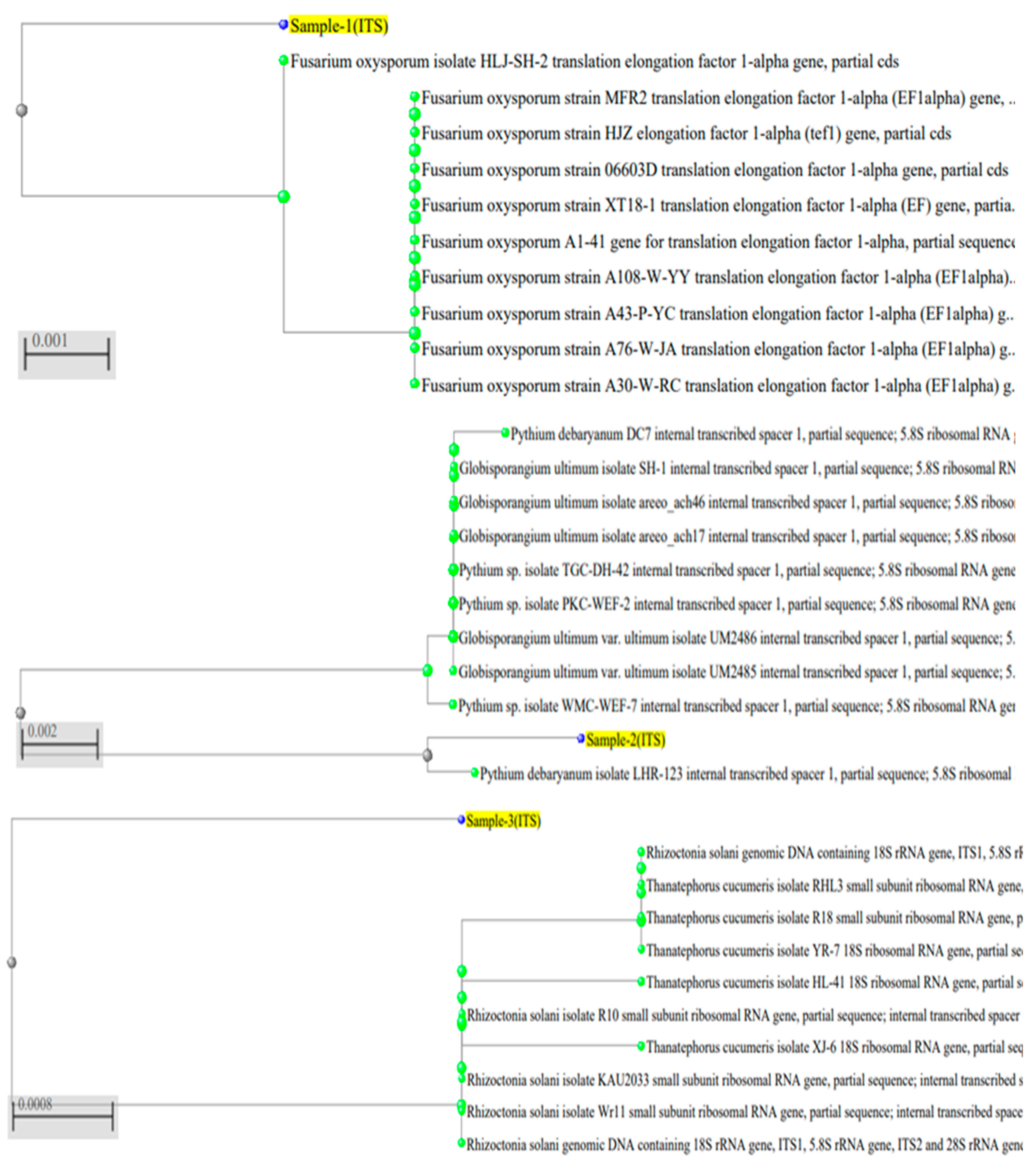
2.1. Identification of Pathogenic Fungi

Three fungal species were isolated from diseased tomato plant samples with damping-off, root rot, and wilting, which were identified based on molecular identification. For sequence analysis, data compared with the 18S rRNA gene showed Fusarium oxysporum, Pythium debaryanum, and Rhizoctonia solani (Figure 1). The sequences of amplified ITS regions were submitted to GenBank and given accession numbers of KT224063 for F. oxysporum (Sample 1), OP823136 for P. debaryanum (Sample 2), and OP823124 for R. solani (Sample 3).

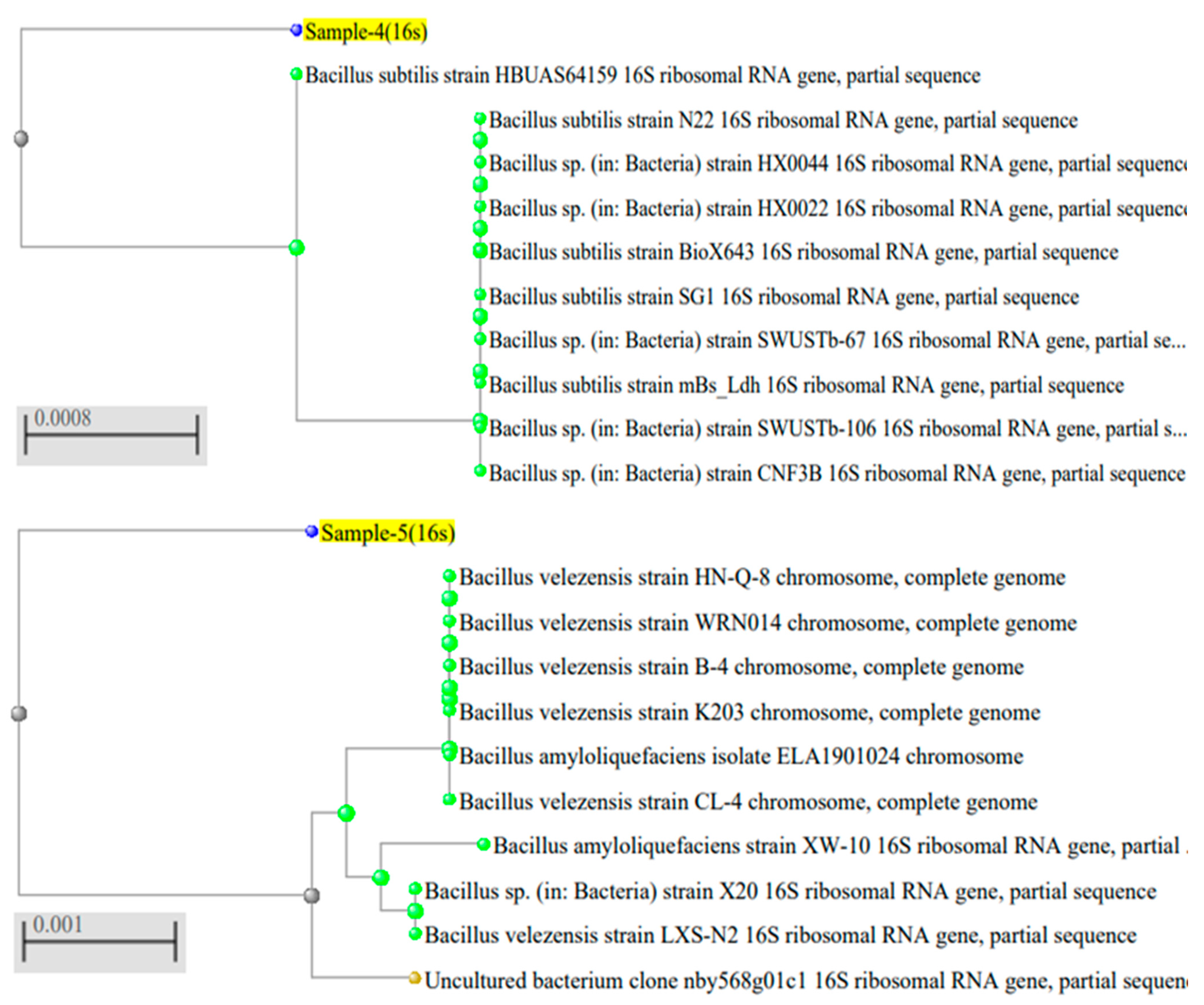
2.2. Identification of Bacillus Isolates

Enrichment of bacteria from the tomato rhizosphere on a nutrient agar medium under aerobic circumstances showed the growth of circular, rough, glistening, and creamy white colonies. In addition, the bacterial cells were spore-forming, motile, and positive for Gram staining, catalase, indole, and starch hydrolysis. Based on the morphological and chemical properties, the two most dominant and active strains were subjected to 16s rRNA analysis, which identified Bacillus subtilis (Accession No. OP823140, Figure 2, Sample 4) and Bacillus amyloliquefaciens (Accession No. OP823147, Figure 2, Sample 5).

2.3. In Vitro Antagonistic Test

On the PDA medium, the effectiveness of B. subtilis and B. amyloliquefaciens was determined to inhibit R. solani, P. debaryanum, and F. oxysporum development in dual culture, for which the inhibitory effect was remarkable after 7 days of incubation (Table 1 and Figure 3). Generally, B. subtilis outperformed B. amyloliquefaciens in resistance to R. solani and F. oxysporum, which were recorded as 28.33 and 33.00 mm. In contrast, B. amyloliquefaciens caused the highest antagonistic effect against the tested P. debaryanum fungus.

2.4. Pot Trial

2.4.1. Antagonistic Effect

The effects of antagonistic bacterial strains (B. subtilis and B. amyloliquefaciens) on the post-emergence damping-off and plant survival % with F. oxysporum, P. debaryanum, and R. solani infections in greenhouse conditions are shown in Figure 4. In comparison to the control treatments (T1, T2, and T3), the findings generally indicated that all inoculation treatments enhanced the percentage of healthy plants and lowered the damping-off %. Other than pesticide treatments (Topsin-M70), the post-emergence % of damping-off plants (45 days) showed notable decreases due to the different studied bacterial strains compared to control; therefore, the most effective treatments were recorded for B. subtilis treatments followed by B. amyloliquefaciens treatments, which decreased post-emergence. On the other hand, the control group generally showed the lowest survival rates for fungi infection, and remarkable enhancements were noticed with Bacillus bacteria treatments. The highest survival rates were recorded for Topsin-M70 and B. subtilis treatments against F. oxysporum during 2022 and 2023 seasons. A similar trend was recorded forTopsin-M70 and B. amyloliquefaciens under R. solani infection (Figure 4).

2.4.2. Photosynthetic Pigments

Table 2 shows that using different inoculation procedures under soil infected with F. oxysporum, P. debaryanum, and R. solani led to a significant (p ≤ 0.05) increase in the levels of photosynthetic pigments, i.e., total Chl, Caro, and TSS. Positive effects were seen in the T7 treatment (seedlings dipped with B. subtilis (90 min) + Soil infested with F. oxysporum (3%)), which was almost 3-fold more effective than the control treatment (T1, T2, and T3), and was recorded as 8.03 and 8.49 mg g−1 FW for total Chl, 1.57 and 1.77 µg g−1 FW for Caro, and 5.25 and 5.85 µg g−1 FW for TSS, during 2022 and 2023 seasons, respectively (Table 2). Therefore, different inoculation treatments were often organized as follows: T7 > T9 > T8 > T11 > T10 > T12 > T6 > T5 > T4 > T2 > T1 > T3.

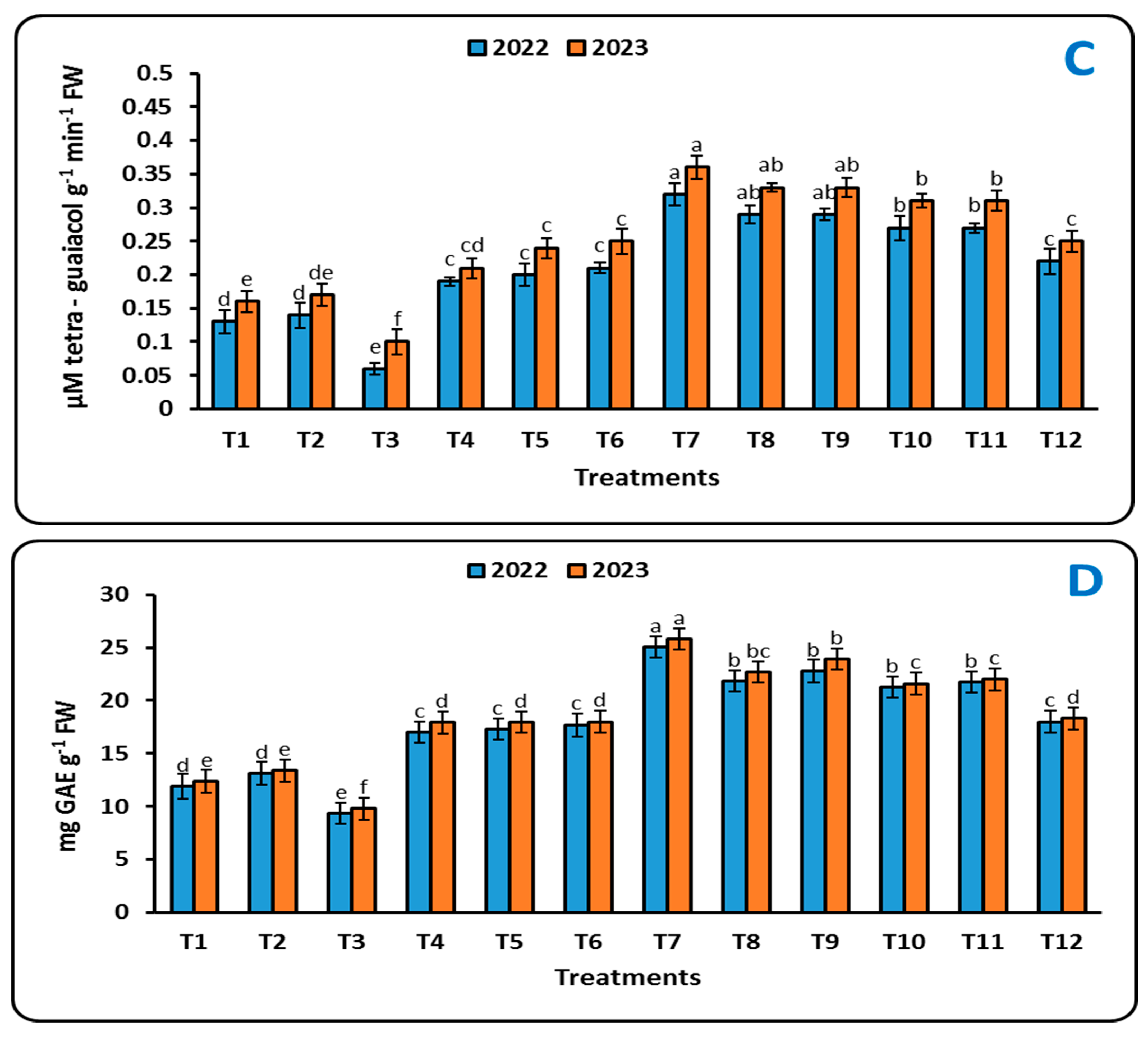
2.4.3. Antioxidant Enzymes

The data shown in Figure 5 demonstrate that, at 60 days after transplanting, the tomato plants treated with inoculation treatments under soil conditions infested with F. oxysporum, P. debaryanum, and R. solani increased their activity of the antioxidant enzymes PO, PAL, PPO, and TPC. As shown in Figure 5A, the maximum values of PO activity (μM H2O2 g−1 FW min−1) in the treated seedlings significantly increased from 0.51 and 0.60 (control, T1) to 4.04 and 4.13 (T7) under soil infested with F. oxysporum, from 0.71 and 0.75 (control, T2) to 3.05 and 3.24 (T8) under soil infested with P. debaryanum, and from 0.24 and 0.27 (control, T3) to 3.021 and 3.35 (T9) under soil infested with R. solani during 2022 and 2023 seasons, respectively (Figure 5A). Meanwhile, the maximum values of PAL enzyme activity (μmoles min−1 g−1 FW) were recorded as 5.43 and 5.58 for T7, followed by 4.81 and 4.89 for T9, compared to the control and other treatment in the first growing seasons (2022) and the second growing seasons (2023), respectively (Figure 5B).
Furthermore, the antioxidant capacity indicated by PPO activity in the untreated tomato plants was decreased by pathogenic-fungus-infested soil, but the detrimental effects of infected soil on antioxidant capacity were lessened when the seedlings were treated with alternative inoculations. The PPO enzyme activity (μM tetra-guaiacol g−1 min−1 FW) was increased by the T7 treatment compared to the other treatments, with values rising from 0.13 and 0.16 (T1) to 0.32 and 0.36 (T7) in 2022 and 2023, respectively (Figure 5C). However, we found that TPC was statistically significant (p ≤ 0.05). The data indicated that, in comparison to the control in the 2022 season, T7 (seedlings dipped with B. subtilis (90 min) + Soil infested with F. oxysporum (3%)) produced 25.07 mg GAE g−1 FW, while T9 (seedlings dipped with B. subtilis (90 min) + Soil infested with R. solani (3%)) produced 22.78 mg GAE g−1 FW. The 2023 season showed a similar pattern (Figure 5D).

2.4.4. N, P, and K (%) of Leaves

The chemical composition of tomato leaves (N, P, and K %) under F. oxysporum, P. debaryanum, and R. solani at 60 days from transplanting during 2022 and 2023 seasons differs significantly (p ≤ 0.05) depending on the different inoculation treatments (Table 3). Positive effects were seen in the T7 treatment (seedlings dipped with B. subtilis (2 h) + Soil infested with F. oxysporum (3%)), which increased compared with the control treatment (T1, T2, and T3) and was recorded as 2.30% for N, 0.187% for P, and 3.24% for K in the 2022 season and 2.48% for N, 0.216% for P, and 3.29% for K in the 2023 season, respectively (Table 3). Therefore, different inoculation treatments were often arranged as follows: T7 > T9 > T8 > T11 > T10 > T12 > T6 > T5 > T4 > T2 > T1 > T3.

2.4.5. Growth Characteristics and Yield

Tomato was used as a model crop in a greenhouse pot experiment to find the best method for applying various inoculations under F. oxysporum, P. debaryanum, and R. solani infection. The results from 12 treatments after 120 days of transplanting showed that tomato plants only had pathogenic fungal infection when compared to other inoculated plants. All growth parameters, such as shoot length, root length, shoot dry weight, and yield were lower than those of those treatments (Table 4). However, this negative impact of pathogenic fungal stress was lessened when tomatoes were inoculated with several Bacillus strains. When compared to other treatments, the T12 treatment (seedlings dipped in B. amyloliquefaciens for 90 min and soil infested with R. solani at 3%) improved the growth dynamics of the tomato plants, as shown in Table 4. For example, the tomato plants’ shoot and root lengths were 94.13 and 31.08 cm plant−1 in the 2022 season, respectively, and 100.62 and 36.20 cm plant−1 in the 2023 season, respectively. Additionally, the same trend was observed for the dry weight of the shoots and roots (Table 4). As a consequence, the T12 treatment regularly increased the rate for yield (g plant−1), which reached 53%, compared to the T2 treatment (Table 4).

3. Discussion

Biological control is globally favored for managing soilborne diseases in vegetable crops, such as Fusarium, Pythium, and Rhizoctonia, due to its eco-friendly, cost-effective, and user-friendly characteristics. Numerous commercial biopesticide products utilizing highly effective strains of antagonistic microorganisms are accessible globally for extensive field application [27]. Nonetheless, the effective biocontrol of plant diseases relies on the fitness of the microbial biocontrol agents present in the soil. Therefore, the focus of our study is on finding native antagonistic bacterial strains that are appropriate for the local environmental conditions.
In our study, we isolated the following three pathogenic fungi from infected tomato plants: F. oxysporum, P. debaryanum, and R. solani. Furthermore, two bacterial strains were isolated from the tomato rhizosphere soil: B. subtilis and B. amyloliquefaciens. Among them, B. subtilis OP823140 exhibited the strongest inhibition of mycelial growth in F. oxysporum KT224063 and R. solani OP823124. Conversely, B. amyloliquefaciens OP823147 exhibited the strongest antagonistic effect against the examined P. debaryanum OP823136 fungus (Table 1 and Figure 3). These bacterial strains’ generation of antifungal metabolites may be the cause of the inhibitory zone’s creation. Several Bacillus species have yielded bacteriocins and bacteriocin-like compounds, such as amylolysin, amisin, subtilin, subtilosin A, subtilosin B, thuricin, entianin, and ericin. Because they produce antibiotics, Bacillus species have been shown in numerous studies to have biocontrol effects on tomato diseases. According to [28], B. pumilus PTB180 and B. subtilis PTB185 produce surfactin (both strains) and iturin and fengycin (B. subtilis PTB185), which have a strong antagonistic effect on a number of plant pathogens, including F. oxysporum, R. solani, S. sclerotiorum, Pythium ultimum, and Phytophthora capsici.
B. subtilis treatments were shown to have the most effective antagonistic impact, followed by B. amyloliquefaciens treatments, which reduced post-emergence and increased plant survival (Figure 4). These findings are consistent with those of [29,30], who found that seedlings were protected from Fusarium sp. and R. solani infection in plants treated with bioagents and bio-fungicides. Because Bacillus species activate soil microbes and decrease the population of F. oxysporum, R. solani, and P. debaryanum, they significantly decreased the incidence of damping-off disease. Additionally, Bacillus species increased the biomass of the root system, which may have a positive effect on disease control, defense mechanism activation, and the accumulation of antimicrobial secondary metabolites [31,32,33].
One essential measure of a plant’s physiological state is its photosynthetic pigment [34]. Under various inoculation treatments, the total soluble sugar, carotenoids, and chlorophyll content of tomato plants were determined (Table 2). In fact, compared to plants treated with Bacillus species, tomato plants infected with F. oxysporum, P. debaryanum, and R. solani had a negative impact on the amount of photosynthetic pigment. Our findings are consistent with those of [35], who demonstrated that the overall chlorophyll content of A. alternate diseased plants was much lower in the diseased tissue—roughly 16 times lower than that found in healthy tissues. Likewise, decreases of 9.96% and 40.34% in total chlorophyll content were reported by [36]. According to [37], the disarray of the plastid membrane during infection may be the cause of this decrease in total chlorophylls and pigments. Kazerooni [36] observed a similar trend of better chlorophylls and pigments, where B. amyloliquefaciens increased total chlorophyll in pepper plants under Botrytis and Alternaria stress conditions by 31.07% and 57.88%, respectively, in comparison to those of infected plants. Furthermore, [38] showed that Raphanus sativus’s chlorophyll content increased as a result of Pseudomonas and Bacillus. The bacteria’s ability to operate as a biofertilizer or the increase in 1-Aminocyclopropane-1-Carboxylate (ACC) deaminase enzymes in PGPR-treated plants, which delay the breakdown of chlorophyll, could be the cause of this increase in chlorophyll concentration. Additionally, the chlorophyll content rose as a result of PGPR’s improved nutrient absorption. Similarly, B. amyloliquefaciens SN13 enhanced carbon assimilation in rice plants with or without R. solani, which is strongly associated with higher dry mass and chlorophyll content, according to [39].
Preserving the integrity of the host plant’s defense mechanisms is one possible method for the biological management of fungal infections (Figure 5). Antioxidant enzymes have a critical role in scavenging ROS and avoiding oxidative stress, which harms numerous sensitive molecules [40,41]. According to [42], peroxidase (PO) is essential at the start of the plant’s defense response against infections because it can either create highly toxic conditions by releasing more ROS and synthesizing phenolic chemicals or by constructing structural barriers (lignin accumulation). Additionally, PO actively participates in growth-related self-regulation processes like photosynthesis and respiration [43]. Numerous studies have documented the various manifestations of PAL. For example, the activity of PAL in wheat was impacted by the Fusarium mycotoxin deoxynivalenol (DON), which resulted in the downregulation of PAL in susceptible cultivars [44]. Although it is challenging to understand how Bacillus inhibits PAL activity in the plant–pathogen–bacillus relationship, PAL biosynthesis from L-phenylalanine may occur in the plant via other pathways, and the PAL reaction is not the only pathway for the body’s defense response [45]. However, PPO is important for disease resistance because of its ability to accelerate the oxidation of phenolic chemicals into quinones and the synthesis of lignin [46]. PPO activity did not directly contribute to the development of mung beans’ resistance to pathogenic fungus, according to [47], who also discovered a negative association between PPO activity and the proportion of plants that survived and no correlation between PPO activity and total phenol. As one of the biggest and most diverse classes of plant active chemicals, phenols play a key role in initiating defense responses to abiotic stress and pathogen infection. Phenolics are involved in controlling the growth of plants. Phenolics are created when plants detect possible pathogens [48]. However, phenol buildup is also influenced by environmental factors such as light and temperature [49], nutritional status, and plant genetics or species [50]. These factors imply that there is a very complicated link between elicitor activity and specificity in a plant–microbe interaction.
The chemical contents of tomato leaves (N, P, and K%) cultivated in soil infected with F. oxysporum, P. debaryanum, and R. solani varied considerably (p ≤ 0.05) based on the various Bacillus inoculation treatments (Table 3). Bacillus uses hydrolysis, chelation, redox, and acidity to solubilize minerals as a plant growth regulator [51]. According to [52], B. methylotrophicus increased the synthesis of chlorophyll and the uptake of NPK nutrients. Additionally, compared to control plants, chickpea plants that were inoculated with M. ciceri IC53 and NUU4 had higher N contents [53]. PGPR strains may directly enhance plant metabolism by enhancing their host plants’ absorption of water, nutrients, and enzyme activity [54]. Furthermore, because of their higher mineral content, plants inoculated with B. amyloliquefaciens RaSh1 may produce more metabolites, proteins, and defense genes [34].
In comparison to the treatments inoculated with Bacillus bacteria, all growth metrics, including shoot length, root length, dry weight of shoots, dry weight of roots, and yield, were lower in circumstances infected solely with pathogenic fungi (Table 4). The findings of earlier studies [55,56,57] are consistent with these findings. Furthermore, the beneficial effects of beneficial PGPR were derived from the observable influence of bacteria on growth, development, and yield. Additionally, by secreting phytohormones like indole acetic acid, bacteria help plants to obtain nitrogen from the atmosphere or other nutrients in the soil [58,59]. Adinarayana [60] showed that tomato plants inoculated with Bacillus subtilis and Bacillus amyloliquefaciens under diseases incidence of A. solani. had an improved growth and yield and were recorded to have 110.07 fruits per plant, 6.42 Kg yield per plant, 179.42 Kg yield per plot and 112.13 tons yield ha−1.

4. Materials and Methods

4.1. Sources of Samples

Diseased tomato plants with their rhizosphere were collected from different regions of the Governorate of Elkharbyia (Gymmiza and Kotor), Egypt, and were subjected to isolation trials following the method described by [61].

4.2. Isolation and Identification of Pathogenic Fungi

The lower parts of the stem and roots of the diseased tomato plants with damping-off, root rot, and wilting were cleaned from soil particles attached, cut into 2 cm pieces, sterilized with 3% sodium hypochlorite (3 min), washed 5 times with sterilized distilled water, and dried using filter sheets. The sterilized plant pieces were incubated in a potato dextrose agar (PDA) medium for 7 days at 27 °C. The growing hyphal tips were moved from the agar plates and purified several times on a new PDA medium using the hyphal tip technique [62].
As stated by [63], the three pure fungi were identified using their morphological and microscopical characteristics and were identified by polymerase chain reaction (PCR) at Sigma Scientific Services Co., Giza, Egypt. The growth mycelia of each fungus were acquired by centrifugation for 10 min at 6000 rpm, homogenized in liquid nitrogen, and their RNA was extracted using total extraction kits (Solarbio, Beijing, China) according to the manufacturer’s guidelines. The PCR was completed in a total amount of 50 µL, comprising 1× reaction buffer, 1.5 mM MgCl2, 1U Taq DNA polymerase (Promega, Madison, WI, USA), 2.5 mM dNTPs, 30 Pmol of each primer (ITS-1 F: 5′-TCCGTAGGTGAACCTGCGG-3′ and ITS-1 R: 5′-TCCTCCGCTTATTGATATGC-3′), and 30 ng genomic DNA. With a Big Dye TM Terminator Cycle Sequencing Kits, the resultant PCR was sequenced automatically in an ABI PRISM 3730XL Analyzer while adhering to the manufacturer’s instructions. Each template underwent single-pass sequencing with the Rbcl Forward primer. The fluorescently tagged fragments were isolated from the unincorporated terminators using an ethanol precipitation process. The samples were resuspended in distilled water and then electrophoresed on an ABI 3730xl sequencer (Microgen Company, Moscow, Russia). The sequences were aligned using Nucleotide BLAST http://www.ncbi.nlm.nih.gov/BLAST (accessed on 14 September 2023).

4.3. Isolation and Identification of Bacillus spp.

Approximately 10 g of tomato rhizosphere soil was added to sterile water (90 mL) and agitated on a rotary shaker at 150 rpm for 45 min. The soil suspension was serially diluted (105–106) and pasteurized in a water bath for 15 min at 65 °C to kill non-spore-forming microbes [64]. After cooling, the soil suspension was cultured on a nutrient agar medium (NA), and colonies appearing after 3 days at 27 °C were isolated and purified using the streaking plate method. The two pure bacteria were identified by 16s rRNA according to [65]. Bacterial colonies were grown for 8 h at 30 °C in a nutrient broth prior to DNA extraction. Centrifuging 1 mL of culture media at 8000× g for 2 min extracted the bacterial cells. The cells were then rinsed twice with 400 µL of STE Buffer (100 mM NaCl, 10 mM Tris/HCl, 1 mM EDTA, pH 8.0) and centrifuged again at 8000× g for 2 min. To lyse the bacterial cells, the generated pellets were again suspended in 200 µL TE buffer (10 mM Tris/HCl, 1 mM EDTA, pH 8.0) together with 100 µL Tris-saturated phenol (pH 8.0) and vortexed for 60 s. The samples were separated into liquid and solid by centrifuging them for five minutes at 4 °C at 13,000× g. A portion of the top liquid, 160 µL, was moved to a 1.5 mL tube with 100 µL chloroform and 40 µL TE buffer. The tube was then centrifuged at 13,000× g for 5 min at 4 °C. The PCR mixture (50 µL) comprised 5 µL of 5× Taq buffer, 200 mmol/L of each dNTP, 10 Pmol of primers 27F (Forward: 5\-AGAGTTTGATCMTGGCTCAG-3\) and 1492R (Reverse: 5\-GGYTACCTTGTTACGACTT-3\), 1.5 U of Taq DNA polymerase (Promega, Madison, WI, USA), 3 mmol/L of MgCl2, and 5 µL of genomic DNA. The PCR condition, thermal cycle structure, and sequence of PCR products are described in Section 2.2.

4.4. In Vitro Antagonistic Assay

Three fungal isolates, including F. oxysporum, P. debaryanum, and R. solani, along with two bacterial strains, B. subtilis and B. amyloliquefaciens, were selected based on the identification process detailed in Section 4.2 and Section 4.3 to perform a dual culture assay to assess the antagonistic effect [66]. A loopful of bacterial broth culture (108 CFU/mL) was streaked diagonally on a Petri dish filled with PDA (without antibiotics), while fungal strains from a 7-day-old PDA culture were spotted at the two edges of the dish, perpendicular to the bacterial line, using sterile toothpicks with 6 mm diameter agar discs. The setup was then incubated for 7 days at 27 °C. The inhibition level in the dual cultures was determined by subtracting the distance (mm) of fungal growth towards the antagonist colony (γ) from the fungal growth radius (γ0) of the control culture, resulting in Δγ = γ0 − γ, as outlined by [67]. The controls comprised fungal mycelial plugs placed in the center of non-inoculated PDA plates.

4.5. Inoculum Preparation

The investigated fungi were inoculated into sterilized 250 mL potato dextrose broth medium in 0.5 L Erlenmeyer flasks, and they were then cultured for 14 days at 27 °C. After thoroughly shaking the flasks, 20 mL of the suspension was moved to a 1 L Erlenmeyer flask that contained 2/3 wet autoclaved grain sorghum. The flask was then kept at 27 °C for four weeks. Soil was infested with pathogenic fungi one week prior to transplanting by incorporating 3% inoculum into the soil of each pot, then irrigating [68]. Conversely, the bacillus strains were cultivated in nutrient broth medium on a rotary shaker for 72 h at 27 °C, and each culture (1 × 10⁸ CFU mL−1) served as inoculum by immersing the seedlings for 90 min [69].

4.6. Greenhouse Trial

A pot experiment was carried out in the greenhouse at the Gymmiza Agricultural Research Station, located in Elkharbyia Governorate, Egypt. Clay soil was gathered, air-dried, sifted through a 10-mesh sieve, assessed for its physical and chemical properties as indicated in Table 5, and subsequently placed into polyethylene bags containing 8 kg of soil following sterilization. During the 2022–2023 seasons, a total of 96 tomato plants (cv. Super Strain B, at 25 days old, acquired from a commercial nursery as sensitive variety) were separated into 12 treatments, each consisting of 8 duplicates with 1 plant. The pots were organized in a randomized block design with the treatments displayed in Table 6. The proposed dosage by the Egyptian Ministry of Agriculture of 10 mL of 4 g L−1 N:P:K (20:20:20) was administered to the plants every three days, in conjunction with weekly irrigation.

4.7. Measurements

4.7.1. Disease Assessment

The percentages of post-emergency damping-off and surviving plants were computed after 45 days of transplant, as follows [70]:
Post-emergency damping-off % = [No. of dead plants/No. of transplanted seedlings] × 100
Surviving plants % = [No. of surviving plants/No. of transplanted seedlings] × 100

4.7.2. Physiological Features

Sixty days post-transplantation and following each treatment, a leaf sample was frozen to evaluate the photosynthetic pigments, total soluble sugars (TSS), antioxidant enzymes, and phenolic content using a UV spectrophotometer (Bibby Scientific, Staffordshire, UK) (Model 6705).

Photosynthetic Pigments

As stated by [71], to assess the total chlorophyll and carotenoids, 0.1 g of leaf samples was ground and subsequently extracted in 5 mL of 80% acetone. The supernatant was analyzed at wavelengths of 663, 645, and 470 nm following centrifugation at 13,000× g for 10 min. The measured and documented values for carotenoids and chlorophylls are µg g−1 FW and mg g−1 FW, respectively.

TSS

We implemented the measures proposed by [72]. Following homogenization in ethanol (5 mL, 80%), a 0.5 g leaf sample was placed in a water bath and heated at 80 °C for 30 min. To assess the TSS concentration at 620 nm, the supernatants were gathered and centrifuged (10,000× g for 10 min). The data were shown as µg g−1 FW, with glucose serving as the standard curve.

4.7.3. Antioxidant Enzymes

Peroxidase Activity (PO)

Utilizing the techniques outlined by [73], the conversion of pyrogallol to purpurgallin with H2O2 was evaluated to measure the peroxidase enzyme’s activity. In the sample cuvette, there was 3.0 mL of dH2O, 0.5 mL of 0.1M sodium phosphate buffer (pH 7), 0.3 mL of the extract (enzyme), 0.05 mL of pyrogallol, and 0.1 mL of H2O2 (10%). The peroxidase enzyme activity was assessed at 425 nm μM H2O2 g−1 FW min−1.

Polyphenol Oxidase Assay (PPO)

As stated by [74], a measurement of the enzyme polyphenol oxidase was conducted. In summary, this included 1.0 mL of sodium phosphate buffer (0.2 M, pH 7), 10.0 mL of catechol (0.001 M), 1.0 mL of extract (enzyme), and 3.0 mL of dH2O. The activity of polyphenol oxidase was assessed at 495 nm μM tetra-guaiacol g−1 min−1 FW.

Phenylalanine Ammonia Lyase Assay (PAL)

The enzyme phenylalanine ammonia lyase was measured following the method outlined by [75]. In summary, the reaction included 1.5 mL of borate buffer (0.2 M) + 1 mL of phenylalanine (1%) + 2.5 mL of dH2O, with a pH of 8.8. A total of 1 mL of deionized water was mixed with phenylalanine to serve as a blank. The blend was incubated at 40 °C for one hour. Subsequently, the reaction was halted by introducing 0.5 mL of HCL (5N), and the enzyme’s activity was assessed at 290 nm μmoles min−1 g−1 FW.

Total Phenolic Content (TPC)

The TPC was estimated based on the approach described by [76]. A total of 10 mL of 80% methanol was utilized to homogenize 1 g of leaf tissue, subsequently stirring the mixture for 15 min at 70 °C. The solution was maintained at 25 °C, consisting of 1 mL of the methanol extract, 5 mL of dH2O, and 250 µL of 1 N Folin Ciocalteau reagent. After adding 1 mL of saturated Na2CO3 and 1 mL of dH2O, the reaction mixture was allowed to incubate for another hour at 25 °C. The absorption of the resulting blue color was recorded at 725 nm. Using the gallic acid calibration curve, TPC was assessed and expressed as mg GAE g−1 FW.

Chemical Contents of Leaves

Dried samples were maintained at 65 °C for 3 days and then ground into a uniform powder (IKa-Werke, M 20 Darmstadt, Germany). N% was measured using the Micro-Kjeldahl method [77], while P and K % were determined using spectrophotometry and atomic absorption spectrometry methods, respectively, as per [78,79]. The chemical composition of the leaves was analyzed 60 days following transplantation.

4.8. Plant Growth and Yield

A total of 120 days after transplantation, the shoot and root length (cm plant−1), dry weight of the shoots and roots (g plant−1), and yield (g plant−1) were determined.

4.9. Statistical Analyses

The SPSS program (version 20; IBM Corp., Armonk, NY, USA) was used to carry out a variance analysis on the data. Duncan’s multiple range testing method was used to examine the mean separations, and p ≤ 0.05 was considered significant [80].

5. Conclusions

According to our study, the pathogens F. oxysporum, P. debaryanum, and R. solani are inhibited by two strains of B. subtilis and B. amyloliquefaciens. B. subtilis was more resistant to R. solani and F. oxysporum than B. amyloliquefaciens. By contrast, the fungus P. debaryanum under investigation was most antagonistically affected by B. amyloliquefaciens. In the greenhouse study, we found that applying each of these antagonistic Bacillus strains individually to tomato plants successfully reduced the incidence of the pathogens’ disease. Moreover, two Bacillus strains used individually considerably enhanced plant growth metrics, boosted macronutrient absorption, and elevated the levels of photosynthetic pigments, total phenolic compounds, and yield. Consequently, employing B. subtilis and B. amyloliquefaciens as potent bio-fungicides can reduce our reliance on harmful synthetic fungicides and chemical fertilizers while also promoting environmentally friendly and sustainable agricultural methods. In addition, we recommend further studies in the field, with the possibility of using it as a commercial product.

Author Contributions

Conceptualization, D.F.I.A., S.E.-N., H.A.H.E.-Z. and A.E.-D.O.; methodology, D.F.I.A., S.E.-N., H.A.H.E.-Z. and A.E.-D.O.; software, D.F.I.A. and A.E.-D.O.; validation, D.F.I.A., S.E.-N., H.A.H.E.-Z. and A.E.-D.O.; formal analysis, A.E.-D.O.; investigation, D.F.I.A., S.E.-N., H.A.H.E.-Z. and A.E.-D.O.; resources, H.A.H.E.-Z. and A.E.-D.O.; data curation, A.E.-D.O.; writing—original draft preparation, D.F.I.A. and A.E.-D.O.; writing—review and editing, A.E.-D.O.; visualization, D.F.I.A., S.E.-N., H.A.H.E.-Z. and A.E.-D.O.; supervision, D.F.I.A. and A.E.-D.O.; project administration, D.F.I.A. and A.E.-D.O.; funding acquisition, D.F.I.A. and A.E.-D.O. All authors have read and agreed to the published version of the manuscript.

Funding

This research received no external funding.

Data Availability Statement

Data sets analyzed during the present study are accessible from the current author upon reasonable request.

Acknowledgments

All the authors are grateful for the support provided by Faculty of Agriculture, Mansoura University, Faculty of Agriculture, Al-Azhar University, and Soils, Water, and Environment Research Institute (SWERI), Agriculture Research Center (ARC), Egypt.

Conflicts of Interest

The authors declare no conflicts of interest.

References

  1. Hashem, A.H.; Abdelaziz, A.M.; Askar, A.A.; Fouda, H.M.; Khalil, A.M.; Abd-Elsalam, K.A.; Khaleil, M.M. Bacillus megaterium-mediated synthesis of selenium nanoparticles and their antifungal activity against Rhizoctonia solani in Faba Bean Plants. J. Fungi 2021, 7, 195. [Google Scholar] [CrossRef] [PubMed]
  2. Abdelaziz, A.M.; Kalaba, M.H.; Hashem, A.H.; Sharaf, M.H.; Attia, M.S. Biostimulation of tomato growth and biocontrol of Fusarium wilt disease using certain endophytic fungi. Bot. Stud. 2022, 63, 34. [Google Scholar] [CrossRef]
  3. FAOSTAT Database. Food and Agriculture Organization Statistics. Available online: https://www.fao.org/faostat/en/ (accessed on 15 November 2023).
  4. Panno, S.; Davino, S.; Caruso, A.G.; Bertacca, S.; Crnogorac, A.; Mandić, A.; Matić, S. A review of the most common and economically important diseases that undermine the cultivation of tomato crop in the Mediterranean Basin. Agronomy 2021, 11, 2188. [Google Scholar] [CrossRef]
  5. Montenegro, I.; Madrid, A.; Cuellar, M.; Seeger, M.; Alfaro, J.F.; Besoain, X.; Martínez, J.P.; Ramirez, I.; Olguín, Y.; Valenzuela, M. Biopesticide activity from drimanic compounds to control tomato pathogens. Molecules 2018, 23, 2053. [Google Scholar] [CrossRef]
  6. Attia, M.S.; El-Wakil, D.A.; Hashem, A.H.; Abdelaziz, A. Antagonistic effect of plant growth-promoting fungi against fusarium wilt disease in tomato: In vitro and in vivo study. Appl. Biochem. Biotechnol. 2022, 194, 5100–5118. [Google Scholar] [CrossRef] [PubMed]
  7. Renu, J. A review of Fusarium oxysporum on its plant interaction and industrial use. J. Med. Plants Stud. 2018, 6, 112–115. [Google Scholar]
  8. Salman, M.; Abuamsha, R. Potential for integrated biological and chemical control of damping-off disease caused by Pythium ultimum in tomato. BioControl 2012, 57, 711–718. [Google Scholar] [CrossRef]
  9. Balghouthi, A.; Jonathan, R.; Gognies, S.; Mliki, A.; Belarbi, A. A new species, Pythium echinogynum, causing severe damping-off of tomato seedlings, isolated from Tunisia, France, and India: Morphology, pathology, and biological control. Ann. Microbiol. 2013, 63, 253–258. [Google Scholar] [CrossRef]
  10. Sarhan, E.A.D.; El-Far, E.M.M.; Ebrahiem, A.M.Y. Systemic resistance in snap bean (Phaseolus vulgaris L.) elicited by some chemicals and biotic inducers against white mold disease caused by Sclerotinia sclerotiorum. Egypt. J. Phytopathol. 2018, 46, 61–84. [Google Scholar] [CrossRef]
  11. Droby, S.; Wisniewski, M.; Macarisin, D.; Wilson, C. Twenty years of postharvest biocontrol research: Is it time for a new paradigm? Postharvest Biol. Technol. 2009, 52, 137–145. [Google Scholar] [CrossRef]
  12. Singh, A.; Jain, A.; Sarma, B.K.; Upadhyay, R.S.; Singh, H.B. Rhizosphere microbes facilitate redox homeostasis in Cicer arietinum against biotic stress. Ann. Appl. Biol. 2013, 163, 33–46. [Google Scholar] [CrossRef]
  13. Gupta, R.; Singh, A.; Srivastava, M.; Singh, V.; Gupta, M.M.; Pandey, R. Microbial modulation of bacoside a biosynthetic pathway and systemic defense mechanism in Bacopa monnieri under Meloidogyne incognita stress. Sci. Rep. 2017, 7, 41867. [Google Scholar] [CrossRef] [PubMed]
  14. Rosenblueth, M.; Martínez-Romero, E. Bacterial endophytes and their interactions with hosts. Mol. Plant Microbe Interact. 2006, 19, 827–837. [Google Scholar] [CrossRef] [PubMed]
  15. Zhang, X.; Gao, J.; Zhao, F.; Zhao, Y.; Li, Z. Characterization of a salt-tolerant bacterium Bacillus sp. from a membrane bioreactor for saline wastewater treatment. J. Environ. Sci. 2014, 26, 1369–1374. [Google Scholar] [CrossRef] [PubMed]
  16. Fan, H.; Zhang, Z.; Li, Y.; Zhang, X.; Duan, Y.; Wang, Q. Biocontrol of bacterial fruit blotch by Bacillus subtilis 9407 via surfactinmediated antibacterial activity and colonization. Front. Microbiol. 2017, 8, 1973. [Google Scholar] [CrossRef]
  17. Zhang, Q.X.; Zhang, Y.; He, L.L.; Ji, Z.L.; Tong, Y.H. Identification of a small antimycotic peptide produced by Bacillus amyloliquefaciens 6256. Pesticide Biochem. Physiol. 2018, 150, 78–82. [Google Scholar] [CrossRef]
  18. Duan, Y.; Chen, R.; Zhang, R.; Jiang, W.; Chen, X.; Yin, C.; Mao, Z. Isolation, identification, and antibacterial mechanisms of Bacillus amyloliquefaciens QSB-6 and its effect on plant roots. Front. Microbiol. 2021, 12, 746799. [Google Scholar] [CrossRef] [PubMed]
  19. Garcia-Fraile, P.; Menendez, E.; Rivas, R. Role of bacterial biofertilizers in agriculture and forestry. AIMS Bioeng. 2015, 2, 183–205. [Google Scholar] [CrossRef]
  20. Kang, S.M.; Radhakrishnan, R.; Lee, I.J. Bacillus amyloliquefaciens subsp. plantarum GR53, a potent biocontrol agent resists Rhizoctonia disease on Chinese cabbage through hormonal and antioxidants regulation. World J. Microbiol. Biotechnol. 2015, 31, 1517–1527. [Google Scholar] [PubMed]
  21. Ahmed, W.; Zhou, G.; Yang, J. Bacillus amyloliquefaciens WS-10 as a potential plant growth-promoter and biocontrol agent for bacterial wilt disease of flue-cured tobacco. Egypt. J. Biol. Pest. Control 2022, 32, 25. [Google Scholar] [CrossRef]
  22. Soliman, S.A.; Khaleil, M.M.; Metwally, R.A. Evaluation of the antifungal activity of Bacillus amyloliquefaciens and B. velezensis and characterization of the bioactive secondary metabolites produced against plant pathogenic fungi. Biology 2022, 11, 1390. [Google Scholar] [CrossRef] [PubMed]
  23. Solanki, M.K.; Singh, R.K.; Srivastava, S.; Kumar, S.; Kashyup, P.L.; Srivastava, A.K. Characterization of antagonistic-potential of two Bacillus strains and their biocontrol activity against Rhizoctonia solani in tomato. J. Basic. Microbiol. 2013, 53, 82–90. [Google Scholar] [CrossRef]
  24. Zouari, I.; Jlaiel, L.; Tounsi, S.; Trigui, M. Biocontrol activity of the endophytic Bacillus amyloliquefaciens strain CEIZ-11 against Pythium aphanidermatum and purification of its bioactive compounds. Biol. Control 2016, 100, 54–62. [Google Scholar] [CrossRef]
  25. Diabankana, R.G.C.; Shulga, E.U.; Validov, S.Z.; Afordoanyi, D.M. Genetic characteristics and enzymatic activities of Bacillus velezensis KS04AU as a stable biocontrol agent against phytopathogens. Int. J. Plant Biol. 2022, 13, 201–222. [Google Scholar] [CrossRef]
  26. Rashad, Y.M.; Abdalla, S.A.; Sleem, M.M. Endophytic Bacillus subtilis SR22 triggers defense responses in tomato against rhizoctonia root rot. Plants 2022, 11, 2051. [Google Scholar] [CrossRef] [PubMed]
  27. Lahlali, R.; Ezrari, S.; Radouane, N.; Kenfaoui, J.; Esmaeel, Q.; El Hamss, H.; Belabess, Z.; Barka, E.A. Biological control of plant pathogens: A global perspective. Microorganisms 2022, 10, 596. [Google Scholar] [CrossRef] [PubMed]
  28. Bouchard-Rochette, M.; Machrafi, Y.; Cossus, L.; Nguyen, T.T.A.; Antoun, H.; Droit, A.; Tweddell, R.J. Bacillus pumilus PTB180 and Bacillus subtilis PTB185: Production of lipopeptides, antifungal activity, and biocontrol ability against Botrytis cinerea. Biol. Control 2022, 170, 104925. [Google Scholar] [CrossRef]
  29. Ahmad, A.G.; Attia, A.Z.; Mohamed, M.S.; Elsayed, H.E. Fermentation, formulation and evaluation of PGPR Bacillus subtilis isolate as a bioagent for reducing occurrence of peanut soil-borne diseases. J. Integr. Agric. 2019, 18, 2080–2092. [Google Scholar] [CrossRef]
  30. Khalifa, N.A.; Saleh, R.A.; Mahmoud, N.A. Efficiency of some bio-control agents and plant extracts against beans (Phaseolus vulgaris L.) damping-off and root rot diseases under greenhouse and field conditions. Egypt. J. Phytopathol. 2021, 49, 152–167. [Google Scholar] [CrossRef]
  31. Samaras, A.; Roumeliotis, E.; Ntasiou, P.; Karaoglanidis, G. Bacillus subtilis MBI600 promotes growth of tomato plants and induces systemic resistance contributing to the control of soilborne pathogens. Plants 2021, 10, 1113. [Google Scholar] [CrossRef]
  32. Gaya Karunasinghe, T.; Hashil Al-Mahmooli, I.; Al-Sadi, A.M.; Velazhahan, R. The effect of salt-tolerant antagonistic bacteria from tomato rhizosphere on plant growth promotion and damping-off disease suppression under salt-stress conditions. Acta Agric. Scand. Sect. B Soil. Plant Sci. 2020, 70, 69–75. [Google Scholar] [CrossRef]
  33. Al-Daghari, D.S.; Al-Sadi, A.M.; Al-Mahmooli, I.H.; Janke, R.; Velazhahan, R. Biological control efficacy of indigenous antagonistic bacteria isolated from the rhizosphere of cabbage grown in biofumigated soil against Pythium aphanidermatum damping-off of cucumber. Agriculture 2023, 13, 626. [Google Scholar] [CrossRef]
  34. Abdelhameed, R.E.; Metwally, R.A.; Soliman, S.A. Prospects of Bacillus amyloliquefaciens (MZ945930) mediated enhancement of Capsicum annuum L. plants under stress of Alternaria alternata in terms of physiological traits, thiol content, antioxidant defense, and phytohormones. J. Plant Growth Regul. 2024, 43, 3265–3281. [Google Scholar] [CrossRef]
  35. Sharma, A.; Sharma, I.; Pati, P. Post-infectional changes associated with the progression of leaf spot disease in Withania somnifera (L.) Dunal. J. Plant Pathol. 2011, 93, 397–405. [Google Scholar]
  36. Kazerooni, E.A.; Maharachchikumbura, S.S.N.; Al-Sadi, A.M.; Kang, S.M.; Yun, B.W.; Lee, I.J. Biocontrol potential of Bacillus amyloliquefaciens against Botrytis pelargonii and Alternaria alternata on Capsicum annuum. J. Fungi 2021, 7, 472. [Google Scholar] [CrossRef] [PubMed]
  37. Alwadi, H.M.; Baka, Z.A.M. Microorganisms associated with Withania somnifera leaves. Microbiol. Res. 2001, 156, 303–309. [Google Scholar] [CrossRef]
  38. Yildirim, E.; Turan, M.; Donmez, M.F. Mitigation of salt stress in radish (Raphanus sativus L.) by plant growth promoting rhizobacteria.—Roman. Biotechnol. Lett. 2008, 13, 3933–3943. [Google Scholar]
  39. Srivastava, S.; Bist, V.; Srivastava, S.; Singh, P.C.; Trivedi, P.K.; Asif, M.H.; Chauhan, P.S.; Nautiyal, C.S. Unraveling aspects of Bacillus amyloliquefaciens mediated enhanced production of rice under biotic stress of Rhizoctonia solani. Front. Plant Sci. 2016, 6, 587. [Google Scholar] [CrossRef] [PubMed]
  40. Metwally, R.A.; Soliman, S.A. Alleviation of the adverse effects of NaCl stress on tomato seedlings (Solanum lycopersicum L.) by Trichoderma viride through the antioxidative defense system. Bot. Stud. 2023, 64, 4. [Google Scholar] [CrossRef]
  41. Omara, A.E.; El-maghraby, F.M. Novel Bioformulations with Trichoderma lixii to Improve the Growth Dynamics and Biocontrol of the Cowpea Damping-Off Disease. Microbiol. Res. 2023, 14, 2041–2066. [Google Scholar] [CrossRef]
  42. Adhilakshmi, M.; Paranidharan, V.; Balachandar, D.; Ganesamurthy, K.; Velazhahan, R. Suppression of root rot of mung bean (Vigna radiata L.) by Streptomyces sp. is associated with induction of peroxidase and polyphenol oxidase. Arch. Phytopathol. Plant Prot. 2014, 47, 71–583. [Google Scholar] [CrossRef]
  43. Kavitha, R.; Umesha, S. Regulation of defense-related enzymes associated with bacterial spot resistance in tomato. Phytoparasitica 2008, 36, 144. [Google Scholar] [CrossRef]
  44. Perincherry, L.; Lalak-Kańczugowska, J.; Stępień, Ł. Fusarium-produced mycotoxins in plant-pathogen interactions. Toxins 2019, 11, 664. [Google Scholar] [CrossRef]
  45. Inayati, A.; Sulistyowati, L.; Aini, L.Q.; Yusnawan, E. Trichoderma virens-Tv4 enhances growth promoter and plant defenserelated enzymes of mungbean (Vigna radiata) against soil-borne pathogen Rhizoctonia solani. Biodiversitas J. Biol. Divers. 2020, 21, 2410–2419. [Google Scholar] [CrossRef]
  46. Constabel, C.P.; Barbehenn, R. Defensive roles of polyphenol oxidase in plants. In Induced Plant Resistance to Herbivory; Schaller, A., Ed.; Springer: Dordrecht, Germany, 2008. [Google Scholar]
  47. Mousa, E.S.; Elbagory, M.; Mahdy, M.E.; Abo-Koura, H.A.; Omara, A.E. Microencapsulation of Bacillus megaterium in Humic Acid-Supplied Alginate Beads Enhances Tomato Growth and Suppresses the Root-Knot Nematode Meloidogyne javanica Under Greenhouse Conditions. Horticulturae 2024, 10, 1284. [Google Scholar] [CrossRef]
  48. Kubalt, K. The role of phenolic compounds in plant resistance. Biotechnol. Food Sci. 2016, 80, 97–108. [Google Scholar]
  49. Løvdal, T.; Olsen, K.M.; Slimestad, R.; Verheul, M.; Lillo, C. Synergetic effects of nitrogen depletion, temperature, and light on the content of phenolic compounds and gene expression in leaves of tomato. Phytochemistry 2010, 71, 605–613. [Google Scholar] [CrossRef]
  50. Bhattacharya, A.; Sood, P.; Citovsky, V. The roles of plant phenolics in defense and communication during Agrobacterium and Rhizobium infection. J. Mol. Plant Pathol. 2010, 11, 705–719. [Google Scholar] [CrossRef]
  51. Hesham, A.E.-L.; Upadhyay, R.S.; Sharma, G.D.; Manoharachary, C.; Gupta, V.K. Fungal Biotechnology and Bioengineering; Springer: Berlin/Heidelberg, Germany, 2020. [Google Scholar]
  52. Radhakrishnan, R.; Lee, I.J. Gibberellins producing Bacillus methylotrophicus KE2 supports plant growth and enhances nutritional metabolites and food values of lettuce. Plant Physiol. Biochem. 2016, 109, 181–189. [Google Scholar] [CrossRef] [PubMed]
  53. Egamberdieva, D.; Wirth, S.J.; Shurigin, V.V.; Hashem, A.; Abd-Allah, E.F. Endophytic bacteria improve plant growth, symbiotic performance of chickpea (Cicer arietinum L.) and induce suppression of root rot caused by Fusarium solani under salt stress. Front. Microbiol. 2017, 8, 1887. [Google Scholar] [CrossRef] [PubMed]
  54. Pérez-Montano, F.; Alias-Villegas, C.; Bellogin, R.A.; del Cerro, P.; Espuny, M.R.; Jimenez-Guerrero, I.; Lopez-Baena, F.J.; Ollero, F.J.; Cubo, T. Plant growth promotion in cereal and leguminous agricultural important plants: From microorganism capacities to crop production. Microbiol. Res. 2014, 169, 325–336. [Google Scholar] [CrossRef] [PubMed]
  55. Almaghrabi, O.A.; Massoud, S.I.; Abdelmoneim, T.S. Influence of inoculation with plant growth promoting rhizobacteria (PGPR) on tomato plant growth and nematode reproduction under greenhouse conditions. Saudi J. Biol. Sci. 2013, 20, 57–61. [Google Scholar] [CrossRef]
  56. Chandrasekaran, M.; Chun, S.C.; Oh, J.W.; Paramasivan, M.; Saini, R.K.; Sahayarayan, J.J. Bacillus subtilis CBR05 for tomato (Solanum lycopersicum) fruits in South Korea as a novel plant probiotic bacterium (PPB): Implications from total phenolics, flavonoids, and carotenoids content for fruit quality. Agronomy 2019, 9, 838. [Google Scholar] [CrossRef]
  57. Rahi, A.A.; Anjum, M.A.; Iqbal Mirza, J.; Ahmad Ali, S.; Marfo, T.D.; Fahad, S.; Datta, R. Yield enhancement and better micronutrients uptake in tomato fruit through potassium humate combined with micronutrients mixture. Agriculture 2021, 11, 357. [Google Scholar] [CrossRef]
  58. Ahemad, M.; Kibret, M. Mechanisms and applications of plant growth promoting rhizobacteria: Current perspective. J. King Saud. Univ. Sci. 2014, 26, 1–20. [Google Scholar] [CrossRef]
  59. Ait Rahou, Y.; Douira, A.; Tahiri, A.; Cherkaoui, E.M.; Benkirane, R.; Meddich, A. Application of plant growth-promoting rhizobacteria combined with compost as a management strategy against Verticillium dahliae in tomato. Can. J. Plant Pathol. 2022, 44, 806–827. [Google Scholar] [CrossRef]
  60. Adinarayana, M.; Narasegowda, A.N.; Devappa, V. Effect of Bacillus subtilis var. amyloliquefaciens on Growth, Yield and Control of Early Blight in Tomato (Solanum lycopersicum L.). Int. J. Curr. Microbiol. Appl. Sci. 2018, 7, 656–664. [Google Scholar]
  61. Sinclair, J.B.; Dhingra, O.D. Basic Plant Pathology Methods; CRC Press: Boca Raton, FL, USA, 2017; 22p. [Google Scholar]
  62. Thi Minh Le, T.; Thi Hong Hoang, A.; Thi Bich Le, T.; Thi Bich Vo, T.; Van Quyen, D.; Hoang Chu, H. Isolation of endophytic fungi and screening of Huperzine A–producing fungus from Huperzia serrata in Vietnam. Sci. Rep. 2019, 9, 16152. [Google Scholar] [CrossRef] [PubMed]
  63. Burgess, N.; Recce, M.; O’Keefe, J. A model of hippocampal function. Neural Netw. 1994, 7, 1065–1081. [Google Scholar] [CrossRef]
  64. Margosch, D.; Gänzle, M.G.; Ehrmann, M.A.; Vogel, R.F. Pressure inactivation of Bacillus endospores. Appl. Environ. Microbiol. 2004, 70, 7321–7328. [Google Scholar] [CrossRef]
  65. Lu, Z.; Guo, W.; Liu, C. Isolation, identification and characterization of novel Bacillus subtilis. J. Vet. Med. Sci. 2018, 80, 427–433. [Google Scholar] [CrossRef] [PubMed]
  66. Sadfi, N.; Cherif, M.; Fliss, I.; Boudabous, A.; Antoun, H. Evaluation of bacterial isolates from salty soils and Bacillus thuringiensis strains for the biocontrol of Fusarium dry rot of potato tubers. J. Plant Pathol. 2001, 83, 101–118. [Google Scholar]
  67. Aghighi, S.; Bonjar, G.S.; Rawashdeh, R.; Batayneh, S.; Saadoun, I. First report of antifungal spectra of activity of Iranian actinomycetes strains against Alternaria solani, Alternaria alternate, Fusarium solani, Phytophthora megasperma, Verticillium dahliae and Saccharomyces cerevisiae. Asian J. Plant Sci. 2004, 3, 463–471. [Google Scholar] [CrossRef]
  68. El-Shabrawy, E.; Heba, S. Controlling maize late-wilt and enhancing plant salinity tolerance by some rhizobacterial strains. Egypt. J. Phytopathol. 2018, 46, 235–255. [Google Scholar] [CrossRef]
  69. El-Nahrawy, S.; Elhawat, N.; Alshaal, T. Biochemical traits of Bacillus subtilis MF497446: Its implications on the development of cowpea under cadmium stress and ensuring food safety. Ecotoxicol. Environ. Saf. 2019, 180, 384–395. [Google Scholar] [CrossRef]
  70. Arafa, M.K. Studies on Fusarium Wilt of Cumin. Master’s Thesis, Assiut University, Asyut, Egypt, 1985. [Google Scholar]
  71. Lichtenthaler, H.K. Chlorophylls and carotenoids: Pigments of photosynthetic biomembranes. In Methods in Enzymology; Academic Press: San Diego, CA, USA, 1987; Volume 148, pp. 350–382. [Google Scholar]
  72. Hendrix, D.L. Rapid extraction and analysis of nonstructural carbohydrates in plant tissues. Crop. Sci. 1993, 33, 1306–1311. [Google Scholar] [CrossRef]
  73. Srivastava, S.K. Peroxidase and polyphenol-oxidase in Brassica juncea plants infected with Macrophomina phaseolina (Tassi): Goid of and their implication in disease resistance. Phytopathology 1987, 120, 249–254. [Google Scholar] [CrossRef]
  74. Matta, A.I.; Dimond, A.F. Symptoms of Fusarium wilt in relation to quantity of fungus and enzyme activity in tomato stems. Phytopathology 1963, 53, 574–578. [Google Scholar]
  75. Zucker, M. Sequential induction of phenylalanine ammonia layse and lyase in inactivating system in potato tuber disks. Plant Physiol. 1968, 43, 365–374. [Google Scholar] [CrossRef] [PubMed]
  76. Zieslin, N.; Ben-Zaken, R. Peroxidase activity and presence of phenolic substances in peduncles of rose flowers. Plant Physiol. Biochem. 1993, 31, 333–339. [Google Scholar]
  77. Page, A.L.; Miller, R.H.; Keeny, D.R. Methods of Soil Analysis, Part II, Agronomy Monographs ASA and SSSA, 2nd ed.; Madison Book Company: Madison, WI, USA, 1982. [Google Scholar]
  78. Murphy, J.; Riley, J.P. A modified single solution method for the determination of phosphate in natural waters. Anal. Chim. Acta 1962, 27, 31–36. [Google Scholar] [CrossRef]
  79. USDA. Soil Survey Laboratory Methods Manual Soil Survey Investigation Report, No. 42, Version 4; USDA-NRCS: Lincoln, NE, USA, 2004. [Google Scholar]
  80. Duncan, D.B. Multiple range and multiple F tests. Biometrics 1955, 11, 1–42. [Google Scholar] [CrossRef]
Figure 1. The phylogenetic trees of Fusarium oxysporum KT224063, Pythium debaryanum OP823136, and Rhizoctonia solani OP823124, identified with molecular identification, compared with sequences retrieved from GenBank.
Stresses 05 00009 g001
Figure 2. The phylogenetic trees identified for Bacillus subtilis OP823140 (Sample 4) and B. amyloliquefaciens OP823147 (Sample 5) by 16S rRNA amplification primers, compared with sequences retrieved from GenBank.
Stresses 05 00009 g002
Figure 3. In vitro antagonistic impacts of Bacillus subtilis and Bacillus amyloliquefaciens against Fusarium oxysporum, Pythium debaryanum, and Rhizoctonia solani.
Stresses 05 00009 g003
Figure 4. Impact of fungicide (Topsin-M70) and Bacillus strains on the post-emergence and plant survival % of tomato plants infected with Fusarium oxysporum, Pythium debaryanum, and Rhizoctonia solani. Values are means ± S.D (n = 3). Numbers within a row with different superscripts vary statistically (p ≤ 0.05). a–g: Duncan’s letters. SL: shoot length; RL: root length; SDW: shoot dry weight; RDW: root dry weight. T1: Seedlings grown in soil infested with F. oxysporum (3%); T2: Seedlings grown in soil infested with P. debaryanum (3%); T3: Seedlings grown in soil infested with R. solani (3%); T4: Seedlings dipped with Topsin-M70 (fungicide, 2 g L−1, 90 min) + Soil infested with F. oxysporum (3%); T5: Seedlings dipped with Topsin-M70 (fungicide, 2 g L−1, 90 min) + Soil infested with P. debaryanum (3%); T6: Seedlings dipped with Topsin-M70 (fungicide, 2 g L−1, 90 min) + Soil infested with R. solani (3%); T7: Seedlings dipped with B. subtilis (90 min) + Soil infested with F. oxysporum (3%); T8: Seedlings dipped with B. subtilis (90 min) + Soil infested with P. debaryanum (3%); T9: Seedlings dipped with B. subtilis (90 min) + Soil infested with R. solani (3%); T10: Seedlings dipped with B. amyloliquefaciens (90 min) + Soil infested with F. oxysporum (3%); T11: Seedlings dipped with B. amyloliquefaciens (90 min) + Soil infested with P. debaryanum (3%); T12: Seedlings dipped with B. amyloliquefaciens (90 min) + Soil infested with R. solani (3%).
Stresses 05 00009 g004
Figure 5. Impact of fungicide (Topsin-M70) and Bacillus strains on antioxidant enzymes PO (A), PAL (B), and PPO (C) and total phenolic content (D) of tomato plants infected with Fusarium oxysporum, Pythium debaryanum, and Rhizoctonia solani at 60 days from transplanting during 2022 and 2023 seasons. Values are means ± S.D (n = 3). Numbers within a row with different superscripts vary statistically (p ≤ 0.05). a–f: Duncan’s letters. SL: shoot length; RL: root length; SDW: shoot dry weight; RDW: root dry weight. T1: Seedlings grown in soil infested with F. oxysporum (3%); T2: Seedlings grown in soil infested with P. debaryanum (3%); T3: Seedlings grown in soil infested with R. solani (3%); T4: Seedlings dipped with Topsin-M70 (fungicide, 2 g L−1, 90 min) + Soil infested with F. oxysporum (3%); T5: Seedlings dipped with Topsin-M70 (fungicide, 2 g L−1, 90 min) + Soil infested with P. debaryanum (3%); T6: Seedlings dipped with Topsin-M70 (fungicide, 2 g L−1, 90 min) + Soil infested with R. solani (3%); T7: Seedlings dipped with B. subtilis (90 min) + Soil infested with F. oxysporum (3%); T8: Seedlings dipped with B. subtilis (90 min) + Soil infested with P. debaryanum (3%); T9: Seedlings dipped with B. subtilis (90 min) + Soil infested with R. solani (3%); T10: Seedlings dipped with B. amyloliquefaciens (90 min) + Soil infested with F. oxysporum (3%); T11: Seedlings dipped with B. amyloliquefaciens (90 min) + Soil infested with P. debaryanum (3%); T12: Seedlings dipped with B. amyloliquefaciens (90 min) + Soil infested with R. solani (3%).
Stresses 05 00009 g005aStresses 05 00009 g005b
Table 1. In vitro antagonistic impacts of Bacillus subtilis and Bacillus amyloliquefaciens against Fusarium oxysporum, Pythium debaryanum, and Rhizoctonia solani.
Pathogenic FungiAntagonistic Impacts (Zone, mm)
Bacillus subtilisBacillus amyloliquefaciens
Rhizoctonia solani28.33 ± 2.08 a23.00 ± 2.00 b
Pythium debaryanum33.33 ± 1.15 b39.00 ± 1.00 a
Fusarium oxysporum33.00 ± 2.00 b15.33 ± 1.52 c
Values are means ± S.D (n = 3). Numbers within a row with different superscripts vary statistically (p < 0.05), a–c: Duncan’s letters.
Table 2. Impact of fungicide (Topsin-M70) and Bacillus strains on photosynthetic pigments of tomato leaves infected with Fusarium oxysporum, Pythium debaryanum, and Rhizoctonia solani at 60 days from transplanting during 2022 and 2023 seasons.
TreatmentsTotal Chlorophyll
(mg g−1 FW)
Carotenoids
(µg g−1 FW)
TSS
(µg g−1 FW)
202220232022202320222023
T13.11 ± 0.85 e2.89 ± 0.49 e0.55 ± 0.09 g0.62 ± 0.10 e1.80 ± 0.32 e1.90 ± 0.85 e
T23.46 ± 0.37 e3.30 ± 0.39 e0.65 ± 0.11 fg0.70 ± 0.17 de1.98 ± 0.39 e2.05 ± 0.88 e
T31.39 ± 0.29 f1.22 ± 0.21 f0.29 ± 0.05 gh0.31 ± 0.07 f1.01 ± 0.28 f1.09 ± 0.27 f
T44.31 ± 0.89 d4.43 ± 0.38 d0.77 ± 0.15 ef0.83 ± 0.19 cde3.13 ± 0.57 d3.20 ± 0.68 d
T54.47 ± 0.97 d4.60 ± 0.49 d0.85 ± 0.19 ef0.93 ± 0.15 cd3.19 ± 0.65 d3.27 ± 0.26 d
T64.80 ± 0.87 d5.01 ± 0.51 d0.91 ± 0.12 e0.94 ± 0.14 cd3.32 ± 0.59 d3.40 ± 0.46 d
T78.03 ± 1.09 a8.49 ± 0.94 a1.57 ± 0.25 a1.77 ± 0.28 a5.25 ± 0.69 a5.58 ± 0.94 a
T86.49 ± 0.94 bc6.52 ± 0.78 c1.27 ± 0.31 bc1.36 ± 0.36 b4.52 ± 0.77 bc4.55 ± 0.91 bc
T97.07 ± 0.94 b7.35 ± 0.91 b1.38 ± 0.29 ab1.50 ± 0.41 b4.79 ± 0.81 b4.83 ± 0.83 b
T106.02 ± 0.91 c6.13 ± 0.76 c1.12 ± 0.22 cd1.28 ± 0.32 b4.27 ± 0.85 c4.34 ± 0.76 c
T116.33 ± 0.83 c6.46 ± 0.79 c1.21 ± 0.32 bc1.32 ± 0.27 b4.35 ± 0.80 bc4.41 ± 0.59 bc
T125.01 ± 0.59 d5.13 ± 0.94 d0.95 ± 0.19 de0.95 ± 0.21 c3.39 ± 0.46 d3.55 ± 0.48 d
LSD 0.050.720.740.190.240.430.41
Values are means ± S.D (n = 3). Numbers within a row with different superscripts vary statistically (p ≤ 0.05). a–h: Duncan’s letters. SL: shoot length; RL: root length; SDW: shoot dry weight; RDW: root dry weight. T1: Seedlings grown in soil infested with F. oxysporum (3%); T2: Seedlings grown in soil infested with P. debaryanum (3%); T3: Seedlings grown in soil infested with R. solani (3%); T4: Seedlings dipped with Topsin-M70 (fungicide, 2 g L−1, 90 min) + Soil infested with F. oxysporum (3%); T5: Seedlings dipped with Topsin-M70 (fungicide, 2 g L−1, 90 min) + Soil infested with P. debaryanum (3%); T6: Seedlings dipped with Topsin-M70 (fungicide, 2 g L−1, 90 min) + Soil infested with R. solani (3%); T7: Seedlings dipped with B. subtilis (90 min) + Soil infested with F. oxysporum (3%); T8: Seedlings dipped with B. subtilis (90 min) + Soil infested with P. debaryanum (3%); T9: Seedlings dipped with B. subtilis (90 min) + Soil infested with R. solani (3%); T10: Seedlings dipped with B. amyloliquefaciens (90 min) + Soil infested with F. oxysporum (3%); T11: Seedlings dipped with B. amyloliquefaciens (90 min) + Soil infested with P. debaryanum (3%); T12: Seedlings dipped with B. amyloliquefaciens (90 min) + Soil infested with R. solani (3%).
Table 3. Impact of fungicide (Topsin-M70) and Bacillus strains on N, P, and K % of tomato plants infected with Fusarium oxysporum, Pythium debaryanum, and Rhizoctonia solani at 60 days from transplanting during 2022 and 2023 seasons.
TreatmentsN (%)P (%)K (%)
202220232022202320222023
T11.21 ± 0.25 e1.34 ± 0.23 e 0.090 ± 0.01 d0.106 ± 0.02 f2.15 ± 0.25 e2.42 ± 1.15 b
T21.30 ± 0.19 de1.44 ± 0.35 de0.094 ± 0.03 d0.126 ± 0.01 ef2.24 ± 0.19 de2.66 ± 1.15 ab
T31.17 ± 0.09 e1.25 ± 0.05 e0.060 ± 0.01 e0.066 ± 0.02 g2.11 ± 0.09 e2.15 ± 0.15 b
T41.39 ± 0.22 cde1.48 ± 0.21 cde0.124 ± 0.02 c0.140 ± 0.03 de2.33 ± 0.22 cde2.66 ± 0.13 ab
T51.61 ± 0.27 bcd1.69 ± 0.19 bcd0.127 ± 0.01 c0.143 ± 0.02 cde2.55 ± 0.27 bcd2.66 ± 0.31 ab
T61.68 ± 0.22 bc1.75 ± 0.21 bcd0.130 ± 0.02 c0.160 ± 0.02 bcd2.62 ± 0.22 bc2.70 ± 0.36 ab
T72.30 ± 0.11 a2.48 ± 0.10 a0.187 ± 0.01 a0.216 ± 0.02 a3.24 ± 0.11 a3.29 ± 0.06 a
T81.82 ± 0.24 b1.92 ± 0.18 b0.171 ± 0.02 ab0.18 ± 0.02 ab2.76 ± 0.24 b2.93 ± 0.29 ab
T91.91 ± 0.15 b1.95 ± 0.11 b0.171 ± 0.01 ab0.206 ± 0.02 a2.85 ± 0.15 b2.95 ± 0.07 ab
T101.72 ± 0.13 b1.82 ± 0.16 b0.160 ± 0.02 b0.170 ± 0.01 bcd2.66 ± 0.13 b2.73 ± 0.05 ab
T111.78 ± 0.18 b1.90 ± 0.13 b0.161 ± 0.03 b0.173 ± 0.02 bc2.72 ± 0.18 b2.85 ± 0.15 ab
T121.71 ± 0.10 bc1.79 ± 0.08 bc0.135 ± 0.01 c0.160 ± 0.02 bcd2.65 ± 0.10 bc2.70 ± 0.12 ab
LSD 0.050.310.290.0190.0310.310.85
Values are means ± S.D (n = 3). Numbers within a row with different superscripts vary statistically (p ≤ 0.05). a–g: Duncan’s letters. SL: shoot length; RL: root length; SDW: shoot dry weight; RDW: root dry weight. T1: Seedlings grown in soil infested with F. oxysporum (3%); T2: Seedlings grown in soil infested with P. debaryanum (3%); T3: Seedlings grown in soil infested with R. solani (3%); T4: Seedlings dipped with Topsin-M70 (fungicide, 2 g L−1, 90 min) + Soil infested with F. oxysporum (3%); T5: Seedlings dipped with Topsin-M70 (fungicide, 2 g L−1, 90 min) + Soil infested with P. debaryanum (3%); T6: Seedlings dipped with Topsin-M70 (fungicide, 2 g L−1, 90 min) + Soil infested with R. solani (3%); T7: Seedlings dipped with B. subtilis (90 min) + Soil infested with F. oxysporum (3%); T8: Seedlings dipped with B. subtilis (90 min) + Soil infested with P. debaryanum (3%); T9: Seedlings dipped with B. subtilis (90 min) + Soil infested with R. solani (3%); T10: Seedlings dipped with B. amyloliquefaciens (90 min) + Soil infested with F. oxysporum (3%); T11: Seedlings dipped with B. amyloliquefaciens (90 min) + Soil infested with P. debaryanum (3%); T12: Seedlings dipped with B. amyloliquefaciens (90 min) + Soil infested with R. solani (3%).
Table 4. Impact of fungicide (Topsin-M70) and Bacillus strains on growth characteristics and yield of tomato plants infected with Fusarium oxysporum, Pythium debaryanum, and Rhizoctonia solani at 120 days from transplanting during 2022 and 2023 seasons.
TreatmentsSL (cm Plant−1)RL (cm Plant−1)SDW (g Plant−1)RDW (g Plant−1)Yield (g Plant−1)
2022 Season
T165.06 ± 7.12 de22.44 ± 2.54 de188.67 ± 20.64 de88.98 ± 9.96 de780.70 ± 85.40 de
T261.28 ± 5.55 e21.08 ± 1.98 e177.70 ± 16.08 e83.69 ± 7.76 e735.33 ± 66.54 e
T362.43 ± 2.65 e21.50 ± 0.95 e181.05 ± 7.68 e85.30 ± 3.71 e749.17 ± 31.77 e
T478.88 ± 6.31 b27.37 ± 2.25 b228.75 ± 18.31 b108.33 ± 8.84 b946.56 ± 75.76 b
T574.09 ± 7.69 bcd23.78 ± 2.75 cde214.85 ± 22.29 bcd101.62 ± 10.76 bcd889.05 ± 92.24 bcd
T675.97 ± 6.27 bc27.20 ± 2.24 bc220.32 ± 18.19 bc104.26 ± 8.78 bc911.67 ± 75.27 bc
T776.89 ± 3.09 bc26.40 ± 1.10 bc222.99 ± 8.97 bc105.55 ± 4.33 bc922.72 ± 37.11 bc
T867.74 ± 7.02 cde23.85 ± 2.51 cde196.44 ± 20.35 cde92.60 ± 9.82 cde812.84 ± 84.19 cde
T977.23 ± 4.39 b25.74 ± 1.57 bcd223.98 ± 12.73 b105.63 ± 6.15 bc926.80 ± 52.67 b
T1082.57 ± 3.84 b27.59 ± 1.37 ab239.46 ± 11.15 b113.10 ± 5.38 b990.88 ± 46.12 b
T1180.14 ± 5.25 b28.03 ± 1.87 ab232.40 ± 15.22 b109.69 ± 7.35 b961.64 ± 62.98 b
T1294.13 ± 2.76 a31.08 ± 0.99 a272.97 ± 8.00 a129.28 ± 3.86 a1129.52 ± 33.12 a
LSD 0.059.333.5227.0713.06112.01
2023 Season
T169.55 ± 7.61 de25.02 ± 2.74 de201.68 ± 22.06 de97.59 ± 10.67 de793.71 ± 86.82 de
T265.50 ± 5.93 e23.57 ± 2.13 e189.96 ± 17.19 e91.92 ± 8.32 e747.58 ± 67.65 e
T366.74 ± 2.83 e24.01 ± 1.02 e193.54 ± 8.21 e93.65 ± 3.97 e761.66 ± 32.30 e
T484.32 ± 6.75 b30.34 ± 2.43 b244.53 ± 19.57 b118.32 ± 9.47 b962.33 ± 77.03 b
T579.20 ± 8.22 bcd28.50 ± 2.96 bcd229.67 ± 23.83 bcd111.13 ± 11.53 bcd903.87 ± 93.78 bcd
T681.21 ± 6.70 bc29.22 ± 2.41 bc235.52 ± 19.44 bc113.96 ± 9.41 bc926.87 ± 76.52 bc
T782.20 ± 3.31 bc29.57 ± 1.19 bc238.37 ± 9.59 bc115.34 ± 4.64 bc938.10 ± 37.73 bc
T872.41 ± 7.50 cde26.05 ± 2.70 cde209.98 ± 21.75 cde101.61 ± 10.52 cde826.39 ± 85.59 cde
T982.56 ± 4.69 b29.71 ± 1.69 b239.42 ± 13.61 b115.85 ± 6.58 b942.24 ± 53.55 b
T1088.27 ± 4.11 b31.76 ± 1.48 b255.98 ± 11.91 b123.86 ± 5.77 b1007.40 ± 46.89 b
T1185.66 ± 5.61 b30.82 ± 2.02 b248.42 ± 16.27 b120.20 ± 7.87 b977.67 ± 64.03 b
T12100.62 ± 2.95 a36.20 ± 1.06 a291.79 ± 8.56 a141.19 ± 4.14 a1148.34 ± 33.67 a
LSD 0.059.973.5928.9314.00113.87
Values are means ± S.D (n = 3). Numbers within a row with different superscripts vary statistically (p ≤ 0.05). a–e: Duncan’s letters. SL: shoot length; RL: root length; SDW: shoot dry weight; RDW: root dry weight. T1: Seedlings grown in soil infested with F. oxysporum (3%); T2: Seedlings grown in soil infested with P. debaryanum (3%); T3: Seedlings grown in soil infested with R. solani (3%); T4: Seedlings dipped with Topsin-M70 (fungicide, 2 g L−1, 90 min) + Soil infested with F. oxysporum (3%); T5: Seedlings dipped with Topsin-M70 (fungicide, 2 g L−1, 90 min) + Soil infested with P. debaryanum (3%); T6: Seedlings dipped with Topsin-M70 (fungicide, 2 g L−1, 90 min) + Soil infested with R. solani (3%); T7: Seedlings dipped with B. subtilis (90 min) + Soil infested with F. oxysporum (3%); T8: Seedlings dipped with B. subtilis (90 min) + Soil infested with P. debaryanum (3%); T9: Seedlings dipped with B. subtilis (90 min) + Soil infested with R. solani (3%); T10: Seedlings dipped with B. amyloliquefaciens (90 min) + Soil infested with F. oxysporum (3%); T11: Seedlings dipped with B. amyloliquefaciens (90 min) + Soil infested with P. debaryanum (3%); T12: Seedlings dipped with B. amyloliquefaciens (90 min) + Soil infested with R. solani (3%).
Table 5. Physical and chemical analysis of soil used in the greenhouse experiment.
SeasonMechanical Analysis (%)TexturepH
(1:2.5)
EC
(dSm−1)
OM
(g Kg−1)
Available Elements
(mg Kg−1)
SandSiltClayNPK
202221.1425.6953.17Clayey7.712.6116.958.968.22391.31
202321.3325.0253.65Clayey7.622.8917.829.328.76372.27
Table 6. Treatment used for the greenhouse experiment.
SymbolDescription
T1Seedlings grown in soil infested with F. oxysporum (3%)
T2Seedlings grown in soil infested with P. debaryanum (3%)
T3Seedlings grown in soil infested with R. solani (3%)
T4Seedlings dipped with Topsin-M70 (fungicide, 2 g L−1, 90 min) + Soil infested with F. oxysporum (3%)
T5Seedlings dipped with Topsin-M70 (fungicide, 2 g L−1, 90 min) + Soil infested with P. debaryanum (3%)
T6Seedlings dipped with Topsin-M70 (fungicide, 2 g L−1, 90 min) + Soil infested with R. solani (3%)
T7Seedlings dipped with B. subtilis (90 min) + Soil infested with F. oxysporum (3%)
T8Seedlings dipped with B. subtilis (90 min) + Soil infested with P. debaryanum (3%)
T9Seedlings dipped with B. subtilis (90 min) + Soil infested with R. solani (3%)
T10Seedlings dipped with B. amyloliquefaciens (90 min) + Soil infested with F. oxysporum (3%)
T11Seedlings dipped with B. amyloliquefaciens (90 min) + Soil infested with P. debaryanum (3%)
T12Seedlings dipped with B. amyloliquefaciens (90 min) + Soil infested with R. solani (3%)
Disclaimer/Publisher’s Note: The statements, opinions and data contained in all publications are solely those of the individual author(s) and contributor(s) and not of MDPI and/or the editor(s). MDPI and/or the editor(s) disclaim responsibility for any injury to people or property resulting from any ideas, methods, instructions or products referred to in the content.

