Review

Regulation, Biosynthesis, and Extraction of Bacillus-Derived Lipopeptides and Its Implications in Biological Control of Phytopathogens

by
Valeria Valenzuela Ruiz
1,
Azucena Gándara-Ledezma
1,
María Fernanda Villarreal-Delgado
1,2,
Eber Daniel Villa-Rodríguez
1,3,
Fannie Isela Parra-Cota
4,
Gustavo Santoyo
5,
Lorena Jacqueline Gómez-Godínez
6,
Luis A. Cira Chávez
1 and
Sergio de los Santos-Villalobos
1,*
1
Instituto Tecnológico de Sonora, 5 de Febrero 818 sur, Ciudad Obregón 85000, Sonora, Mexico
2
Sartorius de México SA de CV, Libramiento Norte de Tepotzotlán S/N Int. 5, Tepotzotlán 54605, Estado de México, Mexico
3
Uvex Agro Internacional, Carretera las Bocas Km 45, Heroica Caborca 83600, Sonora, Mexico
4
Campo Experimental Norman E. Borlaug, INIFAP, Norman E. Borlaug Km. 12, Ciudad Obregón 85000, Sonora, Mexico
5
Instituto de Investigaciones Químico Biológicas, Universidad Michoacana de San Nicolás de Hidalgo, Morelia 58030, Michoacán, Mexico
6
Laboratorio de Recursos Genéticos Microbianos, Centro Nacional de Recursos Genéticos, Instituto Nacional de Investigación Forestales, Agrícolas y Pecuarios, Boulevard de la Biodiversidad 400, Rancho las Cruces, Tepatitlán de Morelos 47600, Jalisco, Mexico
*
Author to whom correspondence should be addressed.
Stresses 2024, 4(1), 107-132; https://doi.org/10.3390/stresses4010007
Submission received: 30 October 2023 / Revised: 11 December 2023 / Accepted: 3 January 2024 / Published: 16 January 2024

Abstract

:
In recent years, the adoption of sustainable pest management strategies has increased interest in the utilization of biopesticides, with a focus on harnessing beneficial microorganisms. Among these, lipopeptides, such as surfactins, iturins, and fengycins produced by the genus Bacillus, have gained significant attention due to their multifaceted biocontrol mechanisms and wide-ranging inhibitory effects. This review aims to address the regulation, biosynthesis, and production of three main lipopeptide families secreted by the genus Bacillus, as well as the identification and quantification analysis used to date, through the omic tools approach. The three families have been identified as key contributors to the biocontrol abilities of these bacteria, with their broad-spectrum activity making them valuable tools in integrated pest management approaches that aim to reduce reliance on chemical pesticides use while maintaining crop health and productivity.

1. Introduction

Over the past decade, there has been a substantial increase in pesticide use per area of cropland by nearly 50 percent compared to the 1990s (increasing from 1.2 to 1.8 kg/ha in agricultural practices), driven by the need to enhance crop yields and ensure food security [1]. Moreover, an increase in synthetic pesticide (organochlorines, organophosphates, carbamates, and pyrethroids) application has not resulted in higher efficiency, leading to the development of resistant pest populations, diminishing the long-term efficacy of these chemicals, and the need to apply even greater quantities of pesticides for desired outcomes [2]. While the adoption of synthetic pesticides has undeniably contributed to higher agricultural productivity and pest control, it has also given rise to a range of ecological and human health concerns [3,4]. The indiscriminate use of pesticides has been associated with environmental issues, including contamination of soil, water, and non-target organisms [5]. In attempts to counteract pesticide resistance, there has been a tendency to apply higher concentrations of synthetic pesticides and develop new pesticide varieties [6]. Therefore, synthetic pesticides have undoubtedly played a role in increasing agricultural output; there is a pressing need for a balanced approach that addresses both the benefits and potential drawbacks associated with their use and emphasizing sustainable alternatives that minimize environmental impacts and protect animal and human health. In this way, the search for sustainable alternatives has gained popularity over time, where the generation and application of biopesticides have been demonstrated to be a suitable alternative [7].
The adoption of sustainable pest management strategies has increased interest in the utilization of biopesticides, with a focus on harnessing beneficial microorganisms, such as the genus Bacillus. This genus is renowned for its versatility and safety and has been extensively investigated for its potential in pest control [8,9,10]. About 74% of the biopesticides used commercially to control diseases contain strains of the genus Bacillus [11]. Bacillus thuringiensis [producer of crystal (cry) toxins], specifically devoted to insect pest control, accounts for >70% of total sales. As for the rest, Bacillus-based products represent about half of the commercially available bacterial biocontrol agents [12]. For example, Bacillus subtilis (Serenade, Integral Clarity Subtiles, Cease) can control fungal (Rhizoctonia, Rosellinia, Botrytis and Fusarium species) and bacterial (Acidovorax citrulli and Pseudomonas syringae) plant pathogens; while Bacillus pumilus (Sonata) controls Podosphaera pannosa [5]. A successful case study of the efficacy of commercial bioformulations of Bacillus subtilis QST 713 form Serenade ASO® (Bayer Crop Science, Barmen, Germany) was presented by Tut et al. [13]; in this case study they demonstrated an efficacy of up to 100% against Botrytis cinerea in lettuce plants under ideal conditions and hypothesized that this inhibitory effect may be driven by lipopeptide production. However, further research is ongoing in the gray areas of determining the timing of biological control agents’ (BCAs) applications (considerations include pathogen infection risks), ensuring viable biocontrol population sizes and the BCA/pathogen dose relationship under specific environmental conditions, and a profound understanding of BCA/pathogen interactions and action mechanism [13]. The foregoing evidence shows the wide use of Bacillus strains associated with the control of diseases that negatively affect crops [14]. Thus, Bacillus species have emerged as prominent biocontrol agents, harnessing a repertoire of intricate mechanisms to combat pests and pathogens in diverse ecosystems through the production of lytic enzymes and antibiotics including the production of lipopeptides, volatile organic compounds, siderophore production, plant systemic response induction as well as competitive interactions with phytopathogens [15,16].
Furthermore, Bacillus species are prolific producers of a diverse range of secondary metabolites, which play a crucial role in their ecological fitness and biocontrol potential [17]. Among these metabolites, lipopeptides have gained significant attention due to their multifaceted biocontrol mechanisms and wide-ranging inhibitory effects. Lipopeptides are amphiphilic molecules consisting of a peptide moiety linked to a lipid chain, and they exhibit antimicrobial, antifungal, and surfactant activities [15]. Notably, Bacillus lipopeptides, such as surfactins, iturins, and fengycins, have been identified as key biocontrol action modes of this bacterial genus. The production of these lipopeptides not only enhances the competitiveness of Bacillus strains but also provides a valuable source of environmentally friendly biopesticides and biostimulants that can contribute to sustainable agricultural practices. This review aims to address the regulation, biosynthesis, and extraction of three main lipopeptide families secreted by the genus Bacillus and their role in the biocontrol of phytopathogens.

2. The Genus Bacillus as a Lipopeptide Producer

The genus Bacillus belongs to the Bacterial Kingdom; phylum Firmicutes; Bacilli class; order Bacillales; and family Bacillaceae. Currently, the genus includes 423 validly published correctly named species, subspecies, and their synonyms according to the List of Prokaryotic Names with Standing in Nomenclature (LPSN) web interface [18].
The genus Bacillus is made up of Gram-positive bacteria, which may be obligate aerobes or facultative anaerobes, presenting a bacillary morphology, flagellar mobility, and a variable size between 0.5 and 10 µm. They present optimal growth at neutral pH, the majority being mesophilic species, and have wide ranges of optimal temperatures [19]. The genus Bacillus is renowned for its capacity to produce an array of secondary metabolites with versatile biocontrol mechanisms, particularly lipopeptides [17]. These amphiphilic molecules, characterized by a peptide moiety linked to a lipid chain, play a pivotal role in antagonistic interactions with pathogens, pests, and other microorganisms. Lipopeptides exhibit a broad spectrum of biological activities, including antimicrobial, antifungal, and surfactant properties, making them potent candidates for sustainable biocontrol strategies [20].
Thus, lipopeptides are characterized by their unique, non-toxic, biodegradable structures, which are promising characteristics for applications in the agricultural sector [21]. The lipopeptides with antimicrobial capacity produced by the genus Bacillus are the most studied, since they influence the ecological capacity of the producing strain for colonization and persistence in the root through the production of biofilm and swarming dependent on the production of biosurfactants (i.e., surfactins) [22]. Furthermore, these lipopeptides play a key role in the beneficial interaction of Bacillus species with plants, by stimulating the hosts’ defense mechanisms, where the different structural characteristics and physicochemical properties of these amphiphilic and active biomolecules on the cytoplasmic membrane surface determine their participation in the biological control of different plant pathogens [23].
As previously mentioned, lipopeptide biological control of phytopathogens may be supported by the following interactions (Figure 1).
Lipopeptides are mainly studied for their antifungal and antibacterial activity, where these disrupt the cell membrane of the phytopathogen, potentially resulting in cytoplasm leakage and hyphae death or inhibition of spore germination [24,25]. These amphiphilic molecules possess both hydrophilic and hydrophobic regions, allowing them to bind to the lipid bilayer of the cell membrane, inducing structural changes in the lipid bilayer, and leading to increased membrane permeability and fluidity [23]. This disruptive action progresses to the formation of pores and channels in the membrane, causing leakage of cellular contents and compromising the structural integrity of the phytopathogen cell [25]. Furthermore, it has also been reported that lipopeptides are effective at disrupting the formation and stability of pathogenic biofilms [26]. On the other hand, indirect bioactivity related to biological control by lipopeptides includes the induction of plant defense mechanisms by activating various signaling pathways and the induced systemic resistance (ISR) through the priming of the host’s defense mechanisms [27]. Thus, this is related to plant hormone signaling, particularly the jasmonic acid (JA), salicylic acid (SA), ethylene, or brassinosteroid signaling pathways that regulate a sophisticated network of defense-related genes in plants [28]. The ISR initiates processes associated with biochemical alterations, encompassing the fortification of plant cell walls, the generation of antimicrobial phytoalexins, and the formation of pathogenesis-related (PR) proteins, including chitinases, β-1,3-glucanases, or peroxidases [29]. Also, lipopeptides can outcompete phytopathogens for space and nutrients on and around plant roots, limiting pathogen growth and impact [30].
Lipopeptides produced by Bacillus are divided into three families according to the structure of the cyclic peptides: surfactins, fengycins, and iturins (Figure 2) [31]. Research has shown that families of individual lipopeptides possess different characteristics and, therefore, perform different functions in interaction with the plant [32]. In the context of biocontrol of plant diseases, three families have been studied for their antagonistic activity against a wide range of potential phytopathogens, including bacteria, fungi, and oomycetes.
These lipopeptides have demonstrated efficacy against a wide range of plant pathogens, including fungi such as Rhizoctonia, Fusarium, Rosellinia, Slerotinia, Botrytis, Bipolaris, Pseudomonas, Pythium, Xanthomonas, Sclerotinia and Pseudocercospora in crops such as sugar beet, wheat, bean, apple, watermelon, Arabidopsis, banana and soybean, and bacteria like Acidovorax citrulli and Pseudomonas syringae [12,24,33,34,35,36,37,38,39,40,41,42,43] (Table 1). This broad-spectrum activity highlights the potential of Bacillus lipopeptides as an integral component of integrated pest management strategies.
The successful utilization of Bacillus lipopeptides in biocontrol applications can be attributed to several factors. Their production by Bacillus strains occurs under various growth conditions, ensuring their availability in diverse ecological niches [44]. Additionally, genetic diversity among Bacillus species contributes to the variation in lipopeptide profiles, allowing for tailored biocontrol solutions [45].
The application of lipopeptides in agriculture involves careful consideration of the specific formulation, the target pathogens, and the crop. This varies between lipopeptide extract application and lipopeptide-producing bacteria application [39,42], as well as in what physiological stage it is applied on the seed, plant or post-harvest fruit (as a preventive strategy before phytopathogen infection [41] or a controlling strategy [12,24,36,37,38,39,40,42,43]) and how it is applied (ranging from foliar spray [24,36], seed treatment [40,43], and root dip [39] among others). Furthermore, in-field studies on applied lipopeptide extracts are very limited and the current research available is mainly guided towards the application of bacterial biopesticides which include bacterial cells and the lipopeptides these produce [42]. It is important to follow the recommended application rates, timings, and guidelines provided by researchers, agricultural extension services, or product manufacturers. Additionally, the formulation of lipopeptides, their concentration, and the timing of application are critical factors in their effectiveness. Proper application ensures optimal results while minimizing any potential adverse effects.

2.1. Surfactin Biosynthesis

Surfactins are the most studied family of lipopeptides [46], which have shown great potential for commercial application in the oil industry, environmental and agricultural applications, detergent products, and the pharmaceutical, cosmetic, and food industries (nanoemulsions and emulsions) [47]. Surfactins are classified as powerful lipopeptide biosurfactants in addition to being versatile bioactive molecules that have demonstrated antifungal, antiviral, antitumor, insecticide, antimycoplasma, and bioremediation agents in soil and water [48]. However, the complexity and high cost of its purification is the most important limitation for extensive commercial use [31].
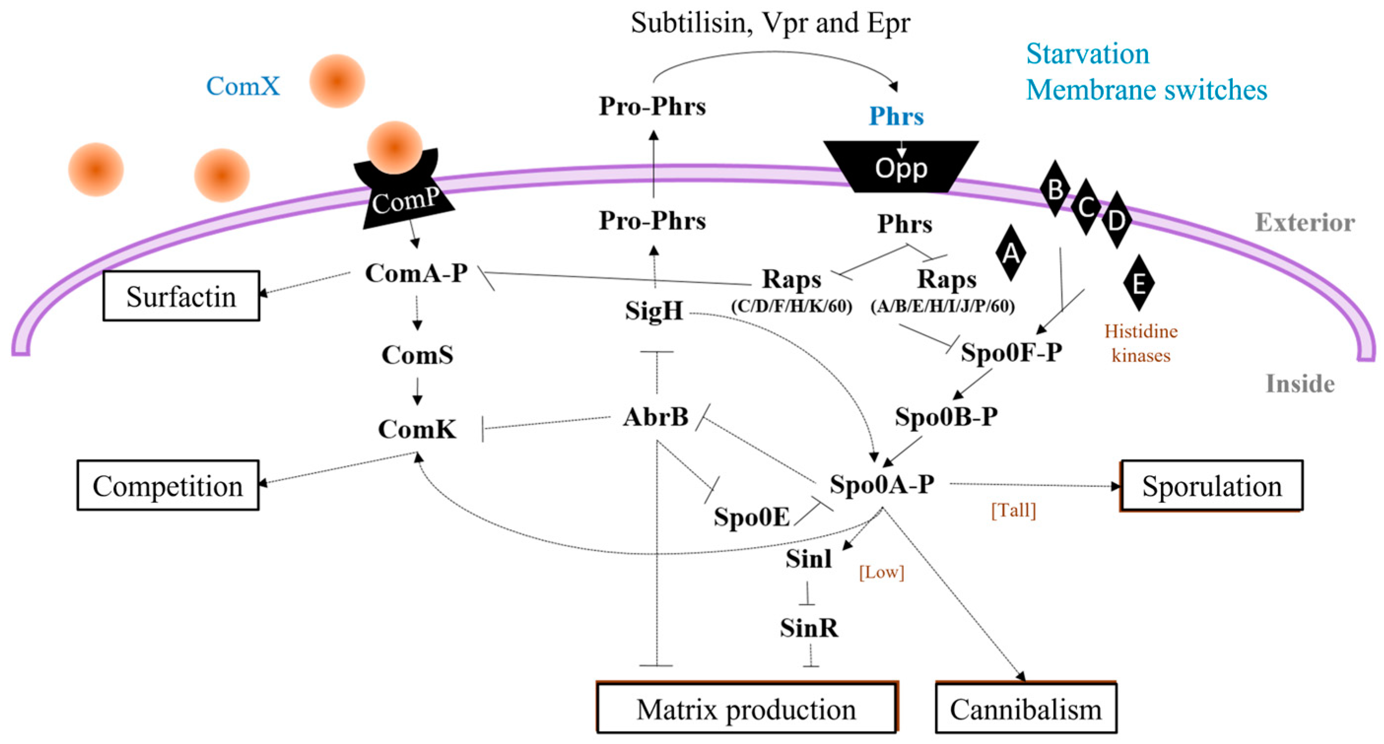
Surfactin production by Bacillus is highly regulated by several factors mainly relating to cell differentiation and is coordinated with the activation of three master regulators: ComA, DegU, and Spo0A, which result in the action of functionally different populations. ComA directs competent and surfactant-producing cell populations; DegU mediates the behavior of cells producing proteases and/or antibiotics; and Spo0A regulates the cells that produce biofilms, cannibals, and spores [49]. The activation of the master regulators is by phosphorylation with the action of different sensorial kinases (Figure 3).
  • ComA (ComA-P): ComX is a pheromone and ComP, a membrane kinase that phosphorylates ComA, induces the pathway for the production of surfactin [50] (Figure 3). In addition, a fraction of the surfactin producers undergo a secondary process of cell differentiation to convert a subpopulations of cells that are competent and capable of incorporating exogenous DNA, to cells with the ability to acquire characteristics that benefit them under stress conditions [51].
  • DegU (DegU-P): DegQ activates cytoplasmic kinase DegS with the addition of ComA-P Spacapan et al., [52] DegS is the one which phosphorylates DegU. Activation of DegU-P leads to the expression of the machinery responsible for the production and secretion of proteases, constituting the subpopulation of miners, and is also responsible for providing more assimilable peptides to the community through the hydrolysis of the most complex molecules [53].
  • Spo0A (Spo0A-P): five kinases (Kin AE) are responsible for sensing the signals that activate the Spo0A-P protein to differentiate subpopulations towards the cell matrix producers and cannibalism when there are low levels of phosphorylated protein in the cell, and when there are levels high Spo0A-P, sporulation genes are induced [54].
The sporulation process generally begins in the stationary phase, where a subpopulation of cells transforms spores, structures with minimal metabolic activity, capable of withstanding long periods and extreme conditions, such as temperatures, desiccation, and ionizing radiation [55]. Later, the spores can germinate in vegetative cells when conditions are appropriate.
Sporulation is primarily stimulated by starvation; however, no single nutritional effect acts as a trigger. The cell has an extremely complex and sophisticated decision-making apparatus, which controls a wide range of internal and external signals [56]. Quorum sensing refers to the diffusion monitoring of bacteria to make decisions and coordinates biological processes with cell density through the accumulation of signaling molecules (generally peptides) that, upon reaching a concentration threshold, mediates responses, and part of the sporulation in Bacillus is regulated in response to the quorum [54]. Among the different peptides that Bacillus recognizes as part of the quorum is ComX, a signaling peptide of surfactin-producing and competent cells. As previously described, ComX is a pheromone sensed by the ComP protein, which is phosphorylated when recognizing it and it transfers its phosphate group to ComA. Once activated, ComA-P binds to the tripartite DNA located within the promoter of the target genes, activating the transcription of 89 genes involved in the development of competition, the production of antibiotics, and the secretion of degrading enzymes [57]. One of the first competition genes is ComS, which encodes an anti-adapter protein responsible for stabilizing ComK. ComK is the primary regulator of late competition genes whose gene products comprise the DNA translocation machinery [23] (Figure 4).
Other signaling proteins that are involved in the regulation of surfactin production are Phr (penta or hexapeptides), which regulate the expression of Rap proteins (regulators of aspartate phosphatases). These proteins regulate the activities of Spo0A and ComA [58]. Phr peptides are synthesized by Pro-Phr precursor proteins that are exported to the environment and once outside, they are processed by extracellular proteases (Subtilisin, Vpr, and Epr); thus, the peptide remains mature and can be recognized by the oligopeptide-permease (Opp) and it enters the cell again, and once inside it inhibits the activity of Rap proteins [59]. Certain Rap proteins (A/B/E/H/I/J/P/60) act as phosphatases by inhibiting Spo0F and Spo0A activity. Also, specific Rap proteins (C/D/F/H/K/60) inhibit the activity of ComA by preventing its binding to DNA [54] (Figure 4).
On the other hand, the sporulation process recognizes different stimuli (Quorum sensing, starvation) through histidine kinases (KinA-E) that indirectly activate Spo0A by the transfer of the phosphate group to two intermediate phosphotransferases, Spo0F and Spo0B [56]. KinA and KinB are considered the main kinases involved in the activation of Spo0A by starvation [54]. KinA autophosphorylates with ATP upon starvation and then transfers a phosphoryl group to downstream components in a His-Asp-His-Asp signaling pathway [60]. While KinB is repressed by CodY (key sensor in guanine nucleotide levels) in the presence of nutrients and starvation, there is a drop in GTP, stimulating phosphorylation by KinB [55]. In addition, it is pointed out that KinA is a much more efficient kinase than KinC, D, or E [51]; thus, depending on which kinase you stimulate, different levels of Spo0A activation will occur, resulting in a different cell fate. High concentrations of Spo0A-P activate the transcription of genes involved in sporulation, while low concentrations activate the transcription of genes involved in biofilm production and cannibalism. Kin C and D have been reported to be involved in biofilm production [61]. Specifically, membrane-disrupting molecules stimulate KinC activity. Compounds that cause potassium leakage, like surfactin, the fungicide nystatin, and the antibiotic valinomycin, have been reported to induce KinC and stimulate the cell matrix of B. subtilis [51]. Likewise, sublethal concentrations of chlorine dioxide, a biocide that collapses the membrane potential, also trigger the activation of KinC [62]. On the other hand, KinD has a dual regulatory role, designated as a phosphatase to maintain low levels of Spo0A-P until the matrix (or one of its components) is detected and later as a kinase to promote sporulation of the bacteria [63]. Regarding KinE, the signaling that induces its activation is not very well known, but studies indicate its control through the sigma-H factor (Figure 4) [64].
Spo0A-P phosphorylation is not dependent on a single kinase but on the contribution of the different kinase changes as a function of the signals present under the growth conditions. Furthermore, the transcription of Spo0A is also modulated by a double repression system through the transition state AbrB and the stationary phase sigma factor SigH ( σ H ) [54].
Furthermore, various conformational studies on lipopeptides of the surfactin family demonstrate that structure plays an important role in its chelating properties [65]. Vass et al. [66] revealed the existence of two domains: (i) a polar domain, consisting of the Glu1 and Asp5 acid side chains, and (ii) a hydrophobic domain, consisting of residue 4, the lipid tail and residues 2 and 6. The carboxyl groups of the Glu1 and Asp5 residues have negative charges that allow them to form saline bridges with divalent cations, which explains the ability of surfactins to bind and differentiate cations such as Ca2+ and Mg2+ [48]. The Ca2+ cation complexes with the surfactins in a 1:1 ratio, creating an intramolecular bridge between the acidic residues, which stabilizes the structure and allows a considerable increase in the surfactant activity [67]. Finally, the size of the acyl chain is related to surfactant activity and antiviral activity, where a 14-carbon acyl chain favors surfactant activity, while a chain of 15 carbon atoms increases antiviral activity [55].
Genetic engineering advancements have demonstrated the ability to enhance surfactin production in Bacillus species. For example, in a study conducted by Zhang et al., [12] genome-reduced strain, GR167, was developed through the deletion of approximately 4.18% of the B. amyloliquefaciens LL3 genome, involving the removal of non-essential genomic regions. GR167 demonstrated enhanced characteristics, including a faster growth rate, elevated transformation efficiency, higher intracellular reducing power levels, and increased capacity for expressing heterologous proteins. The engineered chassis strain, GR167, was further modified to improve surfactin production. Initially, the biosynthetic gene clusters responsible for iturin and fengycin in GR167 were deleted to create GR167ID. Subsequent enhancements involved the replacement of the native srfA promoter in GR167ID with two LL3-derived promoters, PRsuc and PRtpxi, identified through RNA-seq and promoter strength characterization. This resulted in the generation of GR167IDS and GR167IDT. Notably, the most successful mutant, GR167IDS, exhibited a remarkable 678-fold increase in the transcriptional level of the srfA operon compared to GR167ID. Furthermore, it achieved a surfactin production of 311.35 mg/L, marking a significant 10.4-fold improvement over GR167. Furthermore, research is now being made towards the development of a systematic engineering approach to improve the biosynthesis of surfactin [12]. They restored the surfactin biosynthetic activity by integrating a complete sfp gene into the nonproducing Bacillus subtilis 168 strain and obtained a surfactin titer of 0.4 g/L. Then, they reduced competition by deleting biofilm formation-related genes and nonribosomal peptide synthetases/polyketide synthase pathways (3.8% of the total genome), which increased the surfactin titer by 3.3-fold. They also improved cellular tolerance to surfactin by overexpressing potential self-resistance-associated proteins, which further increased the surfactin titer by 8.5-fold. Also, they increased the supply of precursor branched-chain fatty acids by engineering the branched-chain fatty acid biosynthesis pathway, resulting in an increase of the surfactin titer to 8.5 g/L (a 20.3-fold increase). Last, due to the preference of the glycolytic pathway for cell growth, they diverted the precursor acetyl-CoA away from cell growth to surfactin biosynthesis by enhancing the transcription of srfA. The final surfactin titer increased to 12.8 g/L, with a yield of 65.0 mmol/mol sucrose (42% of the theoretical yield) in the metabolically engineered strain. One of the major problems we face in lipopeptide production is the yield obtained by Bacillus species being low for industrial applications, thus these and similar findings would aid that need.

Surfactin Biocontrol Activity

The biological activity of surfactins is related to their effect on the lipid part of the membranes; they can easily associate and firmly anchor within the lipid layers, creating destabilization and/or perforations in the plasma membrane, as well as activating a torrent of events molecules that derive the various defensive responses of plants [46]. Cawoy et al. [68] mention that surfactin has synergistic effects; for example, it supports the colonization of root tissue and promotes the supply of nutrients by surface wetting and detergent properties, based on its strong active surface properties that trigger biofilm formation, essential for the swarm in plant tissues. In a recent study by Le Mire et al. [69], the effectiveness of surfactin was shown to protect wheat by up to 70% against fungi of the species Zymoseptoria tritici, where surfactin itself did not show any antifungal activity, but was shown to stimulate salicylic acid and jasmonic acid-dependent signaling pathways, which are important regulators for plant defenses against biotic stress.

2.2. Iturin Biosynthesis

Iturins are a class of cyclic lipopeptides produced by various species of the genus Bacillus, particularly by B. subtilis and related species. These lipopeptides are characterized by their unique structure, consisting of a cyclic peptide linked to a lipid tail. Iturins are known for their potent antifungal and surfactant properties, making them valuable natural products with diverse applications in agriculture, biotechnology, and other fields [20,35].
Iturins are heptapeptides linked to a β-amino fatty acid chain, with a length of 14 to 17 carbons [20]. Iturins have been classified as the lipopeptide with the highest inhibitory activity against a wide variety of yeasts and fungi, but they have limited antibacterial and antiviral activities, and are considered to be excellent biopesticides [68]. This fungitoxicity is due to the membrane permeabilization properties they possess [70]. Iturins primarily exert their biological activities by disrupting cellular membranes. They insert into lipid bilayers, causing permeabilization, ion imbalance, and leakage of cellular contents. This mechanism underlies their antifungal activity against various plant pathogenic fungi, including Fusarium, Rhizoctonia, and Botrytis [71]. Additionally, iturins have been reported to present limited antibacterial properties but have demonstrated antibacterial efficacy against Ralstonia solanacearum in tomato bacterial wilt [50].
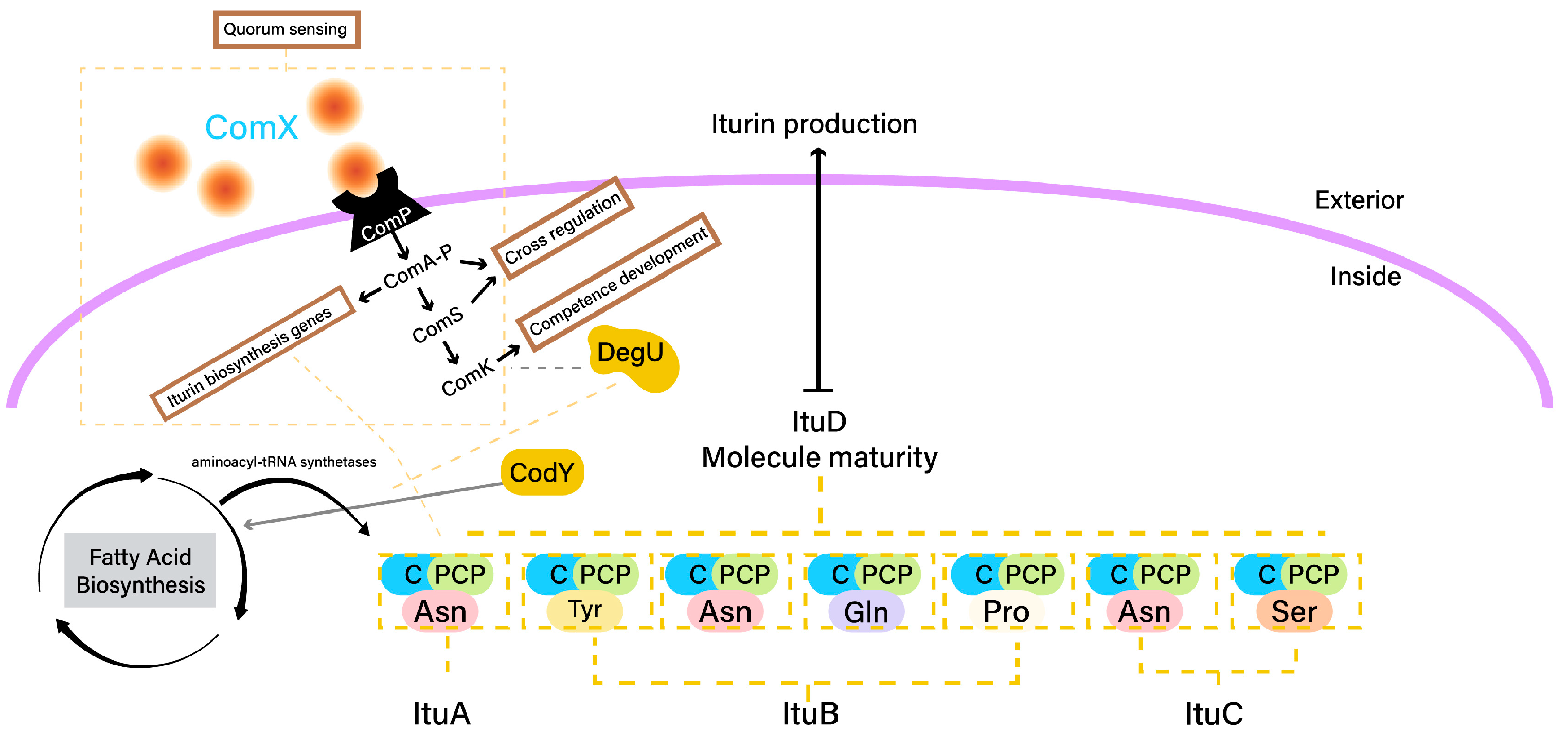
Iturin biosynthesis in Bacillus species involves a series of metabolic activities orchestrated by enzymes encoded by specific genes. These activities are regulated by a complex network of regulatory proteins that respond to environmental cues and cellular signals. The process begins with the activation of specific amino acids that will be incorporated into the iturin structure. The activation of amino acids involves the formation of aminoacyl-AMP intermediates. This activation is catalyzed by enzymes known as aminoacyl-tRNA synthetases. In the context of iturin biosynthesis, the amino acids that will become part of the iturin structure are activated by an aminoacyl-tRNA synthetase enzyme encoded by the ituA gene (Figure 5) [45]. The ituA gene encodes a fatty acid ligase enzyme that functions as an aminoacyl-tRNA synthetase specifically for iturin biosynthesis. This enzyme activates specific amino acids by attaching them to AMP (adenosine monophosphate) to form aminoacyl-AMP intermediates. These activated amino acids serve as substrates for the subsequent steps in iturin biosynthesis, including cyclization and lipidation [72]. The aminoacyl-AMP intermediates formed by the activation process are essential building blocks for the assembly of the cyclic peptide backbone of iturins. These intermediates are subsequently used in the cyclization reaction, where the amino acids are linked together to form the cyclic structure characteristic of iturins. The intermediates also play a crucial role in the lipidation step, where a lipid tail is attached to the cyclic peptide to complete the mature iturin molecule [73].
Then, activated amino acids are cyclized by enzymes encoded by the ituB gene (Figure 5). The ituB gene encodes a condensation enzyme that catalyzes the cyclization of activated amino acids. The ituB gene is responsible for directing the synthesis of this enzyme, which plays a central role in forming the cyclic peptide structure of iturins [72]. The amino acid substrates utilized in the cyclization reaction are activated amino acids that have been previously loaded onto aminoacyl-AMP intermediates, as described in the activation step. The specific arrangement of these amino acids within the iturin peptide sequence determines the cyclization pattern and the resulting cyclic structure. The ituB-encoded cyclase enzyme facilitates the enzymatic bond formation between certain amino acids, leading to cyclization [45]. The cyclization process involves the enzymatic formation of amide bonds between specific amino acids. The specific cyclization pattern determines the sequence of hydrophobic and hydrophilic amino acids in the cyclic structure, contributing to the amphiphilic properties of iturins. This structural diversity plays a crucial role in the biological activities of iturins, including their membrane-disrupting properties [74]. This mechanism is responsible for their potent antifungal and antibacterial activities [75].
After cyclization, the cyclic peptide structure undergoes lipidation, where the hydrophobic lipid tails to the cyclic peptide backbone, resulting in the complete iturin molecule with its unique amphiphilic structure [76]. The hydrophobic lipid tail anchors the iturin molecule in lipid bilayers, while the hydrophilic cyclic peptide remains exposed to the surrounding aqueous environment [44]. This step is catalyzed by a fatty acid synthetase enzyme, encoded by the ituD gene (Figure 5). The ituD gene directs the synthesis of the enzyme responsible for attaching the hydrophobic lipid tail to the cyclic peptide, forming the mature iturin molecule [45]. The lipidation and maturation process contribute to the potent biological activities of iturins.
Furthermore, the biosynthesis of iturins is tightly regulated to ensure their production under appropriate conditions [77]. Several regulatory proteins play a role in modulating the expression of genes involved in iturin biosynthesis (Figure 5).
  • ComA: The response regulator ComA is a key player in quorum sensing, a mechanism that coordinates gene expression based on cell density. When quorum-sensing signaling peptides (i.e., ComX) reach a certain concentration, ComA becomes phosphorylated and binds to the promoters of target genes, including those involved in iturin biosynthesis [78]. This activates the transcription of iturin biosynthetic genes.
  • DegU: The response regulator DegU is part of a two-component regulatory system. Phosphorylated DegU activates the transcription of genes involved in iturin biosynthesis, enhancing their expression [79].
  • CodY: The transcriptional regulator CodY senses nutrient availability. In nutrient-rich conditions, CodY binds to the promoters of genes related to secondary metabolism, including iturin biosynthetic genes, repressing their expression [80].
The interplay between these regulatory proteins allows Bacillus to modulate iturin production in response to changes in environmental conditions, cell density, and nutrient availability.
Furthermore, genetic engineering to increase iturin production by Bacillus species is being researched. In a study conducted by Dang et al. [80], to enhance the transcription of the iturin A biosynthetic genes from Bacillus amyloliquefaciens LL3, a robust constitutive promoter, C2up, was introduced upstream of the itu operon. This genetic modification resulted in the production of iturin A, reaching a titer of 37.35 mg/L. The iturin A extract displayed potent inhibitory activity against several common plant pathogens (A. alternate, B. cinerea, C. gloeosporioides, F. oxysporum, and R. solani). Through optimization of fermentation conditions using response surface methodology, the iturin A yield was further increased to 99.73 mg/L. Additionally, the overexpression of the pleiotropic regulator DegQ led to a notable improvement, resulting in a final iturin A yield of 113.1 mg/L. In another study by Xu et al. [81], they manipulated the promoter of the iturin A synthetase cluster from Bacillus amyloliquefaciens HZ-12. Different promoters, including P43, PbacA, PsrfA, and Pylb, were tested. PbacA was identified as the most efficient, resulting in an iturin A titer of 950.08  ±  19.43 mg/L. Deletion of the regulator gene abrB was then performed to alleviate the repression effect of AbrB on PbacA. Through further optimization of the fermentation medium, the maximum iturin A titer significantly rose by a 392.15% increase. The findings underscored the correlation between enhancing iturin A synthesis and its effectiveness in suppressing A. alternate, making these strains suitable for the industrial production of iturin.

Iturin Biocontrol Activity

The participation of iturins was demonstrated in the antibiosis-based biocontrol activity of Bacillus strains against various pathogens and different plant species; for example, in the case of soil-borne diseases, iturin A produced by B. subtilis RB14 is involved in the damping-off of tomato caused by Rhizoctonia solani, as well as in the control of diseases of the physiology. A contribution of both iturins was recently shown in the antagonism of B. subtilis against Podosphaera fusca in melon leaves [20] as well as strains of B. subtilis that produce a high level of antibiotics, especially iturin A, serving as the main mechanism underlying the control of Fusarium oxysporum and Rosellinia necatrix [14].

2.3. Fengycin Biosynthesis

Fengycins produced by Bacillus belong to the family of cyclic lipopeptides. They consist of a cyclic heptapeptide ring linked to a β-hydroxy fatty acid chain, forming the lipophilic portion [15]. The peptide portion comprises nonpolar, polar, and charged amino acids, which contribute to the amphiphilic nature of fengycins [82].
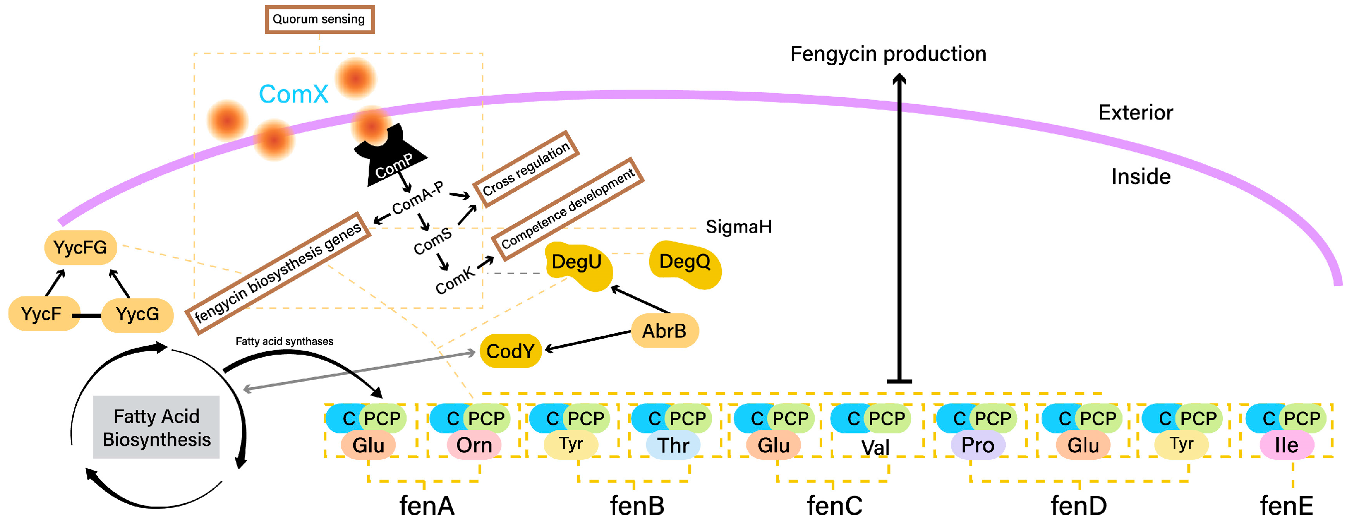
The biosynthesis of fengycins involves several genes and enzymes that work collaboratively. These include nonribosomal peptide synthetases (NRPSs) responsible for assembling the peptide backbone and enzymes like fatty acid synthases (FASs) for fatty acid tail attachment [83]. The cyclization, oxidation, and modification of amino acids also contribute to the complexity of fengycin structures [84]. Fengycin production is governed by a biosynthetic gene cluster (fen cluster) containing genes responsible for amino acid activation, loading, modification, and cyclization. NRPS genes, such as fenA, fenB, fenC, fenD, and fenE, encode modules that activate and load amino acids onto the peptide chain (Figure 6) [83]. These genes work in tandem, with each module incorporating a specific amino acid into the growing fengycin peptide. The order of modules and their interactions determine the amino acid sequence and cyclization pattern in the lipopeptide [70,74]. Genes within the fen cluster encode enzymes responsible for modifying amino acid residues and fatty acid chains. These tailoring reactions contribute to the structural diversity of fengycins [84]. Interactions between these genes result in specific modifications that differentiate various fengycin variants.
The production of fengycin lipopeptides in Bacillus species is tightly regulated to ensure optimal synthesis and bioactivity. The regulation involves a network of genes and transcription factors that respond to environmental cues, growth conditions, and cell population density (Figure 6).
  • CodY is a global regulator that plays a pivotal role in coordinating fengycin production with nutrient availability. CodY responds to changes in intracellular amino acid levels and acts as a sensor for nutrient sufficiency. In response to limiting nutrients, CodY negatively regulates the expression of fengycin biosynthetic genes, thus adjusting fengycin production to match cellular metabolic status [71].
  • The ComA-ComP quorum sensing system is central to fengycin production regulation in response to cell population density. The ComP histidine kinase senses external signaling peptides, and upon reaching a certain threshold, activates ComA, a response regulator. Activated ComA influences the expression of genes, including those involved in fengycin biosynthesis, in a density-dependent manner. This system ensures coordinated fengycin production within a microbial community [85].
  • DegU is a response regulator involved in fengycin regulation and environmental adaptation. The DegU phosphorylation status determines its activity as a transcription factor. In response to specific environmental cues, such as cell wall stress, DegU influences fengycin biosynthesis by directly affecting the expression of fengycin biosynthetic genes and other regulators [86].
  • AbrB is a pleiotropic transcriptional regulator that modulates the activity of CodY and DegU. It indirectly impacts fengycin production by influencing the regulatory cascades controlled by CodY and DegU. AbrB’s role in coordinating various regulatory pathways adds complexity to the control of fengycin biosynthesis [83].
Furthermore, two-component systems, including YycFG, have been implicated in the regulation of fengycin production. YycG serves as a sensor kinase that responds to cell envelope stress, influencing fengycin biosynthesis through its regulatory interactions. These systems contribute to the integration of various signals that impact fengycin production [74]. The regulation of fengycin production is characterized by intricate crosstalk and interactions between various regulatory pathways. CodY, ComA-ComP, DegU, AbrB, and other factors create a network for fengycin production based on a combination of nutrient availability, quorum sensing, and environmental signals [87].
Also, lipid structure is a critical factor in shaping both the structure and functionality of fengycin [88]. Consequently, the primary purpose of the fatty acid biosynthesis system is to facilitate the production of fengycin, particularly emphasizing the generation of branched-chain fatty acids [89]. In this process, the β-ketoacyl-acyl carrier protein synthase catalyzes the amalgamation of a malonyl-acyl carrier protein (ACP) with acetyl-CoA, leading to the formation of β-keto butyryl-ACP, which represents the initial stage of synthesizing straight-chain saturated fatty acids [89]. In this way, the building blocks for branched-chain fatty acid synthesis, such as isobutyl-CoA, isovaleryl-CoA, and α-methylbutyryl-CoA, can be sourced from the branched-chain amino acids L-valine, L-leucine, and L-isoleucine, respectively [88]. As a result, the synthesis of branched-chain amino acids stands as a vital element in the process of fengycin biosynthesis. The cooperation and interplay of these regulators ensure an adaptive response to changing conditions. Additionally, the enzyme system encoded by ilvBN, ilvGM, ilvIH, ilvC, ilvD, and ilvE catalyzes the synthesis of L-isoleucine, L-valine, and L-leucine where Lu et al., [88] elucidated an up-regulation by a factor of 1.5 of ilvBN, suggested that fructose can induce biosynthesis of branched-chain amino acids, thereby promoting the accumulation of fengycin production. Also, they reported that small molecules such as vitamins act as modulators or enzyme auxiliary groups to promote the biosynthesis of the precursors, fengycin. However, fengycin is produced through a non-ribosomal pathway, which means that the biosynthesis of fengycin is minimally influenced by the gene transcription process but mainly controlled by the gene regulatory network [90].
The intricate understanding of the genetic determinants governing fengycin synthesis allows scientists to strategically modify the genetic composition of these bacteria, to optimize the expression of key genes involved in fengycin production. By employing advanced genetic engineering techniques, researchers seek to augment the yield, efficiency, and overall capability of Bacillus strains to produce fengycin. Successful examples of this are those demonstrated by Gao et al. [91], where they presented the capacity of Bacillus subtilis 168 to produce lipopeptides; specifically, fengycin was reinstated resulting in a fengycin titer of 1.81 mg/L. Subsequent enhancements were achieved by knocking out pathways linked to surfactin and bacillaene synthesis, substituting the native promoter (PppsA) with the Pveg promoter, leading to an increased fengycin production of 174.63 mg/L. Further improvements were made by upregulating genes in the fatty acid pathway, resulting in elevated fengycin levels of 258.52 mg/L. Suppressing spore and biofilm formation contributed to a fengycin production level of 302.51 mg/L. The addition of threonine in the optimized culture medium represented a final enhancement, elevating fengycin production to approximately 885.37 mg/L, marking a remarkable 488-fold increase compared to the original strain. In another study conducted by Li et al. [92], they cloned key modules associated with improved fatty acid synthesis of B. subtilis ATCC 21332. The resulting engineered strain, BSA02, demonstrated a 3-fold increase in the fengycin titer, reaching 442.51 mg/L. Furthermore, they added glutamate to further elevate the fengycin titer of BSA034 to 657.55 mg/L, resulting in facilitation of the membrane transport processes in BSA034 which led to a higher fengycin production.

Fengycin Biocontrol Activity

This family of lipopeptides has a strong fungitoxic activity, specifically against filamentous fungi such as Fusarium oxysporum, Colletrotrichum, Alternaria alternata, Gloeosporioides, and Fusarium solani among others, which exhibit potential applications in agriculture, biological control, the food and feed industry [88,93]. However, unlike surfactins, the defense mechanisms promoted by fengycins are specific to certain plant genotypes or pathogen systems [94]. The direct evidence of the role of fengycins as biocontrol derives from experiments that demonstrate their capacity, as in the case of fengycins obtained from Bacillus subtilis against Botrytis cinerea (causing gray mold) in apples, and also in the wilt of the bean crop caused by Pythium ultimum [12]; as well as the results obtained by Liu et al. [93], which indicate that they play an important role in suppressing the mycelial growth of the fungal pathogen Monilinia fructicola. Mechanically, the action of fengycins is less known compared to other lipopeptides, but they also easily interact with lipid layers and, to a certain extent, retain the potential to alter the structure of the cell membrane and permeability, in a dose-dependent manner [82]. They can also interact with plant cells as a bacterial determinant to activate an immune response through stimulation of the phenomenon of induced systemic resistance [95]. Thus, the antifungal effects of fengycin can be described as the disruption of cell membrane integrity [34], interference with bacterial quorum sensing systems [35], and stimulation of cellular apoptosis [6].

3. Extraction of Bacillus Lipopeptides

The extraction and purification of Bacillus lipopeptide-producer strains involves a series of steps to obtain the desired lipopeptides from the bacterial culture [96]. Initially, a suitable Bacillus strain known for lipopeptide production is cultured under optimal conditions to maximize yield [97]. With respect to media optimization, the well-established mineral salt medium used by Cooper et al. [98] and the Landy medium [99] are often used as references. Carbon and nitrogen sources are carefully selected to provide the necessary nutrients for bacterial growth and lipopeptide synthesis [39]. For surfactin, phosphate limitation induces stress and boosts production, while iturin and fengycin benefit from organic nitrogen sources like peptones and yeast extract [100]. Strain selection is a critical consideration, with different Bacillus strains exhibiting variations in lipopeptide productivity [12,24,36,37,38,39,40,41,42,43]. Genetic engineering approaches are employed to enhance strains for increased lipopeptide synthesis, optimizing the genetic machinery responsible for their production [101]. Furthermore, maintaining an optimal pH range is crucial for lipopeptide biosynthesis. Surfactin production is favored in slightly alkaline conditions (pH 7.0–8.0), while iturin and fengycin production thrive in slightly alkaline to neutral (pH 6.0–7.0) conditions [32]. Temperature control is also vital, with surfactin production occurring at higher temperatures (around 37 °C) compared to iturin and fengycin (32–49 °C) [32]. These controlled environmental conditions ensure the bacteria are in an ideal state for lipopeptide production. The culture is allowed to reach the late stationary phase, during which lipopeptide production is at its peak [102]. After harvesting the bacterial biomass, cell lysis methods, such as sonication or enzymatic treatment, are used to release the intracellular lipopeptides [103]. Sonication involves exposing bacterial cells to high-frequency sound waves, leading to the formation and collapse of microscopic bubbles in the culture medium, causing cell disruption and release of intracellular components, including lipopeptides [104]. While sonication is a rapid and efficient method, care must be taken to control the duration and intensity of sonication to prevent excessive heat buildup, which could potentially denature the lipopeptides [104]. On the other hand, enzymatic treatment, specifically with lysozyme, is a gentler method for cell lysis and has vastly been reported in lipopeptide extractions [105]. Lysozyme targets the peptidoglycan layer in bacterial cell walls, catalyzing the hydrolysis of β-1,4 glycosidic bonds and leading to cell wall degradation [106]. Enzymatic methods are advantageous for preserving the bioactivity and structural integrity of the lipopeptides, making them suitable for downstream applications [107]. Also, mechanical disruption methods, such as bead milling, involve the physical grinding of bacterial cells using beads or grinding media [108]. This approach is effective for larger-scale production and can be particularly useful for industrial applications [108]. The choice of cell lysis method is influenced by the need for high yields, preservation of lipopeptide bioactivity, and scalability for large-scale production.
The next crucial step involves organic solvent extraction, where hydrophobic lipopeptides partition into the organic phase, separated from cellular debris [109]. The subsequent organic solvent extraction step involves mixing the lipopeptide-containing liquid with an organic solvent, typically methanol, ethanol, or a solvent mixture [103]. This organic solvent selectively dissolves the lipopeptides from the aqueous phase, facilitating their separation. The organic phase, enriched with lipopeptides, is then isolated, and the solvent is evaporated to obtain a crude lipopeptide extract. This step is followed by purification through chromatography techniques.
A singular approach may prove insufficient for the purification of these substances. Therefore, it is typical to employ a multi-stage process for lipopeptide purification, incorporating techniques such as acid precipitation, the utilization of organic solvents, ultrafiltration, solid phase extraction, and chromatography [15]. Acid precipitation is often employed as an initial step, capitalizing on pH-induced solubility changes to separate lipopeptides from impurities, followed by centrifugation, where the precipitated lipopeptides are separated from the liquid phase [110]. Furthermore, ultrafiltration uses semipermeable membranes with defined pore sizes and concentrates lipopeptides by selectively retaining them while allowing smaller impurities to pass through, removing low-molecular-weight contaminants [111]. Chromatography and solid-phase extraction (SPE) are preferred for achieving high purity in the final stages of lipopeptide purification [15]. Both methods involve passing the lipopeptide mixture through a column, where the compounds of interest interact with the solid phase, ensuring selectivity [112]. The success of these purification methods depends on the resin type and solvent mixture, with the C18 column recognized for its efficiency in adsorbing lipopeptides due to their hydrophobic nature [113]. Despite the structural similarities within specific lipopeptide families, reverse-phase liquid chromatography methods have successfully addressed these challenges. Chromatographic techniques are not only employed for the purification of lipopeptides but are also crucial for their identification and quantification. Various chromatography variants, such as thin-layer chromatography (TLC) [114] and high-resolution liquid chromatography (HPLC), have been developed for comprehensive lipopeptide analysis [115,116].
Thus, the extraction and purification of Bacillus lipopeptides involves a comprehensive process that begins with optimized culture conditions and strain selection. An integrated approach among the discussed strategies for extraction would benefit lipopeptide extraction in regard to yield. This integrated approach ensures the isolation of highly purified Bacillus lipopeptides for further analysis.

4. Identification of Bacillus Lipopeptide-Producer Strains

Current knowledge about the biosynthesis, structure, and bioactivity of Bacillus lipopeptides has allowed the development of a great variety of methods to identify lipopeptide-producer strains across different information levels (i.e., phenotype, DNA, RNA, protein, and metabolites) (Figure 7).
For instance, PCR analysis is directed to identify the presence or absence of genes (gene-level) involved in the biosynthesis and assembly of lipopeptides [117], whereas analytical tools such as liquid chromatography or mass spectrometry are focused on identifying the lipopeptide itself (metabolite-level) [118]. Each method varies based on the purpose and maturity stage of the study. For example, if the study is in the early stages and the objective is to identify lipopeptide-producing strains in a large microbial collection (200–300 strains), high-throughput methods (i.e., PCR or drop-collapse) that allow for reducing the complexity of the collection could be a good starting point to explore lipopeptide-producing strains [119]. On the other hand, if a bioprospecting study is in advanced stages and the objective is to identify the diversity of lipopeptides produced by some interesting strains, more robust methods (ESI-MS/MS) that allow identification of lipopeptides at a homologs level could be the most appropriate approach.

4.1. Phenotype Level

Although lipopeptide production is a conserved characteristic of Bacillus strains, in part because they are metabolites involved in multiple physiological processes (i.e., biofilm formation, quorum sensing, competition for space, sporulation, etc.) [120], it has been identified that Bacillus strains are not able to produce any lipopeptide homologs [68]. In this sense, during screening assays, there are indirect methods that allow potentially lipopeptide-producing strains to be identified (Table 2). The methods used to identify strains that potentially produce lipopeptides are based on detecting changes in surface properties caused by these compounds [121], taking into account their biological activity (hemolysis test) and their amphoteric property (drop-collapse, microplate meniscus formation test, oil atomization). However, these methods have some limitations, for example: (i) it is not possible to know the type of lipopeptide and (ii) it is common to find false positives due to the production method.

4.2. DNA/RNA Level

There are two main methods for ascertaining the lipopeptide synthesis capability of bacterial species and strains: examining the final synthesized product and detecting the presence of the relevant NRPS genes within the genome. In practice, genetic profiling of bacteria is frequently accomplished through the PCR utilizing primers designed for conserved regions of NRPS-encoding genes to confirm their ability to synthesize lipopeptides [126]. The target sequences within the NRPS genes are duplicated, and the PCR amplifies the polymorphic DNA fragments [127]. Within the genus Bacillus, both species and subspecies exhibit diversity in their complement of NRPS genes.
New advances are being made toward the understanding of lipopeptides through the integration of genomic sequencing with cutting-edge bioinformatics tools through the exploration of genes and enzymes associated with lipopeptide biosynthesis, including NRPS and hybrid NRPS–PKS (Polyketide Synthase) [128]. Omic methods, in conjunction with the emergence of high-throughput sequencing technologies and robust bioinformatics pipelines, enable us to uncover the presence and organization of biosynthetic gene clusters (BGCs) [129]. High-throughput sequencing, when coupled with bioinformatics, offers a multifaceted approach to the investigation of LPs including the identification of genes responsible for LP synthesis, the anticipation of the LP’s structural composition based on genomic information, and the proposed functions and ecological roles of the LP product [44]. Among these, the Antibiotics and Secondary Metabolite Analysis Shell (antiSMASH) [130] stands out as a prominent bioinformatics tool specifically designed for BGC identification, functional annotation, and comprehensive analysis [131]. Its algorithm facilitates the alignment of microbial genomic sequences with previously cataloged BGC sequences from its database. AntiSMASH has gained extensive recognition as a screening technique for detecting diverse BGCs, encompassing those affiliated with NRPS and hybrid NRPS–PKS accountable for lipopeptide synthesis [132].
Furthermore, RT-qPCR (Reverse Transcription-quantitative Polymerase Chain Reaction) is a powerful method used for the identification of lipopeptides in Bacillus species by targeting biosynthetic gene clusters including those encoding NRPS and PKS [133]. This approach combines the precision of PCR with the ability to quantify gene expression, allowing for a more comprehensive understanding of the biosynthetic potential of these bacteria. The process begins with the extraction of RNA from Bacillus cultures, which represents the actively transcribed genetic material of these bacteria [134]. This RNA is then reverse transcribed into complementary DNA (cDNA), providing a stable and amplifiable template. Specific primers designed to target the biosynthetic genes associated with lipopeptide production are used for the subsequent qPCR amplification [135]. By comparing the expression of these specific genes across different Bacillus strains, researchers can effectively pinpoint strains actively producing lipopeptides, aiding in the selection of strains with high lipopeptide production potential. RT-qPCR, therefore, plays a pivotal role in both confirming the presence of relevant biosynthetic genes involved in lipopeptide production and providing quantitative data on gene expression, making it an essential tool in the screening and selection of Bacillus lipopeptide-producing strains.

4.3. Protein Level

Identifying lipopeptides at the protein level can be challenging but is essential for understanding their functions and potential applications. When biosurfactant extracts are derived from intricate mixtures following liquid–liquid extraction using organic solvents or precipitation, using mass spectrometry immediately after extraction might not be the most favorable approach [136]. This is primarily due to the potential interference of numerous substances within the extract, resulting in spectra with a high abundance of signals. Within these spectra, the masses corresponding to lipopeptide biosurfactants are obscured by those of other metabolites, leading to the observation of weak signals for lipopeptides [137]. In this context, the utilization of electrophoresis as a preliminary step before mass spectrometry analysis may present an intriguing method for both purifying and identifying lipopeptides, particularly within intricate sample matrices [136]. Thus, sodium dodecyl sulfate-polyacrylamide gel electrophoresis (SDS-PAGE) is a widely used technique for the detection and separation of proteins and peptides, and it can also be applied to the analysis of lipopeptides [138]. This method involves the use of an acrylamide gel, which, when subjected to an electric field, separates molecules based on their size and charge. For lipopeptides, which consist of both a lipid tail and a peptide head, SDS-PAGE can help in their separation and detection. In SDS-PAGE, lipopeptides are first treated with SDS, a detergent that unfolds and imparts a uniform negative charge to the molecules [104]. Post purification, the negatively charged lipopeptides are then loaded onto the polyacrylamide gel and separated based on their size. This method can reveal the presence and approximate molecular weight of lipopeptides in a sample. Additionally, by transferring the separated lipopeptides to a membrane and employing specific antibodies or stains, it is possible to further characterize and confirm the identity of lipopeptides, making SDS-PAGE a valuable tool for the analysis and detection of these compounds in complex samples [136].
Other analytical methods for detecting and quantifying lipopeptides produced from Bacillus species is the tandem mass spectrometry (MS/MS) [139]. During MS/MS analysis, the lipopeptides are first subjected to ionization and fragmentation [104]. Fragmentation patterns reveal structural information about the peptide head and lipid tail, allowing for precise identification [140]. Notably, different classes of lipopeptides, such as surfactins, iturins, and fengycins, have distinct fragmentation patterns, which can be matched against reference spectra to confirm their identity. Furthermore, by monitoring specific transitions between precursor and product ions, tandem mass spectrometry enables the quantification of individual lipopeptides in a sample. This approach enhances the understanding of the diversity and relative abundance of lipopeptides produced by Bacillus species, providing valuable insights into their potential applications in biotechnology and agriculture [141].

4.4. Metabolic Level

The identification and quantification of lipopeptides from Bacillus species at a metabolic level involve the use of various analytical techniques, including liquid chromatography (HPLC) [15], gas chromatography (GC) [121], tandem mass spectrometry (MS/MS) [142] and thin-layer chromatography (TLC) [114]. These methods enable the comprehensive analysis of the metabolic profile of lipopeptides, shedding light on their diversity, abundance, and biosynthetic pathways.
HPLC is a powerful tool for separating lipopeptides extracted from Bacillus cultures. In the case of lipopeptide analysis, reverse-phase HPLC is commonly employed, utilizing a hydrophobic stationary phase that retains lipopeptides [15]. After extraction and purification, the lipopeptides are typically separated based on their hydrophobicity and eluted in order of increasing hydrophobic character. This step helps to isolate individual lipopeptides for further analysis [143].
Another widely used method for Bacillus lipopeptide quantification is GC used to analyze the lipid tails of lipopeptides [144]. Bacillus lipopeptides often consist of fatty acid chains, and GC can separate and quantify these lipid components [145]. After saponification to release the fatty acids, the derivatized samples are injected into the GC system, which separates and quantifies the individual fatty acids in the lipopeptides [146]. This allows for the determination of the composition and abundance of the lipid tails.
Similarly, MS/MS plays a crucial role in the structural elucidation and quantification of lipopeptides from Bacillus species [147,148]. After HPLC or GC separation, the isolated lipopeptides can be analyzed by MS/MS [138]. This technique involves the ionization of the lipopeptides followed by fragmentation of the ions. The resulting fragment ions provide information about the structure of the peptide head and lipid tail. MS/MS can identify the specific lipopeptides present in the sample and determine their relative abundance [104]. Database searching and spectral libraries aid in the identification of known lipopeptides, while de novo sequencing can uncover novel lipopeptides [138].
Each technology described has its advantages and limitations (Table 3) [104,138,149,150]. Thus, the combination of these can conduct a comprehensive metabolic profiling of Bacillus lipopeptides. The obtained data allow for the identification of different classes of lipopeptides, determination of their relative abundances, and insights into the biosynthetic pathways involved. This information is valuable for understanding the potential applications of Bacillus lipopeptides in biocontrol.
Last, thin-layer chromatography (TLC) is a versatile method for lipopeptide analysis, involving the placement of a sample on an aluminum plate coated with an adsorbent-like silica gel [115]. The plate is immersed in a solvent mixture, serving as the mobile phase, and as the solvent rises through capillarity, compounds in the sample distribute differentially across the plate [115]. TLC is valuable for identifying lipopeptide families and quickly assessing compound purity during purification steps [114]. Lipopeptide identification through TLC entails comparing the Rf values between a standard and the test sample. For instance, in the identification of surfactins, iturins, and fengycins in Bacillus extracts, specific Rf values were utilized with chloroform/methanol/water (65:25:4, v/v/v) as the mobile phase and silica gel as the stationary phase [151].
The identification and quantification of lipopeptides from Bacillus species at a metabolic level necessitates a comprehensive analytical approach, employing a repertoire of techniques. Liquid chromatography (HPLC) and gas chromatography (GC) contribute to the separation and quantification of lipopeptides, providing high resolution and sensitivity. Tandem mass spectrometry (MS/MS) further enhances the precision and specificity of identification by elucidating the molecular structure of lipopeptides. Additionally, thin-layer chromatography (TLC) serves as a rapid and cost-effective tool for qualitative analysis and initial screening. The combination of these analytical techniques offers a multifaceted strategy, allowing researchers to gain a holistic understanding of the lipopeptide profiles produced by Bacillus species, crucial for applications in agriculture, medicine, and industrial processes.

5. Conclusions

Bacillus-derived lipopeptides have demonstrated exceptional antimicrobial properties that can combat a range of pathogens affecting crops. These lipopeptides act through multiple modes of action, including disruption of cell membranes, interference with signaling pathways, and modulation of host defense responses. This broad-spectrum efficacy makes them valuable tools in integrated pest management strategies. Despite lipopeptide research still being conducted in vitro, and in plant assays, due to the many limitations this has such as yield production, and complex extraction methods, this serves as a scientific background for future research of in-field applications. It gives insight into the potential effect of lipopeptide extract application as biopesticides or the application of lipopeptide-producing Bacillus species. Currently, commercialized lipopeptide-producing Bacillus species present high inhibition rates against phytopathogens. Much research is still needed to migrate this technology to in-field applications. Thus, understanding the biosynthesis of lipopeptides with biological control bioactivities provides key gene identification in molecular processes that could better the yield obtention of Bacillus lipopeptides and generates a better understanding of how these molecules are produced and their regulation.

Author Contributions

Conceptualization, S.d.l.S.-V.; investigation, V.V.R., A.G.-L., M.F.V.-D. and E.D.V.-R.; writing—original draft preparation, V.V.R., A.G.-L., M.F.V.-D. and E.D.V.-R.; writing—review and editing, V.V.R., A.G.-L., M.F.V.-D., E.D.V.-R., F.I.P.-C., G.S., L.J.G.-G., L.A.C.C. and S.d.l.S.-V.; supervision, S.d.l.S.-V.; project administration, S.d.l.S.-V.; funding acquisition, S.d.l.S.-V. All authors have read and agreed to the published version of the manuscript.

Funding

This research was funded by Asociación de Productores de Hortalizas del Yaqui y Mayo (APHYM) and Patronato para la Investigación y Experimentación Agrícola del Estado de Sonora (PIAES) project “Identificación de agentes de control biológico contra Meloidogyne sp., Fusarium sp. y Streptomyces sp.”, 2022-0364. We thank the Consejo Nacional de Humanidades, Ciencia y Tecnología (CONAHCYT) for master and doctorate Scholarship No. 712969 (Valeria Valenzuela Ruiz).

Data Availability Statement

No new data was created or analyzed in this study. Data sharing is not applicable to this article. This article uses the available information on line and gathers the data in regards to Bacillus lipopeptides.

Acknowledgments

In addition, we would like to profoundly thank all current and past members of the LBRM-COLMENA for their dedication and commitment to the development of all research projects, as well as the external organizations involved for their support and cooperation.

Conflicts of Interest

The authors declare no conflicts of interest.

References

  1. FAO. Pesticides use, pesticides trade and pesticides indicators. In Global, Regional and Country Trends, 1990–2020; FAOSTAT Analytical Briefs; FAO: Rome, Italy, 2022; pp. 1–13. [Google Scholar]
  2. Díaz Rodríguez, A.M.; Parra Cota, F.I.; Santoyo, G.; de los Santos Villalobos, S. Chlorothalonil tolerance of indole producing bacteria associated to wheat (Triticum turgidum L.) rhizosphere in the Yaqui Valley, Mexico. Ecotoxicology 2019, 28, 569–577. [Google Scholar] [CrossRef] [PubMed]
  3. Mishra, S.; Zhang, W.; Lin, Z.; Pang, S.; Huang, Y.; Bhatt, P.; Chen, S. Carbofuran toxicity and its microbial degradation in contaminated environments. Chemosphere 2020, 259, 127419. [Google Scholar] [CrossRef] [PubMed]
  4. Pang, S.; Lin, Z.; Zhang, Y.; Zhang, W.; Alansary, N.; Mishra, S.; Bhatt, P.; Chen, S. Insights into the Toxicity and Degradation Mechanisms of Imidacloprid Via Physicochemical and Microbial Approaches. Toxics 2020, 8, 65. [Google Scholar] [CrossRef]
  5. Córdova-Albores, L.C.; Zelaya-Molina, L.X.; Ávila-Alistac, N.; Valenzuela-Ruíz, V.; Cortés-Martínez, N.E.; Parra-Cota, F.I.; Burgos-Canul, Y.Y.; Chávez-Díaz, I.F.; Fajardo-Franco, M.L.; De los Santos-Villalobos, S. Omics sciences potential on bioprospecting of biological control microbial agents: The case of the Mexican agro-biotechnology. Rev. Mex. Fitopatol. 2020, 39, 147–184. [Google Scholar] [CrossRef]
  6. Hu, J.; Zheng, M.; Dang, S.; Shi, M.; Zhang, J.; Li, Y. Biocontrol Potential of Bacillus amyloliquefaciens LYZ69 against Anthracnose of Alfalfa (Medicago sativa). J. Phytopathol. 2021, 111, 1338–1348. [Google Scholar] [CrossRef]
  7. Samada, L.H.; Tambunan, U.S.F. Biopesticides as promising alternatives to chemical pesticides: A review of their current and future status. Online J. Biol. Sci. 2020, 20, 66–76. [Google Scholar] [CrossRef]
  8. Morales-Cedeño, L.R.; Barajas-Barrera, I.A.; Parra-Cota, F.I.; Valenzuela-Ruiz, V.; de los Santos-Villalobos, S.; Loeza-Lara, P.D.; Herrera-Pérez, A.; del Carmen Orozco-Mosqueda, M.; Santoyo, G. Evaluation of Biocontrol Potential of Bacillus spp. and Pseudomonas fluorescens UM270 against Postharvest Fungal Pathogens. Microbiol. Res. 2023, 14, 1511–1523. [Google Scholar] [CrossRef]
  9. Montoya-Martínez, A.C.; Figueroa-Brambila, K.M.; Escalante-Beltrán, A.; López-Montoya, N.D.; Valenzuela-Ruíz, V.; Parra-Cota, F.I.; Estrada Alvarado, M.I.; de los Santos-Villalobos, S. Biological Control Mechanisms of Bacillus cabrialesii subsp. tritici TSO2T against Fusarium languescens, the Causal Agent of Wilt in Jalapeño Peppers. Horticulturae 2023, 9, 964. [Google Scholar]
  10. Valenzuela-Ruiz, V.; Parra-Cota, F.I.; Santoyo, G.; de los Santos-Villalobos, S. Potential biocontrol mechanisms of Bacillus sp. TSO2 against Bipolaris sorokiniana, spot blotch in wheat. Rev. Mex. Fitopatol. 2022, 40, 230–239. [Google Scholar] [CrossRef]
  11. Mishra, R.K.; Bohra, A.; Kamaal, N.; Kumar, K.; Gandhi, K.; Sujayanand, G.K.; Saabale, P.R.; Satheesh Naik, S.J.; Birinchi Kumar, S.; Dharmendra, K.; et al. Utilization of biopesticides as sustainable solutions for management of pests in legume crops: Achievements and prospects. Egypt. J. Biol. Pest Control 2018, 28, 3. [Google Scholar] [CrossRef]
  12. Ongena, M.; Jacques, P.; Touré, Y.; Destain, J.; Jabrane, A.; Thonart, P. Involvement of fengycin-type lipopeptides in the multifaceted biocontrol potential of Bacillus subtilis. Appl. Microbiol. Biotechnol. 2005, 69, 29–38. [Google Scholar] [CrossRef]
  13. Tut, G.; Magan, N.; Brain, P.; Xu, X. Critical Evaluation of Two Commercial Biocontrol Agents for Their Efficacy against B. cinerea under In Vitro and In Vivo Conditions in Relation to Different Abiotic Factors. Agronomy 2021, 11, 1868. [Google Scholar] [CrossRef]
  14. Hashem, A.; Tabassum, B.; Fathi Abd_Allah, E. Bacillus subtilis: A plant-growth promoting rhizobacterium that also impacts biotic stress. Saudi J. Biol. Sci. 2019, 26, 1291–1297. [Google Scholar] [CrossRef] [PubMed]
  15. Valenzuela-Ruiz, V.; Gálvez-Gamboa, G.T.; Villla-Rodriguez, E.D.; Parra-Cota, F.I.; Santoyo, G.; de los Santos Villalobos, S. Lipopeptides produced by biological control agents of the genus Bacillus: A review of analytical tools used for their study. Rev. Mex. Cienc. Agríc. 2020, 11, 419–432. [Google Scholar]
  16. Santoyo, G.; Sánchez-Yáñez, J.; de los Santos-Villalobos, S. Methods for detecting biocontrol and plant growth-promoting traits in rhizobacteria. In Methods in Rhizosphere Biology Research: Rhizosphere Biology; Reinhardt, D., Sharma, A., Eds.; Springer: Singapore, 2019; pp. 133–149. [Google Scholar]
  17. Salazar, B.; Ortiz, A.; Keswani, C.; Minkina, T.; Mandzhieva, S.; Pratap Singh, S.; Rekadwad, B.; Borriss, R.; Jain, A.; Singh, H.B.; et al. Bacillus spp. as Bio-factories for Antifungal Secondary Metabolites: Innovation Beyond Whole Organism Formulations. Microb. Ecol. 2023, 86, 1–24. [Google Scholar] [CrossRef]
  18. Parte, A.C.; Carbasse, J.S.; Meier-Kolthoff, J.P.; Reimer, L.C.; Göker, M. List of prokaryotic names with standing in nomenclature (LPSN) moves to the DSMZ. Int. J. Syst. Evol. Microbiol. 2020, 70, 5607–5612. [Google Scholar] [CrossRef]
  19. Logan, N.A.; De Vos, P. Bacillus Bergey’s Manual of Systematics of Archaea and Bacteria; John Wiley & Sons, Inc. in Association with Bergey’s Manual Trust: Hoboken, NJ, USA, 2015; 163p. [Google Scholar]
  20. Ongena, M.; Jacques, P. Bacillus lipopeptides: Versatile weapons for plant disease biocontrol. Trends Microbiol. 2008, 16, 115–125. [Google Scholar] [CrossRef]
  21. Jacques, P. Surfactin and Other Lipopeptides from Bacillus spp. In Biosurfactants: From Genes to Applications; Springer: Berlin/Heidelberg, Germany, 2011; pp. 57–91. [Google Scholar]
  22. Villarreal-Delgado, M.F.; Villa-Rodríguez, E.D.; Cira-Chávez, L.A.; Estrada-Alvarado, M.I.; Parra-Cota, F.I.; De los Santos-Villalobos, S. El género Bacillus como agente de control biológico y sus implicaciones en la bioseguridad agrícola. Rev. Mex. Fitopatol. 2018, 36, 95–130. [Google Scholar] [CrossRef]
  23. Hamoen, L.W.; Venema, G.; Kuipers, O.P. Controlling competence in Bacillus subtilis: Shared use of regulators. Microbiology 2003, 149, 9–17. [Google Scholar] [CrossRef]
  24. Villa-Rodriguez, E.; Moreno-Ulloa, A.; Castro-Longoria, E.; Parra-Cota, F.I.; de los Santos-Villalobos, S. Integrated omics approaches for deciphering antifungal metabolites produced by a novel Bacillus species, B. cabrialesii TE3T, against the spot blotch disease of wheat (Triticum turgidum L. subsp. durum). Microbiol. Res. 2021, 251, 126826. [Google Scholar] [CrossRef]
  25. Fira, D.; Dimkic, I.; Beric, T.; Lozo, J.; Stankovic, S. Biological control of plant pathogens by Bacillus species. J. Biotechnol. 2018, 285, 44–55. [Google Scholar] [CrossRef]
  26. Li, R.; Du, W.; Yang, J.; Liu, Z.; Yousef, A.E. Control of Listeria monocytogenes biofilm by paenibacterin, a natural antimicrobial lipopeptide. Food Control 2018, 84, 529–535. [Google Scholar] [CrossRef]
  27. Crouzet, J.; Arguelles-Arias, A.; Dhondt-Cordelier, S.; Cordelier, S.; Pršic, J.; Hoff, G.; Mazeyrat-Gourbeyre, F.; Baillieul, F.; Clément, C.; Ongena, M.; et al. Biosurfactants in Plant Protection against Diseases: Rhamnolipids and Lipopeptides Case Study. Front. Bioeng. Biotechnol. 2020, 8, 1014. [Google Scholar] [CrossRef]
  28. Al-Mutar, D.M.K.; Noman, M.; Abduljaleel Alzawar, N.S.; Azizullah; Li, D.; Song, F. Cyclic Lipopeptides of Bacillus amyloliquefaciens DHA6 Are the Determinants to Suppress Watermelon Fusarium Wilt by Direct Antifungal Activity and Host Defense Modulation. J. Fungi 2023, 9, 687. [Google Scholar] [CrossRef]
  29. García-Gutiérrez, L.; Zeriouh, H.; Romero, D.; Cubero, J.; de Vicente, A.; Pérez-García, A. The antagonistic strain Bacillus subtilis UMAF6639 also confers protection to melon plants against cucurbit powdery mildew by activation of jasmonate- and salicylic acid-dependent defence responses. Microb. Biotechnol. 2013, 6, 264–274. [Google Scholar] [CrossRef]
  30. Andrić, S.; Meyer, T.; Rigolet, A.; Prigent-Combaret, C.; Höfte, M.; Balleux, G.; Steels, S.; Hoff, G.; De Mot, R.; McCann, A.; et al. Lipopeptide Interplay Mediates Molecular Interactions between Soil Bacilli and Pseudomonads. Am. Soc. Microbiol. 2021, 9, e02038-21. [Google Scholar] [CrossRef]
  31. Ma, Y.; Kong, Q.; Qin, C.; Chen, Y.; Chen, Y.; Lv, R.; Zhou, G. Identification of lipopeptides in Bacillus megaterium by two-step ultrafiltration and LC–ESI–MS/MS. AMB Express 2016, 6, 79. [Google Scholar] [CrossRef]
  32. Geissler, M.; Heravi, K.M.; Henkel, M.; Hausmann, R. Lipopeptide Biosurfactants from Bacillus Species. In Biobased Surfactants: Synthesis, Properties, and Applications; AOCS Press: Champaign, IL, USA, 2019; pp. 205–240. [Google Scholar]
  33. Villa-Rodríguez, E.; Parra-Cota, F.; Castro-Longoria, E.; López-Cervantes, J.; de los Santos-Villalobos, S. Bacillus subtilis TE3: A promising biological control agent against Bipolaris sorokiniana, the causal agent of spot blotch in wheat (Triticum turgidum L. subsp. durum). Biol. Control 2019, 132, 135–143. [Google Scholar] [CrossRef]
  34. Lin, L.-Z.; Zheng, Q.-W.; Wei, T.; Zhang, Z.-Q.; Zhao, C.-F.; Zhong, H.; Xu, Q.-Y.; Lin, J.-F.; Guo, L.-Q. Isolation and Characterization of Fengycins Produced by Bacillus amyloliquefaciens JFL21 and Its Broad-Spectrum Antimicrobial Potential against Multidrug-Resistant Foodborne Pathogens. Front. Microbiol. 2020, 11, 579621. [Google Scholar] [CrossRef] [PubMed]
  35. Piewngam, P.; Zheng, Y.; Nguyen, T.H.; Dickey, S.W.; Joo, H.S.; Villaruz, A.E.; Glose, K.A.; Fisher, E.L.; Hunt, R.L.; Li, B.; et al. Pathogen elimination by probiotic Bacillus via signalling interference. Nature 2018, 562, 532–537. [Google Scholar] [CrossRef] [PubMed]
  36. Nikolić, I.; Berić, T.; Dimkić, I.; Popović, T.; Lozo, J.; Fira, D.; Stanković, S. Biological control of Pseudomonas syringae pv. aptata on sugar beet with Bacillus pumilus SS-10.7 and Bacillus amyloliquefaciens (SS-12.6 and SS-38.4) strains. J. Appl. Microbiol. 2019, 126, 165–176. [Google Scholar]
  37. Al-Mutar, D.M.K.; Alzawar, N.S.A.; Noman, M.; Li, D.; Song, F. Suppression of fusarium wilt in watermelon by Bacillus amyloliquefaciens DHA55 through extracellular production of antifungal Lipopeptides. J. Fungi 2023, 9, 336. [Google Scholar] [CrossRef]
  38. Bais, H.P.; Fall, R.; Vivanco, J.M. Biocontrol of Bacillus subtilis against infection of Arabidopsis roots by Pseudomonas syringae is facilitated by biofilm formation and surfactin production. Plant Physiol. 2004, 134, 307–319. [Google Scholar] [CrossRef]
  39. Chen, M.; Wang, J.; Liu, B.; Zhu, Y.; Xiao, R.; Yang, W.; Ge, C.; Chen, Z. Biocontrol of tomato bacterial wilt by the new strain Bacillus velezensis FJAT-46737 and its lipopeptides. BMC Microbiol. 2020, 20, 160. [Google Scholar] [CrossRef]
  40. Preecha, C.; Sadowsky, M.J.; Prathuangwong, S. Lipopeptide surfactin produced by Bacillus amyloliquefaciens KPS46 is required for biocontrol efficacy against Xanthomonas axonopodis pv. glycines. Agric. Nat. Resour. 2010, 44, 84–99. [Google Scholar]
  41. Alvarez, F.; Castro, M.; Príncipe, A.; Borioli, G.; Fischer, S.; Mori, G.; Jofré, E. The plant-associated Bacillus amyloliquefaciens strains MEP218 and ARP23 capable of producing the cyclic lipopeptides iturin or surfactin and fengycin are effective in biocontrol of sclerotinia stem rot disease. J. Appl. Microbiol. 2012, 112, 159–174. [Google Scholar] [CrossRef]
  42. Cuellar-Gaviria, T.Z.; González-Jaramillo, L.M.; Villegas-Escobar, V. Role of Bacillus tequilensis EA-CB0015 cells and lipopeptides in the biological control of black Sigatoka disease. Biol. Control 2021, 155, 104523. [Google Scholar] [CrossRef]
  43. Ongena, M.; Duby, F.; Jourdan, E.; Beaudry, T.; Jadin, V.; Dommes, J.; Thonart, P. Bacillus subtilis M4 decreases plant susceptibility towards fungal pathogens by increasing host resistance associated with differential gene expression. Appl. Microbiol. Biotechnol. 2005, 67, 692–698. [Google Scholar] [CrossRef]
  44. Gutiérrez-Chávez, C.; Benaud, N.; Ferrari, B.C. The ecological roles of microbial lipopeptides: Where are we going? Comput. Struct. Biotechnol. J. 2021, 19, 1400–1413. [Google Scholar] [CrossRef] [PubMed]
  45. Chen, X.H.; Vater, J.; Piel, J.; Franke, P.; Scholz, R.; Schneider, K.; Koumoutsi, A.; Hitzeroth, G.; Grammel, N.; Strittmatter, A.W.; et al. Structural and functional characterization of three polyketide synthase gene clusters in Bacillus amyloliquefaciens FZB 42. J. Bacteriol. 2006, 188, 4024–4036. [Google Scholar] [CrossRef] [PubMed]
  46. Sánchez Pila, F.E. Importance of Bacillus subtilis lipopeptides in the biological control of diseases in crops of high economic value. Bionatura 2016, 1, 135–138. [Google Scholar] [CrossRef]
  47. Sen, R. Surfactin: Biosynthesis, genetics and potential applications. Adv. Exp. Med. Biol. 2010, 672, 316–323. [Google Scholar]
  48. Mulligan, C.N. Environmental applications for biosurfactants. Environ. Pollut. 2005, 133, 183–198. [Google Scholar] [CrossRef]
  49. Kalamara, M.; Spacapan, M.; Mandic-Mulec, I.; Stanley-Wall, N.R. Social behaviours by Bacillus subtilis: Quorum sensing, kin discrimination and beyond. Mol. Microbiol. 2018, 110, 863–878. [Google Scholar] [CrossRef]
  50. Chen, B.; Wen, J.; Zhao, X.; Ding, J.; Qi, G. Surfactin: A Quorum-Sensing Signal Molecule to Relieve CCR in Bacillus amyloliquefaciens. Front Microbiol. 2020, 11, 631. [Google Scholar] [CrossRef]
  51. López, D.; Kolter, R. Extracellular signals that define distinct and coexisting cell fates in Bacillus subtilis. FEMS Microbiol. Rev. 2010, 34, 134–149. [Google Scholar] [CrossRef]
  52. Spacapan, M.; Danevčič, T.; Mandic-Mulec, I. ComX-induced exoproteases degrade ComX in Bacillus subtilis PS-216. Front. Microbiol. 2018, 9, 105. [Google Scholar]
  53. Mielich-Süss, B.; Lopez, D. Molecular mechanisms involved in Bacillus subtilis biofilm formation. Environ. Microbiol. 2015, 17, 555–565. [Google Scholar] [CrossRef] [PubMed]
  54. Boguslawski, K.M.; Hill, P.A.; Griffith, K.L. Novel mechanisms of controlling the activities of the transcription factors Spo0A and ComA by the plasmid-encoded quorum sensing regulators Rap60-Phr60 in Bacillus subtilis. Mol. Microbiol. 2015, 96, 325–348. [Google Scholar] [CrossRef] [PubMed]
  55. Piggot, P.J.; Hilbert, D.W. Sporulation of Bacillus subtilis. Curr. Opin. Microbiol. 2004, 7, 579–586. [Google Scholar] [CrossRef]
  56. Errington, J. Regulation of endospore formation in Bacillus subtilis. Nat. Rev. Microbiol. 2003, 1, 117–126. [Google Scholar] [CrossRef] [PubMed]
  57. Griffith, K.L.; Grossman, A.D. A Degenerate Tripartite DNA-Binding Site Required for Activation of ComA-Dependent Quorum Response Gene Expression in Bacillus subtilis. J. Mol. Biol. 2008, 381, 261–275. [Google Scholar] [CrossRef]
  58. Omer Bendori, S.; Pollak, S.; Hizi, D.; Eldar, A. The RapP-PhrP quorum-sensing system of Bacillus subtilis strain NCIB3610 affects biofilm formation through multiple targets, due to an atypical signal-insensitive allele of RapP. J. Bacteriol. 2015, 197, 592–602. [Google Scholar] [CrossRef] [PubMed]
  59. Lanigan-Gerdes, S.; Dooley, A.N.; Faull, K.F.; Lazazzera, B.A. Identification of subtilisin, Epr and Vpr as enzymes that produce CSF, an extracellular signalling peptide of Bacillus subtilis. Mol. Microbiol. 2007, 65, 1321–1333. [Google Scholar] [CrossRef]
  60. Devi, S.N.; Kiehler, B.; Haggett, L.; Fujita, M. Evidence that autophosphorylation of the major sporulation kinase in Bacillus subtilis is able to occur in trans. J. Bacteriol. 2015, 197, 2675–2684. [Google Scholar] [CrossRef] [PubMed]
  61. Banse, A.V.; Hobbs, E.C.; Losick, R. Phosphorylation of Spo0A by the histidine kinase KinD requires the lipoprotein med in Bacillus subtilis. J. Bacteriol. 2011, 193, 3949–3955. [Google Scholar] [CrossRef]
  62. Shemesh, M.; Kolter, R.; Losick, R. The biocide chlorine dioxide stimulates biofilm formation in Bacillus subtilis by activation of the histidine kinase KinC. J. Bacteriol. 2010, 192, 6352–6356. [Google Scholar] [CrossRef]
  63. Aguilar, C.; Vlamakis, H.; Guzman, A.; Losick, R.; Kolter, R. KinD is a checkpoint protein linking spore formation to extracellular-matrix production in Bacillus subtilis biofilms. mBio 2010, 1, 10–1128. [Google Scholar] [CrossRef]
  64. Jiang, M.; Shao, W.; Perego, M.; Hoch, J.A. Multiple histidine kinases regulate entry into stationary phase and sporulation in Bacillus subtilis. Mol. Microbiol. 2000, 38, 535–542. [Google Scholar] [CrossRef]
  65. Thimon, L.; Peypoux, F.; Wallachb, J.; Michel, G. Ionophorous and sequestering properties of surfactin, a biosurfactant from Bacillus subtilis. Colloids Surf. B Biointerfaces 1993, 1, 57–62. [Google Scholar] [CrossRef]
  66. Vass, E.; Majer, Z.; Hollósi, M.; Besson, F.; Volpon, L. Ca2+-induced changes of surfactin conformation: AFTIR and circular dichroism study. Biochem. Biophys. Res. Commun. 2001, 282, 361–367. [Google Scholar] [CrossRef]
  67. Grau, A.; Gómez Fernández, J.C.; Peypoux, F.; Ortiz, A. A study on the interactions of surfactin with phospholipid vesicles. Biochim. Biophys. Acta 1999, 1418, 307–319. [Google Scholar] [CrossRef] [PubMed]
  68. Cawoy, H.; Debois, D.; Franzil, L.; De Pauw, E.; Thonart, P.; Ongena, M. Lipopeptides as main ingredients for inhibition of fungal phytopathogens by Bacillus subtilis/amyloliquefaciens. Micro. Biotechnol. 2015, 8, 281–295. [Google Scholar] [CrossRef] [PubMed]
  69. Le Mire, G.; Siah, A.; Brisset, M.N.; Gaucher, M.; Deleu, M.; Jijakli, M.H. Surfactin protects wheat against Zymoseptoria tritici and activates both salicylic acid- and jasmonic acid-dependent defense responses. Agriculture 2018, 8, 11. [Google Scholar] [CrossRef]
  70. Bonmatin, J.-M.; Laprévote, O.; Peypoux, F. Diversity Among Microbial Cyclic Lipopeptides: Iturins and Surfactins. Activity-Structure Relationships to Design New Bioactive Agents. Comb. Chem. High Throughput Screen. 2003, 6, 541–556. [Google Scholar] [CrossRef]
  71. Zhang, F.; Huo, K.; Song, X.; Quan, Y.; Wang, S.; Zhang, Z.; Gao, W.; Yang, C. Engineering of a genome-reduced strain Bacillus amyloliquefaciens for enhancing surfactin production. Microb. Cell Fact. 2020, 19, 223. [Google Scholar] [CrossRef]
  72. Gao, L.; She, M.; Shi, J.; Cai, D.; Wang, D.; Xiong, M.; Shen, G.; Gao, J.; Zhang, M.; Yang, Z.; et al. Enhanced production of iturin A by strengthening fatty acid synthesis modules in Bacillus amyloliquefaciens. Front. Bioeng. Biotechnol. 2022, 10, 974460. [Google Scholar] [CrossRef] [PubMed]
  73. Zhou, S.; Liu, G.; Zheng, R.; Sun, C.; Wu, S. Structural and Functional Insights into Iturin W, a Novel Lipopeptide Produced by the Deep-Sea Bacterium Bacillus sp. Strain wsm-1. Appl. Environ. Microbiol. 2020, 86, e01597-20. [Google Scholar] [CrossRef]
  74. Ranjan, A.; Rajput, V.D.; Prazdnova, E.V.; Gurnani, M.; Bhardwaj, P.; Sharma, S.; Sushkova, S.; Mandzhieva, S.S.; Minkina, T.; Sudan, J.; et al. Nature’s Antimicrobial Arsenal: Non-Ribosomal Peptides from PGPB for Plant Pathogen Biocontrol. Fermentation 2023, 9, 597. [Google Scholar] [CrossRef]
  75. Seydlová, G.; Svobodová, J. Review of surfactin chemical properties and the potential biomedical applications. Cent. Eur. J. Med. 2008, 3, 123–133. [Google Scholar] [CrossRef]
  76. Kowalczyk, R.; Harris, P.W.R.; Williams, G.M.; Yang, S.H.; Brimble, M.A. Peptide Lipidation—A Synthetic Strategy to Afford Peptide Based Therapeutics. Adv. Exp. Med. Biol. 2017, 1030, 185–227. [Google Scholar]
  77. Yue, H.; Zhong, J.; Li, Z.; Zhou, J.; Yang, J.; Wei, H.; Shu, D.; Luo, D.; Tan, H. Optimization of iturin A production from Bacillus subtilis ZK-H2 in submerge fermentation by response surface methodology. 3 Biotech 2021, 11, 36. [Google Scholar] [CrossRef]
  78. Dang, Y.; Zhao, F.; Liu, X.; Fan, X.; Huang, R.; Gao, W.; Wang, S.; Yang, C. Enhanced production of antifungal lipopeptide iturin A by Bacillus amyloliquefaciens LL3 through metabolic engineering and culture conditions optimization. Microb. Cell Fact. 2019, 18, 68. [Google Scholar] [CrossRef]
  79. Yu, C.; Qiao, J.; Ali, Q.; Jiang, Q.; Song, Y.; Zhu, L.; Gu, Q.; Borriss, R.; Dong, S.; Gao, X.; et al. degQ associated with the degS/degU two-component system regulates biofilm formation, antimicrobial metabolite production, and biocontrol activity in Bacillus velezensis DMW1. Mol. Plant Pathol. 2023, 24, 1510–1521. [Google Scholar] [CrossRef]
  80. Vahidinasab, M.; Adiek, I.; Hosseini, B.; Akintayo, S.O.; Abrishamchi, B.; Pfannstiel, J.; Henkel, M.; Lilge, L.; Voegele, R.T.; Hausmann, R. Characterization of Bacillus velezensis UTB96, Demonstrating Improved Lipopeptide Production Compared to the Strain B. velezensis FZB42. Microorganisms 2022, 10, 2225. [Google Scholar] [CrossRef] [PubMed]
  81. Xu, Y.; Cai, D.; Zhang, H.; Gao, L.; Yang, Y.; Gao, J.; Li, Y.; Yang, C.; Ji, Z.; Yu, J.; et al. Enhanced production of iturin A in Bacillus amyloliquefaciens by genetic engineering and medium optimization. Process Biochem. 2020, 90, 50–57. [Google Scholar] [CrossRef]
  82. Deleu, M.; Paquot, M.; Nylander, T. Effect of fengycin, a lipopeptide produced by Bacillus subtilis, on model biomembranes. Biophys. J. 2008, 94, 2667–2679. [Google Scholar] [CrossRef]
  83. Tan, W.; Yin, Y.; Wen, J. Increasing fengycin production by strengthening the fatty acid synthesis pathway and optimizing fermentation conditions. Biochem. Eng. J. 2022, 177, 108235. [Google Scholar] [CrossRef]
  84. Gimenez, D.; Phelan, A.; Murphy, C.D.; Cobb, S.L. Fengycin A Analogues with Enhanced Chemical Stability and Antifungal Properties. Org. Lett. 2021, 23, 4672–4676. [Google Scholar] [CrossRef]
  85. Deng, X.; Tian, Y.; Niu, Q.; Xu, X.; Shi, H.; Zhang, H.; Liang, L.; Zhang, K.; Huang, X. The ComP-ComA Quorum System Is Essential For “Trojan horse” Like Pathogenesis in Bacillus nematocida. PLoS ONE 2013, 8, e76920. [Google Scholar] [CrossRef]
  86. Wang, P.; Guo, Q.; Ma, Y.; Li, S.; Lu, X.; Zhang, X.; Ma, P. DegQ regulates the production of fengycins and biofilm formation of the biocontrol agent Bacillus subtilis NCD-2. Microbiol. Res. 2015, 178, 42–50. [Google Scholar] [CrossRef] [PubMed]
  87. Yaseen, Y.; Diop, A.; Gancel, F.; Béchet, M.; Jacques, P.; Drider, D. Polynucleotide phosphorylase is involved in the control of lipopeptide fengycin production in Bacillus subtilis. Arch. Microbiol. 2018, 200, 783–791. [Google Scholar] [CrossRef] [PubMed]
  88. Lu, H.; Li, R.; Yang, P.; Luo, W.; Chen, S.; Bilal, M.; Xu, H.; Gu, C.; Liu, S.; Zhao, Y.; et al. iTRAQ-BASED Proteomic Analysis of the Mechanism of Fructose on Improving Fengycin Biosynthesis in Bacillus amyloliquefaciens. Molecules 2021, 26, 6309. [Google Scholar] [CrossRef]
  89. Alarcon, D.A.; Nandi, M.; Carpena, X.; Fita, I.; Loewen, P.C. Structure of glycerol-3-phosphate dehydrogenase (GPD1) from Saccharomyces cerevisiae at 2.45 Å resolution. Acta Crystallogr. Sect. F Struct. Biol. Cryst. Commun. 2012, 68, 1279–1283. [Google Scholar] [CrossRef]
  90. Desmyttere, H.; Deweer, C.; Muchembled, J.; Sahmer, K.; Jacquin, J.; Coutte, F.; Jacques, P. Antifungal activities of bacillus subtilis lipopeptides to two venturia inaequalis strains possessing different tebuconazole sensitivity. Front. Microbiol. 2019, 10, 2327. [Google Scholar] [CrossRef] [PubMed]
  91. Gao, G.R.; Hou, Z.J.; Ding, M.Z.; Bai, S.; Wei, S.Y.; Qiao, B.; Xu, Q.M.; Cheng, J.S.; Yuan, Y.J. Improved Production of Fengycin in Bacillus subtilis by Integrated Strain Engineering Strategy. ACS Synth. Biol. 2022, 11, 4065–4076. [Google Scholar] [CrossRef]
  92. Li, Y.; Wen, J. Metabolomic analysis of the effect glutamate on fengycin-overproducing Bacillus subtilis ATCC 21332 with an enhanced fatty acid synthesis pathway. Biochem. Eng. J. 2023, 196, 108957. [Google Scholar] [CrossRef]
  93. Liu, J.; Zhou, T.; He, D.; Li, X.Z.; Wu, H.; Liu, W.; Gao, X. Functions of lipopeptides bacillomycin D and fengycin in antagonism of Bacillus amyloliquefaciens C06 towards Monilinia fructicola. Microb. Physiol. 2011, 20, 43–52. [Google Scholar] [CrossRef]
  94. Li, Y.; Héloir, M.C.; Zhang, X.; Geissler, M.; Trouvelot, S.; Jacquens, L.; Henkel, M.; Su, X.; Fang, X.; Wang, Q.; et al. Surfactin and fengycin contribute to the protection of a Bacillus subtilis strain against grape downy mildew by both direct effect and defence stimulation. Mol. Plant Pathol. 2019, 20, 1037–1050. [Google Scholar] [CrossRef]
  95. Farzand, A.; Moosa, A.; Zubair, M.; Khan, A.R.; Massawe, V.C.; Tahir, H.A.S.; Sheikh, T.M.M.; Ayaz, M.; Gao, X. Suppression of Sclerotinia sclerotiorum by the Induction of Systemic Resistance and Regulation of Antioxidant Pathways in Tomato Using Fengycin Produced by Bacillus amyloliquefaciens FZB42. Biomolecules 2019, 9, 613. [Google Scholar] [CrossRef]
  96. Beltran-Gracia, E.; Macedo-Raygoza, G.; Villafaña-Rojas, J.; Martinez-Rodriguez, A.; Chavez-Castrillon, Y.Y.; Espinosa-Escalante, F.M.; Mascio, P.D.; Ogura, T.; Beltran-Garcia, M.J. Production of lipopeptides by fermentation processes: Endophytic bacteria, fermentation strategies and easy methods for bacterial selection. In Fermentation Processes; BoD: Norderstedt, Germany, 2017; pp. 199–222. [Google Scholar]
  97. Sidorova, T.M.; Asaturova, A.M.; Homyak, A.I.; Zhevnova, N.A.; Shternshis, M.V.; Tomashevich, N.S. Optimization of laboratory cultivation conditions for the synthesis of antifungal metabolites by Bacillus subtilis strains. Saudi J. Biol. Sci. 2020, 27, 1879–1885. [Google Scholar] [CrossRef] [PubMed]
  98. Cooper, D.G.; Macdonald, C.R.; Duff, S.J.B.; Kosaric, N. Enhanced production of surfactin from Bacillus subtilis by continuous product removal and metal cation additions. Appl. Environ. Microbiol. 1981, 42, 408–412. [Google Scholar] [CrossRef] [PubMed]
  99. Landy, M.W.G.H.; Warren, G.H.; Rosenman, M.S.B.; Colio, L.G. Bacillomycin: An antibiotic from Bacillus subtilis active against pathogenic fungi. Proc. Soc. Exp. Biol. Med. 1948, 67, 539–541. [Google Scholar] [CrossRef]
  100. Marcelino, P.R.F.; Gonçalves, F.; Jimenez, I.M.; Carneiro, B.C.; Santos, B.B.; da Silva, S.S. Sustainable production of biosurfactants and their applications. Lignocellul. Biorefining Technol. 2020, 159–183. [Google Scholar]
  101. Adetunji, C.O.; Jeevanandam, J.; Anani, O.A.; Inobeme, A.; Thangadurai, D.; Islam, S.; Olaniyan, O.T. Strain improvement methodology and genetic engineering that could lead to an increase in the production of biosurfactants. In Green Sustainable Process for Chemical and Environmental Engineering and Science; Elsevier: Amsterdam, The Netherlands, 2021; pp. 299–315. [Google Scholar]
  102. Barale, S.S.; Ghane, S.G.; Sonawane, K.D. Purification and characterization of antibacteri al surfactin isoforms produced by Bacillus velezensis SK. Amb Express 2022, 12, 7. [Google Scholar] [CrossRef] [PubMed]
  103. Sharma, S.; Pandey, L.M. Production of biosurfactant by Bacillus subtilis RSL-2 isolated from sludge and biosurfactant mediated degradation of oil. Bioresoure Technol. 2020, 307, 123261. [Google Scholar] [CrossRef] [PubMed]
  104. Smyth, T.J.P.; Perfumo, A.; Marchant, R.; Banat, I.M. Isolation and Analysis of Lipopeptide and high molecular weight biosurfactant. In Handbook of Hydrocarbon and Lipid Microbiology; Timmis, K.N., Ed.; Springer: Berlin/Heidelberg, Germany, 2010; pp. 3687–3704. [Google Scholar]
  105. Inès, M.; Dhouha, G. Lipopeptide surfactants: Production, recovery and pore forming capacity. Peptides 2015, 71, 100–112. [Google Scholar] [CrossRef]
  106. Ragland, S.A.; Criss, A.K. From bacterial killing to immune modulation: Recent insights into the functions of lysozyme. PLoS Pathogens 2017, 13, e1006512. [Google Scholar] [CrossRef]
  107. Gerhardt, H.; Sievers-Engler, A.; Jahanshah, G.; Pataj, Z.; Ianni, F.; Gross, H.; Lindner, W.; Lämmerhofer, M. Methods for the comprehensive structural elucidation of constitution and stereochemistry of lipopeptides. J. Chromatogr. A 2016, 1428, 280–291. [Google Scholar] [CrossRef]
  108. Lee, S.Y.; Chew, K.W.; Show, P.L. Cell separation and disruption, product recovery, and purification. In Essentials in Fermentation Technology; Springer: Berlin/Heidelberg, Germany, 2019; pp. 237–271. [Google Scholar]
  109. Venkataraman, S.; Rajendran, D.S.; Kumar, P.S.; Vo, D.V.N.; Vaidyanathan, V.K. Extraction, purification and applications of biosurfactants based on microbial-derived glycolipids and lipopeptides: A review. In Environmental Chemistry Letters; Springer: Berlin/Heidelberg, Germany, 2022; pp. 1–22. [Google Scholar]
  110. Coutte, F.; Lecouturier, D.; Dimitrov, K.; Guez, J.S.; Delvigne, F.; Dhulster, P.; Jacques, P. Microbial lipopeptide production and purification bioprocesses, current progress and future challenges. Biotechnol. J. 2017, 12, 1600566. [Google Scholar] [CrossRef]
  111. Jauregi, P.; Coutte, F.; Catiau, L.; Lecouturier, D.; Jacques, P. Micelle size characterization of lipopeptides produced by B. subtilis and their recovery by the two-step ultrafiltration process. Sep. Purif. Technol. 2013, 104, 175–182. [Google Scholar] [CrossRef]
  112. Poole, C.F. New trends in solid-phase extraction. TrAC-Trends Anal Chem. 2003, 22, 362–373. [Google Scholar] [CrossRef]
  113. Razafindralambo, H.; Paquot, M.; Hbid, C.; Jacques, P.; Destain, J.; Thonart, P. Purification of antifungal lipopeptides by reversed-phase high-performance liquid chromatography. J. Chromatogr. A 1993, 639, 81–85. [Google Scholar] [CrossRef] [PubMed]
  114. Ciura, K.; Dziomba, S.; Nowakowska, J.; Markuszewski, M.J. Thin layer chromatography in drug discovery process. J. Chromatogr. A 2017, 1520, 9–22. [Google Scholar] [CrossRef]
  115. Jamshidi-Aidji, M.; Dimkić, I.; Ristivojević, P.; Stanković, S.; Morlock, G.E. Effect-directed screening of Bacillus lipopeptide extracts via hyphenated high-performance thin-layer chromatography. J. Chromatogr. A 2019, 1605, 460366. [Google Scholar] [CrossRef] [PubMed]
  116. de Souza, C.G.; Martins, F.I.C.C.; Zocolo, G.J.; Figueiredo, J.E.F.; Canuto, K.M.; de Brito, E.S. Simultaneous quantification of lipopeptide isoforms by UPLC-MS in the fermentation broth from Bacillus subtilis CNPMS22. Anal. Bioanal. Chem. 2018, 410, 682–736. [Google Scholar] [CrossRef] [PubMed]
  117. Hussein, W.; Fahim, S. Detection of synthetases genes involved in non ribosomal lipopeptides (NRLPS) biosynthesis from Bacillus species by bioinformatics and PCR degenerated primers and estimation of their production. Int. J. Pharm. Bio Sci. 2017, 8, 116–125. [Google Scholar]
  118. Wang, K.; Qin, Z.; Wu, S.; Zhao, P.; Zhen, C.; Gao, H. Antifungal Mechanism of Volatile Organic Compounds Produced by Bacillus subtilis CF-3 on Colletotrichum gloeosporioides Assessed Using Omics Technology. J. Agric. Food Chem. 2021, 69, 5267–5278. [Google Scholar] [CrossRef]
  119. Burch, A.Y.; Browne, P.J.; Dunlap, C.A.; Price, N.P.; Lindow, S.E. Comparison of biosurfactant detection methods reveals hydrophobic surfactants and contact-regulated production. Microbiology 2011, 13, 2681–2691. [Google Scholar] [CrossRef]
  120. Raaijmakers, J.M.; de Bruijn, I.; Nybroe, O.; Ongena, M. Natural functions of lipopeptides from Bacillus and Pseudomonas: More than surfactants and antibiotics. FEMS Microbiol. Rev. 2010, 34, 1037–1062. [Google Scholar] [CrossRef]
  121. Biniarz, P.; Łukaszewicz, M.; Janek, T. Screening concepts, characterization and structural analysis of microbial-derived bioactive lipopeptides: A review. Crit. Rev. Biotechnol. 2016, 37, 393–410. [Google Scholar] [CrossRef] [PubMed]
  122. Satpute, S.K.; Bhawsar, B.D.; Dhakephalkar, P.K.; Chopade, B.A. Assessment of different screening methods for selecting biosurfactant producing marine bacteria. Indian J. Mar. Sci. 2008, 37, 243–250. [Google Scholar]
  123. Hernández-Salmerón, J.E.; Prieto-Barajas, C.M.; Valencia-Cantero, E.; Moreno-Hagelsieb, G.; Santoyo, G. Isolation and characterization of genetic variability in bacteria with β-hemolytic and antifungal activity isolated from the rhizosphere of Medicago truncatula plants. Genet. Mol. Res. 2014, 13, 4967–4975. [Google Scholar] [CrossRef]
  124. Chen, C.Y.; Baker, S.C.; Darton, R.C. The application of a high throughput analysis method for the screening of potential biosurfactants from natural sources. J. Microbiol. Methods 2007, 70, 503–510. [Google Scholar] [CrossRef] [PubMed]
  125. Burch, A.Y.; Shimada, B.K.; Browne, P.J.; Lindow, S.E. Novel high throughput detection method to assess bacterial surfactant production. Appl. Environ. Microbiol. 2010, 76, 5363–5372. [Google Scholar] [CrossRef] [PubMed]
  126. Maksimov, I.V.; Blagova, D.K.; Veselova, S.V.; Sorokan, A.V.; Burkhanova, G.F.; Cherepanova, E.A.; Sarvarova, E.R.; Rumyantsev, S.D.; Alekseev, V.Y.; Khayrullin, R.M. Recombinant Bacillus subtilis 26DCryChS line with gene Btcry1Ia encoding Cry1Ia toxin from Bacillus thuringiensis promotes integrated wheat defense against pathogen Stagonospora nodorum Berk. and greenbug Schizaphis graminum Rond. Biol. Control 2020, 144, 104242. [Google Scholar] [CrossRef]
  127. Afsharmanesh, H.; Perez-Garcia, A.; Zeriouh, H.; Ahmadzadeh, M.; Romero, D. Afatoxin degradation by Bacillus subtilis UTB1 is based on production of an oxidoreductase involved in bacilysin biosynthesis. Food Control 2018, 94, 48–55. [Google Scholar] [CrossRef]
  128. Safari, N.; Mirabzadeh Ardakani, M.; Hemmati, R.; Parroni, A.; Beccaccioli, M.; Reverberi, M. The Potential of Plant-Based Bioactive Compounds on Inhibition of Aflatoxin B1 Biosynthesis and Down-regulation of aflR, aflM and aflP Genes. Antibiotics 2020, 9, 728. [Google Scholar] [CrossRef]
  129. Djavaheri, M.; Mercado-Blanco, J.; Versluis, C.; Meyer, J.-M.; Van Loon, L.C.; Bakker, P.A.H.M. Ironregulated metabolites produced by Pseudomonas fluorescens WCS374r are not required for eliciting induced systemic resistance (ISR) against Pseudomonas syringae pv. tomato in Arabidopsis. Microbiol. Open 2012, 1, 311–325. [Google Scholar] [CrossRef]
  130. Blin, K.; Shaw, S.; Augustijn, H.E.; Reitz, Z.L.; Biermann, F.; Alanjary, M.; Fetter, A.; Terlouw, B.R.; Metcalf, W.W.; Helfrich, E.J.N.; et al. antiSMASH 7.0: New and improved predictions for detection, regulation, chemical structures, and visualisation. Nucleic Acids Res. 2023, 51, W46–W50. [Google Scholar] [CrossRef] [PubMed]
  131. Kumar, S.; Chauhan, P.S.; Agrawal, L.; Raj, R.; Srivastava, A.; Gupta, S.; Mishra, S.K.; Yadav, S.; Singh, P.C.; Raj, S.K.; et al. Paenibacillus lentimorbus inoculation enhances tobacco growth and extenuates the virulence of cucumber mosaic virus. PLoS ONE 2016, 11, e0149980. [Google Scholar] [CrossRef] [PubMed]
  132. Harun-Or-Rashid, M.; Kim, H.J.; Yeom, S.I.; Yu, H.A.; Manir, M.M.; Moon, S.S.; Chung, Y.R. Bacillus velezensis YC7010 enhances plant defenses against brown planthopper through transcriptomic and metabolic changes in rice. Front Plant Sci. 2018, 9, 1904. [Google Scholar] [CrossRef] [PubMed]
  133. Gond, S.K.; Bergen, M.S.; Torres, M.S.; White, J.F., Jr. Endophytic Bacillus spp. produce antifungal lipopeptides and induce host defence gene expression in maize. Microbiol. Res. 2015, 172, 79–87. [Google Scholar] [CrossRef] [PubMed]
  134. Li, S.; He, P.; Fan, H.; Liu, L.; Yin, K.; Yang, B.; Li, Y.; Huang, S.M.; Li, X.; Zheng, S.J. A real-time fluorescent reverse transcription quantitative PCR assay for rapid detection of genetic markers’ expression associated with Fusarium wilt of banana biocontrol activities in Bacillus. J. Fungi 2021, 7, 353. [Google Scholar] [CrossRef] [PubMed]
  135. Yuan, J.; Zhao, M.; Li, R.; Huang, Q.; Rensing, C.; Shen, Q. Lipopeptides produced by B. amyloliquefaciens NJN-6 altered the soil fungal community and non-ribosomal peptides genes harboring microbial community. Appl. Soil Ecol. 2017, 117, 96–105. [Google Scholar] [CrossRef]
  136. Moldes, A.B.; Álvarez-Chaver, P.; Vecino, X.; Cruz, J.M. Purification of lipopeptide biosurfactant extracts obtained from a complex residual food stream using Tricine-SDSPAGE electrophoresis. Front. Bioeng. Biotechnol. 2023, 11, 1199103. [Google Scholar] [CrossRef] [PubMed]
  137. López-Prieto, A.; Rodríguez-López, L.; Rincón-Fontán, M.; Cruz, J.M.; Moldes, A.B. Characterization of extracellular and cell bound biosurfactants produced by Aneurinibacillus aneurinilyticus isolated from commercial corn steep liquor. Microbiol. Res. 2021, 242, 126614. [Google Scholar] [CrossRef]
  138. Smyth, T.J.; Rudden, M.; Tsaousi, K.; Marchant, R.; Banat, I.M. Protocols for the Isolation and Analysis of Lipopeptides and Bioemulsifiers. In Hydrocarbon and Lipid Microbiology Protocols; McGenity, T., Timmis, K., Nogales, B., Eds.; Springer: Berlin/Heidelberg, Germany, 2014; pp. 3–28. [Google Scholar]
  139. Ma, Z.; Hu, J.; Wang, X.; Wang, S. NMR spectroscopic and MS/MS spectrometric characterization of a new lipopeptide antibiotic bacillopeptin B1 produced by a marine sediment-derived Bacillus amyloliquefaciens SH-B74. J. Antibiot. 2014, 67, 175–178. [Google Scholar] [CrossRef]
  140. Kind, T.; Tsugawa, H.; Cajka, T.; Ma, Y.; Lai, Z.; Mehta, S.S.; Wohlgemuth, G.; Barupal, D.K.; Showalter, M.R.; Arita, M.; et al. Identification of small molecules using accurate mass MS/MS search. Mass Spectrom. Rev. 2018, 37, 513–532. [Google Scholar] [CrossRef]
  141. Elkahoui, S.; Djébali, N.; Karkouch, I.; Ibrahim, A.H.; Kalai, L.; Bachkovel, S.; Tabbene, O.; Limam, F. Mass spectrometry identification of antifungal lipopeptides from Bacillus sp. BCLRB2 against Rhizoctonia solani and Sclerotinia sclerotiorum. Prikl. Biokhim. Mikrobiol. 2014, 50, 184–188. [Google Scholar] [CrossRef] [PubMed]
  142. Olajide, O.E.; Yi, Y.; Zheng, J.; Hamid, A.M. Species-level discrimination of microorganisms by high-resolution paper spray—Ion mobility—Mass spectrometry. Int. J. Mass Spectrom. 2022, 478, 116871. [Google Scholar] [CrossRef]
  143. Zhao, H.; Xu, X.; Lei, S.; Shao, D.; Jiang, C.; Shi, J.; Zhang, Y.; Liu, L.; Lei, S.; Sun, H.; et al. Iturin A-like lipopeptides from Bacillus subtilis trigger apoptosis, paraptosis, and autophagy in Caco-2 cells. J. Cell. Physiol. 2019, 234, 6414–6427. [Google Scholar] [CrossRef] [PubMed]
  144. Dos Santos, J.B.; de Oliveira Cruz, J.; Geraldo, L.C.; Dias, E.G.; Queiroz, P.R.M.; Monnerat, R.G.; Borges, M.; Blassioli-Moraes, M.C.; Blum, L.E.B. Detection and evaluation of volatile and non-volatile antifungal compounds produced by Bacillus spp. strains. Microbiol. Res. 2023, 275, 127465. [Google Scholar] [CrossRef] [PubMed]
  145. Rodríguez-López, L.; Rincón-Fontán, M.; Vecino, X.; Cruz, J.M.; Moldes, A.B. Extraction, separation and characterization of lipopeptides and phospholipids from corn steep water. Sep. Purif. Technol. 2020, 248, 117076. [Google Scholar] [CrossRef]
  146. Rathankumar, A.K.; Saikia, K.; Palanisamy, S.; Ahalliya, R.M.; Arasu, M.V. Purification Assessment and Assay of Biosurfactant Efficacy. In Multifunctional Microbial Biosurfactants; Springer: Berlin/Heidelberg, Germany, 2023; pp. 25–50. [Google Scholar]
  147. Sani, A.; Qin, W.Q.; Li, J.Y.; Liu, Y.F.; Zhou, L.; Yang, S.Z.; Mu, B.Z. Structural diversity and applications of lipopeptide biosurfactants as biocontrol agents against phytopathogens: A review. In Microbiological Research; Elsevier: Amsterdam, The Netherlands, 2023; p. 127518. [Google Scholar]
  148. Favaro, G.; Bogialli, S.; Di Gangi, I.M.; Nigris, S.; Baldan, E.; Squartini, A.; Pastore, P.; Baldan, B. Characterization of lipopeptides produced by Bacillus licheniformis using liquid chromatography with accurate tandem mass spectrometry. Rapid Commun. Mass Spectrom. 2016, 30, 2237–2252. [Google Scholar] [CrossRef]
  149. Ho, Y.P.; Reddy, P.M. Advances in mass spectrometry for the identification of pathogens. Mass Spectrum. Rev. 2011, 30, 1203–1224. [Google Scholar] [CrossRef]
  150. Krásný, L.; Hynek, R.; Hochel, I. Identification of bacteria using mass spectrometry techniques. Int. J. Mass Spectrom. 2013, 353, 67–79. [Google Scholar] [CrossRef]
  151. Geissler, M.; Oellig, C.; Moss, K.; Schwack, W.; Henkel, M.; Hausmann, R. High-performance thin-layer chromatography (HPTLC) for the simultaneous quantification of the cyclic lipopeptides Surfactin, Iturin A and Fengycin in culture samples of Bacillus species. J. Chrom. B 2017, 1044, 214–224. [Google Scholar] [CrossRef]
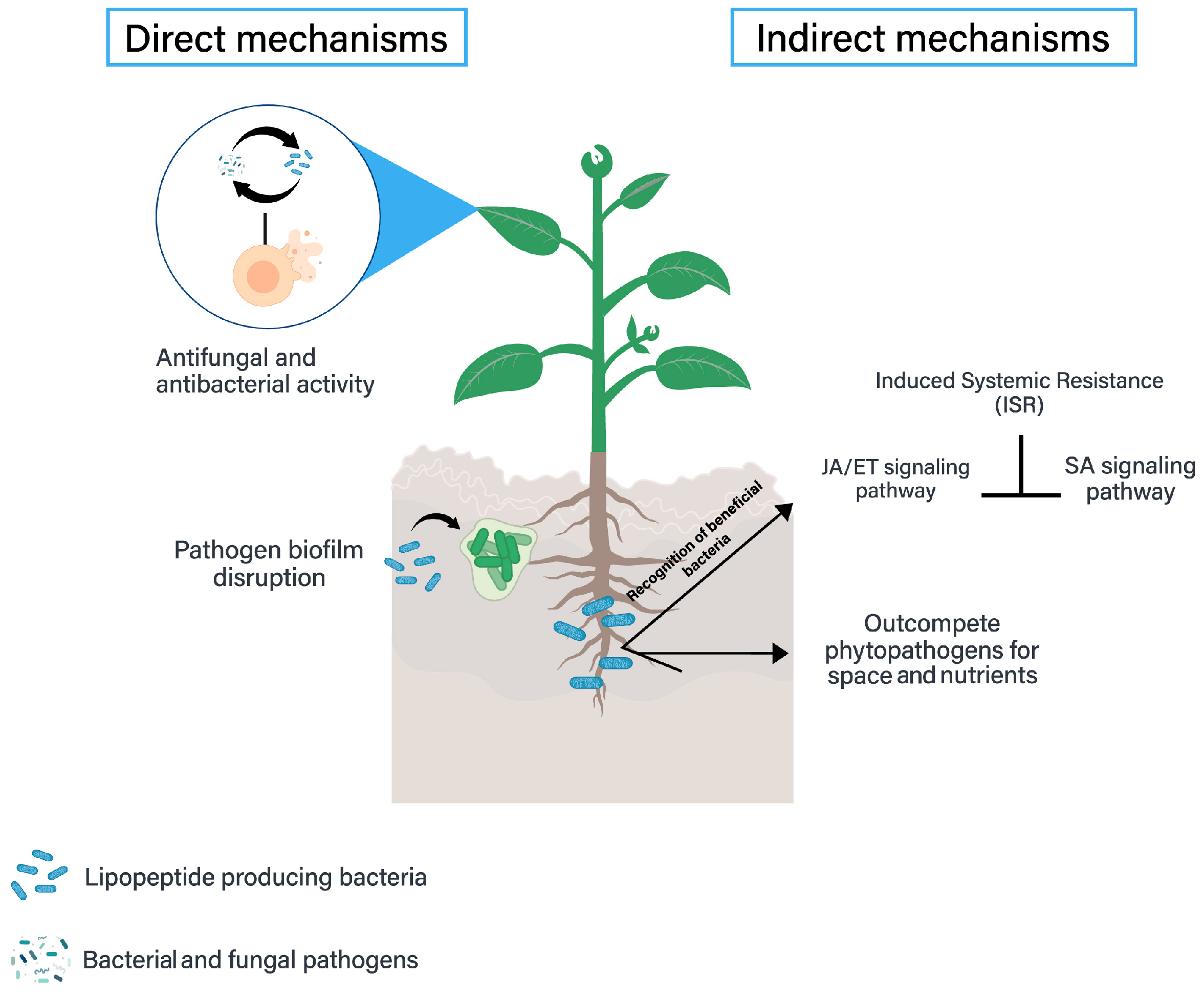
Figure 1. Lipopeptide’s role in the biological control of phytopathogens is divided into direct (including the two main bioactivities for pathogen inhibition) and indirect mechanisms (including induced systemic resistance and competition mechanisms).
Stresses 04 00007 g001
Figure 2. Structures of the three main lipopeptide families produced from Bacillus species.
Stresses 04 00007 g002
Figure 3. Activation of master regulators in cell differentiation of Bacillus. Adapted from Kalamara et al. [49].
Stresses 04 00007 g003
Figure 4. The Bacillus metabolism involved in surfactin production and sporulation. Signage is carried out by ComX, Pro-Phr, and the sporulation process including histidine kinases (A–E). Adapted from Boguslawski et al. [54].
Stresses 04 00007 g004
Figure 5. Biosynthesis of iturin and regulation by Bacillus, initiated by the activation of the ItuA gene and regulated by quorum-sensing-related genes ComX and ComA-P among others.
Stresses 04 00007 g005
Figure 6. Biosynthesis of fengycin and regulation by Bacillus, governed by the fen cluster (fenA, fenB, fenC, fend, and fenE).
Stresses 04 00007 g006
Figure 7. Identification of lipopeptide-producing Bacillus strains.
Stresses 04 00007 g007
Table 1. Description of lipopeptide-producing Bacillus species and phytopathogen inhibition to suppress plant disease in crops of agricultural importance.
Plant DiseasePhytopathogen Lipopeptide Producing SpecieLipopeptide Inhibiting the PhytopathogenApplication MethodInhibition Reference
Gray mold disease of appleBotrytis cinereaBacillus subtilis S499FengycinCell-free lipopeptide applied directly to the infected apple70%[12]
Spot blotch on wheat plantsBipolaris sorokinianaBacillus cabrialesii TE3TSurfactin and fencgycinFoliar application of crude lipopeptide extract93%[24]
Leaf spot on sugar beetPseudomonas syringaeBacillus pumilus (SS—10.7) and Bacillus amyloliquefaciens (SS—12.6 and SS—38.4)Surfactin, fengycin and iturinFoliar application of crude lipopeptide extracts92%[36]
Watermelon wiltFusarium oxysporumBacillus amyloliquefaciens DHA55Surfactin, fengycin and iturinPlants drenched in inoculum suspensions71.50%[37]
Arabidopsis root infectionPseudomonas syringaeBacillus subtilis 6051SurfactinBacterial inoculation on plant70%[38]
Tomato wiltRalstonia solanacearumBacillus velezensis FJAT-46737Surfactin, fengycin and iturinTomato seedling roots were dipped in the crude lipopeptide solution96.20%[39]
Root and foliar diseases of soybeansXanthomonas axonopodis PV. GlycinesBacillus amyloliquefaciens KPS46SurfactinCell-free supernatant treatment of soybean seeds30%[40]
Sclerotinia stem rot diseaseSclerotinia sclerotiorumBacillus amyloliquefaciensSurfactin and fengycinSpray (bacterial cells grown in MOLP) on soybean plants100%[41]
Sigatoka disease of bananaPseudocercospora fijiensisBacillus tequilensis EA-CB0015Surfactin, fengycin and iturinBanana plants were sprayed with liquid culture including biomass of bacteria and the lipopeptides100%[42]
Damping-off beanPythium ultimumBacillus subtilis M4Iturin and fengycinBean seed soaked in cell suspension n of 5 × 10⁸ CFUs98%[43]
Table 2. Indirect screening methods to identify potentially lipopeptide-producing strains.
MethodDescriptionReference
Drop-collapse assayEach well of a microplate is coated with a layer of oil (i.e., mineral oil) before analysis. Then, a drop of the supernatant is added to the center of a well and observed after 1 min. The drop formed (as it is immiscible) will collapse revealing the presence of biosurfactants, including lipopeptides.[122,123]
Hemolytic assayThe bacteria are cultured on blood agar in a Petri dish. After a number of certain days of growth, the formation of a halo around the colony (β-hemolysis) indicates the production of lipopeptides.[123]
Meniscus formation assayOne volume of supernatant is placed in a 96-well microplate. When biosurfactants (including lipopeptides) are present in the supernatant, the surface of a wellbore liquid forms a concave lens that distorts the view of a grid.[124]
Oil atomizationThe bacteria are grown on Petri dish agar. After a number of certain days of growth, a mist of mineral oil is applied to an agar surface with an airbrush. A halo around the colony indicates the production of biosurfactants, including lipopeptides.[119,125]
Table 3. Advantages and limitations of used techniques for identification of lipopeptides.
TechniquePrincipleAdvantagesLimitations
Liquid chromatography (HPLC)Separation based on hydrophobicity.Separates lipopeptides from complex mixtures.Limited to the separation of intact lipopeptides without detailed structural information.
Gas chromatography (GC)Separation based on volatility.Quantifies fatty acid chains of lipopeptides.Requires saponification and derivatization, limited to the analysis of lipid tails.
Tandem mass spectrometry (MS/MS)Fragmentation of ions for structural elucidation.Provides detailed structural information and identifies specific lipopeptides.Requires prior chromatographic separation, and database searching for identification.
Thin-layer chromatography (TLC) Separation based on hydrophobicity.Provides easy visualization of separated compounds.Limited resolution and sensitivity.
Disclaimer/Publisher’s Note: The statements, opinions and data contained in all publications are solely those of the individual author(s) and contributor(s) and not of MDPI and/or the editor(s). MDPI and/or the editor(s) disclaim responsibility for any injury to people or property resulting from any ideas, methods, instructions or products referred to in the content.