Article

The Effects of Altered Blood Flow, Force, Wrist Posture, Finger Movement Speed, and Population on Motion and Blood Flow in the Carpal Tunnel: A Mega-Analysis

1
Occupational Biomechanics Laboratory, Department of Kinesiology, McMaster University, Hamilton, ON L8S 4K1, Canada
2
School of Physical and Health Education, Nipissing University, North Bay, ON P1B 8L7, Canada
*
Author to whom correspondence should be addressed.
Biomechanics 2025, 5(1), 15; https://doi.org/10.3390/biomechanics5010015
Submission received: 19 December 2024 / Revised: 12 February 2025 / Accepted: 26 February 2025 / Published: 3 March 2025
(This article belongs to the Section Injury Biomechanics and Rehabilitation)

Abstract

:
Background/Objectives: Mechanical compression of the median nerve is believed to be responsible for idiopathic carpal tunnel syndrome (CTS) due to fibrosis of the subsynovial connective tissue (SSCT). Vascular consequences have also been observed in structures of the carpal tunnel, raising speculation regarding the role of factors such as ischemia and edema in CTS pathology. Methods: We performed a mega-analysis from our database of over 10 years of studies. Mixed-effects models were used to address the disconnect between mechanical and vascular influences on CTS; the effects of biomechanical factors and CTS status were evaluated on carpal tunnel tissue mechanics and blood flow. Altered blood flow was also induced during tissue motion to draw inferences regarding the cyclical relationship between tissue mechanics and fluid flow changes on CTS pathology. Results: Greater movement speed and flexed wrist postures were found to contribute to greater shear strain. Flexed wrist postures and greater fingertip force were found to increase median nerve blood flow. Greater CTS severity was associated with lower median nerve blood flow. Finally, brachial blood flow restriction as a surrogate for elevated carpal tunnel pressure was found to alter tissue motion and increase carpal tunnel tissue shear strain. Conclusions: Finger movement speed, force application, wrist posture, and altered fluid flow in the carpal tunnel contribute to changes in outcomes associated with the development of CTS. The mechanistic findings from this paper should be incorporated into future research to update the damage model for CTS pathology.

1. Introduction

Carpal tunnel syndrome (CTS) is the most common peripheral neuropathy and is associated with symptoms such as pain, numbness, and tingling of the hand and wrist [1]. Its prevalence is estimated to be about 4% in the general population [1] and 8% in the US working population, with a higher proportion of cases in females than males (10% vs. 6%, respectively) [2]. In the United States, CTS carries an annual financial burden of approximately USD 2 billion [2]. CTS development has been associated with occupational exposures such as computer use in offices [3,4], repetitive work and high forces in industrial settings [5,6], and raw meat processing in poultry plants [7]. Chronic exposure to occupational risk factors, such as excessive force, repetitive exertions, and non-neutral postures have been linked to an increased risk for developing CTS [8]. These factors are known to elevate hydrostatic pressure in the carpal tunnel (CTP), affect the motion of the flexor tendons and sub-synovial connective tissue (SSCT), and alter blood flow, which all affect the function of the median nerve.
Investigations have strived to better understand the etiology of idiopathic carpal tunnel syndrome. Surgical and histological findings largely agree that non-inflammatory synovial tissue fibrosis is the most prevalent tissue-related change in the carpal tunnel [9,10,11,12,13,14]. In recent years, emphasis has been placed on the SSCT, which surrounds the structures within the carpal tunnel. The SSCT is a highly intricate system comprising thick bundles of collagen that run parallel to the tendons and loose interconnecting fibres that join adjacent layers of the SSCT [10,15]. As the finger flexor tendons move through the carpal tunnel, thick bundles of the SSCT in the immediate proximity of the tendons are recruited and displaced. SSCT interconnecting fibres are then pulled taut, recruiting subsequent thick bundles of the SSCT. This process continues with more and more thick bundles—or layers—of the SSCT being recruited to accommodate independent tendon excursion. Fibrosis of the SSCT is characterized by the thickening of its parallel bundles and rupture of its interconnecting fibres [10,13,16].
A shear strain mechanism of injury has been proposed to explain the pathophysiology of the SSCT and its relation to CTS. It is believed that excessive relative motion between the tendons in the carpal tunnel and its adjacent SSCT induces a cycle of rupture, repair, and thickening of the SSCT [9,17]. This is supported by the findings of Ettema et al. [10], who used scanning electron microscopy on samples from patients with CTS and found SSCT damage to be most prominent in its layers closest to the tendon, with superficial layers demonstrating less severe changes. The mechanism is further corroborated by sonographic findings of patients with CTS possessing thicker SSCT [13,16,18,19] and decreased SSCT motion in patients with CTS relative to controls [20]. Together, this suggests origin of injury at the interface between the tendon and SSCT, with accumulated damage presenting as an overall thickening of the SSCT.
To quantify SSCT damage, investigations have been conducted to measure finger tendon movement in various wrist postures, motions (isolated finger, all fingers, or wrist), and speeds [21,22,23,24,25,26,27,28,29]. A flexed wrist posture and greater movement speed were identified to be factors that increase the relative motion between the tendon and its SSCT, quantified by the shear strain index (SSI) and maximum velocity ratio (MVR) [24,26]. Effectively, this indicates that wrist flexion and greater movement speed exacerbate the shear strain mechanism of injury, increasing the relative motion between the finger tendon and its SSCT.
As CTS is characterized by median nerve compression, one of the primary consequences is altered median nerve blood flow. Differences have been observed in median nerve blood flow amongst many groups, yet a consensus relationship is unclear: differences range from higher median nerve blood flow with a greater number of CTS symptoms [30,31] to higher median nerve blood flow in asymptomatic individuals at the proximal carpal tunnel [32] and higher median nerve blood flow in symptomatic individuals only with a flexed wrist posture [33]. Some have even failed to identify significant relationships in median nerve blood flow between individuals with CTS symptoms and healthy individuals [34] and amongst mild, moderate, and severe CTS diagnoses [35]. A clearer relationship exists in the location of blood flow changes, with the segment of the median nerve underneath the transverse carpal ligament (TCL) demonstrating random flow and changing following carpal tunnel release surgery to pulsatile flow [36]. As the segment proximal to the TCL was pulsatile both before and after surgery, Seiler et al. [36] speculated this to be a sign that the segment of the median nerve under the TCL is ischemic.
Early progression of CTS research has been outlined by Sunderland [37], who detailed how the mechanism of injury was investigated from an ischemia perspective before falling out of favour towards the mechanical compression of the median nerve. The current understanding of CTS largely treats these issues separately, with focus divided between the mechanism underlying the elevation of carpal tunnel pressure and the consequences to median nerve blood flow and nerve conduction. What may warrant greater consideration is the ability of these consequences to play an etiological role in CTS development and/or progression. While the shear strain mechanism of injury primarily focuses on tissue mechanics and histology, one of the roles of the SSCT is to provide vascular continuity in the carpal tunnel [15]. Given that CTS samples have shown vascular proliferation, hypertrophy, and obstruction in the SSCT [12], and that SSCT blood vessels have been found to be mechanically deformed with tendon motion [38], it is plausible to assume that fibrosis of the SSCT also implicates its blood vessels and capacity to perform its blood flow function. The extent to which impaired SSCT vascular function impacts the median nerve, however, is less clear.
Recent works have tested the effects of altered blood and fluid flow to determine the plausibility of their influence on the shear strain mechanism of injury. Changes in carpal tunnel tissue motion and strain measures were studied by inflating a brachial blood pressure cuff to alter blood and fluid flow in the carpal tunnel [28,39,40]. While blood flow alteration was demonstrated to have a significant effect on carpal tunnel tissue motion, differences were noted between supradiastolic [28] and subdiastolic [39,40] levels of blood flow alteration.
The purpose of this work was to assess the overall effects of factors influencing CTS-related outcomes to address the discrepancies between the findings of some studies and to reaffirm that relationships for the shear strain mechanism of CTS injury hold true when samples across different study designs and interventions are considered. Of key interest was to evaluate the effects of biomechanical factors on median nerve blood flow and carpal tunnel tissue motion, how these outcomes can influence one another, and to provide a mechanism relating these outcomes to the progression of CTS.

2. Materials and Methods

Participant-level data from 11 studies conducted in the McMaster Occupational Biomechanics Laboratory were consolidated and included where appropriate to evaluate the evidence for factors that influence median nerve blood flow and carpal tunnel tissue motion. The effects of finger movement speed, finger force, wrist posture, and blood flow alteration were tested on different aspects of CTS pathomechanics. Thematically, the analyses investigated the following: (1) biomechanical influences on tendon and SSCT motion, (2) changes in median nerve blood flow, and (3) the effect of induced altered blood flow on tissue mechanics.

2.1. Biomechanical Influences on Tendon and SSCT Motion

Data from 7 studies comprising a total of 128 subjects (76 healthy participants, 33 with CTS, 11 symptomatic patients, and 8 cadavers) assessed the effects of finger movement speed and wrist posture on ultrasound-measured tendon and SSCT motion [23,25,26,27,28,29,40]. Study protocols tested repetitive finger flexion–extension motions at various frequencies and speeds, namely 0.75 Hz, 1 Hz, and 1.25 Hz, a self-selected pace of approximately 60 mm/s tendon velocity, and cadaveric tendon excursion velocities of 50–150 mm/s. Tissue movement speed was recorded using Colour Doppler ultrasound and calculated using the method established by Tat, Kociolek, and Keir [24]. Participants were positioned laying supine, with the shoulder abducted at about 45° and the elbow straight, with their forearm supinated and supported in an apparatus. Data from cadaveric specimens were collected with the forearm in a supine position and affixed to a testing apparatus. The measured tissue movement speed (mean peak trial velocity) irrespective of testing condition was analyzed. Wrist posture was tested at various angles ranging from 30° extension, neutral, to 30° flexion. In instances where finger motion data were separated into flexion and extension phases, the mean across both phases was used. The effects of movement speed (in cm/s) and wrist posture (extension, neutral, and flexion) were tested separately, with outcome variables of mean peak flexor digitorum superficialis (FDS) tendon displacement, mean peak SSCT displacement, and strain measures (relative tendon–SSCT displacement, shear strain index (SSI), and maximum velocity ratio (MVR)). SSI and MVR are metrics that represent potential damage to the SSCT and are calculated as follows: Shear   Strain   Index   SSI   =   Displacement FDS peak Displacement SSCT peak Displacement FDS peak and Maximum   Velocity   Ratio   M V R   =   V e l o c i t y SSCT peak Velocity FDS peak   ×   100 % , where higher SSI and lower MVR are associated with greater damage to the SSCT.

2.2. Median Nerve Blood Flow Changes

The overall effects of biomechanical influences (finger force and deviated wrist posture) and CTS status on median nerve blood flow (ultrasound-measured intraneural blood flow) were evaluated using data from a total of 109 subjects [25,33,34,39]. Participants were seated [25,33,34] or laid in a supine position [39] with their shoulder adducted, elbow flexed, the forearm and hand supported in an apparatus, and the wrist splinted. Wrist postures from individual study protocols varied, including 30° extension, 15° extension, neutral, 15° flexion, and 30° flexion. For analysis, wrist posture was reduced to extension, neutral, and flexion levels. Finger force from individual study protocols were 0 N (absence of force) and 6 N middle finger force levels. The effects of wrist posture, finger force, and the wrist posture × force interaction were tested, with median nerve blood flow as the outcome. Separately, the effect of CTS status on median nerve blood flow was tested using data from 56 healthy participants, 20 participants with possible CTS (experience CTS symptoms but no formal diagnosis), and 33 participants with CTS (16 of which had 6-month follow-up values) [25,33,34]. To determine the effect of CTS status classification, multiple models were tested (1) using healthy, possible, CTS, and follow-up classifications and (2) using the same method as before, but with the participants with CTS split into severities of mild, moderate, and severe. Additionally, a model comparing median nerve blood flow by the presence of symptoms was tested, with healthy subjects in the “No” group, and participants with possible, CTS, and follow-up classifications in the “Yes” group.

2.3. Effect of Induced Blood Flow Alteration

Repetitive finger motions performed by 49 participants [28,39,40] were analyzed to determine the effect of induced blood flow alteration on carpal tunnel tissue motion. The presence of altered blood flow in the carpal tunnel, as well as the type of blood flow alteration (baseline vs. subdiastolic vs. supradiastolic), were tested on tissue displacements (mean peak FDS tendon and SSCT displacement) and strain measures (relative tendon-SSCT displacement, SSI, and MVR). Participants were positioned laying supine with the shoulder abducted at about 45° and the elbow straight, with their forearm supinated and supported in an apparatus. To elicit altered blood flow, a manual blood pressure cuff was inflated and held at a constant pressure on the upper arm. For the supradiastolic condition (also referred to as partial ischemia [28], the cuff was inflated to a level of diastolic BP + 0.25 (systolic–diastolic BP), whereas the subdiastolic condition [39,40] restricted blood flow using 0.8 × diastolic BP.

2.4. Statistics

Analyses were conducted using R (R Core Team, 2021). Linear mixed-effects models were generated using the lmerTest package [41] to assess the effects of factors influencing the outcomes of interest. Models were created with subject nested within study to incorporate both subject and study as random effects to account for some of the variability introduced by different population types and methodological differences. Pairwise comparisons of the least-squares means were computed using t-tests (with the degrees of freedom based on the Satterthwaite method) to assess differences between levels of a factor that was determined to be a significant predictor. Regression assumptions were visually assessed. Variance of the model is represented by conditional R2, defined as the total variance explained by both the fixed and random effects in the model [42]. Regression coefficients are represented with β. Significance was defined at the p < 0.05 level.

3. Results

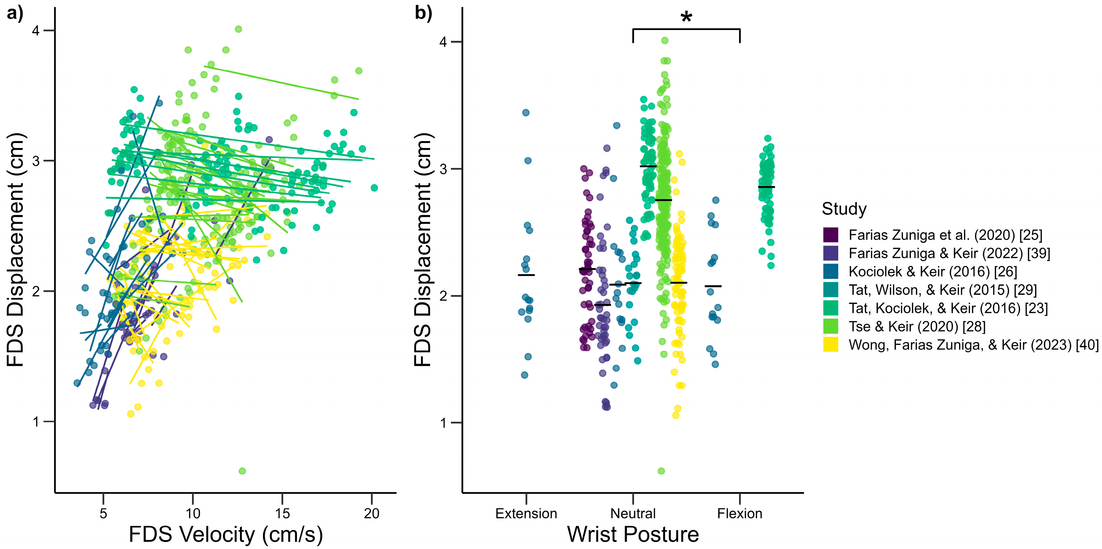
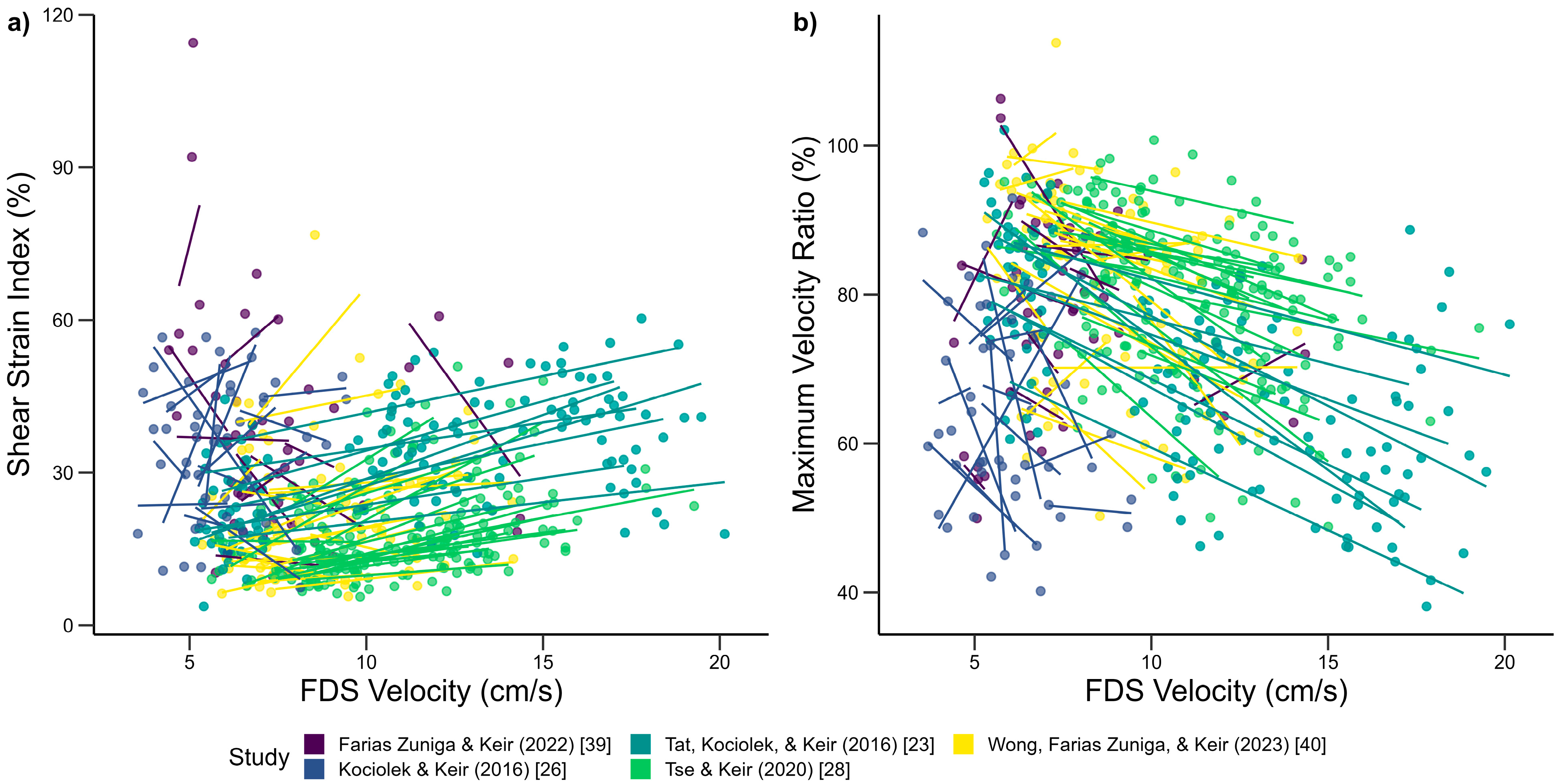
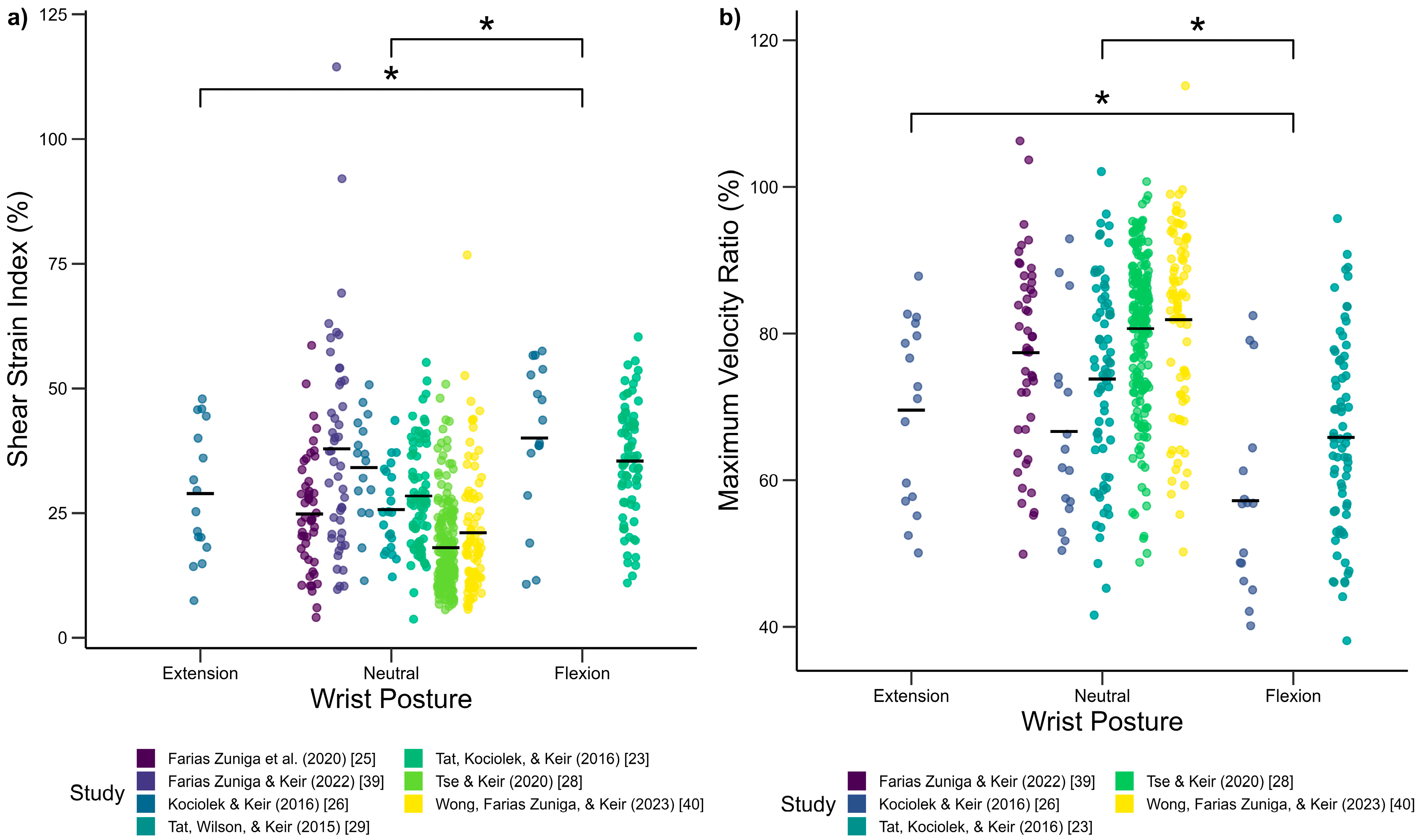
3.1. Biomechanical Influences on Tendon and SSCT Motion

FDS tendon velocity significantly predicted FDS tendon displacement (Figure 1a), where greater tendon velocity was associated with lower mean peak displacement (R2 = 0.804, β = −0.013, p < 0.01). Wrist posture was also found to significantly predict FDS tendon displacement, where wrist flexion, but not extension, was found to reduce the extent of displacement (R2 = 0.746, β = −0.133, p < 0.01) compared to a neutral wrist posture (Figure 1b).
SSCT velocity was found to significantly predict SSCT displacement with a lower mean peak SSCT displacement as the SSCT velocity increased (R2 = 0.746, β = −0.030, p < 0.001) (outliers were removed after being identified using Cook’s distance method of detection). Wrist posture was found to significantly predict SSCT displacement. Compared to the wrist in a neutral posture, wrist flexion was found to reduce SSCT displacement (R2 = 0.645, β = −0.291, p < 0.001).
FDS velocity was found to significantly predict relative displacement (R2 = 0.673, β = 0.035, p < 0.001), the shear strain index (SSI) (R2 = 0.776, β = 1.398, p < 0.001), and the maximum velocity ratio (MVR) (R2 = 0.772, β = −1.933, p < 0.001). As the FDS velocity increased, both the relative displacement and SSI increased, while the MVR decreased (Figure 2). SSCT velocity was not found to predict relative displacement (β = −0.006, p = 0.409), SSI (β = 0.132, p = 0.575), or MVR (β = −0.437, p = 0.078). Upon removal of outliers identified via Cook’s distance, SSCT velocity was found to significantly predict MVR (R2 = 0.663, β = −0.793, p < 0.001).
Wrist posture was found to significantly predict the SSI (R2 = 0.610) and MVR (R2 = 0.562). The SSI was higher in a flexed wrist posture compared to both neutral (β = 6.958, p < 0.001) and extended (β = 11.28, p < 0.001) wrist postures (Figure 3a). The MVR was also found to be lower with a flexed wrist posture compared to neutral (β = −8.543, p < 0.001) and extended (β = −11.26, p < 0.001) wrist postures (Figure 3b).

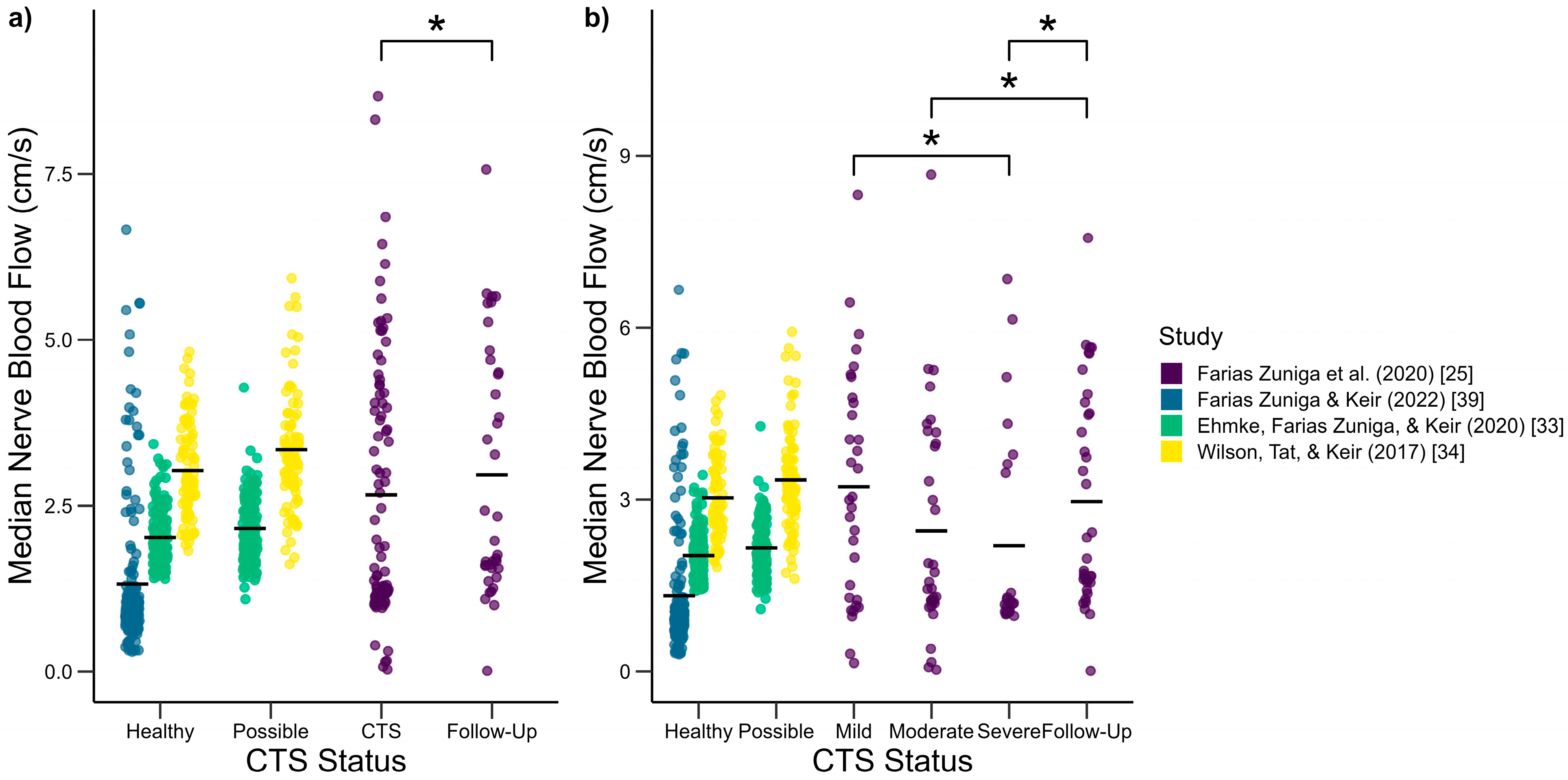
3.2. Median Nerve Blood Flow Changes

Force (β = 0.583, p < 0.001) and wrist posture (β = 0.243, p = 0.01) were found to influence the median nerve blood flow velocity, where wrist flexion and 6 N of force generation were associated with greater median nerve blood flow velocities (R2 = 0.755) (Figure 4).
When CTS status was categorized with the levels (1) healthy, (2) possible, (3) CTS, and (4) follow-up (Figure 5), differences were only detected between the CTS and follow-up groups (R2 = 0.766, β = −0.600, p < 0.001). The effect of CTS status was found to predict the median nerve blood flow when the CTS group was separated into its respective severities of mild, moderate, and severe (R2 = 0.766). Differences were detected between the severe and mild groups (β = 0.832, p < 0.05), between the follow-up and moderate groups (β = −0.614, p < 0.01), and between the follow-up and severe groups (β = −1.172, p < 0.001). Finally, the presence of CTS symptoms alone was insufficient in predicting median nerve blood flow (R2 = 0.727, β = 0.318, p = 0.29).

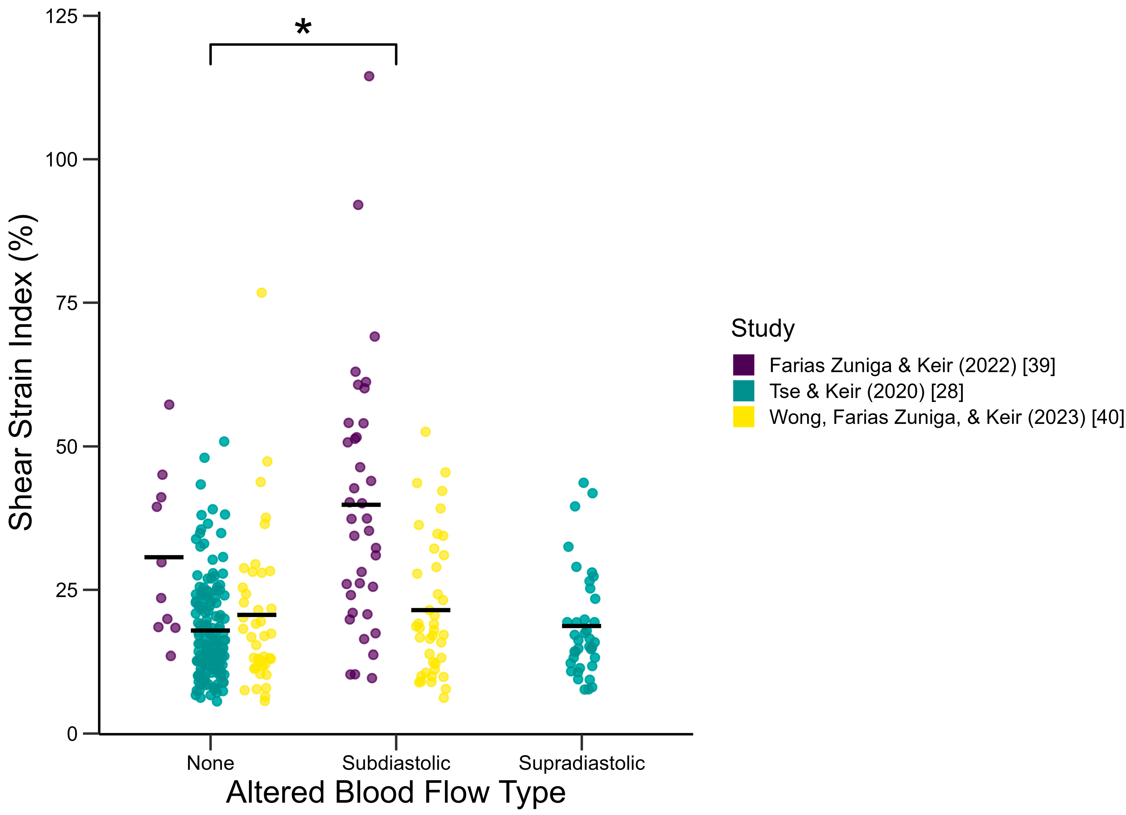
3.3. Effect of Induced Blood Flow Alteration

Altered blood flow was found to significantly predict FDS displacement (R2 = 0.743, β = −0.118, p < 0.01) and SSCT displacement (R2 = 0.647, β = −0.116, p < 0.01), with a reduction of displacement when blood flow alteration occurred (Figure 6). When the type of blood flow alteration was considered, subdiastolic alteration was found to decrease FDS displacement (R2 = 0.736, β = −0.144, p < 0.05), while supradiastolic alteration was found to decrease SSCT displacement (R2 = 0.646, β = −0.122, p < 0.05) compared to baseline conditions.
Regarding strain measures, altered blood flow was found to affect the shear strain index (SSI) (Figure 7). When subjected to subdiastolic blood flow occlusion, the SSI increased relative to the baseline conditions (R2 = 0.737, β = 3.44, p < 0.05). No significant change was detected with the administration of supradiastolic blood flow alteration.

4. Discussion

This paper consolidated findings from studies conducted within the McMaster Occupational Biomechanics Lab. Specifically, studies that investigated tissue motion and median nerve blood flow in relation to the overall theme of carpal tunnel syndrome (CTS) were included in these analyses. Overall, tissues that are associated with CTS pathology—that is, the FDS tendon and surrounding SSCT—demonstrate altered motion from changes to biomechanical factors such as higher tissue velocities and deviated wrist postures. Median nerve blood flow, another metric of interest, was found to differ amongst different groups, with deviated wrist postures and differences in fingertip force. Tissue motion and blood flow are related via local blood flow alteration, which we demonstrated using changes in local blood flow.

4.1. Biomechanical Influences on Tendon and SSCT Motion

Previously, movement speed was treated as a categorical factor with the levels being the speeds or frequencies demanded by subjects in their respective study protocols. In this study, measured tissue velocities were used in place of experimentally defined speed and frequency demands to assess the effect of movement speed. This approach effectively addressed the differences in the wide range of movement speeds, generated a larger dataset, and allowed a more definitive and generalizable statement to be made regarding the movement speed-displacement relationship for the FDS tendon and SSCT. For instance, SSCT motions of up to 16 cm/s were assessed in this paper compared to 0.2–1.0 cm/s reported in the literature [43,44,45]. However, it is important to note that the trade-off in having this greater sample to analyze was the inclusion of not only healthy subjects, but also cadaveric samples and individuals with CTS symptoms as well. Methodological differences amongst studies may also contribute to subsets of the sample forming clusters when evaluating a relationship. After taking these considerations in mind, pooling the data together for analysis was the approach that was taken as it was believed to be more powerful in assessing the relationships of interest.
Both FDS and SSCT velocity, as well as wrist posture, were found to significantly affect their respective tissue displacements, with greater velocity and a flexed wrist posture resulting in lower displacements (Figure 1). This mirrors the findings in cadaveric samples where flexion was found to reduce the extent of tendon excursion [45]. It is possible that volar movement of the FDS tendon with wrist flexion [45,46] reduces the moment arm of the tendon at the wrist, effectively reducing tendon excursion for the same range of finger motion [47]. FDS and SSCT displacements spanned from 1 to 4 cm, which mirrors the broad range of values reported in the literature [43,48,49,50,51,52,53,54,55].
Perhaps it is more important to consider the implications from the shear measure results in which greater tendon movement speeds are associated with higher tendon–SSCT relative displacement, a higher SSI, and a lower MVR (Figure 2). Similarly, greater SSCT movement speed is associated with a higher SSI, and for wrist posture, flexion was found to result in a lower SSI and a higher MVR compared to both neutral and extended wrist postures (Figure 3). As the SSI is calculated as the tendon-SSCT relative displacement normalized to tendon displacement, and the MVR is the ratio of SSCT velocity to tendon velocity, a higher SSI and a lower MVR would be indicative of tissue motion that is less in unison with one another, causing the tissues to experience greater shear strain.
In summary, higher movement speeds and flexed wrist postures can be stated to increase the risk of a shear strain mechanism of injury for CTS.

4.2. Median Nerve Blood Flow Changes

With wrist flexion, median nerve blood flow velocity was found to increase compared to a neutral wrist posture (Figure 4a). With pooled data from various studies, this result gives greater weight to the original findings of Ehmke and colleagues [33] and perhaps indicates elevated median nerve blood flow to be a compensatory mechanism that supports the demands of the muscles innervated by the median nerve when it is compressed. Greater fingertip force has been linked to an elevation in carpal tunnel pressure, with a doubling of pressure from about 20 mmHg at a 0 N force to about 40 mmHg at 6 N of fingertip force in a pronated forearm posture [56]. Coincidentally, the data analyzed for this study measured the median nerve blood flow with 6 N of fingertip force, which was demonstrated to increase median nerve blood flow velocity compared to 0 N of fingertip force (Figure 4b). Overall, increased median nerve blood flow with fingertip force likely acts to support the greater active demands of the muscles it innervates. Meanwhile, passively, median nerve blood flow decreases with greater carpal tunnel pressure and subsequently increases the severity of symptoms in individuals with CTS. The manner in which intraneural blood flow of the median nerve decreases may arise in a multitude of ways, from a net decrease to the nerve due to greater arteriovenous flow through fistulous connections [30] to neovascularization and a greater number of blood vessels [57,58,59].
Through an investigation into the median nerve blood flow based on the classification of CTS status, it was determined that experiencing symptoms alone is insufficient in predicting median nerve blood flow changes. Our results support a stance that there is a decline in median nerve blood flow as CTS severity increases, which matches the findings obtained by Evans and colleagues [32]. Additionally, median nerve blood flow levels in the follow-up group rose to approximately the same levels as those of the mild group (Figure 5b). Physiologically, this may be an indication that insufficient blood flow to the median nerve is responsible for increasing the severity of symptoms experienced by individuals with CTS. This is further supported by findings such as greater contact pressure on the median nerve increasing symptom severity [60] and a dose–response relationship of pressure on median nerve dysfunction having been established in a rabbit model [61].
From these results, flexed wrist postures, fingertip force, and greater CTS severity were found to be associated with lower median nerve blood flow, and in the case of biomechanical factors, they were found to elevate the likelihood of developing CTS.

4.3. Effect of Induced Blood Flow Alteration

To understand how the local fluid environment in the carpal tunnel affects tissue motion and potential injury of carpal tunnel structures, altered blood flow was induced using subdiastolic occlusion or supradiastolic partial ischemia. Notably, any change in blood flow (occlusion or partial ischemia) changed FDS tendon and SSCT displacements (Figure 6), demonstrating the importance of the mechanism. Differences in the effects of the blood flow alteration were observed in tissue motion, where subdiastolic alteration reduced FDS displacement (Figure 6b), and supradiastolic alteration reduced SSCT displacement (Figure 6d). These findings support previous work that found a critical carpal tunnel hydrostatic pressure threshold [8] but expand it to differential effects on tendon and SSCT motion.
Shear strain measures were altered by subdiastolic occlusion, where the SSI was found to increase relative to the baseline conditions (Figure 7). This suggests that altered blood flow affects the local fluid environment in the carpal tunnel to promote shear injury to the SSCT. Physiologically, this may be explained by the hydrophilic properties of the SSCT due to its histological composition [15], which promote tissue swelling of the SSCT and lead to less displacement.
This theme of investigation demonstrates that changes to the carpal tunnel’s local fluid environment can compromise carpal tunnel tissue motion and have consequences on the shear strain mechanism of injury for CTS.

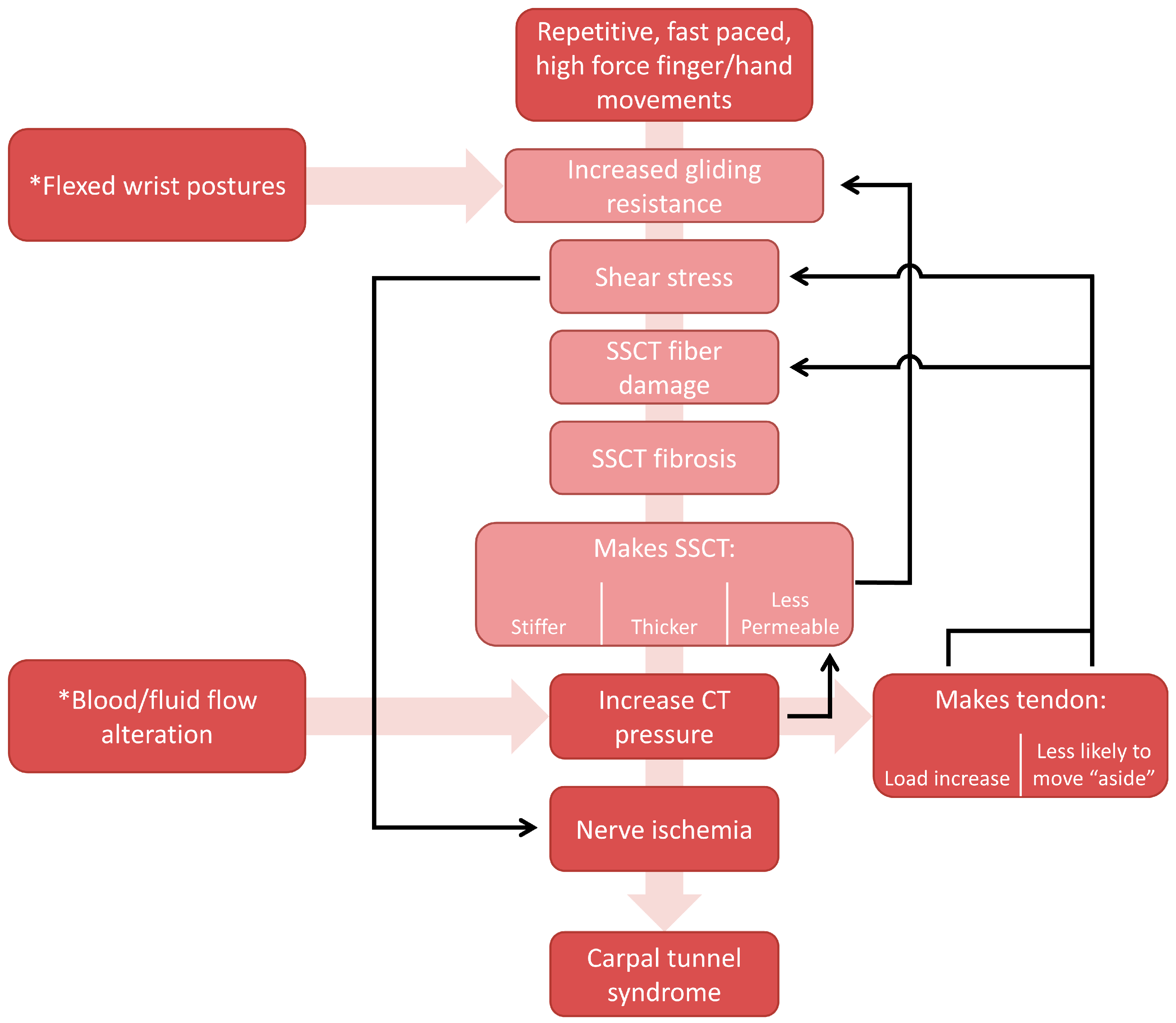
4.4. Updated Mechanism of Injury

The shear strain mechanism of injury mentioned in this study was visualized in a damage model by Festen-Schrier and Amadio [62]. We propose an update to this model that incorporates the mechanistic evidence from our findings (Figure 8). In particular, the experimentally induced altered blood flow interventions provide evidence for alternative sources of increased carpal tunnel pressure (non-SSCT) that feed into CTS pathology.

4.5. Limitations

While cohorts from multiple studies were combined to increase the sample size for analysis, and predictor variables were limited for each model to allow for a greater inclusion of studies, some changes may have been more heavily driven by data from a few cohorts. In relation to this, despite incorporating the study that an observation was sourced from as a random effect in our models—intended to account for differences in the experimenter collecting the data, as well as sample characteristics—ultimately, the experimenter/sonographer and sample type (i.e., healthy vs. cadaveric) were not explicitly defined as random effects in the models. Additionally, many subjects were from a healthy university population, with a smaller subset of the total dataset being from those with CTS symptoms, confirmed subjects with CTS, and cadaveric specimens. This may be beneficial in understanding the mechanisms for CTS pathology in healthy individuals but may limit the generalizability of the results to the broader population that may experience a spectrum of CTS symptoms.

5. Conclusions

This study combined data across a number of investigations into carpal tunnel syndrome to assess the effects of biomechanical influences on carpal tunnel tissue motion and median nerve blood flow. Additionally, it attempted to bridge our understanding of their effects upon one another by analyzing the effect of induced blood flow alteration in the carpal tunnel. Greater movement speeds and flexed wrist postures were associated with greater tissue shear strain. When changes in the median nerve blood flow were assessed, force generation and flexed wrist postures were found to reduce blood flow. The classification of CTS groups was found to be a contributor to differences in median nerve blood flow when CTS status was used as a predictor. Finally, induced blood flow alteration was characterized by greater shear strain and should be considered as a source of elevated carpal tunnel pressure that contributes to the carpal tunnel syndrome mechanism of injury.

Author Contributions

Conceptualization, A.Y.W.W., A.M.K. and P.J.K.; Methodology, A.Y.W.W., A.M.K. and P.J.K.; Software, A.Y.W.W., A.M.K. and P.J.K.; Validation, A.Y.W.W.; Formal Analysis, A.Y.W.W.; Investigation, A.Y.W.W., A.M.K. and P.J.K.; Resources, P.J.K.; Data Curation, A.Y.W.W.; Writing—Original Draft Preparation, A.Y.W.W.; Writing—Review and Editing, A.Y.W.W., A.M.K. and P.J.K.; Visualization, A.Y.W.W.; Supervision, P.J.K.; Project Administration, P.J.K.; Funding Acquisition, P.J.K. All authors have read and agreed to the published version of the manuscript.

Funding

This research was funded by the Natural Sciences and Engineering Council of Canada, Discovery Grant numbers 217382-09 and RGPIN-2016-06460.

Institutional Review Board Statement

This study represents a secondary analysis of data; thus, no institutional review was required. Each study used in the analysis was approved by the Hamilton Integrated Research Ethics Board (HIREB).

Informed Consent Statement

Not applicable.

Data Availability Statement

The original contributions presented in this study are included in the article. Further inquiries can be directed to the corresponding author.

Acknowledgments

We would like to thank all authors who collected these data during their graduate degrees in the lab: Jimmy Tat, Amanda Farias Zuniga, Samantha Ehmke, Calvin Tse, and Kat Wilson.

Conflicts of Interest

The authors declare no conflicts of interest.

References

  1. Atroshi, I.; Gummesson, C.; Johnsson, R.; Ornstein, E.; Ranstam, J.; Rosén, I. Prevalence of Carpal Tunnel Syndrome in a General Population. J. Am. Med. Assoc. 1999, 282, 153–158. [Google Scholar] [CrossRef]
  2. Dale, A.M.; Harris-Adamson, C.; Rempel, D.; Gerr, F.; Hegmann, K.; Silverstein, B.; Burt, S.; Garg, A.; Kapellusch, J.; Merlino, L.; et al. Prevalence and Incidence of Carpal Tunnel Syndrome in US Working Populations: Pooled Analysis of Six Prospective Studies. Scand. J. Work Environ. Health 2013, 39, 495–505. [Google Scholar] [CrossRef] [PubMed]
  3. Shiri, R.; Falah-Hassani, K. Computer Use and Carpal Tunnel Syndrome: A Meta-Analysis. J. Neurol. Sci. 2014, 349, 15–19. [Google Scholar] [CrossRef] [PubMed]
  4. Andersen, J.H.; Thomsen, J.F.; Overgaard, E.; Lassen, C.F.; Brandt, L.P.A.; Vilstrup, I.; Kryger, A.I.; Mikkelsen, S. Computer Use and Carpal Tunnel Syndrome: A 1-Year Follow-Up Study. JAMA 2003, 289, 2963. [Google Scholar] [CrossRef]
  5. Silverstein, B.A.; Fine, L.J.; Armstrong, T.J. Occupational Factors and Carpal Tunnel Syndrome. Am. J. Ind. Med. 1987, 11, 343–358. [Google Scholar] [CrossRef] [PubMed]
  6. Harris-Adamson, C.; Eisen, E.A.; Kapellusch, J.; Garg, A.; Hegmann, K.T.; Thiese, M.S.; Marie Dale, A.; Evanoff, B.; Burt, S.; Bao, S.; et al. Biomechanical Risk Factors for Carpal Tunnel Syndrome: A Pooled Study of 2474 Workers. Occup. Environ. Med. 2015, 72, 33–41. [Google Scholar] [CrossRef] [PubMed]
  7. Musolin, K.M.; Ramsey, J.G. Carpal Tunnel Syndrome Prevalence: An Evaluation of Workers at a Raw Poultry Processing Plant. Int. J. Occup. Environ. Health 2017, 23, 282–290. [Google Scholar] [CrossRef]
  8. Keir, P.J.; Farias Zuniga, A.; Mulla, D.M.; Somasundram, K.G. Relationships and Mechanisms Between Occupational Risk Factors and Distal Upper Extremity Disorders. Hum. Factors 2021, 63, 5–31. [Google Scholar] [CrossRef]
  9. Ettema, A.M.; Amadio, P.C.; Zhao, C.; Wold, L.E.; An, K.-N. A Histological and Immunohistochemical Study of the Subsynovial Connective Tissue in Idiopathic Carpal Tunnel Syndrome. J. Bone Jt. Surg. Am. 2004, 86, 1458–1466. [Google Scholar] [CrossRef]
  10. Ettema, A.M.; Amadio, P.C.; Zhao, C.; Wold, L.E.; O’Byrne, M.M.; Moran, S.L.; An, K.-N. Changes in the Functional Structure of the Tenosynovium in Idiopathic Carpal Tunnel Syndrome: A Scanning Electron Microscope Study. Plast. Reconstr. Surg. 2006, 118, 1413–1422. [Google Scholar] [CrossRef]
  11. Freeland, A.E.; Tucci, M.A.; Barbieri, R.A.; Angel, M.F.; Nick, T.G. Biochemical Evaluation of Serum and Flexor Tenosynovium in Carpal Tunnel Syndrome. Microsurgery 2002, 22, 378–385. [Google Scholar] [CrossRef]
  12. Jinrok, O.; Zhao, C.; Amadio, P.C.; An, K.-N.; Zobitz, M.E.; Wold, L.E. Vascular Pathologic Changes in the Flexor Tenosynovium (Subsynovial Connective Tissue) in Idiopathic Carpal Tunnel Syndrome. J. Orthop. Res. 2004, 22, 1310–1315. [Google Scholar] [CrossRef] [PubMed]
  13. Neal, N.C.; McManners, J.; Stirling, G.A. Pathology of the Flexor Tendon Sheath in the Spontaneous Carpal Tunnel Syndrome. J. Hand Surg. 1987, 12, 229–232. [Google Scholar] [CrossRef] [PubMed]
  14. Phalen, G.S. The Carpal-Tunnel Syndrome. Seventeen Years’ Experience in Diagnosis and Treatment of Six Hundred Fifty-Four Hands. J. Bone Jt. Surg. 1966, 48, 211–228. [Google Scholar] [CrossRef]
  15. Guimberteau, J.C.; Delage, J.P.; McGrouther, D.A.; Wong, J.K.F. The Microvacuolar System: How Connective Tissue Sliding Works. J. Hand Surg. Eur. Vol. 2010, 35, 614–622. [Google Scholar] [CrossRef]
  16. van Doesburg, M.H.M.; Mink van der Molen, A.; Henderson, J.; Cha, S.S.; An, K.N.; Amadio, P.C. Sonographic Measurements of Subsynovial Connective Tissue Thickness in Patients with Carpal Tunnel Syndrome. J. Ultrasound Med. 2012, 31, 31–36. [Google Scholar] [CrossRef]
  17. Werthel, J.-D.; Zhao, C.; An, K.-N.; Amadio, P. Carpal Tunnel Syndrome Pathophysiology: Role of Subsynovial Connective Tissue. J. Wrist Surg. 2014, 03, 220–226. [Google Scholar] [CrossRef]
  18. Kerr, C.D.; Sybert, D.R.; Albarracin, N.S. An Analysis of the Flexor Synovium in Idiopathic Carpal Tunnel Syndrome: Report of 625 Cases. J. Hand Surg. 1992, 17, 1028–1030. [Google Scholar] [CrossRef]
  19. Robben, E.; Dever, J.; De Groef, A.; Degreef, I.; Peers, K. Subsynovial Connective Tissue Thickness in Carpal Tunnel Syndrome: A Systematic Review. Clin. Biomech. 2020, 75, 105002. [Google Scholar] [CrossRef]
  20. Schrier, V.J.M.M.; Evers, S.; Geske, J.R.; Kremers, W.K.; Villarraga, H.R.; Selles, R.W.; Hovius, S.E.R.; Gelfman, R.; Amadio, P.C. Relative Motion of the Connective Tissue in Carpal Tunnel Syndrome: The Relation with Disease Severity and Clinical Outcome. Ultrasound Med. Biol. 2020, 46, 2236–2244. [Google Scholar] [CrossRef]
  21. Kociolek, A.M.; Tat, J.; Keir, P.J. Biomechanical Risk Factors and Flexor Tendon Frictional Work in the Cadaveric Carpal Tunnel. J. Biomech. 2015, 48, 449–455. [Google Scholar] [CrossRef] [PubMed]
  22. Oh, S.; Belohlavek, M.; Zhao, C.; Osamura, N.; Zobitz, M.E.; An, K.N.; Amadio, P.C. Detection of Differential Gliding Characteristics of the Flexor Digitorum Superficialis Tendon and Subsynovial Connective Tissue Using Color Doppler Sonographic Imaging. J. Ultrasound Med. 2007, 26, 149–155. [Google Scholar] [CrossRef] [PubMed]
  23. Tat, J.; Kociolek, A.M.; Keir, P.J. Relative Displacement of the Tendon and Subsynovial Connective Tissue Using Ultrasound Captures Different Phenomena than Mechanical Tendon Shear. J. Biomech. 2016, 49, 3682–3687. [Google Scholar] [CrossRef]
  24. Tat, J.; Kociolek, A.M.; Keir, P.J. Validation of Color Doppler Sonography for Evaluating Relative Displacement Between the Flexor Tendon and Subsynovial Connective Tissue. J. Ultrasound Med. 2015, 34, 679–687. [Google Scholar] [CrossRef]
  25. Farias Zuniga, A.; Ghavanini, A.A.; Israelian, G.; Keir, P.J. Blood Flow Velocity but Not Tendon Mechanics Relates to Nerve Function in Carpal Tunnel Syndrome Patients. J. Neurol. Sci. 2020, 411, 116694. [Google Scholar] [CrossRef] [PubMed]
  26. Kociolek, A.M.; Keir, P.J. Relative Motion between the Flexor Digitorum Superficialis Tendon and Paratenon in Zone V Increases with Wrist Flexion Angle. J. Orthop. Res. 2016, 34, 1248–1255. [Google Scholar] [CrossRef]
  27. Tat, J.; Kociolek, A.M.; Keir, P.J. Repetitive Differential Finger Motion Increases Shear Strain between the Flexor Tendon and Subsynovial Connective Tissue. J. Orthop. Res. 2013, 31, 1533–1539. [Google Scholar] [CrossRef]
  28. Tse, C.T.F.; Keir, P.J. External Compression and Partial Ischemia Decrease Human Finger Flexor Tendon and Subsynovial Connective Tissue Relative Motion. J. Orthop. Res. Off. Publ. Orthop. Res. Soc. 2020, 38, 1038–1044. [Google Scholar] [CrossRef]
  29. Tat, J.; Wilson, K.E.; Keir, P.J. Pathological Changes in the Subsynovial Connective Tissue Increase with Self-Reported Carpal Tunnel Syndrome Symptoms. Clin. Biomech. 2015, 30, 360–365. [Google Scholar] [CrossRef]
  30. Joy, V.; Therimadasamy, A.K.; Chan, Y.C.; Wilder-Smith, E.P. Combined Doppler and B-Mode Sonography in Carpal Tunnel Syndrome. J. Neurol. Sci. 2011, 308, 16–20. [Google Scholar] [CrossRef]
  31. Motomiya, M.; Funakoshi, T.; Ishizaka, K.; Nishida, M.; Matsui, Y.; Iwasaki, N. Blood Flow Changes in Subsynovial Connective Tissue on Contrast-Enhanced Ultrasonography in Patients with Carpal Tunnel Syndrome before and after Surgical Decompression. J. Ultrasound Med. 2018, 37, 1597–1604. [Google Scholar] [CrossRef]
  32. Evans, K.D.; Roll, S.C.; Volz, K.R.; Freimer, M. Relationship Between Intraneural Vascular Flow Measured with Sonography and Carpal Tunnel Syndrome Diagnosis Based on Electrodiagnostic Testing. J. Ultrasound Med. 2012, 31, 729–736. [Google Scholar] [CrossRef] [PubMed]
  33. Ehmke, S.; Farias Zuniga, A.; Keir, P.J. Effect of Force, Posture, and Repetitive Wrist Motion on Intraneural Blood Flow in the Median Nerve. J. Ultrasound Med. 2020, 40, 939–950. [Google Scholar] [CrossRef] [PubMed]
  34. Wilson, K.E.; Tat, J.; Keir, P.J. Effects of Wrist Posture and Fingertip Force on Median Nerve Blood Flow Velocity. BioMed Res. Int. 2017, 2017, 1–8. [Google Scholar] [CrossRef] [PubMed]
  35. Karahan, A.Y.; Arslan, S.; Ordahan, B.; Bakdik, S.; Ekiz, T. Superb Microvascular Imaging of the Median Nerve in Carpal Tunnel Syndrome: An Electrodiagnostic and Ultrasonographic Study. J. Ultrasound Med. 2018, 37, 2855–2861. [Google Scholar] [CrossRef]
  36. Seiler, J.G.; Milek, M.A.; Carpenter, G.K.; Swiontkowski, M.F. Intraoperative Assessment of Median Nerve Blood Flow during Carpal Tunnel Release with Laser Doppler Flowmetry. J. Hand Surg. 1989, 14, 986–991. [Google Scholar] [CrossRef]
  37. Sunderland, S. The Nerve Lesion in the Carpal Tunnel Syndrome. J. Neurol. Neurosurg. Psychiatry 1976, 39, 615–626. [Google Scholar] [CrossRef]
  38. Guimberteau, J.C. New Ideas in Hand Flexor Tendon Surgery; Sauramps Medical: Maisons-Alfort, France, 2001; ISBN 2-84023-268-5. [Google Scholar]
  39. Farias Zuniga, A.; Keir, P.J. Thirty Minutes of Sub-Diastolic Blood Flow Occlusion Alters Carpal Tunnel Tissue Function and Mechanics. Ultrasound Med. Biol. 2022, 48, 1110–1121. [Google Scholar] [CrossRef]
  40. Wong, A.Y.W.; Farias Zuniga, A.; Keir, P.J. Carpal Tunnel Tendon and Sub-Synovial Connective Tissue Mechanics Are Affected by Reduced Venous Return. Clin. Biomech. 2023, 107, 106039. [Google Scholar] [CrossRef]
  41. Kuznetsova, A.; Brockhoff, P.B.; Christensen, R.H.B. lmerTest Package: Tests in Linear Mixed Effects Models. J. Stat. Softw. 2017, 82, 1–26. [Google Scholar] [CrossRef]
  42. Nakagawa, S.; Schielzeth, H. A General and Simple Method for Obtaining R 2 from Generalized Linear Mixed-Effects Models. Methods Ecol. Evol. 2013, 2, 133–142. [Google Scholar] [CrossRef]
  43. Yoshii, Y.; Zhao, C.; Henderson, J.; Zhao, K.D.; An, K.N.; Amadio, P.C. Velocity-Dependent Changes in the Relative Motion of the Subsynovial Connective Tissue in the Human Carpal Tunnel. J. Orthop. Res. 2011, 29, 62–66. [Google Scholar] [CrossRef]
  44. Yoshii, Y.; Zhao, C.; Henderson, J.; Zhao, K.D.; Zobitz, M.E.; An, K.-N.; Amadio, P.C. Effects of Carpal Tunnel Release on the Relative Motion of Tendon, Nerve, and Subsynovial Connective Tissue in a Human Cadaver Model. Clin. Biomech. 2008, 23, 1121–1127. [Google Scholar] [CrossRef] [PubMed]
  45. Yoshii, Y.; Zhao, C.; Zhao, K.D.; Zobitz, M.E.; An, K.N.; Amadio, P.C. The Effect of Wrist Position on the Relative Motion of Tendon, Nerve, and Subsynovial Connective Tissue within the Carpal Tunnel in a Human Cadaver Model. J. Orthop. Res. 2008, 26, 1153–1158. [Google Scholar] [CrossRef]
  46. Gabra, J.N.; Gordon, J.L.; Marquardt, T.L.; Li, Z.M. In Vivo Tissue Interaction between the Transverse Carpal Ligament and Finger Flexor Tendons. Med. Eng. Phys. 2016, 38, 1055–1062. [Google Scholar] [CrossRef] [PubMed]
  47. An, K.-N. Tendon Excursion and Gliding: Clinical Impacts from Humble Concepts. J. Biomech. 2007, 40, 713–718. [Google Scholar] [CrossRef]
  48. Korstanje, J.-W.H.; Scheltens-De Boer, M.; Blok, J.H.; Amadio, P.C.; Hovius, S.E.; Stam, H.J.; Selles, R.W. Ultrasonographic Assessment of Longitudinal Median Nerve and Hand Flexor Tendon Dynamics in Carpal Tunnel Syndrome. Muscle Nerve 2012, 45, 721–729. [Google Scholar] [CrossRef]
  49. van Beek, N.; Gijsbertse, K.; Selles, R.W.; de Korte, C.L.; Veeger, D.J.; Stegeman, D.F.; Maas, H. Tendon Displacements during Voluntary and Involuntary Finger Movements. J. Biomech. 2018, 67, 62–68. [Google Scholar] [CrossRef] [PubMed]
  50. Filius, A.; Thoreson, A.R.; Ozasa, Y.; An, K.N.; Zhao, C.; Amadio, P.C. Delineation of the Mechanisms of Tendon Gliding Resistance within the Carpal Tunnel. Clin. Biomech. 2017, 41, 48–53. [Google Scholar] [CrossRef]
  51. Filius, A.; Thoreson, A.R.; Wang, Y.; Passe, S.M.; Zhao, C.; An, K.-N.; Amadio, P.C. The Effect of Tendon Excursion Velocity on Longitudinal Median Nerve Displacement: Differences Between Carpal Tunnel Syndrome Patients and Controls. J. Orthop. Res. 2015, 33, 483–487. [Google Scholar] [CrossRef] [PubMed]
  52. Yoshii, Y.; Villarraga, H.R.; Henderson, J.; Zhao, C.; An, K.-N.; Amadio, P.C. Speckle Tracking Ultrasound for Assessment of the Relative Motion of Flexor Tendon and Subsynovial Connective Tissue in the Human Carpal Tunnel. Ultrasound Med. Biol. 2009, 35, 1973–1981. [Google Scholar] [CrossRef] [PubMed]
  53. Yoshii, Y.; Zhao, C.; Henderson, J.; Zhao, K.D.; An, K.-N.; Amadio, P.C. Shear Strain and Motion of the Subsynovial Connective Tissue and Median Nerve During Single Digit Motion. J. Hand Surg. Am. 2009, 34, 65–73. [Google Scholar] [CrossRef] [PubMed]
  54. Ettema, A.M.; An, K.-N.; Zhao, C.; O’Byrne, M.M.; Amadio, P.C. Flexor Tendon and Synovial Gliding During Simultaneous and Single Digit Flexion in Idiopathic Carpal Tunnel Syndrome. J. Biomech. 2008, 41, 292–298. [Google Scholar] [CrossRef] [PubMed]
  55. Ugbolue, U.C.; Hsu, W.-H.; Goitz, R.J.; Li, Z.-M. Tendon and Nerve Displacement at the Wrist during Finger Movements. Clin. Biomech. 2005, 20, 50–56. [Google Scholar] [CrossRef] [PubMed]
  56. Rempel, D.; Keir, P.J.; Smutz, W.P.; Hargens, A. Effects of Static Fingertip Loading on Carpal Tunnel Pressure. J. Bone Jt. Surg. 1997, 15, 422–426. [Google Scholar] [CrossRef]
  57. Gupta, R.; Gray, M.; Chao, T.; Bear, D.; Modafferi, E.; Mozaffar, T. Schwann Cells Upregulate Vascular Endothelial Growth Factor Secondary to Chronic Nerve Compression Injury. Muscle Nerve 2005, 31, 452–460. [Google Scholar] [CrossRef]
  58. Mohammadi, A.; Ghasemi-Rad, M.; Mladkova-Suchy, N.; Ansari, S. Correlation between the Severity of Carpal Tunnel Syndrome and Color Doppler Sonography Findings. Am. J. Roentgenol. 2012, 198, 181–184. [Google Scholar] [CrossRef] [PubMed]
  59. Vanderschueren, G.A.K.B.; Meys, V.E.W.H.; Beekman, R. Doppler Sonography for the Diagnosis of Carpal Tunnel Syndrome: A Critical Review. Muscle Nerve 2014, 50, 159–163. [Google Scholar] [CrossRef]
  60. Lundborg, G.; Gelberman, R.H.; Minteer-Convery, M.; Lee, Y.F.; Hargens, A.R. Median Nerve Compression in the Carpal Tunnel—Functional Response to Experimentally Induced Controlled Pressure. J. Hand Surg. 1982, 7, 252–259. [Google Scholar] [CrossRef]
  61. Diao, E.; Shao, F.; Liebenberg, E.; Rempel, D.; Lotz, J.C. Carpal Tunnel Pressure Alters Median Nerve Function in a Dose-Dependent Manner: A Rabbit Model for Carpal Tunnel Syndrome. J. Orthop. Res. 2005, 23, 218–223. [Google Scholar] [CrossRef]
  62. Festen-Schrier, V.J.M.M.; Amadio, P.C. The Biomechanics of Subsynovial Connective Tissue in Health and Its Role in Carpal Tunnel Syndrome. J. Electromyogr. Kinesiol. 2018, 38, 232–239. [Google Scholar] [CrossRef] [PubMed]
Figure 1. Mean peak FDS tendon displacement results for velocity and wrist posture. (a) Velocity and displacement of FDS tendon with lines of best fit for given participant. Model results indicate decrease in displacement with greater velocity. (b) FDS displacement at varying wrist postures. Horizontal bars represent mean value from study in given wrist posture. Wrist flexion was found to decrease FDS displacement compared to neutral position. Significant differences are denoted by an asterisk (*) at the p < 0.05 level [23,25,26,28,29,39,40].
Biomechanics 05 00015 g001
Figure 2. Shear measure results for FDS velocity. (a) Shear strain index of FDS tendon with lines of best fit for given participant. Greater velocity is associated with higher SSI and thus higher shear strain. (b) Maximum velocity ratio of FDS tendon with lines of best fit for given subject. Greater velocity is associated with lower MVR and thus higher shear strain [23,26,28,39,40].
Biomechanics 05 00015 g002
Figure 3. Shear measure results for wrist posture. Horizontal black bars represent group means. (a) Shear strain index for wrist in extended, neutral, and flexed wrist postures. SSI is greater in flexed wrist, indicating greater strain compared to extended and neutral wrist postures. (b) Maximum velocity ratio for wrist in extended, neutral, and flexed wrist postures. MVR is lower in flexed wrist, indicating greater strain compared to extended and neutral wrist postures. Significant differences are denoted by an asterisk (*) at the p < 0.05 level [23,25,26,28,29,39,40].
Biomechanics 05 00015 g003
Figure 4. Median nerve blood flow velocity by (a) wrist posture and (b) force. (a) In a flexed wrist posture, the median nerve blood flow is greater compared to a neutral wrist. (b) With the generation of 6 N of finger force, the median nerve blood flow is greater compared to no finger force. Group means are represented by horizontal black bars. Significant differences are denoted by an asterisk (*) at the p < 0.05 level [25,33,34,39].
Biomechanics 05 00015 g004
Figure 5. Median nerve blood flow velocity by CTS status. Group means are symbolized with horizontal black bars. (a) Greater median nerve blood flow for follow-up subjects compared to CTS subjects. (b) CTS subjects split into respective severities of mild, moderate, and severe. Compared to mild group, severe group presents lower median nerve blood flow velocity. Follow-up group has significantly greater median nerve blood flow compared to moderate and severe groups. Significant differences are denoted by an asterisk (*) at the p < 0.05 level [25,33,34,39].
Biomechanics 05 00015 g005
Figure 6. Tissue displacements by altered blood flow (left column) and altered blood flow type (right column). Mean values are represented by the horizontal black lines. (a) Altered blood flow decreases FDS displacement (N = no blood flow alteration; Y = yes, blood flow alteration). (b) Subdiastolic blood flow alteration decreases FDS displacement. (c) Altered blood flow decreases SSCT displacement. (d) Supradiastolic blood flow alteration decreases SSCT displacement. Significant differences are denoted by an asterisk (*) at the p < 0.05 level [28,39,40].
Biomechanics 05 00015 g006
Figure 7. Shear strain index results for blood flow alteration. Shear strain index increased with subdiastolic blood flow alteration relative to baseline conditions. Group means are represented by the horizontal black bars. Significant differences are denoted by an asterisk (*) at the p < 0.05 level [28,39,40].
Biomechanics 05 00015 g007
Figure 8. Carpal tunnel syndrome pathology and damage model, adapted from Festen-Schrier and Amadio [62]. Components of damage model are coloured in lighter boxes if directly linked to SSCT and in darker boxes if they are causes or have effects on surrounding tissue. Light arrows indicate order of events, and black arrows indicate negative effect on other components which form pathological cycles. Mechanistic evidence from this study used to update damage model are indicated with asterisks (*).
Biomechanics 05 00015 g008
Disclaimer/Publisher’s Note: The statements, opinions and data contained in all publications are solely those of the individual author(s) and contributor(s) and not of MDPI and/or the editor(s). MDPI and/or the editor(s) disclaim responsibility for any injury to people or property resulting from any ideas, methods, instructions or products referred to in the content.

