Perspective

A Perspective on Muscle Synergies and Different Theories Related to Their Adaptation

1
Department of Systems Engineering, University of Arkansas at Little Rock, Little Rock, AR 72204, USA
2
Department of Kinesiology, Northwestern College, Orange City, IA 51041, USA
3
Department of Kinesiology, Colorado Mesa University, Grand Junction, CO 81501, USA
*
Authors to whom correspondence should be addressed.
Biomechanics 2021, 1(2), 253-263; https://doi.org/10.3390/biomechanics1020021
Submission received: 21 May 2021 / Revised: 21 August 2021 / Accepted: 25 August 2021 / Published: 3 September 2021

Abstract

:
The human motor system is a complex neuro-musculo sensory system that needs further investigations of neuro-muscular commands and sensory-motor coupling to decode movement execution. Some researchers suggest that the central nervous system (CNS) activates a small set of modules termed muscle synergies to simplify motor control. Further, these modules form functional building blocks of movement as they can explain the neurophysiological characteristics of movements. We can identify and extract these muscle synergies from electromyographic signals (EMG) recorded in the laboratory by using linear decomposition algorithms, such as principal component analysis (PCA) and non-Negative Matrix Factorization Algorithm (NNMF). For the past three decades, the hypothesis of muscle synergies has received considerable attention as we attempt to understand and apply the concept of muscle synergies in clinical settings and rehabilitation. In this article, we first explore the concept of muscle synergies. We then present different strategies of adaptation in these synergies that the CNS employs to accomplish a movement goal.

1. Introduction

There is an open question that researchers in the field of motor control are trying to answer. This question pertains to how the central nervous system (CNS), from a vast set of solutions to the movement execution problem, identifies an optimal solution to perform a task effectively. Previous investigations using animal models presented evidence that the CNS uses muscle synergies (low dimensional modules) and combines them linearly to perform a motor task [1,2]. The central nervous system (CNS) chooses muscle activations to perform daily tasks and to prevent the stresses and strain that occur in the joints [3,4]. These muscle synergies encode information about muscle coactivation patterns within CNS for movement. Therefore, these muscle synergies are crucial in regulating the kinetics, kinematics, and dynamics of the limb. These muscle synergies are recruited by a control input from the spinal and/or supraspinal regions to form different movements [5].
In muscle synergy analyses, synergies are extracted along with their activation coefficients. The activation coefficient is considered to reflect changes in neural activities [6]. Although the definition of the muscle synergies and activation coefficient is consistent among researchers, they may use different terms to describe them, including motor primitives, motor modules, and muscle modes for muscle synergies, and recruitment profiles, and neural drive for activation coefficients. Mathematical models for synergy extraction have been fairly consistent [7]. A large number of studies aimed to examine muscle synergies and their activation coefficients using linear decomposition algorithms. Principle components analysis (PCA), Independent Component Analysis (ICA), Factor Analysis (FA), and Non-Negative Matrix Factorization (NNMF) are generally applied on EMG signals acquired from different movements and/or from different clinical populations to study motor behavior [8,9].
The scientists who support the synergy hypothesis suggest that synergies and their activation coefficients play a crucial role in demonstrating the changes detected in the EMG signals arising from different movements and/or neurological conditions [10]. The researchers, however, have different opinions on the origin of muscle synergies and whether synergies explain neural changes or non-neural attributes. This led to the debate about the basis of muscle synergies. Kutch and Valero-Cuevas (2012) suggested a non-neural basis using a cadaver experiments and computational models for their study [9]. However, Bizzi and Cheung (2013) in their published opinion addressed several questions in the field of motor control and supported the neural origin [8]. They also indicated possible experimental design constraints for Kutch and Valero-Cuevas (2012), such as low task variability. In addition, it was seen previously that deafferentation in frogs caused changes in the activation coefficients suggesting a correlation between neural changes and activation coefficients. Experiments during early motor and proficiency-based development also indicated a neural basis [11,12]. This evidence advocates significantly stronger views towards the neural prospects of synergies.
Muscle synergies and their recruitment profiles/activation coefficient explain the neurophysiological characteristics of movement and/or neurological conditions observed in the previous studies [6,13]. The activation coefficients (temporal profiles) and muscle synergies (spatial structures) have been thoroughly investigated, but there are unanswered questions and challenges to understand how spatial and temporal structures are changed, adapted, or affected under different conditions. Although there are some well-cited review works of literature on muscle synergies, these review articles do not explain or categorize the specific hypotheses tested to see adaptions in modular control. These articles do not summarize primary theories (hypotheses tested) underlying changes or adaptation in muscle synergies and their activation coefficients [13]. Therefore, the scope of our paper is to discuss muscle synergies adaptation based on primarily tested hypothesis.
There are three main ways in which muscle synergies and their activation coefficients adapt or change. A combination of 1 and 3, or 1 and 2 regarding adaptation can also be observed and tested for hypothesis.
(1)
Changes in the spatial structures and temporal profiles for different conditions.
(2)
No change in spatial structures but changes in the temporal profiles for different conditions.
(3)
Changes in the number of spatial and their temporal profiles for different conditions.

2. Altered Synergies and Altered Recruitment Profiles

The structures of muscle synergies may change under different conditions. Figure 1 shows a basic schematic of muscle synergies organization in the CNS. Figure 2 shows one such case in which a reference synergy is compared against two synergies revealing similarity and alteration. One of these conditions is motor impairments such as stroke and spinal cord injuries, which unsettle the existing composition of muscle synergies and produce abnormal movements. Generally, the composition of muscle synergies alters due to radical variability in their activation coefficients. However, some studies found that the activation coefficients showed no changes after stroke regardless of the changes in synergies and recovery level [14]. It may be argued that the approaches in these studies were constrained towards identifying changes in the temporal shift rather than amplitude because only cross-correlations were studied. At the same time, other measures/methods can be employed to study modulation in the activation coefficients, especially within amplitude [12]. Recently, chaos theory concepts (Complexity, Lyapunov stability, among others) have been included in such analyses to study modulation in activation coefficients [15]. We believe these analytical methods will help us clearly understand the modulation in activation coefficients. Several studies have confirmed their hypotheses regarding alteration in the synergy structures due to alteration in activation coefficients. This alteration could be associated with the modulation in afferent and efferent drives due to challenging task requirements or underlying neurological conditions.
Researchers have extensively investigated the structure of muscle synergies for lower limbs of stroke patients. They found that at a comfortable speed the composition of muscles within synergies alters due to stroke. Researchers have also observed the alteration in the structure of muscle synergies for upper limbs in chronic stroke patients [16]. They found stroke patients’ synergies comprised of isolated activation of the elbow flexors and extensors, which is common among the healthy population (control group) [16]. However, muscle synergies involving proximal muscles (shoulder muscles) exhibited consistent alterations following a stroke in comparison to controls. Unlike controls, the anterior deltoid was coactivated with medial and posterior deltoids within the shoulder abductor/extensor synergy [16]. Shoulder adductor/flexor synergy after stroke was dominated by activation of pectoralis major, with limited anterior deltoid activation. The recruitment of the altered shoulder muscle synergies post stroke was strongly associated with abnormal task performance. Overall, these results suggest that an impaired control of the individual deltoid heads may contribute to the impaired motor function of an arm [16].
Besides stroke patients, changes in muscle synergy structures accompanying poor muscle coordination have also been observed among individuals with cerebral palsy, and spinal injuries during different tasks [17]. Therefore, from these studies, it can be inferred that changes in the composition of muscles within a synergy vector are the result of changes in spinal or supraspinal circuitry due to different neurological conditions and tasks [17,18,19].
The alteration in the structure of muscle synergies is not always bad, and it does not mean poor motor coordination [12]. Within nonclinical populations, it has been revealed that challenging conditions eliciting altered synergy structure is an adaptative strategy for stability [12]. We observed alterations in muscle synergies during normal walking and slacklining experiments. Our work reported task-specific modulation of synergies for postural stability, as some of the muscles showed alteration in their structure while slackline walking in comparison with overground walking. The lower leg muscles were less activated during slacklining than walking. Knee flexors and extensors also showed higher co-activation during slacklining due to cocontraction for stability. Moreover, we suggested that the variability in sensory feedback due to challenging situations is more likely to result in the alteration of synergy structure. This does not negate the fact that even task specific modulation in the synergy structures is governed by the afferent and efferent drives. Muscle synergy analyses during the performance of a certain task can provide a good muscle coordination strategy for that task. Moreover, additional measures for efficient performance such as gait symmetry, step length, margin of stability and boundary of stability can be used to correlate with the altered synergies and validate their role with efficient performance. Hence, altered synergies can be introduced as an adaptive strategy for appropriate muscle coordination for efficient task performance.

3. Preserved Synergies and Altered Recruitment Profiles

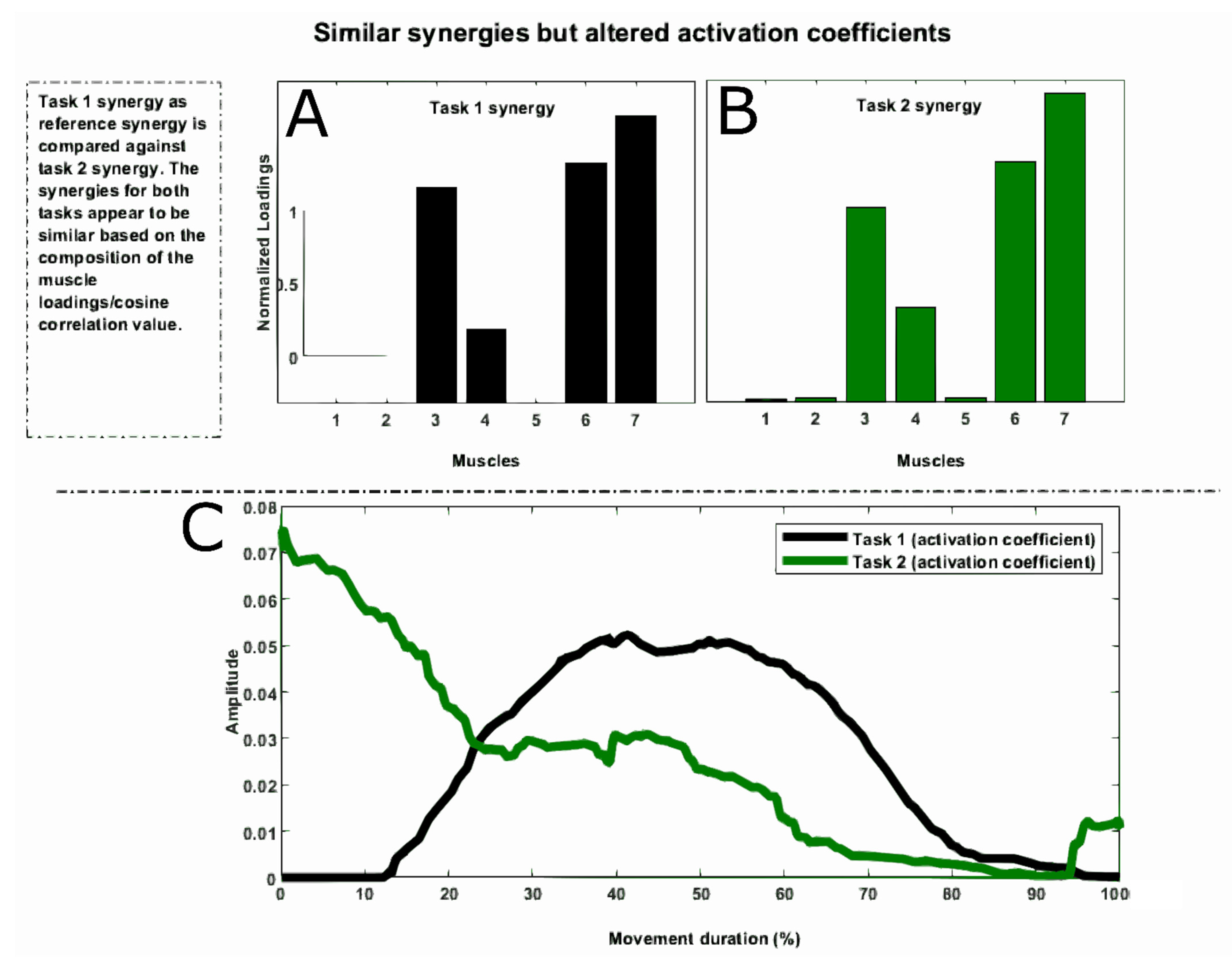
Muscle synergies can potentially be shared across activities as observed previously in animals such as frogs [1,2]. The researchers found that a few synergies were shared between walking, jumping, and swimming. The synergies that appeared to be similar were due to modulation in neural drives that preserve the structures of these synergies. The activation coefficients showed higher variability, as shown in Figure 3. There are several studies done on humans during different biomechanical conditions [20]. The existence of similar synergies relates to having the same mechanical goals for limb movement in different motor tasks. This may result in having the same structure or muscle combinations within a synergy, i.e., shared muscle synergies. Thus, if two different motor tasks share a common mechanical goal such as flexion and extension of limbs in certain tasks, it is likely for a common muscle synergy to appear in both of the synergy sets. Other researchers similarly found that shared synergies across individuals exist between unperturbed, perturbed standing and walking [21]. This may also suggest a common ancestral neural network (primitive behavior) for most of the tasks we perform in activities of daily living. Martino et al. (2015) investigated the synergies of normal walking as well as unstable gait conditions, such as walking on a slippery surface, and reported similarities of those motor behaviors [22]. The researchers assessed the consistency of muscle synergies by calculating the similarity of muscle weights and activation signals across different muscle groups [23,24].
The current question researchers are still investigation is how synergies are preserved or attain a fixed state during upper and lower limb movements. We can think of two ways these synergies show preservation: (1) they exhibit preservation through common ancestral neural networks, and (2) later through motor learning. These studies have shown that during early motor development, two components may correspond to primitive behavior [11]. However, in upper and lower limbs there are more than two synergies or components that explain maximum variance in the locomotion pattern [25]. The additional synergies, that were found to be similar later in life, suggest that years of learning and training played a crucial role in shaping the structure of those synergies, thereby becoming preserved across participants. Some studies have shown that from early development (neonatal) to adulthood stage, muscle synergies during walking become more robust or preserved [26]. Therefore, preservation of these structures is necessary for efficient motor behavior from both evolutionary and motor learning perspectives. We think modulation in spinal and supraspinal level control is an underlying neuro-anatomical mechanism that preserve these synergies. The neural drives are modulated reflecting changes in the activation coefficients while preserving underlying muscle synergies and generating rhythmic patterns of muscle activity required for efficient execution of tasks such as walking, cyclic upper limb motion, etc. [27,28]. Therefore, it is proposed that effective rehabilitation measures can be developed to restore these robust synergies that are affected by neurological conditions or injuries resulting in arrhythmic motor behavior [29,30].

4. Changes in the Number of Muscle Synergies (Modular Organization)

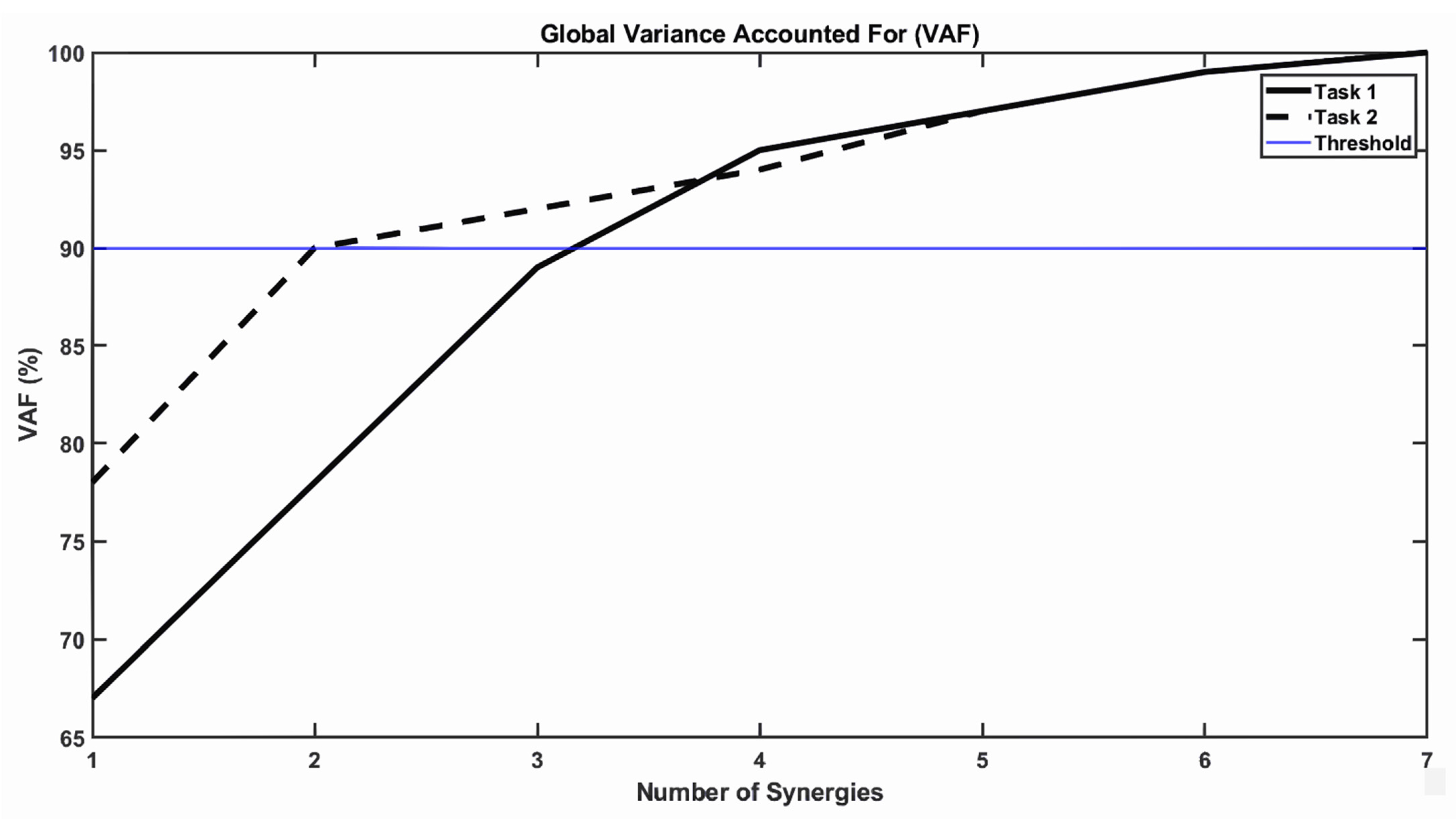
In muscle synergy studies, changes are not only observed in structures (composition of muscles within a synergy) and their recruitment profile. The changes in the dimensionality or the number of synergies has also been observed as shown in Figure 4 and Figure 5. Researchers showed that the numbers of muscle synergies changed across participants during different neurological conditions such as cerebral palsy [19] and stroke [17]. The difference in the number of muscle synergies, especially the merging of synergies among neurological conditions, was attributed to poor muscle coordination [11]. The changes were also observed during various activities, such as slacklining [12], balance beam walking [22], and badminton [31]. The different number of synergies across participants during different activities such as beam walking and slacklining may relate to different proficiency levels [12]. Sawyers et al., and Singh et al., using beam walking and slacklining showed that long-term training modifies the dimensionality of muscle synergies.
Regarding neurological conditions, researchers have observed alteration in both the structure and the number of muscle synergies for stroke, and cerebral palsy patients when compared to healthy subjects [22]. It was observed that the number of muscle synergies in nonparetic legs of stroke patients is larger compared to the paretic leg. The study also compared non stroke patients and found out that variability accounted for by any given number of modules was higher in the control and nonparetic legs compared with the paretic legs of stroke patients, indicating less complexity in the patterned activity of the paretic leg during walking and poor performance [17]. Moreover, (Clark et al., 2010) demonstrated that muscle synergies in stroke patients’ locomotion were changed and the number of synergies in stroke survivors was correlated with various metrics of walking performance, such as preferred speed, speed modulation, step length asymmetry, and propulsive asymmetry [17].
Moreover, patients with cerebral palsy also exhibited reduced number of synergies, altered structures, and asymmetry between legs. Therefore, a current challenge for physiotherapists is to train these groups of patient populations and modify the synergies for effective walking. Several studies were performed that involved patient training and synergy analysis. These studies found that therapy increased the number of modules and showed improved walking performance [15,29]. The researchers found that a higher number of modules after stroke therapy was positively associated with better performance in various clinical and biomechanical assessments of walking. The parameters to understand efficient walking included walking speed, ability to change walking speed, dynamic gait index, step length symmetry, and propulsion symmetry [17,32]. Thus, improvements in muscle coordination during rehabilitation therapy can be indicated by changes in the dimensionality of muscle synergies, possibly through an increase in their number, which may lead to a more normal gait pattern and improved walking performance.
The changing number of muscle synergies can provide the researchers more information about the clinical rehabilitation. Generally, electromyographic signals have issues such as noise, and intra and intersubject variability that can make interpretation difficult. This impacts their clinical utility. Therefore, researchers use computational analyses such as muscle synergies analysis from EMG signals, identifying patterns that may reflect various levels of neural function. We know that the CNS recruits these muscle synergies, which represent motor modules, to perform biomechanical tasks necessary for movement. For example, stroke patients exhibit differences in the number of muscle synergies, which may reflect disturbances in neural pathways and are related to deficits in motor function [29,33].

5. Alternate Theories

There are two alternate theories based on different computational and theoretical models.
  • The uncontrolled manifold hypothesis (UCM). As observed by Bernstein, the degrees of freedom involved in a postural or movement task exceeds the number of control variables necessary to describe the task [34]. The UCM hypothesis states that the CNS focuses on controlling the task relevant DOF and leaves others uncontrolled. The concept provides a basis for distinguishing between different degrees of freedom in terms of their control-theoretical stability [35]. In other words, UCM indicates that the control of essential performance variables among duplications of a task is achieved by taking advantage of the available motor abundance rather than opting for a distinctive solution to the organization problem. According to the UCM theory, collaborations among the outputs of the motor components are arranged such that their variance is essentially limited to a subspace (a UCM) in the state space of those components that corresponds to the desired value of a specific performance variable [36].
  • The equilibrium point hypothesis (EPH). The EPH explains how motor neuron thresholds have been controlled out of the value of lambda (λ), which correlates to an organization for a muscle, joint, or group of joints [37]. Researchers consider the combination of posture and movement control into a single mechanism as a very important characteristic of the equilibrium point hypothesis [37]. Some researchers defined the equilibrium point hypothesis (EPH) as method that the central nervous system utilizes to control the movement of extremities via a simple shift in equilibrium place, and the central nervous system doesn’t need to compensate for task dynamics [38].
  • The internal model theory is one of numerous theories that consider the strategy of the central nervous (CNS) to perform human movements in which the CNS processes neural interpretations of the environment around the human and utilizes these to anticipate and regulate any actions [39,40]. Some researchers state that humans can learn and adapt when the dynamics of extremities change [41].

6. Application of Muscle Synergies

The muscle synergies concept plays a crucial role in the clinical field and rehabilitation. The theory of muscle synergies can be used as a diagnostic technique, and for developing assistive technologies.
For a diagnostic technique, we have presented literature where differences in the number of muscle synergies for hemiparetic stroke patients, cerebral palsy, and spinal cord injury have been observed. The number and structure of muscle synergies have reflected disruptions in descending neural pathways and been associated with the deficits in motor function [42]. Hence, muscle synergy analysis may provide a better view for the clinician, physiotherapist about changes occurring at the CNS and muscular level. This kind of information may help in diagnosis of neurological disorders and assist clinicians and physiotherapist in developing rehabilitation interventions for such patients [29].
For developing assistive technologies, some researchers used the concept of muscle synergies to control myoelectrical assistive technology (AT) because, from a computational point of view, a modular organization based on muscle synergies reduces the computational burden for the controller. This burden is due to the higher dimensionality of neural inputs in the machine interface. This causes nonintuitive control of myoelectrical AT. The people who use e myoelectric controlled assistive technologies (AT) for upper extremities suffer from difficulties in controlling these technologies in daily life because of nonintuitive control. The dimensionality reduction of EMG signals simplifies motor control and learning for myoelectric prostheses, and may contribute to the adaptability observed in biological systems [43]. Roboticists and control engineers are implementing the concept of muscle synergies to develop artificially intelligent controllers for myoelectric devices. In addition to dimensionality reduction, the modularity of such a scheme has the advantage of improving the device performance by introducing additional synergies to the controller. Thus, researchers propose that intuitive control may be accomplished via a limited number of these fixed muscle synergies and can be updated to perform more complex task by adding more synergy or reshaping existing synergies.

7. Conclusions

The muscle synergies hypothesis suggests different strategies of the CNS to perform tasks with and without neurological conditions. This is particularly important in medical fields and especially in rehabilitation. Researchers have defined muscle synergy in different ways. In this study, we explained the concept of muscle synergy and different strategies for their adaptation. We discussed the alteration in muscle synergies for healthy people across different tasks, the alteration of the structure, and the number of synergies between individuals with stroke, cerebral palsy, and spinal injuries. We further discussed how their structure and number of muscle synergies altered compared to healthy people when they perform the same task. We also discussed the importance of the preserved muscle synergies for efficient motor control.

Author Contributions

Conceptualization, A.T.A., R.E.S. and G.W.; methodology, A.T.A., R.E.S., G.W. and K.I.; software, A.T.A. and R.E.S.; validation, A.T.A., R.E.S., G.W. and K.I.; writing—original draft preparation, A.T.A. and R.E.S.; writing—review and editing, A.T.A., R.E.S., G.W. and K.I.; supervision, G.W. and K.I. All authors have read and agreed to the published version of the manuscript.

Funding

This research received no external funding.

Institutional Review Board Statement

Not applicable.

Informed Consent Statement

Not applicable.

Data Availability Statement

Not applicable.

Conflicts of Interest

The authors declare no conflict of interest.

References

  1. D’Avella, A.; Saltiel, P.; Bizzi, E. Combinations of Muscle Synergies in the Construction of a Natural Motor Behavior. Nat. Neurosci. 2003, 6, 300–308. [Google Scholar] [CrossRef]
  2. D’Avella, A.; Bizzi, E. Shared and Specific Muscle Synergies in Natural Motor Behaviors. Proc. Natl. Acad. Sci. USA 2005, 102, 3076–3081. [Google Scholar] [CrossRef] [Green Version]
  3. Alessandro, C.; Rellinger, B.A.; Barroso, F.O.; Tresch, M.C. Adaptation after Vastus Lateralis Denervation in Rats Demonstrates Neural Regulation of Joint Stresses and Strains. eLife 2018, 7, e38215. [Google Scholar] [CrossRef]
  4. Barroso, F.O.; Alessandro, C.; Tresch, M.C. Adaptation of Muscle Activation after Patellar Loading Demonstrates Neural Control of Joint Variables. Sci. Rep. 2019, 9, 20370. [Google Scholar] [CrossRef] [Green Version]
  5. Ting, L.H.; Macpherson, J.M. A Limited Set of Muscle Synergies for Force Control During a Postural Task. J. Neurophysiol. 2005, 93, 609–613. [Google Scholar] [CrossRef] [Green Version]
  6. Cheung, V.C.K. Central and Sensory Contributions to the Activation and Organization of Muscle Synergies during Natural Motor Behaviors. J. Neurosci. 2005, 25, 6419–6434. [Google Scholar] [CrossRef] [PubMed]
  7. Tresch, M.C.; Jarc, A. The Case for and against Muscle Synergies. Curr. Opin. Neurobiol. 2009, 19, 601–607. [Google Scholar] [CrossRef] [PubMed] [Green Version]
  8. Bizzi, E.; Cheung, V.C.K. The Neural Origin of Muscle Synergies. Front. Comput. Neurosci. 2013, 7, 51. [Google Scholar] [CrossRef] [Green Version]
  9. Kutch, J.J.; Valero-Cuevas, F.J. Challenges and New Approaches to Proving the Existence of Muscle Synergies of Neural Origin. PLoS Comput. Biol. 2012, 8, e1002434. [Google Scholar] [CrossRef] [PubMed]
  10. Neptune, R.R.; Clark, D.J.; Kautz, S.A. Modular Control of Human Walking: A Simulation Study. J. Biomech. 2009, 42, 1282–1287. [Google Scholar] [CrossRef] [PubMed] [Green Version]
  11. Yang, Q.; Logan, D.; Giszter, S.F. Motor Primitives Are Determined in Early Development and Are Then Robustly Conserved into Adulthood. Proc. Natl. Acad. Sci. USA 2019, 116, 12025–12034. [Google Scholar] [CrossRef] [PubMed] [Green Version]
  12. Singh, R.E.; White, G.; Delis, I.; Iqbal, K. Alteration of Muscle Synergy Structure While Walking under Increased Postural Constraints. Cogn. Comput. Syst. 2020, 2, 50–56. [Google Scholar] [CrossRef]
  13. Singh, R.E.; Iqbal, K.; White, G.; Hutchinson, T.E. A Systematic Review on Muscle Synergies: From Building Blocks of Motor Behavior to a Neurorehabilitation Tool. Appl. Bionics Biomech. 2018, 2018, 3615368. [Google Scholar] [CrossRef] [PubMed] [Green Version]
  14. Pan, B.; Sun, Y.; Xie, B.; Huang, Z.; Wu, J.; Hou, J.; Liu, Y.; Huang, Z.; Zhang, Z. Alterations of Muscle Synergies During Voluntary Arm Reaching Movement in Subacute Stroke Survivors at Different Levels of Impairment. Front. Comput. Neurosci. 2018, 12, 69. [Google Scholar] [CrossRef]
  15. Santuz, A.; Ekizos, A.; Eckardt, N.; Kibele, A.; Arampatzis, A. Challenging Human Locomotion: Stability and Modular Organisation in Unsteady Conditions. Sci. Rep. 2018, 8, 2740. [Google Scholar] [CrossRef] [Green Version]
  16. Roh, J.; Rymer, W.Z.; Perreault, E.J.; Yoo, S.B.; Beer, R.F. Alterations in Upper Limb Muscle Synergy Structure in Chronic Stroke Survivors. J. Neurophysiol. 2013, 109, 768–781. [Google Scholar] [CrossRef] [Green Version]
  17. Clark, D.J.; Ting, L.H.; Zajac, F.E.; Neptune, R.R.; Kautz, S.A. Merging of Healthy Motor Modules Predicts Reduced Locomotor Performance and Muscle Coordination Complexity Post-Stroke. J. Neurophysiol. 2010, 103, 844–857. [Google Scholar] [CrossRef] [Green Version]
  18. Fox, E.J.; Tester, N.J.; Kautz, S.A.; Howland, D.R.; Clark, D.J.; Garvan, C.; Behrman, A.L. Modular Control of Varied Locomotor Tasks in Children with Incomplete Spinal Cord Injuries. J. Neurophysiol. 2013, 110, 1415–1425. [Google Scholar] [CrossRef] [PubMed] [Green Version]
  19. Steele, K.M.; Rozumalski, A.; Schwartz, M.H. Muscle Synergies and Complexity of Neuromuscular Control during Gait in Cerebral Palsy. Dev. Med. Child Neurol. 2015, 57, 1176–1182. [Google Scholar] [CrossRef] [PubMed] [Green Version]
  20. Torres-Oviedo, G.; Ting, L.H. Subject-Specific Muscle Synergies in Human Balance Control Are Consistent Across Different Biomechanical Contexts. J. Neurophysiol. 2010, 103, 3084–3098. [Google Scholar] [CrossRef] [PubMed] [Green Version]
  21. Chvatal, S.A.; Ting, L.H. Common Muscle Synergies for Balance and Walking. Front. Comput. Neurosci. 2013, 7, 48. [Google Scholar] [CrossRef] [Green Version]
  22. Martino, G.; Ivanenko, Y.P.; d’Avella, A.; Serrao, M.; Ranavolo, A.; Draicchio, F.; Cappellini, G.; Casali, C.; Lacquaniti, F. Neuromuscular Adjustments of Gait Associated with Unstable Conditions. J. Neurophysiol. 2015, 114, 2867–2882. [Google Scholar] [CrossRef] [PubMed] [Green Version]
  23. Barroso, F.; Torricelli, D.; Moreno, J.C.; Taylor, J.; Gomez-Soriano, J.; Esteban, E.B.; Santos, C.; Pons, J.L. Similarity of Muscle Synergies in Human Walking and Cycling: Preliminary Results. In Proceedings of the 2013 35th Annual International Conference of the IEEE Engineering in Medicine and Biology Society (EMBC), Osaka, Japan, 3–7 July 2013; pp. 6933–6936. [Google Scholar] [CrossRef]
  24. Abd, A.T.; Singh, R.E.; Iqbal, K.; White, G. Investigation of Power Specific Motor Primitives in an Upper Limb Rotational Motion. J. Mot. Behav. 2021, 1–12. [Google Scholar] [CrossRef] [PubMed]
  25. Ivanenko, Y.P.; Poppele, R.E.; Lacquaniti, F. Five Basic Muscle Activation Patterns Account for Muscle Activity during Human Locomotion: Basic Muscle Activation Patterns. J. Physiol. 2004, 556, 267–282. [Google Scholar] [CrossRef]
  26. Dominici, N.; Ivanenko, Y.P.; Cappellini, G.; d’Avella, A.; Mondi, V.; Cicchese, M.; Fabiano, A.; Silei, T.; Di Paolo, A.; Giannini, C.; et al. Locomotor Primitives in Newborn Babies and Their Development. Science 2011, 334, 997–999. [Google Scholar] [CrossRef] [PubMed]
  27. Rimini, D.; Agostini, V.; Knaflitz, M. Intra-Subject Consistency during Locomotion: Similarity in Shared and Subject-Specific Muscle Synergies. Front. Hum. Neurosci. 2017, 11, 586. [Google Scholar] [CrossRef] [PubMed] [Green Version]
  28. Abd, A.T.; Singh, R.E.; Iqbal, K.; White, G. Muscle Synergies Are Robust across Participants in Upper Limb Rotational Motion. In Proceedings of the 2020 7th International Conference on Electrical and Electronics Engineering (ICEEE), Antalya, Turkey, 14–16 April 2020; pp. 310–314. [Google Scholar] [CrossRef]
  29. Safavynia, S.; Torres-Oviedo, G.; Ting, L. Muscle Synergies: Implications for Clinical Evaluation and Rehabilitation of Movement. Top. Spinal Cord Inj. Rehabil. 2011, 17, 16–24. [Google Scholar] [CrossRef] [PubMed] [Green Version]
  30. Hong, Y.N.G.; Ballekere, A.N.; Fregly, B.J.; Roh, J. Are Muscle Synergies Useful for Stroke Rehabilitation? Curr. Opin. Biomed. Eng. 2021, 19, 100315. [Google Scholar] [CrossRef]
  31. Matsunaga, N.; Kaneoka, K. Comparison of Modular Control during Smash Shot between Advanced and Beginner Badminton Players. Appl. Bionics Biomech. 2018, 2018, 6592357. [Google Scholar] [CrossRef]
  32. Bowden, M.G.; Clark, D.J.; Kautz, S.A. Evaluation of Abnormal Synergy Patterns Poststroke: Relationship of the Fugl-Meyer Assessment to Hemiparetic Locomotion. Neurorehabil. Neural Repair 2010, 24, 328–337. [Google Scholar] [CrossRef]
  33. Tan, C.K.; Kadone, H.; Watanabe, H.; Marushima, A.; Hada, Y.; Yamazaki, M.; Sankai, Y.; Matsumura, A.; Suzuki, K. Differences in Muscle Synergy Symmetry Between Subacute Post-stroke Patients With Bioelectrically-Controlled Exoskeleton Gait Training and Conventional Gait Training. Front. Bioeng. Biotechnol. 2020, 8, 770. [Google Scholar] [CrossRef] [PubMed]
  34. Bernshtein, N.A. The Coordination and Regulation of Movements; Pergamon Press: Oxford, UK, 1967. [Google Scholar]
  35. Scholz, J.P.; Schöner, G. The Uncontrolled Manifold Concept: Identifying Control Variables for a Functional Task. Exp. Brain Res. 1999, 126, 289–306. [Google Scholar] [CrossRef]
  36. Schoner, G. Recent Developments and Problems in Human Movement Science and Their Conceptual Implications. Ecol. Psychol. 1995, 7, 291–314. [Google Scholar] [CrossRef]
  37. Sainburg, R.L. Should the Equilibrium Point Hypothesis (EPH) Be Considered a Scientific Theory? Motor Control 2015, 19, 142–148. [Google Scholar] [CrossRef]
  38. Hinder, M.R.; Milner, T.E. The Case for an Internal Dynamics Model versus Equilibrium Point Control in Human Movement. J. Physiol. 2003, 549, 953–963. [Google Scholar] [CrossRef] [PubMed]
  39. Ito, M. Control of Mental Activities by Internal Models in the Cerebellum. Nat. Rev. Neurosci. 2008, 9, 304–313. [Google Scholar] [CrossRef]
  40. Kawato, M. Internal Models for Motor Control and Trajectory Planning. Curr. Opin. Neurobiol. 1999, 9, 718–727. [Google Scholar] [CrossRef]
  41. Gillespie, R.B.; Ghasemi, A.H.; Freudenberg, J. Human Motor Control and the Internal Model Principle. IFAC-PapersOnLine 2016, 49, 114–119. [Google Scholar] [CrossRef]
  42. Ting, L.H.; Chiel, H.J.; Trumbower, R.D.; Allen, J.L.; McKay, J.L.; Hackney, M.E.; Kesar, T.M. Neuromechanical Principles Underlying Movement Modularity and Their Implications for Rehabilitation. Neuron 2015, 86, 38–54. [Google Scholar] [CrossRef] [Green Version]
  43. Mussa–Ivaldi, F.A.; Bizzi, E. Motor Learning through the Combination of Primitives. Philos. Trans. R. Soc. Lond. B Biol. Sci. 2000, 355, 1755–1769. [Google Scholar] [CrossRef] [Green Version]
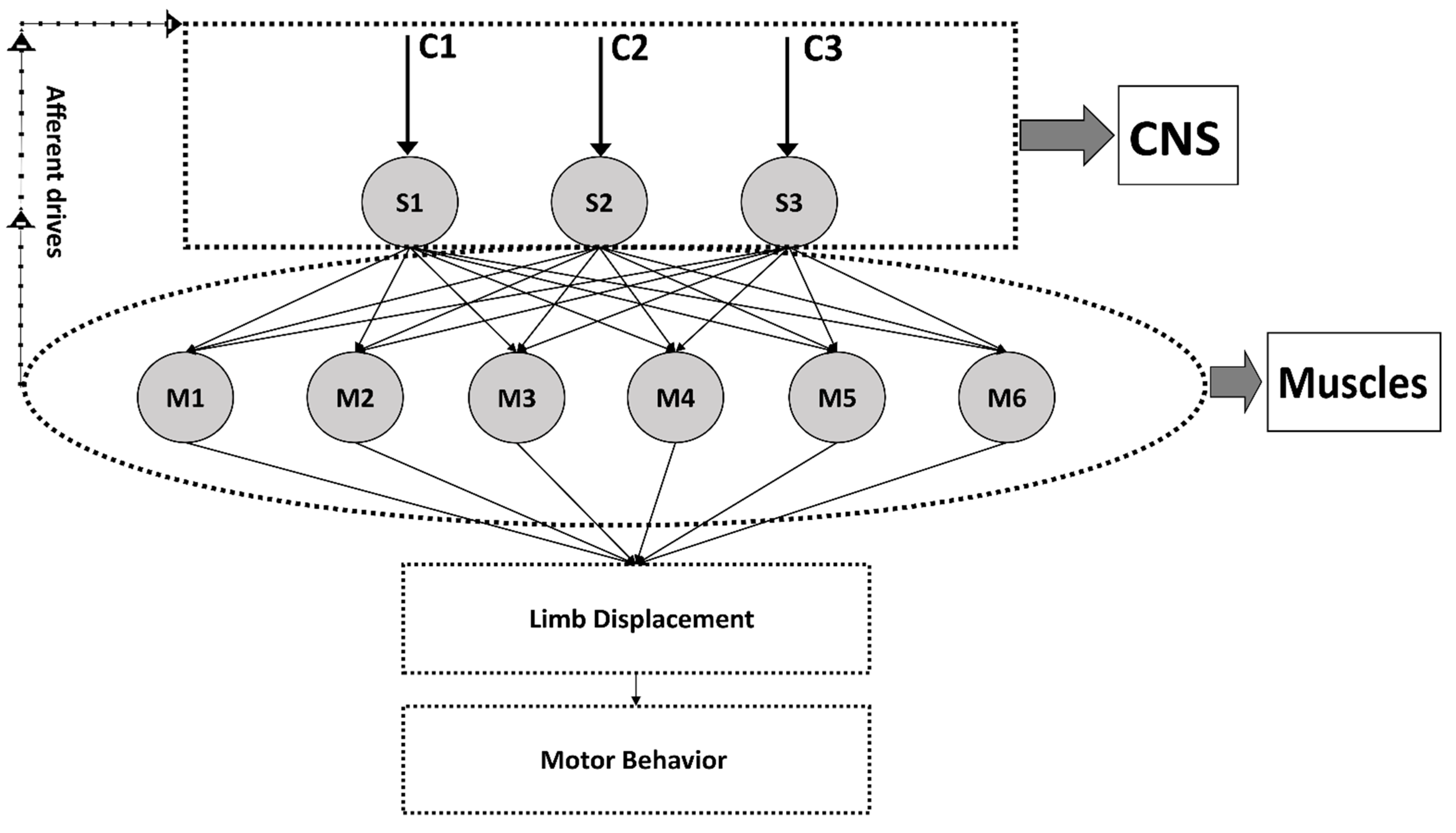
Figure 1. A schematic of muscle synergy theory. Muscle synergies (S1, S2, S3), Activation Coefficient (C1, C2, C3), Muscles (M1, M2–M6). The figure shows feedforward and feedback mechanism of neural drives (efferent and afferent). The alteration in activation coefficients relates to modulation in supraspinal control directly from the cortex. The changes in supraspinal control can also be caused by the modulation in afferent drives carrying feedback information from muscle spindles and surface of skin via afferent drives. The alteration in activation coefficient due to modulation in neural drives can cause changes in the structure of muscle synergies. Note: C1, C2, C3 are not actual neural drives but alteration in them may likely represents modulation in them.
Biomechanics 01 00021 g001
Figure 2. (A) Reference Synergy, (B) Similar Synergy, (C) Altered Synergy. The similarity in these synergies is generally quantified using cosine correlation or Pearson correlation. It is clear from this figure that (C) reveals differences in the muscle composition loading compared to (A,B).
Biomechanics 01 00021 g002
Figure 3. (A) Reference Synergy Task 1; (B) Similar Synergy for Task 2. The x axis displays the number/type of muscles within a muscle synergy and their loading values represented by y axis are normalized between 0 and 1. The number of muscles can be actual muscle groups/type of muscles to understand muscle coactivation pattern and synergistic movement towards a specific movement. (C) Shows dissimilar activation coefficients over movement duration.
Biomechanics 01 00021 g003
Figure 4. Task 1 and Task 2 show differences in the number of synergies based on the VAF threshold of 0.9 or 90%. The minimum number of synergies that account for 90% reconstruction for task 2 is two, and for task 1 is nearly 3. These indicators are used later to extract synergies two and three, respectively. The extracted synergies are later compared for similarity or other analysis depending on the research hypothesis.
Biomechanics 01 00021 g004
Figure 5. (A) Two synergies (task 2) shown against (B) three synergies (task 1) for a sample EMG data of seven muscles.
Biomechanics 01 00021 g005
Publisher’s Note: MDPI stays neutral with regard to jurisdictional claims in published maps and institutional affiliations.