Article

First Molecular Detection of the Poultry Pathogen Protozoan Histomonas meleagridis from Red Mite (Dermanyssus gallinae)

by
Patrícia Kóczán
1,
László Kovács
2,
Adrienn Örkényi
3,
Dorottya Kovács
1,
László Kőrösi
4 and
Edit Eszterbauer
1,*
1
HUN-REN Veterinary Medical Research Institute, Hungária krt 21, 1143 Budapest, Hungary
2
Department of Animal Hygiene, Herd Health and Mobile Clinic, University of Veterinary Medicine Budapest, István u. 2, 1078 Budapest, Hungary
3
Veterinary Diagnostic Directorate, National Food Chain Safety Office, 1143 Budapest, Hungary
4
AgriAL Bt, Béri Balogh Ádám u. 42, 2100 Gödöllő, Hungary
*
Author to whom correspondence should be addressed.
Parasitologia 2025, 5(4), 59; https://doi.org/10.3390/parasitologia5040059
Submission received: 17 September 2025 / Revised: 27 October 2025 / Accepted: 1 November 2025 / Published: 4 November 2025

Abstract

Our study reports the first molecular detection of the protozoan pathogen Histomonas meleagridis, the causative agent of histomonosis, in the poultry red mite Dermanyssus gallinae. Mite samples were collected from a commercial poultry farm and a backyard flock in Hungary and analyzed using PCR assays that targeted the COX1 gene in mites and 18S rRNA gene in H. meleagridis. Phylogenetic analysis confirmed the identity of D. gallinae and revealed distinct genetic lineages between farm and backyard isolates. H. meleagridis DNA was detected in 10 of 17 mite samples, representing multiple variants within Genotype 1. No histomonosis was detected in the examined poultry, although one hen harbored Simplicimonas sp. The findings suggest that D. gallinae could serve as a carrier for H. meleagridis, potentially facilitating the persistence and transmission of the pathogen. This novel host–pathogen association highlights the need for further research into the epidemiological role of poultry ectoparasites in the spread of histomonosis.

Graphical Abstract

1. Introduction

Dermanyssus gallinae (Arachnida, Mesostigmata), commonly known as the poultry red mite, is a widespread haematophagous ectoparasite that infests avian hosts and poses a substantial threat to poultry health and production, contributing to anaemia, reduced egg output and increased susceptibility to infectious diseases [1]. The ectoparasite feeds on the blood of its hosts during dark periods and hides in crevices during the day.
Dermanyssus gallinae is known to spread pathogens, thereby presenting health risks in poultry farming environments [2]. It may carry various pathogens, including Salmonella enterica, Escherichia coli, Klebsiella pneumoniae, Listeria monocytogenes and Staphylococcus aureus bacteria, as well as Candida albicans and Cryptococcus neoformans fungi [3]. There have been documented cases of the spread of viruses such as the avian influenza virus and the Newcastle disease virus [4,5]. Furthermore, red mites have been reported to carry blood parasites such as Plasmodium sp. and Haemoproteus sp., although the exact role of red mites in the transmission of these parasites is not fully understood yet [2,3,6]. To the best of our knowledge, D. gallinae has never been identified as a potential carrier or reservoir of other protozoa, such as Histomonas spp.
Histomonas meleagridis is a parabasalid protozoan parasite of the family Dientamoebidae and the causative agent of histomonosis (blackhead disease) in poultry and other gallinaceous birds (especially in turkeys) [7,8,9,10,11]. Birds affected by histomonosis often appear lethargic, with drooping wings, ruffled feathers, and yellowish droppings. Infected birds typically show inflammation and ulceration in the cecum and liver. Young birds tend to deteriorate rapidly, usually dying within a few days of showing symptoms. In contrast, the disease progresses more slowly in older birds, who may become severely emaciated before eventually succumbing. Turkeys are especially vulnerable to this disease, and once a flock is infected, mortality rates can range from 70% to 100% [12].
The main carrier of H. meleagridis is considered the nematode Heterakis gallinarum, also known as the cecal worm, which lives in the cecum of some galliform birds, particularly chickens and turkeys. The protozoan can enter the nematode eggs in the bird’s cecum. The eggs protect the protozoan, hence it can survive a long period outside its host [13]. Depending on the environment, H. meleagridis survive only for a few hours without a host [14].
The transmission route of H. meleagridis between farms is not fully understood yet. Previous studies [15,16] found that earthworms are possible paratenic hosts for both He. gallinarum and H. meleagridis, but they are not likely to be responsible for transmissions between farms due to their small mobility area. Although various arthropod species could potentially transmit H. meleagridis between poultry farms, they have not been extensively studied [17]. In 1953, Frank [18] found that blowflies (Calliphoridae) and six grasshopper species could carry H. meleagridis for at least 96 h and initiate infection in turkeys. H. meleagridis DNA was detected in flies and a puddle in front of an infected poultry house [19]. The darkling beetle, Alphitobius diaperinus has also been identified as a potential carrier [20]. H. meleagridis and He. gallinarum DNA has been detected in darkling beetles and its larvae, and protozoan DNA was still detectable in litter one year after poultry houses were depopulated [16,21]. After using different trapping methods and sampling regimes every 4 months, Terra et al. [17] found A. diaperinus and four Dipteran species positive for H. meleagridis and/or He. gallinarum with qPCR. On one farm, the samples were positive for 4 months after a histomonosis outbreak. In addition, there were a few H. meleagridis outbreaks documented in the absence of any potential carriers. Son et al. [22] described 10-week-old broiler breeder chickens that died in high numbers caused by H. meleagridis in a poultry house with floor pens, and Cortes et al. [23] reported histomonosis in 6-week-old chickens, without the presence of cecal worms in both cases. This raises the question of how else the infection can spread.
In the present study, poultry red mites were collected from a commercial poultry farm and a backyard flock in Hungary, and tested for the presence of H. meleagridis DNA for the first time. Furthermore, the phylogenetic relationships of both red mites and Histomonas isolates were analyzed.

2. Materials and Methods

2.1. Sample Collection

Red mite samples were collected from a commercial poultry farm (HU/A) and a small backyard poultry flock (HU/B) in Hungary (Table 1). The owners of the farms requested anonymity. At farm HU/A, laying hens were housed in cages. The hens were 35 weeks old in July 2023, when the mite samples were collected. The backyard flock (HU/B) was housed in a deep litter system, and the hens were 43 weeks old when the mites were sampled. There were no reports of disease outbreaks from either poultry flock.
Pathogen-free, individually wrapped, house-made traps were used to collect poultry mites (Figure S1). The trap was a 60 mm long, 16 mm diameter plastic tube containing a rolled-up piece of cardboard. Its shape and function were similar to those of the AviVet mite trap [24]. The design allows mites to crawl inside during dark periods seeking shelter, similar to their natural behaviour. No attractants or ethanol was used; the traps relied solely on physical entrapment and mite phototaxis. The traps were securely fastened to the bottom of the poultry cages and left undisturbed for a week before being collected. The traps were placed along the rows of cages in different parts of the same poultry house. After removal, the traps were individually sealed in airtight plastic bags. The number of mites (adults, nymphs and larvae) was counted using a stereomicroscope. If there were more than 100 mites in the trap, 100 specimens were counted and weighed, and the total number of mites was estimated based on their total weight. All mites in a trap were considered one sample.
Table 1. Summary of the sampling and the DNA sequence analyses of red mite and poultry samples examined in the present study.
SpeciesOriginSampling Date (DD.MM.YYYY)Sample TypeNo. of SamplesConfirmed HIS PCR PositiveHistomonas 18S rDNA NCBI Acc. No.Dermanyssus COX1 NCBI Acc. No.
Dermanyssus gallinaepoultry farm HU/A05.07.2023mite trap138PX210515–PX210523PX210480–PX210485
Dermanyssus gallinaebackyard flock HU/B17.08.2023mite trap42PX210524PX210486
Gallus gallus domesticuspoultry farm HU/A12.04.2024liver, cecum100 *n.d.n.d.
HIS: Histomonas; n.d.: no data; * Simplicimonas sp. was detected in 1 of 10 poultry samples (99.37% identity with Simplicimonas sp. isolate 11/4178, NCBI Acc. No. HG008105 published by Bilic et al. [25]).
The samples were stored at −20 °C until molecular biological examinations were performed, including Histomonas-specific PCR, which took place 8 months after the mite collection. Hens from the same flock at farm HU/A were sampled 9 months later, when the molecular detection showed the presence of Histomonas in the mites (Table 1). A professional on-farm necropsy and sampling of the livers and ceca of hens that died as part of the normal mortality rate were performed. The procedure was carried out in accordance with Hungarian legislation (Section 49 of Act No. XXVIII/1998 on the protection and welfare of animals, and Hungarian Government Decree No. 40/2013), as well as Directive 2010/63/EU on the protection of animals used for scientific purposes.

2.2. DNA Extraction

For DNA extraction, red mite samples from the traps were homogenised in 500 µL 1× PBS buffer using Eppendorf micropestle (Eppendorf, Vienna, Austria) in 1.5 mL microtube, then centrifuged with an Eppendorf 5424R tabletop centrifuge (Eppendorf, Vienna, Austria) at 9400× g for 5 min. DNA extraction was carried out using the IndiSpin Pathogen Kit (Indical Bioscience GmbH, Leipzig, Germany) on the supernatant of 200 µL, following the manufacturer’s protocol.

2.3. Dermanyssus-Specific PCR

The mitochondrial cytochrome c oxidase subunit 1 (COX1) gene of D. gallinae was amplified with a semi-nested PCR assay. The first round used the primer pair FCOIDG (5′ CAT TAA TAT TAA CTG CAC CTG ACA TG 3′) and RCOIDG (5′ CCC GTG GAG TGT TGA AAT TCA TGA 3′) [26], then the second round used the primer pairs OS6F (5′-GTG CAG GGA CTG GAT GAA CT-3′) [27] and RCOIDG. The PCR was performed with Labcycler Basic (SensoQuest, Göttingen, Germany) in a total volume of 25 µL, contained approximately 10 ng template DNA, 250 nM of the forward and reverse primers (IDT, Leuven, Belgium), and 1× DreamTaq Hot Start Green PCR Master Mix (Thermo Fisher Scientific, Waltham, MA, USA). Amplification conditions were the following: initial denaturation at 95 °C for 5 min, then 95 °C for 30 s, 58 °C for 30 s and 72 °C for 1 min for 35 cycles, with a terminal extension at 72 °C for 5 min. The PCR master mix and amplification conditions in the second round were the same as described above.

2.4. Histomonas-Specific PCR and DNA Sequence Analysis

A semi-nested PCR assay developed in the present study was used to amplify parabasalid protozoans, including H. meleagridis 18S rRNA gene. In the PCR round 1 using the primers Hm1f (5′ ACT CGA AAT TCT CGG AGG TG 3′) and HIS5F-rev (5′ CAG CCC AGA GCA TCT AAA GG 3′) [28,29], a 915 bp DNA fragment was amplified. In the second round, the primer pairs were: nHm1f (5′ TCT GCA AGT TTG CTC CCA TA 3′) [28] and HIS5F-rev, which amplified a 778 bp fragment.
The PCR was performed in the total volume of 25 µL, containing approximately 10 ng template DNA, 1× Taq buffer with KCl (Thermo Fisher Scientific, USA), 200 nM of the forward and reverse primers (IDT, Belgium), 0.2 mM dNTPs (Merck-Sigma, Darmstadt, Germany), 3 mM MgCl2 (Thermo Fisher Scientific, Waltham, MA, USA) (all final concentrations), 1.5 U recombinant Taq DNA polymerase (Thermo Fisher Scientific, Waltham, MA, USA). Amplification conditions were the following: initial denaturation at 95 °C for 2 min, then 95 °C for 30 s, 55 °C for 30 s and 72 °C for 1 min for 35 cycles, with a terminal extension at 72 °C for 10 min. The PCR master mix and amplification conditions in the second round were the same as described above, except for the use of 400 nM of the forward and reverse primers and 1.5 mM MgCl2.
The PCR products were purified using the ExoSAP-IT PCR Product Cleanup Reagent (Thermo Fisher Scientific, Waltham, MA, USA) according to the manufacturer’s instructions. Sanger DNA sequencing was performed using the BigDye Terminator v3.1 Cycle Sequencing Kit (Thermo Fisher Scientific, Waltham, MA, USA) on an Applied Biosystems Genetic Analyzer 3500 (Thermo Fisher Scientific, Waltham, MA, USA). For DNA sequencing, the amplification primers of the second round were used.
Consensus DNA sequences were generated with Geneious Prime 2019.2.3. The species were identified based on the mitochondrial COX1 and 18S rRNA gene sequence identity searches, respectively, using the NCBI BLASTn Megablast tool. The DNA sequences of isolates were submitted to NCBI GenBank under accession numbers PX210480–PX210486 (D. gallinae), PX210515–PX210524 (H. meleagridis) (Table 1).

2.5. Phylogenetic Analysis

Phylogenetic trees were predicted using nucleotide sequences: partial sequence alignments of 18S rRNA gene (for H. meleagridis) and COX1 gene (for D. gallinae) served as the molecular marker. Relevant reference sequences were sourced through similarity searches in the NCBI GenBank BLASTn database, using our own nucleotide sequences as queries to identify those with the highest sequence identity. D. gallinae sequence alignment was generated using MAFFT version 7.450 [30], and H. meleagridis alignment was conducted using MUSCLE [31] (Dataset S1 and S2). Evolutionary model selection was performed with ModelTest-NG version 0.2.0, which identified the GTR+I+G model as the best fit for both alignments [32]. Maximum likelihood phylogenies were reconstructed using RAxML-NG version 1.2.2 [33], the branch support was assessed using the transfer bootstrap expectation (TBE) approach with 1000 replicates [34], and a 50% TBE cut-off was applied for the visualization of support values. Tree visualizations were produced using MEGA version 11 and Inkscape version 1.3.2 [35].

3. Results

At the poultry farm (HU/A), 13 of the 15 mite traps could be collected, as two were lost (probably removed by hens) (Table 1 and Table S1). All traps contained mites in varying amounts, from one to 34 mites per trap. Four traps were collected from the backyard flock HU/B, and all contained a high number of mites (over 500 specimens per trap) (Table S1).

3.1. Species Identification of Mites

The Dermanyssus-specific PCR assay successfully amplified the COX1 gene fragment from all of the mites collected. Seven of these DNA fragments could be detected using Sanger sequencing. Based on the DNA sequences, all of the samples were identified as the red mite, D. gallinae. The six COX1 fragments from the poultry farm HU/A were 100% identical to each other (Figure 1 and Dataset S1). A BLAST search revealed the highest identity (99.82%) with D. gallinae isolate DIN14 (MW393108), which was collected from Gallus gallus in France. The COX1 gene of the D. gallinae isolate M23-23AN, which was collected from the backyard flock (HU/B), had only 94.67% identity with the isolates from the farm HU/A. The closest relative of M23-23AN was the D. gallinae isolate BE01S04c (MW392996), which also originated from G. gallus in France.
The phylogenetic analysis of D. gallinae COX1 sequences distinguished the mites from the two sampling sites (M23-23AN from the backyard flock vs. all others from the poultry farm) with high bootstrap support (Figure 1). The COX1 sequence of the only D. gallinae isolate found in Hungary previously (isolate Budapest-EM5; MT812940) originated from rock pigeons (Columba livia) rather than chickens.

3.2. Detection of Histomonas

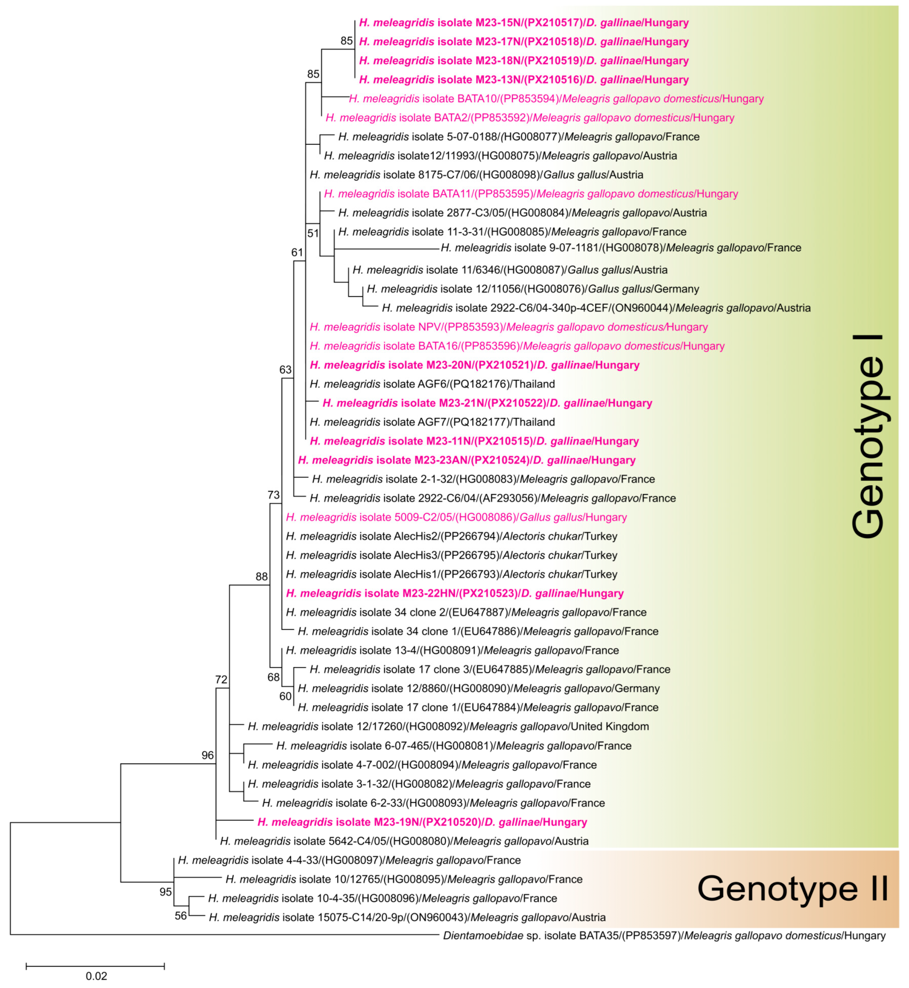
The PCR assay targeting the 18S rRNA gene of parabasalid protozoans produced positive results for most of the red mite samples. In total, the consensus DNA sequences of eight samples could be retrieved from farm HU/A and two from flock HU/B (Table 1). At farm HU/A, Histomonas-positive mite samples were detected in almost every sampling area; therefore, the red mite infection could not be localized to a specific area of the poultry house. Based on the 18S rRNA gene fragments, the presence of the protozoan poultry pathogen, H. meleagridis, was identified. The nucleotide identities among the isolates varied from 95.92% to 99.92%, and numerous single-nucleotide polymorphisms (SNPs) were detected in the nucleotide sequences (Dataset S2).
Histomonas infection could not be detected in the poultry flock. Of the ten hens examined, one sample tested positive for PCR. However, instead of Histomonas, Sanger sequencing revealed the presence of Simplicimonas sp., another parabasalid protozoan parasite often found in wild ducks but also in hens [25]. The highest identity (99.37%) was found with Simplicimonas sp. isolate 11/4178 (HG008105), which was previously detected in the ceca of G. gallus in Austria.

3.3. Histomonas meleagridis Phylogeny and Genotyping

The phylogenetic analysis of the 18S rRNA gene of H. meleagridis revealed clustering by genotype, supported by a high bootstrap value. The two main genotypes were identified, as previously suggested by Bilic et al. [25]. All isolates belonged to Genotype 1, but they were distributed across separate branches (Figure 2). This was partly due to the presence of SNPs in the sequence. In addition, low bootstrap values also indicated a high degree of uncertainty regarding the distinctions within this genotype.
The farm isolates (M23-15N, M23-17N, M23-18N and M23-13N) were grouped with two H. meleagridis isolates (PP853594 and PP853592), which were recently detected in turkeys in Hungary. Isolates M23-11N and M23-20N exhibited 100% nucleotide identity with isolates BATA16 (PP853596) and NPV (PP853893) from turkeys in Hungary, isolate 8175-C7/06 from Austria (HG008098), and two isolates from Thailand (PQ1821176 and PQ182177). Furthermore, the isolate M23-22HN was 100% identical with the H. meleagridis isolate 5009-C2/05 (HG008086), which was also from Hungary but studied over a decade ago [25]. The two samples from the backyard flock (isolates M23-22N and M23-23N) were located on different branches of the phylogenetic tree.
The basal position of isolate M23-19N within Genotype 1 was supported by a high bootstrap value (Figure 2).

4. Discussion

Poultry veterinarians and farmers have long been concerned about the ability of red mites to spread pathogens, which is partly presumed and partly proven [2]. This study was prompted by the need to investigate cases of histomonosis of an unknown origin, given that practical experience shows that red mite infection could be frequent, albeit seasonal, on poultry farms. Red mite is a common ectoparasite in poultry farms worldwide. Infection intensity usually varies widely and often depends on the control management of the farms [2]. Our study was in line with previous findings, as we detected only a moderate infection intensity in poultry farm HU/A, compared to the backyard flock HU/B, where several hundred mites were found in the traps. Furthermore, our study confirmed that the isolates from the two different locations formed distinct genetic lineages, with no genetically similar isolates previously reported in Hungary.
The presence of H. meleagridis could be detected in the red mites from poultry flocks, despite the two sampling sites differing in terms of farm size (commercial vs. backyard flocks) and farming technology (cage vs. deep litter systems). However, the current findings should be considered preliminary in this regard, and a higher number of sampling sites need to be studied before conclusions can be drawn.
Unfortunately, neither Histomonas infection nor histomonosis could be detected in the poultry flocks. As the molecular analyses of the red mite samples were conducted several months after sampling, and the positive outcome of the Histomonas-specific PCR test on such a high proportion of mites was somewhat unexpected, the hens were not examined at the time of mite sampling. As chickens can be infected without displaying any obvious clinical signs of histomonosis, it cannot be ruled out that a subclinical infection was present in the flock. What is confirmed is that, nine months after the mite sampling, all examined hens from the same flock tested negative for H. meleagridis. We acknowledge that the sampling design was retrospective and opportunistic rather than hypothesis-driven. In future work, synchronized sampling of mites and host tissues at multiple time points will be performed to allow for a more systematic evaluation of transmission potential.
In recent years, there has been an increase in outbreaks of histomonosis in poultry flocks, and the disease has become endemic again since antihistomonal drugs were banned in several countries, including the EU and the USA [36]. Szekeres et al. [37] recently detected H. meleagridis in Hungary by examining turkeys and farm-raised pheasants, and found H. meleagridis infection in two of three examined turkey farms. The analysis of the 18S rRNA gene revealed 100% identity with isolates NPV and BATA16 from turkeys and our isolate M23-20N. Moreover, we detected 100% DNA sequence identity between the isolate M23-22HN of the present study and another isolate from Hungary (5009-C2/05) studied by Bilic et al. in 2014 [25]. Thus, our results confirm the conclusion by Szekeres et al. [37] that H. meleagridis has been endemic in Hungary for over a decade and probably much longer.
The role of the red mite in the spread of histomonosis is unclear yet. Our results suggest that the red mite acts as a carrier, but its vector role (i.e., the capability of pathogen transmission to poultry hosts) cannot be ruled out at this stage either. There are some possible explanations for how H. meleagridis, when present in red mite, could reach poultry. The most direct hypothesis is the oral ingestion of infected mites during preening or pecking, or via contaminated litter. In this case, the mites act as mechanical or paratenic carriers, delivering viable protozoa to the bird’s gut. This approach is supported by experimental and field evidence for other arthropods (e.g., flies, grasshoppers and lesser mealworms). Experimental work has demonstrated that ingesting infected arthropods can initiate an infection, and beetles and dipterans have been identified as potential carriers of H. meleagridis [17,18,20]. Alternatively, mites might mechanically transfer the protozoan within the poultry house or redistribute infectious faecal material onto feeders or drinkers, thereby facilitating indirect transmission. However, we only detected Histomonas DNA; it is not yet known whether the protozoan was viable, and whether red mites can transmit the protozoan infection at all. This should be clarified in the future by conducting specific transmission experiments.

5. Conclusions

This is the first study to demonstrate that red mites can carry H. meleagridis, providing important new insights into the epidemiology of histomonosis. While our study indicates that red mites can be a carrier for H. meleagridis, further research is required to determine their exact role in the spread of this protozoan parasite.

Supplementary Materials

The following supporting information can be downloaded at: https://www.mdpi.com/article/10.3390/parasitologia5040059/s1; Figure S1: House-made mite trap used for sample collection; Table S1: Details of the red mite traps collected, including the number of mites in each trap; Dataset S1: A 582 bp nucleotide sequence alignment of Dermanyssus spp. COX1 gene used for phylogenetic analysis; Dataset S2: A 635 bp nucleotide sequence alignment of Histomonas meleagridis 18S rRNA gene used for phylogenetic analysis.

Author Contributions

Conceptualization, E.E. and L.K. (László Kőrösi); methodology, P.K., A.Ö., D.K. and E.E.; validation, P.K. and E.E.; formal analysis, E.E.; investigation, P.K., A.Ö. and E.E.; resources, E.E.; data curation, P.K. and E.E.; writing—original draft preparation, P.K. and E.E.; writing—review and editing, P.K., L.K. (László Kovács) and E.E.; visualization, P.K. and E.E.; supervision, E.E., L.K. (László Kovács) and L.K. (László Kőrösi); project administration, E.E.; funding acquisition, E.E. All authors have read and agreed to the published version of the manuscript.

Funding

This research has been implemented with the support provided by the Ministry of Innovation and Technology of Hungary (legal successor: Ministry of Culture and Innovation of Hungary) from the National Research, Development and Innovation Fund, financed under the TKP2021-EGA-01 funding scheme of the National Research, Development and Innovation Office.

Institutional Review Board Statement

Ethical review and approval were waived for this study, as only animals that died naturally were dissected for the purpose of collecting samples.

Informed Consent Statement

We obtained verbal informed consent from the participants to use the collected samples for publication purposes.

Data Availability Statement

The Supplementary Material of this study is openly available in the ARP Research Data Repository, Hungary (https://hdl.handle.net/21.15109/ARP/WT9TX5). DNA sequences were submitted to GenBank under the accession numbers PX210480–PX210486, and PX210515–PX210524.

Conflicts of Interest

The authors declare no conflicts of interest. The funders had no role in the design of the study; in the collection, analyses, or interpretation of data; in the writing of the manuscript; or in the decision to publish the results.

Abbreviations

The following abbreviations are used in this manuscript:
COX1Cytochrome c oxidase gene subunit I
18S rDNASmall subunit ribosomal RNA gene
RAxMLRandomized Axelerated Maximum Likelihood
TBETransfer Bootstrap Expectation

References

  1. Truong, A.T.; Yoo, M.S.; Woo, S.D.; Lee, H.; Park, Y.; Nguyen, T.T.; Youn, S.Y.; Min, S.; Lim, J.; Yoon, S.S.; et al. Evaluation of acaricidal activity in entomopathogenic fungi for poultry red mite (Dermanyssus gallinae) control. Vet. Parasitol. 2024, 331, 110292. [Google Scholar] [CrossRef]
  2. Schiavone, A.; Pugliese, N.; Otranto, D.; Samarelli, R.; Circella, E.; De Virgilio, C.; Camarda, A. Dermanyssus gallinae: The long journey of the poultry red mite to become a vector. Parasites Vectors 2022, 15, 29. [Google Scholar] [CrossRef] [PubMed]
  3. Ramadan, R.M.; Salem, M.A.; Mohamed, H.I.; Orabi, A.; El-Bahy, M.M.; Taha, N.M. Dermanyssus gallinae as a pathogen vector: Phylogenetic analysis and associated health risks in pigeons. Vet. Parasitol. Reg. Stud. Rep. 2025, 57, 101198. [Google Scholar] [CrossRef]
  4. Sigognault Flochlay, A.; Thomas, E.; Sparagano, O. Poultry red mite (Dermanyssus gallinae) infestation: A broad impact parasitological disease that still remains a significant challenge for the egg-laying industry in Europe. Paras. Vectors 2017, 10, 357. [Google Scholar] [CrossRef]
  5. Sommer, D.; Heffels-Redmann, U.; Köhler, K.; Lierz, M.; Kaleta, E.F. Role of the poultry red mite (Dermanyssus gallinae) in the transmission of avian influenza A virus. Tierarztl. Prax. Ausg. G Grosstiere Nutztiere 2016, 44, 26–33. [Google Scholar] [CrossRef]
  6. Ciloglu, A.; Yildirim, A.; Onder, Z.; Yetismis, G.; Duzlu, O.; Simsek, E.; Inci, A. Molecular characterization of poultry red mite, Dermanyssus gallinae lineages in Turkey and first report of Plasmodium species in the mite populations. Int. J. Acarol. 2020, 46, 241–246. [Google Scholar] [CrossRef]
  7. Clarke, L.L.; Beckstead, R.B.; Hayes, J.R.; Rissi, D.R. Pathologic and molecular characterization of histomoniasis in peafowl (Pavo cristatus). J. Vet. Diagn. Investig. 2017, 29, 237–241. [Google Scholar] [CrossRef]
  8. Friend, M.; Franson, J.C.; Ciganovich, E.A. Field Manual of Wildlife Diseases: General Field Procedures and Diseases of Birds; Ciganovich, E.A., Ed.; U.S. Dept. of the Interior, U.S. Geological Survey: Washington, DC, USA, 1999; ISBN 0607880961.
  9. Lund, E.E.; Chute, A.M. Heterakis and Histomonas infections in young peafowl, compared to such infections in pheasants, chickens, and turkeys. J. Wildl. Dis. 1972, 8, 352–358. [Google Scholar] [CrossRef]
  10. Grabensteiner, E.; Hess, M. PCR for the identification and differentiation of Histomonas meleagridis, Tetratrichomonas gallinarum and Blastocystis spp. Vet. Parasitol. 2006, 142, 223–230. [Google Scholar] [CrossRef]
  11. Hatfaludi, T.; Rezaee, M.S.; Liebhart, D.; Bilic, I.; Hess, M. Experimental reproduction of histomonosis caused by Histomonas meleagridis genotype 2 in turkeys can be prevented by oral vaccination of day-old birds with a monoxenic genotype 1 vaccine candidate. Vaccine 2022, 40, 4986–4997. [Google Scholar] [CrossRef] [PubMed]
  12. Beer, L.C.; Petrone-Garcia, V.M.; Graham, B.D.; Hargis, B.M.; Tellez-Isaias, G.; Vuong, C.N. Histomonosis in Poultry: A Comprehensive Review. Front. Vet. Sci. 2022, 9, 880738. [Google Scholar] [CrossRef] [PubMed]
  13. Lee, D.L. The structure and development of the protozoon Histomonas meleagridis in the male reproductive tract of its intermediate host, Heterakis gallinarum (nematoda). Parasitology 1971, 63, 439–445. [Google Scholar] [CrossRef]
  14. Lotfi, A.R.; Abdelwhab, E.M.; Hafez, H.M. Persistence of Histomonas meleagridis in or on materials used in poultry houses. Avian Dis. 2012, 56, 224–226. [Google Scholar] [CrossRef]
  15. Lund, E.E.; Wehr, E.E.; Ellis, D.J. Earthworm Transmission of Heterakis and Histomonas to turkeys and chickens. J. Parasitol. 1966, 52, 899–902. [Google Scholar] [CrossRef] [PubMed]
  16. Beckmann, J.F.; Dormitorio, T.; Oladipupo, S.O.; Bethonico Terra, M.T.; Lawrence, K.; Macklin, K.S.; Hauck, R. Heterakis gallinarum and Histomonas meleagridis DNA persists in chicken houses years after depopulation. Vet. Parasitol. 2021, 298, 109536. [Google Scholar] [CrossRef] [PubMed]
  17. Terra, M.T.; Macklin, K.S.; Burleson, M.; Jeon, A.; Beckmann, J.F.; Hauck, R. Mapping the poultry insectome in and around broiler breeder pullet farms identifies new potential Dipteran vectors of Histomonas meleagridis. Parasites Vectors 2023, 16, 244. [Google Scholar] [CrossRef]
  18. Frank, J.F. A Note on the Experimental Transmission of Enterohepatitis of Turkeys by Arthropoids. Can. J. Comp. Med. Vet. Sci. 1953, 17, 230. [Google Scholar]
  19. Hauck, R.; Balczulat, S.; Hafez, H.M. Detection of DNA of Histomonas meleagridis and Tetratrichomonas gallinarum in German poultry flocks between 2004 and 2008. Avian Dis. 2010, 54, 1021–1025. [Google Scholar] [CrossRef]
  20. Huber, K.; Gouilloud, L.; Zenner, L. A preliminary study of natural and experimental infection of the lesser mealworm Alphitobius diaperinus (Coleoptera: Tenebrionidae) with Histomonas meleagridis (Protozoa: Sarcomastigophora). Avian Pathol. 2007, 36, 279–282. [Google Scholar] [CrossRef]
  21. Cupo, K.L.; Beckstead, R.B. PCR detection of Heterakis gallinarum in environmental samples. Vet. Parasitol. 2019, 271, 1–6. [Google Scholar] [CrossRef]
  22. Son, H.-Y.; Kim, N.-S.; Ryu, S.-Y.; Shin, H.-J.; Park, M.-K.; Kim, H.-C.; Cho, J.-G.; Park, B.-K. An Outbreak of Chicken Histomoniasis in the Absence of Normal Vectors. J. Vet. Clin. 2009, 26, 591–594. [Google Scholar]
  23. Cortes, P.L.; Chin, R.P.; Bland, M.C.; Crespo, R.; Shivaprasad, H.L. Histomoniasis in the bursa of Fabricius of chickens. Avian Dis. 2004, 48, 711–715. [Google Scholar] [CrossRef]
  24. Lammers, G.A.; Bronneberg, R.G.G.; Vernooij, J.C.M.; Stegeman, J.A. Experimental validation of the AVIVET trap, a tool to quantitatively monitor the dynamics of Dermanyssus gallinae populations in laying hens. Poult. Sci. 2017, 96, 1563–1572. [Google Scholar] [CrossRef]
  25. Bilic, I.; Jaskulska, B.; Souillard, R.; Liebhart, D.; Hess, M. Multi-locus typing of Histomonas meleagridis isolates demonstrates the existence of two different genotypes. PLoS ONE 2014, 9, e92438. [Google Scholar] [CrossRef] [PubMed]
  26. Nishide, Y.; Sugimoto, T.N.; Watanabe, K.; Egami, H.; Kageyama, D. Genetic variations and microbiome of the poultry red mite Dermanyssus gallinae. Front. Microbiol. 2022, 13, 1031535. [Google Scholar] [CrossRef]
  27. Bhowmick, B.; Zhao, J.; Øines, Ø.; Bi, T.; Liao, C.; Zhang, L.; Han, Q. Molecular characterization and genetic diversity of Ornithonyssus sylviarum in chickens (Gallus gallus) from Hainan Island, China. Parasites Vectors 2019, 12, 553. [Google Scholar] [CrossRef] [PubMed]
  28. Hafez, H.M.; Hauck, R.; Lüschow, D.; McDougald, L. Comparison of the specificity and sensitivity of PCR, nested PCR, and real-time PCR for the diagnosis of histomoniasis. Avian Dis. 2005, 49, 366–370. [Google Scholar] [CrossRef]
  29. Huber, K.; Chauve, C.; Zenner, L. Detection of Histomonas meleagridis in turkeys cecal droppings by PCR amplification of the small subunit ribosomal DNA sequence. Vet. Parasitol. 2005, 131, 311–316. [Google Scholar] [CrossRef]
  30. Katoh, K.; Standley, D.M. MAFFT Multiple Sequence Alignment Software Version 7: Improvements in Performance and Usability. Mol. Biol. Evol. 2013, 30, 772–780. [Google Scholar] [CrossRef]
  31. Edgar, R.C. MUSCLE: Multiple sequence alignment with high accuracy and high throughput. Nucleic Acids Res. 2004, 32, 1792–1797. [Google Scholar] [CrossRef]
  32. Darriba, D.; Posada, D.; Kozlov, A.M.; Stamatakis, A.; Morel, B.; Flouri, T. ModelTest-NG: A new and scalable tool for the selection of DNA and protein evolutionary models. Mol. Biol. Evol. 2020, 37, 291–294. [Google Scholar] [CrossRef] [PubMed]
  33. Kozlov, A.M.; Darriba, D.; Flouri, T.; Morel, B.; Stamatakis, A. RAxML-NG: A fast, scalable and user-friendly tool for maximum likelihood phylogenetic inference. Bioinformatics 2019, 35, 4453–4455. [Google Scholar] [CrossRef] [PubMed]
  34. Lemoine, F.; Domelevo Entfellner, J.B.; Wilkinson, E.; Correia, D.; Dávila Felipe, M.; De Oliveira, T.; Gascuel, O. Renewing Felsenstein’s phylogenetic bootstrap in the era of big data. Nature 2018, 556, 452–456. [Google Scholar] [CrossRef] [PubMed]
  35. Tamura, K.; Stecher, G.; Kumar, S. MEGA11: Molecular Evolutionary Genetics Analysis Version 11. Mol. Biol. Evol. 2021, 38, 3022–3027. [Google Scholar] [CrossRef]
  36. Liebhart, D.; Ganas, P.; Sulejmanovic, T.; Hess, M. Histomonosis in poultry: Previous and current strategies for prevention and therapy. Avian Pathol. 2017, 46, 1–18. [Google Scholar] [CrossRef]
  37. Szekeres, S.; Takács, N.; Ózsvári, L.; Tuska-Szalay, B.; Bárdos, K.; Kerek, Á.; Dobra, P.F.; Kovács, L.; Keve, G.; Molnár-Nagy, V.; et al. Molecular investigation of hindgut flagellates from turkeys and pheasants in Hungary confirms the endemicity of a new species closely related to Histomonas meleagridis. Acta Vet. Hung. 2025, 1, 199–206. [Google Scholar] [CrossRef]
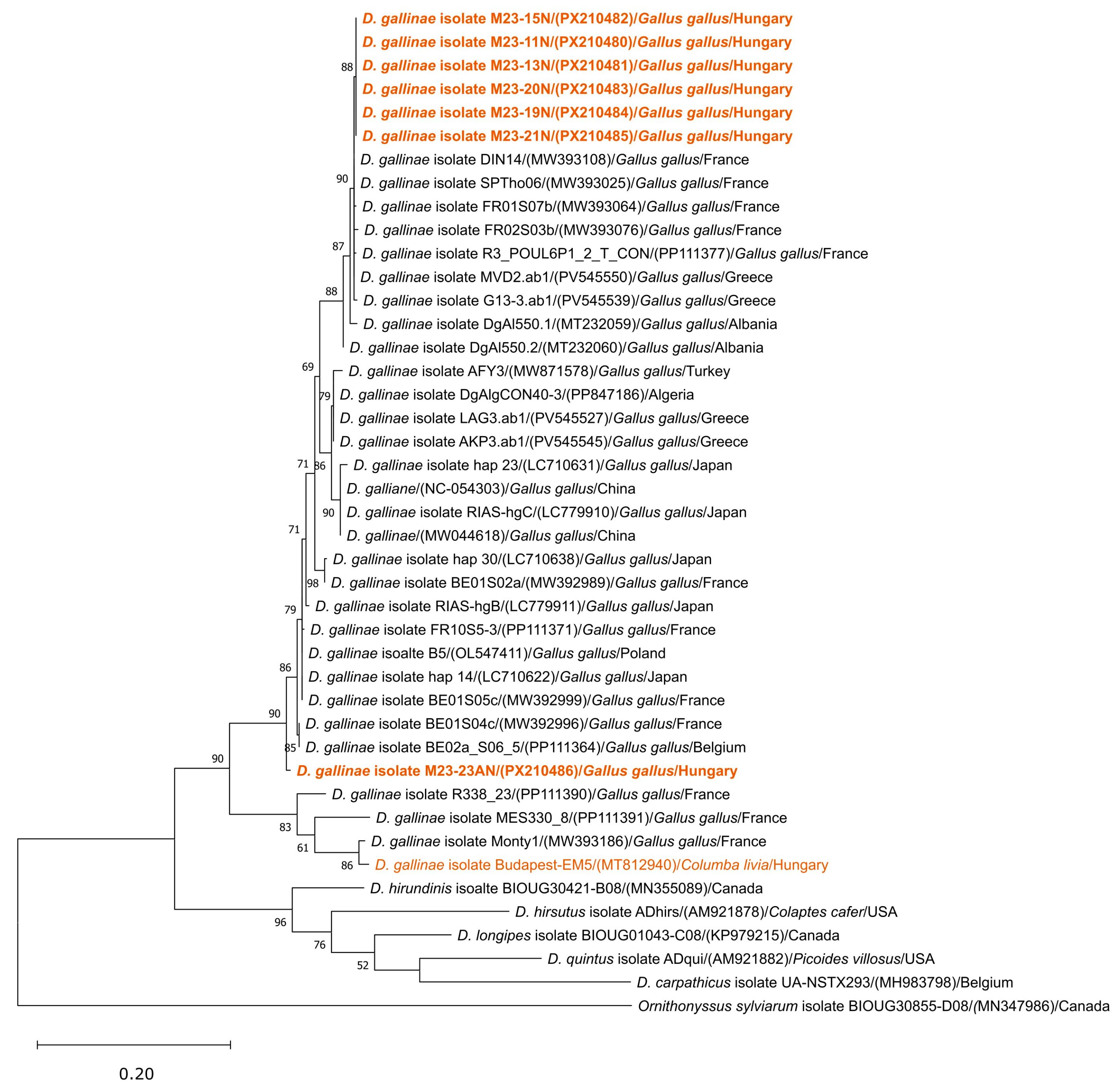
Figure 1. Maximum likelihood phylogenetic tree reconstruction of Dermanyssus gallinae isolates based on a 582 bp nucleotide alignment of COX1 gene (RAxML, evolutionary model GTR+I+G, TBE calculated based on 1000 replicates). Bootstrap values are given as percentages (cut-off value 50%). Ornithonyssus sylviarum (MN347986) was chosen as the outgroup. Isolates from the present study are in bold. Isolates from Hungary are highlighted in brown. NCBI accession numbers are in parentheses. Hosts and geographical origin are listed.
Parasitologia 05 00059 g001
Figure 2. Maximum likelihood phylogenetic tree reconstruction of Histomonas meleagridis isolates based on a 635 bp nucleotide alignment of 18S rRNA gene (RAxML, evolutionary model GTR+I+G, TBE calculated based on 1000 replicates). Bootstrap values are given as percentages (cut-off value 50%). Dientamoebidae sp. isolate BATA35 (PP853597) was chosen as the outgroup. Isolates from the present study are in bold. Isolates from Hungary are highlighted in magenta. Hosts and geographical origins are listed. Genotypes defined by Bilic et al. [25] are labelled.
Parasitologia 05 00059 g002
Disclaimer/Publisher’s Note: The statements, opinions and data contained in all publications are solely those of the individual author(s) and contributor(s) and not of MDPI and/or the editor(s). MDPI and/or the editor(s) disclaim responsibility for any injury to people or property resulting from any ideas, methods, instructions or products referred to in the content.