Article

Investigation of Causative Agents of Vaginitis in Symptomatic and Asymptomatic Women in Konya, Turkey

by
Duygu Beder
1,*,
Fatma Esenkaya Taşbent
2,
Fatma Kılıç Hamzaoğlu
3,
Emine Türen Demir
3,
Mehmet Özdemir
2 and
Gökçe Kader Arslan
4
1
Medical Microbiology, Meram State Hospital, Konya 42090, Turkey
2
Department of Medical Microbiology, Faculty of Medicine, Necmettin Erbakan University, Konya 42080, Turkey
3
Department of Obstetrics and Gynecology, Faculty of Medicine, Necmettin Erbakan University, Konya 42080, Turkey
4
Medical Microbiology, Konya City Hospital, Konya 42020, Turkey
*
Author to whom correspondence should be addressed.
Parasitologia 2025, 5(2), 15; https://doi.org/10.3390/parasitologia5020015
Submission received: 26 February 2025 / Revised: 21 March 2025 / Accepted: 28 March 2025 / Published: 1 April 2025

Abstract

:
A significant portion of women encounter vaginal infections at some point in their lives. These infections have many effects such as infertility, pelvic inflammatory disease (PID), severe disseminated infections, and complications affecting fetal health during pregnancy. Vaginal infections can be symptomatic or asymptomatic, thereby concealing their presence. This study aimed to determine the frequency and causative agents of vaginal infections in symptomatic and asymptomatic women in Konya. The study included 200 patients aged > 18 years who visited the gynecology clinic for any reason and provided informed consent. Patients who reported at least one of the symptoms of vaginal discharge, burning, or itching were categorized into the symptomatic group, while those without any of these complaints were categorized into the asymptomatic group. Vaginal swab samples collected from the patients were evaluated for bacterial vaginosis (BV) using the Nugent scoring method on Gram-stained smears. Trichomonas vaginalis was investigated through Giemsa-stained microscopic examination and polymerase chain reaction (PCR) analysis. Samples were also cultured on routine media to analyze fungal and bacterial growth. A total of 74% of the patients were symptomatic, and 26% were asymptomatic and were admitted to the gynecology outpatient clinic for reasons other than vaginitis. According to the Nugent scoring system, BV was observed in 21.6% of the symptomatic group and 13.5% of the asymptomatic group. T. vaginalis was detected in only seven patients (3.5%) through microscopic examination and PCR. Among the patients with T. vaginalis, five were in the symptomatic group, and two were in the asymptomatic group. The rate of vaginal candidiasis was found to be 34.5% in the symptomatic group and 21.2% in the asymptomatic group. Aerobic vaginitis was observed in 12.8% of the symptomatic group and 7.7% of the asymptomatic group. The positivity rates for BV, vaginal candidiasis, and trichomoniasis in asymptomatic patients are concerning. Considering the fetal complications and chronic effects of vaginal infections, the decision to establish screening programs in the asymptomatic patient group should be based on national data, considering the prevalence of the pathogen in the region and its outcomes.

1. Introduction

The vaginal microbiota has a unique microbial community of about 200 different microorganisms. In vaginitis, where the balance of the vaginal microbiome is disrupted, the most common symptoms include vaginal discharge, foul odor, burning, itching, pain, dyspareunia, and dysuria. The progression of the infection can lead to serious consequences such as infertility and pelvic inflammatory disease (PID). Additionally, if the patient is pregnant, the fetus may be at risk of infection, and if the patient is sexually active, there is potential for infecting the partner. In pregnant women, vaginal infections can result in invasive neonatal infections, miscarriages, and preterm birth. Moreover, since many causative agents of vaginitis can be asymptomatic, they can continue to pose a risk for years without treatment [1,2,3].
In women of reproductive age, the vaginal microbiome is typically composed of various Lactobacillus species. Replacing these lactobacilli with Gardnerella vaginalis and other anaerobic bacteria leads to bacterial vaginosis (BV). BV is not highly inflammatory and is often asymptomatic. Therefore, it is referred to as vaginosis rather than vaginitis. The other two most common types of vaginitis are vaginal candidiasis and trichomoniasis [4]. Another term used to describe infectious vaginitis is aerobic vaginitis. The term “aerobic vaginitis” is used by some clinicians to describe vaginal inflammation believed to be caused by Streptococcus, Staphylococcus, and Escherichia coli. Although the roles of vaginal Streptococcus agalactiae and E. coli in invasive maternal and neonatal infections have been demonstrated in the literature, it is not yet universally accepted that they cause a distinct vaginitis syndrome separate from BV [2,5].
In a woman of reproductive age, the vaginal pH is acidic, ranging between 4 and 4.5. The primary factors contributing to this acidity are estrogen and the lactobacilli in the vaginal microbiota. Estrogen promotes the proliferation of the vaginal epithelium and the storage of glycogen in epithelial cells. The breakdown of glycogen by lactobacilli produces lactic acid, which acidifies the environment. This acidic environment acts as a barrier, preventing the growth of other microorganisms. Additionally, substances like hydrogen peroxide and bacteriocins, produced by many Lactobacillus strains, also inhibit the growth of microorganisms. As women age, the composition of the vaginal microbiota undergoes significant changes along with the structure of the vaginal epithelium, and hormones are suggested to be the primary determinants of these changes. During the postmenopausal period, a decrease in estrogen causes the pH to rise to levels between 6 and 8, creating a more alkaline environment. Compared to acidic environments, alkaline conditions are more conducive to the colonization of many pathogenic microorganisms. However, fungi can still thrive in acidic environments [6,7].
The most significant pathogenesis in vaginitis is vaginal dysbiosis, which reflects a disruption in the microbial community of the vagina, and one of its most prominent features is changes in vaginal pH. In the premenopausal period, pathogenic microorganisms are suppressed in the acidic pH environment. However, if there is a decrease or change in the distribution of microorganisms in the microbiota, candidal infections may exploit the opportunity to multiply. The alkaline environment of the menopausal period presents a potential risk for Trichomonas vaginalis and BV [1].
This study aimed to comparatively investigate the frequency of causative agents of vaginal infection in symptomatic and asymptomatic patient groups, examine the prevalence of these agents in the apparently healthy asymptomatic group, and identify risk factors for these infections. Additionally, the study sought to monitor the distribution of pathogens across different age groups.

2. Materials and Methods

Ethical approval for this study was obtained from the Ethics Committee of Necmettin Erbakan University Faculty of Medicine (Date: 21 February 2025, Decision No: 5551). The study was funded by the Scientific Research Project Coordination Office of Necmettin Erbakan University under project number 211218003.

2.1. Selection of Patients

The study included 200 patients aged > 18 years who presented to the gynecology outpatient clinic of a tertiary university hospital for any reason. After providing the necessary information and obtaining written consent from the participating patients, the prepared questionnaire forms were completed through face-to-face interviews with the patients prior to their examination. The study patients were also divided into two groups: those < 45 years of age in the reproductive period and those ≥ 45 years in the perimenopausal or postmenopausal period. Based on the questionnaire results, patients who reported at least one of the primary symptoms specific to vaginitis—vaginal discharge, burning, or itching—were categorized into the symptomatic group, while those without any of these symptoms were categorized into the asymptomatic group.

2.2. Collection and Storage of Samples

During the gynecological examination, swab samples were collected from the posterior fornix and vaginal sidewalls using sterile cotton-tipped applicators. For microscopic evaluation, the swab sample was spread onto two slides at the patient’s bedside. The smear preparations and swab samples for culture were transported to the laboratory under appropriate conditions. The samples were transferred to a solution-filled sample tube and stored at −80 °C until the time of analysis.

2.3. Stained Microscopic Examination

The smear preparations were stained with Gram and Giemsa stains and examined at ×100 magnification. Gram- and Giemsa-stained smears were evaluated by a single observer. In each immersion field, Gram-positive bacilli resembling Lactobacillus, Gram-negative or Gram-variable cocobacilli resembling Gardnerella, and Gram-negative curved bacilli resembling Mobiluncus were investigated. According to the Nugent scoring system, scores of 0–3 were considered normal, 4–6 were classified as intermediate, and scores of 7–10 were regarded as indicative of BV. Preparations stained with Giemsa that contained one or more organisms matching the morphological description of T. vaginalis (oval shape, with red nuclei, a violet cytoplasm, granular appearance, flagella, an undulating membrane, and axostyles) were considered positive (Figure 1).

2.4. Polymerase Chain Reaction (PCR)

Prior to molecular analysis, nucleic acid isolation was performed using a commercial kit (High Pure PCR Template Preparation Kit, Roche Diagnostics, Basel, Switzerland). Following isolation, T. vaginalis was investigated in the samples with a commercial real-time PCR kit (Nzytech, Lisboa, Portugal). For each sample, 15 μL of the master mix was prepared with 1.5 μL of the primer–probe mix, 1 μL of the enzyme, and 12.5 μL of the buffer. Additionally, 10 μL of the positive control, negative control, or isolated samples were added to the prepared 15 μL master mix. For amplification, a PCR with 40 cycles was programmed with the following steps: initial denaturation at 95 °C for 5 min, denaturation at 90 °C for 1 min, annealing at 60 °C for 30 s, extension at 72 °C for 2 min, and final extension at 72 °C for 7 min. The PCR was performed, and the results were evaluated using the Rotor-Gene Q device (Roche, Basel, Switzerland, 2004). The procedure was conducted in accordance with the kit manufacturer’s recommendations. All procedures were performed within a Class II type biosafety cabinet.

2.5. Culture

The swab samples included in the study were inoculated onto blood agar, eosin methylene blue agar, and Sabouraud dextrose agar. Inoculated plates were incubated at 37 °C and evaluated after 24–48 h. Colonies that grew in culture were examined using Gram staining. The diagnosis of vaginal candidiasis was confirmed by the presence of budding yeast cells and pseudohyphae in the stained preparations, as well as by the growth of yeast colonies in the inoculated media. For bacterial identification, the VITEK 2 Compact System (bioMérieux, Marcy l’Etoile, France), an automated identification system, was used according to the manufacturer’s recommendations. The identified bacteria and Candida spp. growths were recorded. Samples in which aerobic enteric bacteria such as Group B streptococcus (S. agalactiae), Enterococcus faecalis, E. coli, Staphylococcus aureus, and Klebsiella pneumoniae were grown in culture and leukocyte density was observed in the region with Gram and/or Giemsa staining were evaluated as aerobic vaginitis (AV).

2.6. Statistical Analysis

Data were analyzed using SPSS (Statistical Package for the Social Sciences) IBM Software v25. The relationships between categorical variables were examined using Fisher’s exact test, the Fisher–Freeman–Halton exact test, Yates’ correction, and Pearson’s chi-square test. Multiple comparisons of proportions were performed using a Bonferroni-corrected z-test. Frequencies and percentages were used to display categorical variables. The significance level was taken as p < 0.050 (two-tailed).

3. Results

Of the patients, 80% (n = 160) were in the reproductive period, and 20% (n = 40) were in the perimenopausal or postmenopausal period (Table 1). Of the patients, 74% (n = 148) were symptomatic and had at least one of the following complaints: discharge, burning, and itching. The remaining 26% (n = 52) of the patients were asymptomatic and presented to the gynecology outpatient clinic for reasons other than vaginitis (Table 2).
According to the Nugent scoring system, 66.5% (n = 133) of all patients were classified as having normal flora, 14% (n = 28) as having intermediate flora, and 19.5% (n = 39) as having BV. BV was observed in 16.9% (n = 27) of those in the reproductive period and 30% (n = 12) of those in the perimenopausal or postmenopausal period. BV was observed in 21.6% (n = 32) of the symptomatic group and 13.5% (n = 7) of the asymptomatic group.
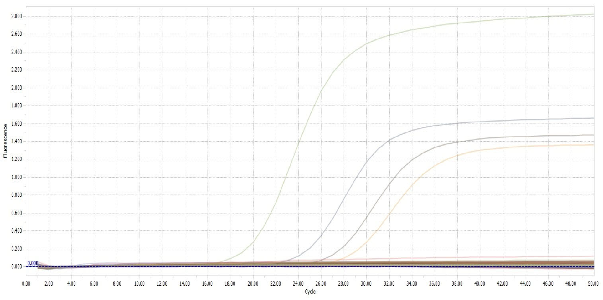
T. vaginalis was detected in a total of seven patients (3.5%), with six (3%) cases through stained microscopic examination, and four (2%) cases through the PCR (Figure 1 and Figure 2).
T. vaginalis was observed in 3.1% (n = 5) of those in the reproductive period and 5% (n = 2) of those in the perimenopausal or postmenopausal period. T. vaginalis was observed in 3.4% (n = 5) of the symptomatic group and 3.8% (n = 2) of the asymptomatic group. In the culture, Candida spp. was detected in 62 patients (31%), E. coli in 11 (5.5%), E. faecalis in 7 (3.5%), S. agalactiae in 4 (2%), and K. pneumoniae in 1 (0.5%). Of the 11 cases that were positive for Candida spp., 4 had BV, 3 had E. coli, 2 had Enterococcus spp., and 1 had S. agalactiae. Among the cases with BV, five also had E. faecalis, and one had E. coli growth. Three cases had T. vaginalis and simultaneous Candida spp. growth, and two cases had T. vaginalis and simultaneous E. coli growth. Candida spp. was observed in 36.9% (n = 59) of those in the reproductive period and 7.5% (n = 3) of those in the perimenopausal or postmenopausal period. Candida spp. was observed in 34.5% (n = 51) of the symptomatic group and 21.2% (n = 11) of the asymptomatic group. AV was observed in 11.9% (n = 19) of those in the reproductive period and 10% (n = 4) of those in the perimenopausal or postmenopausal period. AV was observed in 12.8% (n = 19) of the symptomatic group and 7.7% (n = 4) of the asymptomatic group.
A pathogenic agent was identified in 56% of patients. The distribution of the sociodemographic characteristics of the cases with detected pathogens is shown in Table 3. There was no statistically significant correlation between the presence of pathogens and the sociodemographic and clinical characteristics (p > 0.05).

4. Discussion

Vaginal infections are most commonly caused by BV, vulvovaginal candidiasis, and trichomoniasis [8]. In addition to these, other infectious agents, such as Group A Streptococcus, Group B Streptococcus (S. agalactiae), S. aureus, and Gram-negative bacteria, can also be involved [9].
In BV, the lactobacilli that are normally dominant in the vaginal flora are replaced by anaerobic bacteria such as G. vaginalis, Mobilincus spp., Prevotella spp., and Mycoplasma spp. BV is a very common condition and the most frequent cause of vaginal discharge globally. However, a national survey in the United States has reported that the majority of women with BV are asymptomatic. Vaginal swab culture is the gold standard diagnostic technique for detecting most bacterial infections. However, because the causative agent in BV is difficult to culture, culture is not considered the gold standard [8,10]. The gold standard diagnostic technique for BV is Nugent scoring. However, due to the need for experienced personnel and the time required, it is often preferred for research purposes [11].
Approximately half of BV cases are reported to be asymptomatic. Whether symptomatic or asymptomatic, BV has been strongly associated with obstetric complications, including preterm birth and amnionitis, and gynecological complications, including postoperative infections and other sexually transmitted diseases [12]. The risk of PID in BV patients is 1.53 times higher, and the risk of infertility is 3.32 times higher. BV during pregnancy has been reported to increase the risk of preterm birth by 2.16 times and late miscarriage by 6.32 times due to the increased risk of infection [13]. Additionally, BV is reported to increase the risk of contracting several sexually transmitted infections, including human immunodeficiency virus (HIV), Neisseria gonorrhea, Chlamydia trachomatis, T. vaginalis, and herpes simplex virus-2. The literature also reports a relationship between BV and human papillomavirus [10]. According to the sexually transmitted disease treatment guidelines of the Centers for Disease Control and Prevention (CDC) in the United States, it is recommended that all women with BV be tested for HIV and other sexually transmitted infections [14].
The global prevalence of BV is reported to be between 23% and 29%, while in Germany, BV has been detected in 20% of women participating in preterm birth prevention programs [13]. However, molecular genomic studies have detected higher levels of G. vaginalis and other anaerobes in the vagina compared to classical cultural methods or Nugent scoring. In these studies, G. vaginalis was detected in approximately 50% of vaginal microbial communities of healthy women [15,16]. In another sequencing study conducted on asymptomatic female patients in Estonia, G. vaginalis colonization was found in 70.6% of women, and similar to Gardnerella, Prevotella and Atopobium genera were reported to be common among healthy women participating in the study, with frequencies of 55.8% and 38%, respectively [17]. According to the literature, the prevalence of BV in Turkey detected using classical methods ranges from 1.6% to 15.2% [18,19].
In the present study, findings related to BV were assessed using the Nugent scoring method, and positivity was detected in 39 of 200 patients (19.5%). Of these 39 patients, 7 (~18%) were in the asymptomatic group. Of the seven individuals in the asymptomatic group, five were aged ≥ 45, and two were between 26 and 44 years old. When examining the distribution of BV across age groups, regardless of symptomatic or asymptomatic status, the prevalence was found to be 16.9% in the reproductive age group (<45 years) and 30% in the group aged ≥ 45. As is known, the more alkaline vaginal environment in the postmenopausal period increases the risk of BV [1]. Consistent with this, the present study also found a higher prevalence of BV in the group aged ≥ 45 compared to the group <45 years.
In a study, the normal vaginal microbiota was compared with the microbiota of asymptomatic BV and vulvovaginal candidiasis. Vaginal swabs from 199 asymptomatic participants with urogenital infections were scored using Nugent criteria, showing that 73.9% had normal microbiota, 11.6% had moderate microbiota, and 14.5% had BV. Women with vaginal infections that were asymptomatic (appeared healthy) showed significant disruption in their vaginal microbial communities compared to healthy women. Interestingly and importantly, the distribution of lactobacilli in women with asymptomatic BV was found to be similar to that in symptomatic women. This finding highlighted the importance of screening for asymptomatic infections to avoid further complications [20].
Recommended forms of treatment for BV include metronidazole and clindamycin. BV treatment is usually effective. Studies have reported recovery rates of 80–90% in 1 month compared to baseline However, recurrence rates of BV are high. According to the CDC treatment guidelines, there is insufficient evidence to support the treatment of asymptomatic women, so treatment is recommended only for symptomatic women. The guidelines also state that treatment has limited benefits in non-pregnant women and that it only relieves symptoms. Therefore, asymptomatic women with BV often experience high recurrence rates due to lack of treatment [14,21].
The World Health Organization (WHO) recommends treatment for BV based on microscopy results if persistent vaginal discharge is present. According to the CDC 2021 guidelines for sexually transmitted infections, routine BV screening and treatment for asymptomatic women, including those at high risk for preterm birth, are not recommended. Only the evaluation and treatment of symptomatic women are recommended [14,21]. In the present study, BV was detected in approximately 13.5% of women who appeared completely healthy and asymptomatic. We believe that early detection through screening programs could enable the development of patient-specific monitoring and prevention strategies in cases of BV where the disease can remain asymptomatic at a non-negligible rate.
Candidiasis is another cause of vaginitis and is responsible for approximately one-third of the cases. Vaginal infections caused by Candida species were initially referred to as “acute Candida vaginitis”. However, due to the primary inflammation being localized to the vulva and the source of symptoms being the vulva, the term “vulvovaginal candidiasis” (VVC) has become more commonly used [22]. VVC is considered the second most common cause of vaginitis after BV. It is estimated that approximately 10–15% of asymptomatic women are colonized with Candida, 70–75% of women will experience an episode of VVC during their lifetime, 50% of initially infected women will experience a second VVC infection, and 5–10% of all women will develop recurrent VVC [23].
Candida species live commensally in the vaginas of healthy women and are considered a component of the vaginal microbiota. Using old culture-dependent techniques, researchers have detected vaginal fungi in approximately 20% of asymptomatic women. The dominant portion of the fungal microbiota in the vagina is composed of C. albicans (72–91%), while non-albicans Candida species are also observed. Candida species, although present in healthy women, are still considered opportunistic pathogens due to their high prevalence (85–95%) in patients suffering from vaginal candidiasis, the second most common dysbiosis after BV. To date, it remains unclear why simple colonization by Candida spp. leads to an acute and highly inflammatory infection [7,24]. In a study conducted in Estonia, 494 asymptomatic women of reproductive age were sequenced for bacteria and fungi. In the study, Candida species were detected in 64.5% of the participants, significantly higher than the 20% reported in asymptomatic healthy women using earlier culture-based techniques [7,17].
Recent studies reported that the incidence of VVC in symptomatic women ranges from 12.1% to 57.3%. 35 The highest incidences were reported in African countries, with Nigeria at 57.3% and Tunisia at 48%, while the lowest incidences were reported in European countries, with Greece at 12.1% and Italy at 19.5% [25,26,27,28]. Studies on asymptomatic colonization found that vaginal colonization by Candida species in women without VVC symptoms ranges from 11.6% to 17%. The literature data show that the prevalence of vaginal candidiasis in Turkey ranges from 16% to 40% [8,18,19]. In the present study, while the rate of candidiasis was 34% in the symptomatic patient group, it was 25.7% in the asymptomatic group, which is close to the symptomatic group. The WHO recommends treatment based on microscopy results for patients with vaginal itching and cheese-like discharge complaints but does not recommend screening and treatment for asymptomatic individuals [21].
Vaginal Candida colonization varies during certain periods of life. During prepubertal and postmenopausal periods, lower estrogen levels result in less frequent colonization. Candida vaginitis or colonization occurs due to various factors, including estrogenic activity, pregnancy, immunosuppression, diabetes, antibiotic use, the frequency of sexual activity, and genetic predisposition [29,30]. Almost all epidemiological studies report a higher incidence of VVC in women of reproductive age compared to menopausal women [23]. In estrogen-dominant vaginal flora, classic culture methods have detected Candida spp. in at least 20% of pregnant women and 30% of immunocompromised patients. When non-culture methods like molecular techniques are used, it has been suggested that fungi will be detected in 60% of cases [24].
In the present study, the distribution of candidiasis patients by age was consistent with the literature, revealing 36.9% positivity in women of reproductive age and 7.5% positivity in menopausal women aged ≥ 45 years. In the literature, it has been suggested that the presence of Candida species in the vagina, whether commensal or pathogenic, is related to their dimorphic nature. It has been reported that the yeast form is generally isolated from healthy asymptomatic women, while the hyphal form is consistently found in severe cases of VVC. This supports the association of the yeast form with commensalism and the hyphal form with pathogenicity [31]. Even minor changes in host defenses and vaginal microbiota can lead to opportunistic infections caused by C. albicans. Studies based on the co-culturing of vaginal yeast and bacteria suggest that bacteria inhibit the yeast-to-hypha transition of Candida and compete with Candida cells for binding sites on epithelial receptors due to their higher affinity [7]. However, in the present study, all of the seven patients with co-occurring fungal and bacterial growth in culture were in the symptomatic group. In these patients, Candida spp. growth was accompanied by E. coli in three cases, Enterococcus spp. in two cases, S. agalactiae in one case, and coagulase-negative staphylococci in one case. In the symptomatic patient group, Candida spp. and T. vaginalis positivity was detected in three patients. Similarly, in the symptomatic group, Candida spp. was found alongside BV in two patients, while in the asymptomatic group, one patient had both Candida spp. and BV. Mixed growths were usually seen in the symptomatic group except in one patient.
T. vaginalis, another important causative agent of vaginitis, is reported to be the most common parasitic cause of sexually transmitted infections after viral and bacterial agents [32,33]. Trichomoniasis is usually asymptomatic and can be easily transmitted between sexual partners. The infection has been found to significantly increase the risk of HIV transmission, is associated with pregnancy complications, including preterm birth, and is linked to PID in HIV-infected women. T. vaginalis parasites primarily infect the urethra in men and the vaginal and vulvar regions in women [34].
The prevalence of trichomoniasis varies in different countries and populations. In 2016, the WHO estimated that there were 156 million global cases of T. vaginalis, accounting for nearly half of the global sexually transmitted disease cases that year [35].
In a prospective study of 1025 symptomatic and asymptomatic women in the United States, T. vaginalis positivity was detected in 11.4% of asymptomatic patients, while this rate was 59.9% for symptomatic patients [36]. In Turkey, a study conducted among Syrian refugee women reported a very high positivity rate for T. vaginalis: 36% with Giemsa staining and 21.3% with direct microscopic examination. High positivity rates have been associated with crowded living conditions [32]. In studies conducted in Turkey, the prevalence of T. vaginalis in women has been reported to range from 0.3% to 9% [37]. Most of the research on T. vaginalis has been carried out in symptomatic patients. Another study reported a 14.9% prevalence of VVC, 11.2% of BV, and 1.9% of trichomoniasis among symptomatic patients [37]. In the present study, T. vaginalis was detected in 3.5% of all included patients. When examining the prevalence in symptomatic and asymptomatic cases, approximately 28.5% (2/7) of the T. vaginalis-positive cases were found in the asymptomatic group. T. vaginalis was detected in 3.4% of symptomatic and 3.8% of asymptomatic patients. The higher percentage of positivity in the asymptomatic group is notable. According to CDC guidelines [14], T. vaginalis screening is not recommended in asymptomatic women. Treatment is recommended according to molecular test results [21].
Approximately 90% of the vaginal flora consists of lactobacilli [38]. Aerobic vaginitis is defined as a disruption of the lactobacillary flora, accompanied by signs of inflammation and the presence of a rather scarce, predominantly aerobic microflora composed of enteric commensals or pathogens [39]. Aerobic vaginitis is generally considered an inflammatory condition and has not yet been universally accepted as a distinct cause of vaginitis other than BV [2,5].
In a study, when examining 305 vaginal swab cultures for aerobic bacteria, E. coli was found positive in 8.8%, S. agalactiae in 3.2%, K. pneumoniae in 2.6%, and E. faecalis in 0.3% of the cases [8]. In another study, 5.7% of 314 vaginal swab cultures were positive for causative agents of aerobic vaginitis, and the leading agent was reported to be E. coli, with a rate of 2.2% [18]. In the present study, the rate of aerobic enteric pathogen growth in vaginal swabs was 11.5% (23/200). This rate was 12.8% in symptomatic patients presenting with vaginitis and 7.7% in the asymptomatic group. In the present study, E. coli constituted the majority of isolated Gram-negative bacilli, representing 11 out of 12 cases (91.6%). These pathogens were found at a higher rate in the postmenopausal group, with 4 out of 17 cases (23.5%).
Group B Streptococcus (S. agalactiae) is normally found in the gastrointestinal and genitourinary systems of healthy individuals and rarely causes infections. However, it is a significant cause of morbidity in pregnant women, neonates, and the elderly [40]. S. agalactiae is responsible for serious infections such as sepsis, meningitis, and pneumonia in neonates. In the adult patient population, it causes skin, soft tissue, and urinary tract infections; osteoarticular pathologies; peritonitis; or endocarditis. It is also reported to be a causative agent of vaginitis [5]. The literature data indicate a 0.9% positivity rate for S. agalactiae [18,37]. In the present study, the positivity rate for S. agalactiae was found to be 2%. It was observed that 75% of these cases were <45 years of age.

5. Conclusions

In the present study, the causative agents of vaginitis were investigated in asymptomatic as well as symptomatic patients. Sexually transmitted infection agents and vaginitis etiologies have been predominantly studied in symptomatic patient groups in the literature. Therefore, data from asymptomatic patient groups are important. In this patient group, which did not seek medical attention for vaginitis, there is a risk of developing various gynecological and obstetric complications. Additionally, there could be an increased prevalence of other sexually transmitted infections, including HIV. Current WHO and CDC guidelines do not recommend screening for these agents in asymptomatic patients. Although initially, it may not seem cost effective, pregnancy complications, HIV and its associated clinical issues, and potential secondary problems such as PID can lead to significant costs for this overlooked group. The decision to establish screening programs should be based on national data, considering the prevalence of the pathogen in the region and its outcomes. We believe that more detailed microbiota and long-term prospective studies are needed for asymptomatic patient groups.

Author Contributions

The work described in this manuscript has not been published previously and is not under consideration for publication elsewhere. All authors have approved this submission. Material preparation and data collection were carried out by D.B., F.K.H., E.T.D. and G.K.A. All authors were involved in data analysis and manuscript write-up. D.B., M.Ö. and F.E.T. conceptualized this study and prepared the first draft of this manuscript. All authors have read and agreed to the published version of the manuscript.

Funding

This study was financed by the Necmettin Erbakan University Scientific Research Project Coordination Office under project number 211218003.

Institutional Review Board Statement

The study was conducted in accordance with the Declaration of Helsinki, and approved by Ethics Committee of Necmettin Erbakan University Faculty of Medicine (5551 of 21 February 2025).

Informed Consent Statement

Informed consent was obtained from all subjects involved in the study. Written informed consent has been obtained from the patients to publish this paper.

Data Availability Statement

All data generated by this study are included in this publication.

Conflicts of Interest

All authors declare no competing interests.

References

  1. Chee, W.J.Y.; Chew, S.Y.; Than, L.T.L. Vaginal microbiota and the potential of Lactobacillus derivatives in maintaining vaginal health. Microb. Cell Fact. 2020, 19, 203. [Google Scholar] [CrossRef] [PubMed]
  2. Van de Wijgert, J.H.H.M.; Borgdorff, H.; Verhelst, R.; Crucitti, T.; Francis, S.; Verstraelen, H.; Jespers, V. The vaginal microbiota: What have we learned after a decade of molecular characterization? PLoS ONE 2014, 9, e105998. [Google Scholar] [CrossRef] [PubMed]
  3. Mendling, W. Vaginal microbiota. In Microbiota of the Human Body: Implications in Health and Disease; Schwiertz, A., Ed.; Springer International Publishing: Cham, Switzerland, 2016; pp. 83–93. [Google Scholar]
  4. Dallabetta, G.; Wi, T.E.C.; Nielsen, G. Prevention and control of STD and HIV infection in developing countries. In Sexually Transmitted Diseases, 4th ed.; Holmes, K.K., Sparling, P.F., Stamm, W., Piot, P., Wasserheit, J., Corey, L., Eds.; McGraw-Hill: New York, NY, USA, 2008. [Google Scholar]
  5. Savini, V.; Marrollo, R.; D’Antonio, M.; D’Amario, C.; Fazii, P.; D’Antonio, D. Streptococcus agalactiae vaginitis: Nonhemolytic variant on the Liofilchem® Chromatic StreptoB. Int. J. Clin. Exp. Pathol. 2013, 6, 1693–1695. [Google Scholar] [PubMed]
  6. Acarkan, T. Vajinal flora dysfunction and vaginitis. J. Complement. Med. Reg. Neural. Ther. 2016, 10, 8–12. [Google Scholar]
  7. Kalia, N.; Singh, J.; Kaur, M. Microbiota in vaginal health and pathogenesis of recurrent vulvovaginal infections: A critical review. Ann. Clin. Microbiol. Antimicrob. 2020, 19, 5. [Google Scholar] [CrossRef]
  8. Karakoç, Z.Ç. Does the Etiology Change in Vaginitis? Data Results of Samples from a Single Center. Med. J. Mugla Sitki Kocman Univ. 2021, 8, 18–22. [Google Scholar] [CrossRef]
  9. Aksakal, S.E.; Cırık, D.A.; Ensari, T.; Coskun, B.; Aksu, G.; Mollamahmutoglu, L.; Tapısız, O.L. Comparison of the Effectiveness of VGTest™ and Standard Microbiological Tests in the Diagnosis of the Vaginal Infections. Turk. J. Womens. Health. Neonatol. 2020, 2, 33–40. [Google Scholar]
  10. Coudray, M.S.; Madhivanan, P. Bacterial vaginosis-A brief synopsis of the literature. Eur. J. Obstet. Gynecol. Reprod. Biol. 2020, 245, 143–148. [Google Scholar] [CrossRef]
  11. Bansal, R.; Garg, P.; Garg, A. Comparison of Amsel’s criteria and Nugent’s criteria for diagnosis of bacterial vaginosis in tertiary care centre. Int. J. Reprod. Contracept. Obstet. Gynecol. 2019, 8, 637–640. [Google Scholar] [CrossRef]
  12. Gibbs, R.S. Asymptomatic bacterial vaginosis: Is it time to treat? Am. J. Obstet. Gynecol. 2007, 196, 495–496. [Google Scholar] [CrossRef]
  13. Swidsinski, S.; Moll, W.M.; Swidsinski, A. Bacterial vaginosis-vaginal polymicrobial biofilms and dysbiosis. Dtsch. Arztebl. Int. 2023, 120, 347–354. [Google Scholar] [CrossRef] [PubMed]
  14. Centers for Disease Control and Prevention (CDC). Sexually Transmitted Infections Treatment Guidelines; Centers for Disease Control and Prevention (CDC): Atlanta, GA, USA, 2023. Available online: https://www.cdc.gov/std/treatment-guidelines/bv.htm (accessed on 13 December 2024).
  15. Mikamo, H.; Sato, Y.; Hayasaki, Y.; Hua, Y.X.; Tamaya, T. Vaginal microflora in healthy women with Gardnerella vaginalis. J. Infect. Chemother. 2000, 6, 173–177. [Google Scholar] [CrossRef] [PubMed]
  16. Fredricks, D.N.; Fiedler, T.L.; Marrazzo, J.M. Molecular identification of bacteria associated with bacterial vaginosis. N. Engl. J. Med. 2005, 353, 1899–1911. [Google Scholar] [CrossRef] [PubMed]
  17. Drell, T.; Lillsaar, T.; Tummeleht, L.; Simm, J.; Aaspõllu, A.; Väin, E.; Saarma, I.; Salumets, A.; Donders, G.; Metsis, M. Characterization of the vaginal micro- and mycobiome in asymptomatic reproductive-age Estonian women. PLoS ONE 2013, 8, e54379. [Google Scholar] [CrossRef]
  18. Ünlü, Ö.; Çağlar, T.; Erdem, M.G.; Ekici, S.; Demirci, M. A Three Year Retrospective Analysis of Vaginitis Agents in Patients Admitted to a Private Hospital. Kırıkkale Üniv. Tıp Fak. Derg. 2022, 24, 181–186. [Google Scholar] [CrossRef]
  19. Kanbur, S. Comparison of Screening And Treatment Protocols of Sexually Transmitted Diseases. Zeynep Kamil Tıp Bül. 2018, 49, 15–20. [Google Scholar] [CrossRef]
  20. Pramanick, R.; Mayadeo, N.; Warke, H.; Begum, S.; Aich, P.; Aranha, C. Vaginal microbiota of asymptomatic bacterial vaginosis and vulvovaginal candidiasis: Are they different from normal microbiota? Microb. Pathog. 2019, 134, 103599. [Google Scholar] [CrossRef]
  21. Workowski, K.A.; Bachmann, L.H.; Chan, P.A.; Johnston, C.; Muzny, C.A.; Park, I.; Reno, H.; Zenilman, J.M.; Bolan, G.A. Sexually transmitted infections treatment guidelines. MMWR Morb. Mortal. Wkly. Rep. 2021, 70, 1–187. [Google Scholar] [CrossRef]
  22. Sobel, J.D. Recurrent vulvovaginal candidiasis. Am. J. Obstet. Gynecol. 2016, 214, 15–21. [Google Scholar] [CrossRef]
  23. Gonçalves, B.; Ferreira, C.; Alves, C.T.; Henriques, M.; Azeredo, J.; Silva, S. Vulvovaginal candidiasis: Epidemiology, microbiology and risk factors. Crit. Rev. Microbiol. 2016, 42, 905–927. [Google Scholar] [CrossRef]
  24. Farr, A.; Effendy, I.; Tirri, B.F.; Hof, H.; Mayser, P.; Petricevic, L.; Ruhnke, M.; Schaller, M.; Schaefer, A.P.A.; Sustr, V.; et al. Guideline: Vulvovaginal candidosis (AWMF 015/072, level S2k). Mycoses 2021, 64, 583–602. [Google Scholar] [CrossRef] [PubMed]
  25. Okungbowa, F.I.; Isikhuemhen, O.S.; Dede, A.P.O. The distribution frequency of Candida species in the genitourinary tract among symptomatic individuals in Nigerian cities. Rev. Iberoam. Micol. 2003, 20, 60–63. [Google Scholar] [PubMed]
  26. Amouri, I.; Sellami, H.; Borji, N.; Abbes, S.; Sellami, A.; Cheikhrouhou, F.; Maazoun, L.; Khaled, S.; Khrouf, S.; Boujelben, Y.; et al. Epidemiological survey of vulvovaginal candidosis in Sfax, Tunisia. Mycoses 2011, 54, e499–e505. [Google Scholar] [CrossRef] [PubMed]
  27. Grigoriou, O.; Baka, S.; Makrakis, E.; Hassiakos, D.; Kapparos, G.; Kouskouni, E. Prevalence of clinical vaginal candidiasis in a university hospital and possible risk factors. Eur. J. Obstet. Gynecol. Reprod. Biol. 2006, 126, 121–125. [Google Scholar] [CrossRef]
  28. Tibaldi, C.; Cappello, N.; Latino, M.A.; Masuelli, G.; Marini, S.; Benedetto, C. Vaginal and endocervical microorganisms in symptomatic and asymptomatic non-pregnant females: Risk factors and rates of occurrence. Clin. Microbiol. Infect. 2009, 15, 670–679. [Google Scholar] [CrossRef]
  29. Mills, B.B. Vaginitis: Beyond the Basics. Obstet. Gynecol. Clin. N. Am. 2017, 44, 159–177. [Google Scholar] [CrossRef]
  30. Chatzivasileiou, P.; Vyzantiadis, T.A. Vaginal yeast colonisation: From a potential harmless condition to clinical implications and management approaches-A literature review. Mycoses 2019, 62, 638–650. [Google Scholar] [CrossRef]
  31. Sobel, J.D. Pathogenesis of Candida vulvovaginitis. Curr. Top. Med. Mycol. 1989, 3, 86–108. [Google Scholar] [CrossRef]
  32. Doni, N.Y.; Aksoy, M.; Şimşek, Z.; Gürses, G.; Hilali, N.G.; Zeyrek, F.Y.; Özek, B.; Yıldırımkaya, G. Vajinit yakınmaları olan 15–49 yaş arasındaki suriyeli mülteci kadınlarda Trichomonas vaginalis sıklığının araştırılması. Mikrobiyol. Bul. 2016, 50, 590–597. [Google Scholar] [CrossRef]
  33. Bruni, M.P.; Freitas da Silveira, M.; Stauffert, D.; Bicca, G.L.O.; Caetano Dos Santos, C.; Rosa Farias, N.A.; Golparian, D.; Unemo, M. Aptima Trichomonas vaginalis assay elucidates significant underdiagnosis of trichomoniasis among women in Brazil according to an observational study. Sex. Transm. Infect. 2019, 95, 129–132. [Google Scholar] [CrossRef]
  34. Meites, E.; Gaydos, C.A.; Hobbs, M.M.; Kissinger, P.; Nyirjesy, P.; Schwebke, J.R.; Secor, W.E.; Sobel, J.D.; Workowski, K.A. A review of evidence-based care of symptomatic trichomoniasis and asymptomatic Trichomonas vaginalis infections. Clin. Infect. Dis. 2015, 61, S837–S848. [Google Scholar] [CrossRef] [PubMed]
  35. Van Gerwen, O.T.; Muzny, C.A. Recent advances in the epidemiology, diagnosis, and management of Trichomonas vaginalis infection. F1000Research 2019, 8, 1666. [Google Scholar] [CrossRef]
  36. Schwebke, J.R.; Hobbs, M.M.; Taylor, S.N.; Sena, A.C.; Catania, M.G.; Weinbaum, B.S.; Johnson, A.D.; Getman, D.K.; Gaydos, C.A. Molecular testing for Trichomonas vaginalis in women: Results from a prospective U.S. clinical trial. J. Clin. Microbiol. 2011, 49, 4106–4111. [Google Scholar] [CrossRef]
  37. Yazısız, H.; Özyurt, Ö.; Eryiğit, F.; Özhak, B.; Öngüt, G.; Özekinci, M.; Dönmez, D.; Çolak, D.; Gümüş, S.; Öğünç, D. Evaluation of Microscopic Examination, Culture and Polymerase Chain Reaction Tests in the Diagnosis of Trichomonas vaginalis Infection. Mikrobiyol. Bul. 2020, 54, 135–143. [Google Scholar] [CrossRef]
  38. Kaambo, E.; Africa, C.; Chambuso, R.; Passmore, J.S. Vaginal microbiomes associated with aerobic vaginitis and bacterial vaginosis. Front. Public Health 2018, 6, 78. [Google Scholar] [CrossRef]
  39. Donders, G.; Bellen, G.; Rezeberga, D. Aerobic vaginitis in pregnancy. BJOG Int. J. Obstet. Gynaecol. 2011, 118, 1163–1170. [Google Scholar] [CrossRef] [PubMed]
  40. Raabe, V.N.; Shane, A.L. Group B Streptococcus (Streptococcus agalactiae). Microbiol. Spectr. 2019, 7, 10–1128. [Google Scholar] [CrossRef]
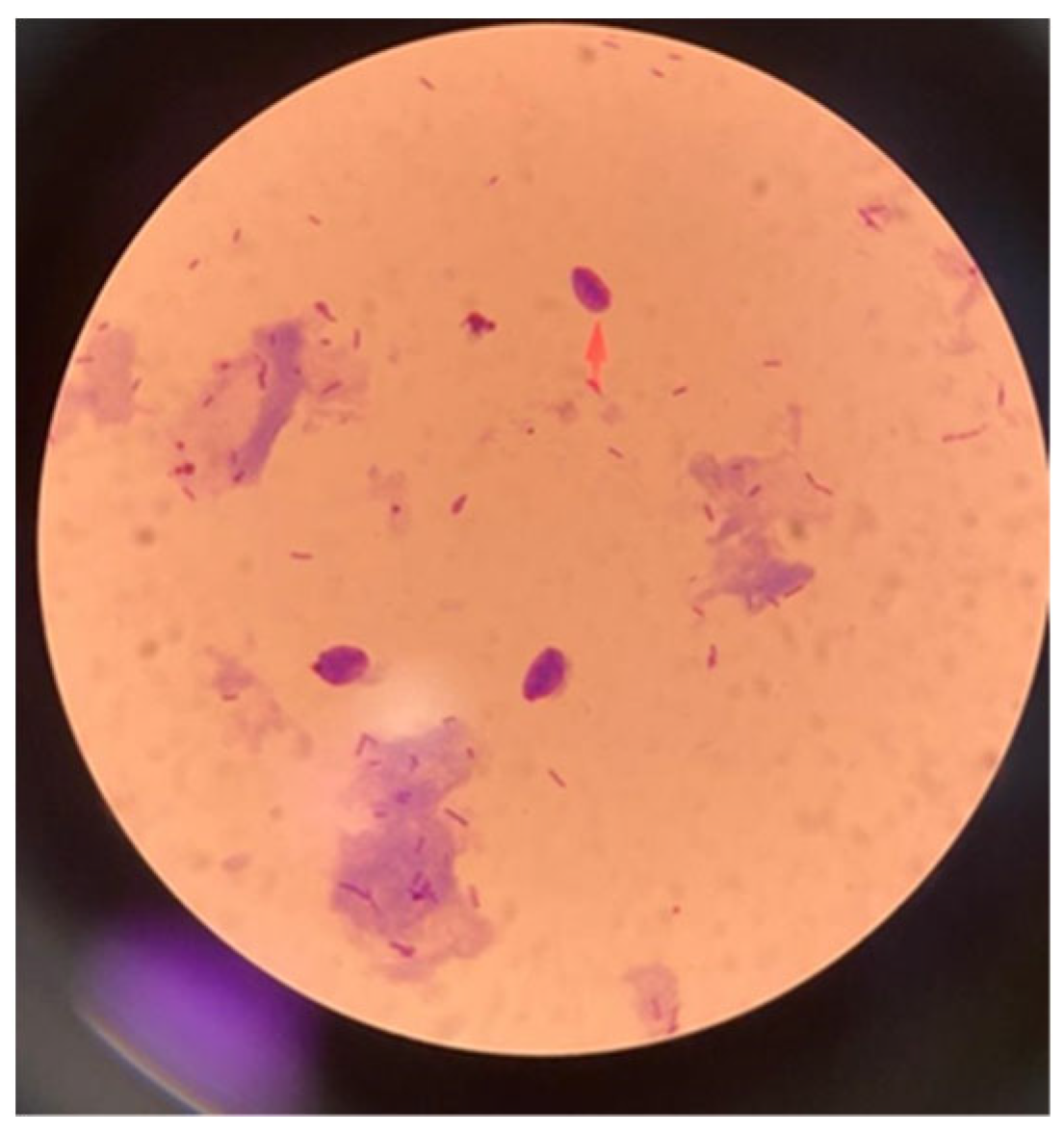
Figure 1. Microscopy image of T. vaginalis-positive patient (×100).
Parasitologia 05 00015 g001
Figure 2. Amplification curves of patient samples found positive for T. vaginalis using real-time PCR. Randomly selected colors were used to display the curves separately for each patient.
Parasitologia 05 00015 g002
Table 1. Distribution of cases with pathogens detected in vaginal swab samples among age groups.
Age < 45 Years (n/N)Age ≥ 45 (n/N)
Number of samples16040
Bacterial vaginosis27/160 (%16.9)12/40 (%30)
Trichomoniasis5/160 (%3.1)2/40 (%5)
Candidiasis59/160 (%36.9)3/40 (%7.5)
Aerobic vaginitis19/160 (%11.9)4/40 (%10)
Table 2. Distribution of pathogens detected in vaginal swabs in symptomatic and asymptomatic patients.
Symptomatic (n/N)Asymptomatic (n/N)
Number of samples14852
Bacterial vaginosis32/148 (%21.6)7/52 (%13.5)
Trichomoniasis5/148 (%3.4)2/52 (%3.8)
Candidiasis51/148 (%34.5)11/52 (%21.2)
Aerobic vaginitis19/148 (%12.8)4/52 (%7.7)
Table 3. Distribution of sociodemographic characteristics in cases with detected pathogens.
Variable Count (n)Number of SamplePercentage (%)p
EducationPrimary school53600.946
Middle school995555.6
High school422354.8
University513058.8
Postgraduate3133.3
ProfessionHousewife1699656.80.436
Employed291448.3
Student22100
Chronic diseaseNo1589459.50.079
Yes421842.9
HemorrhoidNo19410956.21.000
Yes6350
Antibiotic useNo1789553.40.057
Yes221777.3
Age at first sexual intercourse≤18 years412253.70.753
18–25 years old1377957.7
≥25 years221150
Number of partners084500.362
118910555.6
≥233100
Contraceptive methodBirth control pills9555.60.254
Condom382052.6
Spiral (RIA)241875
Other1296953.5
Number of births0382873.70.146
1331545.5
2603355
3472451.1
≥4221254.5
Mode of deliveryNormal1035452.40.776
Cesarean452453.3
Both14642.9
DischargeNo703448.50.120
Yes1307860
RednessNo1528253.90.382
Yes483062.5
BurningNo1045754.80.724
Yes965557.3
ItchingNo773950.60.228
Yes1237359.3
Frequency of changing underwearEvery day1679858.70.127
Every few days331442.4
Daily padNo1488557.40.599
Yes522751.9
Toilet typeSquat toilet1126154.50.873
Western-style toilet613557.4
Squat/Western style271659.3
Frequency of bathing184500.318
2411946.3
≥31518958.9
Frequency of changing pads during menstruation12012600.848
2583153.4
≥31216957
Disclaimer/Publisher’s Note: The statements, opinions and data contained in all publications are solely those of the individual author(s) and contributor(s) and not of MDPI and/or the editor(s). MDPI and/or the editor(s) disclaim responsibility for any injury to people or property resulting from any ideas, methods, instructions or products referred to in the content.