Systematic Review

Advances in Understanding Chronic Traumatic Encephalopathy: A Systematic Review of Clinical and Pathological Evidence

Department of Clinical and Experimental Medicine, Section of Forensic Medicine, University of Foggia, 71122 Foggia, Italy
*
Author to whom correspondence should be addressed.
Forensic Sci. 2025, 5(3), 33; https://doi.org/10.3390/forensicsci5030033
Submission received: 16 May 2025 / Revised: 9 July 2025 / Accepted: 28 July 2025 / Published: 30 July 2025

Abstract

Background/Objectives: Traumatic brain injury is one of the leading causes of death and disability. When traumatic brain injury is repeated over time, it can lead to the development of Chronic Traumatic Encephalopathy, a chronic neurodegenerative disease commonly observed in individuals who engage in contact sports or military personnel involved in activities with a high risk of repeated head trauma. At autopsy, the examination of the brain reveals regional atrophy, corresponding to high concentrations of glutamate receptors. Microscopically, the primary findings are the deposition of neurofibrillary tangles and neuropil threads. The aim of this study is to highlight the clinical and histopathological characteristics of Chronic Traumatic Encephalopathy, providing diagnostic support to forensic pathologists. Additionally, it seeks to aid in the differential diagnosis of similar conditions. Methods: A review of literature was conducted following the PRISMA criteria. Of 274 articles, 7 were selected. Results: According to these papers, most patients were male and exhibited neurological symptoms and neuropsychiatric impairments, and a proportion of them committed suicide or had aggressive behavior. Conclusions: Chronic Traumatic Encephalopathy remains largely underdiagnosed during life. The definitive diagnosis of Chronic Traumatic Encephalopathy is established post-mortem through the identification of pathognomonic tauopathy lesions. Early and accurate antemortem recognition, particularly in at-risk individuals, is highly valuable for its differentiation from other neurodegenerative conditions, thereby enabling appropriate clinical management and potential interventions.

1. Introduction

Traumatic brain injury (TBI) is a major global health concern, recognized as a leading cause of death and disability, incurring substantial human and economic costs [1]. It is a significant cause of morbidity and mortality, particularly among young individuals, and is of critical importance for forensic pathologists [2]. When TBI does not result in immediate death but involves repeated and persistent head trauma over time, it can lead to the development of Chronic Traumatic Encephalopathy (CTE).
CTE is a chronic neurodegenerative disease commonly observed in individuals exposed to repetitive head trauma, such as those engaged in contact sports or military personnel [3,4,5]. In the United States alone, an estimated 4 million sports-related concussions are reported annually [6]. Initially described in boxers as “punch drunk syndrome” or “dementia pugilistica”, CTE’s demographic has expanded to include professional and amateur athletes across various contact disciplines, as well as women who have experienced intimate partner violence [7,8].
While a definitive diagnosis of CTE is made post-mortem, clinical manifestations, collectively known as Traumatic Encephalopathy Syndrome (TES), have established diagnostic criteria [9]. These criteria encompass cognitive, behavioral, and mood symptoms, supplemented by features such as impulsiveness, anxiety, apathy, paranoia, suicidality, headaches, motor signs, documented decline, and delayed onset [10]. A thorough neurological examination, with particular attention to mental status, is crucial [6,11]. Notably, younger individuals with CTE often present with mood and behavioral symptoms, while older individuals typically exhibit cognitive impairment and executive dysfunction [12]. Current treatment options for CTE are limited, underscoring the importance of prevention through adherence to safety protocols and “return-to-play” policies [13,14]. Clinical features of CTE can mimic other neurodegenerative conditions such as Alzheimer’s disease, frontotemporal lobar degeneration, Lewy body disease, cerebral amyloid angiopathy, and Parkinsonism [15].
CTE is characterized by specific gross brain abnormalities, including regional atrophy (most notably in the frontal lobe), ventriculomegaly, wasting of the corpus callosum, and characteristic fenestration of the septum pellucidum [6]. Microscopically, the defining feature of CTE is the deposition of neurofibrillary tangles (NFTs) and neuropil threads, contributing to a distinct tauopathy [6]. These NFTs are typically found perivascularly in the depths of cerebral sulci and are exclusively neuronal [12].
Neuropathological diagnostic and grading criteria for CTE were proposed by McKee, emphasizing patchy, perivascular deposits of phosphorylated tau (p-tau)-positive NFTs and astrocytic tangles within the neocortex, particularly in the depths of cerebral sulci and superficial cortical layers, primarily in the temporal lobe. The pathology is distinguished by an irregular, patchy distribution of NFTs, a prominent perivascular pattern, and preferential involvement of cortical laminae II and III, with a predilection for the depths of the cortical sulci in frontal, temporal, parietal, insular, and septal cortices, while sparing the primary visual cortex. Astrocytic p-tau inclusions (“astrocytic tangles”) are also described in subpial regions and around small blood vessels, alongside dot-like and spindle-shaped neurites. Dense NFTs, ghost tangles, and neurites are observed in various subcortical regions, including the hippocampus, amygdala, and substantia nigra, with less significant involvement of white matter tracts [16].
The National Institute of Neurological Disorders and Stroke (NINDS) and the National Institute of Biomedical Imaging and Bioengineering (NIBIB) have established specific diagnostic criteria for CTE, centered on perivascular p-tau NFTs and astrocytic tangles in the cortex, especially in sulcal depths and superficial layers [9,12]. These criteria were updated in 2021 to clarify that perivascular p-tau aggregates must involve neurons and extend beneath the subpial layer, also introducing “Low CTE” and “High CTE” classifications based on NFT presence in specific brain regions to assist neuropathological evaluations [12]. The diagnosis of CTE is primarily confirmed via autopsy, often facilitated by immunohistochemistry for p-tau [17,18]. The presence of neuronal p-tau correlates significantly with age, duration of repeated head injury exposure, and CTE severity [12].
Four histomorphologic phenotypes associated with CTE have been identified: Type 1 (NFTs and neuropil threads only in cerebral cortex and brainstem); Type 2 (Type 1 features with amyloid-β deposition); Type 3 (NFTs and neuropil threads exclusively in the brainstem); and Type 4 (NFTs and neuropil threads in the cortex, subcortex, brainstem, and basal ganglia, sparing the cerebellum) [19]. Differentiating CTE from other tauopathies like Alzheimer’s or Lewy body dementia is aided by the unique perivascular deposition of tau-immunoreactive astrocytes within the sulcal depths of superficial cortical layers in CTE [17]. CTE NFTs are also larger and often co-localize with TDP-43 aggregates [17,20]. While concurrent neurodegenerative diseases are present in nearly 40% of CTE cases, CTE maintains distinct ultrastructural and microscopic tauopathy features [12].
The pathogenesis of CTE is rooted in the complex interplay of molecular and cellular events initiated by repetitive head trauma. Tau protein is essential for microtubule assembly and axonal integrity; its phosphorylation typically facilitates microtubule binding. However, traumatic insults can lead to calcium influx and glutamate hyperexcitotoxicity, inducing kinase-mediated hyperphosphorylation, misfolding, and aggregation of tau proteins. These altered tau proteins are subsequently cleaved by calpains and caspases, contributing to the formation of NFTs [21,22]. Beyond tau, inclusions of Transactive response DNA-binding protein 43 (TDP-43) and amyloid-β deposition are also observed in CTE.
Neurodegeneration is exacerbated by impaired glymphatic clearance, which results from the perivascular polarization of astroglial aquaporin-4 and prevents the removal of excessive protein deposits. Primary pathogenic factors include oxidative stress, neuroinflammation, and glutamatergic toxicity. Additionally, chronic inflammation disrupts the ubiquitin-proteasome pathway, while microglial priming triggers immune-excitotoxic responses, thereby perpetuating neurodegeneration [22]. Immuno-excitotoxicity plays a crucial role; repeated head injuries cause axonal degeneration and microtubule disintegration, leading to tau oligomerization and NFT formation. These processes disrupt neuronal communication and networking, promoting tau propagation, inflammatory cascades, and impaired blood-brain barrier permeability [23]. Further distinctively, the tau filament in CTE features a unique β-helix region with a hydrophobic cavity [13,24]. Compared to Alzheimer’s disease, CTE shows higher p-tau levels in the CA2 and CA3 regions of the hippocampus, with significantly greater p-tau burden in CA3 and CA4 [12]. Table 1 summarizes the main differences between Alzheimer’s disease and CTE.
The aim of this study is to highlight the clinical and histopathological characteristics of CTE, providing diagnostic support to forensic pathologists. Additionally, it seeks to aid in the differential diagnosis of similar conditions and to raise awareness within the scientific community about the existence of this pathology, stimulating the implementation of effective preventive measures to reduce the incidence of this condition.
Furthermore, this literature review underscores the critical importance of a multidisciplinary collaborative approach in contemporary forensic sciences. As the field rapidly evolves, driven by advanced technologies and the integration of diverse knowledge domains, understanding complex conditions like CTE necessitates expertise beyond traditional boundaries. Forensic investigations, particularly those involving neurological pathologies, increasingly demand insights from biotechnology, psychology, criminology, law, and pathology, alongside emerging fields like artificial intelligence. The definitive diagnosis of CTE, currently possible only post-mortem, highlights the indispensable role of forensic pathologists working with neurologists, neuropsychologists, and other specialists to correlate clinical histories with neuropathological findings. By fostering such collaboration, forensic experts can lead to a more complete understanding of complex cases, improved diagnostic accuracy, and the development of more effective preventive strategies and interventions for debilitating conditions like CTE.

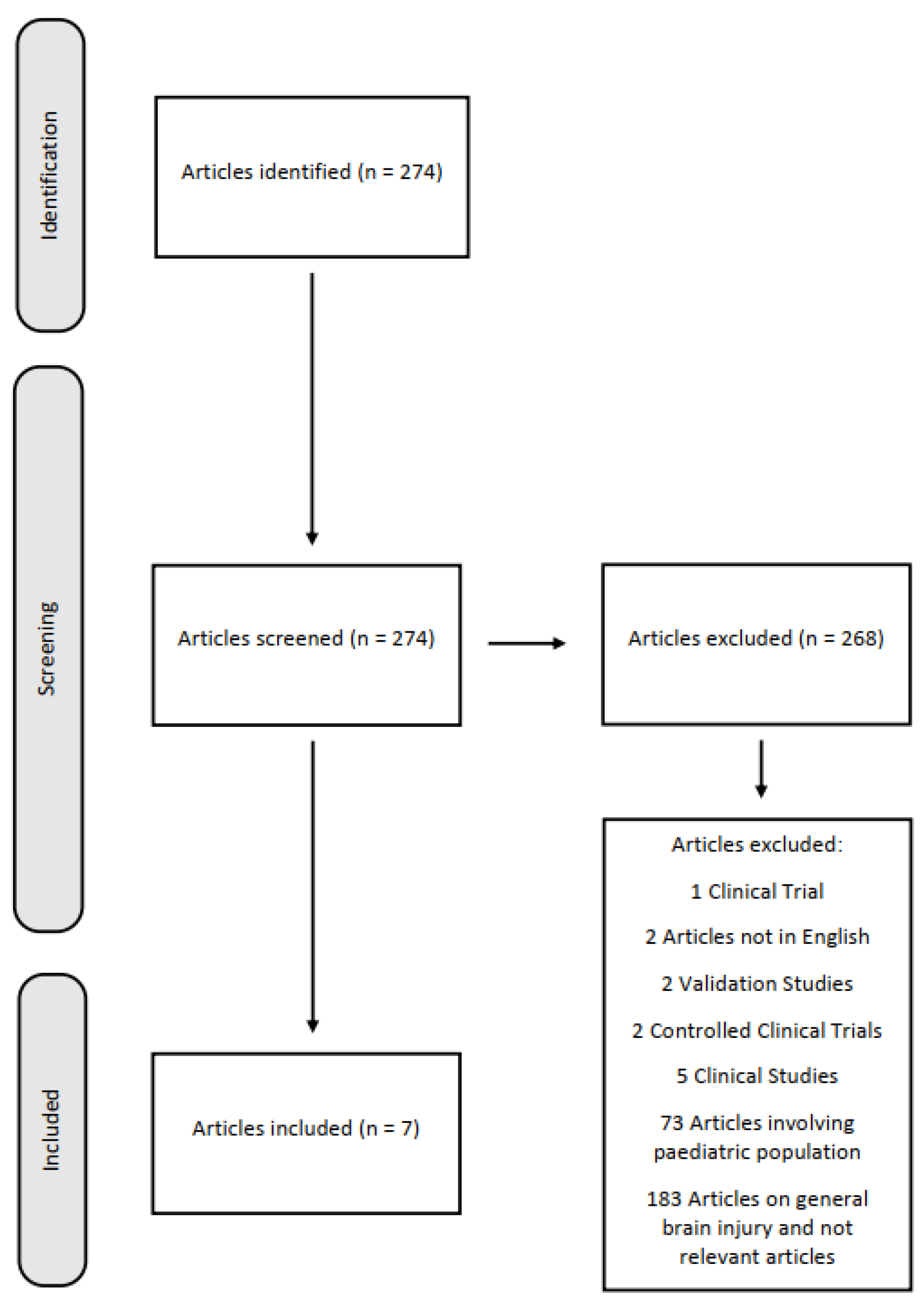
2. Materials and Methods

A review of currently published studies was performed for articles on the CTE secondary to professional and non-professional TBI by evaluating numerous variables, focusing on age at the time of trauma and histopathological studies, following the PRISMA criteria [25]. The literature search was conducted via PubMed and Scopus, and it was carried out for articles published from 2010 onward, a period marked by significant advancements in the understanding and characterization of CTE, which was less well-recognized and investigated prior to this timeframe using the following keywords terms: (chronic traumatic encephalopathy) AND (brain injury) AND (traumatic brain injury)).
We selected articles that discuss cases where the diagnosis of CTE was achieved through post-mortem examinations. In many of these cases, the diagnosis was unexpected, as it was initially believed that the underlying neuropathology, if present, was different. Inclusion criteria were: articles describing or investigating patients with a history of professional or non-professional TBI of any severity, and written in English. Exclusion criteria were: articles written in languages other than English, articles involving pediatric populations, clinical studies, clinical trials, controlled clinical trials, validation studies, and articles on general TBI. The initial electronic data search yielded a total of 274 potentially relevant studies on PubMed and Scopus. The articles were carefully evaluated based on their title, abstracts, and texts. Implementation of these procedures resulted in only 7 eligible papers for inclusion from the initial 274 articles within the search (Figure 1). All the included studies are reported in Table 2.

3. Results

Seven articles were included in the systematic review [3,4,5,8,20,26,27]. According to the examined literature, 29 cases of CTE diagnosed through histological examinations are described. One case was excluded because it was cited twice. Of the analyzed cases, 24 deceased were men (85.7%), while 4 deceased were women (14.2%). One deceased was a professional wrestler, one was an Australian rules football player, four were professional American football players (14.2%), three were military personnel (10.7%), and the professions of 18 were not reported. In 22 cases (78.5%), signs of neurological symptoms and cognitive and neuropsychiatric impairments were reported. Causes of death included: suicide by hanging in one case, myocardial infarction in five cases (17.8%), suicide by ingesting ethylene glycol in one case, suicide by gunshot wound to the head in one case, a traffic accident during a 40-mile-per-hour high-speed police chase in one case, acute pulmonary embolism in one case, glioblastoma in one case, alleged assault in one case, being struck by a motor vehicle in one case, COVID-19 infection in one case, homicide-related death in 4 cases (14.2%), death from complications of chronic alcoholism in two cases (7.1%), death due to smoke inhalation in one case, undetermined death in one case, death by choking on food in one case, death due to probable seizure in one case, death due to acute subdural hematoma in one case, death due to sepsis secondary to scalp wound infection in one case and deaths from multifactorial causes in two cases (7.1%). In all cases, pathognomonic tauopathy for CTE was found; in two cases, amyloid plaques were also found. A history of repeated head traumas due to sports and/or other forms of exposure was reported in 16 cases (57.1%). In two cases (7.1%), a history of head trauma in the context of longstanding intimate partner violence was reported. In one case the individual affected by CTE committed murder-suicide. Only one case reported the results of toxicological analyses.
Moreover, in 6 cases (21.4%), sole alcohol abuse was documented. Concomitant alcohol and polydrug abuse were observed in 4 cases (14.2%), while another 4 cases presented with alcohol abuse coupled with inhalational solvent abuse. A history of perinatal or infantile brain injury was noted in 3 cases, and 2 cases exhibited structural cerebral anomalies of genetic (or presumed genetic) etiology. Within this subgroup of 5, 2 individuals displayed longstanding behavioral disturbances characterized by repetitive cephalic impact over several decades. Excluding individuals with alcohol withdrawal-related seizures, epilepsy was identified in 3 of the total number of cases.
A significant risk of bias in this review stems from the substantial heterogeneity of the enrolled sample. The individuals included vary widely in terms of age, work activity, and other demographic factors, which could influence the presentation and diagnosis of CTE. Furthermore, the diverse modalities leading to death (e.g., suicide, accidents, medical conditions) and the varied instances of abuse or positivity for psychoactive substances and/or alcohol introduce considerable confounding variables. These differences make it challenging to draw consistent conclusions about the causal links between head trauma, substance use, and CTE, as the contributing factors and specific circumstances surrounding each case are highly variable.

4. Discussion

Based on the reviewed literature, most individuals diagnosed with CTE are male. This observation is broadly consistent with the known correlation between CTE and a history of repeated head trauma, an exposure more prevalent in professional and sports categories predominantly involving males. Sports like American football, boxing, and wrestling, alongside professions such as military service, frequently subject individuals to recurrent head impacts, which may contribute to a higher susceptibility in the male population [3,4,5,20,26]. While our current limited dataset, which includes only 4 female cases and two specifically linked to intimate partner violence, does not allow for a comprehensive statistical demonstration of gender prevalence, it highlights an important area for future investigation. The proportion of female patients affected by CTE may be underestimated, underscoring the need for more targeted studies focusing on female cases [28]. This is particularly relevant for victims of longstanding intimate partner violence [8], a group experiencing repeated head trauma whose inclusion in research could significantly enhance the understanding of CTE’s gender-related epidemiology and overall prevalence.
Furthermore, most of the examined patients exhibited neurological symptoms and neuropsychiatric impairments [3,4,20,26]. These symptoms include cognitive deficits, mood disorders, and behavioral changes, which collectively impact the everyday quality of life and functional abilities. These symptoms should serve as a warning sign for early diagnosis, especially in individuals with a history of repeated head traumas, both professional and non-professional, throughout their lives. Early identification of these symptoms could facilitate timely intervention, potentially slowing the progression of the disease and improving patient outcomes. Since these symptoms may mimic other neuropathological conditions (e.g., Alzheimer’s disease, Lewy body dementia, Parkinson’s disease), early diagnosis is crucial. Differential diagnosis through advanced imaging techniques and biomarker identification is essential to distinguish CTE from other neurodegenerative diseases [26].
Among the reported deceased, 25% died by suicide [3,4]. This finding is alarming, as undiagnosed CTE patients may exhibit self-harm, hetero-harm, and engage in criminal behaviors. While evidenced in two reported cases within this review, including a murder-suicide incident, this observation aligns with a growing body of literature highlighting the significant neuropsychiatric sequelae of CTE, which often include aggression, impulsiveness, depression, and executive dysfunction [29]. The prevalence of suicide and violent behavior in these cases underscores the severe psychological impact of CTE, necessitating comprehensive mental health support for individuals at risk. These extreme behavioral changes in individuals with CTE further emphasize the need for early diagnosis and intervention based on the broader clinical understanding of the disease. Implementing multidisciplinary care approaches that include neurological, psychological, and social support could mitigate the adverse outcomes associated with CTE. Despite this, it is important to highlight that suicide cases, and indeed all complex behavioral manifestations, require individual analysis to understand the specific contributions of various neuropsychiatric and psychosocial factors, as the relationship between neuropathology and complex behavior is multifactorial. Histopathological findings of CTE may add little incremental value to such determinations in isolation. Considerations regarding the impact of neurological injury on a specific suicide case are likely to be better informed by more extensive literature that generally describes the association between TBI and suicide [30], a known risk factor for CTE. It is of paramount importance to conduct well-designed, prospective, longitudinal, and large-scale studies to more definitively establish the existence and nature of any possible relationship between CTE and suicide and other violent behaviors [31].
Moreover, homicide-related deaths highlight the potential for aggressive or impulsive behaviors associated with CTE pathology. Deaths resulting from complications of chronic alcoholism and multifactorial causes underscore the significant comorbidities that may be present in individuals with CTE. Isolated instances of mortality were attributed to smoke inhalation, undetermined causes, choking on food, probable seizure, acute subdural hematoma, and sepsis secondary to scalp wound infection, suggesting a range of terminal events unrelated to the primary neurodegenerative process.
Toxicological analysis was available for only one case, limiting our ability to draw conclusions regarding the prevalence and impact of substance use on the clinical presentation and progression of CTE within this cohort [32]. However, the documented history of sole alcohol misuse in 21.4% of cases, concomitant alcohol and polydrug abuse in 14.2%, and alcohol misuse coupled with inhalational solvent abuse in another 14.2% suggests a substantial burden of substance use disorders in this population. The observed history of perinatal or infantile brain injury in 3 cases and structural cerebral anomalies of genetic (or presumed genetic) etiology in 2 cases indicates that pre-existing neurological vulnerabilities may potentially interact with the effects of repetitive head trauma in the development or manifestation of CTE. The finding of longstanding behavioral disturbances characterized by repetitive cephalic impact in two individuals within this subgroup warrants further exploration into the complex interplay between early life neurological factors and the subsequent development of CTE-related behaviors. The presence of epilepsy, excluding alcohol withdrawal seizures, in 3 of the total cases suggests that seizure disorders may be a relevant comorbidity in a subset of individuals with CTE [27].
Histopathologically, it is noteworthy that all the cases examined in this study exhibited lesions indicative of tauopathy pathognomonic for CTE. This significant finding confirms that, at present, the only definitive diagnosis of CTE is provided by autopsy. This evidence underscores the importance of conducting autopsies in individuals who, during their lifetime, exhibited symptoms compatible with CTE, especially if they belong to high-risk professional categories such as contact sport athletes or military personnel.
Indeed, from the analysis of the examined cases, a complex and varied neuropathological picture has emerged, with distinctive and recurring histopathological patterns. In the cases examined, a significant element that emerges is the presence of NFTs, which manifest with variable distributions and characteristics. One case highlighted NFTs and ghost tangles spread across multiple brain regions, involving the neocortex, subcortical ganglia, and substantia nigra, suggesting extensive involvement of the central nervous system.
Of particular interest, one observed case uniquely presented a combination of NFTs and tau-immunopositive neuritic threads co-occurring with amyloid plaques in the neocortex. This specific coexistence of alterations strongly suggests a potential interaction between different underlying pathological mechanisms. Conversely, four other cases clearly demonstrated NFTs and tau-immunopositive neuritic threads in various brain regions but notably lacked any evidence of amyloid plaques, thereby delineating a pathological process that is primarily tau-dependent.
Three cases showed isolated pathognomonic lesions of CTE in the underlying white matter, representing a specific diagnostic pattern of the pathology. Tau pathology characteristically distributed with a predilection for perivascular regions and sulcal depths. Some cases showed specific involvement of cortical areas, such as the dorsolateral frontal cortex.
One case exhibited a complex combination of tau pathology (with characteristic distribution), beta-amyloid plaques, and neuritic plaques, suggesting a possible overlap with other neurodegenerative processes.
The diverse pathological presentations observed suggest that CTE can manifest via various histopathological patterns, possibly indicative of differing disease stages or distinct pathological variants. Across these manifestations, the predominant unifying element is tau-related alterations, characterized by a specific anatomical distribution that stands as a hallmark of the pathology. Gaining this deeper understanding of CTE’s pathological patterns holds significant implications for advancing its future diagnosis and treatment strategies.
Overall, these findings stress the importance of awareness and research into CTE, particularly regarding its diagnosis. Such efforts are essential to develop effective prevention strategies and therapeutic interventions, ultimately reducing the incidence and impact of this debilitating disease.
This study is subject to several limitations. A significant constraint is the underrepresentation of female cases, with only four included in the dataset, two of which are specifically linked to intimate partner violence. This severely limits the ability to draw statistically robust conclusions regarding gender prevalence and potentially underestimates the proportion of affected female patients, highlighting a critical need for more targeted research on women. Furthermore, the analysis of associated factors like substance use is hampered by the limited availability of toxicological data, with only one case providing such information. This prevents a thorough assessment of the prevalence and impact of substance use disorders on CTE’s clinical presentation and progression within this specific cohort. While this study notes high rates of alcohol and polydrug misuse, the absence of comprehensive toxicological data precludes definitive conclusions. Additionally, the discussion on suicide and violent behavior, while alarming, acknowledges that histopathological findings alone may offer limited incremental value in understanding the multifactorial nature of these complex behaviors. Finally, while the histopathological findings consistently confirm tauopathy as pathognomonic for CTE, reinforcing the necessity of autopsy for definitive diagnosis, the diverse neuropathological presentations observed across cases, including overlaps with other neurodegenerative processes (e.g., amyloid plaques), suggest a complex and potentially heterogeneous disease, warranting further investigation into these varying patterns and their clinical implications.

5. Conclusions

Reinforcing current understanding, the definitive diagnosis of CTE remains contingent upon post-mortem neuropathological examination and the identification of pathognomonic tauopathy lesions. Consequently, the autopsy-confirmed diagnoses in all included cases ensure that this review’s findings are fully consistent with the established diagnostic gold standard.
The challenges of antemortem CTE identification are underscored by our analysis, as its symptoms often overlap with those of other neurodegenerative disorders, including Alzheimer’s disease, Lewy body dementia, and Parkinson’s disease. This inherent difficulty in clinical differentiation points to a significant potential for CTE underdiagnosis during a patient’s lifetime. Therefore, comprehensive autopsy protocols are of paramount importance for individuals with a history of repeated head trauma and a clinical presentation indicative of CTE.
This review, primarily examining documented CTE cases in men involved in contact sports, provided a valuable dataset. However, it lacked sufficient data to draw definitive conclusions regarding two key areas: early antemortem diagnostic markers and the prevalence of CTE in broader, less-studied populations, such as women or individuals without a history of athletic head trauma. Consequently, future research, particularly large-scale, prospective studies, is crucial to bridge these diagnostic gaps and gain a comprehensive understanding of CTE’s full spectrum across diverse demographics.
Ultimately, these findings highlight the critical need for ongoing collaboration among forensic pathologists, clinicians, and researchers. This partnership is essential to advance our understanding of CTE pathophysiology and develop effective antemortem diagnostic tools and treatments. Furthermore, healthcare institutions and sports organizations must continue promoting educational initiatives and adopting clear guidelines for managing suspected CTE cases, championing an integrated, multidisciplinary approach to mitigate the disease’s impact.

Author Contributions

Conceptualization, F.O. and S.D.S.; methodology, F.O. and C.C.; validation, L.C., A.C. and G.P.; formal analysis, F.O. and F.C.; resources, F.O., F.C., and R.B.; data curation, F.O.; writing—original draft preparation, F.O.; writing—review and editing, F.O., G.P., and L.C.; supervision, L.C. and S.D.S. All authors have read and agreed to the published version of the manuscript.

Funding

This research received no external funding.

Institutional Review Board Statement

Not applicable.

Informed Consent Statement

Not applicable.

Data Availability Statement

Data are contained within the article.

Conflicts of Interest

The authors declare no conflicts of interest.

Abbreviations

The following abbreviations are used in this manuscript:
TBITraumatic Brain Injury
CTEChronic Traumatic Encephalopathy
NFTsNeurofibrillary Tangles
NINDSNational Institute of Neurological Disorders and Stroke
NIBIBNational Institute of Biomedical Imaging and Bioengineering
TESTraumatic Encephalopathy Syndrome
TDP-43Transactive response DNA-binding protein 43

References

  1. Pinchi, E.; Frati, A.; Cipolloni, L.; Aromatario, M.; Gatto, V.; La Russa, R.; Pesce, A.; Santurro, A.; Fraschetti, F.; Frati, P.; et al. Clinical-Pathological Study on β-APP, IL-1β, GFAP, NFL, Spectrin II, 8OHdG, TUNEL, MiR-21, MiR-16, MiR-92 Expressions to Verify DAI-Diagnosis, Grade and Prognosis. Sci. Rep. 2018, 8, 2387. [Google Scholar] [CrossRef]
  2. Benevento, M.; d’Amati, A.; Nicolì, S.; Ambrosi, L.; Baj, J.; Ferorelli, D.; Ingravallo, G.; Solarino, B. Dura Mater and Survival Time Determination in Individuals Who Died after Traumatic Brain Injury: A Preliminary Study. Forensic Sci. Med. Pathol. 2025, 21, 107–114. [Google Scholar] [CrossRef]
  3. Omalu, B.I.; Fitzsimmons, R.P.; Hammers, J.; Bailes, J. Chronic Traumatic Encephalopathy in a Professional American Wrestler. J. Forensic Nurs. 2010, 6, 130–136. [Google Scholar] [CrossRef]
  4. Omalu, B.I.; Bailes, J.; Hammers, J.L.; Fitzsimmons, R.P. Chronic Traumatic Encephalopathy, Suicides and Parasuicides in Professional American Athletes: The Role of the Forensic Pathologist. Am. J. Forensic Med. Pathol. 2010, 31, 130–132. [Google Scholar] [CrossRef]
  5. Scanlon, M.M.; Shields, M.M.; Perl, D.P.; Priemer, D.S. Chronic Traumatic Encephalopathy Pathognomonic Lesions Occurring in Isolation Adjacent to Infiltrative and Non-Infiltrative White Matter Lesions. J. Neuropathol. Exp. Neurol. 2024, 83, 695–700. [Google Scholar] [CrossRef]
  6. Turk, K.W.; Budson, A.E. Chronic Traumatic Encephalopathy. Continuum 2019, 25, 187–207. [Google Scholar] [CrossRef]
  7. Byard, R.; Tiemensma, M.; Buckland, M.E.; Vink, R. Chronic Traumatic Encephalopathy (CTE)-Features and Forensic Considerations. Forensic Sci. Med. Pathol. 2023, 19, 620–624. [Google Scholar] [CrossRef] [PubMed]
  8. Tiemensma, M.; Byard, R.W.; Vink, R.; Affleck, A.J.; Blumbergs, P.; Buckland, M.E. Chronic Traumatic Encephalopathy (CTE) in the Context of Longstanding Intimate Partner Violence. Acta Neuropathol. 2024, 148, 1. [Google Scholar] [CrossRef] [PubMed]
  9. Katz, D.I.; Bernick, C.; Dodick, D.W.; Mez, J.; Mariani, M.L.; Adler, C.H.; Alosco, M.L.; Balcer, L.J.; Banks, S.J.; Barr, W.B.; et al. National Institute of Neurological Disorders and Stroke Consensus Diagnostic Criteria for Traumatic Encephalopathy Syndrome. Neurology 2021, 96, 848–863. [Google Scholar] [CrossRef] [PubMed]
  10. Eaton, R.G.; Lonser, R.R. History of Biological, Mechanistic, and Clinical Understanding of Concussion. Neurosurg. Focus 2024, 57, E2. [Google Scholar] [CrossRef] [PubMed]
  11. De Simone, S.; Vittorio, S.; Cipolloni, L.; Bibbò, R.; Gurgoglione, G.; Fazio, N.D.; Bosco, M.A. Men’s Suicide by Self-Abdominal Cut and Disembowelment: A Literature Review and Analysis of Three Cases. Available online: https://www.jomh.org/articles/10.31083/j.jomh1807158 (accessed on 17 June 2025).
  12. McKee, A.C.; Stein, T.D.; Huber, B.R.; Crary, J.F.; Bieniek, K.; Dickson, D.; Alvarez, V.E.; Cherry, J.D.; Farrell, K.; Butler, M.; et al. Chronic Traumatic Encephalopathy (CTE): Criteria for Neuropathological Diagnosis and Relationship to Repetitive Head Impacts. Acta Neuropathol. 2023, 145, 371–394. [Google Scholar] [CrossRef]
  13. Fesharaki-Zadeh, A. Navigating the Complexities of Traumatic Encephalopathy Syndrome (TES): Current State and Future Challenges. Biomedicines 2023, 11, 3158. [Google Scholar] [CrossRef]
  14. Saulle, M.; Greenwald, B.D. Chronic Traumatic Encephalopathy: A Review. Rehabil. Res. Pract. 2012, 2012, 816069. [Google Scholar] [CrossRef]
  15. Peacock, W.F.; Kuehl, D.; Bazarian, J.; Singer, A.J.; Cannon, C.; Rafique, Z.; d’Etienne, J.P.; Welch, R.; Clark, C.; Diaz-Arrastia, R. Defining Acute Traumatic Encephalopathy: Methods of the “HEAD Injury Serum Markers and Multi-Modalities for Assessing Response to Trauma” (HeadSMART II) Study. Front. Neurol. 2021, 12. [Google Scholar] [CrossRef] [PubMed]
  16. McKee, A.C.; Cantu, R.C.; Nowinski, C.J.; Hedley-Whyte, E.T.; Gavett, B.E.; Budson, A.E.; Santini, V.E.; Lee, H.-S.; Kubilus, C.A.; Stern, R.A. Chronic Traumatic Encephalopathy in Athletes: Progressive Tauopathy after Repetitive Head Injury. J. Neuropathol. Exp. Neurol. 2009, 68, 709–735. [Google Scholar] [CrossRef] [PubMed]
  17. Tharmaratnam, T.; Iskandar, M.A.; Tabobondung, T.C.; Tobbia, I.; Gopee-Ramanan, P.; Tabobondung, T.A. Chronic Traumatic Encephalopathy in Professional American Football Players: Where Are We Now? Front. Neurol. 2018, 9, 445. [Google Scholar] [CrossRef]
  18. Arciniega, H.; Baucom, Z.H.; Tuz-Zahra, F.; Tripodis, Y.; John, O.; Carrington, H.; Kim, N.; Knyazhanskaya, E.E.; Jung, L.B.; Breedlove, K.; et al. Brain Morphometry in Former American Football Players: Findings from the DIAGNOSE CTE Research Project. Brain 2024, 147, 3596–3610. [Google Scholar] [CrossRef]
  19. Omalu, B.; Bailes, J.; Hamilton, R.L.; Kamboh, M.I.; Hammers, J.; Case, M.; Fitzsimmons, R. Emerging Histomorphologic Phenotypes of Chronic Traumatic Encephalopathy in American Athletes. Neurosurgery 2011, 69, 173–183. [Google Scholar] [CrossRef]
  20. van Amerongen, S.; Kamps, S.; Kaijser, K.K.M.; Pijnenburg, Y.A.L.; Scheltens, P.; Teunissen, C.E.; Barkhof, F.; Ossenkoppele, R.; Rozemuller, A.J.M.; Stern, R.A.; et al. Severe CTE and TDP-43 Pathology in a Former Professional Soccer Player with Dementia: A Clinicopathological Case Report and Review of the Literature. Acta Neuropathol. Commun. 2023, 11, 77. [Google Scholar] [CrossRef]
  21. Castellani, R.J.; Perry, G. Tau Biology, Tauopathy, Traumatic Brain Injury, and Diagnostic Challenges. J. Alzheimer’s Dis. 2019, 67, 447–467. [Google Scholar] [CrossRef]
  22. Ruchika, F.; Shah, S.; Neupane, D.; Vijay, R.; Mehkri, Y.; Lucke-Wold, B. Understanding the Molecular Progression of Chronic Traumatic Encephalopathy in Traumatic Brain Injury, Aging and Neurodegenerative Disease. Int. J. Mol. Sci. 2023, 24, 1847. [Google Scholar] [CrossRef]
  23. Blaylock, R.L.; Maroon, J. Immunoexcitotoxicity as a Central Mechanism in Chronic Traumatic Encephalopathy—A Unifying Hypothesis. Surg. Neurol. Int. 2011, 2, 107. Available online: https://www.semanticscholar.org/paper/Immunoexcitotoxicity-as-a-central-mechanism-in-Blaylock-Maroon/83d4fb51df4b528715345e2d7d819f98c5bbcfcb (accessed on 27 July 2025). [CrossRef] [PubMed]
  24. Falcon, B.; Zivanov, J.; Zhang, W.; Murzin, A.G.; Garringer, H.J.; Vidal, R.; Crowther, R.A.; Newell, K.L.; Ghetti, B.; Goedert, M.; et al. Novel Tau Filament Fold in Chronic Traumatic Encephalopathy Encloses Hydrophobic Molecules. Nature 2019, 568, 420–423. [Google Scholar] [CrossRef]
  25. Page, M.J.; McKenzie, J.E.; Bossuyt, P.M.; Boutron, I.; Hoffmann, T.C.; Mulrow, C.D.; Shamseer, L.; Tetzlaff, J.M.; Akl, E.A.; Brennan, S.E.; et al. The PRISMA 2020 Statement: An Updated Guideline for Reporting Systematic Reviews. BMJ 2021, 372, n71. [Google Scholar] [CrossRef] [PubMed]
  26. Pearce, A.J.; Sy, J.; Lee, M.; Harding, A.; Mobbs, R.; Batchelor, J.; Suter, C.M.; Buckland, M.E. Chronic Traumatic Encephalopathy in a Former Australian Rules Football Player Diagnosed with Alzheimer’s Disease. Acta Neuropathol. Commun. 2020, 8, 23. [Google Scholar] [CrossRef]
  27. Del Bigio, M.R.; Krawitz, S.; Sinha, N. Chronic Traumatic Encephalopathy-Neuropathologic Change in a Routine Neuropathology Service: 7-Year Follow-Up. J. Neuropathol. Exp. Neurol. 2023, 82, 948–957. [Google Scholar] [CrossRef]
  28. De Paola, L.; Tripi, D.; Napoletano, G.; Marinelli, E.; Montanari Vergallo, G.; Zaami, S. Violence against Women within Italian and European Context: Italian “Pink Code”—Major Injuries and Forensic Expertise of a Socio-Cultural Problem: A Narrative Review. Forensic Sci. 2024, 4, 264–276. [Google Scholar] [CrossRef]
  29. Mez, J.; Daneshvar, D.H.; Kiernan, P.T.; Abdolmohammadi, B.; Alvarez, V.E.; Huber, B.R.; Alosco, M.L.; Solomon, T.M.; Nowinski, C.J.; McHale, L.; et al. Clinicopathological Evaluation of Chronic Traumatic Encephalopathy in Players of American Football. JAMA 2017, 318, 360–370. [Google Scholar] [CrossRef]
  30. Brenner, L.A.; Ignacio, R.V.; Blow, F.C. Suicide and Traumatic Brain Injury among Individuals Seeking Veterans Health Administration Services. J. Head Trauma Rehabil. 2011, 26, 257–264. [Google Scholar] [CrossRef]
  31. Wortzel, H.S.; Shura, R.D.; Brenner, L.A. Chronic Traumatic Encephalopathy and Suicide: A Systematic Review. BioMed Res. Int. 2013, 2013, 424280. [Google Scholar] [CrossRef] [PubMed]
  32. Piersanti, V.; Napoletano, G.; David, M.C.; Umani Ronchi, F.; Marinelli, E.; De Paola, L.; Zaami, S. Sudden Death Due to Butane Abuse—An Overview. J. Forensic Leg. Med. 2024, 103, 102662. [Google Scholar] [CrossRef] [PubMed]
Figure 1. Search strategy following PRISMA criteria.
Forensicsci 05 00033 g001
Table 1. Microscopic differences between Chronic Traumatic Encephalopathy and Alzheimer’s Disease [17].
CharacteristicChronic Traumatic EncephalopathyAlzheimer’s Disease
p-tau AggregatesAggregates of hyperphosphorylated tau (p-tau) in glial cells (e.g., astrocytes), neurons, and cell processes, within the vicinity of small vasculature and in the cortical sulci. Typically presents in an irregular, “spot-like” pattern in perivascular spaces. Also found in the superficial layer (II-III) of the cerebral cortex and in areas 2 and 4 of the hippocampus.The locations of p-tau NFTs should largely overlap with those of Aβ amyloid, as well as their presence in the hippocampus.
Neurofibrillary Tangles DimensionsNeurofibrillary Tangles are usually larger in size.Neurofibrillary Tangles are generally smaller in size.
Amyloid-Beta PlaquesAmyloid-Beta plaques may be found in a diffuse pattern in sporadic loci, or may be absent.The primary diagnostic criterion is the post-mortem finding of amyloid-beta plaques and neuritic amyloid plaques in a laminar distribution (in the middle frontal gyrus, the superior and middle temporal gyri, and the inferior parietal lobule). Secondary depositions may also be found in the cerebellum and basal ganglia.
TDP-43 AggregatesTransactive response DNA-binding protein 43 (TDP-43) aggregates are a common finding in the vast majority of Chronic Traumatic Encephalopathy cases, co-localized with p-tau neurofibrillary tangles. Sampling should first be taken from the amygdala and hippocampus.TDP-43 aggregates are present, but are not co-localized with neurofibrillary tangles.
Table 2. Characteristics of the analyzed papers.
AuthorsN. of CasesYear of PublicationSexAge of DeathProfessionNeurological Symptoms and Cognitive and Neuropsychiatric ImpairmentsCause of DeathNeuropathology FindingsToxicological AnalysisHistory of Traumatic Brain Injury/Clinical History
Omalu et al. [3]12010male40 years oldProfessional WrestlerReportedSuicide by hanging after killing his wife and sonNeurofibrillary Tangles and Ghost Tangles in the neocortex, subcortical ganglia, and substantia nigra.Positive for Alprazolam (blood), Hydrocodone (blood), and Testosterone (urine)Reported
Omalu et al. [4]520105 male50 years old
45 years old
44 years old
36 years old
40 years old
4 professional American football players
1 professional American wrestler
5 reported1 myocardial infarction
1 suicide by ingesting ethylene glycol
1 suicide by a gunshot wound of the head
1 died in traffic accident during a 40-mile-per-hour high-speed police chase
1 suicide by hanging
1 tau-immunopositive Neurofibrillary Tangles and neuritic threads in the neocortex, subcortical ganglia, and brainstem nuclei, accompanied by amyloid plaques in the neocortex;
4 tau-immunopositive Neurofibrillary Tangles and neuritic threads in the neocortex, subcortical ganglia, and brainstem nuclei without amyloid plaques.
Not reportedReported
Scanlon et al. [5]320243 male41 years old
46 years old
52 years old
1 former service member of the Navy
1 Service member in the Special Forces
1 Military veteran
Not reported1 acute pulmonary embolism
1 glioblastoma
1 acute myocardial infarction
3 isolated Chronic Traumatic Encephalopathy pathognomonic lesions adjacent to the underlying white matterNot reported1 motor vehicle accident and sustained a traumatic brain injury associated with loss of consciousness
1 combat deployment, participation in boxing
1 participation in wrestling and American Football in High School
Tiemensma et al. [8]220242 femaleFourth decade of life
Fifth decade of life
Not reportedNot reported1 alleged assault
1 struck by motor vehicle
1 perivascular foci of neuronal p-tau immunoreactivity at sulcal depths;
1 perivascular foci of neuronal p-tau immunoreactivity in left dorsolateral frontal cortex and at sulcal depths.
Not reported2 repeated head injury in the context of longstanding Intimate Partner Violence
Van Amerongen et al. [20]12023male63 years oldProfessional soccer playerReportedCOVID-19 infection1 frontal and parietal cortex,
neuronal tau pathology was found with predilection of sulcal depths and perivascular regions
Not reportedDecent had experienced multiple collisions that involved head impact playing soccer, at least once leading to loss of consciousness.
Pearce et al. [26]12020maleNinth decade of lifeAustralian rules football playerReportedNot reported1 Tau pathology was found with predilection of sulcal depths and perivascular regions; Beta-amyloid and neuritic plaquesNot reportedThe decedent had
played more than 350 first-grade matches of Australian Rule Football over
19 years.
Del Bigio et al. [27]16202314 male
2 female
2 35-year-olds
37 years old
42 years old
2 44-year-olds
2 46 years old
47 years old
2 50-year-old
51 years old
2 52-year-olds
54 years old
61 years old
Not reportedReported4 Homicide
1 Sudden death, complications of chronic alcoholism
1 Smoke inalation
1 Hypertrophic cardiomiopaty and congestive heart failure
2 Undetermined
1 Choked on food
1 Probable seizure + dilated cardiomyopathy
1 Acute subdural hematoma with minimal external evidence of head trauma
1 Sepsis secondary to scalp wound infection
1 Alcohol toxicity/hypothermia
1 Accidental head trauma + cardiac arrest + hypothermia
1 Ischemic heart disease + metastatic carcinoma in mediastinum
16 Chronic Traumatic Encephalopathy p-tau immunoreactivity ranging from a single small focus to widespread abnormalities
(7 high Chronic Traumatic Encephalopathy, 9 low Chronic Traumatic Encephalopathy)
Not reported5 Alcohol and drug abuse
1 Alcohol abuse, alcohol withdrawal seizures/post-traumatic epilepsy
3 Alcohol abuse
1 Short limb dwarfism, cognitive
delay, blind from ret-
inal detachments,
seizures
1 Alcohol, cocaine,
methamphetamine,
and marijuana use;
schizophrenia
1 Cerebral palsy with
spastic diplegia and
cognitive delay sec-
ondary to perinatal
hypoxia, suspected
fetal alcohol spectrum disorder
1 Seizure disorder since
infancy, long-time
abuse of solvents
and alcohol, Wer-
nicke-Korsakoff
syndrome
1 Hypoxic brain damage
at birth (breech pre-
sentation and cord
prolapse), cognitive
delay, seizure disor-
der; chronic alcohol
abuse; diabetes mel-
litus type 2
1 Headaches and several
Assaults
1 Abusive head trauma
at 10 months age,
mild hydrocephalus
(shunt nonfunction-
ing), cognitive delay,
chronic alcohol
abuse
Disclaimer/Publisher’s Note: The statements, opinions and data contained in all publications are solely those of the individual author(s) and contributor(s) and not of MDPI and/or the editor(s). MDPI and/or the editor(s) disclaim responsibility for any injury to people or property resulting from any ideas, methods, instructions or products referred to in the content.

