Abstract
Teeth are highly durable and useful in forensic identification and studying the impact of environmental factors could aid forensic investigations. Accurate post-mortem interval (PMI) estimation using dental evidence is critical in legal contexts, it requires further exploration. Aims: This study included a scoping review investigating macroscopic and microscopic changes in teeth in various simulated environments (Part 1) and an experimental study assessing changes in teeth and restorations exposed to distilled water, saline water, acidic soil, and alkaline soil (Part 2). Methods: The scoping review analysed publications from five databases using keywords such as ‘Teeth’, ‘Dental’, ‘Water’, ‘Soil’, ‘Acid’, and ‘Forensic.’ The experimental study involved 40 human teeth photographed before and after a 90-day exposure period to record shade variations and macroscopic changes. Results: Part 1: Twenty-six relevant articles from 10 countries (1987–2022) were reviewed, with most focusing on human teeth (77%), unrestored teeth (54%), macroscopic changes (46%), and high-temperature environments (53%). Part 2: Teeth in distilled water (G1) showed no shade variation. In saline water (G2), 60% of teeth decreased in shade. In acidic soil (G3), 40% showed an increased shade, while 50% showed a decreased shade. In alkaline soil (G4), 70% of teeth showed an increased shade. Restorations exhibited minimal changes across environments. Conclusions: Studies on the macroscopic changes because of high temperature on teeth and dental restorative material are popular. Teeth exposed to alkaline and acidic soil showed the most changes in the structure.
1. Introduction
Teeth, being more durable than the other biometric indicators, can be utilised by a forensic dentist in identification [1]. Dental tissues are typically maintained even when the corpse is skeletonised, decomposing, burnt, or dismembered. Thus, a better understanding of the resilience of natural teeth and the lifespan and durability of dental restorations are of important significance in the field of dentistry [2]. Researchers and dentists are continually working to appreciate the complicated interactions between different materials and the environment, and it is an area that has greater scope of research. It is advisable to imitate these settings to examine the macroscopic changes that take place inside dental structures and restorations when the human mouth is subjected to an extensive range of situations [3]. The study of how heat affects teeth, and restorative material can help establish the temperature the fire reached, investigate the use of an accelerator, and identify whether the individual was already deceased at the time of the fire [4]. Similarly, by studying the reactions of teeth and restorative material in different acids, a forensic investigator can establish the type of acid used and the duration that the deceased was immersed in the acid before further confirmation through biochemical tests [5]. In situations where a corpse is discovered drowned in water or buried, by investigating the colour changes in different restorative materials over time, the forensic expert may differentiate clearly between these restorative materials [6]. This distinction can allow for ease in comparing ante-mortem data with post-mortem data and enhance dental identification [7,8].
The post-mortem interval (PMI) is the interval between an individual’s death and the start of expert examinations of the corpse and is vital in cases that are violent or of a suspicious nature [9]. Constructing a timeline of possible events leading to death can help in finding or ruling out possible suspects [10]. Through the establishment of a timeline of thanatological changes due to the presence of dental remnants, dental findings can be considered an accessible and valuable tool in legal contexts and in finding the cause of death [7]. Though many methods exist for approximating PMI, their accuracy often necessitates an amalgamation of approaches, but teeth are not well explored [11]. Furthermore, there is a need to bridge the gap between research carried out in forensic institutions and the practical application of methods that closely resemble real cases [12]. As the efficiency of dental identification has been proved, there is a necessity to advance our knowledge of the specific components that affect the alteration, degradation, and preservation of dental structures and restorations under various environmental conditions [13,14]. This study, thus, aimed to investigate the effects of chemical exposures on teeth and restorations (dental materials) by a thorough assessment of available studies and experimental investigation.
2. Materials and Methods
2.1. Part-1
The suggested Preferred Reporting Items for Scoping reviews PRISMA guidelines (http://www.prisma-statement.org/scoping/ accessed on 12 May 2022) were followed in this review on the macroscopic and microscopic changes in teeth when subjected to different simulated environments. PubMed, Medline, SCOPUS, Web of Science, and Google Scholar were thoroughly searched, and published peer-reviewed publications were cross-referenced. The search included titles, abstracts, and full-text articles written in English and published up until 12 May 2022 Peer-reviewed publications in other languages, review articles, conference abstracts, book reviews, and editorials were excluded. Three groups (dental, environment, and use in forensic odontology) guided a combination of keywords using different search strings with the Boolean operators ‘AND’ and ‘OR’, as noted in Table 1.

Table 1.
Keywords and search strings used per group.
The extracted data were categorised according to six categories: the title, year of publication, and country, the environment, tooth classification (human or animal), tooth state (restored or unrestored), changes observed (yes or no), and type of research (macroscopic/microscopic/both). The results were quantitatively analysed through descriptive graphs using Microsoft Office Excel (2021).
2.2. Part-2
A total of 40 human teeth obtained from anonymous patients of a private dental clinic in Kerala, India, were exposed to saline water (Group-1; G1), distilled water (Group-2; G2), soil of acidic pH (Group-3; G3), and soil of alkaline pH (Group-4; G4). Each group received 10 teeth in different states, such as sound/decayed (n = 5) and restored (n = 5), as seen in Table 2.
Table 2.
Summary of the four tooth groups and respective conditions placed in different environments (teeth restored with composite, amalgam, and GIC—glass ionomer cement). Class I—caries affecting pits and fissures of posterior teeth, Class II—caries affecting proximal surface of posterior teeth, Class III—caries affecting proximal surface of anterior teeth, Class IV—caries affecting proximal including incisal angles of anterior teeth.
Four solid plastic containers of 15 L capacity were used. One container with distilled water and one container with saline water with the salinity of 33–37 g per litre (simulating the salinity of seawater) were used to create the drowning environment. The burial environment was created using one plastic container containing soil of acidic pH using acetic acid (CH3COOH) with a pH of 2–3, while the other contained soil of alkaline pH that was made alkaline using sodium hydroxide (NaOH) with a pH of 12–13. The soil used was Westland Multi-purpose Topsoil®, which is a rich clay loam soil.
First, photographs of the teeth in multiple views (occlusal, mesial, distal, labial, lingual) were obtained using a LEICA S9i microscope before exposure to the simulated environments for 90 days, from December 2022 until March 2023, and post-study. For both teeth and tooth-coloured restorations such as composite and GIC (glass ionomer cement), the change in shade was recorded using the VITA classical shade guide according to the Shade Guide Unit (Vita Zahnfabrik, Bad Säckingen, Germany, Version 002) before and after the experiment. The teeth were photographed by a single operator with the selected shade by their side using daylight. A comparison of shades in dental structure and dental material before and after exposure to the environment was quantified according to the number of shades changed. For this, a value scale labelled with the numbers 1 through 16 and a stand for the Shade Guide Unit (SGU) was created [15], as seen in Table 3. For instance, a tooth considered B2 whose shade changed to C1 would have moved 3 shades up and was noted as +3, while a tooth considered D4 whose shade changed to C1 would have moved 2 shades down and was noted as −2. Any tooth where no shade changes were seen was noted as 0. Changes in opacity and brightness were analysed descriptively and noted. For amalgam restorations, the changes in the shine and appearance were noted, but no guideline was available. Results were quantitatively analysed using Microsoft Excel 2021 (Redmond, WA, USA).
Table 3.
VITA classical shade guide and each tab with comparable Shade Guide Unit (SGU), with B1 being the first shade and C4 the darkest shade [15].
3. Results
For the scoping review, 123 articles were identified in the search through databases, with 55 articles in Scopus, 38 in PubMed, 29 in Web of Science, 1 in LILACS, and 0 in SciELO. An initial automatic duplicate deletion removed 25 articles, followed by a manual duplicate deletion of 17 articles after screening and of the 81 reports sought for retrieval, 2 were not retrieved. Following the assessment of the 79 remaining articles, 53 were removed as they did not fit the eligibility criteria, and the remaining 26 research articles were analysed for quantitative data, as shown in Figure 1.
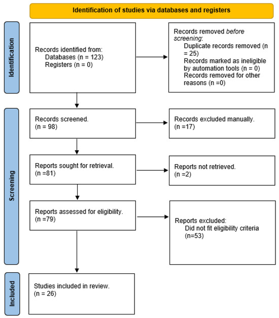
Figure 1.
PRISMA flow diagram.
Studies were conducted in 10 countries with India (n = 9; 35%) being the country with the highest number followed by Brazil (n = 6, 23%); the publication date of the research articles retrieved ranged from 1987 to 2022, with the most articles (n = 3, 12%) published in 2020. Changes to the teeth were noted in 92% (n = 24) of the articles analysed. It was noted that most of the research conducted was macroscopic in nature (n = 12, 46%) followed by both macroscopic and microscopic changes (n = 9, 35%) and then microscopic (n = 5, 19%). In macroscopic changes, colour stability, roughness, and Knoop’s microhardness were commonly studied.
The most-seen simulated environment in the research articles retrieved was that of high temperature (n = 14; 53%) followed by acid (n = 6; 23%). Simulated drowning and burial were uncommon, with only 11% of articles (n = 3) outlining the changes to teeth in burial environments and 15% of articles (n = 4) on drowning, while articles regarding the changes to teeth in low temperatures was the least commonly seen (n = 2; 8%). The most-studied acids were hydrochloric acid, nitric acid, and sulfuric acid.
Unrestored teeth were used in most studies (n = 14, 54%), while both restored and unrestored teeth were used in four articles. Of the teeth used in these studies, human teeth were most used (n = 20; 77%), and the animal teeth (n = 6, 23%) used were bovine. The commonly seen restorations in these restored teeth were silver amalgam, composite restoration, and glass ionomer cement. Further details are outlined in Table 4.
Table 4.
Overall, extracted data was categorised according to six categories.
Considering the results of the experimental study for changes in tooth structure, the colour of 60% of the teeth in distilled water (G1) showed no shade variation, and only three of the teeth moved down in shade. In saline water (G2), it is noted that most of the teeth (60%) decrease a shade and show dry white patches along the tooth surface. In acidic soil (G3), 40% of the teeth showed an increase in shade, while 50% showed a decrease. Distinct brown patchy discolouration was noted along the root surface, and yellowish-brown patches with whitish borders were observed on mainly labial crown surfaces of teeth. In alkaline soil (G4), 70% of the teeth showed an increase in shade. Figure 2 illustrates the overall shade changes in tooth structure in each environment.
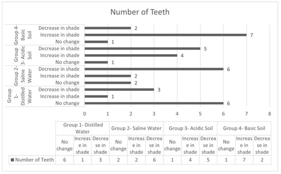
Figure 2.
Graph outlining the shade changes in tooth structure in each environment.
Considering the changes in the tooth-coloured restorations, no significant change in shade was seen in distilled and saline water (G1 and G2, respectively). The shade decreased in two of the three tooth-coloured restorations in the acidic soil (G3). In alkaline soil (G4), all tooth-coloured restorations decreased in shade, as seen in Figure 3. The overall distribution is seen in Table 5, and a visual representation is presented by Figure 4, Figure 5 and Figure 6.
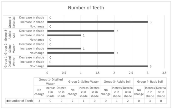
Figure 3.
Graph outlining the shade changes in tooth-coloured restorations in each environment.
Table 5.
A table showing the colour change in each tooth and tooth-coloured restoration. The main changes are highlighted in grey colour (+ indicates an increase in shade, − indicates a decrease in shade, 0 indicates no colour change, * denotes an amalgam-restored tooth, n/a means not applicable).
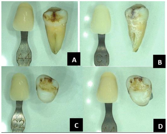
Figure 4.
Group 2, saline water: tooth number nine on labial view showed a decrease of nine shades from A4 to C1 following exposure (A,B). The colour of the composite restoration on the same tooth, as seen in the occlusal view, increased eight shades from C2 to A4 (C,D).
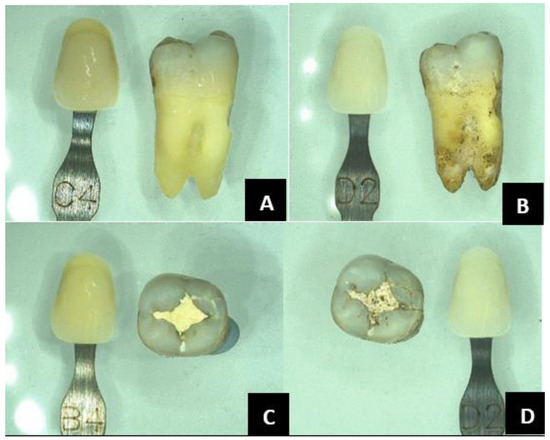
Figure 5.
Group 3, acidic soil: tooth number nine on labial view showed a decrease of twelve shades from C4 to D2 following exposure (A,B). The colour of the restoration on the same tooth, as seen in occlusal view, decrease nine shades from B4 to D2 (C,D).
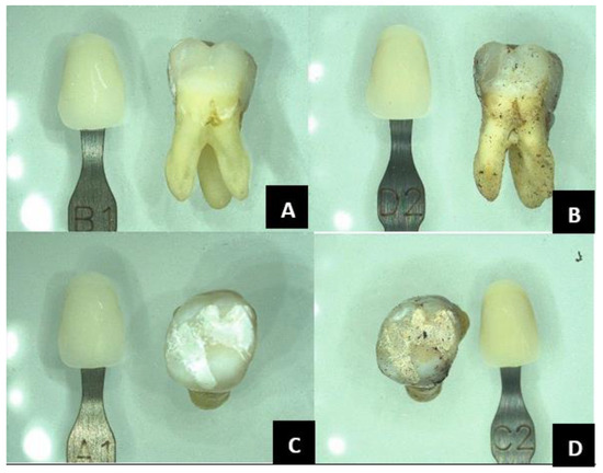
Figure 6.
Group 4, alkaline soil: tooth number two on labial view showed an increase of three shades from B1 to D2 following exposure (A,B). The colour of the restoration on the same tooth, as seen in the occlusal view, increases five shades from A1 to C2 (C,D).
4. Discussion
The first part of this study reported a larger number of studies on teeth and dental materials exposed to high temperatures (53%). This is important because burned skeletal remains can be recovered from a variety of situations, such as transportation accidents involving trains and aeroplanes, suicides, environmental catastrophes, house fires, and terrorist attacks [16,17]. Hence, examining how heat affects teeth and restorations can assist in determining the temperature reached by the fire [3]. According to earlier studies on mass disasters, between 34% and 89% of fire victims’ identities could be verified by dental techniques [18]. An acidic environment was the next most frequently investigated. The applicability could be that the morphological changes in teeth and various restorative materials can aid the forensic investigator in determining how long has passed after the body was submerged in acid, for instance [19]. Furthermore, the incidence of acid assaults is currently highest in various developing countries, with most acid assaults primarily focused on the victim’s face and neck [20]. It was also noted that all six articles on the effects of acid on teeth originated from India, with a growing concern in South Asia regarding acid attacks. They are more likely to utilise an acid that is readily available, affordable, and capable of swiftly destroying the body, and the most widely used acids are HCl, HNO3, and H2SO4 [7].
Twenty of the articles used human teeth, making it the most used for the experimental studies. The use of human teeth has led to more accurate results, but there may be ethical concerns and a lack of homogeneity. It was observed that bovine teeth were the commonly used animal teeth since they are more readily available than human teeth [21]. Aside from the bioethical concerns, bovine teeth facilitate sample standardisation, as they have a homogenous and known history [22], and reduce infection risk [23]. With interest in this discipline increasing significantly over the past 15 years or more, forensic odontology is expanding in India, which has led to the greatest number of articles collected from that country (35%) [24]. articleMost articles noted macroscopic changes, which could signify the relative ease in observing macroscopic changes compared to microscopic changes, which require expensive equipment that is not readily available to all.
The human body has known reactions to death, including morphological changes brought on by taphonomic occurrences like decomposition, skeletonization, and mummification [1]. These reactions are extensively studied in forensic medicine due to their significance in determining the cause of death. Similarly, these thanatological results must be thoroughly examined and recorded by the forensic dentist, but there have been relatively fewer investigations on the effects of soil and water on teeth and restorative materials [25].
Our scoping review found gaps in the research on environmental effects on dental structures. While high temperature was extensively studied (53%), other conditions like burial (11%), simulated drowning (15%), and low temperatures (8%) were underexplored. Research on alkaline and aqueous environments was also limited despite their forensic significance. To address this, we designed an experimental study focusing on underrepresented conditions, particularly burial and water exposure. These environments hold forensic relevance as they are frequently met in drowning and burial cases. By aligning with our review findings, this study aims to bridge research gaps and enhance forensic applicability beyond high-temperature effects. The scoping review was limited in detailed statistical analysis and could not produce in depth quantitative analysis, the selection of studies depended on predefined inclusion/exclusion criteria, which may have introduced selection bias and affected the comprehensiveness of the review.
The second part of this study showed noticeable changes in colour and texture in teeth and tooth-coloured restorative material that were buried in acidic and alkaline soil. That could be useful in finding the area in which a corpse may have been buried; this microenvironment typically starts or slows the decomposition pace, produces signals, and leaves remnants that reveal the nature and circumstances of the soil at the burial site [26]. The phenomenon of pink teeth, for instance, is still up for debate as a probable dental modification associated with damp or moist surroundings and is another common thanatological consequence of individuals submerged in water during or after death in a drowning occurrence. It has also been observed that people whose teeth have been in water for a specific period change colour to a reddish-brown hue [27].
The study utilised 40 human teeth sourced from a single clinic in Kerala, India. This controlled sample reduces confounding variables and allows for a focused analysis of environmental effects. This pilot study primarily aimed to explore broad environmental influences rather than individual variations, as well as to act as a springboard for future research highlighting key trends that warrant further exploration. Future studies will incorporate a larger and more diverse sample, considering variations in tooth type, age, and pathology to enhance statistical robustness and forensic applicability.
The results also showed that the colour of 60% of the teeth in distilled water showed no colour variations, while 30% showed a decrease in shade. In saline water, it is noted that 60% of the teeth showed a decrease in shade, and dry white patches appeared along the tooth surface. This can indicate that teeth from bodies recovered from more saline bodies of water may appear drier and have white patches. Freshwater environments like lakes and rivers have lower salinity compared to marine environments [28]. It was also noted that, in acidic soil, 40% of the teeth moved up in shade, while 50% of the teeth moved down in shade. Distinct brownish patchy discolouration can be identified along the root surface of the teeth recovered from acidic soil that also showed yellowish-brown patches with whitish borders on the crown, primarily labially, that can help distinguish whether a recovered body may have been buried in acidic or alkaline soil.
According to Manoilescu et al. (2015), dentin and enamel may be eroded by the acids that seep into the ground from a variety of sources, including agricultural and industrial wastes, etc. On the labial surface of the crown, close to the neckline, where the enamel is weaker, erosion spots on the enamel are often brown and tend to appear frequently [29]. In this study, 70% of the teeth in alkaline soil moved up in colour along with the tooth-coloured restorations, which also showed the same changes. In the UK, acid soils are more common than alkaline soils and may be found covering huge areas of heathland and coniferous woods. Alkaline soils are often those that sit on top of a bedrock of chalk or limestone [29].
This study indicates that changes can be observed in durations as short as three months. The experimental period for this study was set at 90 days, aligning with forensic recovery timelines and previous research. Vincenti et al. (2018) [7] observed significant colour changes in composite resin and glass ionomer cement within three months of burial, demonstrating that key alterations occur within this timeframe. This study aims to employ short-term experimental periods to assess early-stage changes in dental structures and materials. Future research could explore longer durations to assess progressive and late-stage alterations in dental materials under varied environmental conditions
According to the study conducted by Vincenti SA et al. (2018), after one year of burial and six months of submersion in water, composite resin changed colour considerably differently from GIC and, while the brightness appeared to decline after the first month, the most notable colour changes were noticed after three months and persisted until 12 months following burial [7]. Similarly, de Almeida Salema et al. (2020) also discovered that, depending on the duration of burial, teeth with silver amalgam, composite resin, and glass ionomer cement restorations buried in mangrove habitats exhibit different changes in Knoop microhardness, surface roughness, and colour characteristics [13].
In this study, we used the VITA classical shade guide as it is a widely accepted tool for assessing macroscopic colour changes. Our approach aimed to bridge the gap between forensic research and real-world application by using methodologies that are both scientifically rigorous and practically feasible by avoiding complex methods that may not be readily accessible in routine investigations. Future studies could incorporate advanced techniques to enhance precision while maintaining relevance to forensic casework.
Oliveira Fernandes AP et al. (2021) discovered that drowning conditions can change the radiopacity of endodontically treated teeth, particularly in the middle and cervical thirds, but no evidence has been found to suggest that this also occurs in burial situations. These findings may prove helpful in forensic investigations as a first indication of the setting in which the corpse was exposed to or situated [30]. When corpses are discovered buried or immersed in water, the investigation of the colour change in dental restorative materials and teeth over time may aid forensic experts work by revealing details that enable the differentiation of the materials found in the oral cavity.
The teeth in our study were directly exposed to the environments, as opposed to teeth protected by the lips and cheeks when bodies are buried or drowned, which presents a limitation to the study. The close contact of teeth with various surroundings causes an intensive environmental effect on the dental surface that may overestimate the changes to the teeth. This study focused on environmental impacts in isolation, which is useful in forensic cases where only teeth are found exposed or partially buried. However, we recognise the need for a more realistic experimental design that mimics forensic scenarios. Future research will incorporate cadaveric tissue or artificial simulants to replicate soft tissue protection, enhancing forensic applicability.
In practice, forensic dentists might not receive the information about the base shade ‘ante-mortem’. A larger sample size and increasing exposure time of the teeth to different elements are suggested for further research.
5. Conclusions
Earlier studies proved that tooth tissues undergo unique macroscopic and microscopic alterations in different simulated environments. The greatest number of studies were conducted on the effect of high temperature on teeth and dental restorative material, and macroscopic changes in human teeth. Although there has been a recent rise in the number of scientific articles published on the subject, further research is needed.
Teeth exposed to alkaline and acidic soil showed the most changes in the structure. In alkaline soil, the teeth and tooth-coloured restorations showed a uniform increase in shade. Overall, few changes were observed in dental materials. The teeth retrieved from acidic soil also displayed distinct brownish patchy discoloration and yellowish-white areas. Teeth in saline water showed a decrease in shade and presented dry white patches. These findings should enlighten forensic professionals and encourage more experimental research, especially considering a longer exposure time.
Author Contributions
Conceptualisation, P.K.G., S.M. and H.P.; methodology, P.K.G. and S.M.; validation, P.K.G., S.M. and H.P.; formal analysis, P.K.G.; writing—original draft preparation, P.K.G.; writing—review and editing, S.M. and H.P.; supervision, S.M. and H.P. All authors have read and agreed to the published version of the manuscript.
Funding
This research received no external funding.
Institutional Review Board Statement
Ethical review and approval were waived for this study due to the use of extracted human teeth that were donated specifically for research purposes. The teeth were obtained with informed consent from the patients, ensuring they fully understood the purpose of the donation and its use in scientific research. No additional interventions or procedures were conducted on the donors solely for the purpose of the study, and no personal or identifying information was collected, ensuring the anonymity and privacy of the donors.
Informed Consent Statement
Informed consent was obtained from all subjects involved in the study. Patients who donated their extracted teeth were fully informed about the purpose of the research and its scientific significance. They were provided with detailed information regarding the use of their donated teeth, including assurances of anonymity and confidentiality. Participation was entirely voluntary, and donors had the right to withdraw their consent at any time before the use of the donated teeth in research.
Data Availability Statement
No new data were created or analyzed in this study. Data sharing is not applicable to this article.
Acknowledgments
The authors would like to express their sincere gratitude to the supporting staff at the University of Dundee for their invaluable help and support: Scott McGregor, librarian, Valerie Wilson and Claire Cunnigham, laboratory technicians.
Conflicts of Interest
The authors declare no conflicts of interest.
References
- Adams, B.J. Establishing personal identification based on specific patterns of missing, filled, and unrestored teeth. J. Forensic Sci. 2003, 48, 487–496. [Google Scholar] [CrossRef] [PubMed]
- Weedn, V.W. Postmortem identifications of remains. Clin. Lab. Med. 1998, 18, 115–137. [Google Scholar] [CrossRef]
- Bush, M.A.; Bush, P.J.; Miller, R.G. Detection and classification of composite resins in incinerated teeth for forensic purposes. J. Forensic Sci. 2006, 51, 636–642. [Google Scholar] [CrossRef] [PubMed]
- Muller, M.; Berytrand, M.F.; Quatrehomme, G.; Bolla, M.; Rocca, J.P. Macroscopic and microscopic aspects of incinerated teeth. J. Forensic Odontostomatol. 1998, 16, 1–7. [Google Scholar] [PubMed]
- Kadashett, V.; Shivakumar, K.; Baad, R.; Vibhute, N.; Belgaumi, U.; Bommanavar, S.; Kamate, W. Effect of concentrated acids on teeth: A forensic approach; An In-vitro study. J. Datta Meghe Inst. Med. Sci. Univ. 2021, 16, 283–289. [Google Scholar] [CrossRef]
- Biancalana, R.C.; Vicente, S.A.; Alves da Silva, R.H.; Pires-de-Souza, F.C. Color Stability of Dental Restorative Materials Submitted to Heat Sources, for Forensic Purposes. J. Forensic Sci. 2017, 62, 355–360. [Google Scholar] [CrossRef]
- de Freitas Vincenti, S.A.; Biancalana, R.C.; Alves da Silva, R.H.; De Carvalho Panzeri Pires-de-Souza, F. Colour stability of dental restorative materials submitted to conditions of burial and drowning, for forensic purposes. J. Forensic Odontostomatol. 2018, 36, 20–30. [Google Scholar]
- Biancalana, R.C.; Freitas Vincenti, S.A.; Alves da Silva, R.H.; Carvalho Panzeri Pires-de-Souza, F. Color stability of dental restorative materials submitted to cold temperatures for forensic purposes. J. Forensic Leg. Med. 2017, 51, 63–68. [Google Scholar] [CrossRef]
- Ugrappa, S.; Jain, A. An Emergence of Dental Tissues in the Forensic Medicine for the Postmortem Interval Estimation: A Scoping Review. J. Forensic Sci. Med. 2021, 7, 54–60. [Google Scholar] [CrossRef]
- Kaliszan, M.; Hauser, R.; Kernbach-Wighton, G. Estimation of the time of death based on the assessment of post mortem processes with emphasis on body cooling. Leg. Med. 2009, 11, 111–117. [Google Scholar] [CrossRef]
- Merriam, T.; Kaufmann, R.; Ebert, L.; Figi, R.; Erni, R.; Pauer, R.; Sieberth, T. Differentiation of dental restorative materials combining energy-dispersive X-ray fluorescence spectroscopy and post-mortem, C.T. Forensic Sci. Med. Pathol. 2018, 14, 163–173. [Google Scholar] [CrossRef] [PubMed]
- Sampaio-Silva, F.; Magalhães, T.; Carvalho, F.; Dinis-Oliveira, R.J.; Silvestre, R. Profiling of RNA degradation for estimation of post mortem [corrected] interval. PLoS ONE 2013, 8, e56507. [Google Scholar] [CrossRef] [PubMed]
- de Almeida Salema, C.F.B.; de Barros Silva, P.G.; da Costa Oliveira, P.M.; Lima, J.P.M.; da Silva, R.H.A.; Nobre, T.F.G.; Bezerra, T.P. Forensic study of mechanical properties of dental restoration after burial in mangrove environment. Forensic Sci. Int. 2020, 308, 110166. [Google Scholar] [CrossRef] [PubMed]
- Whittaker, D.K.; MacDonald, D.G. A Colour Atlas of Forensic Dentistry; Wolfe Medical Publications: London, UK, 1989. [Google Scholar]
- Chu, S.J.; Paravina, R.D.; Sailer, I.; Mieleszko, A.J. Color in Dentistry: A Clinical Guide to Predictable Esthetics; Quintessence Publishing: Berlin, Germany, 2017. [Google Scholar]
- Ubelaker, D.H. The forensic evaluation of burned skeletal remains: A synthesis. Forensic Sci. Int. 2009, 183, 1–5. [Google Scholar] [CrossRef]
- Delattre, V.F. Burned beyond recognition: Systematic approach to the dental identification of charred human remains. J. Forensic Sci. 2000, 45, 589–596. [Google Scholar] [CrossRef]
- Kolude, B.; Adeyemi, B.F.; Taiwo, J.O.; Sigbeku, O.F.; Eze, U.O. The role of forensic dentist following mass disaster. Ann. Ib. Postgrad. Med. 2010, 8, 111–117. [Google Scholar] [CrossRef][Green Version]
- Seethapathy, T.; Shylaja, S.; Sekhar, M.S.M.; Manthapuri, S.; Ramanand, O.V.; Reddy, S.K.; Paul, J.B.; Funato, A.; Kubo, K.; Maeda, H.; et al. Effect of acids on teeth and restorative materials: An aid in forensic odontology. J. Hard Tissue Biol. 2019, 28, 21–30. [Google Scholar] [CrossRef]
- Cleary, M.; Visentin, D.C.; West, S.; Say, R.; McLean, L.; Kornhaber, R. Acid burn attacks: Looking beneath the surface. J. Adv. Nurs. 2018, 74, 1737–1739. [Google Scholar] [CrossRef]
- Biancalana, R.C.; Vincenti, S.A.d.F.; da Silva, R.H.A.; Pires-de-Souza, F.C.P. Analysis of the surface roughness and microhardness of dental restorative materials exposed to heat sources and cold temperatures for human identification purposes. Egypt. J. Forensic Sci. 2019, 9, 8. [Google Scholar] [CrossRef]
- Zimmer, S.; Kirchner, G.; Bizhang, M.; Benedix, M. Influence of various acidic beverages on tooth erosion. Evaluation by a New Method. PLoS ONE 2015, 10, e0129462. [Google Scholar] [CrossRef]
- Wang, C.; Li, Y.; Wang, X.; Zhang, L.; Tiantang; Fu, B. The Enamel Microstructures of Bovine Mandibular Incisors. Anat. Rec. 2012, 295, 1698–1706. [Google Scholar] [CrossRef] [PubMed]
- Acharya, A.B. Reflections on setting up forensic odontology department, its activities, and faculty. J. Forensic Dent. Sci. 2019, 11, 167–168. [Google Scholar] [CrossRef] [PubMed]
- Hau, T.C.; Hamzah, N.H.; Lian, H.H.; Hamzah, S. Decomposition process and post mortem changes. Sains Malays. 2014, 43, 1873–1882. [Google Scholar]
- Janaway, R.C.; Wilson, A.S.; Díaz, G.C.; Guillen, S. Taphonomic Changes to the Buried Body in Arid Environments: An Experimental Case Study in Peru. In Criminal and Environmental Soil Forensics; Ritz, K., Dawson, L., Miller, D., Eds.; Springer: Dordrecht, The Netherlands, 2009; pp. 341–356. [Google Scholar] [CrossRef]
- Franco, A.; de Oliveira, M.N.; Gomes-Lima, L.K.; Pereira-de-Oliveira, V.H.F.; Franco, R.; Blumenberg, C.; Silva, R.F.; da Silva, R.H.A.; Makeeva, I.; Santos-Filho, P.C.F.; et al. Case-specific characteristics of pink teeth in dental autopsies—A systematic review. J. Forensic Leg. Med. 2019, 68, 101869. [Google Scholar] [CrossRef]
- Cañedo-Argüelles, M.; Kefford, B.; Schäfer, R. Salt in freshwaters: Causes, effects and prospects—Introduction to the theme issue. Philos. Trans. R. Soc. Lond. B Biol. Sci. 2018, 374, 20180002. [Google Scholar] [CrossRef]
- Manoilescu, I.; Ion, A.; Ioan, B.G. Post-Mortem Changes in Teeth- Forensic Issues. Int. J. Med. Dent. 2015, 5, 249–252. [Google Scholar]
- Oliveira Fernandes, A.P.; Jacometti, B.V.; de Carvalho Panzeri Pires de Souza, F.; Alves da Silva, R.H. Radiographic changes in endodontically treated teeth submitted to drowning and burial simulations: Is it a useful tool in forensic investigation? J. Forensic Odontostomatol. 2021, 1, 9–15. [Google Scholar]


Disclaimer/Publisher’s Note: The statements, opinions and data contained in all publications are solely those of the individual author(s) and contributor(s) and not of MDPI and/or the editor(s). MDPI and/or the editor(s) disclaim responsibility for any injury to people or property resulting from any ideas, methods, instructions or products referred to in the content. |
© 2025 by the authors. Licensee MDPI, Basel, Switzerland. This article is an open access article distributed under the terms and conditions of the Creative Commons Attribution (CC BY) license (https://creativecommons.org/licenses/by/4.0/).