Case Report

Migratory Flows and Endomyocardial Fibrosis: A Mysterious Disease in Western Countries

Section of Legal Medicine, Department of Interdisciplinary Medicine, University of Bari, Piazza Giulio Cesare 11, 70124 Bari, Italy
*
Author to whom correspondence should be addressed.
Forensic Sci. 2025, 5(1), 11; https://doi.org/10.3390/forensicsci5010011
Submission received: 28 December 2024 / Revised: 22 January 2025 / Accepted: 27 February 2025 / Published: 4 March 2025

Abstract

:
Background—Endomyocardial fibrosis (EMF) is a chronic restrictive cardiomyopathy endemic to tropical and subtropical regions, characterized by fibrotic thickening of the endocardium, predominantly affecting the ventricular chambers. As global migration increases, cases of EMF may emerge in non-endemic areas, posing a diagnostic challenge for healthcare professionals unfamiliar with this condition. Methods—We report the case of a 21-year-old man of African origin who experienced sudden collapse while residing in a reception center for asylum seekers in Apulia, Southeastern Italy. His medical history revealed that one week prior, he had been presented with high fever, chest pain, and asthenia, symptoms initially misinterpreted as influenza by a local physician, leading to antibiotic therapy. Results—The definitive diagnosis of EMF was established post-mortem through histological examination, which identified characteristic endocardial fibrosis. This case exemplifies the re-emergence of neglected tropical cardiac diseases in Western countries due to increased migration from endemic regions. Conclusions—This case highlights the need for heightened clinical awareness among healthcare professionals, particularly in cardiology and primary care, to recognize and manage geographically displaced diseases such as EMF. Implementing enhanced medical education and diagnostic protocols is essential to address the evolving epidemiological landscape and improve early identification of rare but significant cardiac pathologies in migrant populations.

1. Introduction

Endomyocardial fibrosis (EMF) is a perplexing and enigmatic tropical cardiovascular disease. Epidemiological studies identify EMF as one of the most prevalent forms of restrictive cardiomyopathy in Sub-Saharan Africa. For this reason, it is also called “tropical endomyocardial fibrosis”. EMF also has a high frequency in Asia (especially in India and China) and South America (especially in Brazil and Colombia) [1]. The first documented cases date back to 1948 in Uganda [2].
EMF exhibits a bimodal age distribution, with peak incidence at 10 and 30 years [2]. Some studies suggest a higher prevalence in males than females. However, no significant differences are observed in the relative occurrence of different types of EMF (biventricular, left-sided, or right-sided) [3].
The prognosis for advanced-stage EMF is inferior. Long-term outcomes remain dismal, with a reported 75% mortality rate within two years of diagnosis. EMF accounts for approximately 20% of hospitalizations due to heart failure in Nigeria, Equatorial Guinea, and Uganda. In these countries, EMF is the second leading cause of pediatric admissions for acquired heart disease, following rheumatic heart disease [2].
Pathologically, EMF is characterized by the fibrous thickening of the endocardium, which may involve one or both ventricles, as well as the atrioventricular valves [2,3]. The fibrotic process often encases the papillary muscles and adheres to the walls of the cardiac chambers, leading to progressive thickening, stiffness, and impaired ventricular compliance. This pathological remodeling results in a restrictive physiology, severely affecting cardiac function. EMF represents one of the most common forms of restrictive cardiomyopathy worldwide [4].
Despite decades of research, the pathogenesis of EMF remains poorly understood. Multiple etiological hypotheses have been proposed, implicating factors such as chronic poverty, malnutrition, parasitic infestations, genetic predisposition, and specific ethnic or geographic clustering [2]. Grimaldi et al. called it a disease of poverty since it afflicts the rural populations of low-income countries [2]. However, the exact interplay of these factors has yet to be elucidated. Many scientific studies have identified eosinophilia as a possible disease trigger [2]. This was based on the similarity of EMF lesions with Löffler’s endocarditis [5].
Clinically, EMF presents an insidious and variable symptomatic profile. The clinical manifestations depend on the cardiac chamber predominantly affected. Right heart involvement typically leads to systemic congestion, presenting with peripheral edema, ascites, and vague gastrointestinal symptoms such as nausea and vomiting. Conversely, left-sided involvement manifests as pulmonary congestion, dyspnea, orthopnea, and an increased risk of thromboembolic events [1]. Usually, there was an active phase with recurrent flare-ups of inflammation evolving to a chronic phase leading to restrictive heart failure [2].
Echocardiography remains the diagnostic gold standard for EMF. The main echocardiographic characteristics of EMF are as follows: apical fibrosis, ventricular wall fibrosis, unilateral or bilateral ventricular apical subvalvular thickening, apical occlusion, apical thrombus calcification in most patients, decreased ventricular long axis, normal or increased transverse diameter, and corresponding atrial dilation, ventricular base showing excessive motion compared with the apex, mild pericardial effusion [6,7,8].
Based on echocardiographic findings, major and minor criteria have been developed for diagnosis and severity estimation. According to this classification, EMF is diagnosed when there are two major criteria or one major criterion with two minor criteria [2,3,9]. Cardiac magnetic resonance (CMR) offers detailed insight into ventricular morphology and function, providing exceptional visualization of the ventricular apex. Moreover, CMR facilitates the detection of myocardial inflammation and fibrosis, making it a pivotal non-invasive tool for diagnosing EMF. Additionally, while an endomyocardial biopsy can yield a definitive diagnosis when positive, it carries potential iatrogenic risks, including thromboembolism and pericardial effusion [10].
In recent years, migratory movements have introduced EMF into Western countries, including Italy, where such diseases were historically unknown. From clinical, etiological, pathological, and therapeutic perspectives, EMF remains a “mystery” due to the scarcity of research and the lack of targeted treatment options. This emerging epidemiological scenario underscores the urgent need for systematic efforts to address this neglected disease. Establishing a dedicated registry for these “imported” cardiac pathologies could enhance diagnostic accuracy and foster the development of specific therapeutic strategies, ultimately improving patient outcomes in non-endemic regions [4].

2. Case

2.1. Patient History

A 21-year-old man of Gambian origin, residing in a reception center for asylum seekers in Apulia, Southeastern Italy, presented with high fever, chest pain, and asthenia. These symptoms were initially attributed to influenza by a local physician, who prescribed antibiotic therapy. However, one week later, the patient experienced a sudden collapse and died unexpectedly.

2.2. Autopsy Finding

Following the sudden death, an external cadaveric examination was performed by a forensic pathologist with 10 years of experience. The body weighed 50 kg and measured 166 cm in height. The inspection examination revealed Janeway’s lesions and evidence of severe cachexia in the absence of traumatic injuries. These findings, while notable, were insufficient to determine the precise cause of death, prompting a judicial autopsy to be conducted.
The autopsy revealed edema in the trachea and lungs. There was a significant increase in the size of paratracheal and para-esophageal lymph nodes. The pericardial cavity contained a few milliliters of dark orange liquid. The spleen had increased shape and volume and weighed 900 g.
During the autopsy, special attention was given to the macroscopic examination of the heart, as the clinical presentation raised suspicions of a cardiac etiology. Unfortunately, no prior medical records or additional clinical data were available, and blood samples could not be obtained for further diagnostic testing.

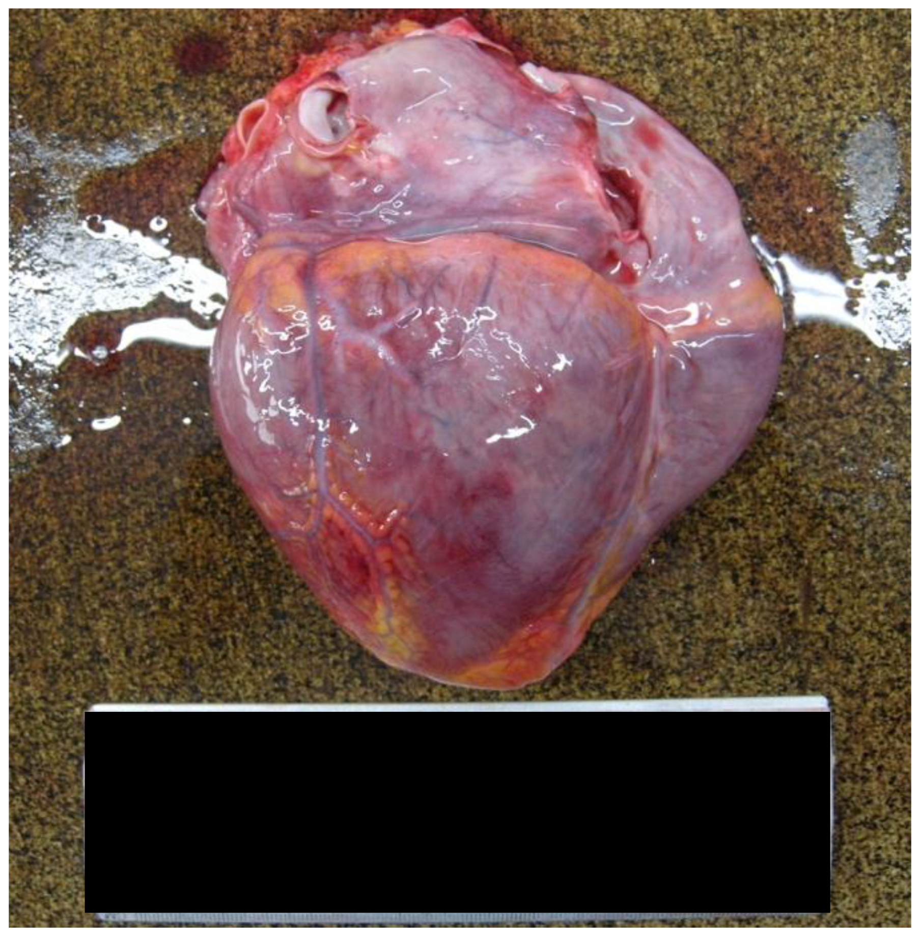
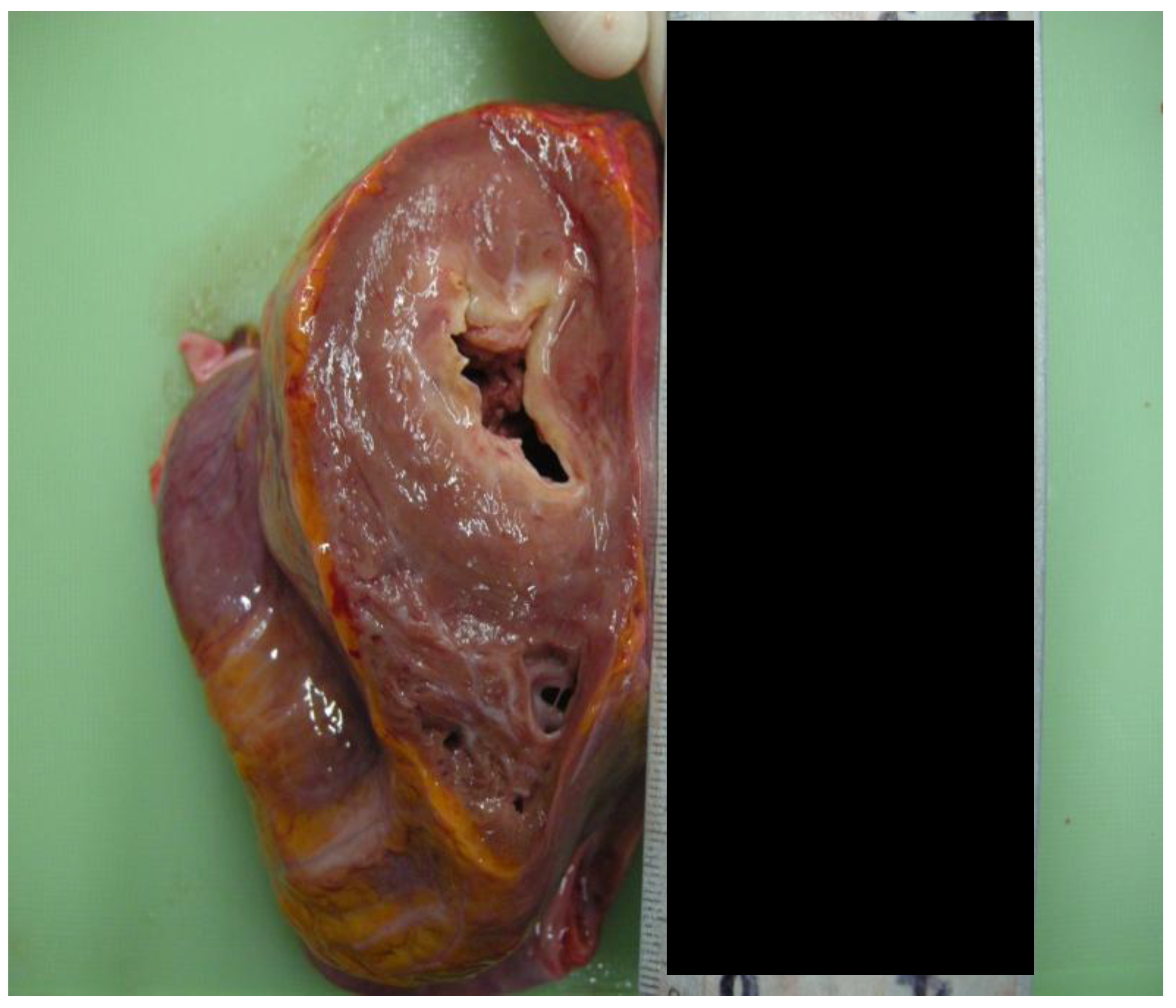
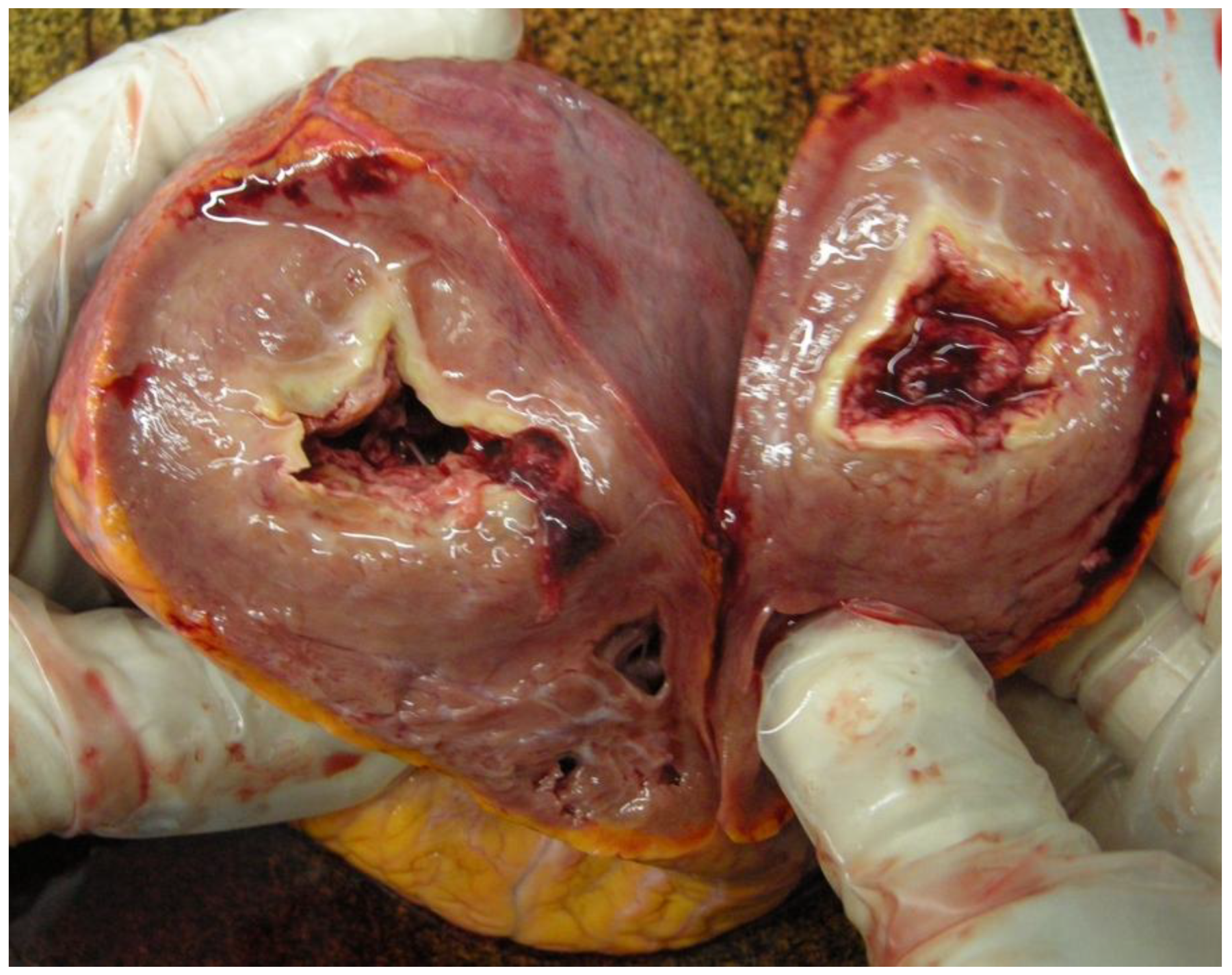
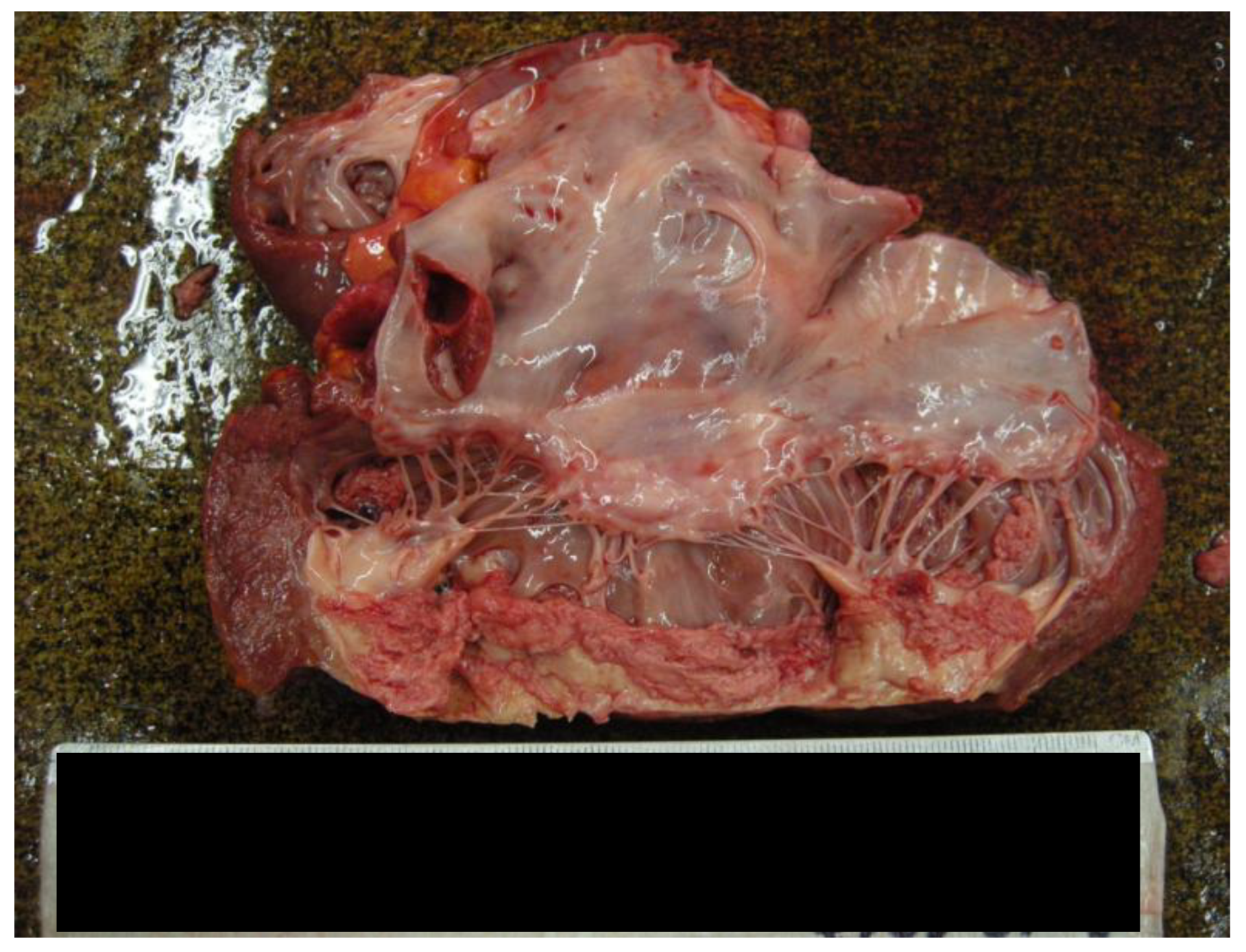
2.3. Macroscopic Examination of the Heart

The macroscopic inspection of the heart revealed the following findings:
-
The heart weighed 350 g, exhibiting a slightly increased size and volume, with dimensions of 9 cm longitudinally, 11.5 cm transversely, and 3 cm anteroposteriorly [Figure 1].
-
The myocardial thickness had increased. The posterior wall measured 1.2 cm, the lateral wall measured 1.8 cm, the anterior wall measured 1.5 cm, the ventricular septal measured 1.6 cm, and the right ventricular wall measured 0.4 cm [Figure 2].
-
Examination of the coronary artery tree showed a subcritical stenosis in the proximal tract of the left anterior descending artery, complicated by an occlusive thrombus. Instead, the right coronary artery remained patent.
-
The right ventricular wall was partially replaced by fibrotic tissue.
-
Both the left atrium and the left ventricle were remodeled, with the apex completely replaced by endoluminal vegetation [Figure 3 and Figure 4].
-
The cusps of the tricuspid and mitral valves appeared slightly retracted, measuring 11 cm and 10.5 cm in length, respectively. The pulmonary valve measured 8 cm, and the aortic valve was 7.5 cm.

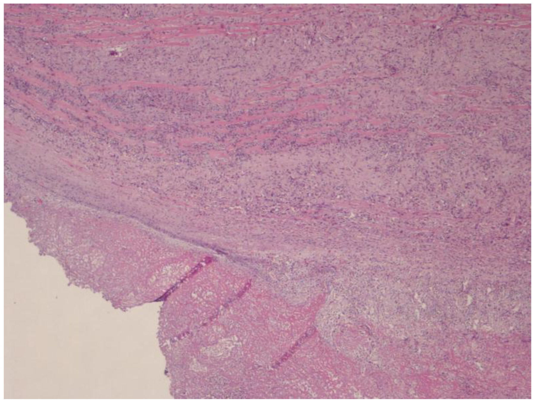
2.4. Histological Finding

Histological analysis confirmed extensive replacement of both ventricular walls by diffuse endomyocardial fibrosis. This fibrotic process was accompanied by an inflammatory infiltrate composed of lymphocytes, plasma cells, and a significant neutrophilic and eosinophilic component [Figure 5]. Additionally, extensive vegetative fibrin–leukocyte thrombi, in the initial phase of organization, were observed. Notably, there were no histological signs of acute ischemia.

2.5. Cause of Death

Based on the findings from the medico-legal investigation, the cause of death was attributed to tropical endomyocardial fibrosis. This condition precipitated sudden death due to acute pulmonary congestion, compounded by hepatic and splenic septic embolization.

3. Discussion

EMF has historically been regarded as a tropical disease, predominantly affecting regions with high prevalence, such as Sub-Saharan Africa, Brazil, and India. However, with increasing global migration, its presence is becoming more evident in Western countries. According to data from the Italian Ministry of the Interior, 158.000 people arrived in Italy between 1 January and 31 December 2023. This represents a 50% increase compared to 2022 and over a 130% increase compared to 2021. Among these, the most represented countries of origin included Ivory Coast, Guinea, Egypt, and Tunisia.
This demographic shift highlights the emergence of previously rare diseases in industrialized nations such as Italy. Conditions such as EMF and Chagas disease, previously confined to endemic regions, now represent a growing global health concern [11]. These diseases are not only rare but are also considered “orphan” and “obsolete” pathologies, characterized by a lack of research, understanding, and specific therapeutic strategies. Cases of EMF in Europe are exceptional. In the literature, only a few cases of EMF are described, most of which are all related to migration [10,12,13,14,15,16]. The only case of EMF in Italy described in the literature dates back to 1985 [5,17].
EMF, in particular, poses significant diagnostic and therapeutic challenges in non-endemic settings. Its clinical signs are often subtle and non-specific, complicating early recognition and leading to delays in appropriate management [9,18].
Recent advancements in echocardiographic techniques, such as myocardial deformation indices obtained through speckle-tracking echocardiography (STE), provide an objective means of identifying abnormalities in cardiac function during the early and subclinical stages of various cardiac diseases. Numerous studies have demonstrated that a reduction in left atrial (LA) function, as assessed by STE, is predictive of the presence of LA fibrosis. By addressing many of the limitations associated with standard echocardiographic methods, such as atrial ejection fraction (EF), transmitral flow, and tissue Doppler imaging (TDI) analysis, STE is increasingly utilized to characterize atrial function. This technique enables the derivation of precise measures of both reservoir and contractile function, with peak atrial longitudinal strain (PALS) being the most commonly used index. The presence of fibrosis in the LA walls significantly reduces PALS, serving as an early indicator of atrial dilation and reflecting impaired atrial compliance [19].
STE analysis should be considered an indirect approach for assessing myocardial fibrosis, as it evaluates the impact of underlying pathologies on tissue function. Various histological and pathophysiological changes affecting the extracellular matrix can alter myocardial mechanics by increasing myocardial stiffness. These alterations in cardiomyocyte mechanics are manifested as global or regional impairments in deformation parameters, as measured by 2D-STE analysis [20].
This diagnostic ambiguity underscores the need for heightened awareness among healthcare professionals, who must consider these emerging cardiomyopathies when evaluating patients from endemic regions.
An epidemiological transition is underway, with the increasing presence of immigrant populations introducing these “importable” diseases to new geographical contexts. In this light, it is crucial to establish a dedicated registry for rare and imported heart diseases. Such an initiative would not only enhance clinical sensitivity but also promote accurate diagnosis and therapeutic interventions.
Differentiating between EMF and other causes of sudden cardiac death, such as genetic mutations affecting the ion channels of the myocardial cell membrane, is essential. Common cardiac channelopathies include congenital conditions such as long QT syndrome (LQTS), Brugada syndrome, catecholaminergic polymorphic ventricular tachycardia (CPVT), and short QT syndrome (SQTS). In these cases, the heart is structurally normal, with the pathology being molecular in origin due to mutations in genes encoding cardiac ion channel proteins. Echocardiography plays a pivotal role in assessing cardiac structures, such as valves, diagnosing cardiomyopathies, and evaluating left ventricular ejection fraction (LVEF). Thus, echocardiography is recommended to distinguish patients with suspected structural cardiac abnormalities from those with molecular pathologies [21].
In endemic regions, where EMF is among the most prevalent forms of restrictive cardiomyopathy, the mainstay of treatment is often surgical intervention. Procedures such as endocardiectomy or valve replacement, while potentially life-saving, carry significant risks, with mortality rates reported to range between 15% and 30%. In contrast, in non-endemic countries, the rarity of the disease translates to limited surgical expertise and therapeutic knowledge. Consequently, most cases encountered in Western settings represent advanced stages of the disease, further complicating management and prognosis.
Medical management, where available, focuses on alleviating symptoms and preventing complications. Diuretics and anticoagulants are commonly employed, but their efficacy is limited by the advanced stage at which the disease is typically diagnosed in non-endemic regions. In addition, a lack of clinical trials further hampers the development of evidence-based therapeutic strategies, perpetuating a cycle of diagnostic and therapeutic uncertainty.
This case exemplifies the severe consequences of a missed diagnosis of EMF in a non-endemic region. The young man presented with non-specific symptoms of high fever, chest pain, and asthenia, which were initially misinterpreted, leading to a tragic outcome of rapid progressive heart failure and sudden death. A definitive diagnosis was only possible postmortem through detailed autopsy and histological examinations.

4. Conclusions

This case highlights the critical need for greater awareness and understanding of EMF and other similarly neglected diseases in industrialized countries. Healthcare professionals must adopt a more comprehensive diagnostic approach, particularly when treating patients from endemic regions. Establishing a registry for rare and imported cardiovascular diseases would be a significant step forward, enabling better diagnostic and therapeutic frameworks and ultimately improving patient outcomes.
The diagnosis, assessment, and management of cardiomyopathy patients require a coordinated, systematic, and personalized approach to ensure optimal care delivered by an experienced multidisciplinary team. Additionally, patients with cardiomyopathy may present with extracardiac manifestations, including neurological, neuromuscular, ophthalmological, or nephrological conditions. Effective patient care relies on close collaboration among various medical specialties.

Author Contributions

L.A. and S.N.: Writing—Original Draft; M.B.: Writing—Review, A.S.: Editing; D.F.: Methodology and Investigation; B.S.: Supervision. All authors have read and agreed to the published version of the manuscript.

Funding

This research received no external funding.

Institutional Review Board Statement

The study did not require ethical approval as it did not involve living humans. As stated in Recital 27 or der GDPR, “This Regulation does not apply to the personal data of deceased persons” (https://gdpr-info.eu/recitals/no-27/, accessed on 1 July 2024).

Informed Consent Statement

Patient consent was waived due to the report does not contain personal data.

Data Availability Statement

Data are contained within the article.

Acknowledgments

The authors acknowledge Francesco Merlanti (University of Bari) for his contribution to the preparation of the histological slides.

Conflicts of Interest

The authors declare no conflicts of interest.

References

  1. Gutierrez, P.S.; Campos, F.P.F.D. Endomyocardial fibrosis. Autops Case Rep. 2017, 7, 3–6. [Google Scholar] [CrossRef] [PubMed]
  2. Grimaldi, A.; Mocumbi, A.O.; Freers, J.; Lachaud, M.; Mirabel, M.; Ferreira, B.; Narayanan, K.; Celermajer, D.S.; Sidi, D.; Jouven, X.; et al. Tropical Endomyocardial Fibrosis: Natural History, Challenges, and Perspectives. Circulation 2016, 133, 2503–2515. [Google Scholar] [CrossRef] [PubMed]
  3. Mocumbi, A.O.; Ferreira, M.B.; Sidi, D.; Yacoub, M.H. A Population Study of Endomyocardial Fibrosis in a Rural Area of Mozambique. N. Engl. J. Med. 2008, 359, 43–49. [Google Scholar] [CrossRef] [PubMed]
  4. De Gaspari, M.; Toscano, G.; Bagozzi, L.; Metra, M.; Lombardi, C.; Rizzo, S.; Angelini, A.; Perazzolo Marra, M.; Gerosa, G.; Basso, C. Endomyocardial fibrosis and myocardial infarction leading to diastolic and systolic dysfunction requiring transplantation. Cardiovasc. Pathol. 2019, 38, 21–24. [Google Scholar] [CrossRef] [PubMed]
  5. Mocumbi, A.O.; Stothard, J.R.; Correia-de-Sá, P.; Yacoub, M. Endomyocardial Fibrosis: An Update After 70 Years. Curr. Cardiol. Rep. 2019, 21, 148. [Google Scholar] [CrossRef] [PubMed]
  6. Wu, B.; Yang, Y.; Zhao, P. Application of Routine Echocardiography Combined with Contrast-Enhanced Echocardiography in the Diagnosis of Endomyocardial Fibrosis. Discov. Med. 2023, 35, 1052. [Google Scholar] [CrossRef] [PubMed]
  7. Tigani, S.E.; Khalil, S.; Saad, H.A.; Khalil, S.I. Endomyocardial fibrosis in Sudan: Clinical and echocardiographic features Cardiovasc. J. Afr. 2017, 28, 208–214. Available online: https://journals.co.za/doi/10.5830/CVJA-2016-079 (accessed on 22 January 2025).
  8. Vásquez-Rodríguez, J.F.; Medina-Mur, R.I.; Giraldo, L.E.; Juan-Guardela, M.L.; Gelves, J.; Jaimes, C.P. Endomyocardial fibrosis: A restrictive cardiomyopathy in developing countries. Arch. De Cardiol. De México 2021, 91, 196–201. [Google Scholar] [CrossRef]
  9. Grimaldi, A.; Alfieri, O.; Camici, P.G.; Zoppei, G.; Olivotto, I. “Mal d’Africa” e cuore: I misteri della fibrosi endomiocardica. G. Ital. Cardiol. 2011, 12, 484–491. [Google Scholar]
  10. Théry, G.; Faroux, L.; Deleuze, P.; Metz, D. Idiopathic endomyocardial fibrosis in a Western European: A case report. Eur. Heart J. Case Rep. 2020, 4, 1–5. [Google Scholar] [CrossRef] [PubMed]
  11. Yusuf, S.; Reddy, S.; Ôunpuu, S.; Anand, S. Global Burden of Cardiovascular Diseases. Circulation 2001, 104, 2746–2753. [Google Scholar] [CrossRef] [PubMed]
  12. Endomyocardial Fibrosis in a European Woman and Its Successful Surgical Treatment-Abstract-Europe PMC. Available online: https://europepmc.org/article/med/916720 (accessed on 18 December 2024).
  13. López Ramón, M.; García de la Calzada, M.D.; Domínguez Cunchillos, M.; Salazar Mena, J. Tropical endomyocardial fibrosis in Spain. An. Pediatr. 2013, 78, 326–329. [Google Scholar] [CrossRef] [PubMed]
  14. Cağli, K.; Uygur, B.; Ozlü, F.; Gölbaşi, Z. Endomyocardial disease: A case report. Turk. Kardiyol. Dern. Ars. 2009, 37, 136–140. [Google Scholar] [PubMed]
  15. Gyr, K.; Fernex, M.; Wick, R.; Burckhardt, D. African endomyocardial fibrosis. Report of a case seen in Basel. Dtsch. Med. Wochenschr. 1970, 95, 2028–2032. [Google Scholar] [CrossRef] [PubMed]
  16. Wagner, G.; Haumer, M.; Poelzl, G.; Wiedemann, D.; Kliegel, A.; Ullrich, R.; Gartlehner, G.; Zuckermann, A.; Müller, L.; Mayr, H.; et al. A case report of a 40-year-old woman with endomyocardial fibrosis in a non-tropical area: From initial presentation to high urgent heart transplantation. BMC Cardiovasc. Disord. 2019, 19, 302. [Google Scholar] [CrossRef] [PubMed]
  17. Pavia, M.; Cugini, A.; Buffa, J. Restrictive cardiomyopathy caused by endomyocardial fibrosis. Description of a case observed in Piedmont. Minerva Cardioangiol. 1985, 33, 357–361. [Google Scholar] [PubMed]
  18. Mocumbi, A.O.; Yacoub, S.; Yacoub, M.H. Neglected tropical cardiomyopathies: II. Endomyocardial fibrosis. Heart 2008, 94, 384–390. [Google Scholar] [PubMed]
  19. Mandoli, G.E.; D’Ascenzi, F.; Vinco, G.; Benfari, G.; Ricci, F.; Focardi, M.; Cavigli, L.; Concetta Pastore, M.; Sisti, N.; De Vivo, O.; et al. Novel Approaches in Cardiac Imaging for Non-invasive Assessment of Left Heart Myocardial Fibrosis. Front. Cardiovasc. Med. 2021, 8, 614235. [Google Scholar] [CrossRef] [PubMed]
  20. Sonaglioni, A.; Nicolosi, G.L.; Rigamonti, E.; Lombardo, M.; La Sala, L. Molecular Approaches and Echocardiographic Deformation Imaging in Detecting Myocardial Fibrosis. Int. J. Mol. Sci. 2022, 23, 10944. [Google Scholar] [CrossRef] [PubMed]
  21. Kumar, A.; Avishay, D.M.; Jones, C.R.; Shaikh, J.D.; Kaur, R.; Aljadah, M.; Kichloo, A.; Shiwalkar, N.; Keshavamurthy, S. Sudden cardiac death: Epidemiology, pathogenesis and management. Rev. Cardiovasc. Med. 2021, 22, 147–158. [Google Scholar] [CrossRef] [PubMed]
Figure 1. The heart dimensions.
Forensicsci 05 00011 g001
Figure 2. The myocardial thickness.
Forensicsci 05 00011 g002
Figure 3. The remodeling of the left ventricle.
Forensicsci 05 00011 g003
Figure 4. The endoluminal vegetation of the left atrium.
Forensicsci 05 00011 g004
Figure 5. The extensive myocardial fibrous scar and residual myocardium with infiltrations by neutrophils and eosinophils (Hematoxylin-Eosin stain, 40× magnification).
Forensicsci 05 00011 g005
Disclaimer/Publisher’s Note: The statements, opinions and data contained in all publications are solely those of the individual author(s) and contributor(s) and not of MDPI and/or the editor(s). MDPI and/or the editor(s) disclaim responsibility for any injury to people or property resulting from any ideas, methods, instructions or products referred to in the content.

