


Intravenous-Therapy-Associated Circulatory Overload: A Retrospective Study of Forensic Cases

 , , , and
Abstract
1. Introduction
2. Materials and Methods
2.1. Study Design
2.2. Statistical Analysis
3. Results
3.1. General Characteristics of the Study Cases
3.2. IV-Infusion-Associated Circulatory Overload (IACO)
3.3. IV Fluids and Clinical Laboratory Diagnostics
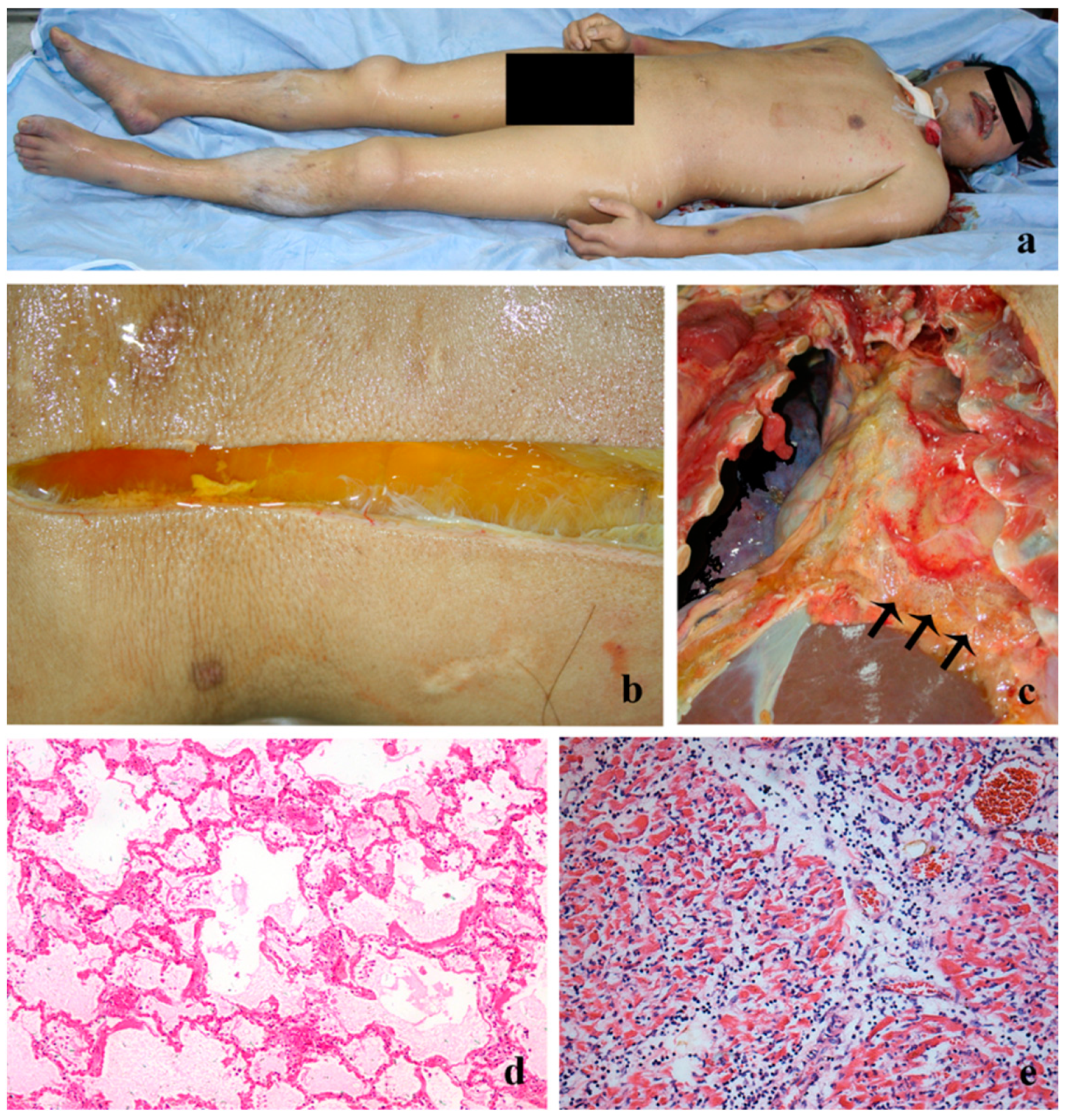
3.4. Autopsy Findings
4. Discussion
5. Conclusions
Author Contributions
Funding
Institutional Review Board Statement
Informed Consent Statement
Data Availability Statement
Acknowledgments
Conflicts of Interest
References
- Kuttab, H.I.; Lykins, J.D.; Hughes, M.D.; Wroblewski, K.; Keast, E.P.; Kukoyi, O.; Kopec, J.A.; Hall, S.; Ward, M.A. Evaluation and Predictors of Fluid Resuscitation in Patients with Severe Sepsis and Septic Shock. Crit. Care Med. 2019, 47, 1582–1590. [Google Scholar] [CrossRef] [PubMed]
- Vincent, J.-L. Fluid management in the critically ill. Kidney Int. 2019, 96, 52–57. [Google Scholar] [CrossRef] [PubMed]
- Meyhoff, T.S.; Hjortrup, P.B.; Wetterslev, J.; Sivapalan, P.; Laake, J.H.; Cronhjort, M.; Jakob, S.M.; Cecconi, M.; Nalos, M.; Ostermann, M.; et al. Restriction of Intravenous Fluid in ICU Patients with Septic Shock. New Engl. J. Med. 2022, 386, 2459–2470. [Google Scholar] [CrossRef] [PubMed]
- WHO. WHO Guideline on the Use of Safety-Engineered Syringes for Intramuscular, Intradermal and Subcutaneous Injections in Health-Care Settings; WHO: Geneva, Switzerland, 2015. [Google Scholar]
- Dong, L.; Yan, H.; Wang, D. Antibiotic prescribing patterns in village health clinics across 10 provinces of Western China. J. Antimicrob. Chemother. 2008, 62, 410–415. [Google Scholar] [CrossRef] [PubMed]
- Yin, X.; Song, F.; Gong, Y.; Tu, X.; Wang, Y.; Cao, S.; Liu, J.; Lu, Z. A systematic review of antibiotic utilization in China. J. Antimicrob. Chemother. 2013, 68, 2445–2452. [Google Scholar] [CrossRef]
- Yuan, S. China should reduce the overuse of intravenous infusion. BMJ 2014, 348, g1262. [Google Scholar] [CrossRef] [PubMed]
- WHO. China-WHO Country Cooperation Strategy (2016-2020); WHO: Geneva, Switzerland, 2016. [Google Scholar]
- Hilton, A.K.; A Pellegrino, V.; Scheinkestel, C.D. Avoiding common problems associated with intravenous fluid therapy. Med. J. Aust. 2008, 189, 509–513. [Google Scholar] [CrossRef] [PubMed]
- Yu, X.; Wang, W. Death caused by circulatory overload in the treatment of traumatic hemorrhagic shock: Two cases report. J. Law Med. 2002, 9, 137–138. [Google Scholar]
- FDA. Fatalities Reported to FDA Following Blood Collection and Transfusion: Annual Summary for Fiscal Year 2009; FDA: Silver Spring, MD, USA, 2010.
- Li, G.; Rachmale, S.; Kojicic, M.; Shahjehan, K.; Malinchoc, M.; Kor, D.J.; Gajic, O. Incidence and transfusion risk factors for transfusion-associated circulatory overload among medical intensive care unit patients. Transfusion 2011, 51, 338–343. [Google Scholar] [CrossRef]
- Li, X.; Huang, J.; Zhang, H. An analysis of hospital preparedness capacity for public health emergency in four regions of China: Beijing, Shandong, Guangxi, and Hainan. BMC Public Health 2008, 8, 319. [Google Scholar] [CrossRef]
- Yu, X.; Wang, H.; Feng, L.; Zhu, J. Quantitative Research in Modern Forensic Analysis of Death Cause: New Classification of Death Cause, Degree of Contribution, and Determination of Manner of Death. J. Forensic Res. 2014, 5, 221. [Google Scholar]
- Arieff, A.I. Fatal postoperative pulmonary edemapathogenesis and literature review. Chest J. 1999, 115, 1371–1377. [Google Scholar] [CrossRef] [PubMed]
- Bai, R.; Yu, X.; Wang, D.; Lv, J.; Xu, G.; Lai, X. The Densities of Visceral Organs and the Extent of Pathologic Changes. Am. J. Forensic Med. Pathol. 2009, 30, 148–151. [Google Scholar] [CrossRef] [PubMed]
- Li, Y.; Xu, J.; Wang, F.; Wang, B.; Liu, L.; Hou, W.; Fan, H.; Tong, Y.; Zhang, J.; Lu, Z. Overprescribing in China, Driven by Financial Incentives, Results in Very High Use of Antibiotics, Injections, and Corticosteroids. Health Aff. 2012, 31, 1075–1082. [Google Scholar] [CrossRef]
- FDA. 2017 Annual Report for National Adverse Drug Reaction Monitoring; FDA: Silver Spring, MD, USA, 2018.
- FDA. 2016 Annual Report for National Adverse Drug Reaction Monitoring; FDA: Silver Spring, MD, USA, 2017.
- Blumenthal, D.; Hsiao, W. Privatization and Its Discontents — The Evolving Chinese Health Care System. New Engl. J. Med. 2005, 353, 1165–1170. [Google Scholar] [CrossRef]
- Ma, X.; Wang, H.; Yang, L.; Shi, L.; Liu, X. Realigning the incentive system for China’s primary healthcare providers. BMJ 2019, 365, l2406. [Google Scholar] [CrossRef]
- Lobo, D.; Dube, M.; Neal, K.; Simpson, J.; Rowlands, B.; Allison, S. Problems with solutions: Drowning in the brine of an inadequate knowledge base. Clin. Nutr. 2001, 20, 125–130. [Google Scholar] [CrossRef]
- Kwan, I.; Bunn, F.; Chinnock, P.; Roberts, I. Timing and volume of fluid administration for patients with bleeding. Cochrane Database Syst. Rev. 2014, 2014, CD002245. [Google Scholar] [CrossRef] [PubMed]
- Monnet, X.; Shi, R.; Teboul, J.L. Prediction of fluid responsiveness. What’s new? Ann. Intensive Care 2022, 12, 46. [Google Scholar] [CrossRef]
- John, G.P.; Arthur, K.; John, O.G. The Textbook of Pharmaceutical Medicine; Wiley-Blackwell: Hoboken, NJ, USA, 2013. [Google Scholar]
- Cotter, G.; Metra, M.; Milo-Cotter, O.; Dittrich, H.C.; Gheorghiade, M. Fluid overload in acute heart failure - Re-distribution and other mechanisms beyond fluid accumulation. Eur. J. Hear. Fail. 2008, 10, 165–169. [Google Scholar] [CrossRef] [PubMed]
- Maldonado, M.; E Villamin, C.; E Murphy, L.; Dasgupta, A.; Bassett, R.L.; Medina, M.C.; Bates, T.S.; Martinez, F.; Couchonal, A.M.K.; Klein, K.; et al. Oncology Patients Who Develop Transfusion-Associated Circulatory Overload: An Observational Study. Lab. Med. 2022, 53, 344–348. [Google Scholar] [CrossRef] [PubMed]
- Lieberman, L.; Maskens, C.; Cserti-Gazdewich, C.; Hansen, M.; Lin, Y.; Pendergrast, J.; Yi, Q.L.; Callum, J. A Retrospective Review of Patient Factors, Transfusion Practices, and Outcomes in Patients with Transfusion-Associated Circulatory Overload. Transfus. Med. Rev. 2013, 27, 206–212. [Google Scholar] [CrossRef] [PubMed]
- Lowell, J.A.; Schifferdecker, C.; Driscoll, D.F.; Benotti, P.N.; Bistrian, B.R. Postoperative fluid overload: Not a benign problem. Crit. Care Med. 1990, 18, 728–733. [Google Scholar] [CrossRef] [PubMed]
- Messina, A.; Robba, C.; Calabrò, L.; Zambelli, D.; Iannuzzi, F.; Molinari, E.; Cecconi, M. Perioperative liberal versus restrictive fluid strategies and postoperative outcomes: A systematic review and metanalysis on randomised-controlled trials in major abdominal elective surgery. Crit. Care 2021, 25, 205. [Google Scholar] [CrossRef] [PubMed]
- Tigabu, B.M.; Davari, M.; Kebriaeezadeh, A.; Mojtahedzadeh, M. Fluid volume, fluid balance and patient outcome in severe sepsis and septic shock: A systematic review. J. Crit. Care 2018, 48, 153–159. [Google Scholar] [CrossRef]
- Boyd, J.H.; Forbes, J.; Nakada, T.-A.; Walley, K.R.; Russell, J.A. Fluid resuscitation in septic shock: A positive fluid balance and elevated central venous pressure are associated with increased mortality. Crit. Care Med. 2011, 39, 259–265. [Google Scholar] [CrossRef]
- Nolan, J. Fluid resuscitation for the trauma patient. Resuscitation 2001, 48, 57–69. [Google Scholar] [CrossRef]
- Gattinoni, L.; Cressoni, M.; Brazzi, L. Fluids in ARDS: From onset through recovery. Curr. Opin. Crit. Care. 2014, 20, 373–377. [Google Scholar] [CrossRef]
- Desborough, J. The stress response to trauma and surgery. Br. J. Anaesth. 2000, 85, 109–117. [Google Scholar] [CrossRef]
- Hu, B.; Wu, Y.; Tong, F.; Liu, J.; Shen, X.; Shen, R.; Xu, G. Apocynin Alleviates Renal Ischemia/Reperfusion Injury Through Regulating the Level of Zinc and Metallothionen. Biol. Trace Element Res. 2016, 178, 71–78. [Google Scholar] [CrossRef]
- Paynejames, J.; Mcgovern, C.; Jones, R. Simpson’s Forensic Medicine, 13th ed.; CRC Press: Boca Raton, FL, USA, 2014. [Google Scholar]
- Xu, G.; Su, R.; Li, B.; Lv, J.; Sun, W.; Hu, B.; Yu, X. Trace element concentrations in human tissues of death cases associated with secondary infection and MOF aftersevere trauma. Biol. Trace Elem. Res. 2015, 168, 335–339. [Google Scholar] [CrossRef] [PubMed]
- Friedrich, M.G.; Sechtem, U.; Schulz-Menger, J.; Holmvang, G.; Alakija, P.; Cooper, L.T.; White, J.A.; Abdel-Aty, H.; Gutberlet, M.; Prasad, S.; et al. Cardiovascular Magnetic Resonance in Myocarditis: A JACC White Paper. J. Am. Coll. Cardiol. 2009, 53, 1475–1487. [Google Scholar] [CrossRef]
- Cooper, L.T., Jr. Myocarditis. N. Engl. J. Med. 2009, 360, 1526–1538. [Google Scholar] [CrossRef] [PubMed]
- Magnani, J.W.; Dec, G.W. Myocarditis: Current trends in diagnosis and treatment. Circulation 2006, 113, 876–890. [Google Scholar] [CrossRef] [PubMed]
- Kenealy, T.; Arroll, B. Antibiotics for the common cold and acute purulent rhinitis. Cochrane Database Syst. Rev. 2013, 2013, CD000247. [Google Scholar] [CrossRef] [PubMed]


| No. | Age/Gender | Cause of Hospitalization | Medical Diagnosis | Other Injury or Disease and Surgical Conditions | Cause of Death | Grade of Hospital |
|---|---|---|---|---|---|---|
| 1 | 28/female | Postpartum hemorrhage | Postpartum hemorrhage shock | Hypoxic ischemic encephalopathy, Lung infection/ Hysterectomy | DIC *, IACO | Clinics and first- and third-tier |
| 2 | 38/female | Postpartum hemorrhage | Postpartum hemorrhage shock | Multiple organ dysfunction syndrome Total abdominal hysterectomy | Hemorrhagic shock, IACO | Clinics and first- and third-tier |
| 3 | 38/female | Traffic accident | Crush injury of back and buttocks | Comminuted fracture of the pelvis, Multiple vascular ruptures and bleeding/Colporrhaphy surgery | Hemorrhagic shock, IACO | Third-tier |
| 4 | 34/female | Gestational hypertension | Uterine rupture | Massive hemorrhage, P2G1/ No surgery | Hemorrhagic shock, IACO | Clinics and second-tier |
| 5 | 12/male | Traffic accident | Blunt abdominal trauma | Mild traumatic shock, Separation of the symphysis pubis Laparotomy surgery | Traumatic shock, IACO | Third-tier |
| 6 | 35/male | Right abdominal pain | Spontaneous rupture of sigmoid colon tumor | Massive hemorrhage/ Laparotomy | Hemorrhagic shock, IACO | Second-tier |
| 7 | 30/female | Abdominal pain | Ectopic pregnancy | Rupture of the left isthmus pregnancy, massive hemorrhage/No surgery | Hemorrhagic shock, IACO | Clinics and first-tier |
| 8 | 49/male | Suffer a serious injury | Left cerebellar hemorrhage, Tentorial notch hernia | Multiple-organ dysfunction syndrome, Hypertension/ No surgery | Multiple organ failure, IACO | Third-tier |
| 9 | 22/female | Injury by fall from height | Compression fractures of Vertebral L1 | Suppurated peritonitis, Toxic shock/ Corpectomy (decompression surgery) | Multiple organ failure, IACO | Third-tier |
| 10 | 20/male | Suffer a serious injury | Extensive stab wounds, Fracture of the right distal ulna | Hemorrhagic shock/Left thoracotomy, Abdominal debridement, Fracture fixation | Multiple organ failure, IACO | Second-tier |
| 11 | 34/male | Metastatic pain in right-lower abdomen | Acute appendicitis | Limitations of peritonitis/ Appendectomy | Septic shock, IACO | Second-tier |
| 12 | 49/male | Suffer a serious injury | Traumatic duodenal perforation | Diffuse purulent peritonitis, pancreatic injury/ No surgery | Septic shock, IACO | Second-tier |
| 13 | 19/female | Upper abdominal pain | Gastric fundus perforation | Diffuse purulent peritonitis/ Open repair of gastric perforation | Septic shock, IACO | Third-tier |
| 14 | 53/male | Traffic accident | Fracture of upper tibia and fibula | Extensive soft tissue injuries, Lung infection/ Fracture fixation | Septic shock, IACO | Second- and third-tier |
| 15 | 45/male | Suffer a serious injury | Suppurative pneumonia | Extensive soft tissue injuries, Systemic inflammatory response syndrome/No surgery | Septic shock, IACO | Second-tier |
| 16 | 38/male | Suffer a serious injury | Blunt abdominal trauma | Jejunum rupture, Secondary suppurative pneumonia/ Laparotomy | Septic shock, IACO | Third-tier |
| No. | Age/Gender | Medical Diagnosis | Clinical Manifestations | Cause of Death | Grade of Hospital |
|---|---|---|---|---|---|
| 1 | 30/male | Heatstroke | Generalized weakness and pain | Acute viral myocarditis, IACO | Clinics |
| 2 | 38/male | Acute gastroenteritis | Dizziness, vomiting, fever, and cough for 3 days | Acute viral myocarditis focal necrosis of myocytes, IACO | Clinics and second-tier |
| 3 | 13/male | Upper respiratory tract infection/Pneumonia | Cough, fever for 2 days | Acute viral myocarditis, IACO | Second-tier |
| 4 | 36/male | Upper respiratory tract infection/Pneumonia | Fever, cough, dizziness, nausea, and vomiting for 2 days | Acute viral myocarditis, IACO | Clinics |
| 5 | 45/male | Gastrointestinal-type cold | Fever, vomiting, and diarrhea for days | Acute viral myocarditis | First-tier |
| 6 | 30/female | Upper respiratory tract infection/Pneumonia | Throat discomfort, chest pain, and tightness for 3 days | Acute lymphocytic pancarditis, focal necrosis of myocytes, IACO | Second-tier |
| 7 | 27/male | Fever | Feeling cold | Acute viral myocarditis, IACO | Clinics |
| 8 | 21/female | Upper respiratory tract infection | Sore throat | Acute viral myocarditis, IACO | Clinics |
| 9 | 9/female | Fever | Abdominal pain and vomiting | Acute viral myocarditis, IACO | Clinics |
| 10 | 28/female | Acute tonsillitis | Sore throat, dizziness, mild chills, and pain in the limbs | Acute lymphocytic pancarditis, IACO | First-tier |
| 11 | 4/female | - | Abdominal pain and vomiting | Acute lymphocytic pancarditis, IACO | Clinics |
| 12 | 35/male | Upper respiratory tract infection/Pneumonia | Cough and fever for 1 day | Acute viral myocarditis, IACO | Clinics |
| 13 | 8/male | Upper respiratory tract infection | Fever and cough for 2 days | Acute viral myocarditis, IACO | Clinics |
| 14 | 43/female | Bronchial asthma | Cough, severe asthma | Acute viral myocarditis, IACO | Clinics |
| 15 | 27/male | - | Cough and weakness | Acute viral myocarditis, IACO | Clinics |
| 16 | 12/female | Fever | Fever | Acute lymphocytic myocarditis, IACO | Clinics |
| 17 | 31/female | - | Fever and dizziness | Acute viral myocarditis, IACO | Second-tier |
| 18 | 19/male | Acute tonsillitis | Sore throat and feeling cold | Acute lymphocytic myocarditis, IACO | First-tier |
| 19 | 36/male | Fever | Fever and pain in the limbs | Acute viral myocarditis, IACO | Clinics |
| 20 | 37/male | Gastrointestinal-type cold | Fever, vomiting, and abdominal pain | Acute viral myocarditis, IACO | Clinics |
| 21 | 30/male | Fever | Fever and feeling cold | Acute viral myocarditis, IACO | Clinics |
| No. | Time | Fluid Intake (mL) | The Ratio of Colloid and Crystal Fluid (mL:mL) | Fluid Output (mL) | Fluid Retention (mL) | Fluid Retention Index (mL/d) |
|---|---|---|---|---|---|---|
| 1 | 59 h | 29,310 | 17,900:11,410 | 8715 | 20,595 | 8378 |
| 2 | 6 h | 10,350 | 3800:6550 | 1500 | 8850 | - |
| 3 | 21 h | 10,560 | 1560:9000 | 320 | 10,240 | - |
| 4 | 30 h | 10,800 | 4800:6000 | 4000 | 6800 | 5440 |
| 5 | 25 h | 15,000 | 1400:13,600 | 600 | 14,400 | 13,824 |
| 6 | 16 h | 11,200 | 5000:6200 | 3500 | 7700 | - |
| 7 | 45 h | 25,120 | 6120:19,000 | 5830 | 19,290 | 10,288 |
| 8 | 10 d | 49,580 | 350:49,230 | 9380 | 40,200 | 4020 |
| 9 | 4 d | 22,025 | 500:21,525 | 4180 | 17,845 | 4461 |
| 10 | 12 d | 88,750 | 6000:82,750 | 5730 | 83,020 | 6918 |
| 11 | 5 d | 32,840 | 1250:31,590 | 5360 | 27,480 | 5496 |
| 12 | 36 h | 14,800 | 1000:13,800 | 5600 | 9200 | 6133 |
| 13 | 22 h | 5454 | 1000:4554 | 1000 | 4554 | - |
| 14 | 3 d | 18,600 | 1500:17,100 | 4000 | 14,600 | 4867 |
| 15 | 5.5 d | 36,660 | 1100:35,560 | 8500 | 28,160 | 5120 |
| 16 | 7 d | 81,585 | 3500:78,085 | 2825 | 78,760 | 11,251 |
| Index | On Admission | Before Death | p-Value |
|---|---|---|---|
| Red blood cells [3.5–4.5 × 10¹²/L] | 3.84 (3.61, 4.14) | 2.23 (1.88, 2.92) | 0.000 |
| Hematocrit [37–54%] | 41.20 (39.30, 47.75) | 22.00 (19.58, 23.50) | 0.000 |
| Hemoglobin [110–160 g/L] | 113.00 (89.25, 147.75) | 65.50 (48.25, 84.00) | 0.000 |
| Platelets [100–300 × 109/L] | 199.00 (168.75, 273.00) | 68.50 (59.75, 81.50) | 0.000 |
| White blood cells [4–10 × 109/L] | 16.35 (11.08, 25.10) | 14.20 (9.55, 22.93) | 0.438 |
| Potassium ions [3.5–5.5 mM] | 5.16 (4.58, 5.90) | 3.15 (2.80, 3.38) | 0.000 |
| Sodium ions [135–145 mM] | 142.40 (138.13, 144.80) | 127.25 (122.83, 131.00) | 0.000 |
| Chloride ions [96–106 mM] | 99.40 (98.08, 103.90) | 87.25 (81.30, 95.72) | 0.000 |
| No. | Time | Fluid Intake (mL) | Infusion Rate Index (Drops/min) |
|---|---|---|---|
| 1 | 3 h | 1360 | 151 |
| 2 | 1.25 h | 1100 | 293 |
| 3 | 3 h | 1500 | 167 |
| 4 | 1.5 h | 1000 | 222 |
| 5 | 1.5 h | 1000 | 222 |
| 6 | 1.25 h | 600 | 160 |
| 7 | 1 h | 500 | 167 |
| 8 | 5 h | 2250 | 150 |
| 9 | 1.5 h | 700 | 156 |
| 10 | 2 h | 750 | 125 |
| 11 | 1.5 h | 1000 | 222 |
| 12 | 1.5 h | 500 | 111 |
| 13 | 1 h | 350 | 117 |
| 14 | 3 h | 1250 | 139 |
| 15 | 2 h | 1250 | 208 |
| 16 | 1.5 h | 750 | 167 |
| 17 | 2 h | 1050 | 175 |
| 18 | 2.5 h | 1500 | 200 |
| 19 | 3 h | 1500 | 167 |
| 20 | 1 h | 700 | 233 |
| 21 | 3.5 h | 2500 | 238 |
| Indicator | The 16 Cases of Massive Fluid Infusion | The 21 Cases of Rapid Infusion | p-Value |
|---|---|---|---|
| Lungs | |||
| Weight (g) | 1656 (1297, 1823) | 1018 (780.5, 1475) | 0.003 |
| Density (g/mL) | 0.975 (0.918, 1.045) | 0.826 (0.731, 0.870) | <0.001 |
| Transudate in the cavity (mL) | |||
| Pleural | 1810 (1513, 1975) | 250 (200, 300) | <0.001 |
| Pericardial | 30 (30, 40) | 32 (22.5, 40) | 0.988 |
| Peritoneal and pelvic | 2090 (1763, 2650) | 0 (0, 87.5) | <0.001 |
Disclaimer/Publisher’s Note: The statements, opinions and data contained in all publications are solely those of the individual author(s) and contributor(s) and not of MDPI and/or the editor(s). MDPI and/or the editor(s) disclaim responsibility for any injury to people or property resulting from any ideas, methods, instructions or products referred to in the content. |
© 2023 by the authors. Licensee MDPI, Basel, Switzerland. This article is an open access article distributed under the terms and conditions of the Creative Commons Attribution (CC BY) license (https://creativecommons.org/licenses/by/4.0/).
Share and Cite
Xu, G.; Su, R.; Lv, J.; Xu, L.; Jin, X.; Chen, D.; Hu, B.; Yu, X. Intravenous-Therapy-Associated Circulatory Overload: A Retrospective Study of Forensic Cases. Forensic Sci. 2023, 3, 34-44. https://doi.org/10.3390/forensicsci3010005
Xu G, Su R, Lv J, Xu L, Jin X, Chen D, Hu B, Yu X. Intravenous-Therapy-Associated Circulatory Overload: A Retrospective Study of Forensic Cases. Forensic Sciences. 2023; 3(1):34-44. https://doi.org/10.3390/forensicsci3010005
Chicago/Turabian StyleXu, Guangtao, Ruibing Su, Junyao Lv, Long Xu, Xin Jin, Deqing Chen, Bo Hu, and Xiaojun Yu. 2023. "Intravenous-Therapy-Associated Circulatory Overload: A Retrospective Study of Forensic Cases" Forensic Sciences 3, no. 1: 34-44. https://doi.org/10.3390/forensicsci3010005
APA StyleXu, G., Su, R., Lv, J., Xu, L., Jin, X., Chen, D., Hu, B., & Yu, X. (2023). Intravenous-Therapy-Associated Circulatory Overload: A Retrospective Study of Forensic Cases. Forensic Sciences, 3(1), 34-44. https://doi.org/10.3390/forensicsci3010005