Abstract
New surveys on the Brazilian tropical coast revealed new occurrences of five species in Styelidae (Stolonia sabulosa, Amphicarpa paucigonas, Polyandrocarpa anguinea Polycarpa insulsa, Styela plicata) and one in Molgulidae (Molgula davidi). The species here described represent either the expansion of their geographic distribution in the country or new records for the country. Some of these species have disjunct or wide geographical distributions, and the possibility of their introduction as exotic fauna is discussed. We also present the first field pictures of Stolonia sabulosa and Amphicarpa paucigonas and a detailed description and figures for all species.
1. Introduction
The currently known biodiversity of Stolidobranchia in Brazil comprises 28 species in Styelidae, 9 species in Pyuridae, and 9 species in Molgulidae [1]. Here, we present six new occurrences from Espírito Santo and Bahia, states located on the central tropical coast of the country. This region presents many habitats favorable to the growth of ascidians, namely shallow reefs fringing the coast, a high concentration of rhodoliths, and small islands close to the shore [2,3], and the presence of international ports, recreational marinas, and artificial reefs suggest the high potential for receiving exotic species.
Very few studies have accounted for the stolidobranch ascidians in this region [4,5,6], with the following species listed in Styelidae (20): Botrylloides giganteus (Pérès, 1949), Botrylloides niger Herdman, 1886, Botryllus planus (Van Name, 1902), Botryllus schlosseri (Pallas, 1766), Botryllus tabori Rodrigues, 1962, Botryllus tuberatus Ritter and Forsyth, 1917, Symplegma brakenhielmi (Michaelsen, 1904), Symplegma rubra Monniot, 1972, Monandrocarpa stolonifera Monniot, 1970, Eusynstyela tincta (Van Name, 1902), Polyandrocarpa anguinea (Sluiter, 1898), Polyandrocarpa zorritensis (Van Name, 1931), Polycarpa arnoldi (Michaelsen, 1914), Polycarpa foresti Monniot, 1970, Polycarpa cf. reviviscens Monniot and Monniot, 2001, Polycarpa spongiabilis Traustedt, 1883, Polycarpa tumida Heller, 1878, Styela canopus (Savigny, 1816), Styela plicata (Lesueur, 1823), Cnemidocarpa irene (Hartmeyer, 1906). In addition, the following is listed in Molgulidae (1): Molgula salvadori Monniot, 1970. All the species here reported represent either new reports for one of the two states or the country.
2. Materials and Methods
The samples were collected between 2011 and 2017 (with a few older samples) from natural and artificial substrates in very shallow sites by free diving or from deeper reefs by autonomous diving. The collection sites in Bahia and Espírito Santo are described in Table 1. The samples were anesthetized with menthol and preserved in 4% formalin. The specimens were dissected and stained with Harris hematoxylin and observed under a stereoscopic microscope following routine methods [7].

Table 1.
Collection sites in Bahia and Espírito Santo, Brazil.
3. Results
- Family STYELIDAE Sluiter, 1895
- Genus Stolonica Lacaze-Duthiers and Delage, 1892
- Stolonica sabulosa Monniot, C., 1972
- Material examined: DZUP STO-01 01 colony, Escalvada Island, Guarapari, Espírito Santo, 20°42′00″ S, 40°24′30″ W, 10 m, Col. R. M. Rocha, 26.01.2012.
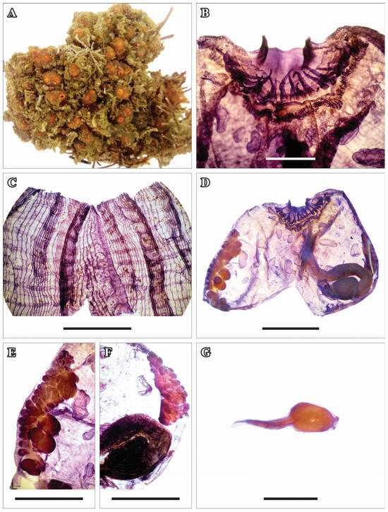
Figure 1.
Stolonica sabulosa Monniot C., 1972. (A) Living colony. (B) The oral siphon region, dissected and stained, highlighting the oral tentacles. (C) Pharynx. (D) Open zooid with the pharynx removed. (E) Gonads (right side). (F) Male gonads (left side). (G) Larva. Scale bars: (B,G) 1.0 mm; (C,E,F) 2.0 mm; (D) 3.0 mm.
The only colony found was 7.0 cm long and was formed by zooids joined by stolons, placed close to each other but mixed with algae and bryozoans. The tunic is smooth and thin but resistant, with little impregnation of sand. The color is orange, especially around the siphons (Figure 1A), but acquires a brown color after fixation. Both siphons are apical and short, with four lobes.
The zooids are 6.5 mm long including the siphons. The body wall is dark brown, thin, and delicate. There are numerous very thin muscular fibers radiating from the oral and atrial siphons and the intersiphonal region. There are 24 simple, thick oral tentacles of three sizes. The prepharyngeal groove does not have any projections, and the peritubercular area is small (Figure 1B and Figure 2A) around a very small (0.1 mm in diameter) dorsal tubercle with a narrow vertical opening. The dorsal lamina is a continuous membrane with a smooth margin, wider in the posterior region, and passes along the left side of the opening of the esophagus. The pharynx has between 21 and 23 rows of stigmata and 2 or 3 stigmata per mesh. There are only three folds, distant from each other, on each side of the pharynx (Figure 1C). The arrangement of the longitudinal blood vessels, from the right to the left side of the body, is as follows: E 5 (10) 5 (10) 6 (12) 7 DL 2 (8) 5 (13) 6 (7) 3 E. There are parastigmatic vessels.
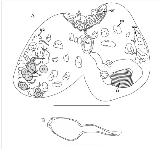
Figure 2.
Stolonica sabulosa Monniot C., 1972. (A) Open zooid with the pharynx removed. (B) Larva. AS—atrial siphon, EN—endocarp, FG—female gonad, MG—male gonad, OT—oral tentacles, ST—stomach. Scale bars: (A) 4.0 mm; (B) 1.0 mm.

Table 2.
Tabular key for the identification of the species here described.
Table 2.
Tabular key for the identification of the species here described.
| 1 | 2 | 3 | 4 | 5 | 6 | 7 | Species |
|---|---|---|---|---|---|---|---|
| C | S | P | R | L | R | P | Stolonica sabulosa Monniot C., 1972 |
| C | S | P | B | L,R | R | 0 | Amphicarpa paucigonas Monniot and Monniot, 1984 |
| C | S | 0 | B | 0 | S | 0 | Polyandrocarpa anguinea (Sluiter, 1898) |
| S | S | P | B | 0 | S | 0 | Polycarpa insulsa (Sluiter, 1898) |
| S | S | P | B | 0 | L | 0 | Styela plicata (Lesueur, 1823) |
| S | B | 0 | B | 0 | R | 0 | Molgula davidi Monniot C., 1972 |
1—Life mode: C—colonial, S—solitary. 2—Oral tentacles: S—simple, B—branched. 3—Endocarps on the body wall: P—present, 0—absent. 4—Hermaphroditic gonads: B—both sides, R—right side only. 5—Male gonads: L—left side, R—right side, 0—absent. 6—Shape of ovaries: L—long tubes, R—round, S—short and oval. 7—Incubation of larvae: P—present, 0—absent.
The esophagus is short and curved. The stomach is horizontal and ovoid, and has 22–24 parallel longitudinal folds, but no cecum was observed. The gut is isodiametric and only forms a very narrow primary loop. The anus is bilobed, and it is anterior to the intestinal loop (Figure 1D and Figure 2A). The atrial siphon has a crown of short tentacles at the base.
Aligned to the ventral margin on the right side, there are seven multilobed testicular follicles located very close to each other, with long sperm ducts (Figure 1E). There are also five ovaries, including 1–2 large and 1–2 smaller oocytes associated with the most posterior testis (Figure 2A). On the left ventral margin, there are only two multilobed testicular follicles, anterior to the intestinal loop (Figure 1F). Bulky endocarps of various sizes project from the body wall (Figure 2A). Some zooids have 1–3 spherical eggs incubated in the atrial cavity on the right side, as well as larvae with an ovoid 1.0 mm long trunk and a short tail of the same size. The larvae have two rounded adhesive papillae supported by short peduncles (Figure 1G and Figure 2B).
Remarks. Stolonica sabulosa Monniot, 1972, was described from a single colony found in Bermuda [8]. After some time, Claude Monniot described samples collected in the Abrolhos region at 15–24 m [9]. He attributed them to this species, as well as material collected on the coast of Piauí at 35 m and described by Millar under the name Polyandrocarpa (Monandrocarpa) stolonifera [10]. The zooids from Bermuda were longer (2 cm) and had round and not lobed testicles, but C. Monniot interpreted this morphology as a non-mature gonad [8], and the Abrolhos zooids had both morphologies. Given that the same author studied both samples and decided that they belonged to the same species, despite the small differences, the long geographic distance, and the few samples available, we decided to maintain this identification for our material. Although Monniot mentions a small cecum is his descriptions [8,9], we could not find one in the Brazilian sample. Two other species found in the Atlantic Ocean are S. socialis Hartmeyer, 1903, and S. conglutinata Sluiter, 1915. Stolonica socialis is found in Ireland, England, the English Channel, Belgium, and the Mediterranean (Spain). It has a third line of gonads along the intestine, which are male and in a rosette shape, which is not present in S. sabulosa [11]. The species S. conglutinata from the African coast also has lobed testicular follicles, but the anus is also deeply divided into six lobes, and there are approximately 14 testes with very short sperm ducts on both sides of the endostyle and a row of additional follicles near the intestine [12].
In the Pacific Ocean, some species of Stolonica also have multilobed testicular follicles. S. truncata Kott, 1972, differs from S. sabulosa by having a stomach with 36 longitudinal folds, the presence of 16 testes on the right and 4 on the left side, and 4 ovaries overlapping the testicles [13]. The species S. reducta (Sluiter, 1904) has only 12 rows of stigmata, a stomach with 20 longitudinal folds, and a curved gastric cecum, and most testes are ovoid with very short sperm ducts [13]. The species S. vesicularis Van Name, 1918, has 11 rows of stigmata, a rather short stomach with 14 longitudinal folds, and 9 slightly lobed testicular follicles on the right side, with relatively short sperm ducts [13].
Distribution: Bermuda [8], Florida [14], Brazil: Piauí? [10], Bahia [9], and Espírito Santo (present study).
Genus Amphicarpa Michaelsen, 1922
Amphicarpa paucigonas Monniot and Monniot, 1984
Material examined: DZUP AMP-05, AMP-11 Ilha Escalvada, Guarapari, Espírito Santo, 20°41′55″ S, 40°24′21″ W, 10 m, Col. R. M. Rocha, 28 and 30.03.2017; DZUP AMP-01 Naufrágio Victory 8B, Guarapari, Espírito Santo, 20°41′23″ S, 40°23′24″ W, 22 m, Col. R. Rocha, 12.02.2011; DZUP AMP-02 Naufrágio Victory 8B, Guarapari, Espírito Santo, 20°41′23″ S, 40°23′24″ W, 20 m, Col. R. M. Rocha, 26.01.2012; DZUP AMP-06, AMP-07 Ilha Rasa de Terra, Guarapari, Espírito Santo, 20°40′31.9″ S, 40°22′1.03″ W, 15 m, Col. R. M. Rocha, 27.03.2017; DZUP AMP-3, AMP-4 Boião da Barra, Salvador, Bahia 13°00′32″ S, 8°32′19″ W, 14 m, Col. R. M. Rocha, 12.12.2007; DZUP AMP-10, Naufrágio Ponta do Cristo Salvador, Bahia, 13°0′43″ S, 38°31′26″ W, 3–4 m, Col. R. M. Rocha, 13.12.2007.
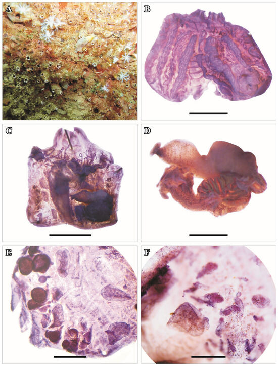
Figure 3.
Amphicarpa paucigonas Monniot and Monniot, 1984. (A) Colony on the Victory 8B shipwreck. (B) Dissected and stained zooid with the pharynx exposed. (C) Dissected zooid with the pharynx removed. (D) Gut with part of the stomach removed. (E) Gonads (right side). (F) Gonads (left side). Scale bars: (B,C) 2.0 mm; (D,E,F) 1.0 mm.
The colonies have zooids close together, united by stolons, that cover large areas > 50 cm in diameter. Many colonies were found on shipwrecks and a few on rocky substrates. The colonies were completely covered by fine sediment or sand, forming a dark background, where both apical siphons are visible because of the white lining interrupted by four longitudinal black lines (Figure 3A). The tunic is thin and resistant, a little wrinkled, and light brown.
The zooids are between 5 and 6 mm long. The body wall is thin and delicate, with an opaque brown color. The muscle fibers are very thin and dense, forming a layer of longitudinal and transverse musculature. There are 23–28 simple oral tentacles, enrolled anteriorly in four orders of size along a conspicuous muscle ring (Figure 3B,C and Figure 4A). A membrane velum covers half the length of the oral siphon anterior to the line of oral tentacles. The prepharyngeal area is wide and without any papillae. The prepharyngeal groove, which is simple and without projections, forms a V around the peritubercular area (Figure 3B,C and Figure 4A). The dorsal tubercle is large and occupies the distance between the oral tentacles and the prepharyngeal groove (approximately 0.5 mm), with an elongated vertical opening. The neural ganglion is round, and the neural gland is voluminous, appearing on both sides of the ganglion and interrupting the first row of stigmata on both sides.
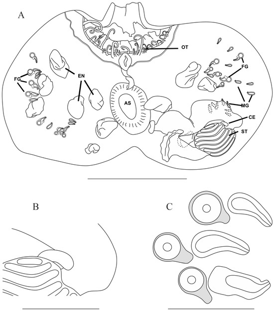
Figure 4.
Amphicarpa paucigonas Monniot and Monniot, 1984. (A) Dissected zooid without the pharynx. (B) Detail of the stomach with the gastric cecum. (C) Three hermaphroditic gonads. AS—atrial siphon, CE—cecum, EN—endocarp, FG—female gonad, MG—male gonad, OT—oral tentacles, ST—stomach. Scale bars: (A) 3.0 mm; (B) 1.0 mm; (C) 0.2 mm.
The dorsal lamina is simple with uniform width throughout its length, passing the right side of the esophagus till the end of the pharynx. The pharynx has 15–16 rows of stigmata and 3–4 very long stigmata per mesh crossed by parastigmatic vessels. The number and organization of the pharyngeal folds vary among the zooids. There can be three or four folds, some of them very flat and marked by the closeness of the longitudinal vessels, and some may disappear on the posterior half of the pharynx (Figure 3B). The arrangement of the longitudinal vessels, from the right to the left side of the body, in three different zooids is as follows:
E 6 (7) 3 (7) 4 (9) 2 DL 3 (12) 6 (8) 2 (4) 0 E
E 7 (6) 3 (2) 7 (6) 4 DL 1 (7) 3 (8) 3 (5) 4 (6) 0 E
E 0 (5) 2 (10) 3 (5) 3 (10) 0 DL 0 (12) 2 (12) 3 (5) 0 E
The abdomen is loosely attached to the body wall by thin filaments. The esophagus is long and broad. The stomach is ovoid and has 13–14 internal parallel longitudinal folds. In the pyloric region, there is a curved gastric cecum (Figure 3D and Figure 4B). The primary intestinal loop is quite closed, approaching the intestine to the stomach in a horizontal position. The intestine has a wide diameter, narrowing very close to the anus. The anus is bilobed and is located at the same height as the intestinal loop when there is no secondary intestinal loop (Figure 3D); but in some zooids, the intestine bends anteriorly and the anus is close to the atrial siphon. The atrial siphon has a crown of small tentacles at its base (Figure 4A).
The gonads are present on both sides, but the number varies among the zooids. We counted, at most, 13–14 hermaphroditic gonads on the right side but usually 5–6 in a line parallel to the endostyle. In one zooid, there are another four male gonads located far posteriorly on the right side. On the left side, there are, at most, 15 hermaphroditic gonads, and they are anterior to the gut, with another 6 to 8 male gonads more or less hidden by the gut (Figure 3E,F and Figure 4C).
Numerous endocarps of various sizes, but usually larger than the gonads, are on the body wall. Inserted into the intestinal loop is a single, voluminous ovoid-shaped endocarp (Figure 4A).
Remarks. There is a dispute about the diagnostic characteristics of the genera Stolonica and Amphicarpa because of the large phenotypic variation, and identification is based mostly on the mature gonads’ pattern and morphology, characteristics that are not always available [14,15]. The Register of Marine Species shows all species of Amphicarpa, but A. duploplicata (Sluiter, 1913) is used as a synonym of Stolonica without any literature to support this decision [16]. Without a detailed genetic analysis, it is almost impossible to evaluate the status of these two genera, and we will maintain the classification proposed for the species in the literature. This is the first record of the genus Amphicarpa on the Brazilian coast, and A. paucigonas is the only species found in the Atlantic Ocean. The main characteristics to distinguish this species are the white siphons in living colonies, the large dorsal tubercle with a vertical aperture and a large neural gland, the stomach with 13–14 folds and a conspicuous curved cecum, and the presence of 5–8 male gonads in an area covered by the gut in ventrally dissected animals. Brazilian zooids have more variable pharyngeal fold patterns with more longitudinal vessels and a larger number of gonads than Caribbean colonies [9].
Distribution. Martinique [9,17] and Brazil: Bahia and Espírito Santo (present study).
Genus Polyandrocarpa Michaelsen, 1904
Polyandrocarpa anguinea(Sluiter, 1898)
Material examined: DZUP PODC-15 01 colony, Escalvada Island, Guarapari, Espírito Santo, 20°42′00″ S, 40°24′30″ W, 13 m, Col. R. M. Rocha, 02.12.2011, DZUP STY-81 Quebra Mar Sul, Salvador, Bahia, 12°58′24″ S, 38°31′04″ W, 9–11 m, Col. R. M. Rocha, 07.08.1999; DZUP STY94 Bahia Marina 12°58′37″ S, 38°31′14″ W, 4–6 m, Col. R. M. Rocha, 03.08.1999.
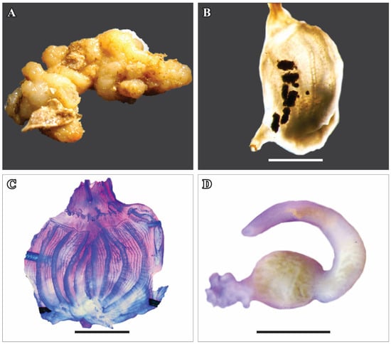
Figure 5.
Polyandrocarpa anguinea (Sluiter, 1898). (A) Colony preserved in formalin. (B) Zooid without the tunic. (C) Dissected and stained zooid. (D) Digestive tract. Scale bars: (B) 5.0 mm; (C) 3.0 mm; (D) 2.0 mm.
The colony is 10 cm in length and 1.3 cm in thickness and consists of a mass of aggregated zooids (Figure 5A). The coloration is light brown, disappearing after fixation. The surface of the colony is rough and leathery, without clear individualization in the position of the zooids. The colony adheres firmly to the substrate, and there is sand impregnation at the base. The body wall is very delicate and creamy yellow (Figure 5B).
The zooids are of different sizes in the same colony, between 1.0 and 1.7 cm long. Both the oral and atrial siphons are apical and elongated, with smooth margins, and they open onto the surface of the tunic. The oral tentacles number approximately 18, in three sizes, the largest being 0.8 mm long (Figure 5C). The dorsal tubercle measures approximately 0.2 mm in diameter, with a “C”-shaped opening, with highly curved ends. The dorsal lamina is slightly prominent and has a jagged margin due to projections formed by the ends of the transversal vessels.
The pharynx has four longitudinal folds on each side, with the following distribution of longitudinal vessels in a zooid 1.0 cm long from the right to the left side: E 4 (12) 5 (12) 5 (12) 5 (10) 5 DL 3 (8) 4 (11) 6 (12) 5 (12) 4 E. Parastigmatic vessels are present.
The digestive tract is located beside the pharynx, occupying the posterior one-third of the left side of the animal. The esophagus is short and slightly curved. The stomach is ovoid, with 16 evident longitudinal sinuous folds in the wall. The intestine is isodiametric but slightly wider at the beginning and undergoes continuous narrowing up to the anus. There is only the primary loop, forming a curvature toward the anus, which opens close and dorsally to the esophagus aperture (Figure 5D). There are no endocarps. The gonads are elongated, small (0.4–0.5 mm), and firmly attached to the body wall. On the right side, there are 10–13 gonads, while on the left side, there are only 4–7.
Remarks. This species exhibits large morphological plasticity [18,19] in almost all characteristics, and a global molecular study is needed to clarify its current distribution. All the species with zooids agglomerating without a specific order in a tough tunic with long siphons protruding from the surface of the colony have been synonymized with P. anguinea [19]. Besides P. anguinea, there are three Polyandrocarpa species known in the Atlantic: P. zorritensis (Van Name, 1931), P. arianae Monniot, F. 2016, and P. aurorae Monniot, F. 2018. All three species have upright zooids covered by sand and linked by their bases only, a very different colony organization compared to P. anguinea.
Distribution of Atlantic populations: Brazil: Espírito Santo [4], Rio de Janeiro [20,21], São Paulo [18,22,23], Paraná [24], and Santa Catarina [25]; Caribbean Sea [26]; Florida, United States of America [27]; Martinique [14,17]; and South Africa [28,29,30].
Genus Polycarpa Heller, 1877
Polycarpa insulsa (Sluiter, 1898)
Material examined: DZUP POLC-76 01 individual, Ilha Rasa de Terra, Guarapari, Espírito Santo, 20°40′32″ S, 40°22′01″ W, 10–15 m, leg. R. M. Rocha, 27.03.2017
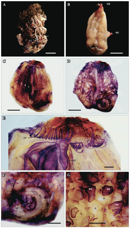
Figure 6.
Polycarpa insulsa (Sluiter, 1898). (A) Preserved individual. (B) Individual without the tunic. (C) Dissected and stained individual, with the four right pharyngeal folds marked. (D) Dissected and stained individual without the pharynx. (E) Dissected and stained anterior region, highlighting the oral tentacles and dorsal tubercle. (F) Detail of the gut with endocarps. (G) Detail of the gonads with endocarps. AN—anus, AS—atrial siphon, DT—dorsal tubercle, EN—endocarp, IN—intestine, G—gonad, OS—oral Siphon, OT—oral tentacles, ST—stomach. Scale bars: (A–D) 1.0 cm; (E) 1.0 mm; (F) 4 mm; (G) 2 mm.
The only individual found is 4.5 cm long and 2.5 cm wide, dark brown, with siphons close to each other. The tunic is 1–2 mm thick, brown internally, and has a rough and wrinkled external surface with little encrustation (Figure 6A). The musculature forms a thick sheet on the body wall, turning it opaque and cream-colored, with the tips of the siphons red (Figure 6B).
The oral siphon is apical, very short, and has a red velum. There are 46 oral tentacles of at least five sizes, the largest being 5 mm long. They are more or less triangular in cross section, with the tip of the triangle facing anteriorly and the rounded base posteriorly. The prepharyngeal area is narrow without any papillae. The peripharyngeal groove is simple and forms a deep V around the dorsal tubercle, whose aperture is a horizontal S (Figure 6E). The dorsal lamina is continuous and narrow with a smooth rim, extending until the esophagus aperture.
There are four pharyngeal folds on each side (Figure 6C). They are very shallow and separated from each other, mainly marked by the concentration of longitudinal vessels. The most dorsal left fold is very short, circa one-fourth of the length of the others, and disappears into the dorsal lamina just or immediately posterior to the peritubercular area. The blood vessel formula is the following, from the right to the left side of the body: E 6 (14) 7 (15) 7 (20) 6 (17) 4 DL 4 (17) 13 (15) 9 (17) 11 (13) 6 E. There are 6–8 stigmata per mesh, but the meshes at the right side of the dorsal lamina are quite irregular. Parastigmata vessels are not common.
The digestive tract is small, occupying less than one-quarter of the left side (Figure 6D). The stomach is round without external folds, followed by a short intestinal loop and vertical rectum, ending in a smooth anus with a typhlosole. No cecum was found. There are six elongated endocarps in the intestinal loop and two between the rectum and esophagus. These endocarps appear to have ramified blood vessels inside (Figure 6F). The velum at the base of the atrial siphon is covered by thin, elongated tentacles.
Oval or elongated endocarps are spread on the body wall of both sides in between the gonads. The gonads are sac-like, embedded in the body wall, and elongated or irregularly shaped (Figure 6G). There are more than 30 on the right side but fewer than on the left side. The gonoducts are short and open side by side.
Remarks. Eight species of Polycarpa are currently known in Brazil [1]; this is the first report on P. insulsa. The characteristics of the animal described here are very similar to the ones given by Van der Sloot [31], C. Monniot [32], F. Monniot [14], and Monniot et al. [33]. Van der Sloot [31] synonymized P. insulsa from the Atlantic and P. circumarata from Indonesia, but Monniot [33] and Kott [13] did not agree and maintained that they were separate species, although Kott considered P. circumarata a junior synonym of P. aurita (Sluiter, 1990). Later, Kott [34] changed her opinion, and because of the large variation in characteristics and the extended geographic distribution of both species, she considered the Atlantic P. insulsa to also be a junior synonym of P. aurita. Descriptions of Atlantic and Indo-Pacific populations show that the latter tend to have fewer oral tentacles (<30), more longitudinal blood vessels (>300), a longer rectum, and a lobed anus. The area covered by the gonads does not include the posterior third of the right side, and the tunic is more translucent and yellowish, while Atlantic populations have a dark brown or black tunic. But there are exceptions to all these trends, and there is no consistency in the presence of a pyloric cecum and a typhlosole along the rectum and anus in any geographical region. A genetic study including populations in all oceans could clarify if there are two species and the real geographical distribution of each.
Distribution of Atlantic populations: Gulf of Mexico, Florida, United States of America [27]; Cuba [35]; Panama [36]; Colombia; Venezuela [26]; Guiana Shelf [37]; Martinique [14]; and Brazil (present study).
Genus Styela Fleming, 1822
Styela plicata (Lesueur, 1823)
Material examined: DZUP STY-151 02 individuals, Guarapari, Espírito Santo, 20°39′50″ S 40°29′41″ W, 1 m, Col. R. M. Rocha, 13.02.2011, DZUP STY-153 Terminal Náutico Salvador, Salvador, Bahia, 12°58′21″ S, 38°30′56″ W, 0.5 m, Col. R. M. Rocha, 02.03.2012; DZUP STY-156 Marina Píer Salvador, Salvador, Bahia, 12°54′44″ S, 38°29′28″ W, 0.5 m, Col. R. M. Rocha, 03.03.2012.
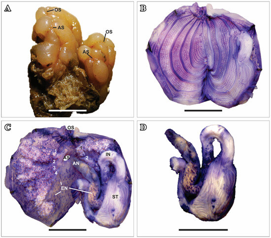
Figure 7.
Styela plicata (Lesueur, 1823). (A) Two preserved individuals. (B) Dissected and stained individual. (C) Dissected and stained individual without the pharynx, showing four gonads on the right side and two on the left. (D) Detail of the gut with endocarps. AN—anus, AS—atrial siphon, EN—endocarp, G—gonad, IN—intestine, OS—oral siphon, OT—oral tentacles, ST—stomach. Scale bars: (A–D) 1.0 cm.
Two young individuals, 3.0 cm long, that were found on the hull of a boat taking divers to the islands in Espírito Santo will be described here. Their tunic is thick, resistant, and leathery, with many grooves and folds. The animal’s coloration is beige, both alive and preserved in formalin. Both siphons are apical, close to each other, and short, with four rounded lobes each (Figure 7A). The body wall is not very thick, slightly translucent, and light beige. There are 25–27 simple oral tentacles of three sizes, enrolled anteriorly. The prepharyngeal groove is double, without projections, forming a V-shaped peritubercular area. The dorsal tubercle measures 0.8–1.1 mm in anteroposterior diameter and has a U-shaped opening with curved tips.
The dorsal lamina is simple, with uniform width throughout its length, ending next to the esophageal opening. The pharynx has, on each side, four folds that are not very high and approximately 65 longitudinal blood vessels (Figure 7B). Parastigmatic vessels are present. The esophagus is long and curved. The stomach is elongated and has numerous thin parallel longitudinal folds. The intestine has very closed primary and secondary loops, covered by endocarps. The lobed anus is situated anteriorly to the esophagus, close to the atrial siphon.
The gonads are present on both sides, four on the right side and two on the left side, and are formed by an elongated tubular ovary surrounded by a multilobed testis well attached to the body wall (Figure 7C,D). Numerous endocarps are present on the body wall and between the gonads.
Remarks. This is a well-known species, easily recognizable by the beige or white tunic, which is smooth and usually with few encrustations, with four expanded rounded lobes around both siphons. There are 73 valid species in Styela [16], among which only seven species have more than two gonads on each side. Two of them have been recently described from the Atlantic: S. multicarpa (Barros and Rocha, 2021) has a brown tunic, the testicle follicles are attached to the body wall by their proximal end only, and the intestine wall has no endocarps [38]; S. multicarpa (Barros and Rocha, 2021) is greenish-brown, and the testicle follicles are attached to the body wall by their proximal end only [38]. From the Pacific, S. maeandria Sluiter, 1904, does not have endocarps, and S. clava Herdman, 1881, has an elongate body with a conspicuous peduncle and protuberances on the tunic surface. Two are deep sea species, with different testicle morphology compared with S. plicata: S. adriatica Monniot F. and Monniot C., 1975, and S. thalassae Monniot C., 1969 (see table 2 in [38]).
Distribution of Atlantic populations. United States [39,40,41]; Bermuda [8]; Puerto Rico [42]; Guadeloupe [17]; Curaçao [43]; Brazil: Bahia [4], Rio de Janeiro [4,20,21], São Paulo [22,23,44], Paraná [45], and Santa Catarina [44]; Senegal [46]; and the Madeira Islands, Portugal [47].
Family MOLGULIDAE Lacaze-Duthiers, 1877
Genus Molgula Forbes, 1848
Molgula davidiMonniot C., 1972
Material examined: DZUP MOL-07 01 individual, Shipwreck Victory 8B, Guarapari, Espírito Santo, 20°41′23″ S, 40°23′24″ W, 20 m, leg. R. M. Rocha, 26.01.2012.
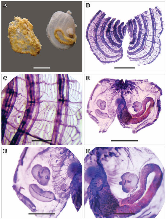
Figure 8.
Molgula davidi Monniot C., 1972. (A) Individual with tunic and without tunic (left side). (B) Stained pharynx. (C) Detail of the pharynx. (D) Dissected animal without the tunic. (E) Detail of the right side (gonad and renal sac). (F) Detail of the left side (digestive gland, intestine, and gonad). Scale bars: (A,B,D) 2.0 mm; (C) 1.0 mm; (E) 1.3 mm; (F) 0.9 mm.
The single individual found is approximately spherical, 5.0 mm in diameter, and was attached to algae on a shipwreck. The tunic is thin but sturdy and semi-transparent, covered by sand. The oral and atrial siphons are apical (Figure 8A,B). Both siphons are short and have six small, rounded projections. The body wall is very thin, delicate, and translucent. The longitudinal muscles originate in both siphons and end anteriorly to the gonads and intestine.
There are approximately 12 oral tentacles flattened laterally that ramify only once. They are distributed in three orders of size and are curved anteriorly. The prepharyngeal groove is double and without projections. The dorsal tubercle measures 0.18 mm anteroposteriorly, with an ovoid opening. The dorsal lamina is slightly prominent, with a smooth margin, displaced to the left side, and extends beyond the opening of the esophagus. The pharynx has seven longitudinal folds on the right side and six on the left side. The folds are distant from each other but closer and shorter in the dorsal region (Figure 8B). The arrangement of the longitudinal blood vessels from the right to the left side is the following: E. 0 (3) 0 (5) 0 (5) 0 (6) 0 (5) 0 (5) 0 (4) 0 DL 0 (4) 0 (4) 0 (5) 0 (7) 0 (7) 0 (4) 0 E. The stigmata have a simple spiral shape; they are interrupted and are counterclockwise. Parastigmatic vessels are present (Figure 8C).
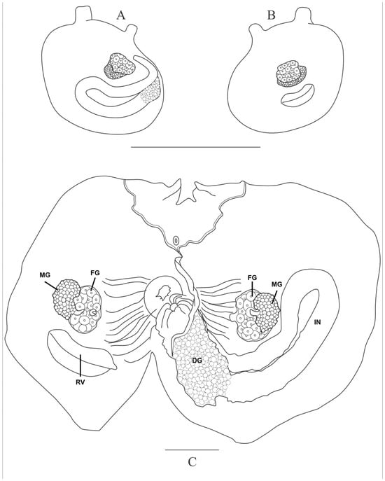
Figure 9.
Molgula davidi Monniot C., 1972. (A) External view of the left side without the tunic. (B) External view of the right side without the tunic. (C) Dissected animal without the pharynx. DG—digestive gland, FG—female gonad, IN—intestine, MG—male gonad, RV—renal vesicle. Scale bars: (A,B) 5.0 mm; (C) 1.0 mm.
The esophagus is curved and very short. The stomach is elongated and is entirely covered by a single yellowish digestive gland. The intestine is narrow and isodiametric; it has a closed and short primary loop and a very open secondary loop (Figure 8D and Figure 9C). There are no endocarps. The anus is circular and opens slightly below the level of the top of the primary intestinal loop.
On each side of the body, there is a gonad tightly attached to the wall. On the right side, it is located in the central region of the body, anteriorly to the renal sac. On the left side, the gonad is inserted in the secondary intestinal loop (Figure 9A,B). The gonads are formed by an oval testicle located ventrally to a small globular ovary containing many oocytes (Figure 8E,F and Figure 9C). Both the oviduct and sperm duct are short and have dorsally oriented apertures. The renal sac is small and elongated, slightly curved, posterior to the right gonad, and separated from it. Larvae were not found.
Remarks. This is the first record of the species on the Brazilian coast. It had been previously reported only from Bermuda [8], the type locality. The Bermuda specimens have only the female gonad on the left side; however, the author points out that perhaps this is an individual variation. The holotype also has a darker body wall, the prepharyngeal groove forms a deeper V around the dorsal tubercle, and fig 10C [8] shows that the siphons seem more distant from each other than in the Brazilian individual. Comparing other characteristics, the specimens studied here agree well with the original description of the species.
Distribution: Bermuda [8], Brazil (present study).
4. Discussion
Among the species reported here, Styela plicata is an invasive species, very common on bivalve farms in South Brazil and piers and marinas in Paraná, São Paulo, and Rio de Janeiro [24,48,49,50]. It is a subtropical species, and it was found only once in Bahia [4] before this study; thus, its occurrence in Espírito Santo was expected. As usual, it was found on artificial substrate in all sites, and not a single individual has been found on natural rocky reefs, suggesting a contained invasion in the region.
Polyandrocarpa anguinea has a wide geographical distribution in the Atlantic, Indian, and Pacific oceans, both in tropical and subtropical regions, and this suggests that the species might be involved in bioinvasion. Unfortunately, no global or regional molecular study has verified the identity of all reports and tried to reveal the probable native origin of the species. In Brazil, the samples collected in Bahia 25 years ago [4] represent the current northern geographical limit of the species, although it is expected to be found in other sites of the northeastern region, given that the species is known from the Caribbean and Florida [14,17,26,27]. The species was never abundant in any of the collection sites.
Polycarpa insulsa is also involved in taxonomic debate about its identity and synonymy with Pacific species, and no genetic study is available to clarify its geographical distribution. It is clearly a tropical species with wide distribution in the Caribbean Sea [14,26,35,36,37]; thus, its presence in Espírito Santo is surprising. Although in a tropical region, this region suffers from upwelling and very cold waters during the summer months [51]. The fact that this is the first individual found after many dives in Espírito Santo and other tropical sites in Brazil means that the species is either very rare in the country or a new non-established introduction.
The other three species reported in this study have a similar disjunctive geographical distribution between Bermuda and Brazil or Caribbean countries and Brazil. Stolonica sabulosa has been found before in south Bahia; thus, it is an established species, but it seems to be rare or difficult to find because it is covered by sediment in deep enough waters to require diving for sampling. Given that only one colony, which had immature gonads, has been found in Bermuda, it is possible that the populations do not belong to the same species. In this case, the Brazilian samples would belong to a new native species.
Amphicarpa paucigonas usually cover large areas on the substrate, and it is established in the region, given its presence in different years. The constant presence on shipwrecks in different sites and in a port area in Guadalupe (Port Saint-François, ref. [9]) suggest an affinity with hulls and that it is possibly transportable by vessels. Despite being covered by sediment, this species is not that difficult to find because of the large size of the colonies and white siphons. Thus, it seems that its distribution will not be much enlarged with a greater sampling effort. A molecular study of this species would also be welcome to clarify the connectivity among sites within Brazil and with Guadalupe and Martinique.
Molgula davidi is a very small species, covered by sand and impossible to spot in nature, and they were collected by chance and recognized only under magnification. It is worth mentioning that both individuals found until now were epibionts on artificial substrata. Thus, the geographic distribution of the species is still unknown but seems restricted to the Tropical West Atlantic.
Author Contributions
Conceptualization, methodology, formal analysis, and writing and editing, G.A.G. and R.M.d.R.; resources and funding acquisition, R.M.d.R. All authors have read and agreed to the published version of the manuscript.
Funding
This research was funded by FAPESP (2010/50190-2 and 13/502288-8), who financed the fieldwork in Bahia and Espírito Santo. R.M.d.R. received a research grant from Conselho Nacional de Pesquisa e Inovação—CNPq (306788/2022-5), and G.A.G. received a scholarship from Coordenadoria de Aperfeiçoamento Pessoal do Ensino Superior—CAPES (financial aid 001).
Data Availability Statement
Voucher specimens were deposited at the Ascidiacea Collection in the Department of Zoology, Federal University of Paraná (DZUP), Brazil.
Acknowledgments
We thank Universidade Federal da Bahia and Universidade Vila Velha for their logistical support during sampling campaigns. We also thank the diving operator Atlantes, who provided all the logistic support during the survey in Guarapari, and Isabela Monteiro Neves, Sandra Vieira de Paiva, and Catherine C. Peruzzolo, who helped during dives and material processing.
Conflicts of Interest
The authors declare no conflicts of interest. The funders were not involved in the study design, collection, analysis, interpretation of data, the writing of this article or the decision to submit it for publication.
References
- Rocha, R.M.; Lotufo, T.M.C.; Bonecker, S.; Oliveira, L.M.; Skinner, L.F.; Carvalho, P.F.C.; Silva, P.C.A. A synopsis of Tunicata biodiversity in Brazil. Zoologia 2024, 41, e23042. [Google Scholar] [CrossRef]
- Instituto Chico Mendes. Plano de Manejo: Refúgio de Vida Silvestre de Santa: Área de Proteção Ambiental Costa das Algas, 1st ed.; Instituto Chico Mendes-ICMBio: Brasília, Brazil, 2023; 104p.
- Monniot, C. Ascidies Phlébobranches et Stolidobranches. Résultats Scientifiques des Campagnes de la ‘Calypso’. Ann. Inst. Océanogr. 1970, 47, 35–59. [Google Scholar]
- Lotufo, T.M.C. Ascidiacea (Chordata: Tunicata) do Litoral Tropical Brasileiro. Ph.D. Thesis, Instituto de Biociências, Universidade de São Paulo, São Paulo, Brazil, 2002; 183p. [Google Scholar]
- Rocha, R.M.; Bonnet, N.Y.K.; Baptista, M.S.; Beltramin, F.S. Introduced and native Phlebobranch and Stolidobranch solitary ascidians (Tunicata: Ascidiacea) around Salvador, Bahia, Brazil. Zoologia 2012, 29, 39–53. [Google Scholar] [CrossRef]
- Rocha, R.M.; Salonna, M.; Griggio, F.; Ekins, M.; Lambert, G.; Mastrototaro, F.; Fiddler, A.; Gissi, C. The power of combined molecular and morphological analyses for the genus Botrylloides: Identification of a potentially global invasive ascidian and description of a new species. Syst. Biodiv. 2019, 17, 509–526. [Google Scholar] [CrossRef]
- Monniot, C.; Monniot, F. Clé mondiale des genres d’ascidies. Arch. Zool. Exp. Gén. 1972, 113, 311–367. [Google Scholar]
- Monniot, C. Ascidies Stolidobranches des Bermudes. Bull. Muséum Natl. D’histoire Nat. 1972, 43, 617–643. [Google Scholar] [CrossRef]
- Monniot, C.; Monniot, F. Ascidies littorales de Guadaloupe VII. Espèces nouvelles et complémentaire à l’inventaire. Bull. Muséum Natl. D’histoire Nat. 1984, 6A, 567–582. [Google Scholar] [CrossRef]
- Millar, R.H. Ascidians (Tunicata: Ascidiacea) from the Northern and Northeastern Brazilian Shelf. J. Nat. Hist. 1977, 11, 169–223. [Google Scholar] [CrossRef]
- Berrill, N.J. The Tunicata with an Account of the British Species; Ray Society: London, UK, 1950. [Google Scholar]
- Sluiter, C.P. Einige neue ascidien von der West-Küste Afrika’s. Tijdschr. Ned. Dierkd. Ver. 1915, 14, 37–57. [Google Scholar]
- Kott, P. The Australian Ascidiacea. Part 1. Phlebobranchia and Stolidobranchia. Mem. Qld. Mus. 1985, 23, 1–440. [Google Scholar]
- Monniot, F. Ascidians collected during the Madibenthos expedition in Martinique: 2. Stolidobranchia, Styelidae. Zootaxa 2018, 4410, 291–318. [Google Scholar] [CrossRef] [PubMed]
- Monniot, C. Ascidies de Nouvelle-Calédonie IV. Styelidae. Bull. Mus. Natl. Hist. Nat. 1988, 10, 163–196. [Google Scholar] [CrossRef]
- Shenkar, N.; Gittenberger, A.; Lambert, G.; Rius, M.; Moreira da Rocha, R.; Swalla, B.J.; Turon, X.; Ascidiacea World Database. Accessed Through: World Register of Marine Species. 2025. Available online: https://www.marinespecies.org/aphia.php?p=taxdetails&id=206939 (accessed on 24 February 2025).
- Monniot, C. Ascidies Littorales de Guadeloupe IV. Styelidae. Bull. Mus. Natl. Hist. Nat. 1983, 5, 423–456. [Google Scholar] [CrossRef]
- Rodrigues, S.A. Notes on Brazilian Ascidians. II: On the Records of Polyandrocarpa anguinea (Sluiter) and P. maxima (Sluiter). Revta Bras. Biol. 1977, 37, 721–726. [Google Scholar]
- Monniot, C.; Monniot, F. Additions to the inventory of eastern tropical Atlantic ascidians; Arrival of cosmopolitan species. Bull. Mar. Sci. 1994, 54, 71–93. [Google Scholar]
- Rocha, R.M.; Costa, L.V.G. Ascidians from Arraial do Cabo, RJ, Brazil. Iheringia Ser. Zool. 2005, 95, 57–64. [Google Scholar] [CrossRef]
- Granthom-Costa, L.V.; Ferreira, C.G.W.; Dias, G.M. Biodiversity of ascidians in a heterogeneous bay from southeastern Brazil. Manag. Biol. Invasions 2016, 7, 5–12. [Google Scholar] [CrossRef]
- Millar, R.H. Some ascidians from Brazil. Ann. Magaz. Nat. Hist. 1958, 13, 497–514. [Google Scholar] [CrossRef]
- Rocha, R.M.; Dias, G.M.; Lotufo, T.M.C. Checklist das ascídias (Tunicata, Ascidiacea) do Estado de São Paulo, Brasil. Biota Neotrop. 2011, 11, 1–11. [Google Scholar] [CrossRef]
- Bumbeer, J.; Cattani, A.P.; Chierigatti, N.B.; Rocha, R.M. Biodiversity of benthic macroinvertebrates on hard substrates in the Currais Marine Protected Area, in southern Brazil. Biota Neotrop. 2016, 16, e20160246. [Google Scholar] [CrossRef]
- Rocha, R.M.; Moreno, T.R.; Metri, R. Ascídias (Tunicata, Ascidiacea) da Reserva Biológica Marinha do Arvoredo, Santa Catarina, Brasil. Revta Brasil. Zool. 2005, 22, 461–476. [Google Scholar] [CrossRef]
- Sluiter, C.P. Tuniciers récueillis en 1896 par la Chazalie dans la Mers des Antilles. Mém. Soc. Zool. Fr. 1898, 11, 5–34. [Google Scholar]
- Van Name, W.G. Ascidians of the West Indian region and southeastern United States. Bull. Am. Mus. Nat. Hist. 1921, 44, 283–494. [Google Scholar]
- Sluiter, C.P. Beiträge zur Kenntniss der Fauna von Süd-Afrika. II. Tunicaten von Süd-Afrika. Zool. Jahrbücher 1898, 11, 1–75. [Google Scholar]
- Millar, R.H. On a collection of ascidians from South Africa. Proc. Zool. Soc. Lond. 1955, 125, 169–221. [Google Scholar] [CrossRef]
- Millar, R.H. Further descriptions of South African ascidians. Ann. S. Afr. Mus. 1962, 46, 113–221. [Google Scholar]
- Van der Sloot, C.J. Ascidians of the family Styelidae from the Caribbean. Stud. Fauna Curaçao Caribb. Islands 1969, 30, 1–57. [Google Scholar]
- Monniot, C. Ascidies de Nouvelle-Calédonie. II Les genres de Polycarpa et Polyandrocarpa. Bull. Mus. Natl. Hist. Nat. 1987, 9, 275–310. [Google Scholar] [CrossRef]
- Monniot, C.; Monniot, F.; Griffths, C.L.; Schleyer, M. South African Ascidians. Ann. S. Afr. Mus. 2001, 108, 1–141. [Google Scholar]
- Kott, P. The Australian Ascidiacea. Phlebobranchia and Stolidobranchia, Supplement. Mem. Qld. Mus. 1990, 29, 267–298. [Google Scholar]
- Hernandez-Zanuy, A. Lista de ascidias cubanas. Poeyana 1990, 388, 1–7. [Google Scholar]
- Rocha, R.M.; Faria, S.B.; Moreno, T.R. Ascidians from Bocas del Toro, Panama. I. Biodiversity. Carib. J. Sci. 2005, 41, 600–612. [Google Scholar]
- Millar, R.H. Ascidians from the Guyana Shelf. Netherl. J. Sea Res. 1978, 12, 99–106. [Google Scholar] [CrossRef]
- Barros, R.C.; Rocha, R.M. Two new species of Styela (Tunicata: Ascidiacea) from the tropical West Atlantic Ocean. Zootaxa 2021, 4948, 275–286. [Google Scholar] [CrossRef]
- Lesueur, C.A. Descriptions of several new species of Ascidia. J. Acad. Nat. Sci. Phila. 1823, 3, 2–8. [Google Scholar] [CrossRef]
- Van Name, W.G. The North and South American ascidians. Bull. Am. Mus. Nat. Hist. 1945, 84, 1–476. [Google Scholar]
- Nydam, M.L.; Stefaniak, L.M.; Lambert, G.; Counts, B.; López-Legentil, S. Dynamics of ascidian-invaded communities over time. Biol. Invasions 2022, 24, 3489–3507. [Google Scholar] [CrossRef]
- Streit, O.T.; Lambert, G.; Erwin, P.M.; López-Legentil, S. Diversity and abundance of native and non-native ascidians in Puerto Rican harbors and marinas. Mar. Poll. Bull. 2021, 167, 112262. [Google Scholar] [CrossRef]
- Goodbody, I. The ascidian fauna of two contrasting lagoons in the Netherlands Antilles: Piscadera Baai, Curaçao, and the lac of Bonaire. Stud. Fauna Curaçao Caribb. Islands 1984, 67, 21–61. [Google Scholar]
- Rodrigues, S.A. Algumas ascídias do litoral sul do Brasil. Bol. Fac. Filos. Ciênc. Let. Univ. São Paulo 1962, 24, 193–216. [Google Scholar] [CrossRef]
- Moure, J.S.; Björnberg, T.K.S.; Loureiro, T.S. Protochordata ocorrentes na entrada da Baía de Paranaguá. Dusenia 1954, 5, 233–242. [Google Scholar]
- Pérès, J.M. Contribution à l’étude des Ascidies de la côte occidentale d’Afrique. Bull. Inst. Fr. Afr. Noire 1949, 11, 159–207. [Google Scholar]
- Ramalhosa, P.; Gestoso, I.; Rocha, R.M.; Lambert, G.; Canning-Clode, J. Ascidian biodiversity in the shallow waters of the Madeira Archipelago: Fouling studies on artificial substrates and new records. Reg. Stud. Mar. Sci. 2021, 43, 101672. [Google Scholar] [CrossRef]
- Rocha, R.M.; Kremer, L.P.; Baptista, M.S.; Metri, R. Bivalve cultures provide habitat for exotic tunicates in southern Brazil. Aquat. Invasions 2009, 4, 195–205. [Google Scholar] [CrossRef]
- Skinner, L.F.; Barboza, D.F.; Rocha, R.M. Rapid Assessment Survey of introduced ascidians in a region with many marinas in the southwest Atlantic Ocean, Brazil. Manag. Biol. Invasions 2016, 7, 13–20. [Google Scholar] [CrossRef]
- Rocha, R.M.; Kremer, L.P. Introduced ascidians in Paranaguá Bay, Paraná, southern Brazil. Rev. Bras. Zool. 2005, 22, 1170–1184. [Google Scholar] [CrossRef]
- Palóczy, A.; Brink, K.H.; Silveira, I.C.; Arruda, W.Z.; Martins, R.P. Pathways and mechanisms of offshore water intrusions on the Espírito Santo Basin shelf (18° S–22° S, Brazil). J. Geophys. Res. Oceans 2016, 121, 5134–5163. [Google Scholar] [CrossRef]
Disclaimer/Publisher’s Note: The statements, opinions and data contained in all publications are solely those of the individual author(s) and contributor(s) and not of MDPI and/or the editor(s). MDPI and/or the editor(s) disclaim responsibility for any injury to people or property resulting from any ideas, methods, instructions or products referred to in the content. |
© 2025 by the authors. Licensee MDPI, Basel, Switzerland. This article is an open access article distributed under the terms and conditions of the Creative Commons Attribution (CC BY) license (https://creativecommons.org/licenses/by/4.0/).
