Article

Integrative Taxonomy of Metarhabditis Associated with Parasitic Otitis in Dairy Cattle

by
Makoto Enoki Caracciolo
1,2,
Beatriz Elise de Andrade-Silva
2,
Victor Hugo Borba
2,3,
Ander Castello-Branco
2,
Hudson Andrade dos Santos
4,
Alena Mayo Iñiguez
3,* and
Eduardo José Lopes-Torres
2,5,*
1
Centro Multiusuário para Análise de Fenômenos Biomédicos, Universidade do Estado do Amazonas, Av. Carvalho Leal, 1777, Manaus 69065-001, AM, Brazil
2
Laboratório de Helmintologia Romero Lascasas Porto, Departamento de Microbiologia, Imunologia e Parasitologia, Universidade do Estado do Rio de Janeiro, Av. Professor Manoel de Abreu, 444, Vila Isabel 20550-170, RJ, Brazil
3
Laboratório de Parasitologia Integrativa e Paleoparasitologia, Fundação Oswaldo Cruz, Instituto Oswaldo Cruz, Av. Brasil 4365, Pavilhão Osório de Almeida, Sala 16, Manguinhos, Rio de Janeiro 21041-250, RJ, Brazil
4
Departamento de Parasitologia, Instituto de Ciências Biológicas—ICB, Universidade Federal de Minas Gerais, Av. Antônio Carlos, 6627-Caixa Postal 486-Pampulha, Belo Horizonte 31270-901, MG, Brazil
5
Laboratório Multiusuário de Parasitologia, Departamento de Microbiologia, Imunologia e Parasitologia, Universidade do Estado do Rio de Janeiro, Av. Professor Manoel de Abreu, 444, Vila Isabel 20550-170, RJ, Brazil
*
Authors to whom correspondence should be addressed.
Taxonomy 2024, 4(3), 464-486; https://doi.org/10.3390/taxonomy4030023
Submission received: 30 April 2024 / Revised: 19 June 2024 / Accepted: 25 June 2024 / Published: 4 July 2024

Abstract

:
Parasitic otitis is an inflammatory process that can affect the external to internal cattle ear, causing discomfort in animals, impairing performance, and even leading to animal death. The infection was initially associated with nematodes of the Rhabditis genus in tropical and subtropical regions. Currently, the nematode species described as associated with bovine otitis are Metarhabditis costai, Metarhabditis freitasi, and, more recently, M. blumi. It is worth highlighting that there is still a lack of robust information regarding the morphological details, ultrastructural aspects, and molecular biology data of these species. The Metarhabditis genus is composed of seven species and two more have recently been added. The objective of this study is to update the morphological data using advanced microscopy techniques to emphasize and clarify the main morphological differences between three species of Metarhabditis currently associated with parasitic otitis. Samples of inflammatory exudate were collected from four adult female Gir cattle (Bos taurus indicus) on a farm in Itabira, Minas Gerais state, Brazil. Specimens were analyzed using light microscopy and scanning electron microscopy. Two species, M. costai and M. freitasi, were morphologically identified, consistent with previous reports. Scanning electron microscopy revealed new structural characteristics of the nematode species involved in parasitic otitis compared with M. blumi obtained from the CGC Center. Significant differences were observed in the male posterior region, bursa, and tail. Molecular analysis was conducted to differentiate these three species, and it was observed that the species first associated with otitis formed distinct clusters compared to M. blumi. However, it is important to note that further studies are needed to genetically characterize species of the Metarhabditis genus.

1. Introduction

Livestock farming plays a central role in sustaining rural communities worldwide, holding significant social, economic, and political importance at both national and international levels. Studies show that the bovine Gir, an Indian-born dairy cattle breed, possesses a certain predisposition for the development of parasitic otitis because of the anatomical structure of the ear, causing cerumen accumulation and creating a favorable condition for the development of microorganisms and nematodes [1,2,3]. The top five milk-producing countries are India, the United States, Pakistan, China, and Brazil. The European Union (EU)-27 is the largest dairy producer globally, accounting for 25.2% of the world’s milk production [4,5]. The specific identification of parasites is fundamental for precise diagnosis and correct treatment. The veterinary impact of helminthiasis caused by nematodes results in significant economic losses due to production delays and costs associated with prophylactic treatment [6]. Taxonomy science is an important discipline to help address these challenges.
Parasitic otitis is an inflammatory process that can affect the external, middle, or inner ear of cattle of all ages, occurring in isolated cases or entire herds [7]. In tropical and subtropical regions, parasitic otitis can be caused by mites and nematodes, often associated with diverse bacterial and fungal microbiota [2,8]. In severe cases, parasitic otitis can promote serious damage to the facial nerves, the formation of abscesses in the central nervous system, and, in grave cases, the death of the animal [9]. The infection caused by nematodes of the Rhabditis Dujardin, 1845 genus can affect both ears and cause a characteristic purulent discharge with parasites. Cases of parasitic otitis were previously documented in African countries but, with limited or no details regarding the morphological and morphometric features, often included only epidemiological studies [10,11]. Additionally, Rhabditis spp. nematodes, likely belonging to a Metarhabditis Tahseen, Hussain, Tomar, Shah and Jairajpuri, 2004 species, were reported in the human outer ear canal in Germany [12].
Rhabditidae is one of the richest nematode groups, whose members are usually terrestrials, preferring saprophyte-dwelling feces and decaying matter [13]. Members of the Rhabditis genus are reported as parasites of earthworm cocoons and insects [14,15]. Several cases of infection in humans by these nematodes were reported, showing their great adaptability and potential risk in other hosts [16,17,18]. The genus Metarhabditis was proposed in 2004 with the description of the Metarhabditis andrassyana Tahseen et al. [19]. Posteriorly, Sudhaus [20] included other species from the genera Rhabditis and Oscheius Andrássy, 1976 into the Metarhabditis genus. The genus Metarhabditis currently has nine known species [19,21].
Parasitic otitis caused by mites of the Raillietia genus and Metarhabditis spp. nematodes is usually associated with diverse bacterial and fungal microbiota [9,22]. Many African countries, characterized by warm and humid climates, have reported cases of parasitic otitis associated with Metarhabditis spp. [10,11,23].
In Brazil, the first report was described by Martins and collaborators [22], and a subsequent study identified two new species, Rhabditis costai Martins, 1985, and Rhabditis freitasi Martins, 1985, from Gir cattle [24]. Posteriorly, both species were reallocated under the genus Metarhabditis [20], and new cases of this nematode associated with parasitic otitis in Gir cattle were reported in Brazil [25,26]. Bossi et al. [27] isolated nematodes from Gir cattle with otitis from farms in the municipality of Itabira in the state of Minas Gerais, Brazil. Using genetic analysis without morphological characterization, they concluded that these nematodes belonged to the species Metarhabditis blumi Sudhaus, 1974. The present study provides a comprehensive characterization of Metarhabditis costai, M. freitasi, M. blumi, and Metarhabditis sp. through integrative taxonomy, incorporating light and scanning electron microscopy (SEM) together with genetic analyses.
The aim of this research is to enhance the morphological data through advanced microscopy techniques and molecular characterization. This effort highlights the critical importance of accurately identifying nematodes linked to parasitic otitis, particularly in dairy cattle.

2. Materials and Methods

2.1. Study Area and Sample Collection

Inflammatory exudate samples were collected from the ear canals of Gir cattle (Bos taurus indicus) with otitis clinical signals using a sterile swab at two farms located in the municipalities of Teófilo Otoni and Itabira, state of Minas Gerais, southeastern Brazil. Specimens of Metarhabditis blumi were acquired from the Caenorhabditis Genetics Center (CGC) and cultivated according to Stiernagle [28]. All nematodes were fixed by immersion in AFA at 60 °C (2% glacial acetic acid, 3% formaldehyde, and 95% ethanol) for light microscopy (LM) and in Karnovsky’s solution for Scanning Electron Microscopy (SEM). Some specimens were frozen immediately and stored at −20 °C for genetic analysis. Sample collection was conducted under the supervision of medical veterinarians and approval was granted by the Animal Use Ethics Committee of UERJ (Date: 11 October 2022—No. 030/2022).

2.2. Light Microscopy (LM)

Approximately 50 specimens were clarified in lactophenol, mounted as temporary slides, and examined using an Olympus BX51 light microscope (Olympus Corporation, Tokyo, Japan). The drawings for the morphometric analyses were made with the aid of a camera lucida coupled with Olympus BX 51. The drawings were made using this equipment, scanned, and later vectorized using Corel Draw 5.0 software. Images were captured with a digital camera (Nikon DS-Ri7 and Olympus DP-030) and using a light microscope (Nikon, Tokyo, Japan and Zeiss, Jena, Germany). Images were edited using Adobe® Photoshop® CS. The species were first identified based on morphological and morphometric characteristics, following the method of Martins [24] and Sudhaus [20,21]. The measurements are given in micrometers (µm).

2.3. Scanning Electron Microscopy (SEM)

For SEM, twenty fixed specimens were post-fixed in 1% OsO4 and 0.8% K3Fe (CN)6, dehydrated in graded ethanol, critical-point dried in CO2, mounted on stubs, coated with gold, and examined using scanning electron microscopes (Zeiss Auriga 600 Compact and FEI Quanta 25).

2.4. Genetic Characterization

Twenty females recovered from ear cattle were isolated in different Petri dishes for 48 h at 37 °C. Males and females were recovered and morphologically identified. Genomic DNA was extracted from a pool of nematodes of M. freitasi. Furthermore, to increase the robustness of our analysis, DNA was also extracted from the nematode species Rhabiditis dudichi Andrássy, 1970 and Caenorhabditi remanei Sudhaus, 1974. M blumi, provided by the Caenorhabditis Genetics Center (CGC), and used as control. For all the samples, the QIAamp DNA Mini Kit (Qiagen, Düsseldorf, Germany) was used, according to the manufacturer’s instructions with modifications [29]. DNA concentrations were estimated using a QuantusTM Fluorometer (Promega, Madison, WI, USA).
DNA amplification using polymerase chain reaction (PCR) was conducted using the D2/D3 regions of the 28S rDNA gene as molecular targets, which were amplified using the following primers: forward #391 (5′-AGCGGAGGAAAAGAAACTAA-3′) and reverse #501 (5′-TCGGAAGGAACCAGCTACTA-3′) [27]. The PCR reactions were carried out in a total volume of 25 μL, containing 12.5 μL of GoTaq® G2 DNA Polymerase Master Mix (Promega, USA), 10 μL of each primer, and 50–100 ng of DNA. PCR cycling parameters included thermal conditions of a step at 94 °C for 3 min followed by 35 cycles of 94 °C for 30 s, 54 °C for 30 s, and 72 °C for 1 min, followed by a post-amplification extension at 72 °C for 7 min. Successfully amplified products were purified using the QIAquick PCR Purification Kit (Qiagen, Germany), following the manufacturer’s protocol. The sequencing reaction was performed following the protocols of the sequencing platform RPT01A/IOC-Fiocruz (Applied Biosystems ABI 3730 sequencer, Waltham, MA, USA) (https://plataformas.fiocruz.br/unidades/RPT01A, accessed on 20 June 2024). Electropherograms were assembled into contigs and edited for errors and ambiguities using the Geneious Prime 2022.2.1 software (https://www.geneious.com, accessed on 20 June 2024). resulting in consensus sequences. Nucleotide sequence data reported in this paper are available in the GenBank database under the accession numbers: PP571891 Metarhabditis blumi, PP571892 Rhabditis dudichi, PP571893 Caenorhabditis remanei, and PP571894 Metarhabditis freitasi.

2.5. Phylogenetic Analyses

Phylogenetic analysis was conducted with the D2/D3 segment of the 28S rDNA gene. The dataset consisted of 20 taxa, including sequences from the present study and those available in GenBank, which are representatives of different genera. As an outgroup, we added tree sequences of genus Heterarhabditis Poinar, 1976: H. indica Poinar, Karunakar and David, 1992 (KU214824), H. zelandica Poinar, 1990 (DQ145666), and H. bacteriophora Poinar, 1976 (KT378445). GenBank accession numbers were appended to the taxa names in the phylogenies. The alignment of 28S rDNA gene sequences was performed using the ClustalW multiple sequence alignment program [30]. The resulting alignment was edited and regions with poor overlap were trimmed using the software Mesquite Version 3.70 [31]. Phylogenetic reconstruction using the Maximum Likelihood (ML) method as the optimality criterion was carried out using the PhyML 3.0 web server [32]. The best-fit nucleotide evolutionary model was calculated under the Akaike Information Criterion (AIC), via SMS (Smart Model Selection) [33]. Branch supports were assessed by the approximate likelihood-ratio test (aLRT) [34] and by bootstrap percentages (ML-BP) after 1000 replicates. Bayesian phylogenetic inference (BI) was carried out using MrBayes version 3.2.7a [35] on XSEDE using the CIPRES Science Gateway [36].
We sampled Markov Chain Monte Carlo (MCMC) for 10,000 generations, with four simultaneous chains, in two runs, at every 100 generations, after discarding an initial burn-in of 25%. The robustness of nodes was assessed using Bayesian posterior probabilities (BPPs) calculated from the sampled trees. To assess the BI sampling adequacy, we used Tracer v1.6 [37] to calculate the Effective Sample Sizes (ESSs) of each parameter. We considered values of over 100 effectively independent samples as adequate.

3. Results

3.1. Taxonomy Identification

3.1.1. Metarhabditis costai (Martins, 1985), Sudhaus, 2011

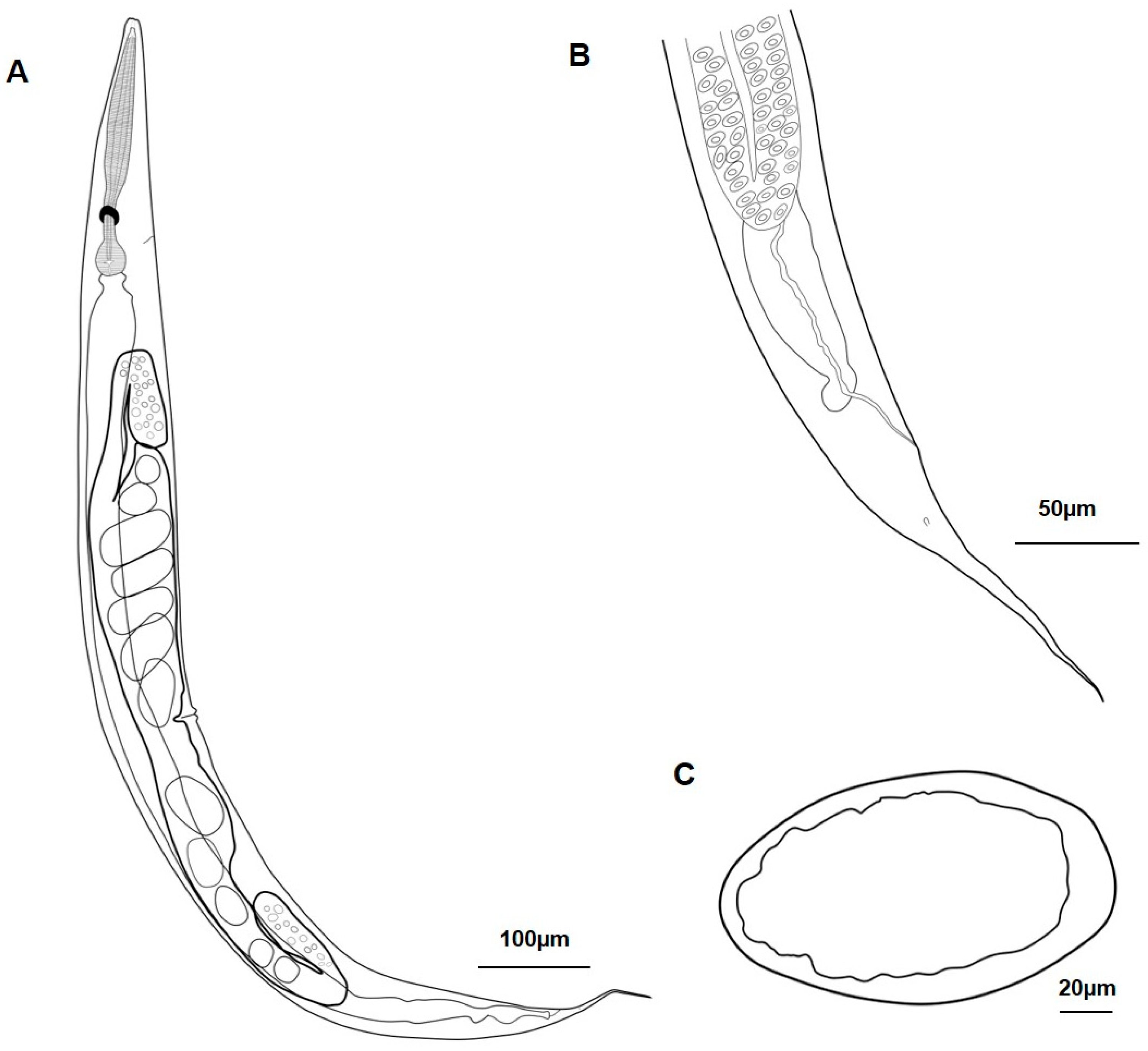
General: The anterior end consists of slightly projected tree lips, the pharynx is without a distinct median bulb, the excretory pore after the never ring at the isthmus and pharynx ends with a piriform basal bulb (Figure 1A,B and Figure 2A,B). A single testicle is reflected ventrally on the left side of the intestine (Figure 1A and Figure 2A); robust spicules are supported by a trough-shaped gubernaculum. The bursa is open and of the leptoderan type, with genital papillae (GP) and phasmids (PH); it has the following configuration: 1 + 1/1/3 + 2 + PH (Figure 1C and Figure 2C,D). The first and second pairs are spaced and pre-cloacal. The third pair is ad cloacal and close to the second pair. The following pairs are post-cloacal. Narrow phasmids are localized posteriorly to the eighth pair, having a filiform aspect.
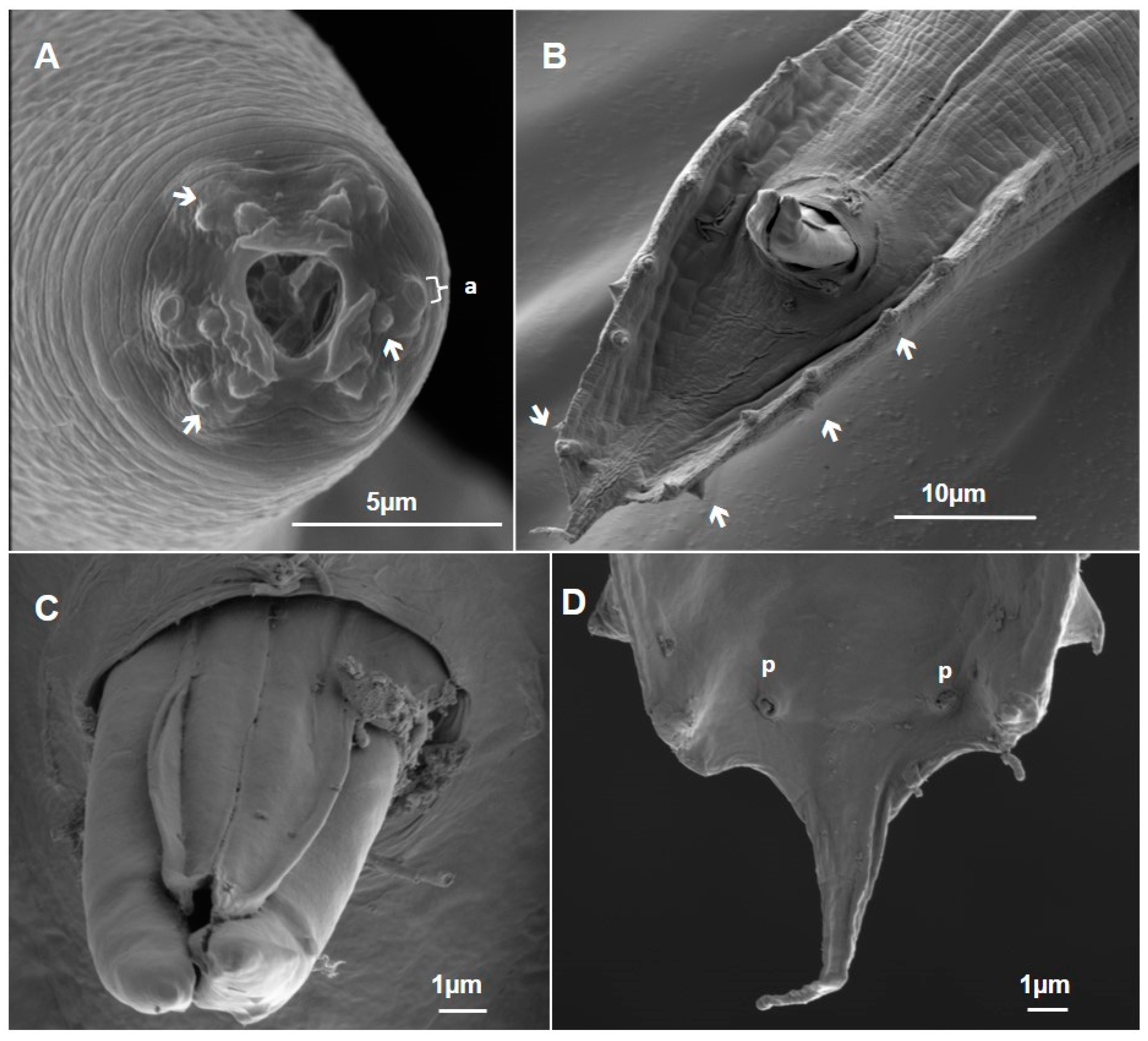
SEM enabled us to detail the anterior end, evidencing the triangularly shaped oral opening with two lateral ventral lips, ornamented by tree papillae and one amphid each (Figure 3A). The surface presents a cuticle with faint transverse striations, covering the body. At the posterior region, the copulatory bursa presents a circular pattern, almost resembling a half-moon format (Figure 3C). The spicules tip resembles an arrow-like structure (Figure 3D). SEM also showed that the fifth and seventh pairs of genital papillae had different orientations to the others. After the cuticular dilatation, there are a pair of phasmids and a small tail projection.
Males—based on six specimens (Table 1): Body length of 993.7–1.221 µm and body diameter of 43.5–63.5 µm. The stoma is 19–27.2 µm long, the pharynx is 206.2–238.7 µm long, the never ring is 150–163.8 µm long, and the basal bulb is 31.9–39.5 µm long. The spicules are 34.7–50.6 µm long and the gubernaculum length is 26–26.5 µm. The tail length is 40.5–43.7 µm and the tail length of the post-copulatory bursa is 1.1–1.7 µm. Demanian values: a, 19.2–22.8; b, 4.7–5.1; and c, 22.8–29.2.

3.1.2. Metarhabditis freitasi (Martins, 1985), Sudhaus, 2011

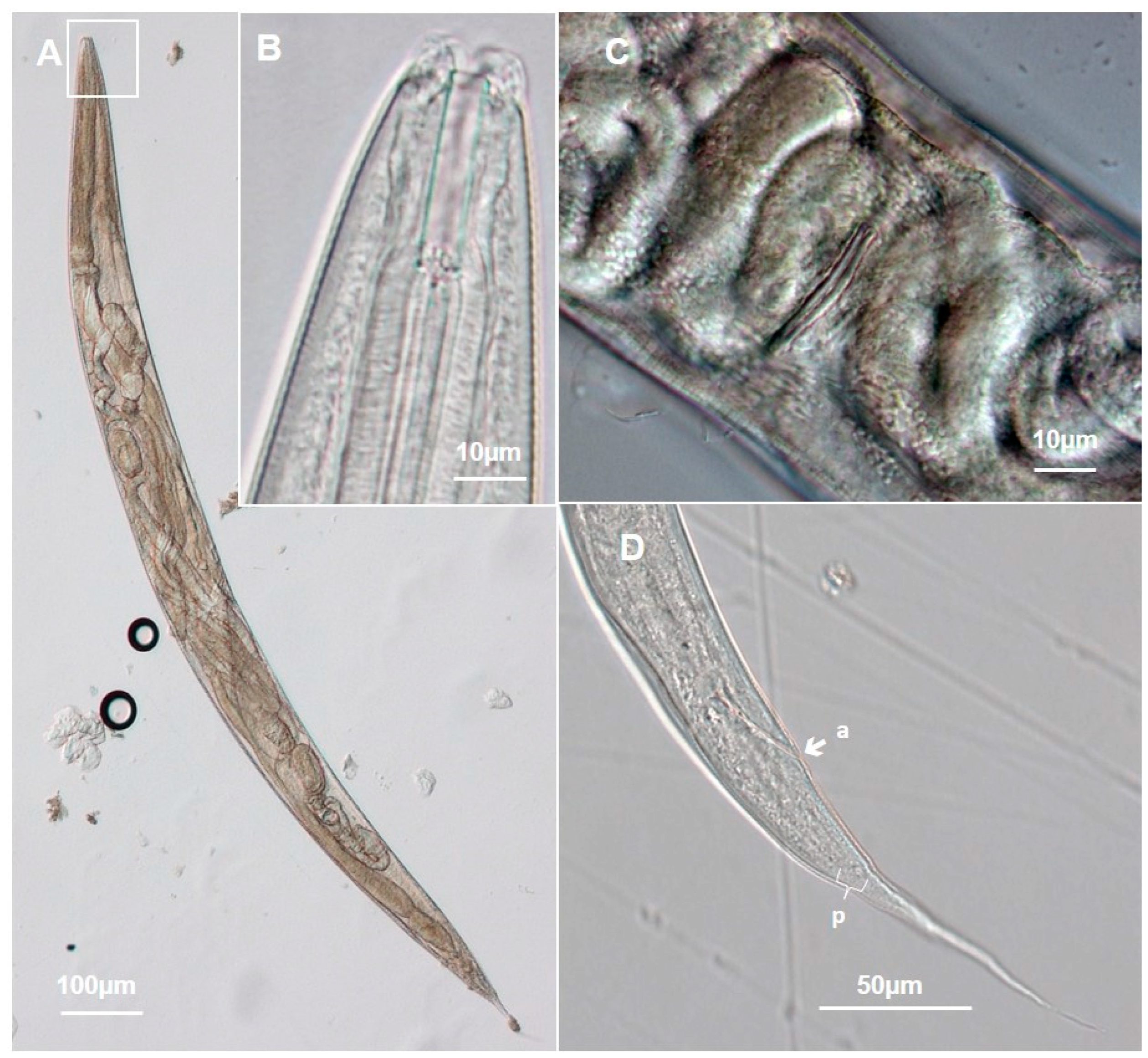
General: Oral opening with a triangular shape, two lateral ventral lips, ornamented by tree papillae and one amphid each, the glotoid apparatus is well-developed, the pharynx corpus does not have an indistinct metacorpus. The excretory pore is located at the level of the isthmus after the nerve ring. The body is fusiform, reaching the maximum body diameter (Figure 4A and Figure 5A,B).
Females have didelphic, amphidelphic vulva, which are located at the mid-body level and dorsally reflexed; the anterior ovary is on the right side of the intestine and the posterior ovary is on the left. The ovaries contain mature eggs with a more cylindrical shape and larvae are present (Figure 4A–C and Figure 5C,D). SEM enabled us to detail the anterior end, showing an oral opening with a triangular shape, provided by three lips, two of which latero-ventral and one is dorsal. Each lateroventral lip has three papillae distributed in a triangular pattern and a well-developed amphid, located at the line that divides the body into ventral and dorsal faces. The dorsal lips are provided by four papillae, arranged in pairs (Figure 6A). The cuticle surface in this region is smoother when compared with the rest of the body and provided by longitudinal and transversal striations (Figure 6B). The vulva is composed of cuticular structures that form a pair of thick lips and are prominent (Figure 6C). In the posterior region, the anus opens in the form of a semicircle and a pair of phasmids can be seen on the laterodorsal surface and tail at the posterior end, with a hair-like tip (Figure 6D–F).
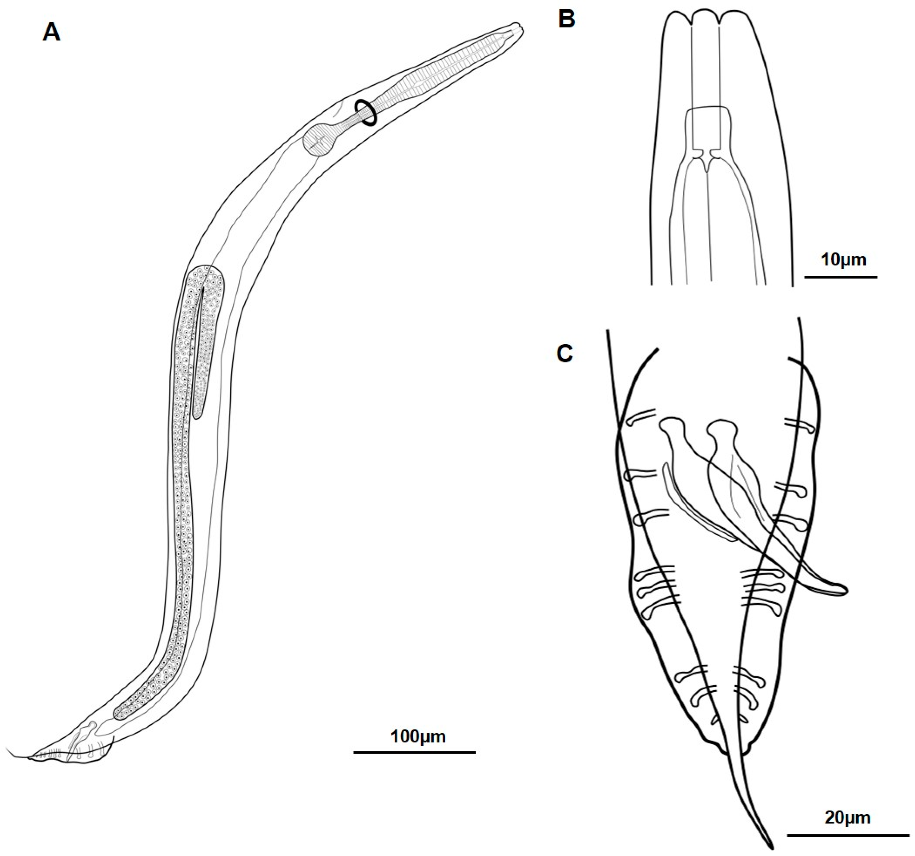
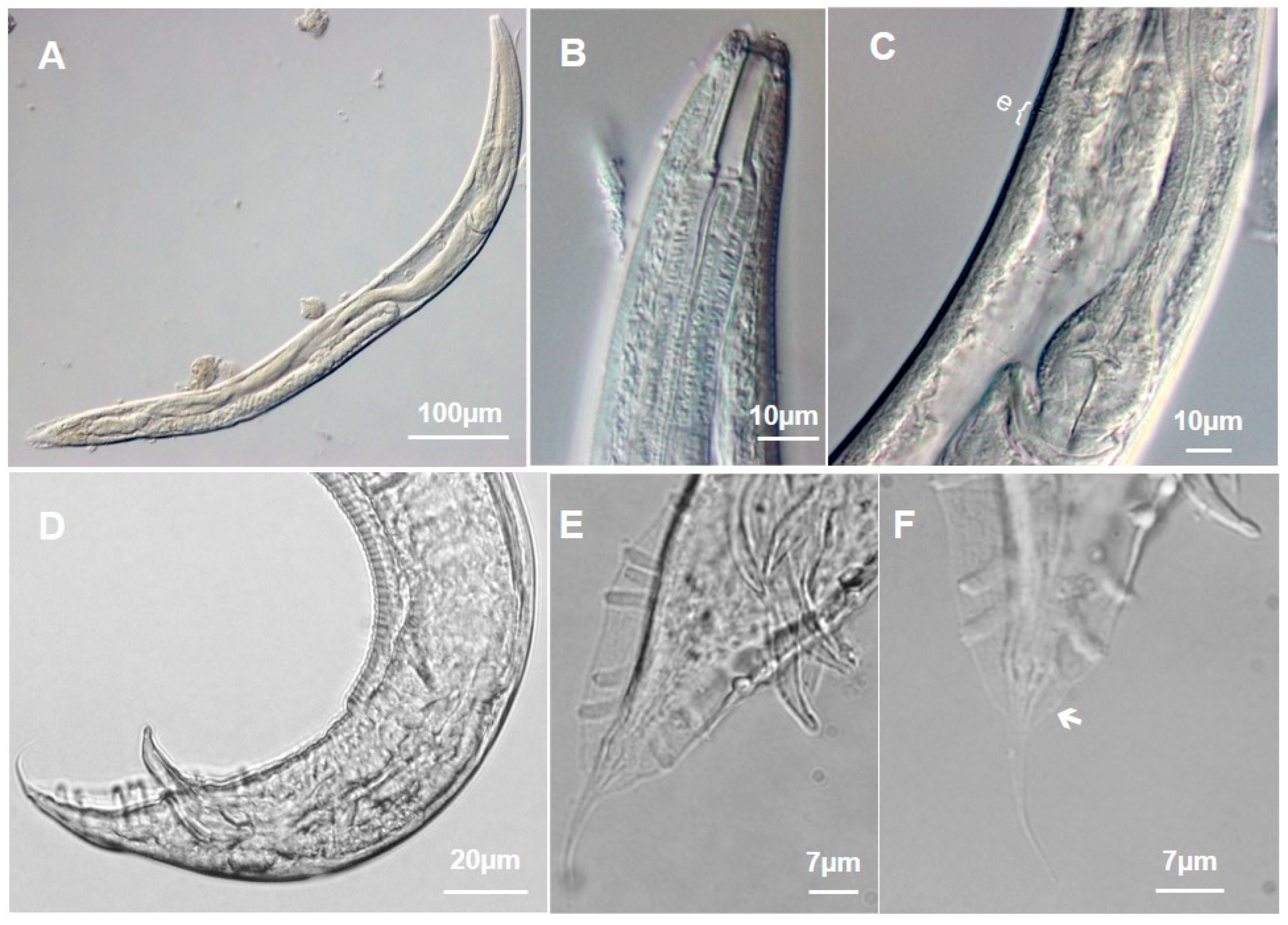
Males are similar to females in general morphology, and the maximum diameter is approximately half the maximum width of these nematodes. A single testis, reflexed ventrally, is located on the left side of the intestine (Figure 7A,B and Figure 8A–C). The bursa is narrow with a triangular shape, beginning at the cloacal opening and ending at the terminal portion of the tail. The cuticle dilatation features a smaller border after the last pair of papillae, where a pair of phasmids are located. On the posterior end of the bursa, a narrow tail tip (appendage) is projected after the cuticle dilatation (Figure 7C and Figure 8D–F). At the anterior end, SEM allowed the oral opening characterization with cephalic papillae, and amphids (Figure 9A), and at the posterior end, the cuticle surface was visualized, which is provided by transversal striations on the dorsal face and is smoother on the dorsal face, particularly at the cloacal opening (Figure 9B). The seventh genital papillae pair is positioned more dorsally than other genital papillae, with a ventral orientation. After the seventh and eighth pairs, a pair of diminutive phasmids are located at the bursa’s posterior end (Figure 9B). Projected spicules were detailed and analyzed, presenting a sheath with a smooth surface, covering the base of robust spicules, and a tip provided by structures with arrow or spear shapes (Figure 9C). At the bursa’s terminal region, the cuticular dilatation closes until a narrow and thin border is shown, before ending in an elongated and narrow free tail (Figure 9D).
Males—based on ten specimens (Table 1): Body length of 861.2–1070 µm and body diameter of 51.3–74.2 µm. The stoma is 24.3–30.6 µm long, the pharynx is 198.3–261.7 µm long; the nerve ring is 137.9–176.6 µm long, and the basal bulb length is 31.9–38.3 µm. The spicules are 33.9–43 µm long, the gubernaculum is 21.8–26.6 µm long, the tail length is 35–48 µm, and the tail length of the post-copulatory bursa is 6–9.8 µm. Demanian values: a, 12.7–21.8; b, 3.6–4.8; and c, 18.8–27.2.
Females—based on ten specimens (Table 2): Body length of 838.7–1118.6 µm and the body diameter is 52.4–67.8 µm long. Stoma length is 23.4–30.54 µm, the pharynx is 216–241 µm, the nerve ring is 149–172 µm long, and the basal bulb is 30–37 µm. The tail length is 137–155 µm and eggs are 49–60 µm long and 30–39 µm wide. Demanian values: a, 16–16.5; b, 4–5; c, 6.1–7.3; and v (%), 47–63.

3.1.3. Metarhabditis blumi (Sudhaus, 1974)

The cultivation of nematodes of the M. blumi species allowed us to have access to a large number of parasites and to carry out morphological and morphometric analysis experiments for comparison with the samples obtained from animals.
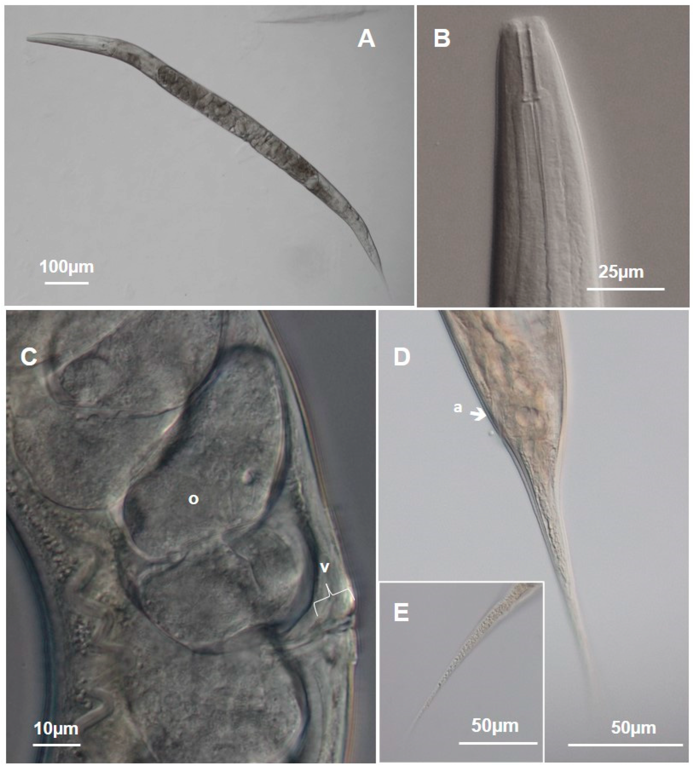
General: The body is cylindrical and fusiform. The anterior end possesses tree lips with cephalic papillae, a cylindrical pharynx with indistinguishable procorpus and metacorpus, and an excretory pore after the never ring at the isthmus that has a pharynx ending with a piriform basal bulb. The lips are projected and the stoma is cylindrical and straight, with the final segment possessing the glotoid apparatus and shaped like an isosceles triangle (Figure 10A,B). The vulva can be seen in the middle of the female’s body (Figure 10C) and, from here, the body gradually tapers towards the anus, after which the tail becomes more tapered, ending in a tail conically and pointed (Figure 10D,E).
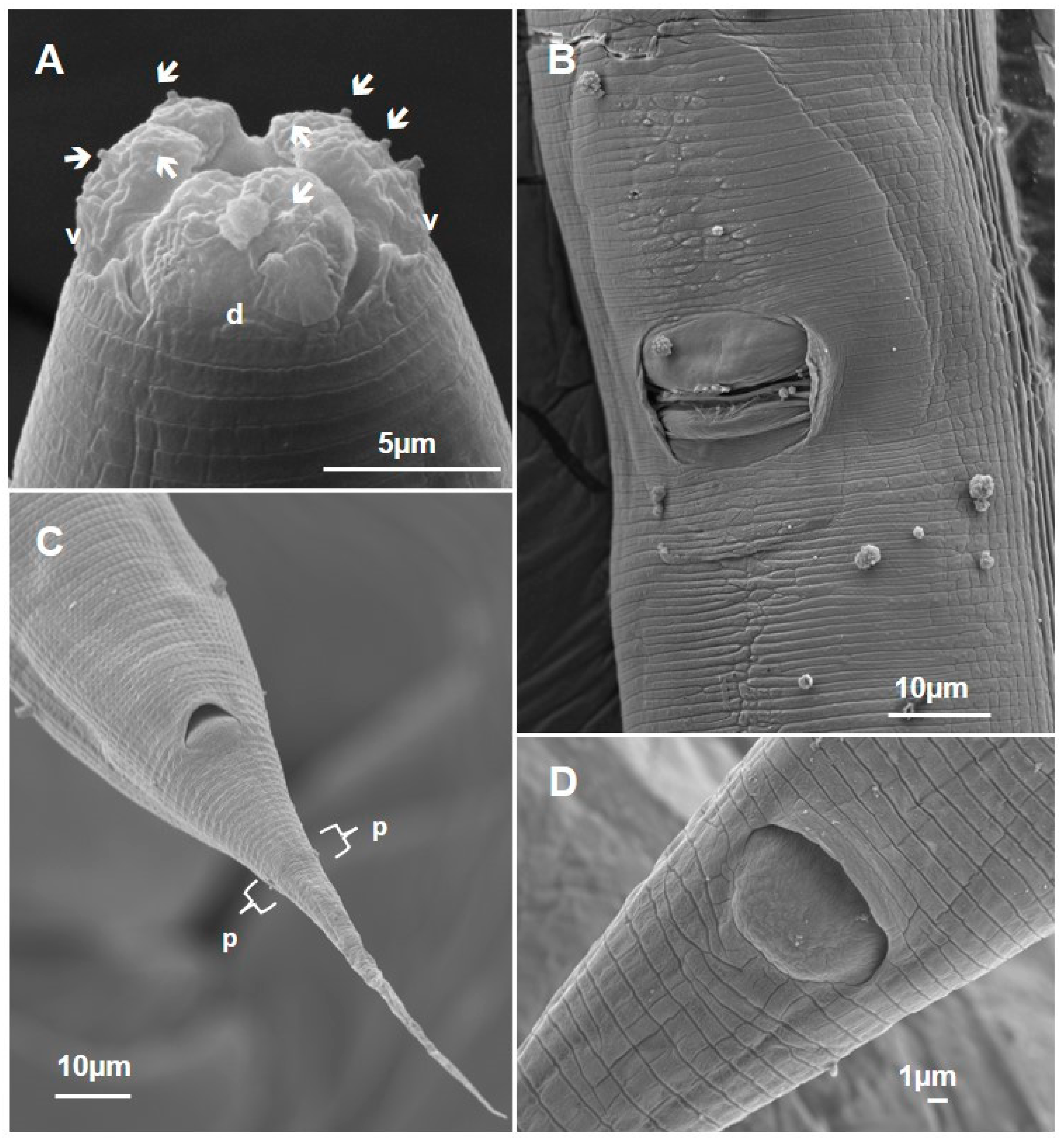
SEM enabled us to detail the anterior end of M. blumi, evidencing a triangular mouth opening (Figure 11A). The external morphology of the vulva shows two simple lips with a cuticular design that surrounds these lips and two cuticular folds lateralizing this vulvar opening (Figure 11B). At the posterior region, we can identify the anus, which is located ventrally. On the lateral side of the tail, we can identify a pair of phasmids (Figure 11C). The anus appears as a semi-circular opening with a cuticular elevation without striations in the most posterior portion (Figure 11D).
Males and females have three lips, one dorsal and two lateroventral, and the female tail is conically pointed (Figure 12A,B). Males present a single testicle reflected ventrally on the left side of the intestine and robust spicules supported by a trough-shaped gubernaculum. The bursa is open, of the leptoderan type, and has genital papillae (GP) and phasmids (PH); it has the following configuration: 1 + 1 + 1/3 + 2 + PH (Figure 12C and Figure 13A,B). The first and second pairs are spaced and pre-cloacal. The third pair is ad cloacal and close to the second pair. The following pairs are post-cloacal. Narrow phasmids are localized posterior to the eighth pair, with a filiform aspect. The posterior region of the male has a copulatory bursa ornamented with seven pairs of genital papillae. This bursa has a cuticular indentation at the end that highlights the post-bursal tail at the end of the tail (Figure 13C–E).
Males and females have three lips, one dorsal and two lateroventral, with the dorsal lip decorated with four cephalic papillae and the two lateroventral lips with two papillae and an amphid each (Figure 14A,B). In the posterior region of the males, the copulatory bursa is distinguished by an evident cuticular dilation. The fourth pair of genital papillae is oriented dorsally in relation to the others (Figure 14C). The spicules are projected (Figure 14D) and on the inner face of the end of the bursa, there are a pair of phasmids, close to the region from which the caudal appendage projects (Figure 14E).
Males—based on ten specimens (Table 1): The body length is 764.5–1045.6 µm and the body diameter is 45.4–80.6 µm long. The stoma is 21.4–30 µm long, the pharynx length is 175–232.7 µm, and the basal bulb is 175–232.7 µm long. The spicules are 37–50 µm long, the gubernaculum is 14.9–18 µm long, and the tail length is 50–60 µm, including the portion located inside the bursa region. The tail length of the post-copulatory bursa is 14–22 µm. Demanian values: a, 10.4–19; b, 4–5; and c, 14.4–25.
Females—based on ten specimens (Table 2): The body length is 948–1371.8 µm and the body diameter is 52–83.1 µm. The stoma is 20–33.8 µm long, the pharynx is 180–256.9 µm long, the nerve ring is 153–193.1 µm long and the basal bulb is 31.5–44.2 µm long. Tail length is 78–128.2 µm and egg length is 39–58.4 µm long. Demanian values: a, 14.6–20; b, 4.1–5.9; c, 10–13.7; and v (%), 47.8–63.3.

3.1.4. Metarhabditis sp.

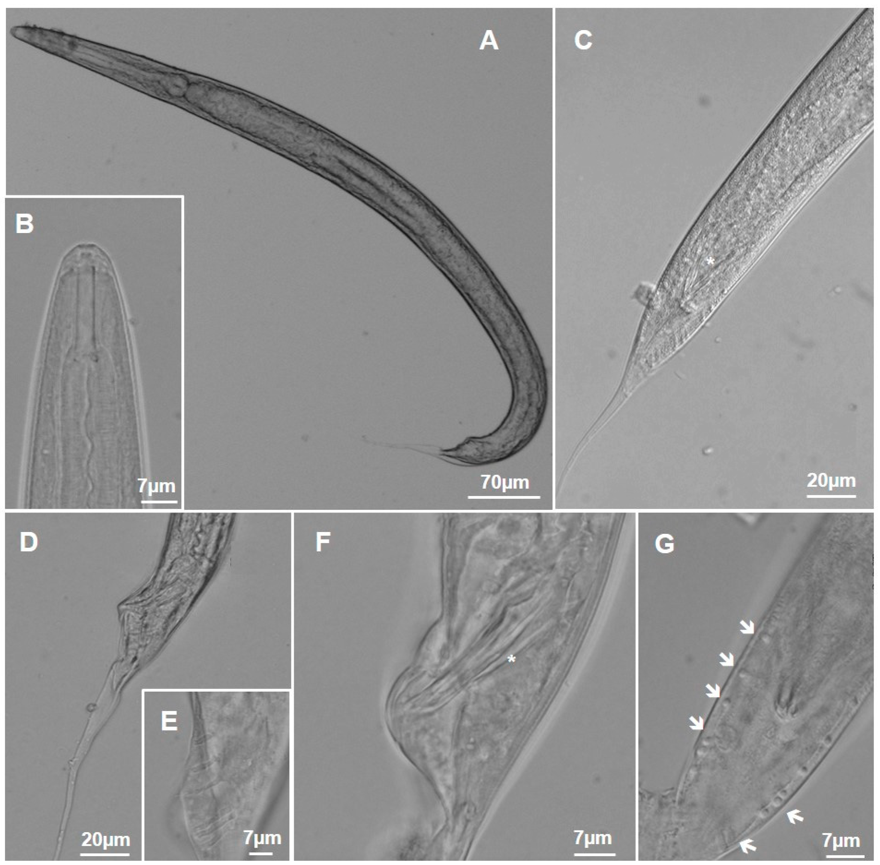
This nematode has slightly undeveloped lips. The stoma encompasses about 50% of the nematode and is surrounded by the esophageal collar. The nerve ring is located at the initial portion of the isthmus. The largest diameter is observed in the middle region of the body, which then gradually tapers towards the initial portion of the tail, which has a conical appearance (Figure 15A,B). The testis is simple, reflected ventrally, and has two unfused spicules. The bursa is of the leptoderan type, reduced in size, and open anteriorly with eight pairs of genital papillae adorning the bursa (Figure 15C–G). The distribution is as follows: the first two pairs are pre-cloacal; the third pair is ad-cloacal; the fourth and fifth pairs are close together, followed by the sixth pair which is together with these pairs; and the last two pairs are located closer to the tip of the tail, forming a group of papillae which are further away from those previously mentioned. Immediately before the end of the bursa and the beginning of the projection of the long caudal appendage, there is a discrete pair of phasmids. The tail is long and accounts for around fifteen percent of the total body length (Figure 15C).
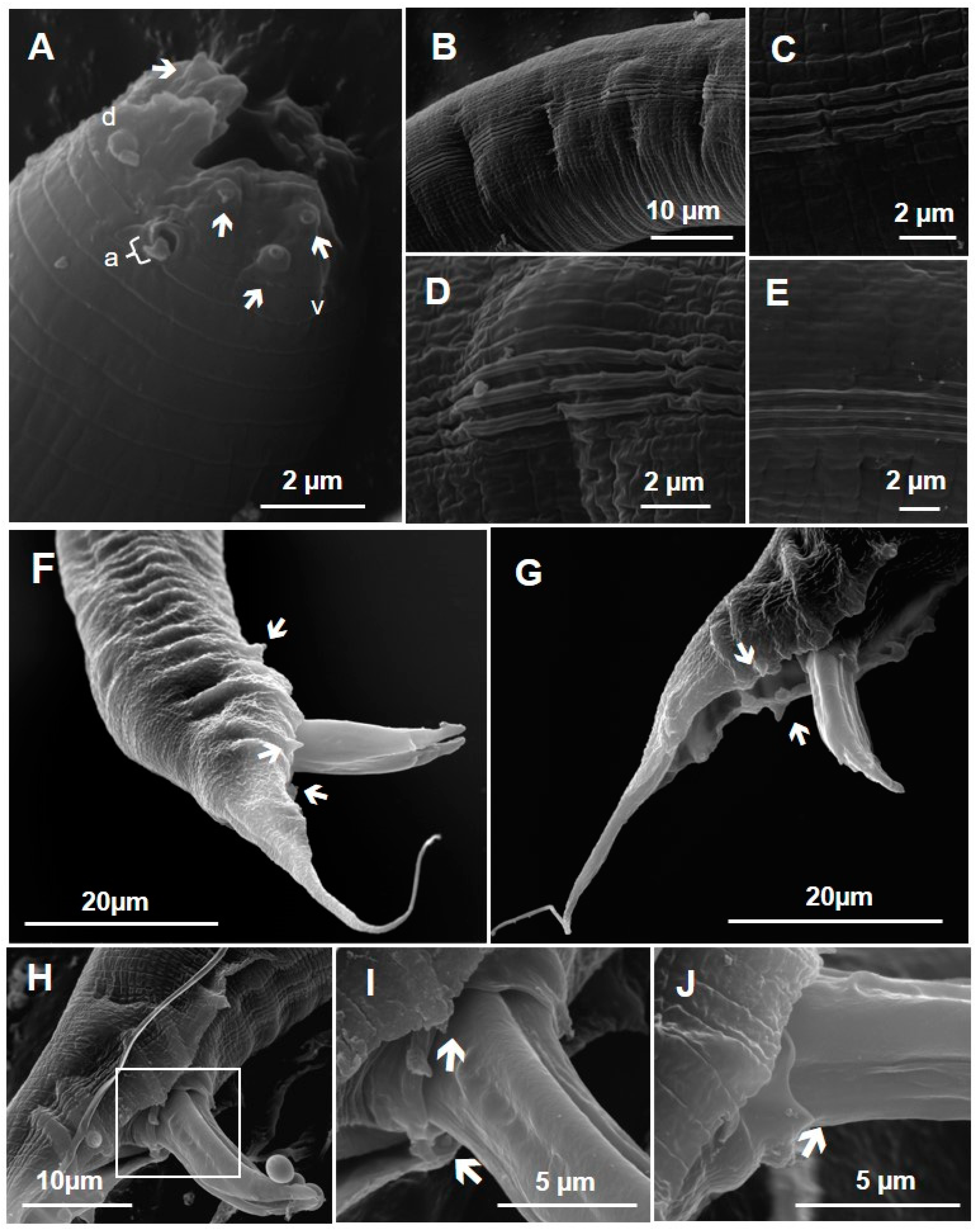
Using SEM, it was possible to detect the details of the anterior end, showing the triangular-shaped oral opening surrounded by three poorly developed lips, one dorsal and two latero-ventral (Figure 16A). Two well-developed amphids lateralize the anterior end and each latero-ventral lip is provided with three papillae, while the dorsal lip is provided with four cephalic papillae, forming a set of 12 sensory structures at the anterior end (ten papillae and two amphids). In this region, the cuticle has discrete cuticular ridges and along the body, it is possible to identify transverse cuticular ridges and a pronounced lateral line made up of three ridges. This structure is identified from the anterior region to the area close to the copulatory bursa (Figure 16B–E). There are spicules projected with a tip in a half-arrow shape and a long and filiform tail (Figure 16F–G). Posterior end in ventral view showed the genital papillae distribution and the cloacal opening morphology (Figure 16H–J).
Males—based on six specimens (Table 1): The body length is 728.6–1000 µm long and body diameter is 33.7–545.5 µm long. The stoma is 19.8–28.1 µm long, the pharynx is 167.7–250 µm long, the never ring is µm long, and the basal bulb is 27.7–40.3 µm long. The spicules are 24.4–38 µm long, the gubernaculum length is 17–18.7 µm, the tail is 100–166.7 µm long, and the tail length of the post-copulatory bursa is 61.4–106.7 µm. Demanian values: a, 20–26.8; b, 3.6–5.2; c, 6.5–9.2.

3.2. Genetic Characterization

The 28S rDNA alignment of the dataset resulted, after trimming, in a dataset of 20 taxa per 940 pairs of bases. The best-fit model, calculated via SMS in PhyML, under AIC, was GTR + G + I, with four free rate categories and a gamma shape parameter α = 1.571000, resulting in an ML tree with a lnL score = −4920.40803. After 25% burn-in removal, MCMC sampling resulted in a mean lnL = −4949.2694 (standard deviation = 5.4577; median = −4948.944), which is the estimated marginal likelihood.
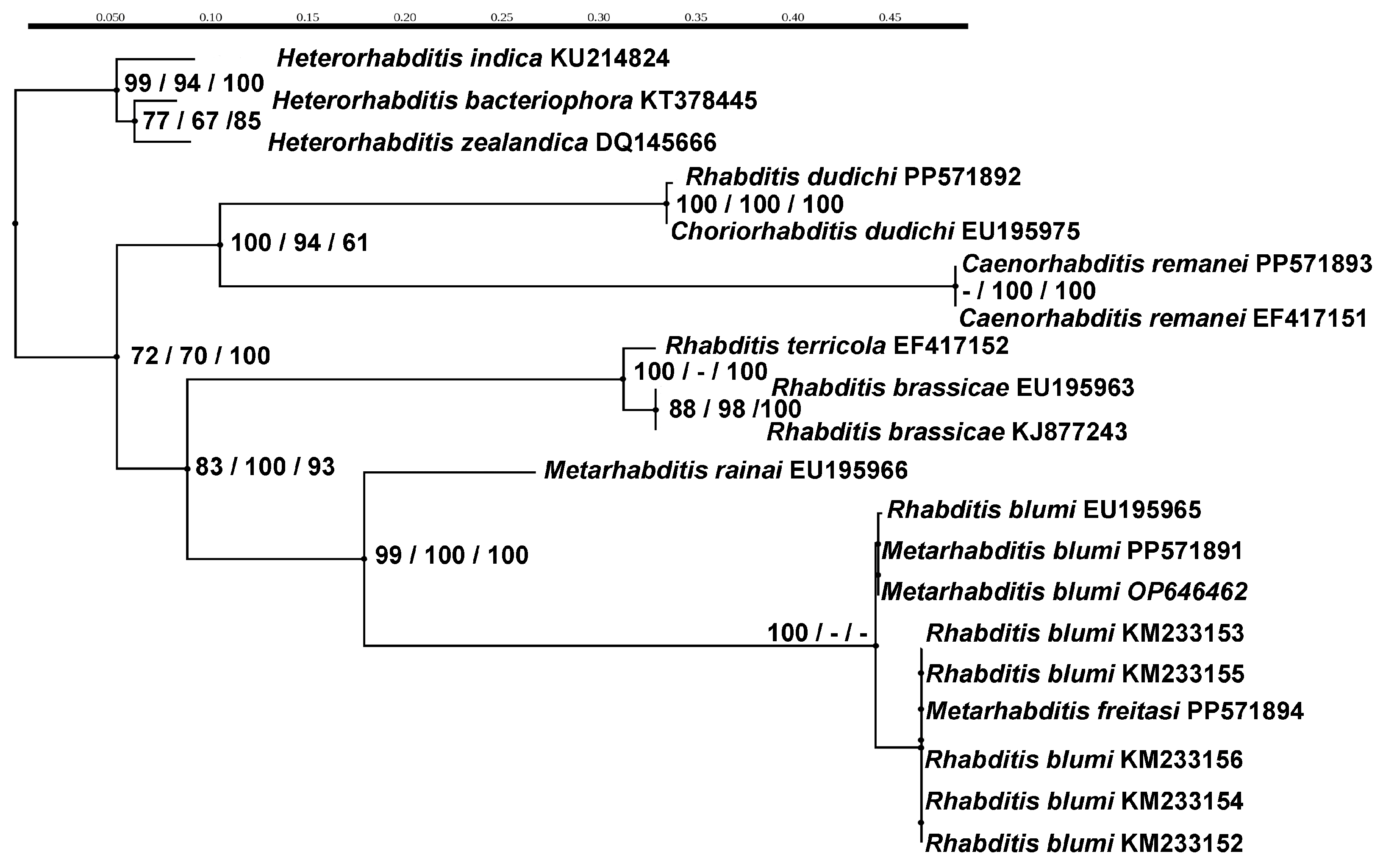
The tree topologies recovered in our phylogenetic analyses were congruent, separating the genera Rhabditis and Metarhabditis, which was highly supported by both ML and BI trees (aLRT = 83, ML-BP = 100, BPP = 93) (Figure 16). Among Metarhabditis species, two well-supported clades were formed, separating M. rainai and M. blumi (aLRT = 99, ML-BP = 100, BPP = 100). The M. freitasi sequence (PP571894) from the present study was grouped with the sequences deposited in the Genbank of M. blumi (KM233152, KM233153, KM233154, KM233155, and KM233156) from Minas Gerais, Brazil [28]. This group formed a polytomy in ML-aLRT, ML-BP, and BI. Improbably, the M. blumi sequences, EU1915965 and OP646462, which came from the USA and Colombia, respectively, and the M. blumi sequence (PP571891) from the USA, provided by the CGC and used as a control in our study, formed a separate clade in ML-aLRT and a polytomy in ML-BP and BI. Furthermore, the sequences of C. remanei (PP571893) and R. dudichi (PP571892) from the USA, which were provided by the CGC, showed two distinct clusters, all grouped with their respective species, with high supports in the two groups (aLRT = 100, ML-BP = 61, BPP = 94). The group formed by species of the genus Rhabditis was moderate to well-supported (aLRT = 83, ML-BP = 100, BPP = 93). The sequences of species belonging to the Heterorhabditis genus (KU214824, DQ145666, and KT378445) were used as outgroups (Figure 17).

4. Discussion

The Rhabditidae family is one of the richest nematode groups, with members who are usually terrestrials and prefer saprophyte-dwelling feces and decaying materials. They are also reported to be associated with earthworms, insects, and humans [12,13,14,15]. The species identified in this study belong to the genus Metarhabditis, established by Tahseen et al. [19], with the type and species Metarhabditis andrassyana. Sudhaus [11] revised the genus Rhabditis and created a new organization, the Metarhabditis genus, based on the presence of a prerectum; a shorter rectum; separate, capitate, dagger-shaped spicules with an angled distal part (reminiscent of a sabre); and an oval gubernaculum in ventral view. Based on these characteristics, the nematodes included in the genus Metarhabditis are Rhabditis costai, Rhabditis freitasi, and another four species. Posteriorly, Sudhaus [21] updated the genus to include two other species, Metarhabditis suklaae [38] and Metarhabditis giennensis [39].
Metarhabditis costai specimens identified in this study showed all of the morphological characteristics of the nematode described by Martins [24]; however, in the present study, we detailed some morphometric intraspecific variations. When compared with the described specimens of M. costai by Martins, the specimens from the present study displayed a lower maximum body diameter, a slight difference in body length, and a slightly lower “a” value. The SEM analysis showed that the lips are not fused (Figure 13A), instead presenting a slight separation and not a bifurcation to characterize a pair, thus differentiating them from the M. costai described by Asif and coauthors [40]. These nematodes, when compared with the recovered specimens in this study, present a stoma with slightly larger and smaller gubernaculum. Metarhabditis costai shows morphologic differences with M. andrassyana [19] regarding the form of the spicules and the robustness of M. andrassyana. Metarhabditis costai differs from M. rainai, as proposed in [41], especially as it does not have a well-developed median bulb. The position of the phasmid is also different (posterior to GP8 versus anterior to GP8) and the bursa type is leptdoran instead of peloderan, with a notable posterior notch, lacking a projected tail tip after the bursa with regards to the latter. Metarhabditis costai presents slight morphological differences from M. freitasi, with the differences in the anterior end. The lips are more robust, and at the posterior end, the bursa is robust and seems to have a half-moon shape and a short caudal appendix. Metarhabditis costai differs from M. blumi, another nematode associated with cases of parasitic otitis in cattle [27,42].
We observed the morphology differences mainly in the posterior end of M. costai male. The differences were in tail length, with the spout of the spicules generally being larger than 50 µm. In addition, the bursa shape consisted of a cuticular opening. The males of M. costai differ from M. giennensis Abolafia and Peña-Santiago [39] mainly at the anterior end, specifically in the stoma region and at the lips. An especially important difference is the lack of gynmostomatal denticles. In the posterior region, the bursa shape and post-bursal tail are shorter. We highlight that our observations using light microscopy (LM) and scanning electron microscopy (SEM) did not show the presence of these denticles in both species identified in this study, as described by Abolafia and Peña-Santiago [39]. In their descriptions, both LM and SEM showed these denticles in the stomata. Therefore, it is suggestive that this characteristic could be used in differentiating M. costai and M. freitasi from M. giennensis. Metarhabditis suklai, as described by Mondal and Manna [37], shares similar morphometric parameters; however, M. costai presents morphological differences in terms of bursal structure and spicules, with an arrow-shape tip confirmed by SEM.
Metarhabditis freitasi differs from Metarhabditis adenobia [20,43] in terms of cuticular bursa dilatation, gurbenaculum morphology, and spicule form. Bhat et al. [44] morphologically and molecularly characterized Metarhabditis amsactae recovered from the soil in India, differentiating it from male M. freitasi due to the less robust bursa and the filiform and robust tail, with a part not covered by the bursa cuticle. Metarhabditis suklai [38] shares similar morphometrical parameters; however, in the males of M. freitasi, differences in mid-body width, gubernaculum length, and tail length are observed. The authors’ drawing, which is based on LM observations, showed a bursa without cuticular dilation. However, it does not provide ultrastructural or molecular characterization. Besides morphometric similarities, M. costai and M. freitasi males can be distinguished by their bursal patterns and spicules with arrow-like tips.
Martins [24] also identified M. freitasi and differentiated M. costai species mainly based on the morphological characteristics of the copulatory bursa. In our results, we also recovered these two species and corroborated this information, magnifying the details with SEM results. M. freitasi presents a triangular morphology of the bursa when compared with M. costai, which shows a more rounded profile. In addition, M. freitasi has a more elongated caudal appendix. Males of M. freitasi differ morphologically from M. blumi for their less developed lips, evidenced by SEM analysis, and the shape of the bursa. The bursa of M. freitasi does not present an indentation in the terminal part of the cuticular dilation of the bursa, the region from where the caudal appendix of M. blumi is projected [40].
The species of M. freitasi and M. blumi share many morphometric similarities; however, they can be differentiated mainly by analyzing the length of the post-bursa tail. This parameter can be used to differentiate M. freitasi, M. costai, and M. blumi. Metarhabditis blumi had the longest post-bursa tail of the three species of the genus analyzed in the present study. In the present study, the cultivated M. blumi had bodies that were relatively smaller than those of the specimens characterized in the original description. The NGM medium with Escherichia coli OP 50 would not the optimal for development, once this standard is used for Caenorhabditis elegans [45].
Females and males of M. freitasi have less developed lips when compared with M. blumi and M. costai, and the bursa of M. freitasi does not have an indentation in the terminal part of the cuticular dilation, the region from which the caudal appendix of M. blumi is projected. As mentioned above, the lips of M. blumi are more robust in both males and females, and in the males of this species, a larger post-bursal tail was observed with other specific characteristics. Using SEM, we were able to confirm the robustness of the lips of M. blumi, and it was possible to use electron microscopy to compare these three species. We also observed differences in the tip of the spicule, where M. blumi does not have an arrow-shaped spicule tip. In addition, M. blumi females have ornamentation surrounding the vulva opening.
In the present study, we recovered a third species of nematode, Metarhabditis sp., which showed similarities with Metarhabditis freitasi and M. costai. These were restricted to the total length of the body, the number of genital papillae, and the length of the spicules, which are important structures in taxonomic characterization. However, they differ in the length of the caudal appendix and, consequently, in the values of Man “a” and “c”, the latter of which is influenced by the measurement of tail length. The tail of Metarhabditis sp. is three times longer than M. costai. We characterized the cuticular surface of these nematodes using SEM and were able to observe that the lips of M. costai are more robust than those of the Metarhabditis sp., and in the posterior region of M. costai, we described a more developed copulatory bursa than that in Metarhabditis sp. The M. freitasi species is similar in terms of the number and distribution of the genital papillae, and also in the position of the fifth and seventh pairs of papillae, which have a dorsal orientation. However, these species differ in the morphology and morphometry of the spicules, as well as in their Man “a” and “c” values, along with the length of the post-bursa caudal appendix.
Metarhabditis sp. differs from other species not associated with parasitic otitis. The main difference between it and M. rainai is the robustness of the lips and the bursa leptoderan. When compared with M. amsactae, as described by Ali et al. [46], the robustness of the tail is observed, as well as the bursa being less robust when compared to Metarhabditis sp. It differs from M. giennensis in that it has more projected lips, Gymnostomatal denticles, a robust spicule shape, and a smaller maximum body diameter. Other species, such as M. adrassyana and M. adenobia, are distinguishable by the length of the tail.
Bossi et al. [27] carried out genetic characterization of the nematodes recovered from the same Gir cattle that we used to collect the samples analyzed in this present work. Based only on genetic analysis using rDNA, the authors concluded that the identification was of the M. blumi species, showing, for the first time, that this species is associated with parasitic otitis. Subsequently, Barbosa et al. [42] again identified the M. blumi species in Gir cattle in the northern region of Brazil, using only molecular biology techniques based on the methodology applied by Bossi et al. [27]. However, morphological and morphometric characterization was not provided.
In the present study, we isolated and identified Metarhabditis spp. nematodes recovered from infected cattle based on morphological and morphometrical taxonomic parameters. Based on the present genetic characterization, we observed the formation of a clade with other species of Metarhabditis, identified as M. blumi collected from the same hosts from Brazil, specifically from sequences obtained by Bossi et al. [27] and Barbosa et al. [42]. The clade with Metarhabditis species recovered in Brazil was distinct from other M. blumi species isolated from the soil and provided by the CGC in other countries, such as the USA and Colombia, which have not been associated with parasitic otitis. Therefore, it is essential to employ morphological techniques to accurately identify species of the genus Metarhabditis. Although the DNA barcoding approach has been widely used to diagnose nematodes in Brazil with accurate and reliable results, we emphasize that the use of a combination of more than one molecular marker makes data analysis more robust. In our analysis, we used one marker, a 28S rDNA nuclear gene, and one of the more conserved regions of DNA, which justifies the need for further studies using different markers for the molecular identification of Metarhabditis species.

5. Conclusions

The present paper presents taxonomic findings on two previously described species, M. freitasi and M. costai, while also providing new data by exploring their morphological characteristics using both Light Microscopy and Scanning Electron Microscopy. Exploring these advanced tools, we also revealed structural characteristics of a third species, Metarhabditis sp., associated with parasitic otitis. Our findings align with previous reports, confirming the presence of Metarhabditis spp. nematodes associated with bovine otitis. Furthermore, our morphological differentiation of M. costai, M. freitasi, and M. blumi, coupled with molecular analyses, allowed us to delineate clades among species collected in Brazil and those from other countries. Notably, all species described in Brazil were associated with parasitic otitis, while those from other countries showed no veterinary impact association. Our phylogenetic data suggest potential geographic isolation, which contributes to this differentiation. On the other hand, the M. blumi identified by Bossi et al. [27] and Barbosa et al. [42] could represent another species within this genus, such as M. freitasi or M. costai, exhibiting genetic similarities in the ribosomal DNA analyzed. Further exploration using complementary markers of rDNA, including mitochondrial DNA, may aid in distinguishing these closely related species.

Author Contributions

E.J.L.-T., M.E.C., A.M.I. and B.E.d.A.-S. contributed to the study conception and design. Material preparation, data collection and analysis were performed by M.E.C., B.E.d.A.-S., V.H.B., A.M.I., H.A.d.S., A.C.-B. and E.J.L.-T. The first draft of the manuscript was written by M.E.C. and B.E.d.A.-S. and all authors commented on previous versions of the manuscript. All authors have read and agreed to the published version of the manuscript.

Funding

This work received financial support from the Fundação de Amparo à Pesquisa do Estado do Rio de Janeiro-FAPERJ under Award Number FAPERJ/JCNE E-26/201.287/2022 (EJLT) and FAPERJ/CNE E-26/201.069/2021 (AMI). Fellowship from Conselho Nacional de Desenvolvimento Científico e Tecnológico-CNPq. Grant Number 315634/2021-9 (AMI). Additionally, PhD scholarship (MEC) was provided by the Coordenação de Aperfeiçoamento de Pessoal de Nível Superior-CAPES under number 103443/2024-0.

Data Availability Statement

The data supporting the findings of this study are accessible from the corresponding author upon request (Lopes-Torres, EJ).

Acknowledgments

We are grateful to the RPT01A/FIOCRUZ sequencing facility for their technical support, as well as to the Centro Nacional de Biologia Estrutural e Bioimagem of the Federal University of Rio de Janeiro (CENABIO-UFRJ), NANOFAB-UERJ, UERJ-Unidade Urogenital, and Plataforma de Microscopia Eletrônica Rudolf Barth/FIOCRUZ for their assistance with the electron microscopy platforms.

Conflicts of Interest

The authors declare the presence of non-financial interests, either directly or indirectly related to this work.

References

  1. Verocai, G.G.; Fernandes, J.I.; Correia, T.R.; Melo, R.M.P.S.; Alves, P.A.M.; Scott, F.B. Otite Parasitária Bovina Por Nematoides Rhabditiformes em Vacas Gir no Estado do Rio de Janeiro, Brasil. Rev. Bras. Parasitol. Vet. 2007, 16, 105–107. [Google Scholar] [PubMed]
  2. Leite, P.V.B.; Cunha, L.M.; Leite, L.B.; Leite, R.C. Farmers’ Perception About Parasitic Otitis in Gyr Breed from Three States of Brazil. Pesq. Vet. Bras. 2012, 32, 855–858. [Google Scholar] [CrossRef]
  3. Cardona, J.A.; González, M.; Álvarez, J. Frequency of clinical parasitic otitis due to Rhabditiform nematodes (Rhabditis sp) in six Gyr breed cattle farms in Cordoba, Colombia. Rev. Colomb. Cienc. Pecu. 2012, 25, 417–421. [Google Scholar]
  4. Bulletin of the International Dairy Federation 446/2010—World Dairy Situation 2010. Available online: https://www.fil-idf.org/wp-content/uploads/woocommerce_uploads/Publications/Sold/Bulletins/446-2010-The-World-Dairy-Situation-2010.pdf (accessed on 8 March 2024).
  5. Douphrate, D.I.; Hagevoort, G.R.; Nonnenmann, M.W.; Lunner Kolstrup, C.; Reynolds, S.J.; Jakob, M.; Kinsel, M. The dairy industry: A brief description of production practices, trends, and farm characteristics around the world. J. Agromed. 2013, 18, 187–197. [Google Scholar] [CrossRef] [PubMed]
  6. Vercruysse, J.; Charlier, J.; Dijk, J.V.; Morgan, E.R.; Geary, T.; Samson-Himmelstjerna, G.V.; Edwin Claerebout, E. Control of helminth ruminant infections by 2030. Parasitology 2018, 145, 1655–1664. [Google Scholar] [CrossRef] [PubMed]
  7. Sobral, S.; Almeida, L.S.; Ferraz, C.M.; Junior, O.L.F.; Lopes, A.D.C.G.; Langoni, H.; Moreira, T.F.; Lima, J.A.C.; Vilela, V.L.R.; Braga, F.R. Infestações por Rhabditis spp.: Uma revisão. Vet. Zoot. 2020, 27, 1–10. [Google Scholar] [CrossRef]
  8. Duarte, E.R.; Hamdan, J.S. Otitis in cattle, an etiological reveiw. J. Vet. Med. Bras. Infect. 2004, 51, 1–7. [Google Scholar] [CrossRef] [PubMed]
  9. Leite, P.V.B.; Cunha, A.P.; Silva, M.X.; Bello, A.C.P.P.; Domingues, L.N.; Junior, A.L.; Leite, R.C. Clinical aspects and dynamics of auricular parasitoses in Gir cattle. Pesq. Vet. Bras. 2013, 33, 319–325. [Google Scholar] [CrossRef]
  10. Msolla, P.; Semuguruka, W.D.; Kasuku, A.A.; Shoo, M.K. Clinical Observations on Bovine parasitic otitis in Tanzania. Trop. Anim. Health. Prod. 1993, 25, 15–18. [Google Scholar] [CrossRef]
  11. Ushewokunze-Obatolu, U.; Pfukenyi, D.M.; USHE, T. A retrospective epidemiological study of parasitic otitis in cattle in the South-East Lowveld of Zimbabwe. Zimb. Vet. J. 1999, 30, 19–24. [Google Scholar] [CrossRef]
  12. Teschner, M.; Würfel, W.; Sedlacek, L.; Suerbaum, S.; De Tappe, D.; Mathias, M.W.; Hornef, W. Outer Ear Canal Infection with Rhabditis sp. Nematodes in a Human. J. Clin. Microbiol. 2014, 52, 1793–1795. [Google Scholar] [CrossRef]
  13. Andrássy, I. A Taxonomic Review of the Auborder Rhabiditina (Nematoda: Sercenentia); ORSTOM: Paris, France, 1983. [Google Scholar]
  14. Taboga, L. Rhabditis terrícola: An Opportunistic Nematode Parasite of Earthworms Cocoons. J. Invertebr. Pathol. 1981, 38, 22–25. [Google Scholar] [CrossRef]
  15. Park, H.W.; Kim, Y.O.; Ha, J.; Youn, S.H.; Kim, H.H.; Bilgrami, A.L.; Shin, C.H. Effects of associated bacteria on the pathogenicity and reproduction of the insect-parasitic nematode Rhabditis blumi (Nematoda: Rhabditida). Can. J. Microbiol. 2011, 57, 750–758. [Google Scholar] [CrossRef]
  16. Lee, W.K.; Choi, W.Y.; Lee, O.R. Rhabditis elongata Schneider, 1866 from Students in Korea. Kisaengchunghak Chapchi 1978, 16, 113–116. [Google Scholar] [CrossRef]
  17. Ahn, Y.K. Rhabditis sp. infect cases in rural schoolchildren. Kor. J. Parasitol. 1985, 23, 1–6. [Google Scholar] [CrossRef] [PubMed]
  18. Campos, D.M.B.; Araújo, J.L.B.; Viera, M.C.M.; Damasceno, F.; Barbosa, A.P. Um caso de parasitismo por Rhabditis sp em criança natural de Goiânia, Goiás, Brasil. Rev. Soc. Bras. Med. Trop. 2002, 35, 519–522. [Google Scholar] [CrossRef]
  19. Tahseen, Q.; Hussain, A.; Tomar, V.; Shah, A.A.; Jairajpuri, M.S. Description of Metarhabditis andrassyana gen. n., sp. n. Nematoda: Rhabditidae from India. Int. J. Nematol. 2004, 14, 163–168. [Google Scholar]
  20. Sudhaus, W. Phylogenetic systematization and catalogue of paraphyletic “Rhabditidae” (Secernentea, Nematoda). J. Nematode Morphol. Syst. 2011, 14, 113–178. [Google Scholar]
  21. Sudhaus, W. An update of the catalogue of paraphyletic ’Rhabditidae’ (Nematoda) after eleven years. Soil. Org. 2023, 95, 95–116. [Google Scholar] [CrossRef]
  22. Martins, W., Jr.; Nunes, I.J.; Ribeiral, L.A.; Rosaz, C.E.E.; Nunes, V.A. Nota sobre a ocorrência da Rhabditidae (Nematoda, Rhabditida) relacionados com otite em bovinos na região geo-econômica de Brasília/DF. Ciênc. Cult. 1971, 23, 248–249. [Google Scholar]
  23. Round, M.C. The Helminths Parasites of Domesticated Animals in Kenya. J. Helminthol. 1962, 36, 375–449. [Google Scholar] [CrossRef] [PubMed]
  24. Martins, W., Jr. Rhabditis (Rhabditis) freitasi sp. n. e Rhabditis (Rhabditis) costai sp. n. (Nematoda-Rhabditidae) isolados de otite bovina. Mem. Inst. Oswaldo Cruz 1985, 80, 11–16. [Google Scholar] [CrossRef]
  25. Leite, R.C.; Nunes, V.A.; Nunes, I.J.; Costa, A.L.; Facchini, J.L.H.; Lopes, C.W.G. Otite parasitária por nematóides rhabditiformes: Aspectos epidemiológicos e clínicos. Rev. Bras. Med. Vet. 1993, 15, 49–51. [Google Scholar]
  26. Abdalla, M.S.; Peixoto, T.C.; Alves, P.A.M.; França, T.N.; Brito, M.F. Aspectos Anátomo-Patológicos da otite Causada por Rhabditis sp. em Bovinos no Estado do Rio de Janeiro, Brasil. In Anais do Congresso Brasileiro de Veterinária; Sovergs: Gramado, Brazil, 2008. [Google Scholar]
  27. Bossi, P.V.; Consoli, E.A.; Rosa, J.M.O.; Leite, L.B.; Leite, R.C.; de Oliveira, C.M.G. Molecular identification and phylogenetic analysis of Metarhabditis blumi (Nematoda: Rhabditida). Vet. Parasitol. 2015, 214, 184–186. [Google Scholar] [CrossRef] [PubMed]
  28. Stiernagle, T. Maintenance of C. elegans. The Online Review of C. elegans Biology. Available online: https://www.ncbi.nlm.nih.gov/books/NBK19649/ (accessed on 8 March 2024).
  29. Pessanha, T.S.; Herrera, H.M.; Jansen, A.M.; Iñiguez, A.M. “Mi Casa, Tu Casa”: The coati nest as a hub of Trypanosoma cruzi transmission in the southern Pantanal biome revealed by molecular blood meal source identification in triatomines. Parasites Vectors 2023, 16, 26. [Google Scholar] [CrossRef]
  30. Thompson, J.D.; Higgins, D.G.; Gibson, T.J. CLUSTAL w: Improving the sensitivity of progressive multiple sequence alignment through sequence weighting, position-specific gap penalties and weight matrix choice. Nucleic. Acids. Res. 1994, 22, 4673–4680. [Google Scholar] [CrossRef] [PubMed]
  31. Maddison, W.P.; Maddison, D.R. Mesquite: A Modular System for Evolutionary Analysis. Version 3.70. 2021. Available online: http://www.mesquiteproject.org (accessed on 8 March 2024).
  32. Guindon, S.; Dufayard, J.F.; Lefort, V.; Anisimova, M.; Hordijk, W.; Gascuel, O. New algorithms and methods to estimate maximum-likelihood phylogenies: Assessing the performance of PhyML 3.0. Syst. Biol. 2010, 59, 307–321. [Google Scholar] [CrossRef] [PubMed]
  33. Lefort, V.; Longueville, J.E.; Gascuel, O. SMS: Smart model selection in PhyML. Mol. Biol. Evol. 2017, 34, 2422–2424. [Google Scholar] [CrossRef]
  34. Anisimova, M.; Gascuel, O. Approximate likelihood-ratio test for branches: A fast, accurate, and powerful alternative. Syst. Biol. 2006, 55, 539–552. [Google Scholar] [CrossRef]
  35. Ronquist, F.; Teslenko, M.; van der Mark, P.; Ayres, D.L.; Darling, A.; Hohna, S.; Larget, B.; Liu, L.; Suchard, M.A.; Huelsenbeck, J.P. MRBayes 3.2: Efficient Bayesian phylogenetic inference and model selection across a large model space. Syst. Biol. 2012, 61, 539–542. [Google Scholar] [CrossRef]
  36. Miller, M.A.; Pfeiffer, W.; Schwartz, T. Creating the CIPRES Science Gateway for inference of large phylogenetic trees. In Proceedings of the 2010 Gateway Computing Environments Workshop (GCE), New Orleans, LA, USA, 14 November 2010; pp. 1–8. [Google Scholar] [CrossRef]
  37. Rambaut, A.; Drummond, A.J.; Xie, D.; Baele, G.; Suchard, M.A. Posterior Summarization in Bayesian phylogenetics using Tracer 1.7. Syst. Biol. 2018, 67, 901–904. [Google Scholar] [CrossRef] [PubMed]
  38. Mondal, S.; Manna, B. Rhabditis suklai sp.ov. (Nematoda:Rhabditida) from Cervus unicolor from Alipore Zoological Garden, Kolkata, India. Flor. Fauna (Jhansi) 2014, 20, 313–319. [Google Scholar]
  39. Abolafia, J.; Peña-Santiago, R. Description of Metarhabditis giennensis sp. N. (Nematoda, Rhabditida, Rhabditidae) from decaying wood of a riverbank forest in the southern Iberian Peninsula. Zootaxa 2019, 4652, 145–154. [Google Scholar] [CrossRef] [PubMed]
  40. Asif, M.; Prassad, J.S.; Khan, R.; Somasekhar, N.; Tahseen, Q. A revision of the genus Metarhabditis (Nematoda: Rhabditidae) with description of three known species, a key to the identification of congeners and discussion of their relationship. J. Nat. Hist. 2013, 47, 2599–2622. [Google Scholar] [CrossRef]
  41. Carta, L.K.; Osbrinkink, W. Rhabditis rainai n. sp. (Nematoda: Rhabditida) associated with the Formosan subterranean termite, Coptotermes formosanus (Isoptera: Rhinotermitidae). Nematology 2005, 7, 863–879. [Google Scholar] [CrossRef]
  42. Barbosa, J.D.; Barbosa, J.; Henrique, D.; Henrique, L.; Araújo, V.; Loiola, L.; Alessandra, S.; Reis, B.; Masiero, F.; and Farias, M.D.E. Detecção e tratamento de otite por Rhabditis blumi em bovinos da região Norte do Brasil. Pesq. Vet. Bras. 2017, 36, 605–610. [Google Scholar] [CrossRef]
  43. Poinar, G.O., Jr. Rhabditis adenobia sp. n. (Nematoda: Rhabditidae) from the Colleterial Glands of Oryctes monoceros L. and Other Tropical Dynastid Beetles (Coleoptera: Scarabaeidae). Proc. Helminthol. Soc. Wash. 1971, 38, 99–108. [Google Scholar]
  44. Bhat, A.H.; Srivastava, S.; Rana, A.; Chaubey, A.K.; Machado, R.A.R.; Abolafia, J. Morphological, morphometrical, and molecular characterization of Metarhabditis amsactae (Ali, Pervez, Andrabi, Sharma and Verma, 2011) Sudhaus, 2011 (Rhabditida, Rhabditidae) from India and proposal of Metarhabditis longicaudata as a junior synonym of M. amsactae. J. Nematol. 2020, 52, 1–23. [Google Scholar] [CrossRef]
  45. Moon, J.-H.; Indong, R.A.; Alcantara, A.V., Jr.; Yoon, K.-H.; Lee, J.I. Comparison of Life Traits in Two Bacterivorous Nematodes Suggest Different Ecological Strategies to Exploit Similar Habitats. Life 2022, 12, 1516. [Google Scholar] [CrossRef]
  46. Ali, S.S.; Prevez, R.; Andrabi, R.; Sharma, R.; Verma, V. Oscheius amsactae n. sp. (Nematoda: Rhabditida), a necromenic associate of red-hairy caterpillar, Amsacta moori (Lepidoptera: Arctiidae) from Kanpur district, India. Arch. Phytopathol. Plant Prot. 2011, 44, 871–881. [Google Scholar] [CrossRef]
Figure 1. Male of Metarhabditis costai (A) Entire body, in lateral view; (B) Anterior end, showing the stoma; (C) Posterior end, in ventral view, showing the copulatory bursa.
Taxonomy 04 00023 g001
Figure 2. Light microscopy of a Metarhabditis costai male. (A) Entire body in ventral view; (B) Anterior end showing the stoma and the anterior segment of the pharynx (C) Posterior end in lateral view; (D) Posterior end in ventral view showing spicules, gubernaculum (*), genital papillae (g), and phasmids (p).
Taxonomy 04 00023 g002
Figure 3. Scanning electron microscopy of a Metarhabditis costai male. (A) Anterior end showing the oral opening, cephalic papillae (arrows), and amphids (a); (B) Posterior region in lateral view, showing the copulatory bursa, genital papillae (arrows), and spicules; (C) Copulatory bursa, in ventral view, showing the genital papillae distribution (arrows); (D) Detail of the spicule tips, in ventral view.
Taxonomy 04 00023 g003
Figure 4. Female Metarhabditis freitasi. (A) Entire body in lateral view; (B) Posterior region in lateral view; (C) Egg.
Taxonomy 04 00023 g004
Figure 5. Light microscopy of female Metarhabditis freitasi. (A) Entire body in lateral view, showing eggs and larvae; (B) Anterior end, showing the stoma; (C) Detail of the vulva and eggs with embryos at mid-body, in ventral view; (D) Posterior region, lateral view, showing the anus (a), phasmid (p), and with the hair-like tip tail.
Taxonomy 04 00023 g005
Figure 6. Scanning electron microscopy of Metarhabditis freitasi female. (A) Anterior end showing the cephalic papillae (arrows) and the amphids (a); (B) Entire body in lateral view; (C) Detail of the vulva in ventral view; (D) Posterior end, showing the anus (arrow) and the tail; (E) Detail of the anus; (F) Detail of the phasmid (p).
Taxonomy 04 00023 g006
Figure 7. Male Metarhabditis freitasi. (A) Entire male body, in lateral view; (B) Anterior end, showing the stoma; (C) Posterior end in ventral view.
Taxonomy 04 00023 g007
Figure 8. Light microscopy of Metarhabditis freitasi male. (A) Entire body, in ventral view; (B) Anterior end, showing the stoma and the anterior segment of the pharynx; (C) Anterior region showing the bulb and excretory pore opening (e); (D) Posterior region in lateral view; (E) Posterior end in ventral view, showing the copulatory bursa, spicules, and genital papillae; (F) Detailed view of the phasmid (arrow).
Taxonomy 04 00023 g008
Figure 9. Scanning electron microscopy of Metarhabditis freitasi male. (A) Anterior end showing the oral opening, cephalic papillae (arrows), and amphids (a); (B) Posterior end, in ventral view, showing the genital papillae (arrows) and spicules of the copulatory bursa; (C) Detailed view of the spicule tips; (D) Detailed view of the phasmid (p) and the tip of the tail in ventral view.
Taxonomy 04 00023 g009
Figure 10. Light microscopy of Metarhabditis blumi female. (A) Entire body in ventral view; (B) Anterior end showing the stoma and the anterior end of the pharynx; (C) Vulva (v) and egg (o) in lateral view; (D) Posterior end showing the anus (a); (E) Detail of the tail tip.
Taxonomy 04 00023 g010
Figure 11. Scanning electron microscopy of a Metarhabditis blumi female. (A) Anterior end showing the lips and the cephalic papillae (arrows), dorsal (d) and latero-ventral (v); (B) Detail of the vulva at the midbody; (C) Posterior end in ventral view, showing the anus and the phasmids (p); (D) Detail of the anus showing the cuticle elevation.
Taxonomy 04 00023 g011
Figure 12. Metarhabditis blumi. (A) Anterior region of male; (B) Posterior end of female; (C) Posterior end of male.
Taxonomy 04 00023 g012
Figure 13. Light microscopy of a Metarhabditis blumi male. (A) Entire body in ventral view; (B) Anterior end showing the stoma; (C) Posterior end in latero-ventral view, showing the copulatory bursa, the genital papillae distribution (arrows), and the cuticular indentation (r); (D) Detail of the spicule and gubernaculum (arrow); (E) Detail of the cuticular indentation at the posterior end of the copulatory bursa.
Taxonomy 04 00023 g013
Figure 14. Scanning electron microscopy of a Metarhabditis blumi male. (A) Anterior end showing the dorsal (d) and latero-ventral (v) lips, cephalic papillae (arrows), and amphids (a); (B) Entire body in lateral view; (C) Posterior end showing the copulatory bursa and genital papillae (arrows). (D) Detail of the spicule; (E) Detail of the phasmids (p) in ventral view and the tip tail.
Taxonomy 04 00023 g014
Figure 15. Light microscopy of a Metarhabditis sp. male. (A) Entire body, in lateral view; (B) Posterior end, showing the stoma, (C) Ventral view of the posterior region; (D) Posterior end, in lateral view; (E) Detail of the genital papillae in lateral view; (F) Detail of the spicules and gubernaculum (*); (G) Distribution pattern of the genital papillae (arrows) in ventral view.
Taxonomy 04 00023 g015
Figure 16. Scanning electron microscopy of a Metarhabditis sp. male. (A) Anterior end in lateral view showing the dorsal (d) and latero-ventral (v) lips, amphid (a), and cephalic papillae (arrows); (B) Lateral line at the anterior region; (CE) Sequence of images of the anterior, middle, and posterior regions of the lateral line; (F) Posterior region in laterodorsal view, showing the genital papillae (arrows), the tail, and the projected spicules; (G) Posterior end in ventral view, arrows highlighting the post-cloacal genital papillae; (H) Posterior end, in latero-ventral view, showing the cloacal opening; (I) Detail of the pair of genital papillae (arrows) at the cloacal opening; (J) Detail of the anterior face of the cloacal opening showing a tip-shaped cut-out (arrow).
Taxonomy 04 00023 g016
Figure 17. Phylogenetic relationships of Metarhabditis spp. Illustrated by a maximum likelihood phylogram based on the D2/D3 fragment of 28S rDNA. Support values at nodes: aLRT/ML-BP/BPP, respectively.
Taxonomy 04 00023 g017
Table 1. Comparison of the morphometrics of male Metarhabditis spp.
M. costai
Present Study
M. costai
Martins, 1985
M. freitasi
Present Study
M. freitasi
Martins, 1985
Metarhabditis blumi.
Present Study
Metarhabditis blumi
Sudhaus, 1974
Body length 1059.2 ± 82.2 (993.7–1221)1012 ± 150 (844–1175)971.2 ± 87 (861.2–1070)1235 ± 135 (1035–1405)920 ± 117.1 (764.5–1045.4)995–1415
Body diameter *52.2 ± 6.6 (43.5–63.5)64 ± 11 (51–79)59 ± 7.9 (51.3–74.2)73 ± 10 (54–88)57.6 ± 10.6 (45.4–80.6)49–68
Stoma length23.8 ± 2.7 (19–27.2)-26 ± 2.1 (24.3–30.6)19.7 ± 2.8 (17.4–23.9)24.8 ± 2.6 (21.4–30)25–27
Stoma width4.2 ± 1 (3.2–5.6)-4.2 0.6 (3.2–5)-4.2 ± 0.6 (3.4–5.2)-
Pharynx length 190.1 ± 12 (183.1–213.7)192 ± 6 (183–198)205.8 ± 14.8 (172.6–215.3)163 ± 16 (146–193)204.9 ± 22.8 (175–232.7)226–276
Pharynx length214.7 ± 12.3 (206.2–238.7)-231.8 ± 15.9 (198.3–261.7)-204.9 ± 22.8 (175–232.7)-
Basal bulbus length34.6 ± 3 (31.9–39.5)-34.41 ± 2.5 (31.9–38.3)34 ± 2.2 (26–35)34.9 ± 3.7(29–41)-
Basal bulbus width29.4 ± 4 (22–33.2)-28.2 ± 2.6 (22–31.2)27 ± 1.5 (24–30)27.2 ± 3.8 (22–35)-
Nerve ring159.7 ± 7.1 (150–163.8)-160 ± 19.9 (137.9–176.6)-146.7 ± 16 (124.5–163.8)-
Spicule length42 ± 5.4 (34.7–50.6)40 ± 6 (33–50)40.6 ± 3.3 (32.8–44.7)47 ± 4.3 (43–52)44.1 ± 3.4 (39–50)45–51
Gubernaculum length26.2 ± 0.3 (26–26.5)21 ± 1.9 (20–24)24.6 ± 2.5 (21.8–26.6)20 ± 1.6 (17–22)17.5 ± 3.7 (15–21)16–22
Tail length41.8 ± 1.1 (40.5–43.7)37 ± 2.8 (35–42)43.8 ± 4.8 (37.6–48.9)41 ± 7 (35–48)55.9 ± 10.2 (31.5–60)50–66
Post-bursa tail1.4 ± 0.2 (1.1–1.7)-7.8 ± 1.5 (6–9.8)-17.5 ± 1.5 (14–22.6)-
a20.4 ± 1.3 (19.2–22.8)16.1 ± 1.35 (14–17)16.7 ± 3 (12.7–21.8)17.2 ± 3.1 (14–25.5)16.4 ± 3.1 (10.4–19)17.8–22.3
b4.9 ± 0.2 (4.7–5.1)5.3 ± 0.8 (4.4–6.7)4.2 ± 0.4 (3.6–4.8)7.6 ± 0.8 (6.7–9.1)4.5 ± 0.3 (4.0–5.0)4.2–5.9
c25 ± 2.2 (22.8–29.2)27.1 ± 3.9 (23.2–34.2)22.3 ± 2.2 (18.8–27.2)25.3 ± 5.1 (23.9–39.5)16.9 ± 3.1 (14–25)17–27
All measurements are in µm and mean ± s.d. (range). * Body diameter was measured at the midbody. Demanian values (de Man, 1880): a = body length/body diameter; b = body length/pharynx length; and c = body length/tail length.
Table 2. Comparison of the morphometrics of female Metarhabditis spp.
M. freitasi
Present Study
M. freitasi
Martins, 1985
M. blumi
Present Study
M. blumi
Sudhaus, 1974
Body length 1014.8 ± 124.5 (838.7–1118.6)1530 ± 155 (1253–1714)1180.4 ± 177.7 (948–1371.8)1324–1819
Body diameter 62.5 ± 7 (52.4–67.8)98 ± 13 (73–117)69.2 ± 13.2 (52–83.1)59–85
Stoma length26.4 ± 3.3 (23.4–30.5)21 ± 2 (19–26)28 ± 4.7 (20–33.8)29–35
Stoma width5 ± 0.3 (4.7–5.3)4.2 ± 6 (3.5–5.2)4.2 ± 0.8 (3–5.2)-
Pharynx length200 ± 8 (192–210)-252.2 ± 22.5 (180–256.9)244–298
Pharynx length226.4 ± 10.4 (216–241)201 ± 25 (176–262)252.2 ± 22.5 (180–256.9)-
Basal bulbus length34.2 ± 11.4 (30–37)32 ± 2.3 (29–35)37.3 ± 5.1 (31.5–44.2)-
Basal bulbus width27.3 ± 3.2 (25–30)30 ± 4.1 (21–35)27.1 ± 3.9 (18.3–32)-
Nerve ring161.7 ± 1.9 (149–172)151 ± 15 (132–165)163.4 ± 19.4 (15.3–193.1)-
Tail length148.8 ± 8.1 (137–155)132 ± 15 (112–154)101.3 ± 16.6 (78–128.2)156–221
Egg length54 ± 6.1 (49–60)50 ± 6.7 (44–66)49.5 ± 7 (39–58.4)-
Egg width33.4 ± 4.1 (30–39)33 ± 3.3 (26–32)28.9 ± 5.9 (20.1–33.8)-
a16.2 ± 0.2 (16–16.5)15.7 ± 1.7 (13.3–18)17.3 ± 2 (14.6–20)18–23.3
b4.5 ± 0.5 (4–5)7.6 ± 1.9 (6.5–8.5)5.2 ± 0.6 (4.1–5.9)4.8–6.1
c6.8 ± 0.6 (6.1–7.3)11.7 ± 1.7 (8.2–14.1)12.1 ± 1.9 (10–13.7)7.0–9.7
v (%)56.4 ± 6.9 (47–63)52 ± 16 (50–56)54.6 ± 4.8 (47.8–63.3)48–52
All measurements are in µm and mean ± s.d. (range). Demanian values (de Man, 1880): a = body length/body diameter; b = body length/pharynx length; c = body length/tail length; c = tail length/anal body diameter; and v = (distance from anterior region to vulva/body length) × 100.
Disclaimer/Publisher’s Note: The statements, opinions and data contained in all publications are solely those of the individual author(s) and contributor(s) and not of MDPI and/or the editor(s). MDPI and/or the editor(s) disclaim responsibility for any injury to people or property resulting from any ideas, methods, instructions or products referred to in the content.