3.1. Taxonomy
Miroculis (Ommaethus) cipoensis sp. n.
Type material: Holotype: ♂ imago, Brazil, Minas Gerais, Conceição do Mato Dentro, Cânion do Peixe Tolo, 19°00′17″ S, 43°36′39″ W, 715 m above sea level (a.s.l.), 23.VII.2019, Salles, F.F. leg. (UFVB). Paratypes: 9 ♂ imagos, same data as holotype (UFVB).
Additional material. 8 ♂ and 3 ♀ imago, Brazil, Minas Gerais, Catas Altas, Cachoeira do Maquiné 20°05′02″ S, 43°25′12″ W, 820 m (a.s.l.), I.2014, Firmiano, K.R. leg. (UFVB). 1 ♂ imago, Brazil, Minas Gerais, Serra do Cipó, km 126, 15.XII.1973, Froehlich, C.G. leg. (UFVB). 1 ♂ and 1 ♀ imago, Brazil, Minas Gerais, Parque Nacional da Serra do Cipó, córrego Indaiá, 19°15′35.10″ S, 43°31′14.60” W, 1279 m (a.s.l.), 03.II.2015, Salles, F.F. and Firmiano, K.R. leg. (UFVB).
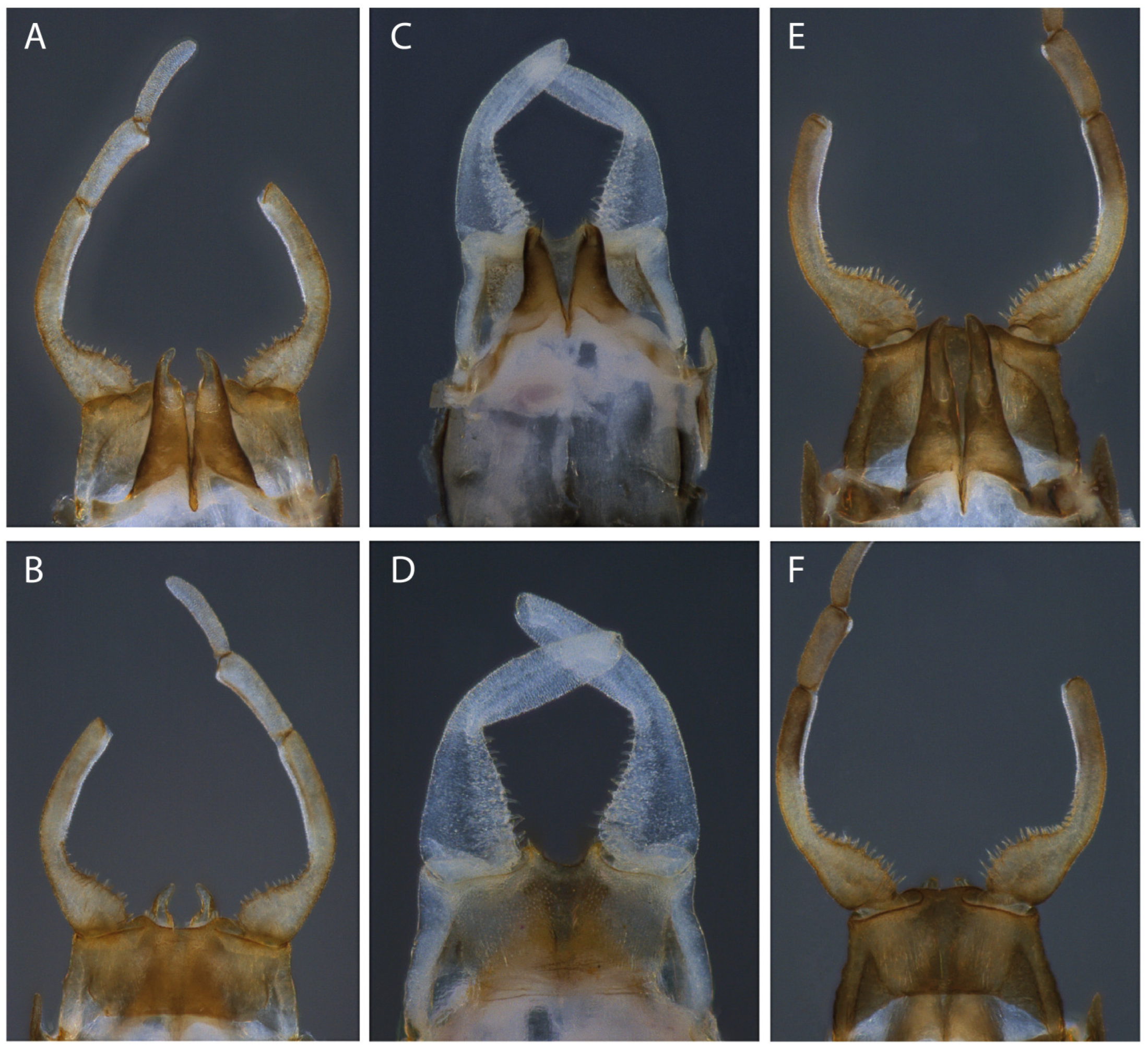
Diagnosis: Male imago (
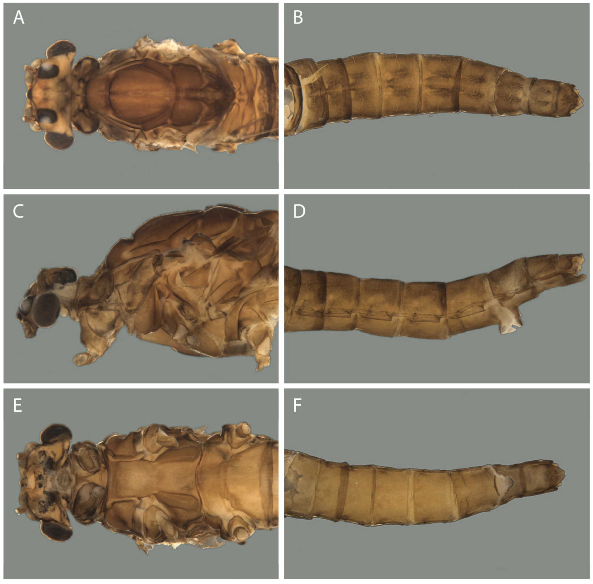
Figure 1,
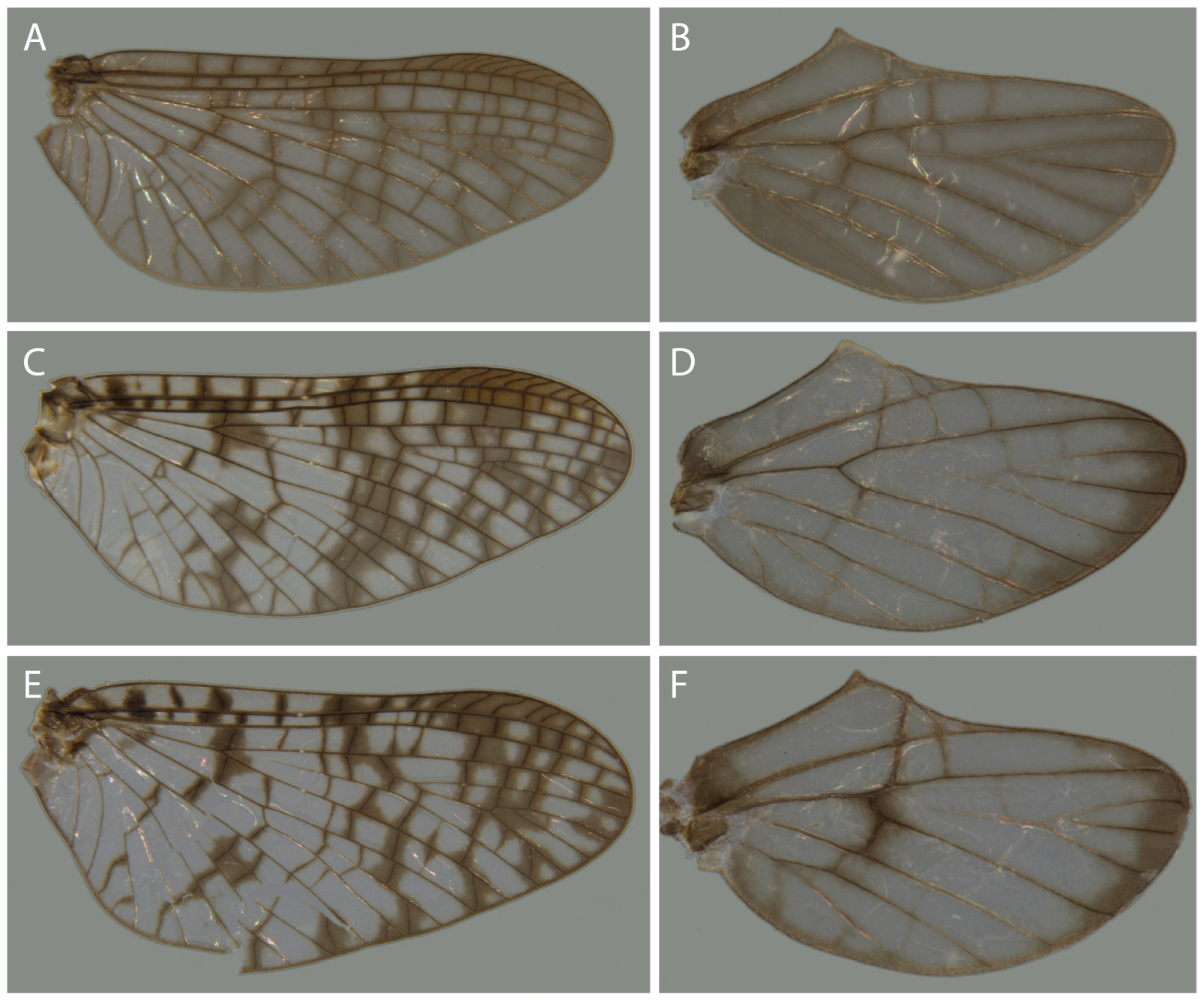
Figure 7A,B,
Figure 8A,B and
Figure 9A): (1) Forewings with membrane brown and dark brown mark around cross veins; (2) Hind wing uniformly brown; (3) Forceps segment I light brown, segment I and III lighter; (4) Penis lobe long (at least ¾ of forceps segment I) and apically rounded and curved on apical ¼; (5) Length of body between 5.0 and 6.3 mm.
Male imago description: Lengths (mm). Body: 5.0–6.3. Forewing: 5.7–6.3; hind wing: 1.2–1.5. Foreleg: 6.3; mid leg: 2.1; hind leg: 2.6. Legs: segments of foreleg: 0.5: 1.00 (2.7 mm): 0.01: 0.30: 0.25: 0.18: 0.07; mid leg: 1.00: 1.00 (0.8 mm): 0.07: 0.07: 0.07:0.11:0.12; hind leg: 1.00: 1.00 (1.1 mm): 0.08: 0.05: 0.05:0.08:0.13. Caudal filaments: 13.0–14.0.
Colouration: Body (
Figure 1): brown. Head (
Figure 1A,C,E). Orange washed with brown. Compound eye with facets of upper portion orange, separated by black grooves. Ocelli white surrounded with black. Antenna with scape and pedicel washed with brown, flagellum pale. Thorax (
Figure 1A,C,E): nota brown, except for a white spot at anterolateral corner of posterior scutal protuberance and at base of anteronotal protuberance, close to the apex of anterolateral scutal costa, and posterior margin of scutellum, dark brown. Pleura and sterna brown, washed with gray. Wings (
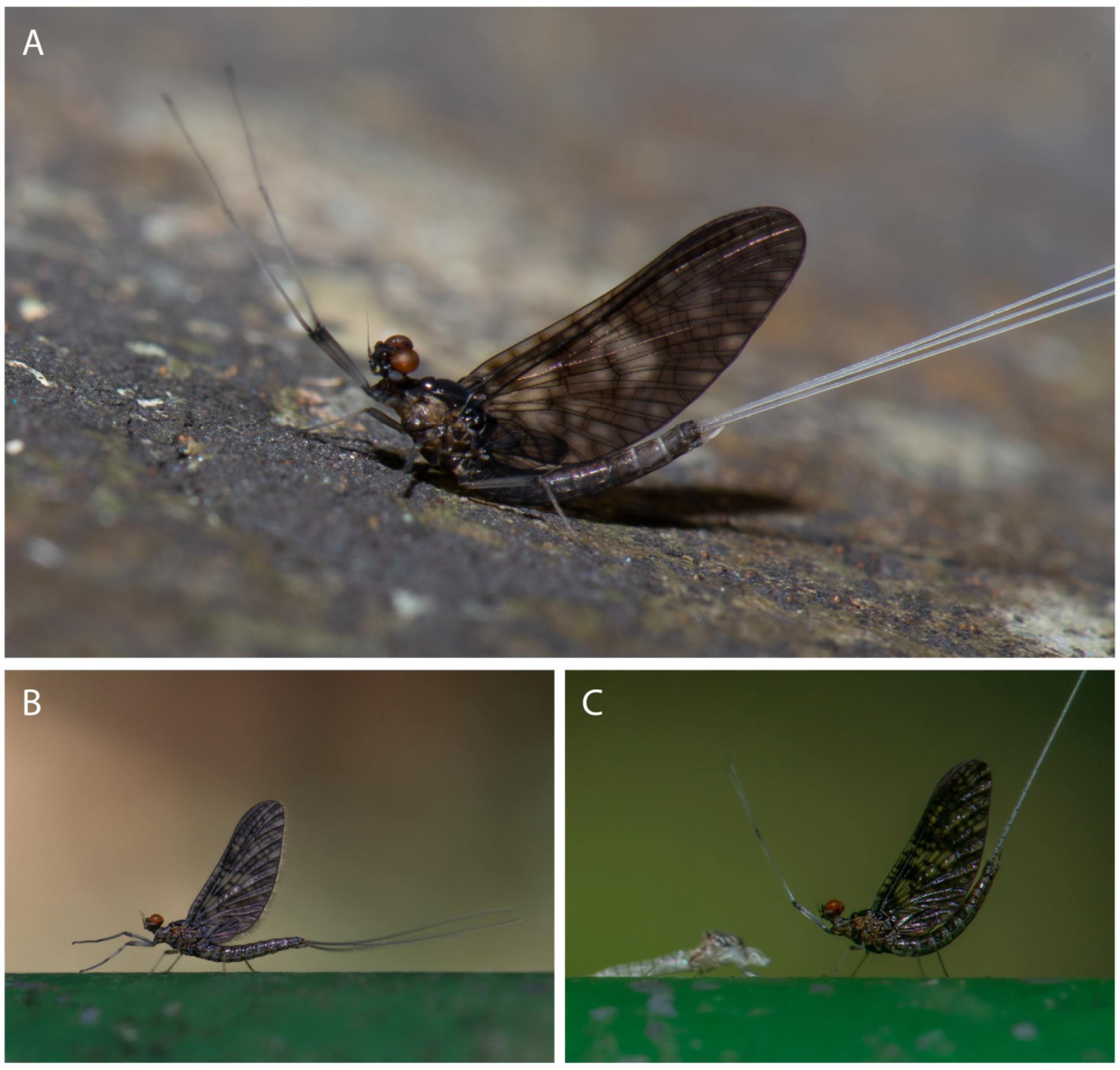
Figure 7A,B): forewing with brown membrane, dark brown on stigmatic field; longitudinal and cross veins dark brown; cross veins surrounded with diffuse dark brown cloud, except on cubito-anal field. Hind wing with brown membrane, longitudinal and cross veins dark brown; apex of wing and cubito-anal field washed with diffuse dark brown. Legs: Foreleg missing on collected material (similar to mid and hind legs according to pictures taken in the field–
Figure 9A). Mid and hind legs brown; femur washed with dark brown and with apical dark brown band, tibiae with dark brown mark on apex. Abdomen (
Figure 1B,D,F): terga translucent brown on terga I to VI and brown on terga VII to X; tergum I completely washed with dark brown; terga II–IX with submedial dark brown mark and dark brown posteriorly. Sterna translucent brown. Genitalia (
Figure 8A,B): styliger plate brown. Forceps segment I light brown, with distomedial margin and outer margin dark brown; Forceps segment II and III lighter, almost white; medial margin of forceps segment II dark brown. Penis brown. Caudal filament: light brown at base, white towards apex.
Morphology: Head (
Figure 1A,C,E): posterior margin V-shaped. Dorsal portion of eye not on stalk; dorsal surface circular with ca. 30 facets on the largest row. Lower portion of compound eye oblong. Wings (
Figure 7A,B): forewing with 3 cross veins basal and 12 distal to bulla; fork of MA2 asymmetric; IMP free basally; MP2 connected to base of MP1 and CuA by a cross vein; CuA connected to CuP by cross vein subbasally. Hind wing with apex rounded; costal projection developed; Sc ends distal to apex of costal projection; fork of R+MA symmetric; MP free basally; CuP present; A absent. Genitalia (
Figure 8A,B): Styliger plate with posteromedial portion broadly concave. Forceps segment I with basal portion abruptly narrowing on basal 1/3. Penis large basally and narrowing toward apex; 0.82× length of forceps segment I; apex rounded, inner margin curved; gonopore relatively large.
Female imago description: Lengths (mm): Body: 4.2. Forewing: 5.2; hind wing: broken off and missing. Legs and caudal filaments broken off and missing.
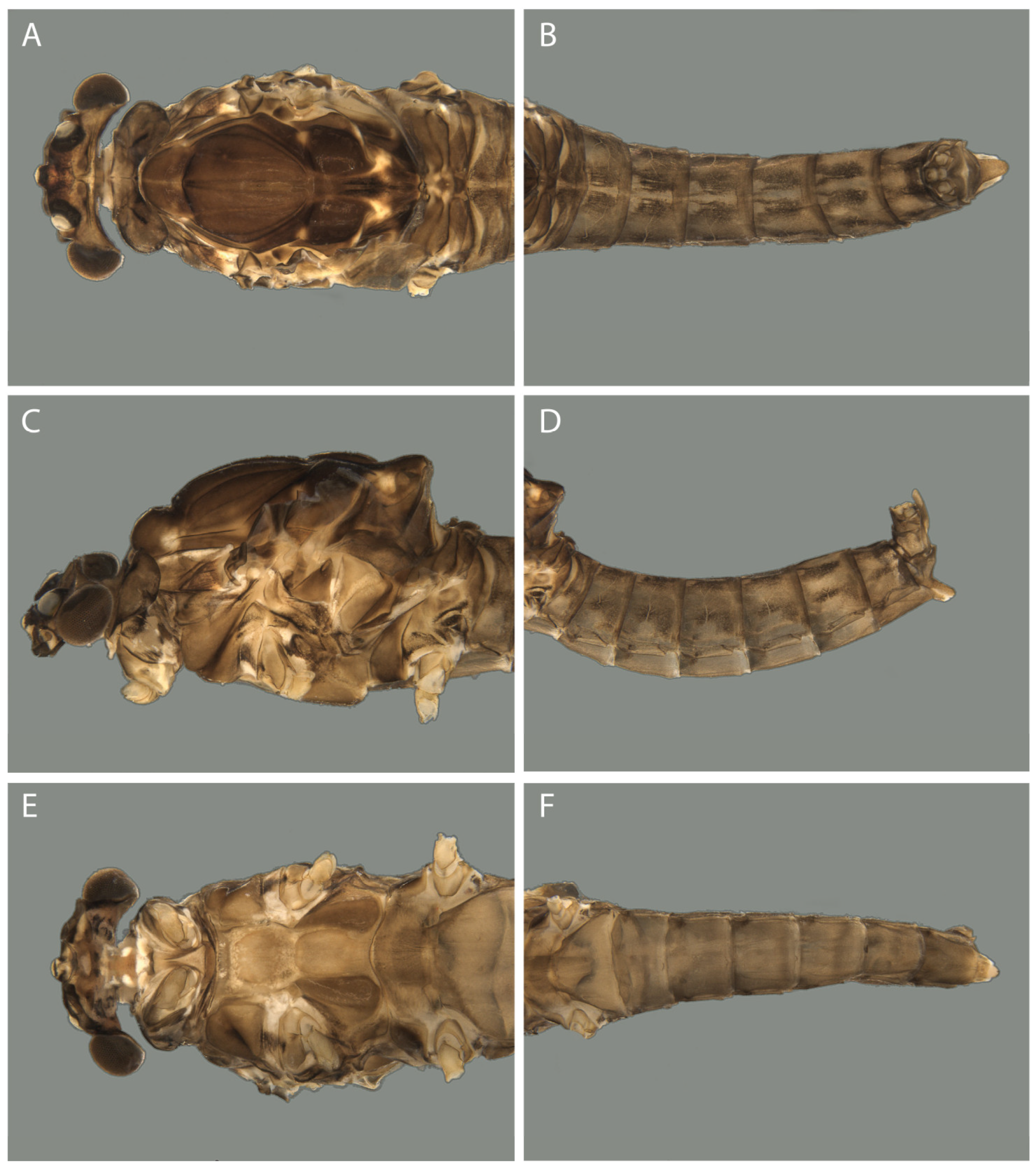
Colouration (
Figure 2): Similar to male imago, except for: head yellowish brown and lighter mesonotum.
Morphology: Wings: forewing with light brown membrane; clouds around cross veins less widespread than in males. Abdomen (
Figure 2B,D,F): sterna VII and VIII forming a small egg guide (little longer than sternum VIII); sternum IX with posterior margin V-shaped.
Nymph: Unknown.
Etymology: From Serra do Cipó, type-locality of the species.
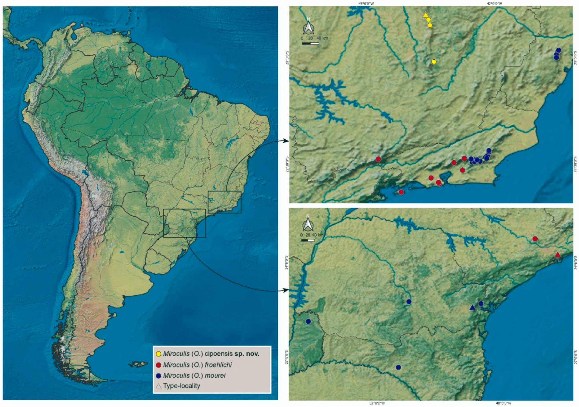
Distribution: BRAZIL: Minas Gerais, Serra do Espinhaço, Conceição do Mato Dentro and Catas Altas.
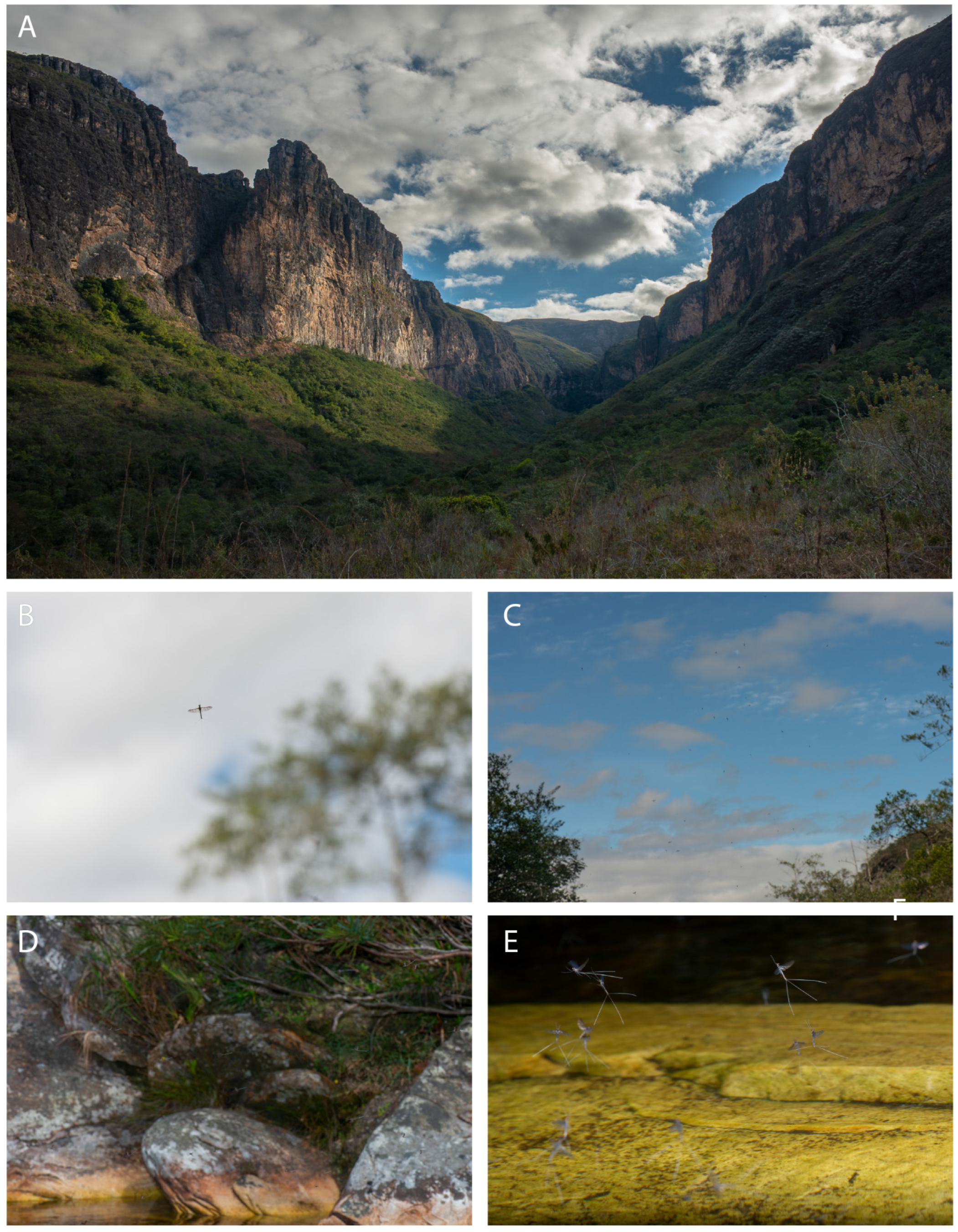
Biology: Two swarms of the new species were observed on consecutive days; one at the type-locality, and another at the Parque Natural Municipal do Tabuleiro, on the trail to the Tabuleiro Waterfall. At the type-locality, around 4:00 PM, a swarm of a few hundred individuals, mostly males, were flying vertically 2 to 3 m above the ground. During that time, few mayflies were seen copulating. About 10 min later, the mayflies were flying horizontally and closer to the water, between 50 cm and 1 m from the water surface. From 5:00 to 5:15 PM, the number of individuals decreased and those that were flying were very close to the water, so close that their filaments often touched the water surface. At this moment they were making short forward and reward movements. At the Parque Natural Municipal do Tabuleiro, we were able to observe them for only 10 min. It was around 3:20 PM and they were flying horizontally, between 50 cm and 1 m from the water.
Figure 1.
M. (O.) cipoensis, male imago. (A) head and thorax (dorsal view); (B) abdomen (dorsal view); (C) head and thorax (lateral view); (D) abdomen (lateral view); (E) head and thorax (ventral view); (F) abdomen (ventral view).
Figure 1.
M. (O.) cipoensis, male imago. (A) head and thorax (dorsal view); (B) abdomen (dorsal view); (C) head and thorax (lateral view); (D) abdomen (lateral view); (E) head and thorax (ventral view); (F) abdomen (ventral view).
Figure 2.
M. (O.) cipoensis, female imago. (A) head and thorax (dorsal view); (B) abdomen (dorsal view); (C) head and thorax (lateral view); (D) abdomen (lateral view); (E) head and thorax (ventral view); (F) abdomen (ventral view).
Figure 2.
M. (O.) cipoensis, female imago. (A) head and thorax (dorsal view); (B) abdomen (dorsal view); (C) head and thorax (lateral view); (D) abdomen (lateral view); (E) head and thorax (ventral view); (F) abdomen (ventral view).
Miroculis (Ommaethus) froehlichi Savage and Peters, 1983
Miroculis (Ommaethus) froehlichi Savage and Peters, 1983 [
2]: 564 (original description—male imago, probable female nymph); Savage, 1983 [
20]: 124 (wing evolution); Da-Silva, 1997 [
16]: 684 (distribution records); Hubbard and Pescador, 1999 [
21]: 138 (list of species); Salles, Da-Silva, Hubbard, and Serrão, 2004 [
22]: 26 (list of species); Domínguez, Molineri, Pescador, Hubbard, and Nieto, 2006 [
23]: 448 (diagnosis—male adults); Mariano and Polegatto, 2011 [
24]: 594 (list of species).
Diagnosis: Male imago (
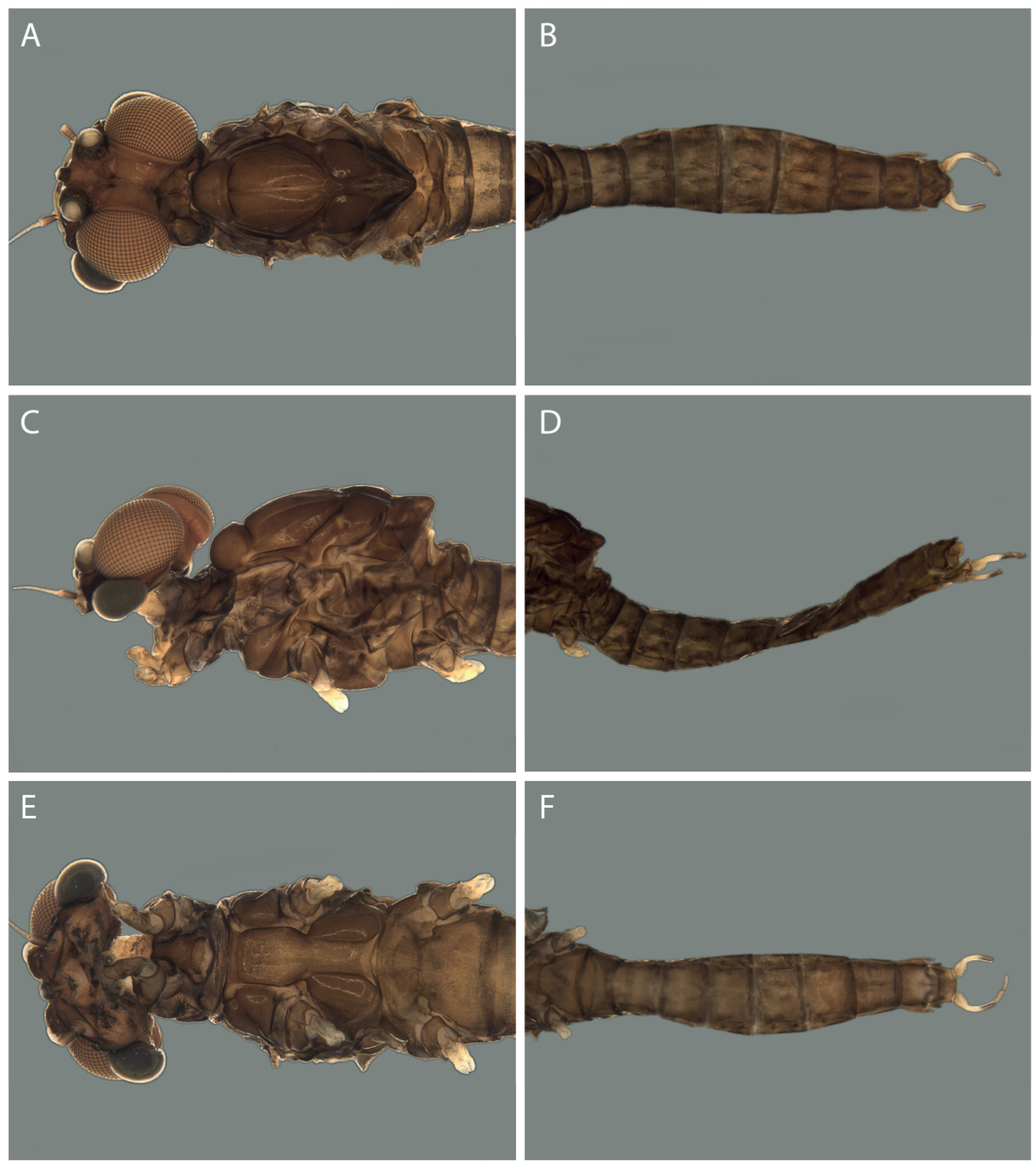
Figure 3,
Figure 7C,D and
Figure 8C,D): (1) Forewings with membrane hyaline and dark brown mark around cross veins; (2) Hind wing tinged with brown on distal margin; (3) Forceps yellowish-white; (4) Penis lobe short (ca. ½ of forceps segment I) and apically pointed; (5) Length of body between 6.2 and 7.8 mm.
Female imago description: Length of body: 6.7 mm, forewing: 7.8 mm, hind wing: broken and missing. Head (
Figure 4A,C,E): brown, with brown mark between ocelli. Eyes brown. Pronotum brown, with sublateral oblique dark brown mark and lateral margins dark brown. Remainder of thorax darker than in male imago, except for absence of white mark anteriorly on scuto-scutellar impression. Wing colouration as in male imagos, but less pigmented on distal half. Legs broken off and missing. Abdomen (
Figure 4B,D,F): brown, terga I to VIII with medial and sublateral bands; sublateral band extending from anterior margin to almost posterior margin. Sterna brown. Sternum VII and VIII with a single, broad, posteriorly projecting medial opening. Caudal filaments broken off and missing.
Comments: The male imago and an immature putative nymph were adequately described by [
2]. Additional material from Jundiaí, São Paulo, around 90 km from the type-locality, was examined and fits exactly the original description. Pictures of the male imago are provided in order to complement the illustrations previously presented and aid in its identification. The holotype, a male imago deposited at MZUSP (MZUSP000009), is faded and forewings are broken and missing.
According to [
16], the species is one of the most common Leptophlebiidae in the State of Rio de Janeiro, being recorded from several areas. We were not able to access these materials, which are composed mainly by nymphs, but imagos of
Miroculis (Ommaethus) examined by us, from other localities in Rio de Janeiro, belong to
M. (O.) mourei (
Figure 9B,C). It is our opinion, based on these facts, that the distribution of
M. (O.) froehlichi is much more restricted (see below).
Material examined: ♂ holotype (MZUSP000009), Brazil, São Paulo State, Estação Biológica de Paranapiacaba, nr. Paranapiacaba 15.X.63 C.G. Froehlich leg. (MZUSP). 2 ♂ and 1 ♀ imago; 1 ♂ and 1 ♀ subimago, Brazil, São Paulo, Jundiaí, REBIO Serra do Japi, 23°14′28″ S, 46°57′55″ W, 1020 m (a.s.l.), 19.XII.2021, Almeida, L. and Taniguti, P. N. leg. (UFVB).
Figure 3.
M. (O.) froehlichi, male imago (fresh material from São Paulo). (A) head and thorax (dorsal view); (B) abdomen (dorsal view); (C) head and thorax (lateral view); (D) abdomen (lateral view); (E) head and thorax (ventral view); (F) abdomen (ventral view).
Figure 3.
M. (O.) froehlichi, male imago (fresh material from São Paulo). (A) head and thorax (dorsal view); (B) abdomen (dorsal view); (C) head and thorax (lateral view); (D) abdomen (lateral view); (E) head and thorax (ventral view); (F) abdomen (ventral view).
Figure 4.
M. (O.) froehlichi, female imago (fresh material from São Paulo). (A) head and thorax (dorsal view); (B) abdomen (dorsal view); (C) head and thorax (lateral view); (D) abdomen (lateral view); (E) head and thorax (ventral view); (F) abdomen (ventral view).
Figure 4.
M. (O.) froehlichi, female imago (fresh material from São Paulo). (A) head and thorax (dorsal view); (B) abdomen (dorsal view); (C) head and thorax (lateral view); (D) abdomen (lateral view); (E) head and thorax (ventral view); (F) abdomen (ventral view).
Miroculis (Ommaethus) mourei Savage and Peters, 1983
Miroculis (Ommaethus) mourei Savage and Peters, 1983 [
2]: 561 (original description—male nymph and imago; female nymph and subimago); Savage, 1983 [
20]: 124 (wing evolution); Salles et al., 2004 [
22]: 26 (list of species); Domínguez et al., 2006 [
23]: 448 (diagnosis—male adults and nymph); Salles et al., 2010 [
17]: 306 (list of species); Salles and Lima, 2011 [
5]: 58 [(distribution records); Brito et al., 2011 [
18]: 104 (sperm ultrastructure); Mariano and Polegatto, 2011 [
24]: 594 (list of species).
Miroculis (Ommaethus) misionensis Domínguez, 2007 syn. n.
Miroculis (Ommaethus) misionensis Domínguez, 2007 [
3]: 99 (original description—male and female imago); Raimundi et al., 2021 [
14]: 390 (description—nymph).
Diagnosis: Male imago (
Figure 5,
Figure 7E,F,
Figure 8E,F and
Figure 9B,C): (1) Forewings with membrane hyaline and dark brown mark around cross veins; (2) Hind wing tinged with brown at base, MA/R fork and distal margin; (3) Forceps brown, apex of segment I and segments I and III darker; (4) Penis lobe long (subequal to forceps segment I), apically rounded and curved on apical ½; (5) Length of body between 3.8 and 6.0 mm [including here data from literature [
1,
2], and examined material].
Comments: The male imago, female subimago and nymphs were adequately described by [
2]. We had access to imagos of this species from Rio de Janeiro State and, more importantly, from Antonina, Paraná. This locality is very close to the type-locality (ca. 30 km) and fits exactly the original description (the straight posterior margin of the styliger plate observed in
Figure 8E,F is a result of a mounting artifact). Pictures of the male and female imago are provided in order to complement the illustrations previously presented and aid in its identification. The holotype, a male imago deposited at MZUSP (MZUSP000010) is in poor condition: the head is separated from the thorax, all legs are missing, except for one mid or hind leg detached from the body, and thorax and abdomen are broken—segments VII to X are missing. A faded right forewing is attached to the holotype, while one hind wing is detached.
After comparing our material from Paraná with the description of
M. (O.) misionensis and with pictures and photographs of the type material, we came to the conclusion that the latter is a junior synonym of
M. (O.) mourei. According to [
3], they could be distinguished by the shape of the dorsal portion of the eye, and by the number of lines on abdominal terga IV–VI. We found no difference concerning the first characteristic and we interpret the difference in the number of lines (four narrower versus two wider) to an intraspecific variation related to the degree of pigmentation of the bands. The same applies to the colouration of forceps segment II, which is darker in the pictures of
M. (O.) misionensis as well as in photographs of the type. Other characteristics, such as colouration of fore and hind wings, and more importantly, the shape of the penis lobe, are exactly the same.
Material examined: ♂ holotype (MZUSP000010), Brazil, Paraná, Rio Ipiranga, Estrada do Itupaua [Itupava], 21–23.II.1969, W.L. and J.G. Peters leg. 2 ♂ and 13 ♀ imago, Brazil, Paraná, Antonina, RPPN Reserva Natural Guariciça (SPVS), Rio do Turvo, 25°17′20″ S, 48°39′11″ W, 88 m (a.s.l.), 31.I–04.II.2022, PPGEnto Entomol. de Campo UFPR leg. (DZUP). 2 ♂ imago, Brazil, Espírito Santo, Santa Teresa, Pinguela 19°52′16″ S, 40°31′43″ W, 700 m (a.s.l.), 26.XI.2009, Salles, F.F. leg. (UFVB). 1 ♂ and 1 ♀ imago, Brazil, Espírito Santo, Santa Teresa, II.2009, Salles, F.F. leg. (UFVB). 1 ♂ subimago (DZRJ 2064), Brazil, Rio de Janeiro, Macaé, Sana, AF. 2º ordem do Rio boa sorte SA, 22°20′41″ S, 42°11′03″ W, 381 m (a.s.l.), 19.II. 2009 (DZRJ). 1 ♀ subimago (DZRJ 2058), Brazil, Rio de Janeiro, Macaé, Arraial do Sana, Rancho Peito de Pombo, Af. 2º ordem do Rio sana SA1, 22°19′17″ S, 42°10′58″ W, 313 m (a.s.l.), 16.II.2009 (DZRJ). 1 ♂ imago (DZRJ 2060), Brazil, Rio de Janeiro, Arraial do Sana, camping do Poço, Rio Sana, SA02, 22°19′20″ S, 42°10′51″ W, 306 m (a.s.l.), 16.II.2009, Gonçalves, I.C. leg. (DZRJ). 1 ♂ subimago (DZRJ 2062), Brazil, Rio de Janeiro, Macaé, Sana, Alto da cabeceira (SA11), Rio Sana (Faz. Agroecologica), 22°16′15″ S, 42°09′20″ W, 459 m (a.s.l.), 18. II. 2009, Sampaio, B.H.L. and Jardim, G.A. leg.; 2 ♂ imago (DZRJ 2034), Brazil, Rio de Janeiro, Nova Friburgo, Macaé de Cima–MC2, Represa, 22°25′51″ S, 42°32′18″ W, 1061 m (a.s.l.), 30.XI.2008, Sampaio, B.H.L. and Santos, A.P.M. leg. (DZRJ). 1 ♂ imago (DZRJ 2049), Brazil, Rio de Janeiro, Nova Friburgo, Rio Bonito de Lumiar, Rio Bonito, RB23, 22°13′47″ S, 42°08′04″ W, 803 m (a.s.l.), 30.X.2009 (DZRJ). 1 ♂ imago (DZRJ 2042), Brazil, Rio de Janeiro, Nova Friburgo, Rio Bonito de Lumiar RB7, AF. 1° ordem do Rio Bonito, 22°24′37″ S, 42°20′42″ W, 676 m (a.s.l.), 30.X.2009, Dumas, L.L. leg. (DZRJ). 1 ♂ imago (DZRJ 2046), Brazil, Rio de Janeiro, Rio Bonito de Cima (Lumiar), AF. do Rio Bonito, RB21, 22°24′15″ S, 42°26′46″ W, 863 m (a.s.l.), Gonçalves, I.C. leg. (DZRJ). 2 ♂ imago, Brazil, Rio de Janeiro, Rio Bonito de Cima (Lumiar), AF. do Rio Bonito, RB21, 22°24′15″ S, 42°26′46″ W, 863 m (a.s.l.), Gonçalves, I.C. leg. (DZRJ).
Figure 5.
M. (O.) mourei, male imago (fresh material from Paraná). (A) head and thorax (dorsal view); (B) abdomen (dorsal view); (C) head and thorax (lateral view); (D) abdomen (lateral view); (E) head and thorax (ventral view); (F) abdomen (ventral view).
Figure 5.
M. (O.) mourei, male imago (fresh material from Paraná). (A) head and thorax (dorsal view); (B) abdomen (dorsal view); (C) head and thorax (lateral view); (D) abdomen (lateral view); (E) head and thorax (ventral view); (F) abdomen (ventral view).
Figure 6.
M. (O.) mourei, female imago (fresh material from Paraná). (A) head and thorax (dorsal view); (B) abdomen (dorsal view); (C) head and thorax (lateral view); (D) abdomen (lateral view); (E) head and thorax (ventral view); (F) abdomen (ventral view).
Figure 6.
M. (O.) mourei, female imago (fresh material from Paraná). (A) head and thorax (dorsal view); (B) abdomen (dorsal view); (C) head and thorax (lateral view); (D) abdomen (lateral view); (E) head and thorax (ventral view); (F) abdomen (ventral view).
Figure 7.
Miroculis (O.) spp., wings of male imago. (A) M. (O.) cipoensis (forewing); (B) M. (O.) cipoensis (hind wing); (C) M. (O.) froehlichi (forewing); (D) M. (O.) froehlichi (hind wing); (E) M. (O.) mourei (forewing); (F) M. (O.) mourei (hind wing).
Figure 7.
Miroculis (O.) spp., wings of male imago. (A) M. (O.) cipoensis (forewing); (B) M. (O.) cipoensis (hind wing); (C) M. (O.) froehlichi (forewing); (D) M. (O.) froehlichi (hind wing); (E) M. (O.) mourei (forewing); (F) M. (O.) mourei (hind wing).
Figure 8.
M. (O.) spp., genitalia of male imago. (A) M. (O.) cipoensis (dorsal view); (B) M. (O.) cipoensis (ventral view); (C) M. (O.) froehlichi (dorsal view); (D) M. (O.) froehlichi (ventral view); (E) M. (O.) mourei (dorsal view); (F) M. (O.) mourei (ventral view).
Figure 8.
M. (O.) spp., genitalia of male imago. (A) M. (O.) cipoensis (dorsal view); (B) M. (O.) cipoensis (ventral view); (C) M. (O.) froehlichi (dorsal view); (D) M. (O.) froehlichi (ventral view); (E) M. (O.) mourei (dorsal view); (F) M. (O.) mourei (ventral view).
Figure 9.
M. (O.) spp., living specimens in the field. (A) M. (O.) cipoensis, male imago; (B) M. (O.) mourei, male subimago; (C) M. (O.) mourei, male imago and subimaginal exuviae. All photographs by F.F. Salles.
Figure 9.
M. (O.) spp., living specimens in the field. (A) M. (O.) cipoensis, male imago; (B) M. (O.) mourei, male subimago; (C) M. (O.) mourei, male imago and subimaginal exuviae. All photographs by F.F. Salles.
Figure 10.
M. (O.) cipoensis, type-locality and swarm. (A) Peixe Tolo Canion, type locality; (B) male flying vertically; (C) swarm of males (and few females) flying vertically; (D) swarm of males (and few females) flying horizontally; (E) same as d, but closer to the water. All photographs by F.F. Salles.
Figure 11.
Map of South America showing details of the known distribution of the species of
Miroculis (Ommaethus) in Southeastern Brazil (
top right) and Southern Brazil (
bottom right), including northern Misiones, in Argentina. Map from QGIS (2024), used according to Open Source Geospatial Foundation. The map of South America is based on data sourced from the website
https://plugins.qgis.org/plugins/OpenTopography-DEM-Downloader/, accessed on 16 March 2024.

