Article

Untangling the Defiant Taxonomy of Physaloptera (Nematoda: Chromadorea: Spirurida: Physalopteridae) Parasites in Reptiles: An Integrative Approach on the Enigmatic P. retusa Suggests Cryptic Speciation

by
Lorena Gisela Ailán-Choke
1,
Vanda Lúcia Ferreira
2,
Fernando Paiva
2,
Luiz Eduardo Roland Tavares
2,
Fabiano Paschoal
3 and
Felipe Bisaggio Pereira
4,*
1
Consejo Nacional de Investigaciones Científicas y Técnicas (CONICET), Centro de Ecología Aplicada del Litoral (CECOAL), Ruta Provincial Número 5, Km 2.5, Corrientes 3400, Argentina
2
Instituto de Biociências, Universidade Federal de Mato Grosso do Sul, Av. Costa e Silva s/n°, Campo Grande 79070-900, Brazil
3
Programa de Pós-Graduação em Biodiversidade e Conservação, Departamento de Oceanografia e Limnologia, Universidade Federal do Maranhão, Av. dos Portugueses, 1966, Bacanga, São Luís 65080-805, Brazil
4
Departamento de Parasitologia, Instituto de Ciências Biológicas, Universidade Federal de Minas Gerais, Av. Presidente Antônio Carlos, 6627, Pampulha, Belo Horizonte 31270-901, Brazil
*
Author to whom correspondence should be addressed.
Taxonomy 2024, 4(2), 326-340; https://doi.org/10.3390/taxonomy4020016
Submission received: 22 February 2024 / Revised: 10 May 2024 / Accepted: 13 May 2024 / Published: 16 May 2024

Abstract

:
(1) Background: Although Physaloptera retusa is one of the most widespread species infecting reptiles in the Americas, numerous taxonomic problems and little genetic data are associated with it. To clarify the taxonomy of this species, we used an integrative approach. (2) Methods: Physaloptera retusa-infecting Erythrolamprus typhlus (snake) from the Pantanal wetlands, Brazil, was morphologically and genetically characterised (18S and 28S rDNA; COI mtDNA) and compared with conspecific sequences available in GenBank, from parasites of Tupinambis teguixin (lizard), using species delimitation methods. Type specimens of P. liophis were re-evaluated given its morphological similarities with P. retusa. (3) Results: The morphology of the present specimens was equal to that of P. retusa, in which the only difference from P. liophis was the relative position of the vulva. Species delimitation methods were more accurate for the COI dataset; all of them (except ABGD) indicated interspecificity among P. retusa sequences. However, a lack of morphological data or voucher material, associated with the deposited sequences, prevented more assertive conclusions. (4) Conclusions: The present results highlight the importance of a clear association between genetic data and morphology of the isolation source, or at least its adequate vouchering. Moreover, P. retusa may represent a species complex in cryptic speciation, since it is widespread and has low hosts specificity.

Graphical Abstract

1. Introduction

With more than a hundred species, Physaloptera Rudolphi, 1819, is the most represented genus within the Physalopteridae in terms of species richness, diversity, and geographic distribution [1,2,3]. These nematodes are commonly found in the gastrointestinal tract of all vertebrate taxa, except Chondrichthyes and Osteichthyes [3]. Despite its great abundance and diversity, the taxonomy of Physaloptera is problematic due to undetailed and inaccurate species descriptions, which results in a large number of taxa with similar morphology and difficult diagnosis [3,4]. Moreover, the morphometry of these parasites may show an apparent wide intraspecific variation, causing even more confusion when taken as a diagnostic feature [3].
The scarcity of genetic data related to the species of Physaloptera prevents the advance on their taxonomic knowledge. Such a lack of information is a real challenge when dealing with the integrative taxonomy of these nematodes. In this sense, from all species of Physaloptera, only 15 have been genetically characterised, based on a few nuclear and mitochondrial markers (mostly 18S rRNA and cox1 mtDNA), in addition to the sequences that are not identified up to the species level (see GenBank, https://www.ncbi.nlm.nih.gov/nucleotide/) (accessed on 20 January 2024). The scenario is even more critical regarding those congeners parasitising reptiles, in which only two species have genetic sequences: namely, P. retusa (Rudolphi, 1819) and P. tupinambae Pereira, Alves, Rocha, Souza Lima & Luque, 2012 [2].
The type species of Physaloptera is P. retusa, one of the most common and widespread in the genus [2,5]. According to recent works that evaluated the taxonomy of P. retusa based on morphology, the species apparently has low host specificity and substantial taxonomic confusions [2,5]. Moreover, the few genetic approaches including P. retusa are inconsistent regarding its phylogenetic position in Physalopteridae, resulting in low nodal supports and polytomies [2]. Most of the Physaloptera parasites in reptiles remain unknown both morphologically and genetically, for example, P. liophis Vicente & Santos, 1974, described in Liophis miliaris (syn. Erythrolamprus miliaris) (Linnaeus, 1758) from the State of Rio de Janeiro, Brazil [6]. It should be mentioned that, since its proposition in 1974, the taxonomy of P. liophis has never been re-valuated in details (see Macedo et al. [5], for some comments on the type material of the species).
During a parasitological study in one specimen of the blind ground snake Erythrolamprus typhlus (Linnaeus, 1758) from the Pantanal wetlands, State of Mato Grosso do Sul, Brazil, some physalopterids were found parasitising its stomach. A morphological evaluation revealed that the parasites were similar to P. retusa. Due to the taxonomic problems associated with this species and the fact that it commonly affects lizards rather than snakes [3], the present specimens were genetically characterised and an integrative taxonomic approach was taken aiming for an accurate identification. Therefore, a more robust species identification framework was taken for P. retusa. Moreover, due to the morphological similarity and closely related hosts between the present specimens and P. liophis, in addition to the poorly detailed morphology associated with the latter, the type series of P. liophis was revisited.

2. Materials and Methods

2.1. Collection, Processing, and Morphological Evaluation of Parasites

In December 2017, one specimen of E. typhlus (Serpentes: Dipsadidae) adult was captured with a pitfall trap in a forest area of the Pantanal wetlands, municipality of Corumbá (18° 59.944′ S, 56° 38.743′ W), State of Mato Grosso do Sul, Brazil. Prior to parasitological examination, the specimen was euthanised with an intraperitoneal injection of barbiturate. Host taxonomic identification was performed based on morphology by a co-author of this manuscript, V.L. Ferreira, who is a herpetologist; its nomenclature and classification were updated according to Grazziotin et al. [7] and Nogueira et al. [8]. The voucher was deposited in the Coleção Zoológica, Reptilia section of the Universidade Federal de Mato Grosso do Sul (ZUFMS-REP05158). Nematodes were found alive in the stomach, washed in saline, fixed in hot 4% formalin, and stored in 70% ethanol for morphological observations. The mid-body of one male specimen was excised, fixed, and stored in 96–100% molecular-grade ethanol for genetic analyses; the anterior and posterior parts were fixed for morphological observation as previously described. The present specimens were deposited as vouchers in the Coleção Helmintológica do Instituto Oswaldo Cruz (acronym CHIOC; accession number CHIOC 39666); the type specimens of P. liophis (CHIOC 31034a–c), deposited in the same collection (CHIOC), were evaluated for morphological confirmation. Access to genetic heritage was registered in the Sistema Nacional de Gestão do Patrimômio Genético e do Conhecimento Tradicional Associado (acronym SisGen), under the number A4B5848, according to Brazilian Federal requirements. Animal use, manipulation, and collection were permitted by the Sistema de Autorização e Informação em Biodiversidade (acronym SISBIO; license number 63916-3) and by the Comissão de Ética no Uso de Animais (acronym CEUA/UFMS; license number 941/2018).
The newly collected nematodes were cleared in glycerin as follows: a drop of glycerin–water solution in a ratio of 20:1 was put on a glass slide; the parasite was placed on it and covered by a coverslip; the slide was transferred to a heating plate at 56 °C; and as the water evaporated, a drop of more concentrated solution was added to the edge of the coverslip, in the concentrations 1:10, 1:5, and 1:2 until only glycerin was left. The slide was then observed, photographed, and measured using a light microscope Eclipse Ei (Nikon), with a camera Intervision 12 mp (Prime Cam) attached to it. Measurements are given in micrometres, unless otherwise indicated. Types of P. liophis were observed and photographed in the facilities of CHIOC, using a microscope Olympus BX51 coupled with a digital camera Olympus UC 30. For the description of male caudal papillae, we used the following abbreviations: p for pedunculate, s for sessile and u for unpaired. In addition, the pairs of papillae were numbered from the most anterior to the most posterior.

2.2. Genetic Procedures

Genomic DNA was isolated using DNeasy Blood & Tissue Kit (QIAGEN, Hilden, Germany), following the manufacturer’s instructions. Three genetic regions were amplified: the 5′ end of the nuclear 18S rDNA, the D2 and D3 domain of the nuclear 28S rDNA, and the barcode region of the mtDNA cytochrome c oxidase subunit I gene (COI). The polymerase chain reactions PCR, cycling conditions, and primers were the same as those in Ailán-Choke et al. [9]. PCR products were purified through an enzymatic treatment with ExoSAP-IT (ThermoFisher, Waltham, MA, USA) and sent for sequencing at ACTGene (Ludwig Biotec, Alvorada, Brazil) with the same primers used in PCR reactions.
Contiguous sequences were assembled and inspected, primers were trimmed, and the consensus was extracted in Geneious Prime 2024.0.5 (Dotmatics, Boston, MA, USA) and deposited in GenBank (see Section 3). A preliminary BLAST search on the GenBank database (https://www.ncbi.nlm.nih.gov/Blast.cgi) (accessed on 25 January 2024) was performed to confirm the genetic proximity between the present samples and those from other physalopterid nematodes.

2.3. Phylogenetic Analyses of Molecular Data

The phylogenetic reconstructions were based on two different datasets, consisting of sequences of 18S and COI, because 28S sequences of physalopterid nematodes are restricted to small initial fragments of the gene and associated with the ITS2 region. Sequences included in the phylogenetic analyses were chosen according to the following criteria, in order to maximise the number of representatives: genetic regions congruent with those obtained in the present study and minimum lengths of 660 bp for 18S and 410 bp for COI (for details, see Table 1). We tried to use as many representatives as possible, including different samples from the same species, for species delimitation and validation analyses. Since COI sequences KP981418 and KT894805 identified as P. mirandai Lent & Freitas, 1937, were 100% similar, only the first was considered in the present analysis, since it could be linked to a morphological reference [10]. We included another sequence also identified as P. mirandai (KT894804) but showing genetic divergence in comparison with the other conspecifics. The outgroup was chosen according to previous broad phylogenies including Physalopteridae (see Maldonado [2] and Černotíková et al. [11]). Sequences were aligned using M-Coffee [12]. The saturation of nucleotide substitution, especially in the alignment using COI sequences, was tested using Xia’s method implemented in DAMBE [13,14].
The phylogenetic hypotheses were inferred using Bayesian inference (BI) in BEAST 2.5 [25]; the best-fit substitution model for each dataset was chosen according to bModelTest [26]; and the molecular clock model was relaxed (log normal), defined using the nested sampling method [27] and the Yule tree priors, selected according to the posterior densities and the effective sample sizes (ESSs), visualised in Tracer [27,28]. This approach was preferred based on its robustness because it provides improved evolutionary pathways in phylogenetic reconstructions without overestimating nodal supports [27]. The posterior estimates of parameter densities, the ESS for each parameter, and the posterior probability for nodal supports in the majority rule consensus phylogenetic trees were determined after running the Markov chain Monte Carlo (MCMC) using 4 chains in 2 runs for 1 to 10 × 106 generations with the sampling frequency at every thousand generation, with 25% burn-in, and saving the last 75% of generated trees. The quality of the analysis (parameter densities, ESS, burn-in) and the chain convergence were examined in Tracer [28]. Genetic divergences were evaluated using patristic distances (abbreviated as p), which were presented in matrices extracted from the phylogenies, in Geneious Prime. The alignment of COI sequences was translated into amino acid residues using this same software.

2.4. Species Delimitation and Validation Approaches

We tested the species hypotheses of physalopterid nematodes mainly focused on the present sample of P. retusa and those conspecifics already deposited in GenBank (KT894814; KT894803) using different approaches detailed as follows.
The tree-based methods Generalized Mixed Yule Coalescent (GMYC), Poisson Tree Process, and its variations (bPTP and mPTP) were used for species delimitation. The GMYC requires an ultrametric guide tree, which uses ML to delimit species, and estimates a transition point before which all nodes reflect species diversification events and after which all nodes represent the population coalescent process; it was run in the webserver https://species.h-its.org/gmyc/ (accessed on 26 January 2024) [29,30,31]. PTP does not require essentially an ultrametric tree but is a phylogenetic-aware method; bPTP uses a BI approach and mPTP is an improvement of PTP that incorporates different levels of intraspecific genetic diversity, which derive from differences in either evolutionary history or sampling of each species; these analyses were run in the webservers https://species.h-its.org/ptp/ (accessed on 26 January 2024) and https://mptp.h-its.org/#/tree (accessed on 26 January 2024) [32,33].
The Automatic Barcode Gap Discovery (ABGD) and its recent improvement Assemble Species by Automatic Partitioning (ASAP) were also used for species delimitation. However, these methods are based on genetic distances, used to create species partitions [34,35]. ABGD and ASAP only require a genetic alignment or a genetic distance matrix. Therefore, we preferred to enter the genetic alignments and chose Kimura (K80) for estimating genetic distances, prior to species partitions, in order to be deliberately different from the tree-based approaches (i.e., GMYC and PTP; see also the Section 3). Moreover, ABGD provides only two additional models (simple distance and Jukes-Cantor), which generate less accurate results than that of K80. ASAP does not require any threshold value for species subset definition [35]. Conversely, ABGD requires a prior specification of an intraspecific genetic distance threshold [34]. These values were selected based on the K80 genetic distance matrices, in which values among conspecific samples were verified and the highest was considered. The values of other parameters included in ABGD analyses were set to default. These analyses were run in the following webservers https://bioinfo.mnhn.fr/abi/public/abgd/abgdweb.html (accessed on 26 January 2024) and https://bioinfo.mnhn.fr/abi/public/asap/ (accessed on 26 January 2024).
GMYC, PTP, ABGD, and ASAP were purposely chosen for species validation, since they are suitable for single-locus genetic analyses based on the not too large datasets, as in the present study, and with special emphasis on the barcoding region of the COI [30,31,32,33,34,35].
To validate the specific entities, we used *BEAST [36] implemented in BEAST 2.5, to generate unrooted (in order to improve species delimitation results; see Zhang et al. [32]) species trees, based on each dataset (minus the outgroup sequences). *BEAST uses the BI approach to generate phylogenies; the method of these phylogenetic reconstructions was the same as the one described in the previous topic. *BEAST also requires priors for species validation, in which different samples must be set as a same specific entity. These conspecific entities were defined following their taxonomic identification to the species level, indicated in GenBank. For those samples not identified to the species level, the prior conspecific definition was based on the results of GMYC, PTP, ABGD, and ASAP. Branch supports were estimated as previously described and used for terminal lineage validation when the posterior probability was higher than 0.90.

3. Results

3.1. Morphological Analysis of Parasites
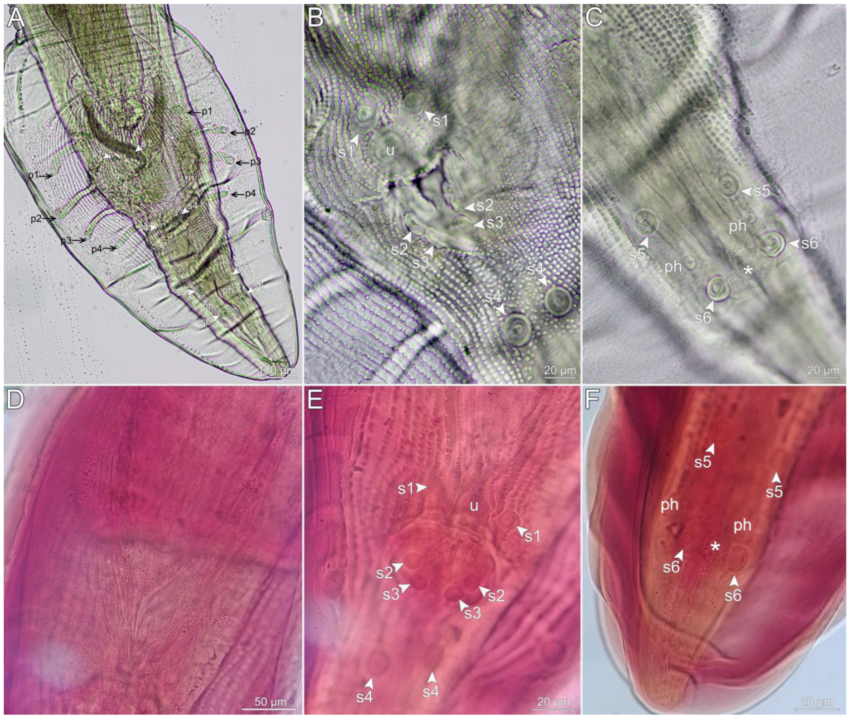
All parasites were collected sexually mature, in the stomach of E. typhlus, totalling five males, in which a tissue sample from one specimen was taken for DNA isolation and the specimen was not included in the morphological analysis, and four females. The morphometry of these specimens is detailed in Table S1 and was within the wide variation observed for P. retusa. A comparative table including morphometric data from all taxonomic reports of P. retusa is also given in Table S1. The main diagnostic features that were observed in the present specimens are as follows (described in telegraphic style).
Anterior end with two lateral dome-shaped pseudolabia, each bearing outer large triangular tooth, internal tripartite tooth of similar size as outer one, pair of large cephalic papillae (one subdorsal and one subventral), and lateral small amphidial pore. Cuticle reflected at pseudolabia base forming conspicuous cephalic collar. Conspicuous pointed deirids somewhat at posterior end of muscular oesophagus. Excretory pore at same level of deirids or slightly posterior to it. Oesophagus divided in anterior shorter muscular and posterior longer glandular parts. Nerve ring encircling posterior half of muscular oesophagus. Females with vulva located about first 1/3 of body and tail conical. Males with well-developed, vesicular caudal bursa and 21 caudal papillae: 4 pairs p supporting caudal bursa, 6 pairs s, and 1 u (Figure 1A). Pair p1and p4pairs subventral, p2 and p3 laterally directed (Figure 1A). Pair s1 subventral and precloacal; s2 and s3 adcloacal and located at posterior margin of cloacal labium (Figure 1B); s4, s5, and s6 pairs posterior to cloaca, subventral, arranged in two longitudinal lines, s5 and s6 closer to each other and slightly far from s4 (Figure 1A). Ventral and large u papilla, slightly posterior to s1 (Figure 1B). Papilla like phasmids small, laterally placed between s5 and s6 (Figure 1A,C). Conspicuous rounded protuberance, located between s6 (Figure 1C). Spicules with blunt proximal and pointed distal ends; similar in length and different in shape; left spicule with lateral expansions at distal half, right spicule without lateral expansion (Figure 1A).
The type series of P. liophis (CHIOC 31034a–c, holotype and one paratype male, and allotype female) was stained and mounted in permanent slides, making it difficult to observe some of the morphological traits. However, most of the diagnostic features previously described for the newly collected physalopterids were also observed in the type specimens of P. liophis, including the shape of the spicules (Figure 1D), the number and the general arrangement of caudal papillae (Figure 1E,F), and the rounded protuberance between the s6 (Figure 1F). The most discrepant difference was the position of the vulva, which was at the mid-body of the allotype female (vs. at the first 1/3 of the body in P. retusa). In addition, the u in the holotype male of P. liophis was smaller and at the same level as the s1 (Figure 1E). Moreover, in this same holotype male, we observed the outline of the excretory pore, although without full certainty due to the colour of the stain. This outline was slightly posterior to the deirids as in the newly collected specimens. The morphology of labial teeth was not completely clear due to the condition of the type specimens, although it seems to resemble that observed in the present physalopterids. The general morphometry was confirmed as described by Vicente & Santos [6] and, consequently, not mentioned here.
Figure 1. Physaloptera retusa male: (A) posterior end; (B) cloacal region; (C) last pairs of sessile papillae (asterisk indicates papilla-like protuberance), all in ventral view. Physaloptera liophis male holotype: (D) outline of spicules; (E) cloacal region; (F) last pairs of sessile papillae (asterisk indicates papilla-like protuberance), all in ventral view. Abbreviations: p: pedeunculate papilla; ph: phasmid; s: sessile papilla; u: unpaired sessile papilla. The pairs of papillae are numbered in crescent order, from the most anterior to the most posterior.
Taxonomy 04 00016 g001

3.2. Genetic Characterisation, Phylogeny, and Species Delimitation

Genetic sequences representing partial fragments of the 18S (879 bp; GenBank PP750392), 28S (809 bp; GenBank PP753775), and COI (414 bp; GenBank PP750553) were obtained for the newly collected parasites. The overlapping fragments of 18S sequences (about 794 bp) from the present specimens, P. retusa (KT894814) parasite of Tupinambis teguixin (Linnaeus, 1758), P. mirandai (KT894815, KT894816) parasite of Metachirus nudicaudatus (Desmarest, 1817), and Physaloptera sp. (KT894816) parasite of Cerradomys subflavus (Wagner, 1842), all from Brazil, showed no genetic divergence (p = 0). In fact, 18S sequences were highly similar among species of Physaloptera, in which that of the present specimens; those of P. amazonica Maldonado et al., 2020 (MK312472), P. bispiculata Vaz & Pereira, 1935 (KT894817), and P. tupinambae (MT810006) from Brazil, P. turgida (Rudolphi, 1819) (DQ503459, KP208673, KT894819, MH748145) from Brazil and the USA; and Physaloptera sp. from Costa Rica (MG808040) showed low genetic divergence (p < 0.02). In contrast, the COI sequence of P. retusa (KT894803), parasitic in T. teguixin, was less divergent in relation to that of the present specimens (p = 0.22), although considered high for an intraspecific variation. Similarly, the COI sequences of P. mirandai (KT894804; KP981418) showed high intraspecific genetic divergence (p = 0.24). In contrast, the sequences of P. hispida (MH782844; MH782845) and Abbreviata caucasica (Linstow 1902) (MT231294; MT231295) had low intraspecific genetic divergences (p < 0.02). The patristic distance matrices are given in Tables S2 and S3. There were 29 polymorphisms between the present COI sequence and that of P. retusa (KT894803), but only two amino acid substitutions (alanine [GCC] by threonine [ACG]; leucine [TGG] by methionine [ATG]). The genetic alignments were not saturated by nucleotide substitution and, consequently, adequate for phylogenetic reconstructions, as the Xia’s test indicated (Iss < Iss.c and p < 0.001 in all alignments). The 18S alignment was 804 bp long and included 32 sequences, in which the sequence from Proleptus obtusus Dujardin, 1845 (KY411575) was the shortest (690 bp) and most of the others were 804 bp long; there were 485 invariable and 319 variable sites. The COI alignment was 415 bp long and included 27 sequences, all of them with the same length (415 bp); there were 154 invariable and 261 variable sites.
Phylogenetic reconstructions using 18S and COI sequences showed somewhat similar topologies, when considering the relationships of the taxa common to both datasets (Figure 2A,B). The best-fit substitution model for both datasets was TN93 + G, with four substitution rate categories and unequal base frequencies. The patterns of the nodal supports were different, in which terminal nodes were better supported in the phylogeny inferred from COI sequences, and outer nodes tended to show better resolution in the 18S phylogeny; however, these values were generally low (Figure 2A,B). In this sense, the monophyletic lineage formed by the present sequences and those of P. retusa was weakly and fully supported in the phylogenies inferred from 18S and COI, respectively (Figure 2A,B). The phylogenetic position of this lineage was uncertain within Physalopteridae (Figure 2A,B).
The correspondence between different species delimitation methods and the specific lineages present in the analysis was more accurate when COI sequences were used (Figure 2C). Regarding the dataset of 18S sequences, no species delimitation method recognised P. retusa as a single specific entity; in fact, there was no accuracy of these methods in identifying the species of the poorly supported clade, formed by P. amazonica, P. mirandai, P. retusa, and P. tupinambae from Brazil and two unidentified Physaloptera spp. from Brazil and Costa Rica (Figure 2A,B). Moreover, there was no species validation, according to *BEAST, for the previously mentioned taxa, in addition to P. alata Rudolphi, 1819; P. apivori Desportes, 1946; P. bispiculata; P. praeputialis Linstow, 1889; P. turgida; and other unidentified Physaloptera (Figure 2A). Conversely, the correspondence between the species delimitation results and the specific taxa was higher in the phylogeny generated from COI sequences. However, all species delimitation methods recognised the different samples of P. retusa and P. mirandai as interspecific, and *BEAST did not validate them as same species (Figure 2C,D). Overall, the results of species delimitation were similar among the different methods, but those of GMYC were different from the others regarding the 18S dataset (Figure 2A,C). Furthermore, minor discrepancies were noted in the results of ASAP and ABGD, in comparison with those of GMYC and PTP regarding the COI dataset (Figure 2A,C). The *BEAST species validation results were similar to those of GMYC and PTP for the COI sequences (Figure 2B,D).

4. Discussion

Physaloptera retusa was redescribed in two recent works [3,5]. Despite these apparent growing concerns regarding the taxonomy of this common and widespread species, knowledge gaps about its population genetics are persistent. In fact, such a situation is extensive to the Physalopteridae as a whole (see Lopes-Torres et al. [10]; Maldonado et al. [2]; Chen et al. [37]). In this sense, physalopterids with wide host and geographic ranges (e.g., P. retusa) need to be associated with well-detailed morphological data and, if possible, diverse genetic characterisation. This is an efficient way to shed light on the complicated systematics of these nematodes. In the present work, we provided the first 28S rDNA sequence for P. retusa, in addition to the 18S and COI sequences, the first genetic data for the species in the Pantanal wetlands from Brazil, and sufficient morphological detailing for the specific diagnosis.
At first, it would be plausible to presume that the present nematodes parasitising the snake E. typhlus in Brazil would belong to P. liophis, which has the congener E. miliaris as its type host, collected in the same country [6]. However, after observation of the type specimens of P. liophis, we concluded that this species and P. retusa are morphologically similar. However, two major differences between them could be observed in the size and position of the unpaired papillae in males and in the relative position of vulva that is more posterior in P. liophis females (see the Section 3 for details). Based on these differences, which have been considered important for species diagnosis of Physaloptera [3], and due to the fact that the morphology of the newly collected specimens corresponds to the redescriptions of P. retusa [3,5], the present material was identified as P. retusa, and P. liophis is tentatively maintained valid until parasites from its type host in the type locality are re-evaluated and genetically characterised. It should be mentioned that P. liophis has no genetic characterisation and its original description is inconsistent regarding the number of caudal papillae (for details, see Macedo et al. [5]). Moreover, an integrative approach that evaluates the importance of the relative position of vulva in females of Physaloptera as a diagnostic feature would be helpful for the resolution of these taxonomic deadlocks.
We expected the present relationships between nodal supports and the genetic marker used for phylogenetic reconstructions, in which inner divergence events were better shown by the mitochondrial marker in comparison with the nuclear one. This is an effect of the nucleotide substitution rates in metazoans that is higher for COI than for 18S [38]. Therefore, 18S is more adequate for evaluating relationships among higher taxa (e.g., genera and family) and, consequently, will not be addressed in the discussion on species delimitation. We also justify this decision based on the fact that the correspondence between species delimitation results and the nominal species in 18S phylogeny was substantially imprecise.
The 18S phylogeny was poorly resolved in general, in comparison with that based on COI sequences. However, the 18S sequence-based phylogeny revealed a few patterns that seem to be consistent in Physaloteridae, generating similar results to those present in other phylogenetic frameworks [2,37]. The family is monophyletic, and parasites of fish (i.e., Heliconema, Paraleptus, and Proleptus) form a basal lineage (see Maldonado et al. [2]; Chen et al. [37]). Moreover, Turgida Travassos, 1919, and Physaloptera seem to be closely related [2,37,39]. It should be mentioned that the validity of Turgida and its synonymy with Physaloptera has been debated [2,10,37]. However, the phylogenetic comprehension of Physalopteridae remains restricted due to the scarcity of genetic data and problems with taxonomic accuracy of the sources from which sequences have been generated. For example, the 18S sequence MH748145 referred to as P. turgida (=Turgida turgida) (Rudolphi, 1819) in GenBank was isolated from a parasite of the lizard Anolis sagrei Duméril & Bibron, 1837. Nonetheless, this nematode parasitises opossums (Didelphis), and the previously mentioned sequence from a lizard was most likely misidentified.
Species delimitation methods have been important for understanding the systematics of nematode parasites from plant to animals (see Jansen et al. [40]; Qing et al. [41]; Ailán-Choke et al. [9,42]). Although each method has its limitations (e.g., overlumping and oversplitting; see Puillandre et al. [34]; Dellicour & Flot [43]), recent improvements such as mPTP and ASAP are providing more accuracy and less limitations [33,35]. Therefore, discrepancies among the results of each method are expected. Here, ABGD generated the most divergent results, probably because this is a barcode gap-based approach, which depends on a prior intraspecific divergence value that is not established for physalopterid nematodes. In this sense, it is appropriate to use different species delimitation approaches, make a comparative evaluation of their results, and confront these results with the conspecific components of the datasets. Considering the present analysis, it can be believed that a number of unidentified Physaloptera spp. makes it difficult to assess the accuracy of species delimitation. However, most of the methods showed similar results, which strengthens the evidence. For now, it is important to highlight that the samples considered conspecific by species delimitation methods showed patristic distances from 0.01 to 0.04 and those from P. retusa and P. mirandai that were considered interspecific were as high as 0.2. Although these are preliminary results, they represent an initial but crucial step for better understanding the evolutionary history of Physaloptera spp.
As previously commented upon, the morphology of the present specimens corroborated their assignment to P. retusa. In contrast, the genetic evaluation challenged this specific diagnosis, since all species delimitation methods indicated no conspecificity of the COI sequences from P. retusa. However, such a result should be carefully interpreted, since there are no morphological data or voucher (hologenophore) specimens associated with the sequence of P. retusa previously deposited in GenBank, making it impossible to confirm its morphological identity. On the other hand, as the present specimens were found in the snake E. typhlus from Pantanal and the other sequences of P. retusa were from parasites of the lizard T. teguixin, probably with a geographic origin distant from the Pantanal, the process of cryptic speciation cannot be discarded. It should be mentioned that T. teguixin, after taxonomic rearrangements [44,45], is a species that does not occur in Pantanal wetlands.
Similar data were observed for COI sequences identified as P. mirandai (KT894804; KP981418) and considered different species according to the delimitation methods. These sequences are originally from the same host species but from two completely different geographic regions and biomes, namely the States of Acre (Amazon biome) and Espirito Santo (Atlantic Forest biome). In addition, only one sequence (KP981418) could be associated with the morphological reference, and no specimens were deposited in biological collections. Moreover, they formed distinct and well-supported lineages in a previous phylogeny using COI as a genetic marker [2]. Similarly to P. retusa, no conclusion can be achieved between the incorrect taxonomic determination and cryptic speciation of P. mirandai. Unfortunately, 18S sequences could not be used as complementary evidence, since they were identical among all representatives of P. retusa (KT894814; PP750392) and P. mirandai (KT894815; KT894816).
The intraspecific genetic distance from the COI sequences of P. retusa and P. mirandai, seems to corroborate the species delimitation results. As previously demonstrated, the patristic distance between samples of P. retusa was 0.22 and that between samples of P. mirandai was 0.24. These values can be considered substantially high as intraspecific variations, since among the present physalopterids, the highest intraspecific distance was 0.04. Moreover, according to a similar approach on Spiruromorpha (Camallanidae) nematodes [9], the intraspecific genetic distances of COI sequences ranged from 0.002 to 0.09, consequently being no higher than 0.10. It should be mentioned that the intraspecific polymorphisms in COI sequences evaluated by Ailán-Choke et al. [9] were “silent”, whereas in the present sequences of P. retusa, they resulted in two amino acid changes (see the Section 3).
The present evidences may tip the scales toward the interspecificity of COI sequences from P. retusa. However, the lack of morphological testimony to assist the specific confirmation of the sequence KT894803, similar to the case of P. mirandai, represents an impediment for reaching definitive conclusions. Physaloptera retusa seemingly has low host specificity and wide geographic distribution, parasitising all orders of reptiles (except Rhynchocephalia) and even amphibians (see the supplementary information by Alves et al. [3]). Thus, the remaining questions are “Are they the same specific entity under the name P. retusa? Are some of them in a cryptic speciation process? Do they hold morphological traits that can be used for a differential diagnosis?” These questions can only be answered once a good amount of detailed morphological and genetic data is available for an integrative analysis. Therefore, it is crucial to deposit vouchers (or hologenophores) that are associated with their original genetic data or at least with their adequate morphological description. In this sense, we urge researchers to pay attention to this issue, especially regarding species with broad host and geographic occurrences as in the case of P. retusa. The integrative taxonomy has been opening doors for a better understanding of parasitic nematodes, yet it still needs adequate datasets to work on.

Supplementary Materials

The following supporting information can be downloaded at https://www.mdpi.com/article/10.3390/taxonomy4020016/s1, Table S1: Comparative measurements of Physaloptera retusa from the different taxonomic studies [3,5,6,39,46,47,48,49]; Table S2: Patristic distance matrix based on 18S rDNA sequences of physalopterids used in the present study, plus the outgroup (Gnathostoma turgidum); superscript numbers indicate correspondence with data in Table 1; Table S3: Patristic distance matrix based on COI mtDNA sequences of physalopterids used in the present study, plus the outgroup (Gnathostoma turgidum); superscript numbers indicate correspondence with data in Table 1.

Author Contributions

Conceptualisation and study design: F.B.P. and L.G.A.-C. Investigation and sampling: F.B.P., F.P. (Fernando Paiva), V.L.F. and L.E.R.T. Host identification: V.L.F. Laboratory analyses: F.B.P., L.G.A.-C. and F.P. (Fabiano Paschoal) Data analyses and compilation: F.B.P., L.G.A.-C. and F.P. (Fabiano Paschoal) Preparation of figures: F.B.P. Writing of the original draft: F.B.P., L.G.A.-C. and F.P. (Fabiano Paschoal) Review of the manuscript final version: L.G.A.-C., F.P. (Fernando Paiva), V.L.F., L.E.R.T., F.P. (Fabiano Paschoal) and F.B.P. General supervision of the work: F.B.P. All authors have read and agreed to the published version of the manuscript.

Funding

This work was supported by Fundação de Amparo à Pesquisa do Estado de Minas Gerais (FAPEMIG nº 01/2021—DEMANDA UNIVERSAL, APQ-01179-21) and partially by the Conselho Nacional de Desenvolvimento Científico e Tecnológico of the Brazilian Government (CNPq #409003/2018-2). Lorena G. Ailán-Choke was supported by a Post-doctoral fellowship from the Consejo Nacional de Investigaciones Científicas y Técnicas (CONICET, Argentina) and Fondo para la Investigación Científica y Tecnológica (FONCyT PICT-2020-02399). Fabiano Paschoal was supported by Fundação de Amparo à Pesquisa e ao Desenvolvimento Científico e Tecnológico do Maranhão (FAPEMA, process no. 84516/2022), Brazil.

Data Availability Statement

All data and materials used are available in this manuscript.

Acknowledgments

Authors thank Marcelo Knoff and Daniela de Almeida, curator and assistant curator, respectively, of the Coleção Helmintológica do Instituto Oswaldo Cruz for the access to the type specimens of P. liophis.

Conflicts of Interest

The authors declare no conflicts of interest.

References

  1. Pereira, F.B.; Alves, P.V.; Rocha, B.M.; de Souza Lima, S.; Luque, J.L. Physaloptera bainae n. sp. (Nematoda: Physalopteridae) parasitic in Salvator merianae (Squamata: Teiidae), with a key to Physaloptera species parasitizing reptiles from Brazil. J. Parasitol. 2014, 100, 221–227. [Google Scholar] [CrossRef] [PubMed]
  2. Maldonado, A.; Simões, R.O.; São Luiz, J.; Costa-Neto, S.F.; Vilela, R.V. A new species of Physaloptera (Nematoda: Spirurida) from Proechimys gardneri (Rodentia: Echimyidae) from the Amazon rainforest and molecular phylogenetic analyses of the genus. J. Helminthol. 2020, 94, e68. [Google Scholar] [CrossRef] [PubMed]
  3. Alves, P.V.; Couto, J.V.; Pereira, F.B. Redescription of the two most recorded Physaloptera (Nematoda: Physalopteridae) parasitizing lizards in the Americas: First step towards a robust species identification framework. Syst. Parasitol. 2022, 99, 63–81. [Google Scholar] [CrossRef] [PubMed]
  4. Pereira, F.B.; Alves, P.V.; Rocha, B.M.; de Souza Lima, S.; Luque, J.L. A new Physaloptera (Nematoda: Physalopteridae) parasite of Tupinambis merianae (Squamata: Teiidae) from southeastern Brazil. J. Parasitol. 2012, 98, 1227–1235. [Google Scholar] [CrossRef]
  5. Macedo, L.C.; Willkens, Y.; Silva, L.M.O.; Gardner, S.L.; Melo, F.T.D.V.; Santos, J.N.D. “Revisiting the past”: A redescription of Physaloptera retusa (Nemata, Physalopteridae) from material deposited in museums and new material from Amazon lizards. Rev. Bras. Parasitol. Vet. 2023, 32, e017422. [Google Scholar] [CrossRef] [PubMed]
  6. Vicente, J.J.; Santos, E. Sobre um novo nematodeo do gênero Physaloptera Rudolphi, 1819 parasito de cobra d’água (Nematoda, Spiruroidea). Atas Soc. Biol. Rio Jan. 1974, 17, 69–71. [Google Scholar]
  7. Grazziotin, F.G.; Zaher, H.; Murphy, R.W.; Scrocchi, G.; Benavides, M.A.; Zhang, Y.P.; Bonatto, S.L. Molecular phylogeny of the New World Dipsadidae (Serpentes: Colubroidea): A reappraisal. Cladistics 2012, 28, 437–459. [Google Scholar] [CrossRef]
  8. Nogueira, C.C.; Argôlo, A.J.S.; Arzamendia, V.; Azevedo, J.A.; Barbo, F.E.; Bérnils, R.S.; Bolochio, B.E.; Borges-Martins, M.; Brasil-Godinho, M.; Braz, H.; et al. Atlas of Brazilian Snakes: Verified point-locality maps to mitigate the Wallacean shortfall in a megadiverse snake fauna. S. Am. J. Herpetol. 2019, 14, 1–274. [Google Scholar] [CrossRef]
  9. Ailán-Choke, L.G.; Tavares, L.E.; Luque, J.L.; Pereira, F.B. An integrative approach assesses the intraspecific variations of Procamallanus (Spirocamallanus) inopinatus, a common parasite in Neotropical freshwater fishes, and the phylogenetic patterns of Camallanidae. Parasitology 2020, 147, 1752–1764. [Google Scholar] [CrossRef]
  10. Lopes-Torres, E.J.; Girard-Dias, W.; Mello, W.N.; Simões, R.O.; Pinto, I.S.; Maldonado, A.; de Souza, W.; Miranda, K. Taxonomy of Physaloptera mirandai (Nematoda: Physalopteroidea) based in three-dimensional microscopy and phylogenetic positioning. Acta Trop. 2019, 195, 115–126. [Google Scholar] [CrossRef]
  11. Černotíková, E.; Horák, A.; Moravec, F. Phylogenetic relationships of some spirurine nematodes (Nematoda: Chromadorea: Rhabditida: Spirurina) parasitic in fishes inferred from SSU rRNA gene sequences. Folia Parasitol. 2011, 58, 135–148. [Google Scholar] [CrossRef]
  12. Notredame, C.; Higgins, D.G.; Heringa, J. T-Coffee: A novel method for fast and accurate multiple sequence alignment. J. Mol. Biol. 2000, 302, 205–217. [Google Scholar] [CrossRef] [PubMed]
  13. Xia, X.; Xie, Z.; Li, W.H. Effects of GC content and mutational pressure on the lengths of exons and coding sequences. J. Mol. Evol. 2003, 56, 362–370. [Google Scholar] [CrossRef] [PubMed]
  14. Xia, X. DAMBE7: New and improved tools for data analysis in molecular biology and evolution. Mol. Biol. Evol. 2018, 35, 1550–1552. [Google Scholar] [CrossRef] [PubMed]
  15. Park, J.K.; Sultana, T.; Lee, S.H.; Kang, S.; Kim, H.K.; Min, G.S.; Eom, K.S.; Nadler, S.A. Monophyly of clade III nematodes is not supported by phylogenetic analysis of complete mitochondrial genome sequences. BMC Genom. 2011, 12, 392. [Google Scholar] [CrossRef] [PubMed]
  16. Tang, L.S.; Gu, X.H.; Wang, J.H.; Ni, X.F.; Zhou, K.F.; Li, L. Morphological and molecular characterization of Paraleptus chiloscyllii Yin & Zhang, 1983 (Nematoda: Physalopteridae) from the brownbanded bambooshark Chiloscyllium punctatum Müller & Henle (Elasmobranchii: Orectolobiformes). Parasitol. Int. 2022, 87, 102511. [Google Scholar] [CrossRef] [PubMed]
  17. Rentería-Solís, Z.; Ramilo, D.W.; Schmäschke, R.; Gawlowska, S.; Correia, J.; Lopes, F.; Madeira de Carvalho, L.; Cardoso, L.; Pereira da Fonseca, I. Morphological and Molecular Identification of Physaloptera alata (Nematoda: Spirurida) in a Booted Eagle (Aquila pennata) from Portugal. Animals 2023, 13, 1669. [Google Scholar] [CrossRef] [PubMed]
  18. Honisch, M.; Krone, O. Phylogenetic relationships of Spiruromorpha from birds of prey based on 18S rDNA. J. Helminthol. 2008, 82, 129–133. [Google Scholar] [CrossRef]
  19. Laetsch, D.R.; Heitlinger, E.G.; Taraschewski, H.; Nadler, S.A.; Blaxter, M.L. The phylogenetics of Anguillicolidae (Nematoda: Anguillicoloidea), swimbladder parasites of eels. BMC Evol. Biol. 2012, 12, 60. [Google Scholar] [CrossRef]
  20. Smythe, A.B.; Sanderson, M.J.; Nadler, S.A. Nematode small subunit phylogeny correlates with alignment parameters. Syst. Biol. 2006, 55, 972–992. [Google Scholar] [CrossRef]
  21. Nadler, S.A.; Carreno, R.A.; Mejía-Madrid, H.; Ullberg, J.; Pagan, C.; Houston, R.; Hugot, J.P. Molecular phylogeny of clade III nematodes reveals multiple origins of tissue parasitism. Parasitology 2007, 134, 1421–1442. [Google Scholar] [CrossRef] [PubMed]
  22. Prosser, S.W.; Velarde-Aguilar, M.G.; León-Règagnon, V.; Hebert, P.D. Advancing nematode barcoding: A primer cocktail for the cytochrome c oxidase subunit I gene from vertebrate parasitic nematodes. Mol. Ecol. Resour. 2013, 13, 1108–1115. [Google Scholar] [CrossRef] [PubMed]
  23. Silva, C.; Veríssimo, A.; Cardoso, P.; Cable, J.; Xavier, R. Infection of the lesser spotted dogfish with Proleptus obtusus Dujardin, 1845 (Nematoda: Spirurida) reflects ontogenetic feeding behaviour and seasonal differences in prey availability. Acta Parasitol. 2017, 62, 471–476. [Google Scholar] [CrossRef] [PubMed]
  24. An, O.V.; Van Ha, N.; Greiman, S.E.; Tram, Q.A.; Tuan, P.A.; Binh, T.T. Description and molecular differentiation of a new Skrjabinoptera (Nematode: Physalopteridae) from Eutropis macularia (Sauria: Scincidae) in North-Central Vietnam. J. Parasitol. 2021, 107, 172–178. [Google Scholar] [CrossRef] [PubMed]
  25. Bouckaert, R.; Vaughan, T.G.; Barido-Sottani, J.; Duchêne, S.; Fourment, M.; Gavryushkina, A.; Drummond, A.J. BEAST 2.5: An advanced software platform for Bayesian evolutionary analysis. PLoS Comput. Biol. 2019, 15, e1006650. [Google Scholar] [CrossRef] [PubMed]
  26. Bouckaert, R.R.; Drummond, A.J. bModelTest: Bayesian phylogenetic site model averaging and model comparison. BMC Evol. Biol. 2017, 17, 42. [Google Scholar] [CrossRef] [PubMed]
  27. Russel, P.M.; Brewer, B.J.; Klaere, S.; Bouckaert, R.R. Model selection and parameter inference in phylogenetics using nested sampling. Syst. Biol. 2019, 68, 219–233. [Google Scholar] [CrossRef] [PubMed]
  28. Rambaut, A.; Drummond, A.J.; Xie, D.; Baele, G.; Suchard, M.A. Posterior summarization in Bayesian phylogenetics using Tracer 1.7. Syst. Biol. 2018, 67, 901–904. [Google Scholar] [CrossRef]
  29. Pons, J.; Barraclough, T.G.; Gomez-Zurita, J.; Cardoso, A.; Duran, D.P.; Hazell, S.; Kamoun, S.; Sumlin, W.D.; Vogler, A.P. Sequence-based species delimitation for the DNA taxonomy of undescribed insects. Syst. Biol. 2006, 5, 595–609. [Google Scholar] [CrossRef]
  30. Fujisawa, T.; Barraclough, T.G. Delimiting species using single-locus data and the Generalized Mixed Yule Coalescent approach: A revised method and evaluation on simulated data sets. Syst. Biol. 2013, 62, 707–724. [Google Scholar] [CrossRef]
  31. Razkin, O.; Sonet, G.; Breugelmans, K.; Madeira, M.J.; Gómez-Moliner, B.J.; Backeljau, T. Species limits, interspecific hybridization and phylogeny in the cryptic land snail complex Pyramidula: The power of RADseq data. Mol. Phylogenet. Evol. 2016, 101, 267–278. [Google Scholar] [CrossRef] [PubMed]
  32. Zhang, J.; Kapli, P.; Pavlidis, P.; Stamatakis, A. A general species delimitation method with applications to phylogenetic placements. Bioinformatics 2013, 29, 2869–2876. [Google Scholar] [CrossRef] [PubMed]
  33. Kapli, P.; Lutteropp, S.; Zhang, J.; Kobert, K.; Pavlidis, P.; Stamatakis, A.; Flouri, T. Multi-rate Poisson tree processes for single-locus species delimitation under maximum likelihood and Markov chain Monte Carlo. Bioinformatics 2017, 33, 1630–1638. [Google Scholar] [CrossRef] [PubMed]
  34. Puillandre, N.; Lambert, A.; Brouillet, S.; Achaz, G.J.M.E. ABGD, Automatic Barcode Gap Discovery for primary species delimitation. Mol. Ecol. 2012, 21, 1864–1877. [Google Scholar] [CrossRef] [PubMed]
  35. Puillandre, N.; Brouillet, S.; Achaz, G. ASAP: Assemble species by automatic partitioning. Mol. Ecol. Resour. 2021, 21, 609–620. [Google Scholar] [CrossRef]
  36. Heled, J.; Drummond, A.J. Bayesian inference of species trees from multilocus data. Mol. Biol. Evol. 2010, 27, 570–580. [Google Scholar] [CrossRef] [PubMed]
  37. Chen, H.X.; Zeng, J.L.; Gao, Y.Y.; Zhang, D.; Li, Y.; Li, L. Morphology and genetic characterization of Physaloptera sibirica Petrow & Gorbunov, 1931 (Spirurida: Physalopteridae), from the hog-badger Arctonyx collaris Cuvier (Carnivora: Mustelidae), with molecular phylogeny of Physalopteridae. Parasit. Vectors 2023, 16, 227. [Google Scholar] [CrossRef]
  38. Allio, R.; Donega, S.; Galtier, N.; Nabholz, B. Large variation in the ratio of mitochondrial to nuclear rate across animals: Implications for genetic diversity and the use of mitochondrial DNA as a molecular marker. Mol. Biol. Evol. 2017, 34, 2762–2772. [Google Scholar] [CrossRef]
  39. Ortlepp, R.J. The nematode genus Physaloptera Rudolphi. Proc. Zool. Soc. Lond. 1922, 4, 999–1107. [Google Scholar]
  40. Janssen, T.; Karssen, G.; Orlando, V.; Subbotin, A.S.; Bert, W. Molecular characterization and species delimiting of plant-parasitic nematodes of the genus Pratylenchus from the penetrans group (Nematoda: Pratylenchidae). Mol. Phylogenet. Evol. 2017, 117, 30–48. [Google Scholar] [CrossRef]
  41. Quing, X.; Bert, W.; Gamliel, A.; Bucki, P.; Duvrinin, S.; Alon, T.; Miyara, B. Phylogeography and molecular species delimitation of Platylenchus capsici n. sp., a new root-lesion nematode in Israel on papper (Capsicum annum). Phytopathology 2019, 109, 847–858. [Google Scholar] [CrossRef] [PubMed]
  42. Ailán-Choke, L.G.; Davies, D.; Malta, L.S.; Couto, J.V.; Tavares, L.E.R.; Luque, J.L.; Pereira, F.B. Cucullanus pinnai pinnai and C. pinnai pterodorasi (Nematoda Cucullanidae): What does the integrative taxonomy tell us about these species and subspecies classification? Parasitol. Res. 2023, 122, 557–569. [Google Scholar] [CrossRef] [PubMed]
  43. Dellicour, S.; Flot, J.F. The hitchhiker’s guide to single-locus species delimitation. Mol. Ecol. Resour. 2018, 18, 1234–1246. [Google Scholar] [CrossRef] [PubMed]
  44. Harvey, M.B.; Ugueto, G.N.; Gutberlet, R.L., Jr. Review of teiid morphology with a revised taxonomy and phylogeny of the Teiidae (Lepidosauria: Squamata). Zootaxa. 2012, 3459, 1–156. [Google Scholar] [CrossRef]
  45. Silva, M.B.; Ribeiro-Jr, M.A.; Ávila-Pires, T.C. A new species of Tupinambis Daudin, 1802 (Squamata: Teiidae) from Central South America. J. Herpetol. 2018, 52, 94–110. [Google Scholar] [CrossRef]
  46. Cristófaro, R.; Guimarães, J.F.; Rodrigues, H.O. Alguns nematódeos do Tropidurus torquatus (Wied) e Ameiva ameiva (L.)—Fauna helmintológica de Salvador, Bahia. Atas Soc. Biol. Rio Jan. 1976, 18, 65–70. [Google Scholar]
  47. Vicente, J.J. Helmintos de Tropidurus (Lacertilia, Iguanidae) da Coleção Helmintológica do Instituto Oswaldo Cruz. II. Nematoda. Atas Soc. Biol. Rio Jan. 1981, 22, 7–18. [Google Scholar]
  48. Poinar, G.O.; Vaucher, C. Cycle larvaire de Physaloptera retusa Rudolphi, 1819 (Nematoda, Physalopteridae), parasite d’un Lézard sudamérican. Bull. Mus. Natl. Hist. Nat. 1972, 95, 1321–1327. [Google Scholar]
  49. Ocampo-Salinas, J.M.; Rosas-Valdez, R.; Martínez-Salazar, E.A. Gastrointestinal helminths of four lizard species (Squamata: Phrynosomatidae and Teiidae) from Mixteca Region, Oaxaca, Mexico. Comp. Parasitol. 2021, 88, 51–66. [Google Scholar] [CrossRef]
Figure 2. Phylogenies inferred using Bayesian inference from sequences of 18S rDNA (A) and COI mtDNA (C), and StarBeast species validation trees of 18S rDNA (B) and COI mtDNA (D). Nodal supports in (A,C) are indicated as posterior probability values. Red stars in (B,D) indicate lineages recognised as conspecific by StarBeast, and blue shaded lineages were not recognised as conspecific. Bars indicate the species delimitation method results (PTP bars indicate results of PTP, bPTP and mPTP, which were all congruent). Superscript numbers indicate correspondence with information in Table 1. Sequences from the present study are in bold red.
Taxonomy 04 00016 g002
Table 1. Species whose sequences were used in phylogenetic reconstructions, associated with their hosts (habitat), localities, GenBank accession numbers, genetic regions, and reference when available. Superscript numbers indicate correspondence with indications in Figure 1; interrogations indicate missing information.
Parasite SpeciesHost Species (Habitat)Locality18S rDNACOI mtDNAReference
Abbreviata caucasicaPan troglodytes verus (terrestrial)Senegal MT231294 1 MT231295 2Unpublished
Unpublished
Gnathostoma turgidumDidelphis aurita (terrestrial)BrazilZ96948KT894798Maldonado et al. [2]
Heliconema longissimumAnguilla sp.
(freshwater)
JapanJF803949 1 Černotíková et al. [11]
Anguilla japonica
(freshwater)
MadagascarJF803926 2 Černotíková et al. [11]
?? GQ332423Park et al. [15]
Paraleptus chiloschylliiChiloscyllium punctatum
(marine)
ChinaOK482081MZ958984 1
MZ958985 2
Tang et al. [16]
Physaloptera alata??AY702703 Unpublished
Physaloptera alataHieraaetus pennatus (terrestrial)Portugal MZ391893Rentería-Solís et al. [17]
Physaloptera amazonicaProechimys gardneri (terrestrial)BrazilMK312472MK309356Maldonado et al. [2]
Physaloptera apivoriPernis apivorus (terrestrial)GermanyEU004817 Honisch and Krone [18]
Physaloptera bispiculataNectomys squamipes
(terrestrial)
BrazilKT894817KT894806Unpublished
Physaloptera hispidaSigmodon hispidus (terrestrial)United States MH782844 1 MH782845 2Unpublished
Physaloptera mirandaiMetachirus nudicaudatus (terrestrial)BrazilKT894815 1
KT894816 2
KT894804 1
KP981418 2
Lopes-Torres et al. [10]; Maldonado et al. [2]
Physaloptera praeputialisFelis catus (terrestrial)IndiaMW410927 Unpublished
Physaloptera raraCanis lupus familiarisUnited StatesMH938367 Unpublished
Physaloptera retusaErythrolamprus typhlus (terrestrial)BrazilPP750392 1PP750553 1Present study
Physaloptera retusaTupinambis teguixinBrazilKT894814 2KT894803 2Unpublished
Physaloptera thalacomysPerameles gunniiAustraliaJF934734 Laetsch et al. [19]
Physaloptera tupinambaeSalvator merianae (terrestrial)BrazilMT810006 Unpublished
Physaloptera turgidaDidelphis aurita
(terrestrial)
BrazilKP208673 1 Unpublished
Anolis sagrei (terrestrial)United StatesMH748145 2 Unpublished
Didelphis aurita (terrestrial)BrazilKT894819 3 Unpublished
Didelphus virginiana (terrestrial)United StatesDQ503459 4 Smythe et al. [20]
Didelphis aurita (terrestrial)Brazil KT894808Unpublished
Physaloptera sp.Cerradomys subflavus (terrestrial)BrazilKT894818 1 Unpublished
Macaca fascicularis (terrestrial)ChinaHM067978 2 Unpublished
Cebus capucinus (terrestrial)Costa ricaMG808040 3MG808042 15Unpublished
Hieraaetus pennatus (terrestrial)?MN855524 4 Unpublished
Felis catus (terrestrial)IndiaMW411349 5 Unpublished
Funambulus palmarum
(Terrestrial)
IndiaLC706442 6 Unpublished
Mephitis mephitis (terrestrial)?EF180065 7 Nadler et al. [21]
Anolis sagrei (terrestrial)United States MH752202 8Unpublished
Cerradomys subflavus (terrestrial)Brazil KT894807 9Unpublished
?India MW517846 10Unpublished
?United States LC596961 11Unpublished
Dubious host *United States LC596962 12Unpublished
Trimorphodon biscutatus (terrestrial)Mexico KC130690 13,14Prosser et al. [22]
Physalopteroides sp.Hemidactylus brooki
(terrestrial)
IndiaKP338605 Unpublished
Proleptus obtususScyliorhinus canicula
(marine)
PortugalKY411575 Silva et al. [23]
Proleptus sp.Trygonorrhina fasciata
(marine)
AustraliaJF934733 Laetsch et al. [19]
Skrjabinoptera vietnamensisEutropis macularia (terrestrial)VietnamMW016950 An et al. [24]
Turgida torresiDasyprocta punctata
(terrestrial)
Costa RicaEF180069 Nadler et al. [21]
Turgida sp.Didelphis virginiana (terrestrial)Mexico KC130680Prosser et al. [22]
* It is most likely a typo. The correct name should be Psoloessa texana, a grasshopper.
Disclaimer/Publisher’s Note: The statements, opinions and data contained in all publications are solely those of the individual author(s) and contributor(s) and not of MDPI and/or the editor(s). MDPI and/or the editor(s) disclaim responsibility for any injury to people or property resulting from any ideas, methods, instructions or products referred to in the content.