Review

Research Progress and Perspectives on Curved Image Sensors for Bionic Eyes

1
Air Force Logistics Academy, Xuzhou 221000, China
2
School of Materials Science and Engineering, Nanyang Technological University, Singapore 639798, Singapore
3
Department of Materials Science and Engineering, National University of Singapore, Singapore 119077, Singapore
*
Authors to whom correspondence should be addressed.
Solids 2025, 6(3), 34; https://doi.org/10.3390/solids6030034
Submission received: 7 June 2025 / Revised: 5 July 2025 / Accepted: 8 July 2025 / Published: 10 July 2025

Abstract

Perovskite bionic eyes have emerged as highly promising candidates for photodetection applications to their wide-angle imaging capabilities, high external quantum efficiency(EQE), and low-cost fabrication and integration. Since their initial exploration in 2015, significant advancements have been achieved in this field, with their EQE reaching 27%. Nevertheless, intrinsic challenges such as the oxidation susceptibility of perovskites and difficulties in curved surface growth hinder their further development. Addressing these issues necessitates a comprehensive and systematic understanding of the preparation mechanisms for hemispherical perovskite, as well as the development of effective mitigation strategies. In this review, a review published provides a detailed overview of the research progress in hemispherical perovskite photodetectors, with a particular focus on the fundamental properties and fabrication pathways of hemispherical perovskites. Furthermore, various strategies to enhance the performance of hemispherical perovskite and overcome preparation challenges are thoroughly discussed. Finally, existing challenges and perspectives are presented to further advance the development of eco-friendly hemispherical perovskite.

1. Introduction

Biological organisms interact with their environment through multimodal sensory mechanisms, among which visual perception stands out as the most critical environmental sensing modality for most species, owing to its efficiency, non-contact nature, and information richness. With the rapid development of intelligent robotics and biomedical technologies, artificial vision systems (AVS) have demonstrated impressive attributes such as high frame rates, superior resolution, and wide dynamic ranges [1,2,3,4,5]. These advancements have solidified their pivotal role across a broad spectrum of artificial intelligence applications [6,7,8,9,10,11,12]. However, as smart electronic devices evolve, conventional photodetectors—such as silicon- and InGaAs-based systems—face mounting challenges in practical implementation, including complex optical components, high costs, and integration difficulties. Two limitations are particularly critical: optical aberrations and computational redundancy.
The first limitation arises from the inherent mismatch between the wide-angle optical images collected by lenses and the planar configuration of image sensors [13], leading to distortions in captured data. To mitigate this, traditional imaging systems rely on multiple sensors and intricate optical setups to acquire multi-spectral or multi-perspective information, resulting in bulky architectures and prohibitive costs. The second issue, computational redundancy [14], stems from the generation of vast amounts of frame-based data with limited actionable information. This necessitates the use of multiple computing units for feature extraction and classification, alongside extensive signal transfers between units. Consequently, such systems demand excessive computational resources, suffer from high energy consumption, and exhibit significant latency. These challenges underscore the urgent need to re-engineer AVS designs to enhance performance and processing efficiency.
The rapid progress in intelligent robotics and biomedical engineering has propelled visual bionics to the forefront of interdisciplinary research, garnering widespread academic and industrial attention. Human eye-inspired systems exhibit remarkable features, including hemispherical geometries, adaptive optical components, and high-density neuronal networks connecting the retina and visual cortex. These attributes collectively enable aberration-corrected vision with an ultra-wide field of view (FOV), reliable spectral resolution, superior adaptability, and visual information preprocessing. By efficiently capturing and processing visual signals—through retinal neuronal enhancement and feature extraction—such systems facilitate advanced tasks like recognition and decision-making. Consequently, bio-inspired vision systems have become a focal point in AVS innovation. Advances in nanomaterials have further driven the development of diverse bionic electronic devices. These systems structurally or functionally emulate the sophisticated visual mechanisms of nature, delivering unparalleled perceptual capabilities while overcoming the inherent limitations of conventional imaging technologies.
In this review, we explore the integration of nanomaterial-enabled bionic electronics into AVS. We begin by analyzing conventional systems reliant on rigid architectures, emphasizing their optical and computational constraints (Figure 1). We then discuss groundbreaking approaches, such as hemispherical detectors mimicking biological eye structures and neuromorphic optoelectronics replicating retinal signal processing. By providing a comprehensive overview of this rapidly evolving field, we aim to guide future research directions and accelerate the development of next-generation AI-driven vision technologies.

2. The Fundamental Performance Parameters of Curved Image Sensors

In order to compare the performance differences and characteristics of curved photodetectors, some performance indicators are rigorously defined to evaluate the performance of the device. In this section, the definitions of the basic performance parameters and their physical implications are introduced.

2.1. Responsivity (R)

The R quantifies the photocurrent output per unit incident photon flux in photonic detection systems, serving as a critical metric for evaluating quantum efficiency at specific wavelengths. This parameter is mathematically expressed as:
R = I p h P i n S = I l i g h t I d a r k P i n S
The photogenerated current (Iph) is mathematically derived from the differential current between illuminated (Ilight) and dark-state conditions (Idark). Pin is the incident optical power density and S is the effective area of the photodetector exposed to light.

2.2. Specific Detectivity (D*)

The D* is fundamentally derived from the reciprocal relationship with the noise equivalent power (NEP), which represents the sensitivity of the photodetector for minimum signal detection. NEP is defined as the incident light power at the signal-to-noise ratio of 1, which refers to the noise current (in) divided by R. The D* can be expressed as follows:
D * = S f 1 / 2 N E P = R S f 1 2 i n  
where Δf is the operational bandwidth. When the dark current of the photodetector is the dominant contributor of the noise, the D* can be simplified as follows:
D * = R 2 e I d a r k 1 2

2.3. External Quantum Efficiency (EQE)

The EQE is defined as the ratio of collected charge carriers to incident photons at specific wavelengths, which is related to the incident light wavelength (λ). The EQE can be calculated as follows:
E Q E = N C N I = h c e λ R  
NC and NI represent the number of photogenerated carriers and incident photons, respectively.

3. Bionic Eye Image Sensors

3.1. Bionic Eye Design

Breakthroughs in nanomaterials have significantly enhanced the sensing capabilities of AVS. Inspired by biological vision systems, biomimetic visual sensors have spurred two notable innovations: curved image sensors and neuromorphic optoelectronic devices.
Curved image sensors, designed using biomimetic principles, demonstrate substantial potential in correcting optical aberrations. These bionic-eye devices achieve wide FOV imaging with high resolution under operational conditions. However, most conventional image sensors rely on rigid silicon-based components, leading to fabrication complexity and structural inflexibility. Such rigidity results in high production costs and limited application scenarios. Leveraging established micro/nanofabrication technologies, innovative nanomaterials, and advanced manufacturing strategies now enable the fabrication of high-performance curved image sensors.

3.2. Curved Image Sensors

In the domain of biological vision-inspired systems, image sensors have undergone iterative optimization to meet diverse application requirements based on their distinct structural and functional characteristics [15,16,17,18,19,20,21,22,23,24,25,26,27,28,29,30,31,32,33,34]. Planar image sensors, owing to their long-standing development, are extensively employed in fields including autonomous driving, healthcare, security and surveillance, aerospace, and industrial manufacturing. Their design features a simple yet effective optical chamber. An optical lens controls incident light direction and intensity across angles, focusing photons onto rear-located photosensitive elements [35]. Photodetector arrays, typically fabricated from optoelectronic materials, generate electrical signals by absorbing incident photons. Each pixel operates independently within the array, enabling simultaneous multi-signal detection and conversion. Signal-processing circuits analyze electrical outputs for storage or transmission to computing units.
This configuration achieves high resolution and responsivity, critical for external environmental perception. Notably, the optical lens serves as the primary light-bending medium, directing focused illumination onto photodetectors. Incident light attenuates while traversing optical media, where material selection dictates photon flux reaching the sensing layer.
In contrast, curved image sensors predominantly emerge in biomimetic vision systems, exemplified by animal eyes. The human eye—a quintessential biological vision system—adopts a single-chambered structure. Light refracted by the cornea converges onto the lens for image formation. The iris and pupil dynamically regulate incoming light intensity. The retina transduces optical signals into electrical impulses. The optic nerve transmits visual information from the retina to the brain.
Distinct from human eyes, insects possess compound eyes composed of numerous small, independent photoreceptive units called ommatidia. In compound eyes, each ommatidium functions as an autonomous photoreceptive device [36]. Specifically, the convex corneal lens at the outermost layer of each ommatidium focuses light onto photoreceptor cells deeper within the unit. Positioned directly beneath the corneal lens, the crystalline cone further refracts and concentrates light onto photosensitive cells. Photoreceptor cells with central rhabdomere-based and rod-like structures absorb light and convert it into electrochemical signals. Pigment cells surrounding each ommatidium isolate it from neighboring units, minimizing optical crosstalk and enhancing image resolution.
Owing to the spherical arrangement of ommatidia, compound eyes provide insects with an exceptionally wide field of view. Their segmented architecture enables the detection of subtle movements—such as light pattern shifts across multiple ommatidia—critical for predator evasion. However, each ommatidium captures only a narrow segment of the visual field, resulting in a composite image assembled from fragmented snapshots. Certain insects exhibit compound eyes capable of polarized light detection and environmental adaptation, making them exceptionally suited for complex habitats [37].
For signal processing, single-chambered eyes employ layered retinas with specialized neurons for distinct functions. Compound eyes utilize distributed processing, where each ommatidium operates as an independent visual unit, integrated through decentralized networks in the optic lobes.
Overall, single-chambered eyes excel in delivering high-resolution, focused vision ideal for precision tasks and depth perception. Conversely, compound eyes provide panoramic environmental coverage optimized for motion tracking and rapid navigation.
For image sensors, the two most critical aspects are sensor architecture and material selection. During the evolution of imaging technology, charge-coupled devices (CCDs) were first widely adopted, followed by the emergence of complementary metal-oxide-semiconductor (CMOS) sensors, which expanded capabilities in signal conversion, enhanced compatibility with processing units, and improved energy efficiency. The high resolution of CCD and CMOS sensors is attributable to advances in silicon-based materials.
However, silicon’s long-range crystalline order imparts extreme rigidity and brittleness during fabrication, rendering it resistant to bending or curved configurations. Consequently, these sensors are inherently restricted to planar geometries. This limitation poses severe constraints when interfacing with the curved focal planes of convex lenses. To mitigate this mismatch, imaging systems employ arrays of optical lenses to flatten incident images onto planar sensor surfaces. Yet this approach introduces two fundamental The first issue was incomplete aberration correction: Optical aberrations cannot be fully resolved by lens systems alone, necessitating supplementary digital processing. The second is peripheral distortion degradation: Aberrations intensify toward the image periphery, constraining the effective field of view.
Furthermore, the complexity of multi-lens assemblies escalates manufacturing costs and process challenges, impeding the miniaturization of imaging devices. Therefore, finding the right material to prepare a curved surface image sensor becomes a top priority.

4. Perovskite Photodetectors for Image Sensing

As the core device for optoelectronic conversion, photodetectors play increasingly critical roles in optical communications, environmental monitoring, military systems, and medical diagnostics. Particularly in defense applications, infrared photodetectors serve pivotal functions in missile guidance and thermal imaging systems (Figure 2). Their capacity for precise target detection and tracking provides essential support throughout missile target acquisition, identification, and engagement processes. Recent advances in perovskite-based photodetectors—leveraging high detectivity, superior responsivity, low cost, and compact form factors—have enabled their deployment in military intelligent detection systems, substantially enhancing combat capabilities.
Perovskite materials have emerged as promising candidates for bioinspired optoelectronic sensors due to their unique combination of excellent optoelectronic properties and mechanical flexibility. Their high optical absorption coefficients, tunable bandgaps, and long carrier diffusion lengths enable efficient photodetection across a broad spectral range, supporting high sensitivity and fast response times in ultrathin, flexible devices. These characteristics make perovskites particularly suited for artificial vision systems that require dynamic imaging capabilities and operation under low-light conditions. Furthermore, the solution processability and compatibility of perovskites with curved and stretchable substrates allow for the fabrication of biomimetic hemispherical photodetector arrays that mimic the structure and function of the human retina, offering wide fields of view and reduced optical aberrations. In addition to their photonic properties, perovskite materials exhibit ionic migration and resistive switching behaviors that can be harnessed for in-sensor memory and neuromorphic computing, paving the way for integrated vision-processing systems capable of edge detection, pattern recognition, and adaptive response. Compared with conventional silicon-based or III–V semiconductor photodetectors, perovskite-based systems combine high performance with mechanical adaptability and neuromorphic functionality, positioning them as key enablers for next-generation artificial visual perception technologies.
The use of lead-based perovskites in bioinspired optoelectronic devices raises critical concerns regarding toxicity and environmental safety, particularly for applications involving direct skin contact, implantation, or long-term exposure. Lead ions (Pb2+), which can be released from perovskite materials under moisture, heat, or light exposure, pose significant risks to human health and ecosystems. To mitigate these risks, substantial efforts have been devoted to the development of robust encapsulation strategies, including multilayer polymer barriers, inorganic thin films, and hybrid approaches that effectively prevent moisture ingress and ionic leakage while maintaining device flexibility. The integration of lead-sequestering materials—such as thiol-functionalized polymers or phosphate-based scavengers—within encapsulation layers offers an additional safeguard by capturing any released lead ions in the event of device degradation or mechanical failure. Wu et al. synthesized patterned all-inorganic CsPbBr3 perovskite films as the active layer [38]. The use of waterproof paraxylene-C film as the substrate and encapsulation layer effectively protects the perovskite film from the penetration of polar liquids during the peeling process, as well as strong mechanical stability during bending or 50% compressive strain [39]. Parallel to these engineering approaches, lead-free perovskite alternatives, including tin-based (Sn2+) and bismuth-based compositions, have been investigated to eliminate toxicity at the material level. Although these alternatives currently exhibit lower efficiencies and stability compared to lead-based counterparts, advances in defect passivation, dimensional engineering, and compositional optimization are progressively narrowing this performance gap [39]. Furthermore, the design of degradable device architectures, in which perovskite layers and encapsulants can undergo controlled disassembly and material recovery at the end of the device′s lifecycle, represents an emerging strategy towards sustainable and environmentally responsible artificial vision systems. Moving forward, addressing toxicity through a combination of material innovation, device architecture optimization, and lifecycle management will be essential for the safe deployment of perovskite-based bioinspired sensors in practical applications.
According to the perovskite-based photodetectors’ basic structure, they are mainly divided into planar image sensors and curved image sensors.

4.1. Planar Perovskite Photodetectors

Planar photodetectors are fundamentally categorized into two structural configurations: Schottky-barrier type and PIN junction type (or PN junction type). Compared to the former, the latter architecture significantly suppresses dark current while enhancing carrier transport, primarily through the implementation of dedicated charge transport layers that suppress carrier recombination. This optimization substantially elevates device performance, particularly in on/off current ratios and detectivity [39,40,41,42,43,44,45]. Planar photodetectors are typically categorized by device architecture into PIN-type and Schottky-type configurations. The PIN structure represents one of the most prevalent architectures in perovskite photodetectors, comprising P-type and N-type semiconductor layers sandwiching a perovskite interlayer. Upon photon absorption within the perovskite layer, photogenerated electron–hole pairs undergo rapid separation under the built-in electric field at the junction region, thereby enabling optoelectronic conversion. To address interfacial chemical reactions and ion migration challenges, Tian et al. introduced a TiN corrosion-resistant interlayer between perovskite films and metal electrodes [46]. After 572 h of thermal aging at 85 °C, the device retained 72% of its initial responsivity, demonstrating exceptional thermal stability. Notably, this configuration exhibited a ∼2-order-of-magnitude enhancement in detectivity compared to TiN-free counterparts (Figure 3). Schottky-type photodetectors leverage the nonlinear electrical characteristics of Schottky junctions for photoelectric conversion. Based on band structure variations, these devices are classified into metal-semiconductor-metal and metal-semiconductor configurations. Schottky-barrier photodetectors offer significant advantages, including simplified fabrication, broad bandwidth, and a high-frequency response stemming from rapid electron thermalization in metallic contacts. To enhance EQE and infrared detection capabilities, Feng et al. developed hybrid tin–lead perovskite photodetectors incorporating novel passivating antioxidants [47]. This approach simultaneously improved crystallization quality, suppressed Sn2+ oxidation, and reduced film defects. The resulting flexible photodetector achieved remarkable performance at 840 nm: 75% EQE with a detectivity of 1.8 × 10¹² Jones. To eliminate the limitation of finite energy resolution, Zhang et al. engineered a Schottky-type perovskite α-particle detector. By solution-processing high-quality CsPbBr3 crystals with deliberate thinning of surface CsPb2Br5 layers, they simultaneously suppressed dark current and enhanced high-temperature stability. This approach ultimately achieved an energy resolution of 6.9% [48].
Beyond conventional designs, narrowband photodetectors have gained significant research momentum to enhance recognition accuracy, finding broad applications in imaging, medical sensing, chemical analysis, and visible light communications [49,50,51,52]. Zhao et al. engineered a narrowband planar heterojunction photodetector featuring spectral-tunable responsivity through PC71BM and small-molecule-doped perovskite layers. This device achieved exceptional performance metrics: a narrow full-width-at-half-maximum (FWHM) of 32 nm with remarkably high EQE reaching 1700% [53]. Concurrently, Yun et al. developed high-quality 2D perovskite BDAPbI4 crystals via low-temperature-gradient crystallization [54]. The resulting planar photodetectors demonstrated outstanding optoelectronic characteristics: a wide dynamic linear range (150 dB), high responsivity (927 mA W−1), and a rapid response speed. Combined with DFT theoretical calculations, the superior crystalline quality substantially reduced defect density in BDAPbI4, suppressing ion migration and thereby enhancing photoresponse performance.
However, narrowband photodetectors suffer from low device efficiency due to charge collection-narrowing mechanisms (where increased detector thickness exacerbates carrier recombination). Addressing this limitation, Ma et al. engineered a narrowband photodetector featuring the monolithic integration of a perovskite single-crystal absorption layer with an independent multiplication layer. By precisely controlling defect state density within the multiplication layer, the device achieved exceptional performance metrics: 2259% EQE, 38 nm FWHM, and specific detectivity of 4.84 × 10¹² Jones [55]. Planar photodetectors exhibit superior compatibility with conventional electronic systems, driving rapid technological adoption. However, challenges persist in traditional fabrication processes, including operational complexity and costly micro/nanoscale processing. Consequently, photodetectors incorporating one-dimensional and two-dimensional nanostructures have emerged as promising alternatives, offering significant performance enhancements over conventional planar architectures [56].
One-dimensional crystalline nanostructures have been thoroughly utilized as active materials in photodetectors because of their unique optoelectronic properties, including large charge carrier mobilities and an abundance of interfaces for exciton separation, as the high surface-to-bulk ratio minimizes the diffusion length from the bulk to the interface where exciton separation typically occurs [57,58,59]. Moreover, their high surface-to-volume ratio makes them extremely sensitive to external stimuli such as electromagnetic fields [60,61,62,63]. Among the optoelectronic devices, organic phototransistors have been proven to be extremely effective in combining light detection and signal magnification in a single device due to their high photosensitivity resulting from an internal amplification of photocurrent [64,65,66,67,68]. In phototransistors, one-dimensional nanostructures exhibited much higher electron mobility and photoresponse when compared with their corresponding spin-coated films because of their greater crystallinity [69]. Interestingly, it was found that the photoresponse of perylenebis(dicarboximide) derivatives could be largely modulated by changing the device geometry. By reducing the channel length, PDI multifiber-based phototransistors displayed a record average responsivity value of 4.08 ± 1.65 × 10⁵ A W−1 for a 2.5 μm channel length, with this being 2–3 orders of magnitude higher compared with the corresponding thin-film phototransistors.
Compared with one-dimensional nanostructures, planar perovskite structures exhibit unique advantages in terms of physical, chemical, and mechanical properties, as evidenced in the superlative characteristics displayed by their prototypical scaffold, that is, graphene [70,71,72,73,74,75]. Their increased dimensionality offers improved percolation pathways for charge transport around defects, thus allowing them to achieve high light responsivity. Additionally, by taking advantage of their ultrathin structures, the active channel can be fully depleted, resulting in the suppression of the dark current, thereby boosting the sensitivity. Fu et al. proposed a novel “phase separation” molecular design strategy to prepare millimeter-sized monolayer or few-layered 2D molecular crystals (2DMCs) by using solution self-assembly [76]. The designed molecule comprises a rigid π-conjugated core decorated by soft alkyl chains exposed above and below the molecular core. As a result of the nanoscale phase segregation between the rigid core and the soft alkyl chains, it was possible to assemble, via layer-by-layer growth, several-layers-thick 2DMCs with lateral sizes on the millimeter scale. Phototransistors based on the 2DMCs showed photosensitivity as high as 2.58 × 10⁷, a high responsivity of 1.91 × 10⁴ A W−1, and detectivity of 4.93 × 10¹⁵  Jones. Impressively, the detectivity of the 2DMC-based phototransistors outperforms most of the organic photodetectors, demonstrating that planar photodetector crystals have huge potential for fabricating high-performance organic photodetectors. With further exploration, curved image sensors with a larger field of view have been gradually explored.

4.2. Curved Perovskite Photodetectors

Optical microcavities have emerged as advanced spectral filters for perovskite narrowband photodetectors (PDs), enabling targeted photon transmission without complex optical systems [39,77]. Inspired by the tapetum structure in butterfly compound eyes, Cao et al. engineered bionic narrowband PDs with selective near-infrared (NIR) detection via integrated optical microcavities [52]. The microcavity comprised alternating LiF (n = 1.33) and NPB (N,N′-Bis(naphthalen-1-yl)-N,N′-bis(phenyl)benzidine, n = 2.1) layers to mimic biological tapetum cells. Precise thickness control of these layers allowed tunable transmission peak wavelengths.
Employing CsPb0.5Sn0.5I3 photodiodes for NIR detection (700–900 nm), the integrated devices demonstrated tunable narrowband responses with FWHM < 50 nm. A maximal detectivity of 5.4 × 10¹⁴ Jones was achieved, surpassing commercial silicon-based PDs.
Alternatively, Tsai et al. developed metal/dielectric/metal microcavities where narrowband transmission is modulated through material selection and layer thickness [78]. The Ag/TAPC/TmPyPB/Ag structure—with TAPC [4,4′-cyclohexylidenebis(N,N-bis(4-methylphenyl)benzenamine)] and TmPyPB [1,3,5-tri(3-pyridylphen-3-yl)benzene] dielectric layers—enabled spectral tuning. Critically, the metal layer thickness (t1) governed FWHM, while the dielectric thickness (t2) controlled peak wavelengths. The resultant perovskite PDs exhibited human photoreceptor-like tunable responses to red/green/blue light.
An additional challenge concerns the inherently limited spectral response range of conventional curved photodetectors. To broaden detectable wavelengths, significant research interest has focused on developing dual-band PDs capable of simultaneous visible and ultraviolet (UV) detection for secure communications, chemical analysis, and environmental monitoring. A straightforward and effective strategy integrates perovskite materials with traditional UV-sensitive semiconductors, such as SnO2, ZnO, and GaN. Wu et al. demonstrated this approach using Cs2AgBiBr6/SnO2 heterojunctions, achieving distinct spectral response peaks at 350 nm and 435 nm [39]. The device attained a high responsivity of 0.11 A W−1 at 350 nm with a rapid response time < 3 ms, significantly outperforming other oxide-semiconductor-heterojunction UV PDs.
Nevertheless, most reported heterojunction-based dual-band PDs require complex fabrication processes that inevitably introduce defects in depletion layers. Addressing this limitation, Fang et al. recently engineered dual-band PDs using a Cs3Bi2Br9/Cs3BiBr6 perovskite bulk heterojunction [79]. The device exhibited dual response peaks at 360 nm (59.4 mA W−1) and 450 nm (3.09 mA W−1), with exceptional UV-band performance: detectivity of 1.2 × 10¹² Jones, an on/off current ratio of 188,104, and an ultrafast response (rise/fall times: 1.4/2 μs).
Further investigation reveals that conventional curved image sensors suffer from low recognition rates. Since curved image sensing relies on single-pixel imaging techniques, the detector must traverse an x–y biaxial stage to collect optical signals from discrete spatial coordinates across a 2D image. This methodology requires prohibitively long reconstruction times for high-fidelity imaging and fundamentally cannot support dynamic imaging scenarios. Consequently, single-pixel imaging remains restricted to laboratory environments for validating PD imaging potential.
A viable alternative employs multipixel image sensors where each PD functions as an individual pixel, converting optical signals into electrical outputs. All electrical signals are aggregated and computationally reconstructed into the original 2D images through algorithmic processing. Leveraging the spectral discrimination capabilities of wavelength-selective perovskite PDs, image-sensing modalities operating within a taxonomic framework obtained single-/dual-color imaging, multicolor imaging, and X-ray imaging. Therefore, hemispherical perovskite photodetectors have been further explored.

4.3. Perovskite Hemispherical Photodetectors

Advanced photodetectors capable of sensing specific optical wavelengths have been extensively deployed across diverse domains, including facial recognition, autonomous vehicle navigation, intraoperative surgical guidance, surveillance systems, and inspection/rescue robotics [80,81,82,83,84]. However, planar sensors exhibit inherent limitations in expanding viewing angles for enhanced information acquisition, with standard planar devices typically constrained to 60° fields of view [85]. Although fisheye lenses can extend this value beyond 180°, their complex optical paths introduce significant integration costs for wide-angle systems, which frequently suffer from fragility, collimation instability, and limited focal depth [86]. Inspired by the compound eye architectures of arthropods, perovskite-based hemispherical photodetectors present a promising solution to address this limitation through their ultrawide field-of-view capabilities [87].
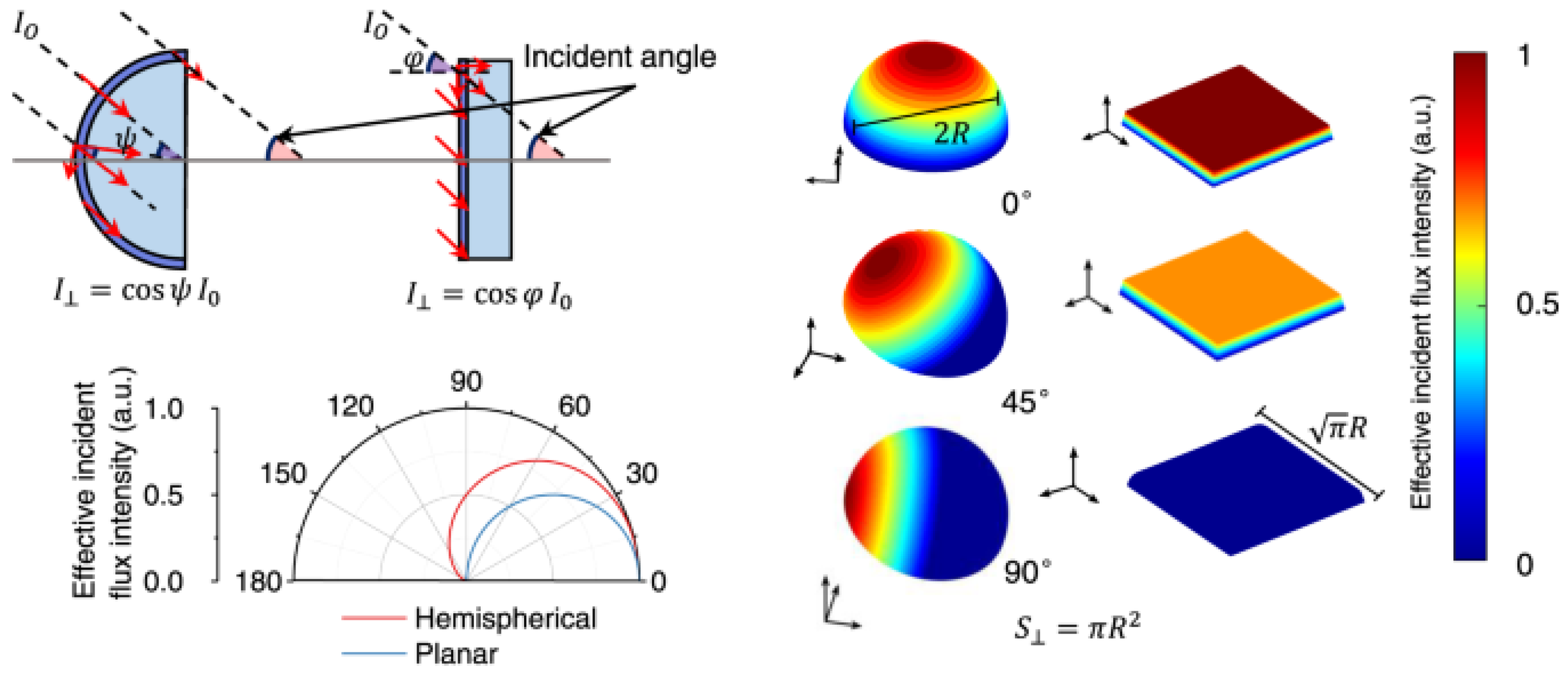
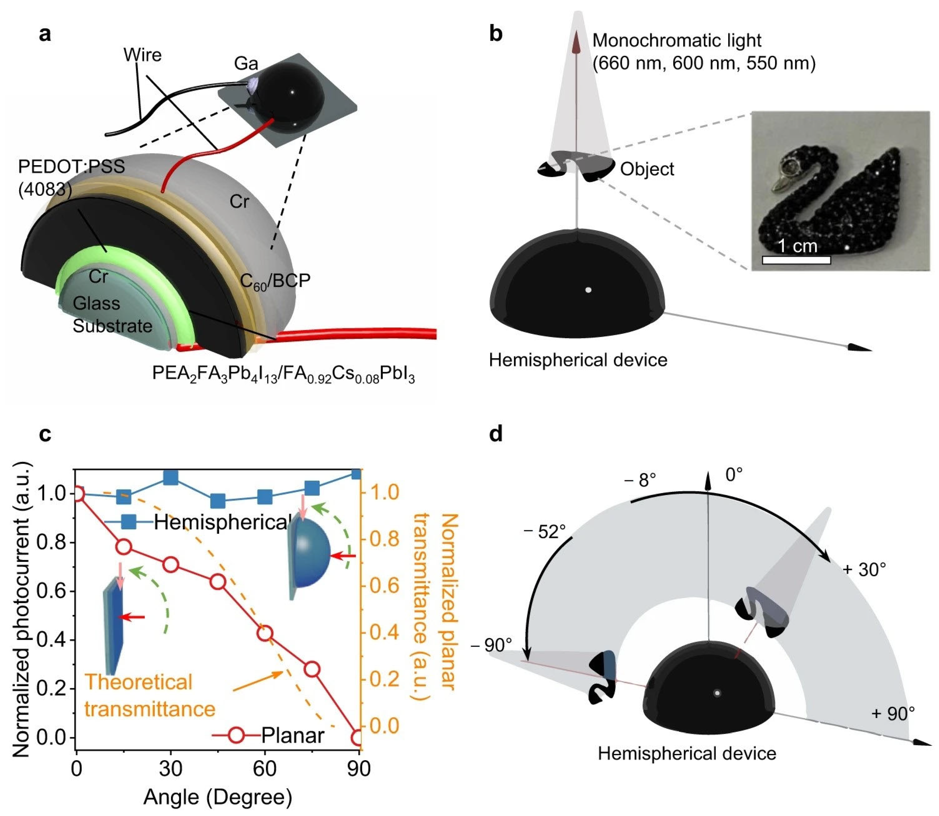
Hemispherical surfaces exhibit variable curvature and detection distances, resulting in substantial responsivity variations to distributions of incident light intensity, wavelength, object distance information, and other parameters [88,89]. Furthermore, hemispherical photodetectors demonstrate inherent wide-angle detection capabilities in lensless systems, effectively emulating the compound-eye morphology of micro-arthropods. Experimental studies confirm that spray-coated perovskite films on hemispherical substrates achieve charge-collection narrowing effects and wide-angle responses through the controlled modulation of film composition, thickness, and charge carrier dynamics.
Feng et al. fabricated quasi-2D phenethylammonium/formamidinium lead halide (PEA2FAn−1PbnX3n+1) perovskite hemispherical photodetectors via spray-coating [90]. By controlling perovskite composition, spraying cycles, and the solution concentration, they regulated crystallization kinetics and film thickness, achieving a narrowband photoresponse (FWHM < 20 nm) from visible to near-infrared wavelengths. The lensless hemispherical detectors enabled imaging across ≈180° fields of view, exhibiting responsivity of 13.8 mA W−1 and specific detectivity of 1011 Jones at target wavelengths.
Additionally, charge transport layers effectively inhibit deleterious physical/chemical reactions between electrodes and perovskite films, enhancing operational stability [91,92,93,94,95,96]. Morphologically engineered transport layers further reduce optical reflectance to boost photon utilization. Sargent′s team demonstrated that full-texture architectures minimize reflection losses while maximizing light-trapping efficiency, critical for device performance [97]. Tian et al. designed nested inverse opals to optimize electron transport layers for enhanced light harvesting [98], achieving 473 mA W−1 responsivity and 1.35 × 10¹³ Jones detectivity at zero bias (Figure 4).
Building on this, Pan et al. engineered perovskite photodetectors with monolayer ZnO hemispherical arrays. Anti-reflective charge transport layers broadened the light-field distribution while suppressing reflection, facilitating efficient carrier generation/separation in perovskites [99]. The devices delivered exceptional performance: a 120.3 dB linear dynamic range and 4.2 × 10¹² Jones detectivity.
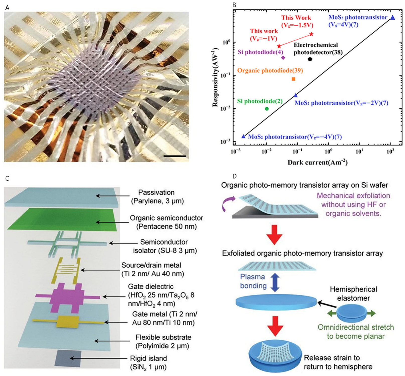
To enable rapid fabrication of hemispherical image sensors with tunable spectral response and low-temperature processing, organic materials present a viable solution. Kim et al. developed hemispherical imaging arrays using organic thin-film photo-memory transistors at a density of 308 pixels per cm2 [100]. This design incorporates only a single photo-memory transistor as the active pixel, contrasting with conventional architectures requiring select/readout/reset transistors plus a photodiode per pixel. The organic photo-memory transistors combine photosensitive semiconductors with charge-trapping dielectrics, delivering linear photoresponse (1–50 W m−2 intensity range) and responsivity up to 1.6 A W−1 (λ = 465 nm) at 0.24 A m−2 drain current (VD = −1.5 V). Concurrently, lensless single-pixel color imaging remains challenging for hemispherical photodetectors. Li et al. engineered perovskite hemispherical detectors capable of filter-free single-pixel color imaging [101]. Through the passivation of reactive vacancies and anti-site defects with functional chemical groups, trap densities were significantly suppressed, yielding stable devices with a current fluctuation standard deviation of 0.69 nA. By integrating Bayer templates with Fourier patterns and designing corresponding color-recovery algorithms, high-resolution 256 × 256-pixel color imaging was achieved. The hemispherical device geometry has greater compatibility with biomimetic eye devices and therefore has great potential for technical applications. However, there were considerable difficulties in the conception of their structure.

5. Structural Engineering of Curved Image Sensors

5.1. Single-Chambered Eyes

Unlike planar image sensors, curved image sensors prioritize emulating the structural and functional characteristics of biological vision systems. These sensors typically adopt an ocular-inspired concave or hemispherical geometry, optimizing light focusing while minimizing optical aberrations. Inspired by nature’s eye designs, two archetypal bionic-eye devices have now been developed, exhibiting high structural and functional similarities to their biological counterparts [13,39,40,41,42,43,44,45,46,47,102,103].
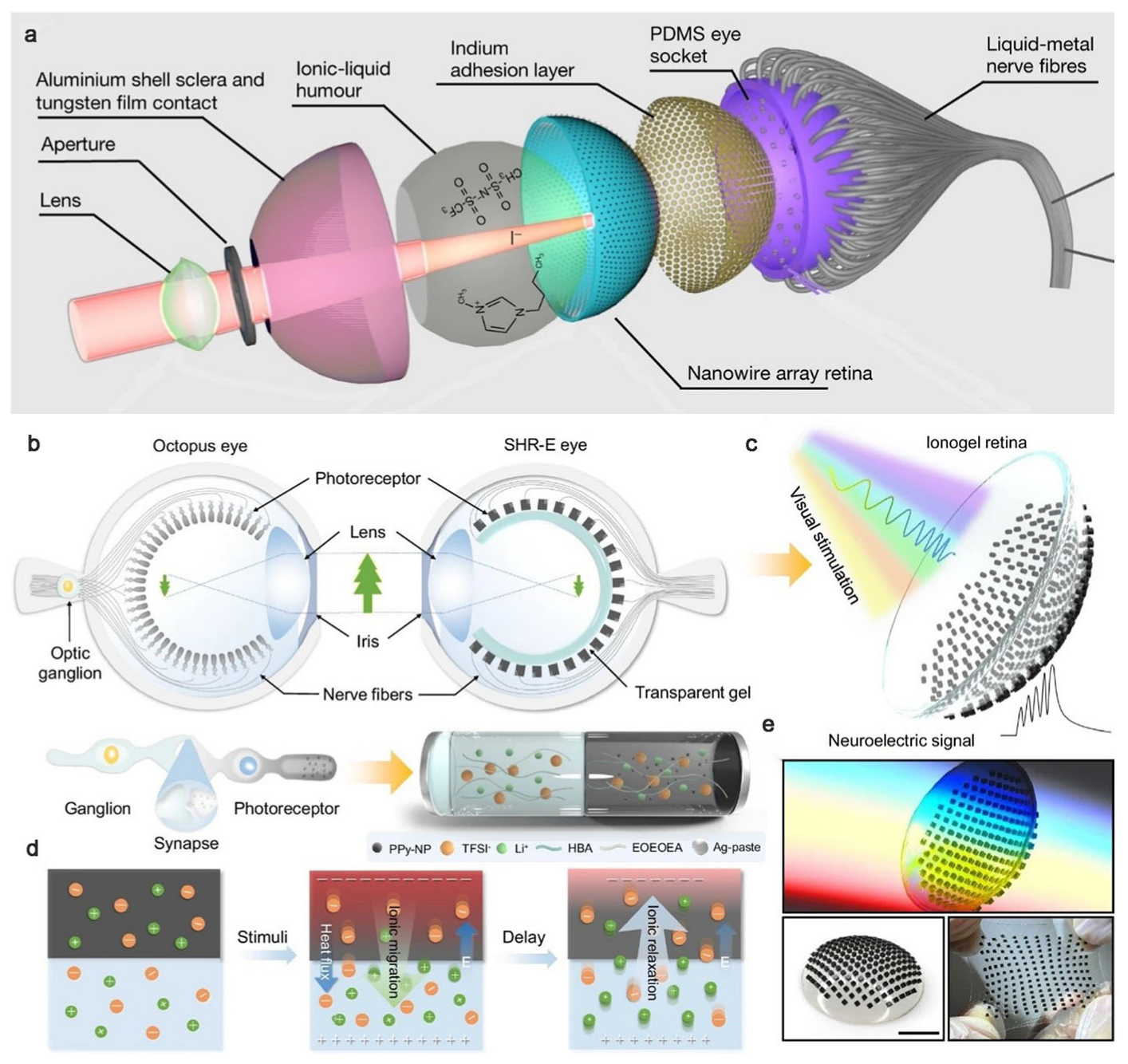
Similar to the human eye, the single-chambered structure incorporates an optical lens that further focuses incident light onto a hemispherical retinal array. Exemplified by Gu et al.′s spherical biomimetic electrochemical eye (EC-Eye), this system features a hemispherical retina mirroring the human ocular anatomy [104] (Figure 5a). The nano-sensitive spot density of the EC-Eye artificial retina is 4.6 × 108 cm−2, which is much higher than the photosensitive density of the human retina (about 107 cm−2), so a higher image resolution can be achieved. High-density perovskite nanowires—grown via physical vapor deposition on a hemispherical porous alumina substrate—serve as photoreceptors, detecting incident photons while converting optical signals into electrical impulses, analogous to biological photoreceptor cells [105].
Critically, an ionic liquid electrolyte acts as a front-side common electrode for the nanowires, while liquid–metal wires function as back contacts, replicating the vitreous humor and neural fibers of biological eyes, respectively [106,107,108,109]. These liquid–metal interconnects transmit electrical signals from perovskite nanowires to processing circuits, enabling individual addressing and processing of each photodetector unit. Concurrently, Choi et al. developed a high-density hemispherical vision sensor array through optimized fabrication, achieving a dramatically reduced thickness (a mere 51 nm) without structural fractures [77]. The encapsulation of this device enabled precise infrared image capture.
The integration of advanced materials and innovative architectures positions these systems for future breakthroughs in resolution and complex image processing [110]. In 2023, driven by autonomous vehicle demands, a self-powered hemispherical retinomorphic eye (SHR-E) emerged, composed of hetero-bilayered ionogel pillar forests as retinal photoreceptors [111]. Inspired by octopus vision, the SHR-E system employs a vertically inverted retina that positions photosensitive layers anterior to optic pathways, eliminating blind spots (Figure 5b). Nanoscale ionogel pillars, implanted on a transparent hemispherical surface (Figure 5c), form a retinal pillar forest capable of simultaneous photoconversion (Figure 5d) and neuroplastic emulation. All-soft components confer exceptional conformality and stretchability, enabling adhesion to complex geometric surfaces (Figure 5e).

5.2. Compound Eyes

Diverging from single-chambered eyes, compound-eye-inspired systems replicate the fundamental architecture of insect vision through arrays of integrated optical units. Each of thousands of individual optical units functions autonomously, concentrating light onto dedicated photoreceptors. These units comprise corneal lenses, crystalline cones, rhabdomeres, and photodetectors that collectively enable decentralized vision.
Exemplifying this approach, Song et al. engineered a hemispherical artificial compound eye using photodetector arrays [13]. An elastomeric microlens array with convex geometry serves as the corneal lens system, focusing light onto photodiodes to mimic biological photoreception. The photodetection subsystem employs thin silicon photodiodes and blocking diodes in a mesh configuration, enabling matrix addressing. The photodiodes emulate arthropod rhabdomeres for photodetection. Precision alignment ensures focused light delivery from each microlens to its corresponding photodiode. Serpentine metal interconnects permit mechanical stretching without electrical failure. Hydraulic inflation transforms planar constructs into hemispherical geometries, replicating the wide FOV of biological compound eyes. Black silicone shielding mimics screening pigments to prevent optical crosstalk between ommatidia, enhancing image contrast.
In parallel, Lee et al. developed a graphene-based hemispherical photodetector array using a fractal-inspired mesh design [108]. Graphene-composite detectors were grown via chemical vapor deposition on silicon substrates. The array was transferred to a hemispherical template, with metal nanowires interconnecting photodetectors. PDMS encapsulation protects individual optical units from environmental interference. Precision imaging was validated using diminishing light spots, achieving micron-scale spatial resolution.
These curved image sensors enable ultra-wide FOV imaging unobtainable with planar systems. Regarding functional adaptations in biological systems, Drosophila utilizes open rhabdomeres where each optical unit operates independently. Honeybees employ fused rhabdomeres that share visual fields across units.

6. Material-Heterogeneous Curved Image Sensors

Curved image sensors and photodetectors, which closely emulate the hemispherical morphology of biological retinas, serve as core components of artificial vision systems. Nanomaterials across dimensionalities have been leveraged in these sensors. According to the characteristics of zero-dimensional to one-dimensional, two-dimensional, and three-dimensional materials, the influence of different dimensions of material properties on curved surface image sensors is analyzed.

6.1. Zero-Dimensional (0D) Nanomaterials

Owing to the influence of the quantum confinement effect, the 0D perovskite material can precisely control the perovskite band gap and luminescence wavelength by adjusting the size and shape. 0D nanomaterials are nanoscale substances with all three dimensions confined to <100 nm. This confinement yields point-like morphologies, high surface area-to-volume ratios, and quantum effects that significantly alter their electronic/optical properties versus bulk materials. Quantum dots (QDs) exhibit exceptional quantum efficiency and tunable bandgaps due to quantum confinement, enabling efficient absorption and responses to multi-wavelength light [112]. For instance, a size-mixed QD layer—comprising controlled ratios of CdSe/CdS QDs with red (R), green (G), and blue (B) bandgaps—enhances color discrimination and pixel resolution in photodetectors, mimicking human retinal cones and enabling multispectral imaging [113] (Figure 6). Replacing long-chain ligands with short metal chalcogenides improves electronic coupling between QDs, facilitating carrier transport in thin-film transistors. This strategy enabled a 7 × 7 pixelated thin-film transistor array to perform filter-free color recognition. Furthermore, a shape-tunable phototransistor array achieved precise RGB pattern imaging on flat/curved surfaces using an intrinsically stretchable QD-based nanocomposite (organic semiconductor polymer + size-tunable CdSe–ZnS core–shell QDs + elastomeric matrix) [114]. The interfacial energy disparities drive distinctive nanoscale quantum dot assemblies within hybrid architectures, facilitating optimized carrier transport and photoresponse amplification mechanisms.
Although 0D nanostructures demonstrate exceptional responsivity, spectral tenability, and flexible substrate compatibility advantages in optoelectronic sensing applications, their industrial scalability remains constrained by inherent material degradation pathways and biological safety concerns.

6.2. One-Dimensional (1D) Nanomaterials

Compared with 0D perovskite materials, 1D perovskite materials have stronger chemical stability and simplicity of fabrication. 1D nanomaterials feature one dimension extended to µm/mm scales while the other two remain <100 nm, including nanotubes, nanorods, and nanowires. Their high electron mobility and aspect ratios enable biomimicry of retinal photoreceptors [115]. Vertically oriented Au–TiO2 nanowire arrays act as artificial photoreceptors, generating photovoltage and triggering neuronal spikes to restore vision in degenerated retinas [116,117] (Figure 7a). Though growable on planar FTO/flexible polymer substrates (~109 cm−2 density), their limited substrate curvature tolerance restricts compact curved imager applications. High-density (~5 × 108 cm−2) perovskite nanowires in hemispherical porous alumina templates structurally/functionally emulate retinas [6,104,110] (Figure 7b), enabling concave/convex sensors for bionic single-chambered and compound eyes.
1D nanomaterials provide dense, uniform photosensitive coverage. Particularly, bioinspired hemispherically distributed nanowire assemblies demonstrate unprecedented integration density in curvilinear electronic platforms, overcoming the planar growth limitations inherent to conventional lithographic processes. This breakthrough in nonplanar nanotechnology directly addresses the critical need for high-fidelity artificial retinal emulation in next-generation bionic vision systems. Future efforts must enhance operational stability and biocompatibility.

6.3. Two-Dimensional (2D) Nanomaterials

Currently, advanced 2D materials exhibit many excellent properties, such as electrical coordination, optical tunability, and mechanical strain, which are different from 0D and 1D materials, and have great prospects in the field of bionic vision. The structure of 2D nanomaterials is characterized by atomic/molecular-scale thicknesses (<few nm) with lateral dimensions up to a µm scale, including graphene, MoS2, h-BN, and black phosphorus [118]. Graphene/MoS2 offers excellent conductivity, high fracture strain, and facile conformality to curved surfaces (Figure 8a). A curved MoS2–graphene photodetector array exhibits low strain due to its ultrathin thickness and optimal interlayer contact [119]. Truncated icosahedron designs enable near-complete hemispherical coverage. Similarly, MoS2/organic PV3D3 heterostructure-based curved sensors transfer-printed onto hemispheres demonstrate photo-synapse behavior [120] (Figure 8b). Zhang et al. engineered a Ruddlesden–Popper quasi-two-dimensional perovskite photodetector, which exhibited a significant light response with a responsivity of 0.41 A W−1 at 430 nm and a response speed of 161 ns/1.91 us through the dual cation release strategy [121].
Ultrathin 2D photodetectors are promising for AVS due to their gate-tunable optoelectronic properties and atomic-scale conformability. However, they suffer from complex fabrication, mechanical instability, limited pixel counts, and constrained spatial resolution.

6.4. Three-Dimensional (3D) Nanomaterials

Compared with the problem of two-dimensional perovskite, 3D nanomaterials are simple to prepare, have high crystalline quality, and are widely used in curved image sensors. Three-dimensional nanomaterials extend beyond 100 nm in all dimensions, including polycrystals, thin films, and aerogels. Their geometry enables high-performance curved sensors via two strategies. Pre-fabricated units are attached to curved surfaces [122,123]. Planar thin-film sensors deformed into curvature via mechanical strain, origami, or kirigami [103,124,125]. For the first strategy, a silicon thin-film photodiode array was lithographically fabricated and transferred to pre-deformed PDMS, achieving hemispherical shapes after release. In the second strategy, a kirigami-based planar Si pixel array was transferred to concave/convex surfaces via conformal balloon stamping [14]. Similarly, organic pentacene phototransistor arrays were plasma-bonded to pre-stretched elastomers, reverting to hemispheres after strain release [100] (Figure 9).
3D nanomaterials have great potential for their integration into curved image sensors. However, they are limited owing to their instability upon deformation and limited pixel numbers. In addition, there is a large amount of unused space between the imaging pixels, which is necessary for stress relief but reduces the effective pixel density and imaging efficiency. For example, direct deformation into a hemispherical configuration generates substantial residual stress, which poses a risk of mechanical instability during operation [126]. Stacking photosensitive layers can be an effective approach to increase pixel density [127,128]. This strategy has been reported in planar image sensors and should translate well to curved image sensors. With the wide application of flexible perovskite devices in smart wearables, joint applications, and aircraft and aerospace fields, the bending or strain of perovskite materials will inevitably affect the performance of perovskite devices. Hemispherical perovskites are subjected to fixed mechanical external forces, and the strain leads to the formation of deep or shallow trap states, which disrupt the structural stability of perovskite materials and lead to their degradation [129,130].

7. Conclusions

Metal halide perovskites have emerged as highly promising materials for bioinspired artificial vision systems owing to their unique combination of outstanding optoelectronic properties, mechanical flexibility, and structural tunability. Their intrinsically high absorption coefficients and long carrier diffusion lengths enable efficient photon-to-electron conversion in ultrathin films, which is essential for achieving lightweight, low-power photodetectors with high sensitivity and rapid response times. The bandgap tunability of perovskites through compositional engineering allows for broad spectral responsivity, covering ultraviolet, visible, and near-infrared regions, thereby closely mimicking the multispectral perception capabilities of the human retina. In addition to their superior photonic performance, perovskites are solution-processable at low temperatures, enabling facile fabrication on flexible, curved, and stretchable substrates that are critical for constructing hemispherical imaging arrays with wide fields of view and reduced optical aberration—key features for artificial retinas and robotic vision systems. Moreover, the ionic nature of perovskites offers unique opportunities for integrating sensing, memory, and neuromorphic computing functionalities within the same material platform. For example, perovskite-based devices can exhibit resistive switching and dynamic conductance modulation, which are essential for in-sensor computing and real-time pattern recognition, moving beyond passive photodetection toward active, adaptive artificial vision. Compared with conventional silicon or III–V semiconductor technologies, perovskite-based systems combine high optoelectronic performance with mechanical compliance and functional versatility, positioning them as a key material class for the next generation of biomimetic vision systems that aim to integrate sensing, processing, and actuation in compact, energy-efficient formats.
With the rapid advancement of bionics, curved image detectors have achieved remarkable progress. These devices integrate seamlessly with artificial ocular systems, eliminating aberrations caused by mismatches between conventional planar detectors and hemispherical eyeballs. Diverse photoelectric materials and architectures now enhance detector performance. By summarizing the performance of curved image sensors and comparing their key performance improvements, it can be seen that there is still some room for improvement in the performance of curved image sensors (Table 1). Despite reported breakthroughs, critical challenges persist.
First, regarding the response speed, current systems struggle to match the sub-millisecond photoresponse of biological photoreceptors, limiting real-time applications.
Second, concerning the detectable spectrum, narrow operational bandwidths (typically 400–700 nm) hinder infrared/UV detection capabilities essential for multispectral imaging.
Future developments should prioritize enhancing photoresponse kinetics, broadening detectable spectrum ranges, and expanding fields of view (>180°). These advances will drive innovations in artificial intelligence, biomedical devices, bionics, and photonics, while accelerating progress toward high-performance biomimetic robotics. The development of curved image sensors has also led to breakthroughs in bionic eye equipment, such as the wide FOV image that combines the hemispherical photodetector itself to correct the optical phase difference, and the optical image can be collected without subsequent image stitching and correction, and the imaging speed is faster. At the same time, the hemispherical perovskite photodetector with excellent optical sensitivity can realize richer visual information capture in different scenes, and combined with the breakthrough of the optical sensor of the nervous system, it can bring a significant improvement in imaging efficiency [131].
In conclusion, hemispherical perovskite photodetectors that integrate advanced optoelectronics and biomimetic optics can provide powerful performance, which will further contribute to the excellence of bioelectronic devices.

Author Contributions

Conceptualization, Q.L. and X.S.; writing—original draft preparation, T.H.; writing—review and editing, T.H. and X.S.; supervision, Q.L. All authors have read and agreed to the published version of the manuscript.

Funding

This research received no external funding.

Data Availability Statement

Data availability is not applicable to this article as no new data were created or analyzed in this study.

Conflicts of Interest

The authors declare no conflict of interest.

Abbreviations

The following abbreviations are used in this manuscript:
EQEExternal Quantum Efficiency
AVSArtificial Vision Systems
FOVfield of view
CCDsCharge-Coupled Devices
CMOSComplementary Metal-Oxide-Semiconductor
FWHMFull-Width-at-Half-Maximum
2DMCs2D Molecular Crystals
PDsphotodetectors
NIRNear-Infrared
UVUltraviolet
EC-EyeElectroChemical Eye
SHR-ESelf-powered Hemispherical Retinomorphic Eye
QDsQuantum dots
0D\1D\2D\3DZero-dimensional\One-dimensional\Two-dimensional\Three-dimensional

References

  1. Zhu, S.; Xie, T.; Lv, Z.; Leng, Y.-B.; Zhang, Y.-Q.; Xu, R.; Qin, J.; Zhou, Y.; Roy, V.A.L.; Han, S.-T. Hierarchies in visual pathway: Functions and inspired artificial vision. Adv. Mater. 2024, 36, 2301986. [Google Scholar] [CrossRef]
  2. Chai, Y. In-sensor computing for machine vision. Nature 2020, 579, 32–33. [Google Scholar] [CrossRef]
  3. Kim, M.S.; Kim, M.S.; Lee, G.J.; Sunwoo, S.-H.; Chang, S.; Song, Y.M.; Kim, D.-H. Bio-inspired artificial vision and neuromorphic image processing devices. Adv. Mater. Technol. 2022, 7, 2100144. [Google Scholar] [CrossRef]
  4. Zhou, T.; Wu, W.; Zhang, J.; Yu, S.; Fang, L. Ultrafast dynamic machine vision with spatiotemporal photonic computing. Sci. Adv. 2023, 9, eadg4391. [Google Scholar] [CrossRef] [PubMed]
  5. Mennel, L.; Symonowicz, J.; Wachter, S.; Polyushkin, D.K.; Molina-Mendoza, A.J.; Mueller, T. Ultrafast machine vision with 2D material neural network image sensors. Nature 2020, 579, 62–66. [Google Scholar] [CrossRef]
  6. Zhou, Y.; Sun, Z.; Ding, Y.; Yuan, Z.; Qiu, X.; Cao, Y.B.; Wan, Z.; Long, Z.; Poddar, S.; Kumar, S.; et al. An ultrawide field-of-view pinhole compound eye using hemispherical nanowire array for robot vision. Sci. Robot. 2024, 9, eadi8666. [Google Scholar] [CrossRef] [PubMed]
  7. Yurtsever, E.; Lambert, J.; Carballo, A.; Takeda, K. A survey of autonomous driving: Common practices and emerging technologies. IEEE Access 2020, 8, 58443–58469. [Google Scholar] [CrossRef]
  8. Hsieh, M.-R.; Lin, Y.-L.; Hsu, W.H. Drone-based object counting by spatially regularized regional proposal network. In Proceedings of the 2017 IEEE International Conference on Computer Vision (ICCV), Venice, Italy, 22–29 October 2017; pp. 4165–4173. [Google Scholar]
  9. Yang, S.; Luo, P.; Loy, C.C.; Tang, X. WIDER FACE: A face detection benchmark. In Proceedings of the 2016 IEEE Conference on Computer Vision and Pattern Recognition (CVPR), Las Vegas, NV, USA, 27–30 June 2016; pp. 5525–5533. [Google Scholar]
  10. He, X.; Yan, S.; Hu, Y.; Niyogi, P.; Zhang, H.-J. Face recognition using Laplacianfaces. IEEE Trans. Pattern Anal. Mach. Intell. 2005, 27, 328–340. [Google Scholar]
  11. Alonso, V.; Dacal-Nieto, A.; Barreto, L.; Amaral, A.; Rivero, E. Industry 4.0 implications in machine vision metrology: An overview. Procedia Manuf. 2019, 41, 359–366. [Google Scholar] [CrossRef]
  12. Golnabi, H.; Asadpour, A. Design and application of industrial machine vision systems. Robot. Comput. Integr. Manuf. 2007, 23, 630–637. [Google Scholar] [CrossRef]
  13. Song, Y.M.; Xie, Y.; Malyarchuk, V.; Xiao, J.; Jung, I.; Choi, K.-J.; Liu, Z.; Park, H.; Lu, C.; Kim, R.-H.; et al. Digital cameras with designs inspired by the arthropod eye. Nature 2013, 497, 95–99. [Google Scholar] [CrossRef] [PubMed]
  14. Zidan, M.A.; Strachan, J.P.; Lu, W.D. The future of electronics based on memristive systems. Nat. Electron. 2018, 1, 22–29. [Google Scholar] [CrossRef]
  15. Sousounis, K.; Ogura, A.; Tsonis, P.A. Transcriptome analysis of nautilus and pygmy squid developing eye provides insights in lens and eye evolution. PLoS ONE 2013, 8, e78054. [Google Scholar] [CrossRef] [PubMed]
  16. Hanke, F.D.; Kelber, A. The eye of the common octopus (Octopus vulgaris). Front. Physiol. 2020, 10, 01637. [Google Scholar] [CrossRef]
  17. Frech, B.; Vogtsberger, M.; Neumeyer, C. Visual discrimination of objects differing in spatial depth by goldfish. J. Comp. Physiol. A 2012, 198, 53–60. [Google Scholar] [CrossRef]
  18. Navarro, R. The optical design of the human eye: A critical review. J. Optom. 2009, 2, 3–18. [Google Scholar] [CrossRef]
  19. Wässle, H. Optical quality of the cat eye. Vis. Res. 1971, 11, 995–1006. [Google Scholar] [CrossRef]
  20. Müller, B.; Glösmann, M.; Peichl, L.; Knop, G.C.; Hagemann, C.; Ammermüller, J. Bat eyes have ultraviolet-sensitive cone photoreceptors. PLoS ONE 2009, 4, e6390. [Google Scholar] [CrossRef]
  21. Reymond, L. Spatial visual acuity of the eagle Aquila audax: A behavioural, optical and anatomical investigation. Vis. Res. 1985, 25, 1477–1491. [Google Scholar] [CrossRef]
  22. Jezeera, M.A.; Tichit, P.; Balamurali, G.S.; Baird, E.; Kelber, A.; Somanathan, H. Spatial resolution and sensitivity of the eyes of the stingless bee, Tetragonula iridipennis. J. Comp. Physiol. A 2022, 208, 225–238. [Google Scholar] [CrossRef]
  23. Land, M.F.; Gibson, G.; Horwood, J.; Zeil, J. Fundamental differences in the optical structure of the eyes of nocturnal and diurnal mosquitoes. J. Comp. Physiol. A 1999, 185, 91–103. [Google Scholar] [CrossRef]
  24. Lee, K.C.; Yu, Q.; Erb, U. Mesostructure of ordered corneal nano-nipple arrays: The role of 5–7 coordination defects. Sci. Rep. 2016, 6, 28342. [Google Scholar] [CrossRef] [PubMed]
  25. Nilsson, D.-E.; Odselius, R. Regionally different optical systems in the compound eye of the water-flea Polyphemus (Cladocera, Crustacea). Proc. R. Soc. London. B 1983, 217, 163–175. [Google Scholar]
  26. Pix, W.; Zanker, J.M.; Zeil, J. The optomotor response and spatial resolution of the visual system in male Xenos vesparum (Strepsiptera). J. Exp. Biol. 2000, 203, 3397–3409. [Google Scholar] [CrossRef] [PubMed]
  27. Wardill, T.J.; Fabian, S.T.; Pettigrew, A.C.; Stavenga, D.G.; Nordström, K.; Gonzalez-Bellido, P.T. A novel interception strategy in a miniature robber fly with extreme visual acuity. Curr. Biol. 2017, 27, 854–859. [Google Scholar] [CrossRef]
  28. Schwarz, S.; Narendra, A.; Zeil, J. The properties of the visual system in the Australian desert ant Melophorus bagoti. Arthropod Struct. Dev. 2011, 40, 128–134. [Google Scholar] [CrossRef] [PubMed]
  29. Marshall, N.J.; Land, M.F.; Cronin, T.W. Shrimps that pay attention: Saccadic eye movements in stomatopod crustaceans. Philos. Trans. R. Soc. B 2014, 369, 20130042. [Google Scholar] [CrossRef]
  30. Land, M.; Layne, J. The visual control of behaviour in fiddler crabs. J. Comp. Physiol. A 1995, 177, 81–90. [Google Scholar] [CrossRef]
  31. Sun, X.; Wang, F.; Guo, X.; Wu, J.; Li, S.; Shi, Y.; Pan, L. Flexible Photodetector Arrays Based on Polycrystalline CsPbI3-xBrx Perovskite Films. IEEE Electron Device Lett. 2024, 45, 621–624. [Google Scholar] [CrossRef]
  32. Sun, X.; Zhao, C.; Li, H.; Yu, H.; Zhang, J.; Qiu, H.; Liang, J.; Wu, J.; Su, M.; Shi, Y.; et al. Wearable Near-Field Communication Sensors for Healthcare: Materials, Fabrication and Application. Micromachines 2022, 13, 784. [Google Scholar] [CrossRef]
  33. Lee, G.J.; Choi, C.; Kim, D.-H.; Song, Y.M. Bioinspired artificial eyes: Optic components, digital cameras, and visual prostheses. Adv. Funct. Mater. 2018, 28, 1705202. [Google Scholar] [CrossRef]
  34. Bertozzi, M.; Broggi, A.; Cellario, M.; Fascioli, A.; Lombardi, P.; Porta, M. Artificial vision in road vehicles. Proc. IEEE 2002, 90, 1258–1271. [Google Scholar] [CrossRef]
  35. Regal, S.; Troughton, J.; Djenizian, T.; Ramuz, M. Biomimetic models of the human eye, and their applications. Nanotechnology 2021, 32, 302001. [Google Scholar] [CrossRef] [PubMed]
  36. Cheng, Y.; Cao, J.; Zhang, Y.; Hao, Q. Review of state-of-the-art artificial compound eye imaging systems. Bioinspir. Biomim. 2019, 14, 31002. [Google Scholar] [CrossRef]
  37. Muller, K.J. Photoreceptors in the crayfish compound eye: Electrical interactions between cells as related to polarized-light sensitivity. J. Physiol. 1973, 232, 573–595. [Google Scholar] [CrossRef]
  38. Wu, W.; Han, X.; Li, J.; Wang, X.; Zhang, Y.; Huo, Z.; Chen, Q.; Sun, X.; Xu, Z.; Tan, Y.; et al. Ultrathin and Conformable Lead Halide Perovskite Photodetector Arrays for Potential Application in Retina-Like Vision Sensing. Adv. Mater. 2021, 33, 2006006. [Google Scholar] [CrossRef]
  39. Wu, W.; Lu, H.; Han, X.; Wang, C.; Xu, Z.; Han, S.-T.; Pan, C. Recent Progress on Wavelength-Selective Perovskite Photodetectors for Image Sensing. Small Methods 2023, 7, 2201499. [Google Scholar] [CrossRef]
  40. Yao, Q.; Xue, Q.; Li, Z.; Zhang, K.; Zhang, T.; Li, N.; Yang, S.; Brabec, C.J.; Yip, H.-L.; Cao, Y. Graded 2D/3D perovskite heterostructure for efficient and operationally stable MA-free perovskite solar cells. Adv. Mater. 2020, 32, 2000571. [Google Scholar] [CrossRef]
  41. Liu, R.; Zhou, H.; Song, Z.; Yang, X.; Wu, D.; Song, Z.; Wang, H.; Yan, Y. Low-reflection, (110)-orientation-preferred CsPbBr3 nanonet films for application in high-performance perovskite photodetectors. Nanoscale 2019, 11, 9302–9309. [Google Scholar] [CrossRef]
  42. Cao, M.; Tian, J.; Cai, Z.; Peng, L.; Yang, L.; Wei, D. Perovskite heterojunction based on CH3NH3PbBr3. single crystal for high-sensitive self-powered photodetector. Appl. Phys. Lett. 2016, 109, 233303. [Google Scholar] [CrossRef]
  43. Li, X.; Wu, G.; Wang, M.; Yu, B.; Zhou, J.; Wang, B.; Zhang, X.; Xia, H.; Yue, S.; Wang, K.; et al. Water-assisted crystal growth in quasi-2D perovskites with enhanced charge transport and photovoltaic performance. Adv. Energy Mater. 2020, 10, 2001832. [Google Scholar] [CrossRef]
  44. Wang, T.; Lian, G.; Huang, L.; Zhu, F.; Cui, D.; Wang, Q.; Meng, Q.; Jiang, H.; Zhou, G.; Wong, C.-P. A crystal-growth boundary-fusion strategy to prepare high-quality MAPbI3 films for excellent vis-NIR photodetectors. Nano Energy. 2019, 64, 103914. [Google Scholar] [CrossRef]
  45. Shen, K.; Li, X.; Xu, H.; Wang, M.; Dai, X.; Guo, J.; Zhang, T.; Li, S.; Zou, G.; Choy, K.-L.; et al. Enhanced performance of ZnO nanoparticle decorated all-inorganic CsPbBr3. quantum dot photodetectors. J. Mater. Chem. A 2019, 7, 6134–6142. [Google Scholar] [CrossRef]
  46. Sun, T.; Chen, T.; Chen, J.; Lou, Q.; Liang, Z.; Li, G.; Lin, X.; Yang, G.; Zhou, H. High-performance p-i-n perovskite photodetectors and image sensors with long-term operational stability enabled by a corrosion-resistant titanium nitride back electrode. Nanoscale 2023, 15, 7803–7811. [Google Scholar] [CrossRef] [PubMed]
  47. Liu, F.; Liu, K.; Rafique, S.; Xu, Z.; Niu, W.; Li, X.; Wang, Y.; Deng, L.; Wang, J.; Yue, X.; et al. Highly Efficient and Stable Self-Powered Mixed Tin-Lead Perovskite Photodetector Used in Remote Wearable Health Monitoring Technology. Adv. Sci. 2023, 10, 2205879. [Google Scholar] [CrossRef] [PubMed]
  48. Zhang, X.; Bai, R.; Fu, Y.; Hao, Y.; Peng, X.; Wang, J.; Ge, B.; Liu, J.; Hu, Y.; Ouyang, X.; et al. High energy resolution CsPbBr3 alpha particle detector with a full-customized readout application specific integrated circuit. Nat. Commun. 2024, 15, 6333. [Google Scholar] [CrossRef]
  49. Kim, J.H.; Liess, A.; Stolte, M.; Krause, A.M.; Stepanenko, V.; Zhong, C.; Bialas, D.; Spano, F.; Würthner, F. An Efficient Narrowband Near-Infrared at 1040 nm Organic Photodetector Realized by Intermolecular Charge Transfer Mediated Coupling Based on a Squaraine Dye. Adv. Mater. 2021, 33, 2100582. [Google Scholar] [CrossRef]
  50. García de Arquer, F.P.; Armin, A.; Meredith, P.; Sargent, E.H. Solution-processed semiconductors for next-generation photodetectors. Nat. Rev. Mater. 2017, 2, 16100. [Google Scholar] [CrossRef]
  51. Zhao, Z.; Xu, C.; Ma, Y.; Yang, K.; Liu, M.; Zhu, X.; Zhou, Z.; Shen, L.; Yuan, G.; Zhang, F. Ultraviolet Narrowband Photomultiplication Type Organic Photodetectors with Fabry−Pérot Resonator Architecture. Adv. Funct. Mater. 2022, 32, 2203606. [Google Scholar] [CrossRef]
  52. Xing, S.; Nikolis, V.C.; Kublitski, J.; Guo, E.; Jia, X.; Wang, Y.; Spoltore, D.; Vandewal, K.; Kleemann, H.; Benduhn, J.; et al. Miniaturized VIS-NIR Spectrometers Based on Narrowband and Tunable Transmission Cavity Organic Photodetectors with Ultrahigh Specific Detectivity above 1014 Jones. Adv. Mater. 2021, 33, 2102967. [Google Scholar] [CrossRef]
  53. Zhao, Z.; Xu, C.; Ma, Y.; Ma, X.; Zhu, X.; Niu, L.; Shen, L.; Zhou, Z.; Zhang, F. Filter-Free Narrowband Photomultiplication-Type Planar Heterojunction Organic Photodetectors. Adv. Funct. Mater. 2023, 33, 2212149. [Google Scholar] [CrossRef]
  54. Zhang, Y.; Liu, Y.; Xu, Z.; Yang, Z.; Liu, S. 2D Perovskite Single Crystals with Suppressed Ion Migration for High-Performance Planar-Type Photodetectors. Small 2020, 16, 2003145. [Google Scholar] [CrossRef]
  55. Ma, Y.; Xu, X.; Li, T.; Wang, Z.; Li, N.; Zhao, X.; Wei, W.; Zhan, X.; Shen, L. Amplified narrowband perovskite photodetector enable by independent multiplicaiotn layers for anti-interference light detection. Sci Adv. 2025, 11, eadq1127. [Google Scholar] [CrossRef] [PubMed]
  56. Yao, Y.; Chen, Y.; Wang, H.; Samorì, P. Organic photodetectors based on supramolecular nanostructures. Smart Mat. 2020, 1, 1. [Google Scholar] [CrossRef]
  57. Tian, B.; Zheng, X.; Kempa, T.J.; Fang, Y.; Yu, N.; Yu, G.; Huang, J.; Lieber, C.M. Coaxial silicon nanowires as solar cells and nanoelectronic power sources. Nature 2007, 449, 885–889. [Google Scholar] [CrossRef]
  58. Wicklein, A.; Ghosh, S.; Sommer, M.; Wurthner, F.; Thelakkat, M. Self-assembly of semiconductor organogelator nanowires for photoinduced charge separation. ACS Nano 2009, 3, 1107–1114. [Google Scholar] [CrossRef] [PubMed]
  59. Zhang, L.; Zhong, X.; Pavlica, E.; Li, S.; Klekachev, A.; Bratina, G.; Ebbesen, T.W.; Orgiu, E.; Samorì, P. A nanomesh scaffold for supramolecular nanowire optoelectronic devices. Nat. Nanotechnol. 2016, 11, 900–906. [Google Scholar] [CrossRef]
  60. Zhou, Y.; Wang, L.; Wang, J.; Pei, J.; Cao, Y. Highly sensitive, air-stable photodetectors based on single organic sub-micrometer ribbons self-assembled through solution processing. Adv. Mater. 2008, 20, 3745–3749. [Google Scholar] [CrossRef]
  61. Mukherjee, B.; Mukherjee, M. One-step fabrication of ordered organic crystalline array for novel optoelectronic applications. Org. Electron. 2011, 12, 1980–1987. [Google Scholar] [CrossRef]
  62. Roy, S.; Kumar Maiti, D.; Panigrahi, S.; Basak, D.; Banerjee, A. A new hydrogel from an amino acid-based perylene bisimide and its semiconducting, photo-switching behaviour. RSC Adv. 2012, 2, 11053–11060. [Google Scholar] [CrossRef]
  63. Rekab, W.; Stoeckel, M.A.; El Gemayel, M.; Gobbi, M.; Orgiu, E.; Samorì, P. High-performance phototransistors based on PDIF-CN2 solution-processed single fiber and multifiber assembly. ACS Appl. Mater. Interfaces 2016, 8, 9829–9838. [Google Scholar] [CrossRef] [PubMed]
  64. Guo, Y.; Du, C.; Yu, G.; Di, C.; Jiang, S.; Xi, H.; Zheng, J.; Yan, S.; Yu, C.; Hu, W.; et al. High-performance phototransistors based on organic microribbons prepared by a solution self-assembly process. Adv. Funct. Mater. 2010, 20, 1019–1024. [Google Scholar] [CrossRef]
  65. Mukherjee, B.; Mukherjee, M.; Sim, K.; Pyo, S. Solution processed, aligned arrays of TCNQ micro crystals for low-voltage organic phototransistor. J. Mater. Chem. 2011, 21, 1931–1936. [Google Scholar] [CrossRef]
  66. Mukherjee, B.; Sim, K.; Shin, T.J.; Lee, J.; Mukherjee, M.; Ree, M.; Pyo, S. Organic phototransistors based on solution grown, ordered single crystalline arrays of a π-conjugated molecule. J. Mater. Chem. 2012, 22, 3192–3200. [Google Scholar] [CrossRef]
  67. Hu, M.; Liu, J.; Zhao, Q.; Liu, D.; Zhang, Q.; Zhou, K.; Li, J.; Dong, H.; Hu, W. Organic single-crystal phototransistor with unique wavelength-detection characteristics. Sci. China Mater. 2019, 62, 729–735. [Google Scholar] [CrossRef]
  68. Gu, P.; Hu, M.; Ding, S.; Zhao, G.; Yao, Y.; Liu, F.; Zhang, X.; Dong, H.; Wang, X.; Hu, W. High performance organic transistors and phototransistors based on diketopyrrolopyrrole-quaterthiophene copolymer thin films fabricated via low-concentration solution processing. Chin. Chem. Lett. 2018, 29, 1675–1680. [Google Scholar] [CrossRef]
  69. El Gemayel, M.; Treier, M.; Musumeci, C.; Li, C.; Müllen, K.; Samorì, P. Tuning the photoresponse in organic field-effect transistors. J. Am. Chem. Soc. 2012, 134, 2429–2433. [Google Scholar] [CrossRef]
  70. Novoselov, K.S. Electric field effect in atomically thin carbon films. Science 2004, 306, 666–669. [Google Scholar] [CrossRef]
  71. Dong, R.; Han, P.; Arora, H.; Ballabio, M.; Karakus, M.; Zhang, Z.; Shekhar, C.; Adler, P.; Petkov, P.S.; Erbe, A.; et al. High-mobility band-like charge transport in a semiconducting two-dimensional metal-organic framework. Nat. Mater. 2018, 17, 1027–1032. [Google Scholar] [CrossRef]
  72. Sahabudeen, H.; Qi, H.; Glatz, B.A.; Tranca, D.; Dong, R.; Hou, Y.; Zhang, T.; Kuttner, C.; Lehnert, T.; Seifert, G.; et al. Wafer-sized multifunctional polyimine-based two-dimensional conjugated polymers with high mechanical stiffness. Nat. Commun. 2016, 7, 13461. [Google Scholar] [CrossRef]
  73. Arora, H.; Dong, R.; Venanzi, T.; Zscharschuch, J.; Schneider, H.; Helm, M.; Feng, X.; Cánovas, E.; Erbe, A. Demonstration of a broadband photodetector based on a two-dimensional metal-organic framework. Adv. Mater. 2020, 32, 1907063. [Google Scholar] [CrossRef] [PubMed]
  74. Li, C.G.; Wang, Y.S.; Zou, Y.; Zhang, X.T.; Dong, H.L.; Hu, W.P. Two-dimensional conjugated polymer synthesized by interfacial Suzuki reaction: Towards electronic device applications. Angew. Chem. Int. Ed. Engl. 2020, 59, 9403–9407. [Google Scholar] [CrossRef] [PubMed]
  75. Yang, F.; Cheng, S.; Zhang, X.; Ren, X.; Li, R.; Dong, H.; Hu, W. 2D organic materials for optoelectronic applications. Adv. Mater. 2018, 30, 1702415. [Google Scholar] [CrossRef] [PubMed]
  76. Fu, B.; Wang, C.; Sun, Y.; Yao, J.; Wang, Y.; Ge, F.; Yang, F.; Liu, Z.; Dang, Y.; Zhang, X.; et al. A “phase separation” molecular design strategy towards large-area 2D molecular crystals. Adv. Mater. 2019, 31, 1901437. [Google Scholar] [CrossRef]
  77. Chun, D.H.; Choi, Y.J.; In, Y.; Nam, J.K.; Choi, Y.J.; Yun, S.; Kim, W.; Choi, D.; Kim, D.; Shin, H.; et al. Halide perovskite nanopillar photodetector. ACS Nano 2018, 12, 8564–8571. [Google Scholar] [CrossRef]
  78. Lee, M.M.; Teuscher, J.; Miyasaka, T.; Murakami, T.N.; Snaith, H.J. Efficient hybrid solar cells based on meso-superstructured organometal halide perovskites. Science 2012, 338, 643–647. [Google Scholar] [CrossRef]
  79. Li, L.; Chen, H.; Fang, Z.; Meng, X.; Zuo, C. An electrically modulated single-color/dual-color imaging photodetector. Adv. Mater. 2020, 32, 1907257. [Google Scholar] [CrossRef]
  80. Tyagi, D.; Wang, H.; Huang, W.; Hu, L.; Tang, Y.; Guo, Z.; Ouyang, Z.; Zhang, H. Recent advances in two-dimensional-material-based sensing technology toward health and environmental monitoring applications. Nanoscale 2020, 12, 3535–3559. [Google Scholar] [CrossRef]
  81. Wang, H.; Kim, D.H. Perovskite-based photodetectors: Materials and devices. Chem. Soc. Rev. 2017, 46, 5204–5236. [Google Scholar] [CrossRef]
  82. Xu, X.; Han, Z.; Zou, Y.; Li, J.; Gu, Y.; Hu, D.; He, Y.; Liu, J.; Yu, D.; Cao, F.; et al. Miniaturized multispectral detector derived from gradient response units on single MAPbX3 microwire. Adv. Mater. 2022, 34, 2108408. [Google Scholar] [CrossRef]
  83. Feldmann, J.; Youngblood, N.; Karpov, M.; Gehring, H.; Li, X.; Stappers, M.; Le Gallo, M.; Fu, X.; Lukashchuk, A.; Raja, A.S.; et al. Parallel convolutional processing using an integrated photonic tensor core. Nature 2021, 589, 52–58. [Google Scholar] [CrossRef] [PubMed]
  84. Jung, I.; Xiao, J.; Malyarchuk, V.; Lu, C.; Li, M.; Liu, Z.; Yoon, J.; Huang, Y.; Rogers, J.A. Dynamically tunable hemispherical electronic eye camera system with adjustable zoom capability. Proc. Natl Acad. Sci. USA 2011, 108, 1788–1793. [Google Scholar] [CrossRef] [PubMed]
  85. Galvez, F.; Monton, C.; Serrano, A.; Valmianski, I.; de la Venta, J.; Schuller, I.K.; Garcia, M.A. Effect of photodiode angular response on surface plasmon resonance measurements in the Kretschmann-Raether configuration. Rev. Sci. Instrum. 2012, 83, 093102. [Google Scholar] [CrossRef]
  86. Arbabi, A.; Arbabi, E.; Kamali, S.M.; Horie, Y.; Han, S.; Faraon, A. Miniature optical planar camera based on a wide-angle metasurface doublet corrected for monochromatic aberrations. Nat. Commun. 2016, 7, 13682. [Google Scholar] [CrossRef]
  87. Yu, H.; Li, H.; Sun, X.; Pan, L. Biomimetic Flexible Sensors and Their Applications in Human Health Detection. Biomimetics 2023, 8, 293. [Google Scholar] [CrossRef] [PubMed]
  88. Zhang, K.; Jung, Y.H.; Mikael, S.; Seo, J.-H.; Kim, M.; Mi, H.; Zhou, H.; Xia, Z.; Zhou, W.; Gong, S.; et al. Origami silicon optoelectronics for hemispherical electronic eye systems. Nat. Commun. 2017, 8, 1782. [Google Scholar] [CrossRef]
  89. Kogos, L.C.; Li, Y.; Liu, J.; Li, Y.; Tian, L.; Paiella, R. Plasmonic ommatidia for lensless compound-eye vision. Nat. Commun. 2020, 11, 1637. [Google Scholar] [CrossRef]
  90. Feng, X.; He, Y.; Qu, W.; Song, J.; Pan, W.; Tan, M.; Yang, B.; Wei, H. Spray-coated perovskite hemispherical photodetector featuring narrow-band and wide-angle imaging. Nat. Commun. 2022, 13, 6106. [Google Scholar] [CrossRef]
  91. Chow, P.C.Y.; Someya, T. Organic photodetectors for next-generation wearable electronics. Adv. Mater. 2020, 32, e1902045. [Google Scholar] [CrossRef]
  92. Ball, J.M.; Lee, M.M.; Hey, A.; Snaith, H.J. Low-temperature processed meso-superstructured to thin-film perovskite solar cells. Energy Environ. Sci. 2013, 6, 1739–1743. [Google Scholar] [CrossRef]
  93. Snaith, J.H. Perovskites: The emergence of a new era for low-cost, high-efficiency solar cells. J. Phys. Chem. Lett. 2013, 4, 3623–3630. [Google Scholar] [CrossRef]
  94. Chiang, C.H.; Tseng, Z.L.; Wu, C.G. Planar heterojunction perovskite/PC71BM solar cells with enhanced open-circuit voltage via a (2/1)-step spin-coating process. J. Mater. Chem. A 2014, 2, 15897–15903. [Google Scholar] [CrossRef]
  95. Liang, Z.; Zhang, Q.; Jiang, L.; Cao, G. Zno cathode buffer layers for inverted polymer solar cells. Energy Environ. Sci. 2015, 8, 3442–3476. [Google Scholar] [CrossRef]
  96. Leung, S.F.; Ho, K.T.; Kung, P.K.; Hsiao, V.K.; Alshareef, H.N.; Wang, Z.L.; He, J.H. A self-powered and flexible organometallic halide perovskite photodetector with very high detectivity. Adv. Mater. 2018, 30, 1704611. [Google Scholar] [CrossRef] [PubMed]
  97. Hou, Y.; Aydin, E.; De Bastiani, M.; Xiao, C.; Isikgor, F.H.; Xue, D.-J.; Chen, B.; Chen, H.; Bahrami, B.; Chowdhury, A.H.; et al. Efficient tandem solar cells with solution-processed perovskite on textured crystalline silicon. Science 2020, 367, 1135–1140. [Google Scholar] [CrossRef] [PubMed]
  98. Tian, W.; Min, L.; Cao, F.; Li, L. Nested inverse opal perovskite toward superior flexible and self-powered photodetection performance. Adv. Mater. 2020, 32, 1906974. [Google Scholar] [CrossRef]
  99. Pan, X.; Zhang, J.; Zhou, H.; Liu, R.; Wu, D.; Wang, R.; Shen, L.; Tao, L.; Zhang, J.; Wang, H. Single-Layer ZnO Hollow Hemispheres Enable High-Performance Self-Powered Perovskite Photodetector for Optical Communication. Nano-Micro Lett. 2021, 13, 70. [Google Scholar] [CrossRef]
  100. Kim, Y.; Zhu, C.; Lee, W.-Y.; Smith, A.; Ma, H.; Li, X.; Son, D.; Matsuhisa, N.; Kim, J.; Bae, W.-G.; et al. A Hemispherical Image Sensor Array Fabricated with Organic Photomemory Transistors. Adv. Mater. 2023, 35, 2203541. [Google Scholar] [CrossRef]
  101. Li, C.; Li, W.; Qu, W.; Hu, H.; Zong, J.; Wei, H. Stable and Lead-Free Perovskite Hemispherical Photodetector for Vivid Fourier Imaging. Adv. Sci. 2024, 12, 2414430. [Google Scholar] [CrossRef]
  102. Ko, H.C.; Stoykovich, M.P.; Song, J.; Malyarchuk, V.; Choi, W.M.; Yu, C.-J.; Geddes III, J.B.; Xiao, J.; Wang, S.; Huang, Y.; et al. A hemispherical electronic eye camera based on compressible silicon optoelectronics. Nature 2008, 454, 748–753. [Google Scholar] [CrossRef]
  103. Ying, S.; Li, J.; Huang, J.; Zhang, J.-H.; Zhang, J.; Jiang, Y.; Sun, X.; Pan, L.; Shi, Y. A Flexible Piezocapacitive Pressure Sensor with Microsphere-Array Electrodes. Nanomaterials. 2023, 13, 1702. [Google Scholar] [CrossRef] [PubMed]
  104. Gu, L.; Poddar, S.; Lin, Y.; Long, Z.; Zhang, D.; Zhang, Q.; Shu, L.; Qiu, X.; Kam, M.; Javey, A.; et al. A biomimetic eye with a hemispherical perovskite nanowire array retina. Nature 2020, 581, 278–282. [Google Scholar] [CrossRef] [PubMed]
  105. Dai, B.; Zhang, L.; Zhao, C.; Bachman, H.; Becker, R.; Mai, J.; Jiao, Z.; Li, W.; Zheng, L.; Wan, X.; et al. Biomimetic apposition compound eye fabricated using microfluidic-assisted 3D printing. Nat. Commun. 2021, 12, 6458. [Google Scholar] [CrossRef] [PubMed]
  106. Rao, Z.; Lu, Y.; Li, Z.; Sim, K.; Ma, Z.; Xiao, J.; Yu, C. Curvy, shape-adaptive imagers based on printed optoelectronic pixels with a kirigami design. Nat. Electron. 2021, 4, 513–521. [Google Scholar] [CrossRef]
  107. Hu, Z.-Y.; Zhang, Y.-L.; Pan, C.; Dou, J.-Y.; Li, Z.-Z.; Tian, Z.-N.; Mao, J.-W.; Chen, Q.-D.; Sun, H.-B. Miniature optoelectronic compound eye camera. Nat. Commun. 2022, 13, 5634. [Google Scholar] [CrossRef]
  108. Lee, M.; Lee, G.J.; Jang, H.J.; Joh, E.; Cho, H.; Kim, M.S.; Kim, H.M.; Kang, K.M.; Lee, J.H.; Kim, M.; et al. An amphibious artificial vision system with a panoramic visual field. Nat. Electron. 2022, 5, 452–459. [Google Scholar] [CrossRef]
  109. Ding, Y.; Liu, G.; Long, Z.; Zhou, Y.; Qiu, X.; Ren, B.; Zhang, Q.; Chi, C.; Wan, Z.; Huang, B.; et al. Uncooled self-powered hemispherical biomimetic pit organ for mid-to long-infrared imaging. Sci. Adv. 2022, 8, eabq8432. [Google Scholar] [CrossRef] [PubMed]
  110. Long, Z.; Qiu, X.; Chan, C.L.J.; Sun, Z.; Yuan, Z.; Poddar, S.; Zhang, Y.; Ding, Y.; Gu, L.; Zhou, Y.; et al. A neuromorphic bionic eye with filter-free color vision using hemispherical perovskite nanowire array retina. Nat. Commun. 2023, 14, 1972. [Google Scholar] [CrossRef]
  111. Luo, X.; Chen, C.; He, Z.; Wang, M.; Pan, K.; Dong, X.; Li, Z.; Liu, B.; Zhang, Z.; Wu, Y.; et al. A bionic self-driven retinomorphic eye with ionogel photosynaptic retina. Nat. Commun. 2024, 15, 3086. [Google Scholar] [CrossRef]
  112. Bao, J.; Bawendi, M.G. A colloidal quantum dot spectrometer. Nature 2015, 523, 67–70. [Google Scholar] [CrossRef]
  113. Jo, C.; Kim, J.; Kwak, J.Y.; Kwon, S.M.; Park, J.B.; Kim, J.; Park, G.-S.; Kim, M.-G.; Kim, Y.-H.; Park, S.K. Retina-inspired color-cognitive learning via chromatically controllable mixed quantum dot synaptic transistor arrays. Adv. Mater. 2022, 34, 2108979. [Google Scholar] [CrossRef] [PubMed]
  114. Song, J.-K.; Kim, J.; Yoon, J.; Koo, J.H.; Jung, H.; Kang, K.; Sunwoo, S.-H.; Yoo, S.; Chang, H.; Jo, J.; et al. Stretchable colour-sensitive quantum dot nanocomposites for shape-tunable multiplexed phototransistor arrays. Nat. Nanotechnol. 2022, 17, 849–856. [Google Scholar] [CrossRef] [PubMed]
  115. Yan, R.; Gargas, D.; Yang, P. Nanowire photonics. Nat. Photonics 2009, 3, 569–576. [Google Scholar] [CrossRef]
  116. Tang, J.; Qin, N.; Chong, Y.; Diao, Y.; Yiliguma; Wang, Z.; Xue, T.; Jiang, M.; Zhang, J.; Zheng, G. Nanowire arrays restore vision in blind mice. Nat. Commun. 2018, 9, 786. [Google Scholar] [CrossRef]
  117. Yang, R.; Zhao, P.; Wang, L.; Feng, C.; Peng, C.; Wang, Z.; Zhang, Y.; Shen, M.; Shi, K.; Weng, S.; et al. Assessment of visual function in blind mice and monkeys with subretinally implanted nanowire arrays as artificial photoreceptors. Nat. Biomed. Eng. 2023, 8, 1018–1039. [Google Scholar] [CrossRef]
  118. Konstantatos, G. Current status and technological prospect of photodetectors based on two-dimensional materials. Nat. Commun. 2018, 9, 5266. [Google Scholar] [CrossRef]
  119. Choi, C.; Choi, M.K.; Liu, S.; Kim, M.; Park, O.K.; Im, C.; Kim, J.; Qin, X.; Lee, G.J.; Cho, K.W.; et al. Human eye-inspired soft optoelectronic device using high-density MoS2-graphene curved image sensor array. Nat. Commun. 2017, 8, 1664. [Google Scholar] [CrossRef]
  120. Choi, C.; Leem, J.; Kim, M.; Taqieddin, A.; Cho, C.; Cho, K.W.; Lee, G.J.; Seung, H.; Bae, H.J.; Song, Y.M.; et al. Curved neuromorphic image sensor array using a MoS2-organic heterostructure inspired by the human visual recognition system. Nat. Commun. 2020, 11, 5934. [Google Scholar] [CrossRef]
  121. Zhang, X.; Li, Z.; Hong, E.; Yan, T. Effective Dual Cation Release in Quasi-2D Perovskites for Ultrafast UV Light-Powered Imaging. Adv. Mater. 2025, 37, 2412014. [Google Scholar] [CrossRef]
  122. Feng, X.; Li, C.; Song, J.; He, Y.; Qu, W.; Li, W.; Guo, K.; Liu, L.; Yang, B.; Wei, H. Differential perovskite hemispherical photodetector for intelligent imaging and location tracking. Nat. Commun. 2024, 15, 577. [Google Scholar] [CrossRef]
  123. Li, H.; Yu, H.; Wu, D.; Sun, X.; Pan, L. Recent Advances in Bioinspired Vision Sensor Arrays Based on Advanced Optoelectronic Materials. APL Mater. 2023, 11, 080601. [Google Scholar] [CrossRef]
  124. Sim, K.; Chen, S.; Li, Z.; Rao, Z.; Liu, J.; Lu, Y.; Jang, S.; Ershad, F.; Chen, J.; Xiao, J.; et al. Three-dimensional curvy electronics created using conformal additive stamp printing. Nat. Electron. 2019, 2, 471–479. [Google Scholar] [CrossRef]
  125. Yang, H.; Ding, S.; Wang, J.; Sun, S.; Swaminathan, R.; Ng, S.W.L.; Pan, X.; Ho, G.W. Computational Design of Ultra-Robust Strain Sensors for Soft Robot Perception and Autonomy. Nat. Commun. 2024, 15, 1636. [Google Scholar] [CrossRef]
  126. Long, Z.; Zhou, Y.; Ding, Y.; Qiu, X.; Poddar, S.; Fan, Z. Biomimetic Optoelectronics with Nanomaterials for Artificial Vision. Nat. Rev. Mater. 2025, 10, 128–146. [Google Scholar] [CrossRef]
  127. Park, J.; Kim, M.S.; Kim, J.; Chang, S.; Lee, M.; Lee, G.J.; Song, Y.M.; Kim, D.-H. Avian eye-inspired perovskite artificial vision system for foveated and multispectral imaging. Sci. Robot. 2024, 9, eadk6903. [Google Scholar] [CrossRef] [PubMed]
  128. Qiu, X.; Ding, Y.; Sun, Z.; Ji, H.; Zhou, Y.; Long, Z.; Liu, G.; Wang, P.; Poddar, S.; Ren, B.; et al. A tetrachromatic sensor for imaging beyond the visible spectrum in harsh conditions. Device 2024, 2, 100357. [Google Scholar] [CrossRef]
  129. Liang, H.; Yang, W.; Xia, J.; Gu, H.; Meng, X.; Yang, G.; Fu, Y.; Wang, B.; Cai, H.; Chen, Y.; et al. Strain Effects on Flexible Perovskite Solar Cells. Adv. Sci. 2023, 10, 2304733. [Google Scholar] [CrossRef]
  130. Chen, Z.; Cheng, Q.; Chen, H.; Wu, Y.; Ding, J.; Wu, X.; Yang, H.; Liu, H.; Chen, W.; Tang, X.; et al. Perovskite Grain-Boundary Manipulation Using Room-Temperature Dynamic Self-Healing “Ligaments” for Developing Highly Stable Flexible Perovskite Solar Cells with 23.8% Efficiency. Adv. Mater. 2023, 35, 2300513. [Google Scholar] [CrossRef]
  131. Lu, H.; Wu, W.; He, Z.; Han, X.; Pan, C. Recent progress in construction methods and applications of perovskite photodetector arrays. Nanoscale Horiz. 2023, 8, 1014–1033. [Google Scholar] [CrossRef]
Figure 1. Temporal and spatial properties of the human visual system. (a) The center-surround antagonism receptive field can be applied for the detection of edges. (b) The hemispherical retina can effectively reduce aberrations compared to a plane detector array. (c) Synaptic connections between nerve cells in the retina and brain. The spike-timing dependent plasticity of synapses. (d) Light and dark adaptability of biological vision. Copyright 2023, Advanced Func Materials. Reproduced with permission.
Solids 06 00034 g001
Figure 2. Schematic diagram of the vertical component of incident light on hemispherical and planar surfaces. The incident light (red arrow) is decomposed into components parallel and perpendicular to the surface. ψ and φ are angles to evaluate the angle of incident light. Copyright 2022, Springer Nature. Reproduced with permission.
Solids 06 00034 g002
Figure 3. Functional passivating antioxidants strategy. (a) The chemical structure of thiophene-2-carbohydrazide (TAH) molecule. (b) Schematic representation of fabricated perovskite films with TAH molecule. (c) Schematic showing the passivating mechanism between TAH and mixed Sn–Pb perovskite. Copyright 2022, Wiley–VCH GmbH. Reproduced with permission.
Solids 06 00034 g003
Figure 4. (a) The design of the hemispherical photodetector. (b) The images captured by hemispherical photodetectors with different I/Br ratios. (c) The photocurrent of hemispherical photodetectors under irradiation from different angles. (d) The images captured by planar and hemispherical photodetectors based on different angles of incident light. Copyright 2023, CCC. Reproduced with permission.
Solids 06 00034 g004
Figure 5. Single-chambered eye devices. (a) Exploded view of EC-Eye. Copyright 2025, Springer Nature. Reproduced with permission. (b). Anatomy of SHR-E eye with ionogel photoreceptor (SHR-E: self-powered hemispherical retinomorphic eye). (c) Schematics of our hemispherical retina with neuromorphic signal processing from ionic heterogel pillar forest. (d) Process mechanism of optical-to-electrical converter. (e) Photographs of the hemispherical retina with multicolor light exposure, high surface conformal and stretchability. Copyright 2024, Springer Nature. Reproduced with permission.
Solids 06 00034 g005
Figure 6. Quantum dot (QD)-based detector color-recognizable human visual system.The arrow symbols is an enlarged view of R-QDs, G-QDs, and B-QDs Copyright 2025, John Wiley and Sons. Reproduced with permission.
Solids 06 00034 g006
Figure 7. (a). Schematic of an Au–TiO2 nanowire array emulating the photoreceptor structure in an eye. (b) Perovskite nanowire arrays mimicking photoreceptors in human and insect retinas. Copyright 2018, Springer Nature. Reproduced with permission.
Solids 06 00034 g007
Figure 8. (a). Schematic illustration of the structure of 2D nanomaterials. (b). pV3D3-PTr based image sensors. Copyright 2020, Springer Nature. Reproduced with permission.
Solids 06 00034 g008
Figure 9. Hemispherical photodetector sensors. (A) Organic phototransistor-based hemispherical image sensor. (B) pV3D3-PTr-based image sensors. (C) Schematic diagram of the layers in sensors. (D) The transfer process of our hemispherical organic optical memory transistor array. Copyright 2025, John Wiley and Sons. Reproduced with permission.
Solids 06 00034 g009
Table 1. The comparison of performance parameters between this work and other reported curved image sensors.
MaterialsDetectivity (Jones)EQEResponsivity
(W/A)
Ref.
Hybrid tin-lead perovskite1.8 × 101275%0.51[47]
PEDOT-PSS perovskite bulk1.35 × 1013 0.47[98]
ZnO/CsPbBr3 hemispherical arrays4.2 × 1012 0.1[99]
lead-free hemispherical1.49 × 1013 0.188[101]
FAPbI3 hemispherical2 × 10131000%5.1[122]
PEA2FAn−1PbnX3n+1 hemispherical≈10115%13.8[123]
MAPbI3−xClx film9.4 × 1011 2.17[131]
Disclaimer/Publisher’s Note: The statements, opinions and data contained in all publications are solely those of the individual author(s) and contributor(s) and not of MDPI and/or the editor(s). MDPI and/or the editor(s) disclaim responsibility for any injury to people or property resulting from any ideas, methods, instructions or products referred to in the content.