Review

Polyoxometalates’ Progress for the Treatment of Alzheimer’s Disease

by
Manuel Aureliano
1,2,*,
João Mateus
3 and
David Manjua Rijo
3
1
Faculdade de Ciências e Tecnologia (FCT), Campus de Gambelas, Universidade do Algarve, 8005-139 Faro, Portugal
2
Centro de Ciências do Mar do Algarve (CCMAR/CIMAR LA), Campus de Gambelas, Universidade do Algarve, 8005-139 Faro, Portugal
3
Faculty of Medicine and Biomedical Sciences, University of Algarve, 8005-139 Faro, Portugal
*
Author to whom correspondence should be addressed.
BioChem 2025, 5(4), 41; https://doi.org/10.3390/biochem5040041
Submission received: 29 August 2025 / Revised: 17 October 2025 / Accepted: 17 November 2025 / Published: 20 November 2025
(This article belongs to the Special Issue Feature Papers in BioChem, 2nd Edition)

Abstract

Alzheimer’s disease (AD) signifies a devastating impact on the quality of life of patients and their families. At a biomolecular level, AD is characterized by the deposition of extracellular plaques of β-amyloid (Aβ), affecting language, spatial navigation, recognition abilities and memory. Among the selected 30 articles about polyoxometalates (POMs) and AD published from 2011 to 2025, pure POMs, hybrid POMs and POM nanoparticles can be found. The majority of POMs are polyoxotungstates (62%), the Keggin-type SiW11O39 being the most studied in AD. The main effect described is the inhibition of Aβ aggregates. Other effects include reversing the neurotoxicity induced by Aβ aggregates, decreasing ROS production and neuroinflammation, restoring memory and sequestering Zn2+ and Cu2+, among others, features that are well known to be associated with the pathology of AD. POMs have also shown the ability to induce the disaggregation of Aβ fibrils, particularly after irradiation, and to inhibit acetylcholinesterase activity at an nM range. Putting it all together, this review highlights a predominant trend in the exploration of POMs to act directly at the level of the formation and/or disaggregation of Aβ aggregates in the treatment of AD.

1. Introduction

The 21st century poses new challenges with regard to the growing prevalence of diseases associated with aging, particularly neurodegenerative diseases [1]. Neurodegenerative diseases define disorders characterized by the progressive dysfunction of synapses, neurons and glial cells, inducing the decline of the central nervous system (CNS) and, therefore, leading to debilitating conditions that impair the cognitive and physical characteristics of individuals [2]. At a more advanced stage, these disorders culminate in situations of total loss of autonomy and, eventually, death [3]. Studies identify changes in the physiological conformation of certain proteins [4], the formation of protein aggregates (intra- or extracellular) [5], mitochondrial dysfunction [6] and oxidative stress [7] as crucial elements that contribute to neurological diseases such as Parkinson’s disease, amyotrophic lateral sclerosis, multiple sclerosis, Huntington’s disease and Alzheimer’s disease, among others [8].
At the molecular level, AD is characterized, in the majority of the cases, by the deposition of extracellular senile plaques of β-amyloid (Aβ), the formation of intracellular neurofibrillary tangles of hyperphosphorylated tau protein (NFT) and, in rarer cases, the formation of intracellular actin aggregates and actin-associated proteins in neurons, predominantly in the hippocampus, although the cause of AD is not yet known [9]. Approved medications include acetylcholinesterase inhibitors such as donepezil, rivastigmine and galantamine, which increase the concentration of acetylcholine in the synaptic cleft and are indicated for the treatment of mild to moderate symptoms [10]. Normally, these drugs are often associated with side effects, which may include nausea, vomiting and heart problems, limiting their usefulness in certain patients with associated comorbidities [11]. Therefore, the development of new treatments for AD is challenging due to multiple factors, including the need for long follow-up periods to assess the effectiveness of treatments, as well as the difficulty in recruiting and retaining participants [12,13].
In recent decades, POMs have played an increasingly important role as a new age treatment for diseases. The wide range of POM uses in medicine may be due to the modulation of several proteins, as referred to elsewhere [14], possibly being the reason why these metal-based compounds, particularly in cancer treatment, offer unique mechanisms of action and have spurred research into alternative and enhanced therapies [14,15,16]. Beyond oncology, POMs are also being explored for their potential in treating viral infections, bacterial resistance to antibiotics and neurodegenerative disorders, among other incurable diseases [14,15,16]. Herein, we provide an overview from an inorganic biochemistry perspective of the evolution of the utilization of POMs in AD studies, focusing on the diverse types of POM structures, the POMs’ effects and future directions in the field.

2. Alzheimer’s Disease

AD represents one of the main global public health challenges due to its devastating impact on the quality of life of patients and their families, as well as the costs associated with providing healthcare [17]. Approximately 57 million people suffer from dementia, with prospects for this number to triple by 2050 due to population growth and aging [18]. According to data from the World Health Organization (WHO), AD and other types of dementia are the 7th leading cause of deaths worldwide. Women are disproportionately represented, accounting for 65% of deaths [17]. The pathophysiology of AD encompasses both anatomical changes in the brain and biomolecular changes that disrupt neuronal function [19]. AD is characterized by generalized brain atrophy, which begins in regions involved in memory and cognition and progressively covers larger areas of the brain [19]. The hippocampus, entorhinal cortex and other medial temporal lobe structures, essential for forming and retrieving memories, are among the first to exhibit neurodegeneration [20].
Alzheimer’s disease was first described in 1906 by Alois Alzheimer as “a severe pathological process peculiar to the cerebral cortex” [21]. AD is currently an irreversible and incurable neurodegenerative disease and the leading cause of dementia in adults over 65 years of age (50–70%). In an initial phase, AD manifests itself as a dysfunction in short-term memory, which progressively evolves into more serious clinical conditions characterized by dementia, aphasia, temporal and spatial disorientation, cognitive deficits, behavioral changes such as apathy, agitation, aggression and irritability, mood changes such as depressive and anxiety syndromes, mutism, insomnia, seizures, paranoia and hallucinations, among others [22].
Although the main risk factor for the development of AD is age, there are a multitude of factors associated with the development of the pathology, namely a family history of dementia, genetic factors such as mutations in the genes coding for amyloid precursor protein (APP), presenilin-1 (PS1) and presenilin-2 (PS2), environmental factors, race, low socioeconomic and/or educational level, hypertension, diabetes, dyslipidemia, obesity and traumatic brain injury, among others [23].
β-amyloid (Aβ) is a peptide, originating from APP, that plays a central role in the pathogenesis of AD. Aβ accumulates in the brain, forming senile plaques, one of the main pathological biomarkers of the disease [24]. APP is a transmembrane protein expressed in various parts of the organism, particularly in neurons [25]. Synapses, fundamental in neurotransmission and neuroplasticity, are particularly sensitive to the presence of Aβ aggregates and NFTs (neurofibrillary tangles), two major pathological hallmarks of AD [26]. The loss of neural connectivity due to Aβ aggregates is reflected in memory decline, changes in executive capabilities and a decrease in the ability to process information [26]. In parallel, the activity of microglia and astrocytes, glial cells with supportive and protective functions in the central nervous system, is also stimulated by Aβ and NFTs [27]. This hyperactive state results in the excessive production of reactive oxygen species (ROS) and pro-inflammatory cytokines, which favor chronic states of neuroinflammation and oxidative stress. These factors, together with the disturbance of calcium homeostasis, contribute to neurotoxicity and cell death [27,28].
Currently, treatments for AD focus primarily on managing the cognitive and behavioral symptoms of the disease [11]. Memantine, an N-methyl-D-aspartate (NMDA) receptor antagonist that inhibits glutamate-induced calcium-mediated excitotoxicity, is prescribed to alleviate moderate to severe symptoms [29]. However, these therapies offer limited benefits and do not prevent disease progression [11]. Acetylcholinesterase inhibitors, for example, may temporarily improve cognitive function or stabilize symptoms in some patients, but do not alter the underlying course of neurodegeneration [10,11]. In short, the complexity of the pathological mechanisms involved (some of which are certainly yet to be discovered) makes the therapeutic target extremely challenging [30]. At the same time, individual variability in the progression and symptoms of the disease makes it difficult to create treatments that are effective across the board [30]. Another significant obstacle is the blood–brain barrier (BBB) [31]. This consists of a highly selective structure that separates the systemic circulation from the brain tissue, protecting the brain from potentially toxic substances present in the blood, allowing the passage of the essential nutrients and metabolites necessary for normal brain functioning [31]. Even so, the blood–brain barrier also prevents many drugs from reaching brain tissue in therapeutic concentrations [13,31]. This panorama demonstrates the urgent need for new therapeutic approaches that not only prevent the symptoms but additionally can act on the original causes of AD (Figure 1). In fact, one of the take-home messages of the present review is to bring together the biochemistry side of AD (left side of Figure 1), illustrated by the synapses, the APP, the Aβ and Aβ aggregates, the neurons and the brain, and AD symptoms (right side of Figure 1), illustrated by the loss of memories of anniversaries and keys, among others. Alzheimer’s caregivers, doctors, psychologists, nurses and family members are in the middle between AD biochemistry and symptoms, and the current review is also aimed at them (Figure 1).

3. Polyoxometalates

POMs represent a fascinating and diverse class of inorganic compounds that have captured the attention of researchers due to their unique structures, chemical versatility and wide range of potential applications [32]. These compounds consist of molecular clusters formed by transition metal oxoanions, such as tungsten (W), molybdenum (Mo) and vanadium (V), linked by oxygen atoms [14,15,32]. The concept of POMs as a distinct class of compounds dates back to 1826 through the studies of Swedish chemist Jöns Jacob Berzelius, who extensively studied metallic acids and their properties [33]. Since then, the practically unlimited ability to chemically modify, adjust properties and structures and form hybrid systems with other particles, compounds or molecules has turned the study of POMs into a vibrant and constantly evolving area of research [14,15]. The structure of POMs is remarkably diverse, varying in size, shape and composition, which is due to the highly modular nature of oxoanions [32]. The basic skeleton of a POM generally consists of a central cluster of metal atoms surrounded by oxygen atoms, forming polyhedral units such as tetrahedra and/or octahedra, among others [32,34]. These units can be linked in different ways, removed, added or even replaced by other elements, creating structures that range from simple rings or chains to complex three-dimensional clusters [32,34].
The applications of POMs are as diverse as their structures [14,15,34]. Due to their catalytic action, POMs can be used to facilitate chemical reactions such as the oxidation of organic compounds and the degradation of environmental pollutants [35,36,37]. In the energy industry, POMs have been explored as components of energy storage devices, such as batteries and supercapacitors, due to their ability to reversibly accept and donate electrons [38,39]. In the field of medicine, the antiviral, antibacterial, antifungal, anticancer and antidiabetic properties (among many others), as well as the inhibition of the formation of Aβ aggregates, of certain POMs pave the way for the development of new pharmaceutical treatments [14,15,16,40,41,42,43,44,45,46,47].

4. Polyoxometalate Studies in Alzheimer’s Disease

On 18 August 2025, the number of articles published with the keyword “polyoxometalate” was 15,894, determined after a literature search on the Web of Science. The number of articles found with the keywords “polyoxometalate” and “Alzheimer’s disease” was 33, also considering review articles. From the articles analyzed, about 65 POMs were found (Table S1). Most POMs found in AD research studies are polyoxotungstates, POTs (62%), followed by polyoxomolybdates, POMos (32%), with minor contributions from polyoxoniobates, PONbs (5%), and polyoxovanadates, POVs (2%). From these, several POM types and structures that were studied in AD were selected for this study (Figure 2). As can be observed in Figure 2, not all were just pure POMs—for example, the one described in the first studies (2011) [48]—but also POM nanoparticles with a peptide with specificity for Aβ [49] (2013); Wells–Dawson POMds with histidine-chelating metals [50] (2014); systems combining cerium nanoparticles with POMs [51] (2016); POM gold nanorods (AuNRs) [52] (2017); hybrid POMs [53,54] (2018, 2019) and those functionalized with a platinum-substituted POT [55] (2019); other pure POMs [56] (2020); and a nanohybrid system consisting of gold nanoparticles covered with POMs and polyethylene glycol [57] (2023), among others (Figure 2). Not all are pure POMs (46%), and, as described below, hybrid POMs (27%) and hybrid nanoparticle POM systems (27%) can also be found.

4.1. Pure Polyoxometalate Studies in Alzheimer’s Disease

In the first study described, in 2011, Geng et al. explored the inhibition of Aβ aggregate formation between different POMs [48]. The results suggest that POTs such as K8[P2CoW17O61], K7[PTi2W10O40], α-Na9H[SiW9O34] and K8[β-SiW11O39] prevent Aβ fibrillation by establishing electrostatic interactions with the peptide, with IC50 values of inhibition between 10 and 39 µM. On the other hand, polyoxomolybdates (POMos) such as H3[PMo12O40] and Na5[IMo6O24] did not demonstrate an inhibitory activity. The study highlighted that POMs appear to specifically bind to charged recognition motifs in the structure of the Aβ peptide, suggesting that this interaction is crucial for preventing fibril formation. In the same year, Zhou et al. [58] showed that two representative POMs, the Keggin-type [W12O42]12− (abbreviated W12) and the donut-shaped [P5W30O110]14− (abbreviated P5W30), interact with Aβ1–40, affecting the degree of aggregation, being W12 more potent than P5W30, and inhibiting its fibrillization upon 5 days of incubation in PBS medium at pH 7.4 [58]. A thioflavin T (ThT) fluorescence assay was used. Upon the binding of ThT, a fluorescent dye, to the β sheet conformation of Aβ fibrils, a fluorescence emission at 480 nm is observed that is proportional to the number of fibrils formed [58]. Most relevant were the findings that both POMs dramatically prevent the Aβ1–40 random coil changes to the β sheet secondary structure and therefore prevent the formation of aggregates. In order to understand the POM binding patterns of interaction with Aβ1–40, a complete characterization was performed using dynamic light scattering (DLS) and isothermal titration calorimetry (ITC). Different binding patterns were found for the two POMs (Figure 3), and the mode of interaction was mainly electrostatic [58].
Two years later, Li et al. [59] used a phosphotungstate (K8[P2CoW17O61]) to test a new therapeutic approach specifically focused on Aβ aggregation. The innovative aspect of this study lies in the application of the photocatalytic properties of phosphotungstate to inhibit Aβ aggregation. Moreover, it induces the degradation of preformed Aβ monomers and oligomers, while also decreasing ROS production [59]. The phosphotungstate compounds used in this study demonstrated the ability not only to inhibit the aggregation of Aβ peptides, but also to degrade both Aβ monomers and oligomers when exposed to UV light radiation. In the same year, Iqbal et al. described several POMs as inhibitors of acetylcholinesterase and butyrylcholinesterase activities [60]. They state that [H2W12O42]10− and [TeW6O24]6− are the most potent acetylcholinesterase inhibitors, with IC50 values of 0.29 and 0.31 µM, respectively, while [(O3PCH2PO3)4W12O36]16− was the most potent and selective inhibitor of butyrylcholinesterase, with an IC50 value of 0.18 µM, pointing to POMs as a new class of acetyl and butyrylcholinesterase inhibitors [60].
In 2014, Chen et al. [61] described the inhibition of Aβ aggregation by three spherically shaped anionic POMo nanoclusters, based in a cluster skeleton with a basic Mo (Mo5) unit and built up of 12 Mo11 fragments. All three POMo nanoclusters prevent Aβ aggregation, induced by Zn2+ and by Cu2+. Circular dicroism (CD) spectroscopy studies also showed that these POMos prevent the conformational conversion of Aβ40 from random coils to β sheets. The PC12 cell neurotoxicity induced by Aβ40 (20 µM) with or without Zn2+ or Cu2+ is partially reverted by the POMs (10 µg/mL), from about 50% to almost 80% for the most potent POM (the POM cluster 3). It was also observed that Zn2+ induces Aβ40 aggregates, which in turn also promote H2O2 production, both these effects being inhibited by these POMos. It was suggested that Aβ40 induces PC12 cell death through apoptosis. Moreover, the depolarization of mitochondria was reduced from 74% to 6–12% (depending on the POMo) when the cells were incubated with these POMos [61]. In the same year, Gao et al. [50] designed a series of transition metal-functionalized POM derivatives, Wells–Dawson POMds such as K8[P2NiW17O61] and K8[P2CuW17O61] with defined histidine-chelating metals (such as Ni, Co, Cu, Fe and Mn) (Figure 4), which have much better Aβ aggregation inhibition and peroxidase-like activity inhibition effects than the parent POM [50] and the ones found previously [48].
In 2017, two Keggin-type heteropolytungstates, K7[Ti2PW10O40]·6H2O and K6H[SiV3W9O40]·3H2O, with different inhibitory potencies toward acetylcholinesterase activity (IC50 values of 1.04 µM and 4.80 mM, respectively) were evaluated regarding their toxicity [62]. Although no severe toxicity was verified, further toxicological studies are needed [62]. Two years later, Li et al. used nanoclusters of MoO7 in neurons to test its protective properties against oxidative stress in a cerebral ischemia/reperfusion injury (I/R) model [63]. These POMo clusters were intrathecally administrated and effectively scavenged ROS. It was also found that the treatment with these POMos recovered superoxide dismutase (SOD) and lipid peroxidation (LPO) levels, in addition to inflammatory parameters such as TNFά and IL6. Moreover, edema volume in the brain was also reduced upon POM administration [63]. The authors point out that POMs play a role in ROS and LPO.
Recently, several features of ROS and LPOs induced in vivo by POMs were reviewed, particularly polyoxovanadates (POVs), where associations with cancer, diabetes and neurological diseases were also analyzed [64]. It was suggested that, because POMs can directly and indirectly induce ROS formation, the study of LPOs arising from increased ROS should analyze the contribution of both processes [64]. Therefore, by inhibiting amyloid aggregation, POMs might also prevent, indirectly, the neurotoxicity induced by Aβ aggregates, decreasing ROS production and neuroinflammation, among other effects.
In 2020, a POT (H4[SiW12O40] (WSiA) with an nM potency (72.3 nM) of achetylcolinesterase (AChE) inhibition was described [56], at least 2.5× and 13× more potent than was described before [60,62]. A new allosteric binding site, termed the β-allosteric site (β-AS), which was considered responsible for the inhibition of AChE by POMs, was deduced through a molecular docking approach [56]. No POM genotoxic effects in human lymphocytes were observed, which indicates their potential to be used as medicinal drugs. In fact, as discussed above, acetylcholinesterase inhibitors may temporarily improve cognitive function or stabilize symptoms in some patients [10,11].
However, at pH 8, both studied POTs (H4[SiW12O40] (WSiA) and H3[PW12O40] (WPA)) suffered transformations in the experimental medium solution. That is, WSiA was converted into [SiW11O39]7−, while WPA was converted into PW9O34]11−. The studies were performed in a phosphate buffer at pH 8. A speciation analysis deduced that [SiW11O39]7−, a derivative of WSiA, and [PW9O34]11−, a derivative of WPA, were considered as the active species present in the solution [56]. Thus, care must be taken to deduce which POM chemical species are responsible for the effects observed [43,65]. In fact, the stability of the POM compounds at all experimental conditions and times upon exposition should be ascertained, highlighting the importance of POMs’ speciation for deducing their biological effects [43,65,66]. Determining POMs’ speciation and stability under physiological conditions is critical for understanding their biological effects. Without this knowledge, POM biomedical applications could be compromised [66,67].
One year later, Chandry et al. studied the effects of decaniobate [Nb10O28]6− (abbreviated Nb10) and monotitanoniobate [TiNb9O28] (abbreviated TiNb10) on S100A9 protein amyloide assembly (Figure 5) [68]. S100A9 is pro-inflammatory and amyloidogenic protein central to the amyloid-neuroinflammatory cascade in neurodegenerative diseases. The inhibition of fibril formation from 37.5 to 700 µM for Nb10 and TiNb9, respectively, was verified. These polyoxoniobates (PONbs) inhibit amyloid aggregation at an early stage of the aggregation process of the S100A9 protein, and not when added to preformed amyloid fibrils. In fact, these POMs do not disaggregate preformed fibrils [68].
It was suggested that these PONb interactions are favored by a Lys-rich cluster in the S100A9 surface. The S100A9-Nb10 complex interacts with Lys50, Lys51 and Lys54, while the S100A9-TiNb9 complex forms interactions additionally with Lys106 [68] (Figure 5). Aureliano’s group and collaborators extensively studied the interactions of POMs with proteins [14,43,44,64,65] such as Nb10 and, particularly, the isostructural V10 [69]. In several cases, these POM–protein interactions are mainly electrostatic in nature between the negatively charged metal clusters and the positively charged regions of proteins [43,65]. For specific proteins such as actin, it was determined that V10 prevents G-actin aggregation into F-actin [43,65], a feature that can explain, at least in part, many of the POMs’ associated biological activities, such as the ones preventing amyloid assembly. However, in the case of G-actin, it was found that the POMs’ interaction is prevented by the native protein ligand ATP [69].
In 2022, Blasco et al. [70] focused on the dual functionality of POMs K4[α-SiW12O40] and K8[α-SiW11O39] as copper-chelating agents and inhibitors of Aβ aggregate formation [70]. Their results suggest that these POTs can prevent the deleterious effects of Cu(II) (Aβ) interactions through Cu(II) sequestration, giving rise to redox-inert complexes, thus inhibiting the production of ROS associated with Cu(II)-bound Aβ. Furthermore, they demonstrated that POMs can induce the formation of less toxic Aβ aggregates [70]. One year later, Wang et al. [71] described a K8{[Co(H2O)4][HP2Mo5O23]2}2 Strandberg-type POM (abbreviated CoPOM) with the ability to inhibit Aβ self-induced misfolding [71]. It is suggested that the CoPOM prevents Aβ40 from misfolding into itself. Thus, it interferes with the formation of β sheets. While CoPOM was able to reduce Aβ40 misfolding into fibrils, induced by Zn2+, it has also reduced ROS formation in Aβ40 induced by Cu2+. However, it is suggested that CoPOM works as an interfering agent, rather than a metal ion chelator [71].
In the same year, Lei et al. studied the antioxidant properties of Keggin-type POMos [72]. They analyzed the effects of 11 POMos for the scavenging capacities of ROS, particularly the anion superoxide and hydroxyl radical, in addition to measuring the total antioxidant capacity. As discussed above, POMos have been used for preventing oxidative stress in the brains of mice suffering from I/R [63]. Thus, it was verified that POMos present in vitro antioxidant activity, PMo12 being the one with the most potent scavenger activity [72]. On the contrary, as discussed above, POMs can induce ROS formation both directly and indirectly [64]. However, as previously reviewed, POMs’ effects on oxidative stress may differ between in vitro and in vivo studies [43,64].
Recently (2025), molecular docking was used to predict the binding pose and affinities of POTs such as ZrPW11O393−, SiW11O397− and TeW6O246− to the top 10 targets associated with AD, such as Aβ, acetylcholinesterase (AChE) and butyryl acetylcholinesterase (BChE), among others [73]. POMs can synergistically address multiple hallmarks of AD by acting as multi-target agents. This includes their established role in inhibiting Aβ aggregation and simultaneously inhibiting acetylcholinesterase (AChE) by binding to its catalytic site. Through such binding, POMs offer the potential to mitigate APP cleavage, Aβ oligomer neurotoxicity and Aβ aggregation, thereby attenuating disease progression [74].
As mentioned above, molecular docking studies represent a powerful tool facilitating the development of novel treatments for AD [73]. In order to elucidate specific POM–peptide interactions, molecular docking and multiscale simulations can be used as a strategy to accelerate the rational design and optimization of next-generation POMs against AD [75].
The major side effects of clinically approved acetylcholinesterase inhibitors for Alzheimer’s include a loss of appetite and weight loss, followed by agitation, insomnia and depression [76]. Although, as mentioned above, the toxicity of POMs with anti-acetylcholinesterase activity cannot be considered as severe, it was suggested that the potential clinical application would require a more complex toxicological study [62]. These studies reinforce the necessity for rigorous toxicological and pharmacokinetic POM studies in AD to be conducted in parallel with efficacy assessments in preclinical models. Therefore, establishing a robust safety profile is essential for any potential clinical development. Moreover, a translational strategy for POM-based therapeutics and the development of POM informatics should promote the developed of appropriate drugs for fighting against AD, among others diseases and POMs applications [77,78].

4.2. Hybrid Polyoxometalate Studies in Alzheimer’s Disease

Modified POMs, such as those with peptide functionalization, show an enhanced ability to target Aβ and block metal-induced aggregation, as described elsewhere [40]. In 2018, Ma et al. introduced a modified POV “CAM” [CoL(H2O)]2[CoL]2[HAsVMoV6MoVI6O40], where L = 2-(1H-pyrazol-3-yl)pyridine and functions as a group targeting Aβ, giving it specificity [53]. This hybrid POV acts as a conformational modulator, converting the Aβ structure, rich in β sheets, into other less toxic conformers and potentially disrupting the aggregation pathway. It has been demonstrated that CAM prevents the formation of self-induced and metal-induced Aβ aggregates (Cu2+ and Zn2+) through the formation of hydrogen bonds with β sheets.
One year later, Gao et al. [54] showed that d/l-amino acid-functionalized POMo derivatives bind to the Aβ13–23 segment and exhibit a strong enantioselectivity in inhibiting Aβ aggregation. A series of d- and l-amino acid-modified POMo (MnMo8O26) derivatives were synthesized, including positively charged amino acids (d-His and l-His) and negatively charged (d-Glu and l-Glu) and hydrophobic amino acids (d-Leu, l-Leu, d-Phe and l-Phe), to modulate Aβ aggregation [54]. It was suggested that chiral POT binds to the cationic cluster from His13 to Lys16 [54]. In the same year, Zhao et al. [55] explored the use of a platinum-substituted POT (Me4N)3[PW11O40(SiC3H6NH2)2PtCl2) that prevents the oligomerization and fibril formation of Aβ [55]. The study highlights the interaction of Pt2+ platinum with an amine group of Aβ42 through electrostatic interactions, hydrogen bonds and Van der Waals forces, which not only inhibited Aβ deposition but also rescued memory loss in APP/PS1 transgenic mice (used as AD models).
In 2022, Hua et al. [79] studied the effects of a nanoscale POT base on a tetra-Cd cluster, sandwiched by trivacant Keggin-type tungstoarsenate POT, (H2dap)6[Cd4-Cl2(B-α-AsW9O34)2]·8H2O (dap = 1,2-diaminopropane, abbreviated as CdAW), in the modulation of beta sheet-rich fibrils of Aβ peptides. CD was used to verify that CdAW can block the beta sheet conformation with or without Cu2+/Zn2+ (Figure 6). Moreover, using the ThT assay, it was verified that, after the incubation of the Aβ40 solution (20 μM) with CdAW, a gradual decrease in fluorescence was observed from 15 to 120 min. As mentioned before, the ThT assay has been widely used to detect the β sheet content in Aβ aggregates [80]. These results point out that CdAW not only inhibits the formation of β sheet-rich aggregates but also has an ability to reverse the formed β sheet conformation [79].
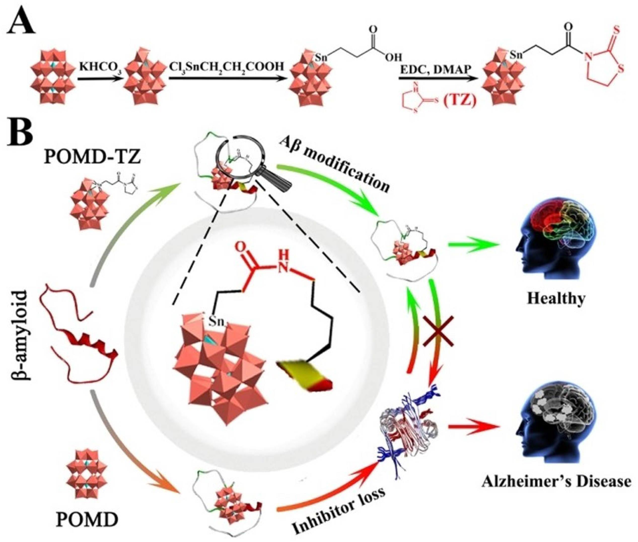
In the same year, Gao et al. [81] followed previous studies but now presented a new approach to inhibiting amyloid aggregation through the chemical post-translational modification (PTM) of protein modification using POMs as inhibitors of Aβ aggregates [81]. Thus, after the POMs were modified with thiazolidinethione (TZ), the resulting POMD-TZ (POMD is α-K6P2W18O62·14H2O) covalently modified Aβ site-selectively at Lys16. Thus, a new way of regulating amyloid aggregation by preventing inhibitor loss was suggested (Figure 7).
Also in 2022, Hu et al. [82] suggested novel chiral Aβ inhibitors based on POTs [(CH3)2NH2]15{[α-P2W15O55(H2O)]Zr3(μ3-O)(H2O)(L-tartH)[α-P2W16O59]} (L-POM) and [(CH3)2NH2]15{[α-P2W15O55(H2O)]Zr3(μ3-O)(H2O)(D-tartH)[α-P2W16O59]} (D-POM). It was found that, in contrast to L-POT, D-POT displayed a higher Aβ40-binding affinity, greater brain biodistribution and the ability to reduce Aβ40 accumulation, scavenge ROS and better rescue memory deficits in a mouse model of AD. Thus, chiral POMs can improve the enantioselectivity and specificity for the Aβ protein and for AD treatment [81].
One year earlier, Díaz et al. [83] focused on the effects of decavanadate (V10) and metforminium decavanadate on cognitive decline associated with pathophysiological processes of aging, such as increased ROS and neuroinflammation (also present in AD) [83]. As mentioned above, V10, belonging to the class of POVs, is perhaps the most studied POM in biological systems, as well in biomedical applications [14,43,44,64,67], although studies in AD, to our knowledge, were not yet described. Their results indicated that these POMs modulate Nrf2 activity, a transcription factor that regulates the expression of antioxidant enzymes promoting redox balance and reducing oxidative stress and neuroinflammation [84]. POMs mitigate neurodegeneration by inhibiting microglial activation and modulating the Nrf2 signaling pathway by preventing ROS production and iron overload, thus reducing the formation of aggregates. Moreover, POMs’ Nrf2 pathway activation leads to an increase in antioxidant enzymes, reducing the oxidative stress and inflammation that contribute to neurodegenerative diseases [85].
In 2025, it was shown that a nano-linear nickel-substituted Strandberg-type phosphomolybdate (H2en)6{[Ni(H2O)4](P2Mo5O23)}3 (abbreviated as NiPM) (en = ethanediamine) can modulate the β sheet-rich conformation of Aβ aggregates [86]. The interaction between NiPM and Aβ involves metal ions in NiPM coordinating with the imidazole group of histidine residues present in the Aβ peptide. This interaction can influence Aβ’s aggregation process, potentially affecting the formation of amyloid plaques associated with AD [86]. In another recent study, in order to further explore the inhibitory mechanisms of action of POMs in AD, a multiscale computational strategy integrating active-learning Bayesian Optimization (BO) and density functional theory (DFT) was employed to explore low-energy configurations of isolated amino acids, [PMo12O40]3−–amino acid complexes and [PMo12O40]3−–peptide systems [87]. The simulations predict that the cluster preferentially targets hydrophobic amino acids with alkyl chains (valine, lysine, leucine, isoleucine) located in peptide regions critical for Aβ aggregation, pointing out the role of multivalent weak interactions in POM-mediated inhibition [87].

4.3. Nanoparticle-Based Polyoxometalate Studies in Alzheimer’s Disease

Besides using pure POMs [48], Li et al. [49] reported in another study the putative AD applications of a self-assembly of Aβ15–20 peptides and a POT (K8[P2CoW17O61]) formation of hybrid colloidal spheres in water. These POM@P hybrid particles were tested as bifunctional Aβ aggregation inhibitors (Figure 8). As evidenced by AFM and spectral analysis and demonstrated in PC12 cells, the POT@P was effective in inhibiting the aggregation of Aβ1–40 [49].
One year later (2014), Gao et al. studied a hybrid system that acts as a multifunctional inhibitor of Aβ [88]. This system combines POMs, gold nanorods (AuNRs) and a peptide with a specificity for Aβ. A synergistic effect was observed between the constituents of the system, culminating in the inhibition of Aβ aggregation, the dissociation of preformed Aβ fibrils and the decrease in Aβ-mediated peroxidase activity and Aβ-induced cytotoxicity. Furthermore, AuNRs functioned as a vehicle for crossing the blood–brain barrier [88]. In 2016, another study from Gao et al. also involved a hybrid system, this time Cerium/POMs, describing proteolytic and antioxidant effects, the latter similar to superoxide dismutase (SOD) [51]. This hybrid system was shown to effectively degrade Aβ aggregates and decrease oxidative stress (due to SOD activity), attenuating Aβ-mediated toxicity. Notably, it has also demonstrated the ability to inhibit microglial activation, thereby reducing neuroinflammation, and also to cross the blood–brain barrier (BBB) [51].
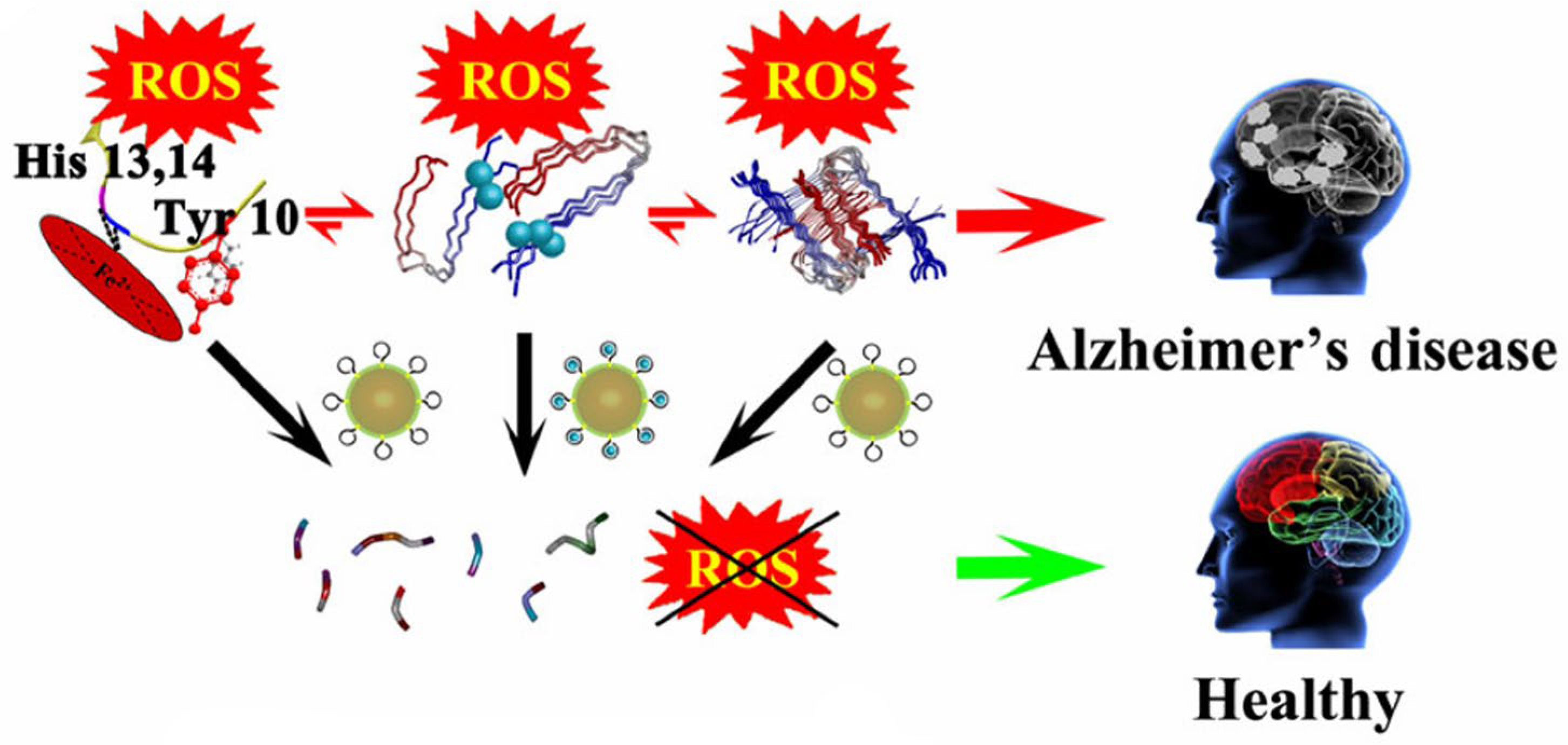
Two years later, also with proteolytic and antioxidant abilities as described previously [51], but now with a completely different approach, Gao et al. described a POM-based nanozyme, AuNPs@POMD, and octa-peptides, AuNPs@POMD-8pep, both with a protease-like activity for depleting Aβ aggregates (Figure 9). As before, an SOD-like activity for scavenging Aβ-mediated ROS was described. Furthermore, this nanozyme acts as a metal chelator to remove Cu from Cu-induced Aβ oligomers [89].
In 2017, four years after the study described above by Li et al. [49], the same research group published a new study where they again studied photothermal therapy, while also using a hybrid system (AuP; Figure 10A) that combines the benefits of gold nanoparticles (AuNRs), POT (K8[P2CoW17O61) and a specific Aβ inhibitor, Aβ15–20 [52]. This system relies on the strong NIR (near-infrared) optical absorption of AuNRs to generate localized hyperthermia, which effectively disaggregates the preformed Aβ fibrils (Figure 10B).
The results demonstrated the ability of AuP to disaggregate preformed Aβ fibrils after NIR irradiation (Figure 10D), reduce Aβ-induced cytotoxicity by about 45% and also cross the blood–brain barrier. The Aβ1–40 fibrils were incubated in an aggregation buffer (10 mM Tris, 150 mM NaCl, pH 7.3) with AuP for 20 min at 37 °C. Next, the morphology of Aβ fibrils was observed through atomic force microscopy (AFM). It was observed that Aβ15–20 cannot establish stable bonds with AuNRs. Thus, the POT (P2CoW17O61) has the dual function of a link between AuNRs and Aβ15–20, and also functions as an inhibitor of fibril formation [52]. It was possible to observe long well-defined fibrils on the left side of the image, as well as clusters of fibrils near the right margin (Figure 10C). Dispersed Aβ segments of reduced size are observed in comparison to the fibrils present in C, without it being possible to observe evident aggregates, suggestive of the photocatalytic activity of AuP after NIR irradiation (Figure 10D).
One year later, Ma et al. [90] developed a redox-activated POM-based nanoplatform sensitive to NIR radiation [90]. The nanoplatform, (rPOMs@MSNs@copolymer), silica nanoparticles (MSNs), rPOMs (rPOMs with various structures, which included Wells–Dawson structure (rPOMDs), Keggin structure (rPOMKs) and Anderson structure (rPOMAs)) and the thermal responsive copolymer poly(N-isopropylacrylamide-co-acrylamide) are activated by NIR radiation, which induces a thermal response capable of disaggregating the Aβ fibrils. This response is particularly advantageous, as it allows non-invasive activation and a localized effect, reducing off-target effects. Furthermore, the nanoplatform demonstrated antioxidant properties that give it the ability to inhibit the production of ROS induced by Aβ, as well as the ability to cross the blood–brain barrier [90].
Compared with unmodified POMs, POM-based inorganic–organic hybrids and POM-based nanocomposite structures show a significantly enhanced bioactivity and reduced side effects in AD [40]. Moreover, POM-based nanomedicine developed hybrid and nanoparticle systems that enhance BBB penetration, increase efficiency and reduce toxicity. In fact, nanotechnology has emerged as a transformative strategy for precise brain-targeted treatment [91,92].
In 2019, Liu et al. verified that Zn induces Aβ aggregation, but this is prevented by peptide@Mo-POM nanoparticles [93]. Only with the peptide–POM is a strong prevention of beta aggregation observed. By preventing ROS production induced by Aβ, the peptide@Mo-POM nanoparticles block neuron shrinkage. It was also observed that the peptide enhanced BBB penetration and the affinity for Aβ species. No morphological changes and no toxicity effects were evaluated in several tissues. Moreover, it was suggested that Mo-POMs can also sequester Zn2+ [93].
In the year 2023, Perich et al. [57] developed a nanohybrid system (Figure 11A) consisting of gold nanoparticles covered with [β2-SiW11O39]8− (Figure 11B) and polyethylene glycol (PEG), AuNPs@POM@PEG, with the aim of reducing the fibrillization of Aβ [61]. In order to evaluate the inhibitory capacity of AuNPs@POM@PEG in vitro, Aβ1–42 peptides were incubated with increasing concentrations of AuNPs@POM@PEG (1, 2.5 and 5 nM) for 2 h at 37 °C. The results demonstrated a strong inhibitory effect of AuNPs@POM@PEG, with an inhibition of approximately 75% for the highest concentration (5 nM) compared to the untreated Aβ sample (Figure 10C).
The above recent insights into the use of POMs in the treatment of AD has demonstrated a growing potential in different forms of action, particularly with regard to the inhibition of the formation of aggregates, one of the main pathophysiological hallmarks of the disease. Reflecting the increasing interest of the putative applications of POMs in AD treatment and research, several review papers have been published recently pointing out different points of view [40,45,75,94]. Moreover, POM nanoparticle-based systems have also emerged as promising tools for AD treatment not only because they improve drug delivery across the blood–brain barrier, but also because they improve efficiency, stability and bioavailability [92,95,96].
In summary, from the selected papers described above in Figure 2, in addition to Aβ aggregation, we can find other effects induced by POMs, such as the following: (1) the disaggregation of Aβ fibrils; (2) acetylcholinesterase inhibition activity; (3) SOD activity; (4) reduced neuroinflammation; (5) reduced ROS; (6) reduced Aβ citotoxicity; (7) reduced peroxidase-like activity; (8) the prevention of Aβ conformational shift (Figure 12).
However, several other effects were described, such as the chelation of transition metals (4 articles) and the improvement of memory (2 articles), among others (Figure 13). The main effect observed in the selected articles was exactly the inhibition of Aβ aggregation (76%), with the majority of the studies (23 articles, from 30) describing this result [48,49,50,51,52,53,54,55,56,57]. Other major effects found to be promoted by POMs included the following: (i) the decrease in neurotoxicity induced by Aβ (66%); (ii) the crossing of the blood–brain barrier (53%); (iii) the decrease in ROS (33%); and (iv) Aβ degradation (26%). Other effects, such as the inhibition of AChE (13%) and transition metal chelators (13%) among other effects, were described only in a few studies (Figure 13).
Globally, the majority of the POM/AD studies used POTs (62%), but also POMos (32%), PONbs (5%) and POVs (2%) (Table S1). However, in the first seven years (2011–2018), we found that the majority of the studies were performed with POTs (80%), with minor contributions from POMos (13%) and POVs (7%), whereas, in the last seven years (2019–2025), POMos (44%) increased but were still below POTs (50%), while PONbs (6%) also had a fresh contribution. Among the POMs described above, the ones most referred to in the AD studies were W11 (six articles) followed by W17 and W12 (three articles). The first one (W11) was said to (1) prevent Aβ fibrillation (2011); (2) be used in a platinum-substituted POT (Me4N)3[PW11O40(SiC3H6NH2)2PtCl2) that prevents the oligomerization and fibril formation of Aβ [55] (2018); (3) induce achetylcolinesterase inhibition (2020); (4) be a copper-chelating agent (2022) [58]; (5) be also present in a nanohybrid system consisting of gold nanoparticles covered with [β2-SiW11O39]8− and polyethylene glycol (AuNPs@POM@PEG), with the aim of reducing the fibrillization of Aβ [61] (2023); and (6) be analyzed in docking studies as a POM target for AD. Conversely, the Keggin-type W12 also interacts with Aβ1–40, affecting the degree of aggregation (2011) [50], and also is a potent acetylcholinesterase inhibitor (2011) [60] and a copper-chelating agent and inhibitor of Aβ aggregate formation (2020) [68]. Regarding the POMs present in the nanohybrid systems, the most common POM used is the Wells–Dawson-type P2W17 (Table S1).
While much AD research has focused on amyloid-beta and tau proteins, the role of POMs in modulating calcium signaling offers a promising but underexplored alternative therapeutic strategy [97]. In fact, calcium dysregulation also plays a key role in AD progression [98]. As described above, AD chronic states of neuroinflammation and oxidative stress, together with the disturbance of calcium homeostasis, contribute to neurotoxicity and cell death [27]. A growing number of studies identify the dysregulation of calcium (Ca2+) homeostasis, particularly the anomalous increase in cytosolic Ca2+ concentrations, as being implicated in the cascade of pathophysiological events that trigger the clinical picture of AD [28]. Recent studies suggest that calcium dysregulation is not just a consequence of the pathological processes of AD, but rather a central player in the pathogenesis of the disease [99]. It is becoming increasingly clear that strategies aimed at correcting or modulating calcium signaling pathways constitute promising approaches for the study of therapeutic alternatives for AD [100]. Recently, polyoxometalates (POMs) were described as presenting agonistic properties on purinergic P2 receptors from neuron cells [47]. Thus, POMs inhibited the P-type ATPases [46] and also modulated the cytosolic calcium concentrations in neurons, proving to be a useful tool in the studies of pathological processes of AD [46,47], as well as in others neurodegenerative diseases [101].
Taking into consideration the articles described in the present review, interdisciplinary collaborations are fundamental for bringing together experienced and also young researchers for the evolution of the applications of POMs in the treatment of AD. Moreover, we believe that inorganic biochemistry also represents an extraordinary platform for promoting communication between Alzheimer’s caregivers, doctors, psychologists, nurses and family members, just like a key cog in the “clock of the knowledge”, dynamically moving chemistry, biology and medicine, among others, to promote innovation and urgent developments in the treatment of AD [102,103].
As summarized in Figure 12, the POM studies in AD started in 2011, with POMs such as W11 and W17 being pure or in nanoparticle (NP) systems and finished also with W11 that was pure and/or in NPs, but now with a higher potency of inhibition, that is, with ranging nM concentrations as inhibitors of AChE activity and Aβ aggregation, well-known hallmarks of AD. Therefore, POMs’ progress in AD shows an increased efficiency that, together with a decreased toxicity, potentiate the development of more effective and sustainable applications in this challenging 21st-century neurodegenerative disease (Figure 14). In fact, Figure 14 was based on the observation of a relative with AD who, upon arriving at the beach, sat with her back to the sea, as if disoriented. It is suggested that POMs can reverse this disorientation and allow a return to the sun (Figure 14). However, it should be realized that POMs’ putative applications in the treatment of AD are yet in the early stages. Collaborations from several fields and points of view are absolutely essential for the progress of the usage of new drugs against this disease, which is devastating to the lives of patients and their families.

5. Conclusions and Outlook

Polyoxometalates are evolving in AD treatment through an increased understanding of their roles in disease and the development of POM-based therapies, including hybrids and POM nanoparticles (NPs@POMs) for preventing Aβ aggregates and the subsequent neurotoxicity induced by β-amyloid aggregates and decreasing ROS production and neuroinflammation, in addition to restoring memory and sequestering transition metals ions such as Zn2+ and Cu2+, features that are well known to be associated with the pathology of Alzheimer’s disease.
Herein, we investigated the potential of POMs in modulating the pathophysiology of Alzheimer’s disease, with a special focus on the possible associated neuroprotective effects. From an in-depth analysis of the studies, relevant data emerged on how polyoxometalates interact with the pathogenic mechanisms of Alzheimer’s disease. The majority of the AD studies use POTs (62%), but also POMos (32%), PONbs (5%) and POVs (2%), W11 being the most studied. Not all are pure POMs (46%), and we also found hybrid POMs (27%) and hybrid nanoparticle POM systems (27%).
Notably, most studies have focused on interactions with Aβ aggregates, which play a central role in the cascade of pathological events present in Alzheimer’s disease. POMs have the ability to reverse the formed β sheet conformation and also to induce the disaggregation of Aβ fibrils, particularly after NIR and/or UV/Vis irradiation. POMs were also described as a novel class of acetylcholinesterase inhibitors known to improve cognitive function in AD. Thus, by inhibiting acetylcholinesterase and the formation of protein aggregates, or by removing the formed aggregates, POMs represent a promising therapeutic strategy for AD.
In the pathology of Alzheimer’s disease, other processes can be prevented by POMs, pointing to putative applications in AD treatment. Future studies will point out POMs’ ability to inhibit microglial activation, thereby reducing neuroinflammation, and also to cross the blood–brain barrier. Still, applications of POMs in AD could be associated with many other mechanisms of action. For instance, we cannot exclude the possibility that POMs can be used as the modulators of neurons for calcium homeostasis, since it is also known that neurodegenerative diseases are associated with calcium dysregulation.
Putting it all together, the studies performed so far suggested that, in several ways, POMs can be used in AD treatment, though it should be stated that putative applications in the treatment of the disease are yet in their early stages. In fact, while the applications of POMs in cancer and in bacterial and/or viral infections are clearly in progress, POM studies in neurodegenerative diseases are still very few. Moreover, a current limitation, besides the paucity of in vivo validation, is the need for greater interdisciplinary collaboration between chemistry, neuroscience and pharmacology. We believe that the present review, with an inorganic biochemistry perspective on the evolution of the utilization of POMs in AD studies, will push forward those interdisciplinary collaborations, particularly with neurosciences, in order to promote new insights into POM applications in AD, among other neurodegenerative diseases with overlapping proteinopathies.

Supplementary Materials

The following supporting information can be downloaded at https://www.mdpi.com/article/10.3390/biochem5040041/s1. Table S1: POM studies associated with Alzheimer´s disease (2011–2025). Year, POM information and POM composition, referred to as pure (P), combined with a organic ligand (H) or POM nanoparticles-based compounds (NP), and the classes of POMs, namely polyoxotungstates (POTs), polyoxomolybdates (POMos), polyoxobiobates (PONbs) and polyoxovanadates (POVs) [48,49,50,51,52,53,54,55,56,57,58,59,60,61,62,63,68,70,71,72,73,79,81,82,86,87,88,89,90,93].

Author Contributions

Conceptualization, M.A.; methodology, M.A., D.M.R. and J.M.; validation, M.A.; formal analysis, M.A.; investigation, M.A., D.M.R. and J.M.; resources, M.A.; data curation, M.A.; writing—M.A.; writing—review and editing, M.A., D.M.R. and J.M.; visualization, M.A., D.M.R. and J.M.; supervision, M.A.; project administration, M.A.; funding acquisition, M.A. All authors have read and agreed to the published version of the manuscript.

Funding

Thanks to Algarve University and to Portuguese national funds from FCT—Foundation for Science and Technology, through contracts UID/04326/2025, UID/PRR/04326/2025 and LA/P/0101/2020 (DOI:10.54499/LA/P/0101/2020).

Data Availability Statement

The original contributions presented in this study are included in the article/Supplementary Material. Further inquiries can be directed to the corresponding author.

Acknowledgments

M.A. thanks Algarve University and Portuguese national funds from FCT—Foundation for Science and Technology through contracts UID/04326/2025, UID/PRR/04326/2025 and LA/P/0101/2020.

Conflicts of Interest

The authors declare no conflicts of interest.

Abbreviations

The following abbreviations are used in this manuscript:
ADAlzheimer’s disease
AChEAchetylcolinesterase
AFMAtomic force microscopy
APPAmyloid precursor protein
AuNRsGold nanorods
β-amyloid
BBBBlood–brain barrier
BOBayesian Optimization
CDCircular dicroism
CNSCentral nervous system
DFTDensity functional theory
DLSDynamic light scattering
IC50Half maximal inhibitory concentration
IL6Interleukin-6
ITCIsothermal titration calorimetry
LPOLipid peroxidation
NFTNeurofibrillary tangles
NIRNear-infrared radiation
NMDAN-methyl-D-aspartate
NPsNanoparticles
Nrf2Transcription factor
P2Purinergic P2 receptors
PBSPhosphate-buffered solution
PC12Cell line from rat pheochromocytoma
PEGPolyethylene glycol
POMoPolyoxmolybdates
POMsPolyoxometalates
POVsPolyoxovanadates
PS1Presenilin-1
PS2Presenilin-2
PTMPost-translational modification
ROSReactive oxygen species
SODSuperoxide dismutase
TNFTumor necrosis factor
TZThiazolidinethione
WHOWorld Health Organization

References

  1. Morris, J.C. Neurodegenerative Disorders of Aging: The Down Side of Rising Longevity. Mo Med. 2013, 110, 393–3944. [Google Scholar]
  2. Dugger, B.N.; Dickson, D.W. Pathology of Neurodegenerative Diseases. Cold Spring Harb. Perspect. Biol. 2017, 9, a028035. [Google Scholar] [CrossRef]
  3. Heemels, M.T. Neurodegenerative diseases. Nature 2016, 539, 179. [Google Scholar] [CrossRef] [PubMed]
  4. Wakabayashi, K. Cellular pathology of neurodegenerative disorders. Clin. Neurol. 2013, 53, 609–617. [Google Scholar]
  5. Davis, A.A.; Leyns, C.E.G.; Holtzman, D.M. Intercellular Spread of Protein Aggregates in Neurodegenerative Disease. Annu. Rev. Cell Dev. Biol. 2018, 34, 545–568. [Google Scholar] [CrossRef]
  6. Lin, M.T.; Beal, M.F. Mitochondrial dysfunction and oxidative stress in neurodegenerative diseases. Nature 2006, 443, 787–795. [Google Scholar] [CrossRef]
  7. Chen, X.; Guo, C.; Kong, J. Oxidative stress in neurodegenerative diseases. Neural Regen. Res. 2012, 7, 376. [Google Scholar]
  8. Lamptey, R.N.L.; Chaulagain, B.; Trivedi, R.; Gothwal, A.; Layek, B.; Singh, J. A Review of the Common Neurodegenerative Disorders: Current Therapeutic Approaches and the Potential Role of Nanotherapeutics. Int. J. Mol. Sci. 2022, 23, 1851. [Google Scholar] [CrossRef]
  9. Breijyeh, Z.; Karaman, R. Comprehensive Review on Alzheimer’s Disease: Causes and Treatment. Molecules 2020, 25, 5789. [Google Scholar] [CrossRef] [PubMed]
  10. Anand, P.; Singh, B. A review on cholinesterase inhibitors for Alzheimer’s disease. Arch. Pharm. Res. 2013, 36, 375–399. [Google Scholar] [CrossRef] [PubMed]
  11. Passeri, E.; Elkhoury, K.; Morsink, M.; Broersen, K.; Linder, M.; Tamayol, A.; Malaplate, C.; Yen, F.T.; Arab-Tehrany, E. Alzheimer’s Disease: Treatment Strategies and Their Limitations. Int. J. Mol. Sci. 2022, 23, 13954. [Google Scholar] [CrossRef]
  12. Frozza, R.L.; Lourenco, M.V.; de Felice, F.G. Challenges for Alzheimer’s Disease Therapy: Insights from Novel Mechanisms Beyond Memory Defects. Front. Neurosci. 2018, 12, 37. [Google Scholar] [CrossRef] [PubMed]
  13. Pardridge, W.M. Treatment of Alzheimer’s Disease and Blood–Brain Barrier Drug Delivery. Pharmaceuticals 2020, 13, 394. [Google Scholar] [CrossRef]
  14. Aureliano, M.; Mitchell, S.G.; Yin, P. Editorial: Emerging polyoxometalates with biological, biomedical, and health applications. Front. Chem. 2022, 10, 977317. [Google Scholar] [CrossRef]
  15. Čolović, M.B.; Lacković, M.; Lalatović, J.; Mougharbel, A.S.; Kortz, U.; Krstić, D.Z. Polyoxometalates in Biomedicine: Update and Overview. Curr. Med. Chem. 2020, 27, 362–379. [Google Scholar] [CrossRef] [PubMed]
  16. Woźniak-Budych, M.J.; Staszak, K.; Bajek, A.; Pniewski, F.; Jastrząb, R.; Staszak, M.; Tylkowski, B.; Wieszczycka, K. The future of polyoxymetalates for biological and chemical apllications. Coord. Chem. Rev. 2023, 493, 215306. [Google Scholar] [CrossRef]
  17. Georges, J.; Bintener, C.; Miller, O. Dementia in Europe Yearbook 2019: Estimating the prevalence of dementia in Europe; Alzheimer Europe: Sandweiler, Luxembourg, 2020; pp. 1–108. [Google Scholar]
  18. GBD 2019 Dementia Forecasting Collaborators. Estimation of the global prevalence of dementia in 2019 and forecasted prevalence in 2050: An analysis for the Global Burden of Disease Study 2019. Lancet Public Health 2022, 7, e105–e125. [Google Scholar] [CrossRef]
  19. DeTure, M.A.; Dickson, D.W. The neuropathological diagnosis of Alzheimer’s disease. Mol. Neurodegener. 2019, 14, 32. [Google Scholar] [CrossRef]
  20. Igarashi, K.M. Entorhinal cortex dysfunction in Alzheimer’s disease. Trends Neurosci. 2023, 46, 124. [Google Scholar] [CrossRef]
  21. Hippius, H.; Neundörfer, G. The discovery of Alzheimer’s disease. Dialogues Clin. Neurosci. 2003, 5, 101–108. [Google Scholar] [CrossRef]
  22. Madnani, R.S. Alzheimer’s disease: A mini-review for the clinician. Front. Neurol. 2023, 14, 1178588. [Google Scholar] [CrossRef] [PubMed]
  23. Silva, M.V.F.; Loures, C.d.M.G.; Alves, L.C.V.; De Souza, L.C.; Borges, K.B.; Carvalho, M.G. Alzheimer’s disease: Risk factors and potentially protective measures. J. Biomed. Sci. 2019, 26, 33. [Google Scholar] [CrossRef]
  24. Chen, G.F.; Xu, T.H.; Yan, Y.; Zhou, Y.R.; Jiang, Y.; Melcher, K.; Xu, H.E. Amyloid beta: Structure, biology and structure-based therapeutic development. Acta Pharmacol. Sin. 2017, 38, 1205–1235. [Google Scholar] [CrossRef]
  25. Coronel, R.; Bernabeu-Zornoza, A.; Palmer, C.; Muñiz-Moreno, M.; Zambrano, A.; Cano, E.; Liste, I. Role of Amyloid Precursor Protein (APP) and Its Derivatives in the Biology and Cell Fate Specification of Neural Stem Cells. Mol. Neurobiol. 2018, 55, 7107–7117. [Google Scholar] [CrossRef]
  26. Griffiths, J.; Grant, S.G.N. Synapse pathology in Alzheimer’s disease. Semin. Cell. Dev. Biol. 2023, 139, 13–23. [Google Scholar] [CrossRef]
  27. Woodburn, S.C.; Bollinger, J.L.; Wohleb, E.S. The semantics of microglia activation: Neuroinflammation, homeostasis, and stress. J. Neuroinflamm. 2021, 18, 258. [Google Scholar] [CrossRef] [PubMed]
  28. Joshi, M.; Joshi, S.; Khambete, M.; Degani, M. Role of calcium dysregulation in Alzheimer’s disease and its therapeutic implications. Chem. Biol. Drug Des. 2023, 101, 453–468. [Google Scholar] [CrossRef] [PubMed]
  29. Liu, J.; Chang, L.; Song, Y.; Li, H.; Wu, Y. The Role of NMDA Receptors in Alzheimer’s Disease. Front. Neurosci. 2019, 13, 43. [Google Scholar] [CrossRef]
  30. Tatulian, S.A. Challenges and hopes for Alzheimer’s disease. Drug Discov. Today 2022, 27, 1027–1043. [Google Scholar] [CrossRef]
  31. Pardridge, W.M. Alzheimer’s disease drug development and the problem of the blood-brain barrier. Alzheimer’s Dement. 2009, 5, 427. [Google Scholar] [CrossRef]
  32. Gao, Y.; Choudhari, M.; Such, G.K.; Ritchie, C. Polyoxometalates as chemically and structurally versatile components in self-assembled materials. Chem. Sci. 2022, 13, 2510–2527. [Google Scholar] [CrossRef]
  33. Hill, C.L. Introduction: Polyoxometalates Multicomponent Molecular Vehicles To Probe Fundamental Issues and Practical Problems. Chem. Rev. 1998, 98, 1–2. [Google Scholar] [CrossRef] [PubMed]
  34. Gumerova, N.I.; Rompel, A. Synthesis, structures and applications of electron-rich polyoxometalates. Nat. Rev. Chem. 2018, 2, 112. [Google Scholar] [CrossRef]
  35. Chen, Y.; Li, F.; Li, S.; Zhang, L.; Sun, M. A review of application and prospect for polyoxometalate-based composites in electrochemical sensor. Inorg. Chem. Commun. 2022, 135, 109084. [Google Scholar] [CrossRef]
  36. Liu, R.; Streb, C. Polyoxometalate-Single Atom Catalysts (POM-SACs) in Energy Research and Catalysis. Adv. Energy Mater. 2021, 11, 2101120. [Google Scholar] [CrossRef]
  37. Samaraj, E.; Balaraman, E.; Manickam, S. Functional POM-catalyst for selective oxidative dehydrogenative couplings under aerobic conditions. Mol. Catal. 2021, 502, 111396. [Google Scholar] [CrossRef]
  38. Asif, H.M.; Shakoor, Z.; Ibraheem, S.; Salama, A.M.; Khan, M.A.; Nguyen, T.A.; Yasin, G. Chapter 12—Polyoxometalate-based metal organic frameworks (POMOFs) for lithium-ion batteries. In Micro and Nano Technologies; Metal-Organic Framework-Based Nanomaterials for Energy Conversion and Storage; Gupta, R.K., Nguyen, T.A., Yasin, G., Eds.; Elsevier: Amsterdam, The Netherlands, 2022; pp. 245–268. [Google Scholar] [CrossRef]
  39. Zhang, Y.; Li, Y.; Guo, H.; Guo, Y.; Song, R. Recent advances in polyoxometalate-based materials and their derivatives for electrocatalysis and energy storage. Mater. Chem. Front. 2024, 8, 732–768. [Google Scholar] [CrossRef]
  40. Wang, X.; Wei, S.; Zhao, C.; Li, X.; Jin, J.; Shi, X.; Su, Z.; Li, J.; Wang, J. Promising application of polyoxometalates in the treatment of cancer, infectious diseases and Alzheimer’s disease. J. Biol. Inorg. Chem. 2022, 27, 405–419. [Google Scholar] [CrossRef]
  41. Li, B.; Xu, X.; Lv, Y.; Wu, Z.; He, L.; Song, Y.F. Polyoxometalates as Potential Artificial Enzymes toward Biological Applications. Small 2024, 20, e2305539. [Google Scholar] [CrossRef]
  42. Bijelic, A.; Aureliano, M.; Rompel, A. Polyoxometalates as Potential Next-Generation Metallodrugs in the Combat Against Cancer. Angew. Chem. Int. Ed. 2019, 58, 2980–2999. [Google Scholar] [CrossRef]
  43. Aureliano, M.; Gumerova, N.I.; Sciortino, G.; Garribba, E.; Rompel, A.; Crans, D.C. Polyoxovanadates with Emerging Biomedical Activities. Coord. Chem. Rev. 2021, 447, 214143. [Google Scholar] [CrossRef]
  44. De Sousa-Coelho, A.L.; Fraqueza, G.; Aureliano, M. Repurposing Therapeutic Drugs Complexed to Vanadium in Cancer. Pharmaceuticals 2024, 17, 12. [Google Scholar] [CrossRef]
  45. Gonzalez-Cano, S.I.; Flores, G.; Guevara, J.; Morales-Medina, J.C.; Treviño, S.; Diaz, A. Polyoxidovanadates a new therapeutic alternative for neurodegenerative and aging diseases. Neural Regen. Res. 2024, 19, 571–577. [Google Scholar] [CrossRef]
  46. Aureliano, M.; Fraqueza, G.; Berrocal, M.; Cordoba-Granados, J.J.; Gumerova, N.I.; Rompel, A.; Gutierrez-Merino, C.; Mata, A.M. Inhibition of SERCA and PMCA Ca2+-ATPase Activities by Polyoxotungstates. J. Inorg. Biochem. 2022, 236, 111952. [Google Scholar] [CrossRef] [PubMed]
  47. Poejo, J.; Gumerova, N.I.; Rompel, A.; Mata, A.M.; Aureliano, M.; Gutierrez-Merino, C. Unveiling the Agonistic Properties of Preyssler-Type Polyoxotungstates on Purinergic P2 Receptors. J. Inorg. Biochem. 2024, 259, 112640. [Google Scholar] [CrossRef] [PubMed]
  48. Geng, J.; Li, M.; Ren, J.; Wang, E.; Qu, X. Polyoxometalates as Inhibitors of the Aggregation of Amyloid β Peptides Associated with Alzheimer’s Disease. Angew. Chem. Int. Ed. 2011, 50, 4184–4188. [Google Scholar] [CrossRef]
  49. Li, M.; Xu, C.; Wu, L.; Ren, J.; Wang, E.; Qu, X. Self-assembled peptide-polyoxometalate hybrid nanospheres: Two in one enhances targeted inhibition of amyloid B-peptide aggregation associated with Alzheimer’s disease. Small 2013, 9, 3455–3461. [Google Scholar] [CrossRef] [PubMed]
  50. Gao, N.; Sun, H.; Dong, K.; Ren, J.; Duan, T.; Xu, C.; Qu, X. Transition-metal-substituted polyoxometalate derivatives as functional anti-amyloid agents for Alzheimer’s disease. Nat Commun. 2014, 5, 3422. [Google Scholar] [CrossRef] [PubMed]
  51. Guan, Y.; Li, M.; Dong, K.; Gao, N.; Ren, J.; Zheng, Y.; Qu, X. Ceria/POMs hybrid nanoparticles as a mimicking metallopeptidase for treatment of neurotoxicity of amyloid-β peptide. Biomaterials 2016, 98, 92–102. [Google Scholar] [CrossRef]
  52. Li, M.; Guan, Y.; Zhao, A.; Ren, J.; Qu, X. Using Multifunctional Peptide Conjugated Au Nanorods for Monitoring β-amyloid Aggregation and Chemo-Photothermal Treatment of Alzheimer’s Disease. Theranostics 2017, 7, 2996–3006. [Google Scholar] [CrossRef]
  53. Ma, X.; Hua, J.; Wang, K.; Zhang, H.; Zhang, C.; He, Y.; Guo, Z.; Wang, X. Modulating Conformation of Aβ-Peptide: An Effective Way to Prevent Protein-Misfolding Disease. Inorg. Chem. 2018, 57, 13533–13543. [Google Scholar] [CrossRef]
  54. Gao, N.; Du, Z.; Guan, Y.; Dong, K.; Ren, J.; Qu, X. Chirality-Selected Chemical Modulation of Amyloid Aggregation. J. Am. Chem. Soc. 2019, 141, 6915–6921. [Google Scholar] [CrossRef]
  55. Zhao, J.; Li, K.; Wan, K.; Sun, T.; Zheng, N.; Zhu, F.; Ma, J.; Jiao, J.; Li, T.; Ni, J.; et al. Organoplatinum-Substituted Polyoxometalate Inhibits β-amyloid Aggregation for Alzheimer’s Therapy. Angew. Chem. Int. Ed. Engl. 2019, 58, 18032–18039. [Google Scholar] [CrossRef]
  56. Bondžić, A.M.; Lazarević-Pašti, T.D.; Leskovac, A.R.; Petrović, S.Ž.; Čolović, M.B.; Parac-Vogt, T.N.; Janjić, G.V. A new acetylcholinesterase allosteric site responsible for binding voluminous negatively charged molecules—The role in the mechanism of AChE inhibition. Eur. J. Pharm. Sci. 2020, 151, 105376. [Google Scholar] [CrossRef]
  57. Perxés Perich, M.; Palma-Florez, S.; Solé, C.; Goberna-Ferrón, S.; Samitier, J.; Gómez-Romero, P.; Mir, M.; Lagunas, A. Polyoxometalate-Decorated Gold Nanoparticles Inhibit β-Amyloid Aggregation and Cross the Blood–Brain Barrier in a µphysiological Model. Nanomaterials 2023, 13, 2697. [Google Scholar] [CrossRef]
  58. Zhou, Y.; Zheng, L.; Han, F.; Zhang, G.; Ma, Y.; Yao, J.; Keita, B.; Oliveira, P.; Nadjo, L. Inhibition of amyloid-β protein fibrillization upon interaction with polyoxometalates nanoclusters, Colloids Surf. A Physicochem. Eng. Asp. 2011, 375, 97–101. [Google Scholar] [CrossRef]
  59. Li, M.; Xu, C.; Ren, J.; Wang, E.; Qu, X. Photodegradation of β-sheet amyloid fibrils associated with Alzheimer’s disease by using polyoxometalates as photocatalysts. Chem. Commun. 2013, 49, 11394–11396. [Google Scholar] [CrossRef] [PubMed]
  60. Iqbal, J.; Barsukova-Stuckart, M.; Ibrahim, M.; Ali, S.U.; Khan, A.A.; Kortz, U. Polyoxometalates as potent inhibitors for acetyl and butyrylcholinesterases and as potential drugs for the treatment of Alzheimer’s disease. Med. Chem. Res. 2013, 22, 1224–1228. [Google Scholar] [CrossRef]
  61. Chen, Q.; Yang, L.; Zheng, C.; Zheng, W.; Zhang, J.; Zhou, Y.; Liu, J. Mo polyoxometalate nanoclusters capable of inhibiting the aggregation of Aβ-peptide associated with Alzheimer’s disease. Nanoscale 2014, 6, 6886–6897. [Google Scholar] [CrossRef]
  62. Čolović, M.B.; Medić, B.; Ćetković, M.; Kravić Stevović, T.; Stojanović, M.; Ayass, W.W.; Mougharbel, A.S.; Radenković, M.; Prostran, M.; Kortz, U.; et al. Toxicity evaluation of two polyoxotungstates with anti-acetylcholinesterase activity. Toxicol. Appl. Pharmacol. 2017, 333, 68–75. [Google Scholar] [CrossRef] [PubMed]
  63. Li, S.; Jiang, D.; Ehlerding, E.B.; Rosenkrans, Z.T.; Engle, J.W.; Wang, Y.; Liu, H.; Ni, D.; Cai, W. Intrathecal Administration of Nanoclusters for Protecting Neurons against Oxidative Stress in Cerebral Ischemia/Reperfusion Injury. ACS Nano. 2019, 13, 13382–13389. [Google Scholar] [CrossRef]
  64. Aureliano, M.; De Sousa-Coelho, A.L.; Dolan, C.C.; Roess, D.A.; Crans, D.C. Biological Consequences of Vanadium Effects on Formation of Reactive Oxygen Species and Lipid Peroxidation. Int. J. Mol. Sci. 2023, 24, 5382. [Google Scholar] [CrossRef]
  65. Aureliano, M.; Gumerova, N.I.; Sciortino, G.; Garribba, E.; McLauchlan, C.C.; Rompel, A.; Crans, D.C. Polyoxidovanadates’ Interactions with Proteins: An Overview. Coord. Chem. Rev. 2022, 454, 214344. [Google Scholar] [CrossRef]
  66. Gumerova, N.I.; Rompel, A. Polyoxometalates in Solution: Speciation under Spotlight. Chem. Soc. Rev. 2020, 49, 7568–7601. [Google Scholar] [CrossRef]
  67. Gumerova, N.I.; Rompel, A. Speciation atlas of polyoxometalates in aqueous solutions. Sci Adv. 2023, 9, eadi0814. [Google Scholar] [CrossRef] [PubMed] [PubMed Central]
  68. Chaudhary, H.; Iashchishyn, I.A.; Romanova, N.V.; Rambaran, M.A.; Musteikyte, G.; Smirnovas, V.; Holmboe, M.; Ohlin, C.A.; Svedružić, Z.M.; Morozova-Roche, L.A. Polyoxometalates as effective nano-inhibitors of amyloid aggregation of pro-inflammatory S100A9 protein involved in neurodegenerative diseases. ACS Appl. Mater. Interfaces 2021, 13, 26721–26734. [Google Scholar] [CrossRef]
  69. Sciortino, G.; Aureliano, M.; Garribba, E. Rationalizing the Decavanadate(V) and Oxidovanadium(IV) Binding to G-Actin and the Competition with Decaniobate(V) and ATP. Inorg. Chem. 2021, 60, 334–344. [Google Scholar] [CrossRef] [PubMed]
  70. Atrian-Blasco, E.; de Cremoux, L.; Lin, X.; Mitchell-Heggs, R.; Sabater, L.; Blanchard, S.; Hureau, C. Keggin-type polyoxometalates as Cu(II) chelators in the context of Alzheimer’s disease. Chem. Commun. 2022, 58, 2367–2370. [Google Scholar] [CrossRef]
  71. Wang, M.; Hua, J.; Zheng, P.; Tian, Y.; Kang, S.; Chen, J.; Duan, Y.; Ma, X. A Nanoscale Cobalt Functionalized Strandberg-Type Phosphomolybdate with b-Sheet Conformation Modulation Ability in Anti-Amyloid Protein Misfolding. Inorganics 2023, 11, 442. [Google Scholar] [CrossRef]
  72. Lei, S.; Yang, H.; Li, J.X.; Li, Y.; Wang, L.; Chen, B.N.; Li, J. A study of the antioxidant properties of Keggin-type polyoxometalates. Dalton Trans. 2023, 52, 9673–9683. [Google Scholar] [CrossRef]
  73. Ben Zaken, K.; Bouhnik, R.; Omer, N.; Bloch, N.; Samson, A.O. Polyoxometalates bind multiple targets involved in Alzheimer’s disease. J. Biol. Inorg. Chem. 2025, 30, 299–309. [Google Scholar] [CrossRef]
  74. Ramesh, M.; Balachandra, C.; Andhare, P.; Govindaraju, T. Rationally Designed Molecules Synergistically Modulate Multifaceted Aβ Toxicity, Microglial Activation, and Neuroinflammation. ACS Chem. Neurosci. 2022, 13, 2209–2221. [Google Scholar] [CrossRef] [PubMed]
  75. Ma, M.; Liu, Z.; Zhao, H.; Zhang, H.; Ren, J.; Qu, X. Polyoxometalates: Metallodrug agents for combating amyloid aggregation. Natl. Sci. Rev. 2024, 11, nwae226. [Google Scholar] [CrossRef] [PubMed] [PubMed Central]
  76. Bittner, N.; Funk, C.S.M.; Schmidt, A.; Bermpohl, F.; Brandl, E.J.; Algharably, E.E.A.; Kreutz, R.; Riemer, T.G. Psychiatric Adverse Events of Acetylcholinesterase Inhibitors in Alzheimer’s Disease and Parkinson’s Dementia: Systematic Review and Meta-Analysis. Drugs Aging 2023, 40, 953–964. [Google Scholar] [CrossRef]
  77. Kondinski, A.; Gumerova, N.I.; Rompel, A.; Falcaro, P.; Schreck, T. Data-Driven Polyoxometalate Chemistry. Chem. Eur. J. 2025, 31, e01528. [Google Scholar] [CrossRef] [PubMed]
  78. Hardman, T.C.; Aitchison, R.; Scaife, R.; Edwards, J.; Slater, G. The future of clinical trials and drug development: 2050. Drugs Context. 2023, 12, 2023-2-2. [Google Scholar] [CrossRef] [PubMed] [PubMed Central]
  79. Hua, J.A.; Wei, X.M.; Ma, X.; Jiao, J.Z.; Chai, B.H.; Wu, C.B.; Zhang, C.L.; Niu, Y.L. A {Cd4Cl2O14} cluster functionalized sandwich-type tungstoarsenate as a conformation modulator for misfolding Aβ peptides. Cryst. Eng. Comm. 2022, 24, 1171–1176. [Google Scholar] [CrossRef]
  80. Xue, C.; Lin, T.Y.; Chang, D.; Guo, Z. Thioflavin T as an amyloid dye: Fibril quantification, optimal concentration and effect on aggregation. R. Soc. Open Sci. 2017, 4, 160696. [Google Scholar] [CrossRef]
  81. Gao, N.; Liu, Z.; Zhang, H.; Liu, C.; Yu, D.; Ren, J.; Qu, X. Site-Directed Chemical Modification of Amyloid by Polyoxometalates for Inhibition of Protein Misfolding and Aggregation. Angew. Chem. Int. Ed. Engl. 2022, 61, e202115336. [Google Scholar] [CrossRef] [PubMed]
  82. Hu, S.; Ning, X.; Lv, J.; Wei, Y.; Zhang, H.; Li, M. Enantioselective modulation of amyloid burden and memory deficits by chiralpolyoxometalates for Alzheimer’s disease treatment. Inorg. Chem. Front. 2023, 10, 5347–5356. [Google Scholar] [CrossRef]
  83. Diaz, A.; Muñoz-Arenas, G.; Venegas, B.; Vázquez-Roque, R.; Flores, G.; Guevara, J.; Gonzalez-Vergara, E.; Treviño, S. Metforminium Decavanadate (MetfDeca) Treatment Ameliorates Hippocampal Neurodegeneration and Recognition Memory in a Metabolic Syndrome Model. Neurochem. Res. 2021, 46, 1151–1165. [Google Scholar] [CrossRef] [PubMed]
  84. Copple, I.M. The Keap1-Nrf2 Cell Defense Pathway—A Promising Therapeutic Target? Adv. Pharmacol. 2012, 63, 43–79. [Google Scholar]
  85. la Torre, A.; Lo Vecchio, F.; Angelillis, V.S.; Gravina, C.; D’Onofrio, G.; Greco, A. Reinforcing Nrf2 Signaling: Help in the Alzheimer’s Disease Context. Int. J. Mol. Sci. 2025, 26, 1130. [Google Scholar] [CrossRef]
  86. Ma, X.; Li, H.; Zhao, Z.; Zhang, L.; Su, H.; Zhao, Y.; Sun, Y.; Li, C.; Wang, Y.; Hua, J. An interesting nano-linear nickel-modified Strandberg-type phosphomolybdate acting as a conformation modulator anti-misfolding peptide. J. Mol. Struct. 2025, 1329, 141498. [Google Scholar] [CrossRef]
  87. Fang, L.; Peng, R.; Xia, L.; Zhuang, G.-L. Active Learning-Assisted Exploration of [PO40Mo12]3− for Alzheimer’s Therapy Insights. Adv. Sci. 2025, 12, e08702. [Google Scholar] [CrossRef] [PubMed]
  88. Gao, N.; Sun, H.; Dong, K.; Ren, J.; Qu, X. Gold-Nanoparticle-Based Multifunctional Amyloid-β Inhibitor against Alzheimer’s Disease. Chem. A Eur. J. 2015, 21, 829–835. [Google Scholar] [CrossRef]
  89. Gao, N.; Dong, K.; Zhao, A.; Sun, H.; Wang, Y.; Ren, J.; Qu, X. Polyoxometalate-based nanozyme: Design of a multifunctional enzyme for multi-faceted treatment of Alzheimer’s disease. Nano Res. 2016, 9, 1079–1090. [Google Scholar] [CrossRef]
  90. Ma, M.; Gao, N.; Sun, Y.; Du, X.; Ren, J.; Qu, X. Redox-Activated Near-Infrared-Responsive Polyoxometalates Used for Photothermal Treatment of Alzheimer’s Disease. Adv. Healthc. Mater. 2018, 7, e1800320. [Google Scholar] [CrossRef]
  91. Gao, L.; Wang, J.; Bi, Y. Nanotechnology for Neurodegenerative Diseases: Recent Progress in Brain-Targeted Delivery, Stimuli-Responsive Platforms, and Organelle-Specific Therapeutics. Int. J. Nanomed. 2025, 20, 11015–11044. [Google Scholar] [CrossRef] [PubMed] [PubMed Central]
  92. Liu, L.; He, H.; Du, B.; He, Y. Nanoscale drug formulations for the treatment of Alzheimer’s disease progression. RSC Adv. 2025, 15, 4031–4078. [Google Scholar] [CrossRef] [PubMed]
  93. Liu, Y.; Sun, J.; Gong, Y.; Zhou, H.; Chen, X.; Zhu, X.; Zhao, Y.; Wen, Y.; Qin, X.; Liu, J. Peptide-modified Mo polyoxometalate nanoparticles suppress Zn2+-induced Aβ aggregation. ChemNanoMat 2019, 5, 897–910. [Google Scholar] [CrossRef]
  94. Díaz, A.; Vázquez-Roque, R.; Carreto-Meneses, K.; Moroni-González, D.; Moreno-Rodríguez, J.A.; Treviño, S. Polyoxidovanadates as a pharmacological option against brain aging. Polyoxidovanadates as a pharmacological option against brain aging. J. Chem. Neuroanat. 2023, 129, 102256. [Google Scholar] [CrossRef]
  95. Huang, Y.; Chang, Y.; Liu, L.; Wang, J. Nanomaterials for Modulating the Aggregation of β-Amyloid Peptides. Molecules 2021, 26, 4301. [Google Scholar] [CrossRef]
  96. Lo, C.H.; Cheong, L.Y.T.; Zeng, J. Nanoplatforms Targeting Intrinsically Disordered Protein Aggregation for Translational Neuroscience Applications. Nanomaterials 2025, 15, 704. [Google Scholar] [CrossRef] [PubMed]
  97. Baracaldo-Santamaría, D.; Avendaño-Lopez, S.S.; Ariza-Salamanca, D.F.; Rodriguez-Giraldo, M.; Calderon-Ospina, C.A.; González-Reyes, R.E.; Nava-Mesa, M.O. Role of Calcium Modulation in the Pathophysiology and Treatment of Alzheimer’s Disease. Int. J. Mol. Sci. 2023, 24, 9067. [Google Scholar] [CrossRef] [PubMed]
  98. Chaudhary, B.; Kumari, S.; Dhapola, R.; Sharma, P.; Paidlewar, M.; Vellingiri, B.; Medhi, B.; HariKrishnaReddy, D. Calcium dysregulation in Alzheimer’s disease: Unraveling the molecular nexus of neuronal dysfunction and therapeutic opportunities. Biochem Pharmacol. 2025, 242 Pt 2, 117211. [Google Scholar] [CrossRef]
  99. Alzheimer’s Association Calcium Hypothesis Workgroup. Calcium Hypothesis of Alzheimer’s disease and brain aging: A framework for integrating new evidence into a comprehensive theory of pathogenesis. Alzheimer’s Dement. 2017, 13, 178–182.e17. [Google Scholar] [CrossRef]
  100. Popugaeva, E.; Bezprozvanny, I.; Chernyuk, D. Reversal of calcium dysregulation as potential approach for treating Alzheimer’s disease. Curr. Alzheimer Res. 2020, 17, 344. [Google Scholar] [CrossRef]
  101. Chen, J.; Yang, W.-Z.; Chen, H.; Ding, X.; Chen, H.; Zhan, C.-H.; Jin, Z. Targeting protein aggregation: The promising application of polyoxometalates in neurodegenerative diseases. Inorg. Chem. Front. 2024, 11, 7238–7255. [Google Scholar] [CrossRef]
  102. Maret, W. The quintessence of metallomics: A harbinger of a different life science based on the periodic table of the bioelements. Metallomics 2022, 14, mfac051. [Google Scholar] [CrossRef] [PubMed]
  103. Aureliano, M.; Ma, B. Feature Papers in BioChem. BioChem 2025, 5, 17. [Google Scholar] [CrossRef]
Figure 1. Alzheimer’s disease, “a severe pathological process peculiar to the cerebral cortex”. In an initial phase, it manifests itself as a dysfunction in short-term memory, which progressively evolves into more detrimental clinical conditions characterized by dementia, aphasia, temporal and spatial disorientation, cognitive deficits and behavioral changes such as apathy and agitation, among others. β-amyloid becomes a central figure in the pathogenesis of AD. Alzheimer’s caregivers, doctors, psychologists, nurses and family members are in the middle between AD biochemistry and symptoms, and the current review is also aimed at them.
Biochem 05 00041 g001
Figure 2. Timeline of selected POM structures included in this review. POMs alone (2011, 2013, 2014 and 2020) or combined in nanoparticles and/or nanorods (2016, 2017), and hybrid POMs (2018, 2019) with a platinum-substituted (2019) and nanohybrid system (2023) showed promising applications in AD.
Biochem 05 00041 g002
Figure 3. Different binding patterns upon W12 and P5W30 interaction with Aβ1–40 [58]. Reproduced from Ref. [58] with permission from Elsevier.
Biochem 05 00041 g003
Figure 4. Structures of POM and POMds. (Left) Wells–Dawson structure; (Right) Wells–Dawson POMds with histidine-chelating metals (such as Ni, Co, Cu, Fe and Mn). The PO4 and WO6 polyhedra are shown in blue and light pink, respectively. The histidine-chelating metal is shown as a purple ball. The O, C, N and H atoms are shown as red, gray, dark blue and white balls, respectively [50]. Reproduced from Ref. [50] with permission from Springer Nature.
Biochem 05 00041 g004
Figure 5. Niobium POMs, Nb10 and TiNb9, can act as potent inhibitors of S100A9 amyloid assembly [68]. Reproduced from Ref. [68] with permission from American Chemical Society.
Biochem 05 00041 g005
Figure 6. CD spectra of Aβ40 (20 μM) and Aβ40 in the presence of Cu2+ or Zn2+ (20 μM), with or without CdAW (20 μM), after incubation at 37 °C for 24 h [79]. Reproduced from Ref. [79] with permission from Royal Society of Chemistry.
Biochem 05 00041 g006
Figure 7. (A) Synthetic route to POMD-TZ (the chemical composition of POMD is α-K6P2W18O62·14H2O). (B) POMD-TZ can specifically modify Aβ at the Lys16 site and inhibit Aβ aggregation [81]. Reproduced from Ref. [81] with permission from Royal Society of Chemistry.
Biochem 05 00041 g007
Figure 8. Schematic representation of the peptide–POT conjugates used for AD treatment. Upper panel: The schematic illustration of self-assembly of Aβ15–20 and POMs to hybrid spheres. Lower panel: The assembled peptide and POM nanoparticles can effectively inhibit Aβ1–40 aggregation [49]. Reproduced from Ref. [49] with permission from Wiley.
Biochem 05 00041 g008
Figure 9. Proteolytic degradation of amyloid-β (Aβ) aggregates and clearance of Aβ-induced reactive oxygen species (ROS) by a POM based nanozyme, AuNPs@POMD, for the treatment of AD [89]. Reproduced from Ref. [89] with permission from Springer Nature.
Biochem 05 00041 g009
Figure 10. Degradation of Aβ fibrils by AuP. (A) Representative scheme of the AuP hybrid system; (B) representative scheme of the degradation mechanism of Aβ fibrils through NIR irradiation; (C) morphology of Aβ1–40 fibrils in the presence of AuP, without NIR irradiation. (D) Morphology of Aβ1–40 fibrils in the presence of AuP, with NIR irradiation. Reproduced from Ref. [52] with permission from Ivyspring International.
Biochem 05 00041 g010
Figure 11. Inhibition of Aβ aggregation by AuNPs@POM@PEG. (A) Representative scheme of the AuNPs@POM@PEG system, where AuNP is the central gold nanoparticle, the surrounding blue structures are the POM and the intercalated filaments are the PEG. (B) Schematic representation of the POM structure. In yellow you can see the silicon core (Si) and in blue the tungstates. The red spheres correspond to oxygen atoms. (C) The % aggregation of Aβ fibrils compared to untreated Aβ. **** p < 0.0001. Reproduced from Ref. [57] with permission from MDPI.
Biochem 05 00041 g011
Figure 12. Timeline of POMs’ effects in Alzheimer’s disease [48,49,50,51,52,53,54,55,56,57].
Biochem 05 00041 g012
Figure 13. POMs’ effects in Alzheimer’s disease studies from 2011 to 2025. Note that the numbers in parentheses refer to the number of articles, from a total of 30.
Biochem 05 00041 g013
Figure 14. Sunflower on the beach: Alzheimer’s disease and polyoxometalates.
Biochem 05 00041 g014
Disclaimer/Publisher’s Note: The statements, opinions and data contained in all publications are solely those of the individual author(s) and contributor(s) and not of MDPI and/or the editor(s). MDPI and/or the editor(s) disclaim responsibility for any injury to people or property resulting from any ideas, methods, instructions or products referred to in the content.