Opinion

An Opinion on the Supplementation of Folic Acid 1 mg + Iron (Ferrous Sulfate) 90 mg in the Prevention and Treatment of Anemia

by
João Gomes
1,
Joana Brandão Silva
2,3,
César Vinícius José
2 and
Hugo Ribeiro
2,4,5,6,*
1
Service of Hematology, Local Health Unit of Coimbra, 3004-561 Coimbra, Portugal
2
Faculty of Medicine, University of Coimbra, 3000-548 Coimbra, Portugal
3
Abel Salazar School of Medicine and Biomedical Sciences, University of Porto, 4050-313 Porto, Portugal
4
Community Palliative Care Support Team Gaia, 4400-129 Vila Nova de Gaia, Portugal
5
Centre for Innovative Biomedicine and Biotechnology, 3000-548 Coimbra, Portugal
6
Faculty of Medicine, University of Porto, 4200-319 Porto, Portugal
*
Author to whom correspondence should be addressed.
BioChem 2025, 5(3), 30; https://doi.org/10.3390/biochem5030030
Submission received: 20 June 2025 / Revised: 3 September 2025 / Accepted: 4 September 2025 / Published: 8 September 2025
(This article belongs to the Special Issue Feature Papers in BioChem, 2nd Edition)

Abstract

Introduction: Anemia, characterized by a reduction in hemoglobin concentration, is a widespread health concern globally, impacting individuals across various demographics. Iron deficiency, often compounded by inadequate folic acid levels, is a primary driver. This review aims to consolidate current evidence and offer a practical recommendation regarding the role of folic acid 1 mg + iron (ferrous sulfate) 90 mg supplementation in both preventing and treating anemia. Objective: We aimed to provide a comprehensive review and recommendation regarding the use of folic acid 1 mg + iron (ferrous sulfate) 90 mg supplementation in the prevention and treatment of anemia in adults, based on current evidence and clinical experience. Methods: A thorough literature review was conducted, encompassing studies, guidelines, and meta-analyses related to iron deficiency, anemia, and folic acid supplementation. This review incorporated data from sources such as the World Health Organization (WHO), the European Hematology Association (EHA), and Cochrane Database. Clinical experience of the authors was also taken into account. Results: Anemia, a prevalent hematological condition, affects a significant portion of the global population. The risk factors for iron deficiency and iron deficiency anemia include age, menstruation, pregnancy, dietary restrictions, chronic diseases, and inflammatory conditions. Accurate diagnosis of anemia involves reticulocyte count, morphological classification, and identification of the underlying etiology. Oral iron salts, particularly ferrous sulfate, are the first-line treatment for uncomplicated iron deficiency anemia, with lower doses or alternate-day dosing improving tolerability. Adequate folic acid availability is crucial for erythropoiesis, and supplementation is safe and enhances treatment response, especially in mixed deficiency anemia. A fixed-dose combination of folic acid 1 mg + iron (ferrous sulfate) 90 mg is effective and well-tolerated for the treatment of iron deficiency anemia, mixed nutritional anemia, and iron deficiency without anemia in adults. Conclusions: Based on extensive scientific evidence and clinical experience, the combination of folic acid 1 mg + iron (ferrous sulfate) 90 mg is a valuable therapeutic option for the prevention and treatment of anemia. This combination should be indicated for iron and folic acid deficiency during pregnancy, lactation, and the postpartum period and for the prophylaxis and treatment of anemia during pregnancy and in adults in general. This approach enables correction of folate deficiencies, optimizing treatment response and ensuring sufficient folic acid levels, particularly in cases of incomplete adherence or missed doses, and is critical during pregnancy to minimize the risk of neural tube defects.

1. Introduction

Anemia, characterized by a reduction in hemoglobin concentration, poses a significant global health challenge [1]. Its origins lie in a variety of pathophysiological mechanisms, including iron deficiency, impaired erythropoiesis, chronic inflammation, and genetic abnormalities [2,3,4,5]. Understanding these underlying mechanisms is crucial for tailoring effective treatment strategies. This article delves into the intricate pathophysiology of anemia, exploring the interplay of factors such as iron metabolism, folic acid and vitamin B12 deficiencies, erythropoietin regulation, and the impact of inflammatory processes on red blood cell production and survival.
A comprehensive review of the current evidence base regarding the most effective treatments for anemia is presented, considering the role of oral iron supplementation, folic acid and vitamin B12 replacement, erythropoiesis-stimulating agents, and emerging therapeutic approaches, consolidating current evidence from studies, guidelines, and meta-analyses and incorporating data from organizations such as the World Health Organization (WHO), European Hematology Association (EHA), and Cochrane Database. These interventions are examined in light of specific anemia subtypes and patient populations, with a focus on optimizing treatment outcomes and minimizing adverse effects.
This review integrates the clinical experience of the authors, who have over a decade of expertise in hematology and primary care. Their insights, gained from using a fixed-dose combination of folic acid 1 mg + iron (ferrous sulfate) 90 mg, offer a practical perspective on the real-world application of evidence-based strategies for the prevention and treatment of anemia in diverse clinical settings.

2. Erythropoiesis

Erythropoiesis is the process of producing and maturing red blood cells occurring in the bone marrow, followed by their release into the bloodstream [6]. This process involves several growth factors such as interleukin-3 (IL-3), erythropoietin (EPO), and substrates like iron, vitamin B12, folic acid, and heme [7,8], as shown in Figure 1.
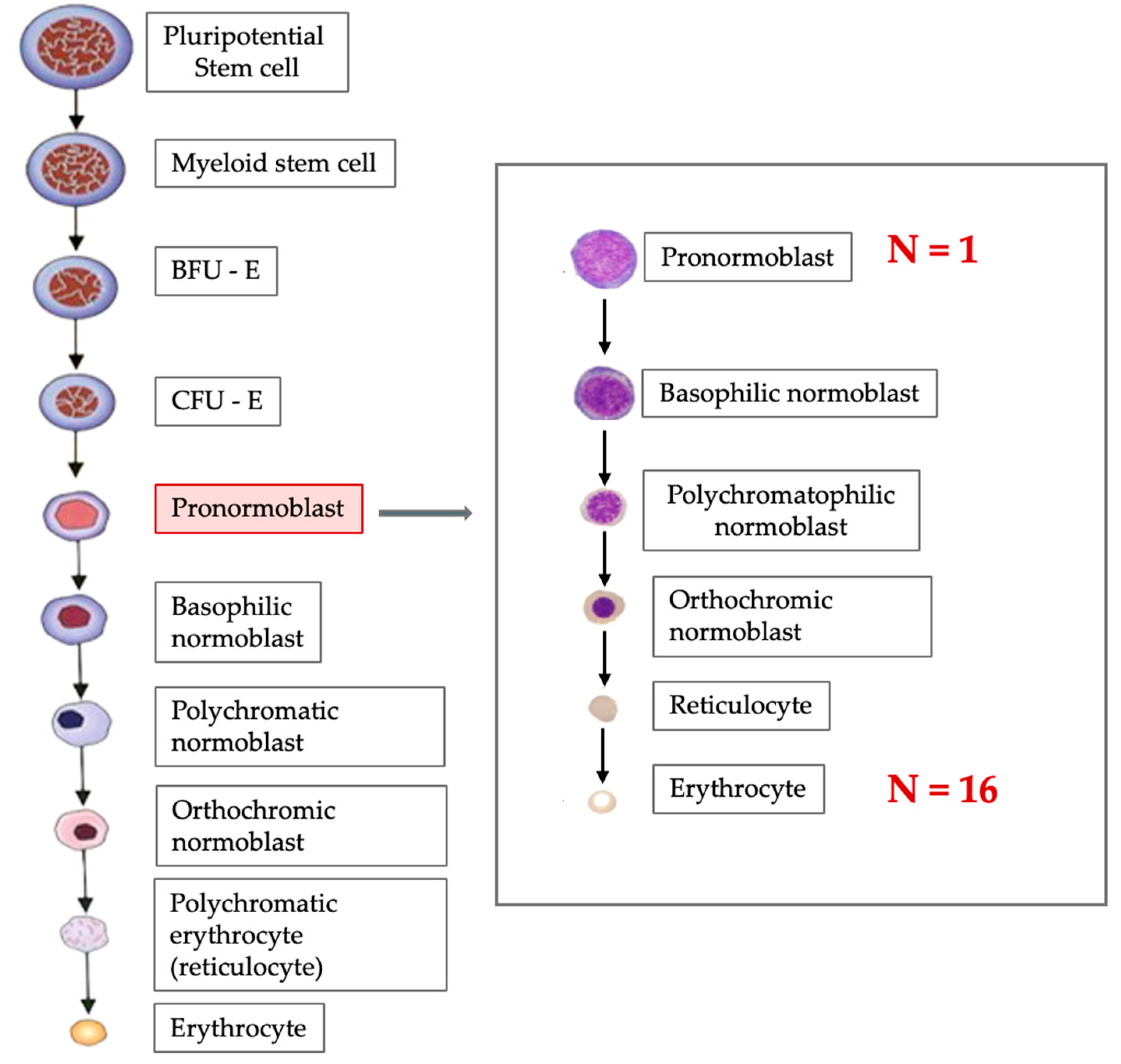
The well-established model indicates that erythropoiesis starts with hematopoietic stem cells in the bone marrow differentiating into myeloid progenitors. They then develop into erythroid progenitors, specifically burst-forming unit-erythroid (BFU-E) and colony-forming unit-erythroid (CFU-E). CFU-E cells further mature through several stages—proerythroblast, basophilic, polychromatic, and orthochromatic erythroblast—where they produce hemoglobin and prepare for enucleation. During this process, the orthochromatic erythroblast ejects its nucleus, forming a reticulocyte. This reticulocyte then matures into an erythrocyte in approximately one week [9].
Under normal physiological conditions, bone marrow can produce approximately 200 billion red blood cells per day, with each pronormoblast developing into 16 erythrocytes [10], as illustrated in Figure 2. This process can be divided into three distinct phases, cellular differentiation, proliferation, and maturation—each regulated by specific mediators and regulatory factors [11,12,13].
During the maturation process, the cell decreases in size and volume, followed by the disappearance of the nucleus and a proportional increase in cytoplasmic volume and pigmentation (shifting from bluish to reddish), thereby becoming more acidophilic [6].
Iron is a fundamental substrate for erythropoiesis. It exists in inorganic form (as ferric [Fe3+] and ferrous [Fe2+] ions) and in organic form (bound to hemoglobin and myoglobin, and found in sources such as meat, eggs, and certain vegetables) [14,15].
Most dietary iron exists as [Fe3+], which needs to be reduced to [Fe2+] before it can be absorbed through the brush border membrane via divalent metal ion transporter 1 (DMT1). This reduction is probably facilitated by the brush border duodenal cytochrome b reductase (DcytB), although other reductases might also play a role. Organic iron is absorbed through the intestinal epithelium by the heme carrier protein 1 (HCP1), where it is degraded by heme oxygenase [14,15].
Hephaestin oxidase converts iron into its ferric form [Fe3+], and transferrin binds two Fe3+ ions, transporting them to transferrin receptors in hepatocytes, where the iron is stored in ferritin subunits and can be mobilized as needed [16,17].
Folic acid is also crucial for erythropoiesis. It is found in green leafy vegetables, other vegetables, and fruits. In the enterocyte, it is converted into dihydrofolate (DHF), then into tetrahydrofolate (THF), and finally into methylfolate—the most bioactive form—which is then distributed to body tissues [18,19].
One of the main regulators of erythropoiesis is oxygen. When the partial pressure of O2 (pO2) decreases, adult kidneys, particularly the interstitial peritubular cells (IPCs), activate EPO (erythropoietin) production. EPO is secreted into the plasma, stimulating bone marrow erythroid progenitors through the EPO receptor to mature into red blood cells (RBCs) [7,20,21]. The interaction of EPO with the EPO receptor in proerythroid cells triggers proliferation, enhances cell survival, and promotes erythroid-specific differentiation.
Folic acid, vitamin B12, and vitamin B3 are involved in nucleic acid synthesis (during the proliferation phase) [6,8,13,18]. In the maturation phase, substrates such as iron, copper, and vitamin B6 are necessary, along with adequate hormonal levels, including androgens, corticosteroids, growth hormone, and thyroid hormones [6,7,8,13,16].
Erythropoiesis takes approximately 8 days, and erythrocytes have an expected lifespan of 120 days [6].

3. Anemias—Identification and Classification

Anemia is a common hematological condition in adults in clinical practice. While anemia is a global health concern, this review focuses specifically on the epidemiology, diagnostic approaches, and treatment recommendations relevant to the Portuguese population. We will examine Portuguese-specific data and guidelines to provide a contextually relevant perspective on the management of anemia in this region.
The World Health Organization (WHO) estimates that approximately 15% of the Portuguese population is affected by anemia, while the study of prevalence of anemia and iron deficiency in Portugal (EMPIRE) reported a prevalence of 20% [22]. Among individuals aged 80 years and older, the prevalence rate rises to approximately 31.4% [22,23].
Anemia is defined by the WHO as a reduction in hemoglobin concentration in peripheral blood below the normal values for age and sex, less than 12 g/dL in women and 13 g/dL in men [1]. In the elderly population, there is a higher prevalence of predisposing conditions for anemia [24,25], as well as an increased frequency of associated complications, as shown in Table 1.
In the elderly population, anemia represents a significant factor for morbidity, mortality, and frailty. However, in many cases, it is underestimated and undertreated [24].
The main causes of anemia are iron deficiency (29%), chronic diseases (27.5%), hemorrhagic losses (17.5%), and hemolysis (17.5%), while other causes account for about 9% of cases [24,25].
Table 2 presents the most common causes of anemia in older adults and their respective prevalence rates.
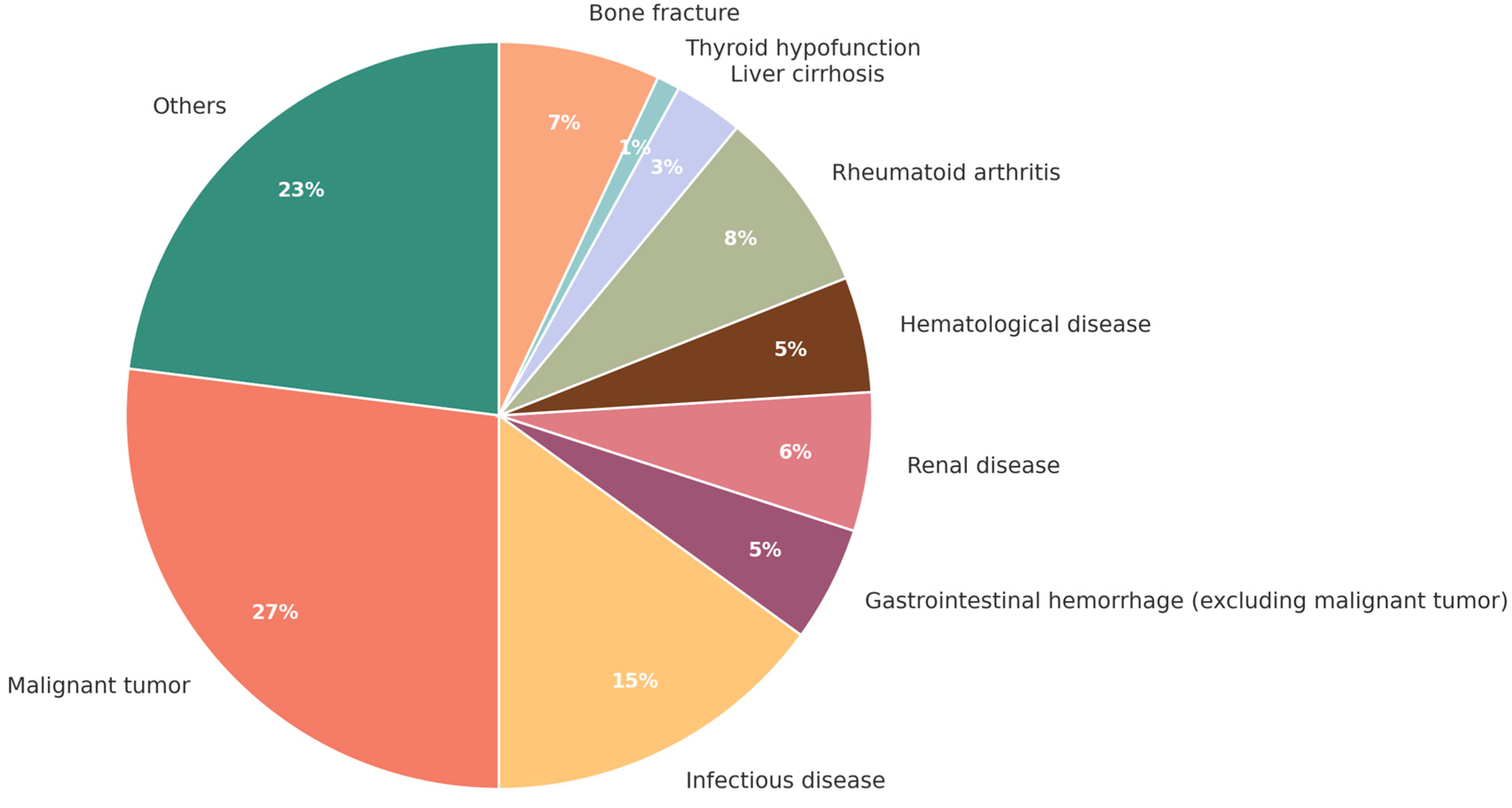
Another study conducted in Japan identified slightly different causes of anemia [24], as shown in Figure 3.
For an accurate etiological diagnosis of anemia, classification is essential [1]. One of the most relevant classification systems is based on reticulocyte count, which allows differentiation between non-regenerative and regenerative anemia [2,27], as illustrated in Table 3.
Another important classification is the morphological one [3,4,28], which is based on the analysis of mean corpuscular volume (MCV), as illustrated in Table 4.
Laboratory diagnosis includes a complete blood count (including RDW), a peripheral blood smear, iron metabolism evaluation, measurements of vitamin B12 and folic acid, erythropoietin (EPO), thyroid-stimulating hormone (TSH), C-reactive protein (CRP), and creatinine. In some cases, a bone marrow aspirate and biopsy may berequired—for example, in situations of pancytopenia, monoclonal gammopathy, suspected myelodysplastic syndrome, or unexplained progressive anemia [2,4].
Table 5 offers a systematic overview of all the steps needed for accurately diagnosing anemia, from collecting clinical history to laboratory assessment [2,3,4,5].
Therefore, the diagnostic approach to anemia involves three key steps: (1) reticulocyte count, which differentiates between a central production problem and increased peripheral destruction; (2) morphological classification of the anemia; and (3) identification of the underlying etiology.
Practically, the reticulocyte count serves as a critical decision point guiding subsequent diagnostic investigations, as depicted in Table 6.
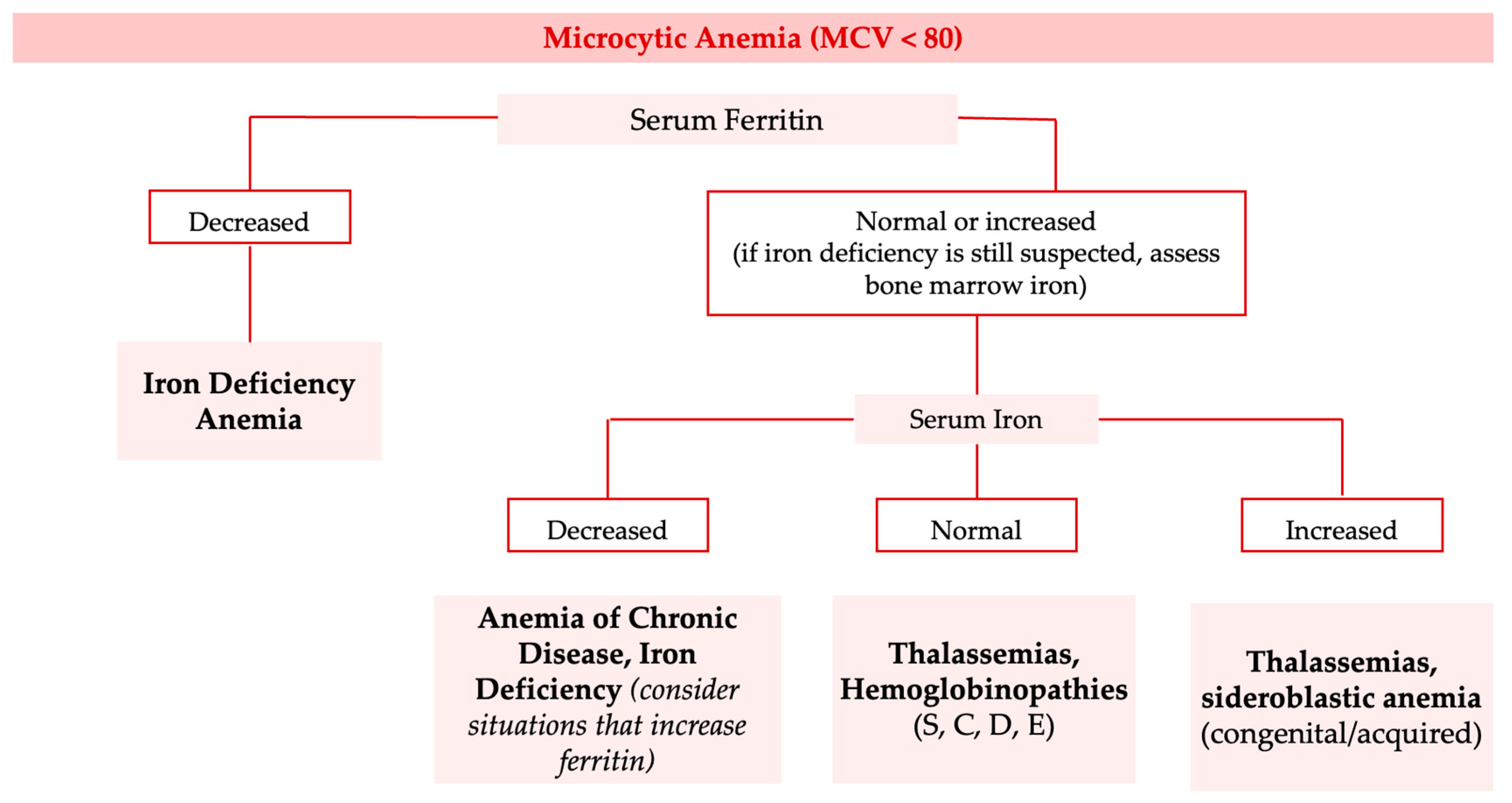
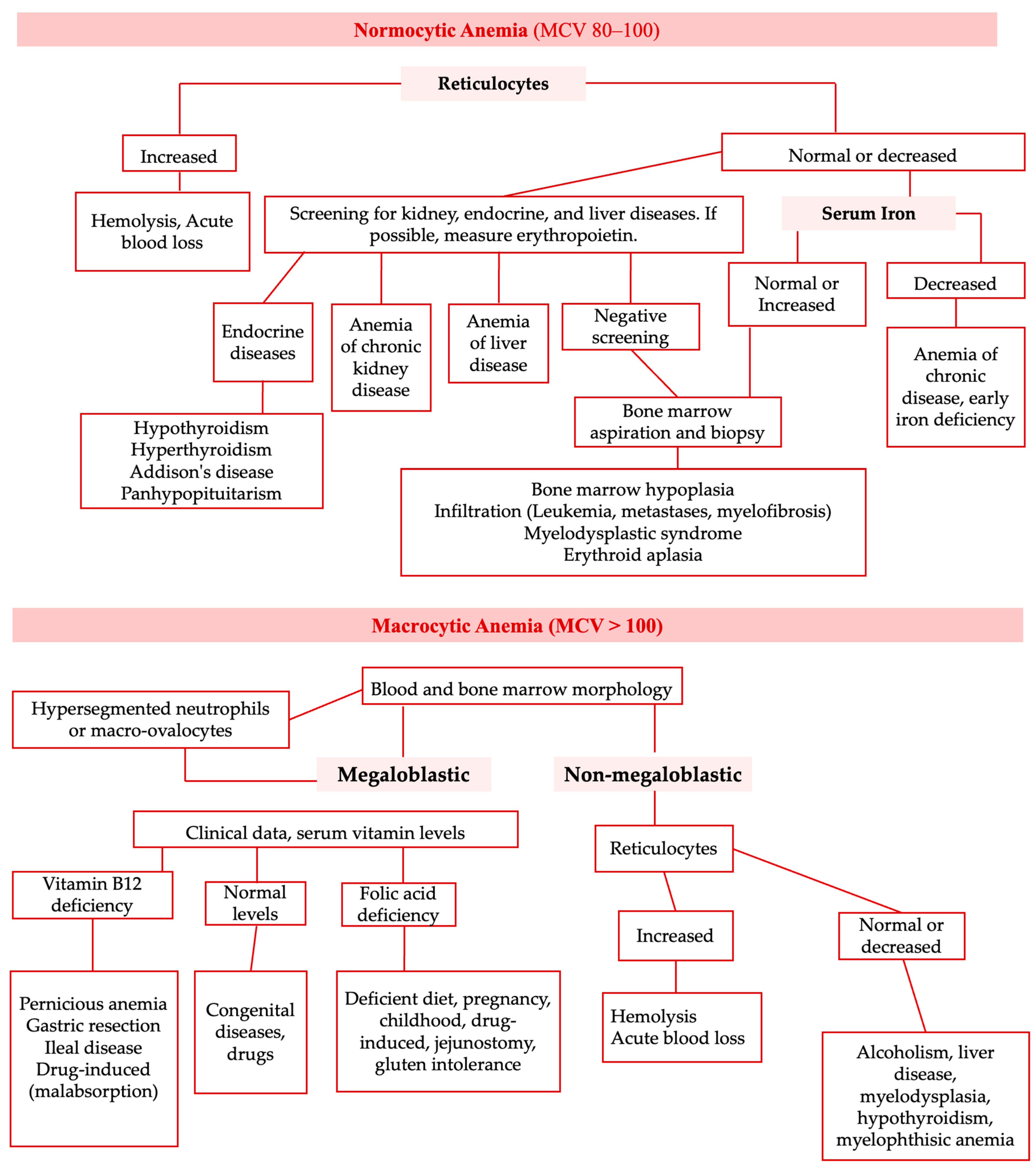
After the morphological classification of anemia, further important steps are needed to identify the underlying cause(s) and to guide a more precise treatment plan, as shown in Figure 4.

4. Prevention of Anemia

The primary risk groups for iron deficiency and iron deficiency anemia include children and adolescents (due to increased needs related to growth), premature infants, menstruating women of reproductive age, pregnant women, older adults, frequent blood donors, and those on strict vegetarian or restrictive diets. People with chronic inflammatory diseases, inflammatory bowel disease, malabsorption syndromes, or chronic hemorrhagic diathesis are also at higher risk for iron deficiency [29].
Pregnant women, older adults, and individuals with inflammatory bowel disease are also at increased risk of mixed nutritional anemias, with concurrent folic acid deficiency [29]. The World Health Organization (WHO) recommends oral iron and folic acid supplementation for the prevention and treatment of iron deficiency anemia in women of reproductive age in countries with high prevalence of this condition [30,31].
A Cochrane review on daily oral iron supplementation during pregnancy—initially published in 2006 and subject to successive updates, the most recent in 2024 [32]—consistently found that such supplementation is associated with a reduction in maternal anemia and iron deficiency at term. Based on 40 clinical trials, the review also concluded that daily oral iron supplementation during pregnancy probably reduces the risk of low birth weight. The included studies used iron supplementation with ferrous sulfate, either alone or combined with folic acid. The authors recommended this strategy as an effective preventive measure for reducing the risk of anemia and iron deficiency during pregnancy [32].
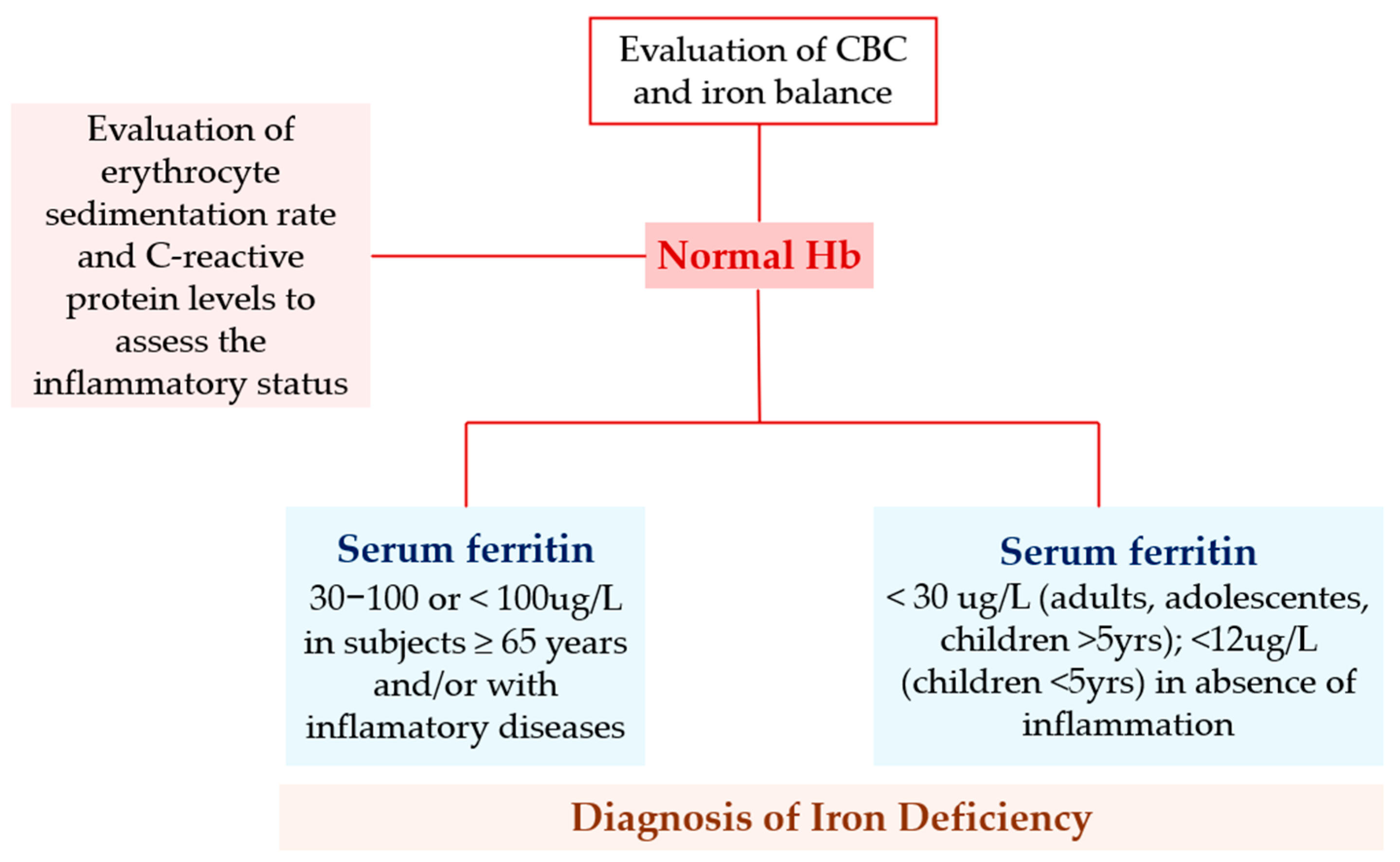
The WHO recommends a daily oral intake of at least 30–60 mg of elemental iron and 400 µg of folic acid during pregnancy to prevent maternal anemia, postpartum sepsis, low birth weight, and preterm delivery, with ferrous salts such as ferrous sulfate being the first-line treatment for this purpose [33]. Although it has some limitations—especially when inflammation is present—ferritin remains the most specific biomarker for evaluating iron stores and aiding in the diagnosis of iron deficiency, even without anemia [29]. Figure 5, adapted from the latest recommendations of the European Hematology Association (EHA) on preventing and treating iron deficiency and iron deficiency anemia [29], summarizes the diagnostic criteria for iron deficiency without anemia, establishing serum ferritin cutoff values to support diagnosis across different age groups and clinical settings.
In cases where iron deficiency is identified in the absence of anemia, the WHO recommends treatment with daily oral iron and folic acid for a duration of 3 months [31], or alternatively, using lower doses such as 40 mg of elemental iron per day or less frequent dosing regimens such as 120 mg of elemental iron every other day [30,34], as a strategy to prevent the development of anemia. The use of lower iron doses or less frequent dosing schedules has been associated with improved tolerability, while remaining effective in preventing anemia in iron-deficient populations [34,35,36].
The recommendation to treat iron deficiency even in the absence of anemia is broadly accepted for the general population and particularly emphasized during pregnancy and in preoperative settings [37].
Potential causes of iron deficiency should always be investigated and addressed, including heavy menstrual bleeding in women of reproductive age, occult gastrointestinal bleeding, or dietary factors (such as low intake of bioavailable iron-rich foods, and excessive consumption of dairy products, tea, or iced tea) [38].
In addition to preventing anemia, these interventions are also important for minimizing other adverse effects of iron deficiency, such as worsening cognitive decline in older adults [29] or delays in neurodevelopment during childhood [39].

5. Treatment of Anemia

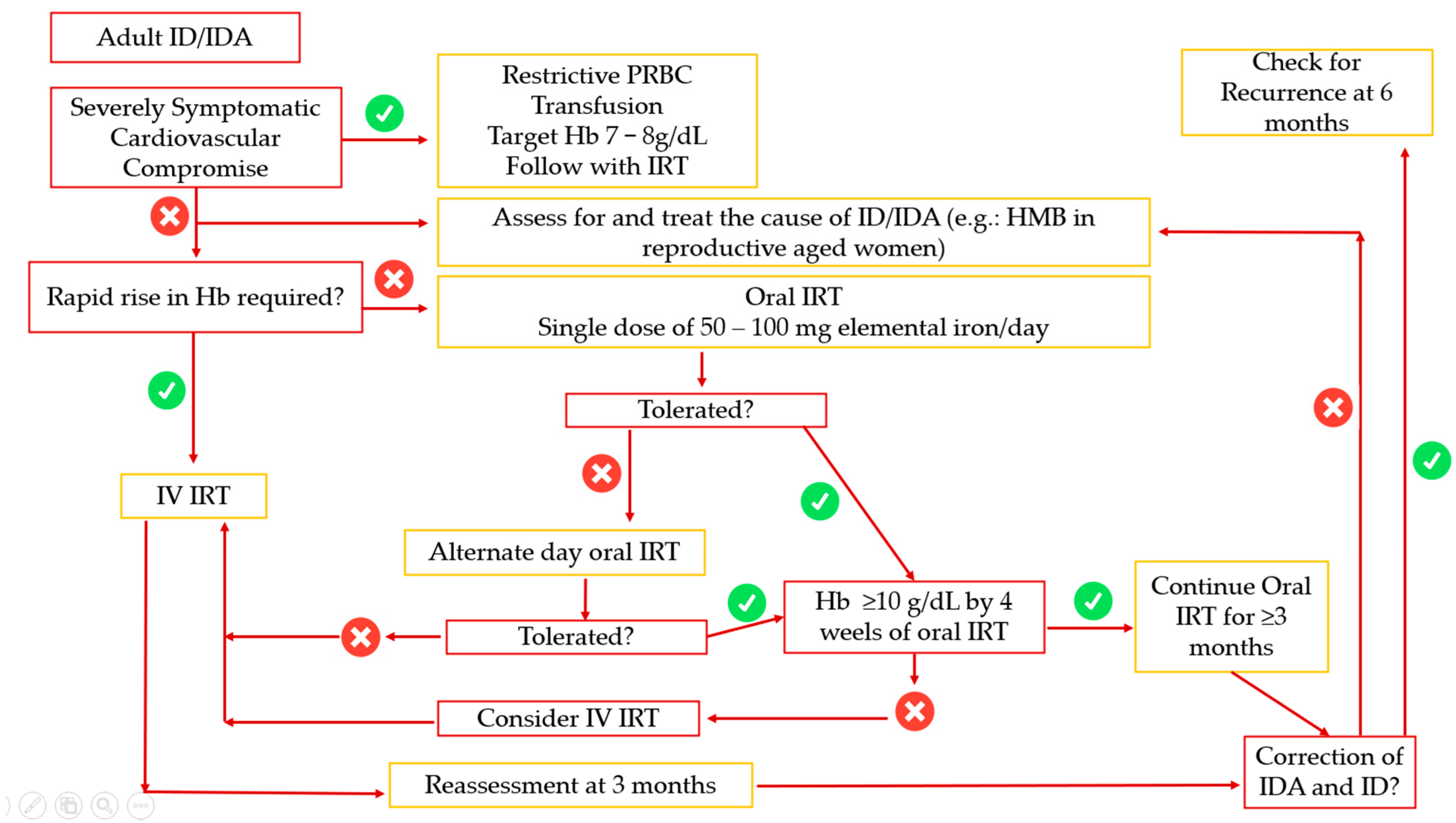
The updated guidelines from the European Hematology Association (EHA) for the treatment and prevention of iron deficiency and iron deficiency anemia, published in July 2024, recommend oral iron salts—particularly ferrous sulfate—as first-line treatment in the vast majority of cases of uncomplicated iron deficiency anemia in adults [29].
Although elemental iron doses of 100–200 mg/day were traditionally recommended, the authors now indicate that lower daily doses or alternate-day dosing regimens of elemental iron are associated with similar or even improved absorption, better gastrointestinal tolerability, and greater adherence, with fewer adverse effects [29].
Figure 6, adapted from these same guidelines, outlines the therapeutic approach to iron deficiency anemia.
Although some studies have suggested a marginal increase in absorption with alternate-day dosing regimens, a recent randomized controlled trial found no significant differences in efficacy or tolerability between daily and alternate-day administration of ferrous sulfate [40].
Oral iron treatment should be continued until anemia is corrected and then maintained for at least an additional 3 months to replenish iron stores and minimize the risk of recurrence [29,41]. The management of iron deficiency anemia should always include investigation and, whenever possible, treatment of the underlying causes [41]. Intravenous iron formulations should be reserved for cases requiring a rapid increase in hemoglobin levels or when oral iron is not tolerated or adequately absorbed [29,42]. Intravenous iron is thus considered a complementary strategy to oral therapy—first-line in most cases—with distinct indications, and not a replacement.
Oral ferrous sulfate is the most widely used treatment option globally and may be considered the gold standard first-line therapy for iron deficiency anemia [42]. This is reflected in several international guidelines, including those of the EHA [29], the British Society of Gastroenterology [34], and the American Gastroenterological Association [43]. The main adverse effects of this formulation are gastrointestinal and are comparable to those of other oral iron formulations. When administered as a single daily dose or on alternate days, ferrous sulfate shows a lower incidence of adverse effects that promotes treatment maintenance [42]. Some authors also note its advantages over other formulations due to its lower cost, extensive clinical experience, and the availability of robust data supporting the use of lower doses or intermittent dosing regimens [42,43].
Proper treatment of iron deficiency anemia has been associated with increased hemoglobin levels, reduction in symptoms and comorbidities attributable to anemia, and decreased need for blood component transfusions [44].
Iron replacement therapy in iron deficiency anemia is associated with an erythropoietic response, characterized by an increase in reticulocyte count that can be observed within the first days or weeks after initiating supplementation [45].
This stimulation of erythropoiesis requires sufficient folic acid availability [46]. Since it is safe to supplement orally—even at daily doses above 5 mg [47]—folic acid can be used alongside iron salts without concern. This combination not only promotes erythropoiesis but also improves treatment response in cases of mixed deficiency anemia [29]. In Portugal, the following fixed-dose formulations are authorized:
  • A fixed combination of folic acid 0.35 mg + ferrous sulfate 247.25 mg, approved for the prophylaxis and treatment of iron and folic acid deficiencies during pregnancy, postpartum, and prolonged lactation, or in cases where iron and folic acid were not regularly taken during lactation;
  • A fixed combination of folic acid 0.35 mg + ferrous sulfate 325 mg, approved for the prophylaxis and treatment of anemia during pregnancy, for treating iron deficiency, and for preventing concurrent folic acid deficiency in adults. The safety profile of folic acid is well established, with no significant toxicity reported. Concerns raised in the past regarding potential harmful (e.g., carcinogenic) effects of high folic acid concentrations after supplementation have not been confirmed. A large meta-analysis involving 49,621 participants found no such risk [48]. On the contrary, prenatal folic acid supplementation has shown protective effects not only against neural tube defects but also against various pediatric cancers, reducing the incidence of leukemia, brain tumors, and neuroblastoma [49].
Doses ranging from 400 µg to 5 mg of folic acid per day are considered non-toxic and can be safely used during pregnancy. For pregnant women at higher risk of folic acid deficiency, neural tube defects, or poor treatment adherence, higher doses such as 5 mg/day are recommended [50].

6. Limitations and Future Perspectives

While this review provides a comprehensive overview of the use of folic acid 1 mg + iron (ferrous sulfate) 90 mg in the treatment of anemia, it is important to acknowledge several limitations in the existing evidence base.
Firstly, there is a relative paucity of high-quality, randomized controlled trials specifically evaluating the effectiveness of this fixed-dose combination, particularly in certain subpopulations such as the elderly, pregnant women, and individuals with chronic inflammatory conditions. Many of the recommendations are based on observational studies, expert opinion, and extrapolations from studies of iron or folic acid supplementation alone. Further research is needed to directly compare the efficacy and safety of this combination to alternative treatment strategies, such as higher doses of iron or separate supplementation with iron and folic acid.
Secondly, although this review focuses on the Portuguese context, there is a lack of specific data on the prevalence and management of anemia within this population. Further studies are needed to assess the effectiveness of current treatment guidelines and to identify any unique challenges or opportunities for improving anemia care in Portugal.
Thirdly, the studies included in this review were heterogeneous in terms of patient characteristics, diagnostic criteria, interventions, and outcome measures. This heterogeneity makes it difficult to draw firm conclusions about the overall effectiveness of the folic acid 1 mg + iron (ferrous sulfate) 90 mg combination and limits the generalizability of the findings.
Finally, there is a lack of long-term data on the safety and efficacy of this combination. Future studies should assess the long-term effects on patient-centered outcomes such as quality of life, functional status, and cognitive function, as well as the potential for adverse effects with prolonged use.
Future research should focus on the following: conducting well-designed, randomized controlled trials to directly compare the folic acid 1 mg + iron (ferrous sulfate) 90 mg combination to alternative treatment strategies in diverse patient populations; performing studies to assess the cost-effectiveness of this combination compared to other treatments; investigating the optimal duration of treatment with this combination; identifying biomarkers that can predict response to treatment; developing strategies to improve adherence to treatment; and conducting research to better understand the underlying mechanisms by which iron and folic acid affect erythropoiesis.
Addressing these limitations through rigorous scientific investigation will help to refine our understanding of the role of folic acid and iron supplementation in the treatment of anemia and to optimize clinical practice guidelines for the benefit of patients.

7. Suggestions and Recommendations Based on Clinical Studies/Experiences

While the evidence reviewed supports the use of a fixed-dose combination of folic acid 1 mg + iron (ferrous sulfate) 90 mg for the treatment of iron deficiency anemia, further studies are needed to specifically evaluate the clinical benefits and long-term outcomes associated with this particular dosage regimen, especially in diverse patient populations and in comparison to other available formulations.
The authors have over 10 years of extensive experience in both hospital-based settings (Clinical Hematology) and community-based care (Primary Health Care and Palliative Care), using a fixed oral daily dose of Folic Acid 1 mg + Iron (Ferrous Sulfate) 90 mg for the treatment of iron deficiency anemia, mixed nutritional anemia (iron and folic acid deficiency), and iron deficiency without anemia in adults.
This clinical experience demonstrates good therapeutic efficacy and satisfactory tolerability, allowing for strong adherence to treatment. The most commonly observed adverse effects are gastrointestinal (constipation, diarrhea, and, less frequently, nausea), which are generally manageable through adjustments to the administration schedule—such as taking the supplement with meals rather than on an empty stomach (which improves tolerability), provided the food contains vitamin C (e.g., citrus fruits), in order not to significantly impair iron absorption in the gastrointestinal tract.
The groups with the highest prevalence of iron deficiency anemia in the Portuguese population are women of reproductive age [22] and the elderly [23]. In the authors’ clinical practice, switching from formulations containing only oral iron salts to fixed combinations of oral iron with folic acid (with or without concomitant vitamin B12 supplementation, depending on the clinical context) frequently leads to improved anemia response.
Additionally, the authors’ extensive clinical experience in the treatment of chronic hemolytic anemias supports the safety of oral folic acid supplementation, both at a dose of 1 mg and at doses up to 5 mg per day, with no observed toxicity or adverse effects attributable to folic acid—even in patients who have been taking daily supplementation for several years or even throughout their entire lives.
Therefore, the use of 1 mg folic acid, being free from toxicity or increased risk, is considered by the authors to be a valuable therapeutic option. It enables correction of folate deficiencies—an essential factor in hematopoiesis—optimizing treatment response and ensuring sufficient folic acid levels even in cases of incomplete adherence or missed doses. This is particularly important during pregnancy, where adequate supplementation helps minimize the risk of neural tube defects. Nevertheless, specific evaluation and recommendations appropriate for the pediatric age group are required.

8. Recommendation

Based on the scientific evidence presented throughout this document, the fact that ferrous sulfate is considered the gold standard in the treatment of iron deficiency anemia, its inclusion as a first-line therapy for both iron deficiency anemia and non-anemic iron deficiency in major and recent international guidelines, as well as the authors’ many years of clinical experience in the treatment and prevention of iron deficiency anemia, non-anemic iron deficiency, and mixed nutritional anemias, the authors consider there to be robust evidence supporting the recommendation of Folic Acid 1 mg + Iron (Ferrous Sulfate) 90 mg as a treatment option.
This combination should be indicated not only for the prevention and treatment of iron and folic acid deficiency during pregnancy, lactation, and the postpartum period but also for the prophylaxis and treatment of anemia during pregnancy and in adults in general.

Author Contributions

Conceptualization—J.G. and H.R.; Methodology—J.G. and H.R.; Software—H.R.; Validation—J.B.S., C.V.J. and H.R.; Formal analysis—J.G. and H.R.; Investigation—J.G. and H.R.; Resources—HR; Data Curation—J.G., J.B.S. and H.R.; Writing—Original Draft—J.G. and H.R.; Writing—Review and Editing—J.B.S. and C.V.J.; Visualization—C.V.J. and H.R.; Supervision—H.R.; Project administration—H.R.; Funding acquisition—H.R. All authors have read and agreed to the published version of the manuscript.

Funding

This research received no external funding.

Data Availability Statement

Data can be obtained upon reasonable request.

Conflicts of Interest

J.G. and H.R. declare that they received a grant to conduct a review of current evidence of Folic Acid 1 mg + Iron (Ferrous Sulfate) 90 mg by Bial.

References

  1. World Health Organization. Guideline on Haemoglobin Cutoffs to Define Anaemia in Individuals and Populations; WHO: Geneva, Switzerland, 2024. [Google Scholar]
  2. Garcia-Casal, M.N.; Dary, O.; Jefferds, M.E.; Pasricha, S. Diagnosing anemia: Challenges at the public health level. Ann. N. Y. Acad. Sci. 2023, 1524, 37–50. [Google Scholar] [CrossRef] [PubMed]
  3. Cappellini, M.D.; Motta, I. Anemia in Clinical Practice—Does Hemoglobin Change with Aging? Semin. Hematol. 2015, 52, 261–269. [Google Scholar] [CrossRef]
  4. Kundrapu, S.; Noguez, J. Laboratory Assessment of Anemia. Adv. Clin. Chem. 2018, 83, 197–225. [Google Scholar] [CrossRef]
  5. De Franceschi, L.; Iolascon, A.; Taher, A.; Cappellini, M.D. Clinical management of iron deficiency anemia in adults: Systemic review on advances in diagnosis and treatment. Eur. J. Intern. Med. 2017, 42, 16–23. [Google Scholar] [CrossRef]
  6. Hillman, R.S.; Finch, C.A. Erythropoiesis. N. Engl. J. Med. 1971, 285, 99–101. [Google Scholar] [CrossRef]
  7. Chung, E.Y.; Palmer, S.C.; Saglimbene, V.M.; Craig, J.C.; Tonelli, M.; Strippoli, G.F. Erythropoiesis-stimulating agents for anaemia in adults with chronic kidney disease: A network meta-analysis. Cochrane Database Syst. Rev. 2023, 2023, CD010590. [Google Scholar] [CrossRef]
  8. Zivot, A.; Lipton, J.M.; Narla, A.; Blanc, L. Erythropoiesis: Insights into pathophysiology and treatments in 2017. Mol. Med. 2018, 24, 11. [Google Scholar] [CrossRef]
  9. Cha, H.J. Erythropoiesis: Insights from a genomic perspective. Exp. Mol. Med. 2024, 56, 2099–2104. [Google Scholar] [CrossRef] [PubMed]
  10. Andrès, E.; Serraj, K.; Federici, L.; Vogel, T.; Kaltenbach, G. Anemia in elderly patients: New insight into an old disorder. Geriatr. Gerontol. Int. 2012, 13, 519–527. [Google Scholar] [CrossRef]
  11. Jagannathan-Bogdan, M.; Zon, L.I. Hematopoiesis. Development 2013, 140, 2463–2467. [Google Scholar] [CrossRef] [PubMed]
  12. Orkin, S.H.; Zon, L.I. Hematopoiesis: An Evolving Paradigm for Stem Cell Biology. Cell 2008, 132, 631–644. [Google Scholar] [CrossRef]
  13. Orkin, S.H. Hematopoiesis: How does it happen? Curr. Opin. Cell Biol. 1995, 7, 870–877. [Google Scholar] [CrossRef]
  14. Anderson, G.J.; Frazer, D.M.; McLaren, G.D. Iron absorption and metabolism. Curr. Opin. Gastroenterol. 2009, 25, 129–135. [Google Scholar] [CrossRef]
  15. Piskin, E.; Cianciosi, D.; Gulec, S.; Tomas, M.; Capanoglu, E. Iron Absorption: Factors, Limitations, and Improvement Methods. ACS Omega 2022, 7, 20441–20456. [Google Scholar] [CrossRef] [PubMed]
  16. Conrad, M.E.; Umbreit, J.N. Iron absorption and transport: An update. Am. J. Hematol. 2000, 64, 287–298. [Google Scholar] [CrossRef]
  17. von Siebenthal, H.K.; Moretti, D.; Zimmermann, M.B.; Stoffel, N.U. Effect of dietary factors and time of day on iron absorption from oral iron supplements in iron deficient women. Am. J. Hematol. 2023, 98, 1356–1363. [Google Scholar] [CrossRef]
  18. Shulpekova, Y.; Nechaev, V.; Kardasheva, S.; Sedova, A.; Kurbatova, A.; Bueverova, E.; Kopylov, A.; Malsagova, K.; Dlamini, J.C.; Ivashkin, V. The Concept of Folic Acid in Health and Disease. Molecules 2021, 26, 3731. [Google Scholar] [CrossRef] [PubMed]
  19. Donnelly, J.G. Folic Acid. Crit. Rev. Clin. Lab. Sci. 2001, 38, 183–223. [Google Scholar] [CrossRef]
  20. Tsiftsoglou, A.S. Erythropoietin (EPO) as a Key Regulator of Erythropoiesis, Bone Remodeling and Endothelial Transdifferentiation of Multipotent Mesenchymal Stem Cells (MSCs): Implications in Regenerative Medicine. Cells 2021, 10, 2140. [Google Scholar] [CrossRef]
  21. Jelkmann, W. Physiology and Pharmacology of Erythropoietin. Transfus. Med. Hemother. 2013, 40, 302–309. [Google Scholar] [CrossRef] [PubMed]
  22. Fonseca, C.; Marques, F.; Nunes, A.R.; Belo, A.; Brilhante, D.; Cortez, J. Prevalence of anaemia and iron deficiency in Portugal: The EMPIRE study. Intern. Med. J. 2016, 46, 470–478. [Google Scholar] [CrossRef]
  23. Robalo Nunes, A.; Fonseca, C.; Marques, F.; Belo, A.; Brilhante, D.; Cortez, J. Prevalence of anemia and iron deficiency in older Portuguese adults: An EMPIRE substudy. Geriatr. Gerontol. Int. 2017, 17, 1814–1822. [Google Scholar] [CrossRef]
  24. Katsumi, A.; Abe, A.; Tamura, S.; Matsushita, T. Anemia in older adults as a geriatric syndrome: A review. Geriatr. Gerontol. Int. 2021, 21, 549–554. [Google Scholar] [CrossRef]
  25. Stauder, R.; Thein, S.L. Anemia in the elderly: Clinical implications and new therapeutic concepts. Haematologica 2014, 99, 1127–1130. [Google Scholar] [CrossRef]
  26. Masatsugu, O.H.T.A. Management of Anemia in the Elderly. JMAJ 2009, 52, 219–223. [Google Scholar] [CrossRef]
  27. Vohra, R.; Hussain, A.; Dudyala, A.K.; Pahareeya, J.; Khan, W.; Mumtaz, W. Multi-class classification algorithms for the diagnosis of anemia in an outpatient clinical setting. PLoS ONE 2022, 17, e0269685. [Google Scholar] [CrossRef]
  28. Clinton, O.; Micheal, K.; Namyalo, A.K.; Mary, M.; Mike, M.; Muwanguzi, E.; Okongo, B.; Wagubi, R. Anaemia, Morphological Classification and Its Associated Risk Factors Among Lactating Mothers at Mbarara City Council Health Centre IV, Southwestern Uganda. J. Blood Med. 2022, 13, 473–481. [Google Scholar] [CrossRef]
  29. Iolascon, A.; Andolfo, I.; Russo, R.; Sanchez, M.; Busti, F.; Swinkels, D.; Martinez, P.A.; Bou-Fakhredin, R.; Muckenthaler, M.U.; Unal, S.; et al. Recommendations for diagnosis, treatment, and prevention of iron deficiency and iron deficiency anemia. HemaSphere 2024, 8, e108. [Google Scholar] [CrossRef] [PubMed]
  30. World Health Organization. Guideline: Intermittent Iron and Folic Acid Supplementation in Menstruating Women; WHO: Geneva, Switzerland, 2011. [Google Scholar]
  31. Roche, M.L.; Samson, K.L.; Green, T.J.; Karakochuk, C.D.; Martinez, H. Perspective: Weekly Iron and Folic Acid Supplementation (WIFAS): A Critical Review and Rationale for Inclusion in the Essential Medicines List to Accelerate Anemia and Neural Tube Defects Reduction. Adv. Nutr. Int. Rev. J. 2021, 12, 334–342. [Google Scholar] [CrossRef]
  32. Finkelstein, J.L.; Cuthbert, A.; Weeks, J.; Venkatramanan, S.; Larvie, D.Y.; De-Regil, L.M.; Garcia-Casal, M.N.; Cochrane Central Editorial Service. Daily oral iron supplementation during pregnancy. Cochrane Database Syst. Rev. 2024, 2024, CD004736. [Google Scholar] [CrossRef] [PubMed]
  33. World Health Organization. WHO Recommendations on Antenatal Care; WHO: Geneva, Switzerland, 2016. [Google Scholar]
  34. Stoffel, N.U.; von Siebenthal, H.K.; Moretti, D.; Zimmermann, M.B. Oral iron supplementation in iron-deficient women: How much and how often? Mol. Asp. Med. 2020, 75, 100865. [Google Scholar] [CrossRef]
  35. Simic, S.; Karczewski, M.; Klapdor, S.; Nowak, A.; Schubert, M.; Moretti, D.; Swinkels, D.W.; Beuschlein, F.; Saleh, L.; Suter, P.; et al. Krayenbuehl PA. Effectiveness of low-dose iron treatment in non-anaemic iron-deficient women: A prospective open-label single-arm trial. Swiss Med. Wkly. 2023, 153, 40079. [Google Scholar] [CrossRef] [PubMed]
  36. Rimon, E.; Kagansky, N.; Kagansky, M.; Mechnick, L.; Mashiah, T.; Namir, M.; Levy, S. Are we giving too much iron? Low-dose iron therapy is effective in octogenarians. Am. J. Med. 2005, 118, 1142–1147. [Google Scholar] [CrossRef]
  37. Al-Naseem, A.; Sallam, A.; Choudhury, S.; Thachil, J. Iron deficiency without anaemia: A diagnosis that matters. Clin. Med. 2021, 21, 107–113. [Google Scholar] [CrossRef] [PubMed]
  38. Santoyo-Sánchez, A.; Aponte-Castillo, J.A.; Parra-Peña, R.I.; Ramos-Peñafiel, C.O. Dietary recommendations in deficiency anaemia. Rev. Med. Hosp. Gen. Mex. 2015, 78, 144–150. [Google Scholar]
  39. Lozoff, B. Iron Deficiency and Child Development. Food Nutr. Bull. 2007, 28 (Suppl. 4), S560–S571. [Google Scholar] [CrossRef]
  40. Pasupathy, E.; Kandasamy, R.; Thomas, K.; Basheer, A. Alternate day vs daily oral iron: A randomized trial. Sci. Rep. 2023, 13, 1818. [Google Scholar]
  41. Snook, J.; Bhala, N.; Beales, I.L.P.; Cannings, D.; Kightley, C.; Logan, R.P.; Pritchard, D.M.; Sidhu, R.; Surgenor, S.; Thomas, W.; et al. British Society of Gastroenterology guidelines for the management of iron deficiency anaemia in adults. Gut 2021, 70, 2030–2051. [Google Scholar] [CrossRef]
  42. Pantopoulos, K. Oral iron supplementation: New formulations, old questions. Haematologica 2024, 109, 2790–2801. [Google Scholar] [CrossRef]
  43. DeLoughery, T.G.; Jackson, C.S.; Ko, C.W.; Rockey, D.C. AGA Update on Iron Deficiency Anemia. Clin. Gastroenterol. Hepatol. 2024, 22, 1575–1583. [Google Scholar] [CrossRef]
  44. Clevenger, B.; Gurusamy, K.; Klein, A.A.; Murphy, G.J.; Anker, S.D.; Richards, T. Systematic review and meta-analysis of iron therapy in anaemic adults without chronic kidney disease: Updated and abridged Cochrane review. Eur. J. Heart Fail. 2016, 18, 774–785. [Google Scholar] [CrossRef] [PubMed]
  45. Vollset, S.E.; Clarke, R.; Lewington, S.; Ebbing, M.; Halsey, J.; Lonn, E.; Armitage, J.; Manson, J.E.; Hankey, G.J.; Spence, J.D.; et al. Effects of folic acid supplementation on overall and site-specific cancer incidence during the randomised trials: Meta-analyses of data on 50,000 individuals. Lancet 2013, 381, 1029–1036. [Google Scholar] [CrossRef] [PubMed]
  46. Koury, M.J.; Ponka, P. The Roles of Folate, Vitamin B12, and Iron. Annu. Rev. Nutr. 2004, 24, 105–131. [Google Scholar] [CrossRef] [PubMed]
  47. E Butterworth, C.; Tamura, T. Folic acid safety and toxicity: A brief review. Am. J. Clin. Nutr. 1989, 50, 353–358. [Google Scholar] [CrossRef]
  48. Parodi, E.; Giraudo, M.T.; Davitto, M.; Ansaldi, G.; Mondino, A.; Garbarini, L.; Franzil, A.; Mazzone, R.; Russo, G.; Ramenghi, U. Reticulocyte Parameters. J. Pediatr. Hematol. 2012, 34, e249–e252. [Google Scholar] [CrossRef]
  49. Barua, S.; Kuizon, S.; Junaid, M.A. Folic acid in pregnancy and disease. J. Biomed. Sci. 2014, 21, 77. [Google Scholar] [CrossRef]
  50. Bar-Oz, B.; Koren, G.; Nguyen, P.; Kapur, B.M. Folate fortification—Are we there yet? Reprod. Toxicol. 2008, 25, 408–412. [Google Scholar] [CrossRef]
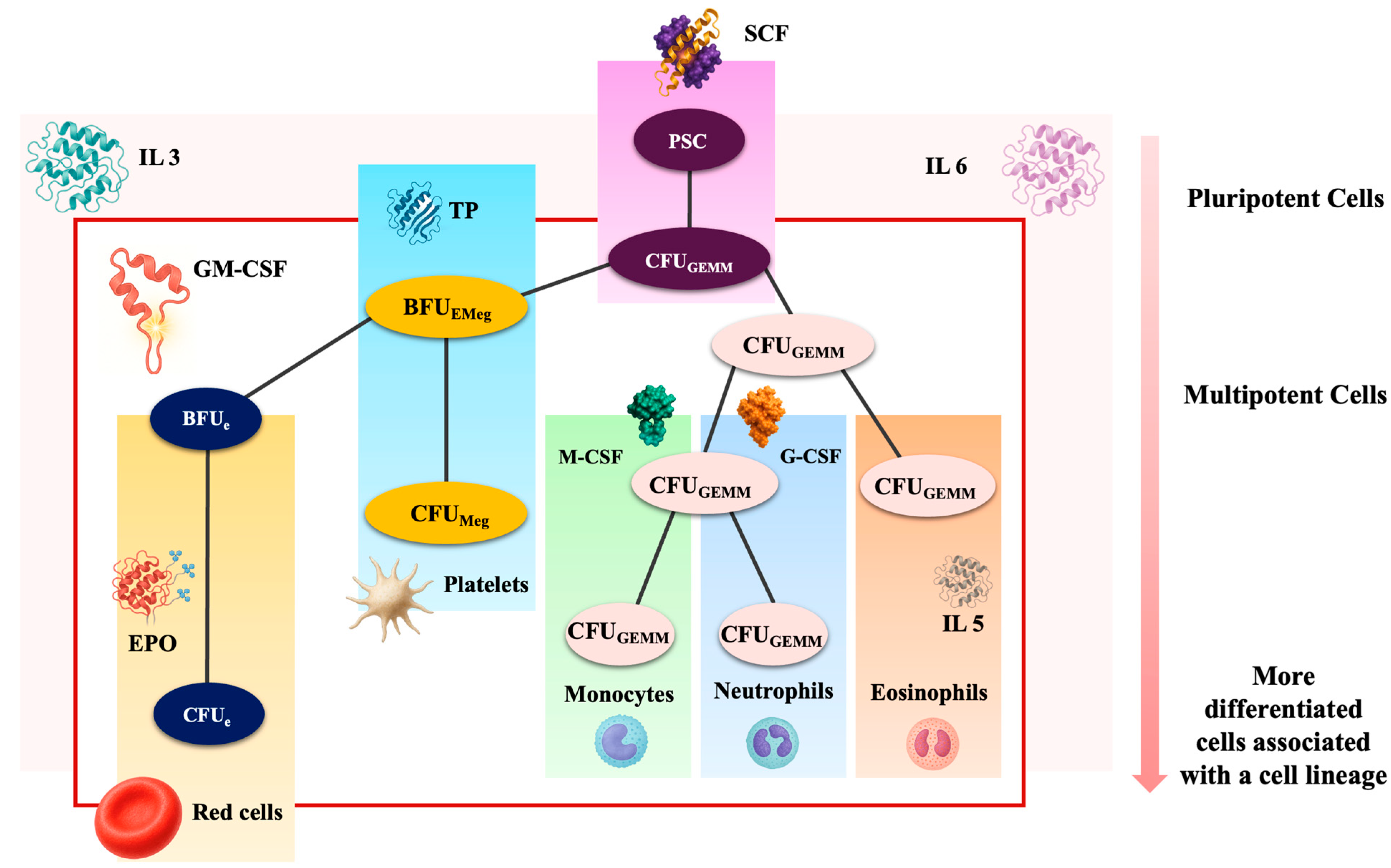
Figure 1. Hematopoiesis and growth factors SCF: stem cell factor; CSF: colony-stimulating factor; GM: granulocyte–macrophage; M: monocyte; G: granulocyte; EPO: erythropoietin; IL: interleukin; TP: thrombopoiesis; Meg: Megakaryocyte; BFUE: burst forming unit-erythroid; CFU: colony forming unit; CFUE: colony forming unit-erythroid; GEMM: granulocyte, erythrocyte, monocyte, megakaryocyte.
Biochem 05 00030 g001
Figure 2. Erythropoiesis—from pluripotent stem cell to erythrocyte. BFU-E: burst forming unit-erythroid; CFU-E: colony forming unit-erythroid.
Biochem 05 00030 g002
Figure 3. Causes and prevalence of anemia in the elderly according to a Japanese study [26].
Biochem 05 00030 g003
Figure 4. Subsequent laboratory evaluations to be considered after morphological classification of anemia.
Biochem 05 00030 g004aBiochem 05 00030 g004b
Figure 5. Diagnosis of iron deficiency without anemia.
Biochem 05 00030 g005
Figure 6. European Hematology Association guidelines on therapeutic approach to iron deficiency anemia [29]. IDA—Iron Deficiency Anemia; ID—Iron Deficiency; IRT—Iron Replacement Therapy; PRBC—Packed Red Blood Cells; Hb—Hemoglobin; HMB—Heavy Menstrual Bleeding; IV—Intravenous.
Biochem 05 00030 g006
Table 1. Consequences of anemia in the elderly (adapted from Emmanuel Andrès et al., 2013 [10]).
Anemia in Elderly Patients
Alteration of cognitive function
Iatrogenic injury
Descompasation of underlying disorders
Hospitalization
Increased mortality
Increased debility
Reduction in mobility and bone density; Increased risk of falls and fractures
Reduction of quality of life
Table 2. Causes and prevalence of anemia in the elderly [22,23].
CausePercentage of All Anemia Cases
Deficiency of iron16.6
Deficiency of folate6.4
Deficiency of vitamin B125.9
Deficiency of folate and vitamin B122.0
Deficiency of iron, folate and vitamin B123.4
Renal insufficiency8.2
Inflammation (chronic disease)19.7
Renal insufficiency and inflammation4.3
Table 3. Classification of anemias based on regenerative capacity.
Non-RegenerativeRegenerative
Aplastic anaemiaHaemolysis
Pure red cell aplasiaImmune
Myelodysplasic syndromeNon-immune
  • Congenital: membrane, SS, thalassaemia, enzymophathies, unstable Hb
  • Acquired: PNH, drugs, microangiopathy, hyperslpenism
Deficiency states
Marrow infiltration/fibrosis
Inflammatory anaemia
Erytropoietin underproductionHaemorrhage
Table 4. Morphological classification of anemias.
Normocytic (MCV 80-100 fL)Microcytic (MCV < 80 fL)Macrocytic (MCV >100 fL)
- Hemorrhagic anemia
- Early iron deficiency anemia
- Anemia of chronic disease
- Anemia associated with bone marrow suppression
- Anemia associated with chronic renal insufficiency
- Anemia associated with endocrine dysfunction
- Autoimmune hemolytic anemia
- Anemia associated with hypothyroidism or hypopituitarism
- Hereditary spherocytosis
- Hemolytic anemia associated with paroxysmal nocturnal hemoglobinuria
- Iron deficiency anemia
- Thalassemias
- Anemia of chronic disease
- Sideroblastic anemia
- Anemia associated with copper deficiency
- Anemia associated with lead poisoning
- Folic acid deficiency anemia
- Anemia associated with vitamin B12 deficiency
- Drug-induced hemolytic anemia
- Anemia associated with reticulocytosis
- Anemia associated with liver disease
- Anemia associated with etanol abuse
- Anemia associated with acute myelodysplastic syndrome
Table 5. Diagnostic algorithm for the evaluation of a patient with anemia.
Evaluation
HistoryPhysical ExaminationLaboratory Investigations
  • Time of onset
  • Severity
  • Age
  • Dissociation Curve Hb/O2
  • Positive family history of anemia
  • Ethnic origin
  • Acuteness of onset of anemia
  • Bleeding: stool, urine, lungs, menses
  • Infections: Parvovírus B19, Hepatitis, HIV
  • Phlebotomy
  • Jaundice, dark urine
  • Petechiae
  • Symptoms from other organs (CNS, gastrites)
  • Masses, nodes
  • Alchohol consumption
  • Diet:low fruit, no meat
  • Pica (abdnormal food habits)
  • Drugs
  • Previus transfusion
  • Pallor, ictereus, petechiae
  • Temperature
  • Lymph nodes
  • Enlarged spleen
  • Tachycardia, hypotension
  • WBC and platelet count (aplastic anaemia)
  • Reticulocyte count (red cell aplasia)
  • MCV (iron or vitamin deficiency, thalassaemias)
  • RBC morphology:
    double RBC population (transfusion, sideroblastic anaemia, high HbF)
    RBC agglutination (cold agglutinins)
    schizocytes (microangiopathy)
    spherocytes (imune haemolysis)
  • Bilirubin, LDH, haptoglobin, Coombs test (haemolysis)
  • Kidney function (EPO deficiency)
  • Occult blood in the stools
  • C-reactive protein
  • Iron studies + vitamins (serum iron, TIBC, ferritinm B12, folates) depending on MCV
Table 6. Causes of anemia according to reticulocyte count.
Reticulocyte Count
LowHigh
Hypoproliferative anemia:
  • Iron deficiency
  • B12 or folate deficiency
  • Anemia of chronic inflammation
  • Cytotoxic chemotherapy
  • Renal Insufficiency
  • Myeolophtisic anemia
Increased loss of red blood cells:
  • Acute hemorrhage
  • Auto-imune hemolytic anemia
  • Microangiopatic anemia
Disclaimer/Publisher’s Note: The statements, opinions and data contained in all publications are solely those of the individual author(s) and contributor(s) and not of MDPI and/or the editor(s). MDPI and/or the editor(s) disclaim responsibility for any injury to people or property resulting from any ideas, methods, instructions or products referred to in the content.