Article

Ethyl 2-(3,5-Dioxo-2-p-tolyl-1,2,4-thiadiazolidin-4-yl) Acetate: A New Inhibitor of Insulin-Degrading Enzyme

1
College of Biological Science and Medical Engineering, Donghua University, Shanghai 201600, China
2
College of Chemistry and Chemical Engineering, Donghua University, Shanghai 201600, China
*
Authors to whom correspondence should be addressed.
These authors contributed equally to this work.
BioChem 2025, 5(3), 27; https://doi.org/10.3390/biochem5030027
Submission received: 27 May 2025 / Revised: 26 August 2025 / Accepted: 28 August 2025 / Published: 30 August 2025

Abstract

Background: Insulin-degrading enzyme (IDE) has become an essential target for the clinical treatment of various important diseases, including type 2 diabetes, Alzheimer’s disease, and breast cancer, owing to its diverse substrate specificity. Particularly in cancer therapy, IDE inhibitors have received significant attention. Methods: We evaluated the in vitro inhibitory activity (IC50) of ethyl 2-(3,5-dioxo-2-p-tolyl-1,2,4-thiadiazolidin-4-yl) acetate (1) against wild-type IDE. The mechanism of action was investigated using Lineweaver–Burk double reciprocal plots and molecular docking analyses. Additionally, we examined the structure–activity relationship, cytotoxicity, selectivity, and effects on cell migration to assess its potential druggability. Based on molecular docking results, we prepared the mutant protein T142A and compared its inhibitory effects with those of the wild-type and mutant proteins. Results: Compound 1 exhibited an inhibitory effect on IDE (IC50 = 3.60 μM). This compound exerts its inhibitory effect through competitive binding to the catalytic site of IDE. Compound 1 demonstrated selective cytotoxicity toward cancer cells compared to normal cells, effectively inhibiting IDE at concentrations ≤ 10 μM. At a concentration of 3.6 μM, the inhibitory effect of the compound on cancer cell migration was significantly stronger than that observed in normal cells. Although the T142A mutant retained catalytic hydrolysis activity with a similar Km value, its reaction rate was markedly lower than that of the wild-type enzyme. Conclusions: Compound 1 exhibits a competitive inhibitory effect on IDE, selectively targeting IDE with greater toxicity toward cancer cells compared to normal cells. It also inhibits cancer cell migration. Notably, 1 demonstrates significantly stronger inhibitory activity against the T142A mutant than the wild-type IDE, indicating that the Thr142 residue plays a crucial role in the interaction between the IDE hydrophobic pocket and 1. These findings suggest that 1 holds potential as a chemotherapeutic agent for treating IDE-related cancers, including breast, prostate, and pancreatic cancers.

1. Introduction

Insulin-degrading enzyme (IDE) is a highly conserved zinc-ion metalloprotease that was initially identified and named for its ability to degrade insulin [1,2,3,4]. Subsequently, research has extensively investigated the functions of IDE in cancer [5,6]. IDE has additional substrates, including Insulin-like Growth Factor 2, glucagon, calcitonin, β-endorphin, somatostatin, β-amyloid, enkephalin, CC-chemokine ligand 3 (CCL3), CCL4, bradykinin, transforming growth factor-α, and atrial natriuretic peptide [7,8,9,10]. This highlights IDE’s significant role in the pathophysiological processes regulated by these peptides.
Recent studies have revealed the significant role of IDE in various cancers and its potential as a therapeutic target. IDE is expressed in various solid tumors and hematologic tumor cell lines [11,12,13,14,15]. A study on breast cancer found that IDE overexpression is a risk factor for recurrence [16]. Furthermore, suppressing IDE expression impacts the ubiquitin–proteasome system (UPS), which in turn hinders the proliferation and viability of neuroblastoma cells [17]. Insulin can induce angiogenesis and promote tumor growth by increasing circulating levels and through mitogenic signaling. Given the substrate hydrolysis activity of IDE, it may influence cancer progression [18,19,20,21]. Specifically, the IDE maintains the activity of the tumor suppressor retinoblastoma (Rb) by degrading insulin in the cytoplasm, thereby preventing its nuclear translocation and subsequent inactivation of Rb [22,23,24,25]. Moreover, Atrial Natriuretic Peptide (ANP) and C-Type Natriuretic Peptide (CNP), as IDE substrates, interact with specific receptors to regulate tumorigenesis-related pathways such as the RAS-MEK1/2-ERK1/2, Wnt/β-catenin, and VEGF/VEGFR2. They exhibit anti-tumor effects in cancers such as breast, prostate, and pancreatic cancer. Nonetheless, the hydrolysis of these peptides by IDE may compromise their anti-tumor potential [26,27,28,29]. Considering these findings, designing inhibitors to modulate IDE activity is crucial. Such inhibitors provide new strategies for treating and managing cancers associated with IDE. Given IDE’s therapeutic potential in tumor progression, regulating its activity has become a highly significant research topic in recent years. The development of IDE inhibitors is therefore essential for advancing cancer therapy and improving patient outcomes.
Early inhibitors of insulin-degrading enzyme (IDE), such as bacitracin and N-ethylmaleimide, displayed low affinity and poor specificity, which limited their clinical application [30,31]. Subsequent research identified several promising IDE inhibitors with improved pharmacological properties. Leissring et al. developed Li1, a peptide hydroxamate inhibitor, through combinatorial peptide library screening and focused compound analysis [32]. Charton et al. identified BDM41367, an imidazole derivative, by conducting high-throughput screening of a 2000-compound drug-like library [33,34]. Deprez-Poulain et al. synthesized BDM44768 using kinetic target-guided synthesis, while Maianti et al. [35] discovered 6bk from a DNA-templated macrocyclic library [36]. Despite their therapeutic potential, most inhibitors target the Zn2+-binding site, leading to off-target effects against other metalloproteases. Notably, 6bk and BDM44768 improved oral glucose tolerance but induced intolerance upon intraperitoneal injection, highlighting complex in vivo metabolic interactions. While IDE inhibitors show promise for treating multiple diseases, future breakthroughs depend on enhancing specificity and substrate selectivity, with peptide-based and substrate-selective strategies emerging as key approaches.
Thiazolidinedione (TZD) is a commonly found heterocyclic pharmacophore [37]. Its unique structure provides extensive opportunities for chemical modification and has demonstrated a variety of significant pharmacological activities [38,39,40]. Nevertheless, its clinical use has been restricted due to potential side effects [37]. In this study, we screened a series of thiadiazolidinediones (TDZDs) derivatives, which are bioelectronic equivalents of thiazolidinediones (TZDs). TDZDs and TZDs are two important heterocyclic compounds that are frequently employed in medicinal chemistry for the design of kinase inhibitors and metabolic modulators. Despite their similar names and structural resemblance, they exhibit significant differences in chemical properties, biological activities, and clinical applications. In the present study, we focused on investigating their inhibitory activities against IDE in vitro. Among them, 1 demonstrated superior IDE activity inhibition and specificity. Furthermore, its significant inhibition of cancer cell migration and cell viability suggests its potential as a candidate for IDE-related tumor therapy (Figure 1).

2. Materials and Methods

2.1. Synthesis of Thiazolidinedione Derivatives

General procedure for the synthesis of 1,2,4-Thiadiazolidine-3,5-dione: In a 50 mL round-bottom flask, isocyanate (5 mmol), isothiocyanate (5 mmol), hexane (5 mL), and dichloromethane (5 mL) were combined and cooled to 0 °C. Sulfuryl chloride (5 mmol) was then slowly added to the solution. The mixture was stirred at room temperature overnight. Following this, the solution was exposed to air for 30 min to fully react with moisture, after which the solvent was removed under reduced pressure. The residue was diluted with ethyl acetate (30 mL) and saturated NaHCO3 aqueous solution (10 mL), and the aqueous phase was extracted with ethyl acetate (20 mL) three times. The combined organic phase was dried (Na2SO4) and removed by rotary evaporation. Flash chromatography (From PE to PE:EA = 8:2) was used to purify the crude reaction mixture. Finally, the synthesized compounds were characterized by nuclear magnetic resonance (NMR) and mass spectrum.

2.2. Preparation of IDE

The full-length IDE gene from a mouse was cloned into the ppSUMO expression vector, and the recombinant plasmid was transformed into E. coli BL21 (DE3). His-SUMO-tagged IDE was expressed and purified by using Ni-NTA resin (Sangon Biotech, Shanghai, China). Following this, ULP1 protease was added to the eluate to remove the His-SUMO tag. The IDE protein was then subjected to further purification using HiTrap QFF and HiLoad 16/60 Superdex 200 pg (GE Healthcare, Chicago, IL, USA).

2.3. Enzymatic Assay

IDE activity was assessed using the fluorescent peptide substrate Mca-RPPGFSAFK(DNP)-OH, a bradykinin-derived quenched peptide (Nanjing Peptide, Nanjing, China). The assay was performed in a 96-well plate by sequentially adding 97 μL of reaction buffer (25 mM Tris-HCl, 500 mM NaCl, 5% glycerol, pH 7.3), 1 μL of 1 mM substrate, 1 μL of inhibitor at a 1:100 dilution, and 1 μL of IDE sample (0.1 mg/mL). The final volume could be adjusted to achieve the desired concentration. The plate was incubated at 37 °C, and fluorescence was monitored every minute for 1 h using a FlexStation II (Molecular Devices, San Jose, CA, USA, excitation at 320 nm, emission at 405 nm). All reactions were conducted in triplicate.

2.4. Molecular Docking

The molecular docking simulations were performed using AutoDock4.2 to predict the binding conformation of 1 with IDE. The crystal structure of IDE (PDB code: 2JG4) was obtained from the Protein Data Bank. The inhibitors were constructed using ChemDraw (ChemDRAW 20), and their energy was minimized using Chem3D (Chem3D 20.0.0). For docking, IDE’s domain 1 was taken as the grid box since it contains the catalytic site. The Lamarckian genetic algorithm was employed for the search parameters. Docking was constructed with protein as a rigid receptor and inhibitor as a flexible ligand.

2.5. Cytotoxicity Assay and Cell Migration

2.5.1. Cytotoxicity Assay

L929 cells and 4T1 cells were inoculated in 96-well plates at a density of 5000 cells per well and incubated overnight at 37 °C with 5% CO2. Following adherence, the medium was replaced with fresh medium containing various concentrations of inhibitors, and the cells were cultured for an additional 24 h. Next, the original medium was discarded, and CCK-8 solution was mixed with serum-free medium at a ratio of 1:9 before being added to the wells. The plates were then incubated at 37 °C for 2 h. Finally, the optical density of the samples was measured at 450 nm using a microplate reader (Thermo ScientificTM Multiskan SkyHigh, Waltham, MA, USA).

2.5.2. Cell Migration

L929 cells and 4T1 cells were inoculated in 24-well plates at a density of 20,000 cells/well and incubated in a 37 °C with 5% CO2 incubator until the cells reached 80–90% confluence. A scratch was created using a 200-μL pipette tip, followed by two washes with D-PBS. Subsequently, serum-free medium containing 3.60 μM of 1 inhibitor was added to the treatment group, while the control group received only serum-free medium, and the cells were further incubated in a 37 °C with 5% CO2 incubator. Images were captured using a 4× objective lens at 0 h, 12 h, and 24 h. The images were then processed using ImageJ (×64, 1.46 ) software.

2.6. Statistical Analyses

Statistical analysis was performed using Excel and Prism 9.0.0 (GraphPad Software 9.5.1 Inc., Boston, MA, USA). Data are presented as means ± SEM. Comparisons between two groups were performed using the unpaired Student’s t-test. Differences were considered significant at p < 0.05.

3. Results

3.1. Characterization of TDZD Compounds

3.1.1. Characterization of TDZD Compound 1

1H NMR (400 MHz, CDCl3) δ ppm: 7.39 (d, J = 8.4 Hz, 2H), 7.23 (d, J = 8.2 Hz, 2H), 4.49 (s, 2H), 4.27 (q, J = 7.1 Hz, 2H), 2.37 (s, 3H), 1.31 (t, J = 7.1 Hz, 3H). 13C NMR (151 MHz, CDCl3) δ ppm: 166.35, 165.11, 150.53, 137.57, 132.93, 130.15, 123.87, 62.21, 42.80, 21.04, 14.11. HR ESI MS: m/z calcd for C13H15N2O4S [M + H]+, 295.0747, found, 295.074.

3.1.2. Characterization of TDZD Compound 2

1H NMR (400 MHz, CDCl3) δ 7.44–7.28 (m, 5H), 4.82 (s, 2H), 4.43 (s, 2H), 4.26 (q, J = 7.2 Hz, 2H), 1.30 (t, J = 7.2 Hz, 3H). 13C NMR (101 MHz, CDCl3) δ 166.40, 165.70, 152.48, 134.26, 129.11, 128.90, 128.39, 62.16, 48.74, 42.71, 14.08. HR ESI MS: m/z calcd for C13H15N2O4S [M + H] +, 295.0747, found, 295.0746

3.1.3. Characterization of TDZD Compound 3

1H NMR (400 MHz, CDCl3) δ 7.41 (d, J = 7.2 Hz, 2H), 7.34–7.24 (m, 3H), 4.81 (s, 2H), 4.28 (s, 2H), 4.19 (q, J = 7.1 Hz, 2H), 1.23 (t, J = 7.2 Hz, 3H). 13C NMR (101 MHz, CDCl3) δ 167.12, 166.02, 153.83, 135.04, 128.74, 128.66, 128.30, 62.17, 46.08, 45.62, 14.06. HR ESI MS: m/z calcd for C13H 15N2O4S [M + H] +, 295.0747, found, 295.0747.

3.2. Discovery of TDZD Hit Compound

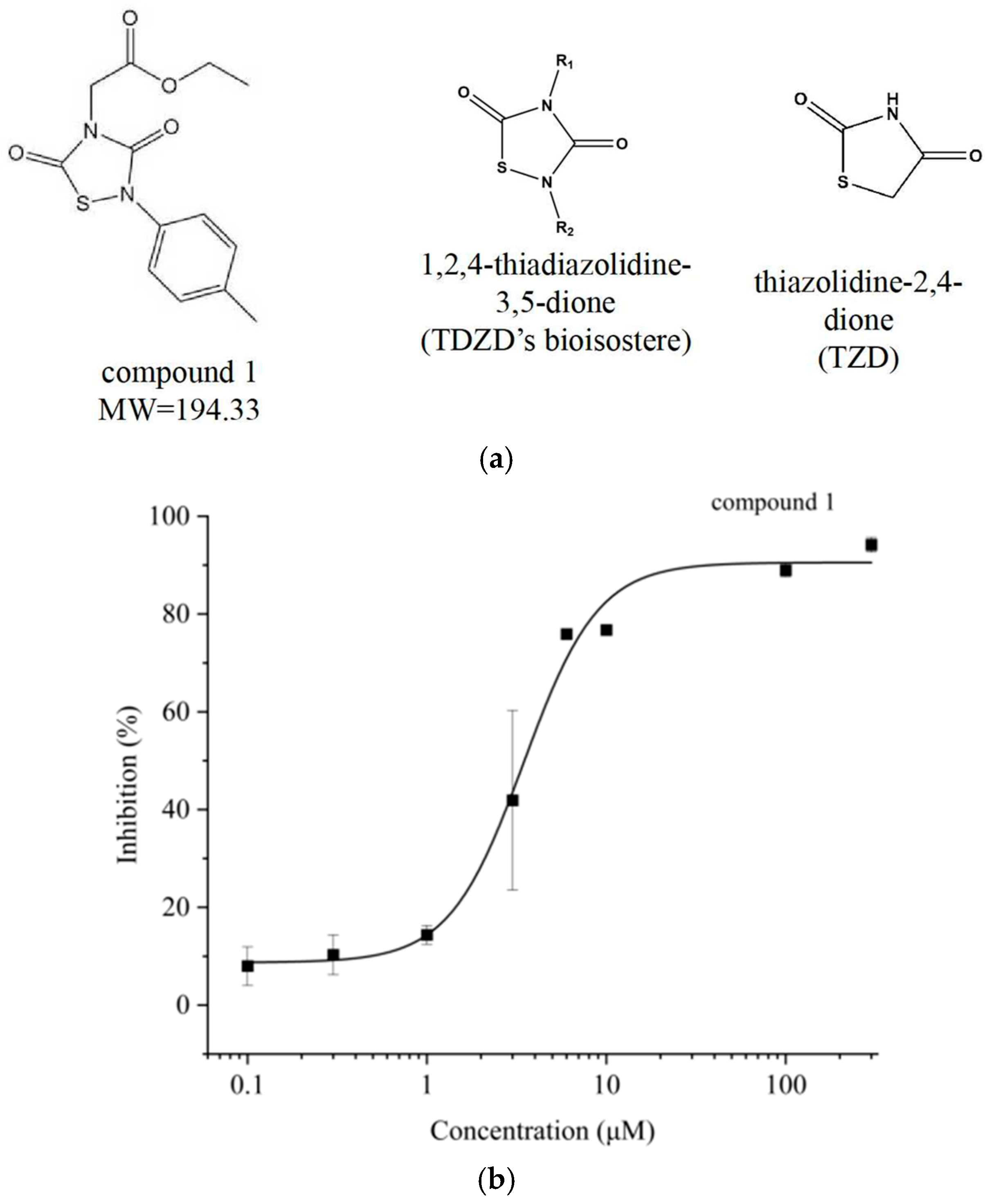
To investigate the potential of TDZD derivatives as inhibitors of IDE (Figure 2a), we evaluated the inhibitory effects of a series of TDZD compounds on IDE activity in vitro. Through screening, we identified a promising compound, designated as 1, which demonstrated IDE inhibitory activity. Compound 1 is a small molecule with 1,2,4-thiadiazolidine-3,5-dione as the core backbone, with ethyl acetate and p-tolyl at the R1 and R2 positions, respectively. This compound exhibited significant inhibition of IDE at concentrations above 10 μM, while showing low inhibition below 1 μM (Figure 2b). The fitted IC50 value for 1 was determined to be 3.60 μM against IDE. Although 1 shows lower affinity for IDE compared to well-known macrocyclic inhibitors (e.g., 6bk TFA, IC50 = 50 nM), its smaller molecular weight (MW = 294.33) suggests it may inhibit IDE through a distinct mechanism compared to larger macrocyclic inhibitors.

3.3. Compound 1 Inhibits IDE Competitively

To further determine the inhibition type of 1, Michaelis–Menten kinetic curves were measured in a substrate concentration range of 5 to 40 μM (Figure 3a). When inhibited by 3 μM, the initial reaction rate V0 decreased significantly. Inhibition by 3 μM of 1 led to a significant decrease in the initial reaction rate V0. Moreover, V0 exhibited an almost linear increase with substrate concentration, suggesting that 1 increases the Km of the reaction. As a result, even at a substrate V concentration of 40 μM, the initial reaction rate did not approach saturation. The Lineweaver–Burk plot revealed that the Vmax remained constant while Km increased under the inhibitory effect of 1 (Figure 3b). These findings indicate that 1 exerts reversible competitive inhibition on IDE by binding competitively to the substrate-binding site of IDE.

3.4. Structure–Activity Relationship (SAR) Analysis of TDZDs

To further investigate the relationship between TDZD’s activity and structure, compounds with different side chains were tested for their inhibition of IDE. These compounds exhibited varying degrees of inhibitory effects on IDE. Notably, ethyl 2-(2-benzyl-3,5-dioxo-1,2,4-thiadiazolidin-4-yl) acetate (2) showed IDE inhibitory activities comparable to those of 1. Based on the insights provided by the structure–activity relationship from the results of side-chain substitution, we introduced a halogen group into the structure of 1, attempting to enhance its interaction with the binding site of IDE by modifying the charge distribution of the compound. The results showed that ethyl 2-(2-(4-chlorophenyl)-3,5-dioxo-1,2,4-thiadiazolidin-4-yl) acetate (3) exhibited only a slight reduction in activity compared to 1. This suggests that introducing a chlorine atom at the terminal p-tolyl group of the R2 side chain did not significantly enhance its inhibitory activity (Table 1).
Compound 2 shares structural similarities with compound 1, particularly in the R1 and R2 side-chain residues. The presence of benzyl or phenylmethyl groups in 2 suggests potential hydrophobic interactions with the receptor protein during binding. Additionally, the carbonyl oxygen atoms in the ethyl acetate group may form hydrogen bonds with amino acid side chains in the binding region. Since the introduction of halogen groups did not significantly reduce inhibitory activity, it is likely that strong charge interactions occur in the thiadiazolidinedione or ethyl acetate side-chain regions rather than the benzyl group when these molecules bind to IDE. This finding implies that halogen modification at the benzyl group terminus could be a viable strategy for increasing drug lipophilicity and prolonging its duration of action in future drug design, without compromising inhibitory activity.
Collectively, these findings indicate that IDE’s recognition pattern for this class of drugs is primarily influenced by the side-chain conjugation effects and the overall molecular structure.

3.5. Docking of 1 in IDE

The competitive inhibition by 1 indicates that it directly interacts with the substrate-binding region. For the small peptide substrate V, it binds exclusively to the catalytic site of IDE without engaging with the exo-site. Consequently, we defined the docking pocket at the structural catalytic site, and the potential binding conformation of 1 was determined using AutoDock 4.2. The results show that 1 acts directly on the catalytic site with a binding energy of -6.11 kcal/mol (Figure 4a). Ser 96, Ala 140, Ser 143, Gly 144, Trp 199, Phe 202, Lys 206, Gly 219 and Thr 220 in the 4 Å range around 1 together constitute the binding hydrophobic pocket of 1 (Figure 4b). Among them, the benzene ring of the Phe 202 side chain likely engages in a strong hydrophobic interaction with the p-tolyl group of 1, while 1 interacts with residues crucial for substrate hydrolysis, including Glu 111, Phe 141, and Thr 142. The carbonyl oxygen of 1′s side chain is predicted to form a hydrogen bond with Thr 142, which is part of IDE’s β6-strand involved in substrate binding and 1 directly interferes with the normal recognition and binding of substrates to IDE. It may also inhibit the formation of the catalytic triad in this region by interacting with His 108 and Glu 189, thereby exerting its inhibitory effect.
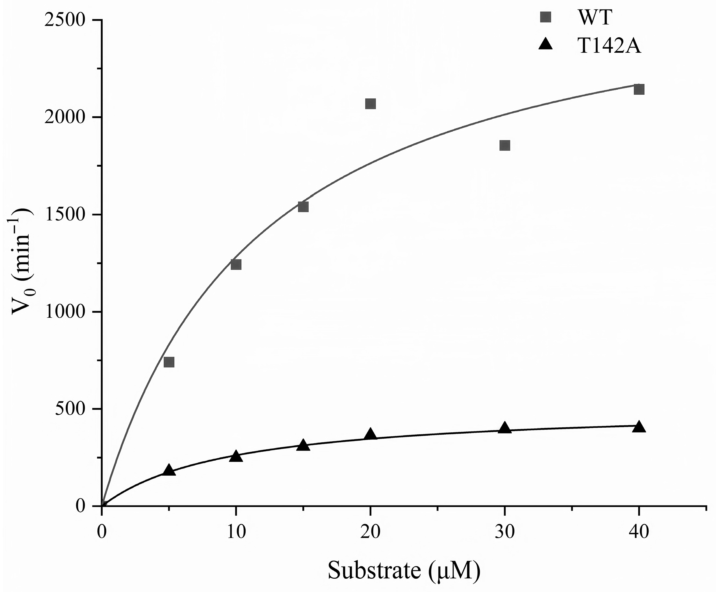
Based on these docking results, we replaced Thr 142, which forms a hydrogen bond with 1 in the docking conformation, with Ala. By evaluating 1′s inhibitory activity on the T142A mutant protein, we determined whether this mutation affects 1′s affinity for IDE, thus verifying if 1 acts at this site. In this experiment, the Michaelis–Menten kinetics curve for T142A degrading the fluorescent substrate Mca-RPPGFSAFK(DNP)-OH was analyzed to evaluate its substrate-degrading activity. The wild-type IDE exhibits a Vmax of 2814.32 min−1 and a Km of 11.93 μM. In contrast, the T142A mutant displays a Vmax of 501.05 min−1 and a Km of 9.01 μM (Figure 5, Table 2). These results indicate that while the T142A mutant retains catalytic hydrolysis activity with a similar Km value, its reaction rate is significantly lower compared to that of the wild-type enzyme.
In addition to the aforementioned studies, we further compared the IC50 values of Compound 1 for various IDE mutants and observed that the IC50 value for the T142A mutant was significantly lower than that of the wild-type enzyme, indicating that the substitution of Thr142 with Ala enhances the inhibitory effect of Compound 1 on IDE. This enhancement may be attributed to the fact that, in the docking conformation, the hydrogen bond formed between Thr142 and the inhibitor involves the amino group hydrogen atom rather than the side-chain hydrogen atoms. Consequently, even though the side-chain structure is altered upon substitution with Ala, the ability to form a hydrogen bond with Compound 1 is retained. Moreover, the substitution of Thr142 with Ala induces structural changes in the entire binding pocket, reducing the steric hindrance experienced by Compound 1 and facilitating its binding at this site. Collectively, these factors contribute to the increased inhibitory activity of Compound 1 observed for the T142A mutant.

3.6. Cytotoxicity and Selectivity

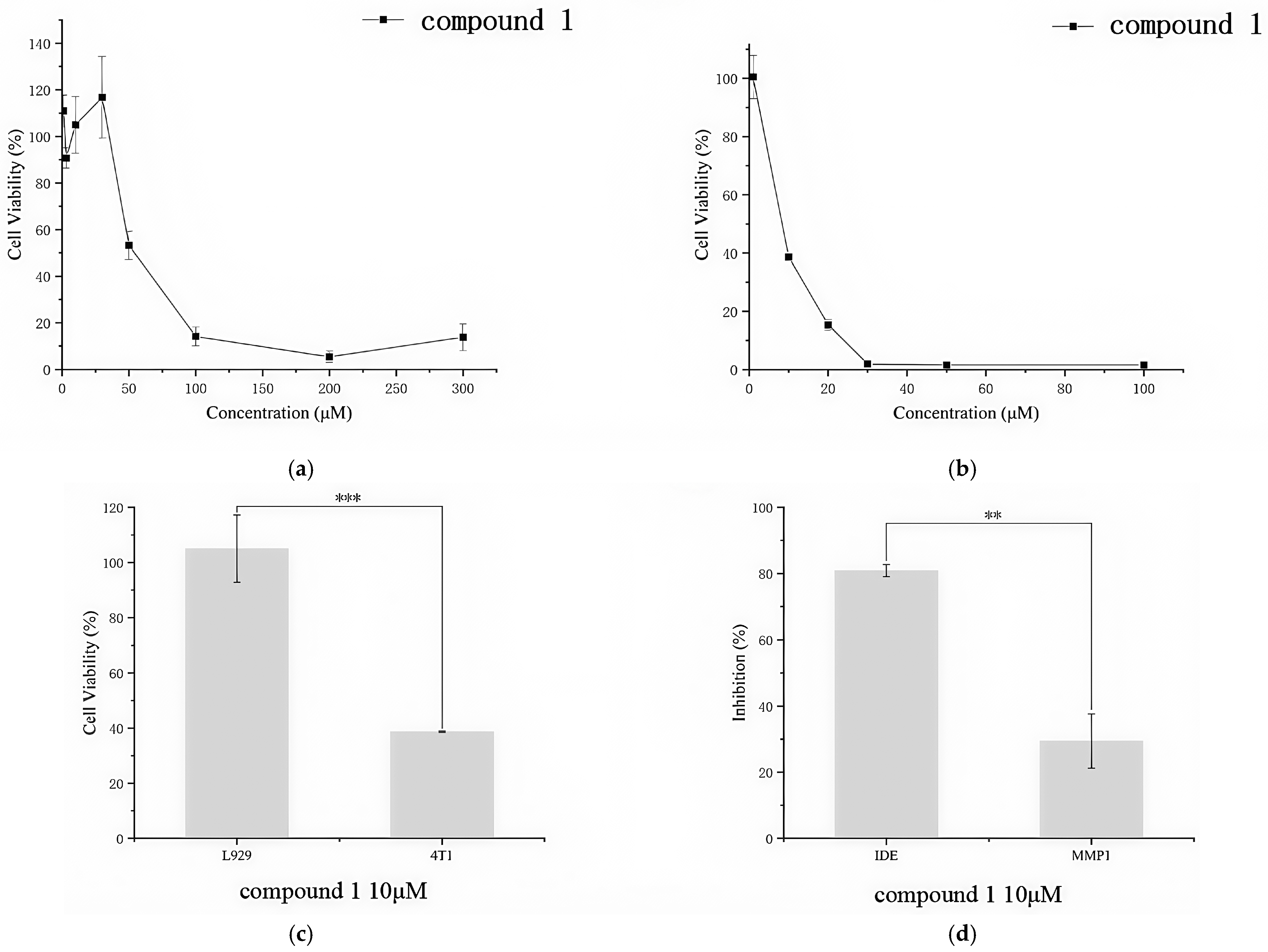
To evaluate the drug potential of TDZDs, we selected L929 mouse fibroblasts and 4T1 mouse breast cancer cells as models for assessing the cytotoxicity of 1. Compound 1 was applied to L929 cells at concentrations ranging from 1 to 300 μM for 24 h, and to 4T1 cells at concentrations ranging from 1 to 100 μM for 24 h. The inhibitory effects on cell proliferation were measured using a CCK-8 assay kit (Beyotime Biotech Inc., Shanghai, China). At concentrations below 30 μM, 1 did not exhibit significant cytotoxicity (Figure 6a). However, when the compound concentration reached 50 μM, cell viability decreased to 53.30%. At 100 μM, 1 exhibited higher cytotoxicity, markedly inhibiting cell growth. For the mouse breast cancer cell 4T1, 1 demonstrated highly significant cytotoxicity (Figure 6b), reducing cell viability to 1.94% at 30 μM. This effect was notably different from its impact on mouse normal tissue cells L929. Even at lower concentrations, such as 10 μM, 1 showed a significant difference in cell viability between L929 and 4T1 cells (Figure 6c).
To further evaluate the selectivity of 1, we also assessed its effects on insulin-degrading enzyme (IDE) and zinc-dependent matrix metalloproteinase 1 (MMP-1) using their respective fluorescent short peptide substrates. Fluorescence assays showed that 10 μM of compound 1 had significantly different inhibitory effects on IDE and MMP-1, indicating some specificity of the compound towards IDE (Figure 6d).

3.7. Cell Migration

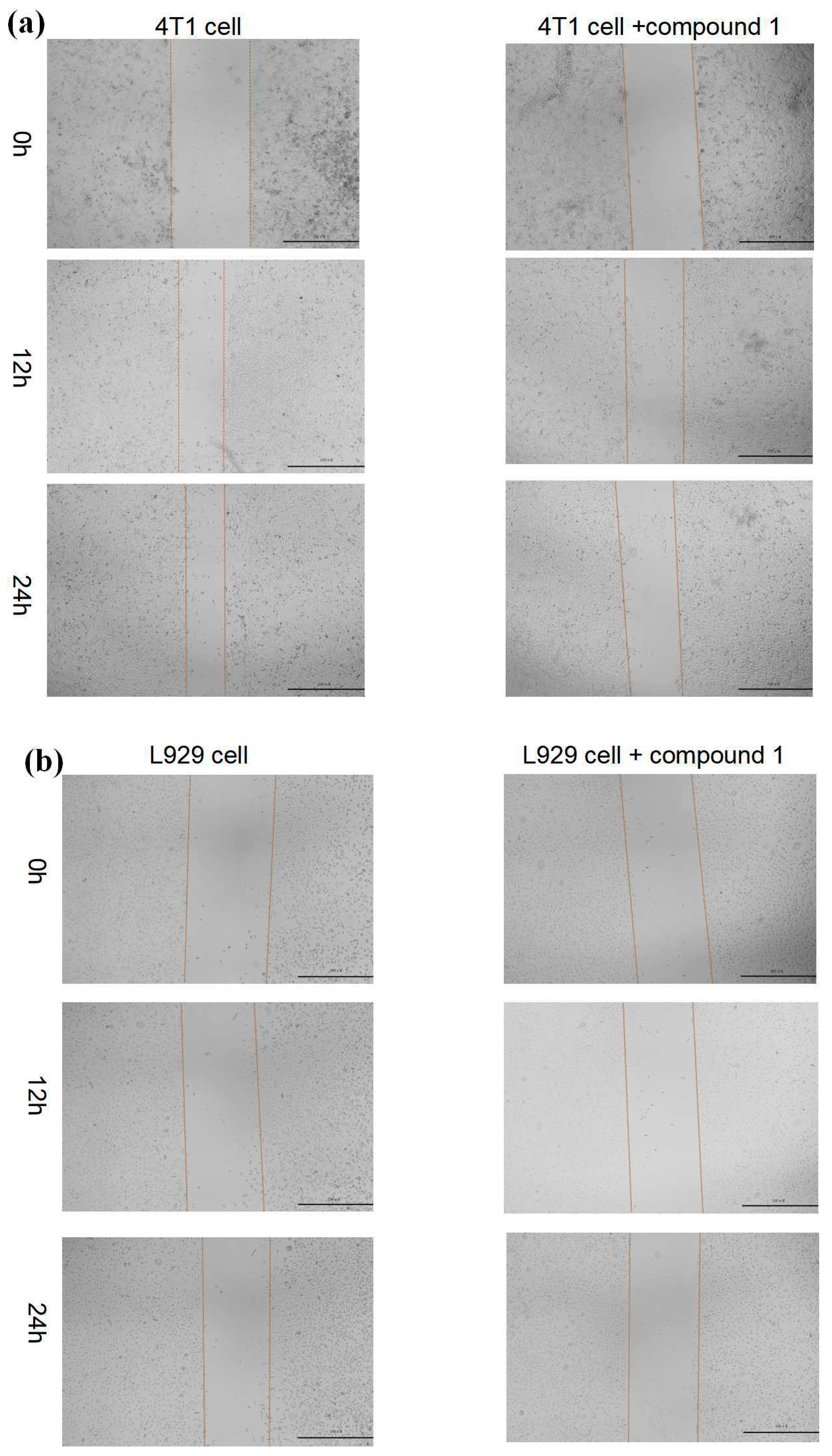
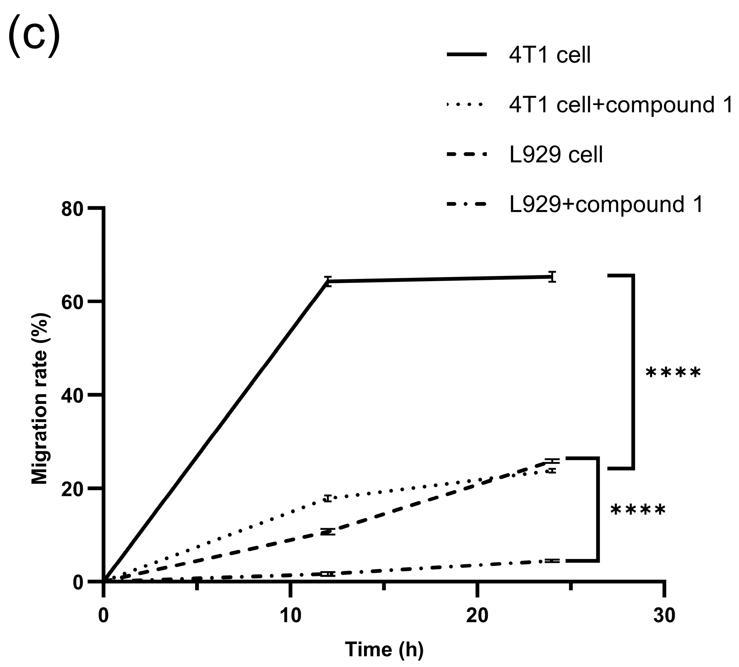
Given the potential of IDE in breast cancer treatment, we selected L929 mouse fibroblasts and 4T1 mouse breast cancer cells as models for evaluating 1’s effects on cell migration. Using a wound healing assay, we assessed how 1 influenced the migration of both L929 and 4T1 cells. The results showed that treating cells with 3.60 µM of 1 for 24 h significantly inhibited migration in both cell types compared to the control group (cells cultured for 24 h post-scratch without any inhibitor). Notably, 1 showed a particularly significant inhibitory effect on the migration of 4T1 cells compared to the control group (Figure 7a). For L929 cells, 1 demonstrated an inhibitory effect on cell migration compared to the control group (Figure 7b). This inhibitory effect was also observed after 24 h of culture in blank medium. This inhibitory effect was more pronounced in cancer cells than in normal cells (Figure 7c).
Based on these experimental results, we hypothesize that 1 may exhibit a selective bias toward IDE in cells, potentially disrupting the physiological function of IDE in cancer cells by inhibiting zinc-ion-dependent metalloproteinases, particularly IDE itself. This selective mechanism could result in increased cytotoxicity and enhanced inhibition of migration in cancer cells compared to normal cells.

4. Discussion

In this study, we investigated the potential of TDZD derivatives as inhibitors of insulin-degrading enzyme (IDE). In vitro assays revealed that 1 exhibited inhibitory activity against IDE-mediated degradation of a fluorescent substrate (IC50 = 3.60 µM). To elucidate the mechanism of inhibition, enzyme kinetic analysis, including Lineweaver–Burk plots, were conducted. The results indicated that 1 acts as a competitive inhibitor of IDE, suggesting it likely interacts with the substrate-binding region of the enzyme. Further structural comparisons and activity assessments provided insights into the structure–activity relationships of these inhibitors. Introducing groups such as toluene or benzyl enhances hydrophobic interactions within the IDE binding pocket, potentially improving inhibitory potency. Conversely, incorporating functionalities like ethyl acetate, which provide a carbonyl oxygen atom, may enable the formation of hydrogen bonds with residues in the binding site, further modulating the inhibitor’s efficacy. Molecular docking studies revealed that 1 binds effectively to the catalytic site of IDE, with a favorable binding energy of -6.11 kcal/mol. Notably, 1 demonstrates a significant cytotoxic effect on cancer cells compared to normal cells at low doses. It also demonstrates selectivity for IDE over other zinc-ion-dependent metalloproteins and shows promise in inhibiting cancer cell migration. These properties suggest its potential as a chemotherapeutic agent for treating cancers associated with IDE.
Although the inhibition of TDZDs with a smaller molecular weight is not as outstanding as many macrocyclic inhibitors that exhibit strong affinity to IDE, it still holds potential as a building block for developing substrate-specific IDE inhibitors—a key direction in current IDE inhibitor research. In future drug development, it may be possible to attach other active groups to the molecular framework of TDZDs and screen for compounds that exhibit substrate specificity, such as those that selectively inhibit insulin degradation processes. Therefore, the discovery of TDZD-like inhibitors offers a new structural template for designing drugs targeting IDE.

5. Conclusions

The objective of this work is to discover novel IDE-selective inhibitors, provide a new structural template for designing anti-IDE drugs, and explore new strategies for managing IDE-related diseases. In vitro experiments confirmed that 1 inhibits IDE during the degradation of a commercialized fluorescent substrate. Further analysis revealed a competitive inhibitory effect of compound 1 on IDE, indicating its interaction with the IDE catalytic site. This was supported by molecular docking studies, which demonstrated that 1 binds to the IDE catalytic site with favorable binding energy (-6.11 kcal/mol). Evaluations of compound 1′s inhibitory potency against various IDE mutants revealed significantly stronger inhibition of the T142A mutant than wild-type IDE, suggesting Thr142′s crucial role in IDE’s hydrophobic pocket for interaction with compound 1.
Subsequently, we modified the side-chain residues to investigate the mechanism of action of 1, particularly the influence of these residues on its inhibitory activity. The results indicated that the side-chain groups of this inhibitor exhibit structural conservation. To assess the drug potential of 1, CCK8 assays demonstrated the significant cytotoxicity of 1 against mouse breast cancer cell line 4T1 compared to normal mouse tissue cells. Cell migration assays revealed that 1 effectively inhibited the migratory capacity of cancer cells, showing a notably stronger inhibitory effect on 4T1 cells than L929 cells. Additionally, by comparing the effects of 1 on IDE and MMP-1 activities, it was found that 1 has a selective preference for IDE. This suggests that the TDZD molecular scaffold holds promise as a candidate for future drug development.
In particular, the TDZD molecular framework serves as a promising template for developing IDE-specific inhibitors. This scaffold can offer novel design insights for creating more effective and selective IDE inhibitors, which may play a crucial role in future treatments of IDE-related cancers, including breast and prostate cancer.

Author Contributions

T.C. designed the overall research. Y.Z. contributed to the cell experiments and drafted the manuscript. S.X. and H.M. were responsible for preparing all the proteins and assaying enzyme activity. Z.S. designed and completed the synthesis of the compounds. C.L. and Q.Z. contributed by revising the manuscript. All authors made substantial contributions to data analysis and/or interpretation, drafted and/or revised the manuscript, and approved the final version for publication. All authors have read and agreed to the published version of the manuscript.

Funding

Grant of the Ministry of Science and Technology of China (No. 9-11; No.10-6).

Acknowledgments

Thank Xueyan Cao for providing cell (L929, 4T1) and cytotoxicity and cell migration test methods.

Conflicts of Interest

The authors declare no conflicts of interest.

References

  1. Leissring, M.A.; González-Casimiro, C.M.; Merino, B.; Suire, C.N.; Perdomo, G. Targeting Insulin-Degrading Enzyme in Insulin Clearance. Int. J. Mol. Sci. 2021, 22, 223. [Google Scholar] [CrossRef]
  2. Manolopoulou, M.; Guo, Q.; Malito, E.; Schilling, A.B.; Tang, W.J. Molecular basis of catalytic chamber-assisted unfolding and cleavage of human insulin by human insulin-degrading enzyme. J. Biol. Chem. 2009, 284, 14177–14188. [Google Scholar] [CrossRef]
  3. Affholter, J.A.; Fried, V.A.; Roth, R.A. Human insulin-degrading enzyme shares structural and functional homologies with E. coli protease III. Science 1988, 242, 1415–1418. [Google Scholar] [CrossRef]
  4. Duckworth, W.C.; Hamel, F.G.; Bennett, R.; Ryan, M.P.; Roth, R.A. Human red blood cell insulin-degrading enzyme and rat skeletal muscle insulin protease share antigenic sites and generate identical products from insulin. J. Biol. Chem. 1990, 265, 2984–2987. [Google Scholar] [CrossRef] [PubMed]
  5. Tundo, G.; Ciaccio, C.; Sbardella, D.; Boraso, M.; Viviani, B.; Coletta, M.; Marini, S. Somatostatin modulates insulin-degrading-enzyme metabolism: Implications for the regulation of microglia activity in AD. PLoS ONE 2012, 7, e34376. [Google Scholar] [CrossRef]
  6. Bennett, R.G.; Hamel, F.G.; Duckworth, W.C. Insulin inhibits the ubiquitin-dependent degrading activity of the 26S proteasome. Endocrinology 2000, 141, 2508–2517. [Google Scholar] [CrossRef]
  7. Ciaccio, C.; Tundo, G.R.; Grasso, G.; Spoto, G.; Marasco, D.; Ruvo, M.; Gioia, M.; Rizzarelli, E.; Coletta, M. Somatostatin: A novel substrate and a modulator of insulin-degrading enzyme activity. J. Mol. Biol. 2009, 385, 1556–1567. [Google Scholar] [CrossRef]
  8. Fernández-Gamba, A.; Leal, M.C.; Morelli, L.; Castaño, E.M. Insulin-degrading enzyme: Structure-function relationship and its possible roles in health and disease. Curr. Pharm. Des. 2009, 15, 3644–3655. [Google Scholar] [CrossRef] [PubMed]
  9. Garcia, J.V.; Gehm, B.D.; Rosner, M.R. An evolutionarily conserved enzyme degrades transforming growth factor-alpha as well as insulin. J. Cell Biol. 1989, 109, 1301–1307. [Google Scholar] [CrossRef]
  10. Liang, W.G.; Ren, M.; Zhao, F.; Tang, W.J. Structures of human CCL18, CCL3, and CCL4 reveal molecular determinants for quaternary structures and sensitivity to insulin-degrading enzyme. J. Mol. Biol. 2015, 427 Pt B, 1345–1358. [Google Scholar] [CrossRef] [PubMed]
  11. Yfanti, C.; Mengele, K.; Gkazepis, A.; Weirich, G.; Giersig, C.; Kuo, W.L.; Tang, W.J.; Rosner, M.; Schmitt, M. Expression of metalloprotease insulin-degrading enzyme insulysin in normal and malignant human tissues. Int. J. Mol. Med. 2008, 22, 421–431. [Google Scholar] [PubMed]
  12. Radulescu, R.T.; Hufnagel, C.; Luppa, P.; Hellebrand, H.; Kuo, W.L.; Rosner, M.R.; Harbeck, N.; Giersig, C.; Meindl, A.; Schmitt, M.; et al. Immunohistochemical demonstration of the zinc metalloprotease insulin-degrading enzyme in normal and malignant human breast: Correlation with tissue insulin levels. Int. J. Oncol. 2007, 30, 73–80. [Google Scholar] [CrossRef] [PubMed]
  13. Weirich, G.; Mengele, K.; Yfanti, C.; Gkazepis, A.; Hellmann, D.; Welk, A.; Giersig, C.; Kuo, W.L.; Rosner, M.R.; Tang, W.J.; et al. Immunohistochemical evidence of ubiquitous distribution of the metalloendoprotease insulin-degrading enzyme (IDE; insulysin) in human non-malignant tissues and tumor cell lines. Biol. Chem. 2008, 389, 1441–1445. [Google Scholar] [CrossRef]
  14. Sbardella, D.; Fasciglione, G.F.; Gioia, M.; Ciaccio, C.; Tundo, G.R.; Marini, S.; Coletta, M. Human matrix metalloproteinases: An ubiquitarian class of enzymes involved in several pathological processes. Mol. Asp. Med. 2012, 33, 119–208. [Google Scholar] [CrossRef]
  15. Grasso, G.; Pietropaolo, A.; Spoto, G.; Pappalardo, G.; Tundo, G.R.; Ciaccio, C.; Coletta, M.; Rizzarelli, E. Copper(I) and copper(II) inhibit Aβ peptides proteolysis by insulin-degrading enzyme differently: Implications for metallostasis alteration in Alzheimer’s disease. Chemistry 2011, 17, 2752–2762. [Google Scholar] [CrossRef]
  16. Gennari, A.; Sormani, M.; Puntoni, M.; Martini, V.; Amaro, A.; Bruzzi, P.; Pfeffer, U. Identification of a Prognostic Signature Based on the Expression of Genes Related to the Insulin Pathway in Early Breast Cancer. Breast Care 2021, 16, 299–306. [Google Scholar] [CrossRef]
  17. Tundo, G.R.; Sbardella, D.; De Pascali, S.A.; Ciaccio, C.; Coletta, M.; Fanizzi, F.P.; Marini, S. Novel Platinum(II) compounds modulate insulin-degrading enzyme activity and induce cell death in neuroblastoma cells. JBIC J. Biol. Inorg. Chem. 2015, 20, 101–108. [Google Scholar] [CrossRef] [PubMed]
  18. Lesire, L.; Leroux, F.; Deprez-Poulain, R.; Deprez, B. Insulin-Degrading Enzyme, an Under-Estimated Potential Target to Treat Cancer? Cells 2022, 11, 1228. [Google Scholar] [CrossRef]
  19. Tundo, G.R.; Sbardella, D.; Ciaccio, C.; Bianculli, A.; Orlandi, A.; Desimio, M.G.; Arcuri, G.; Coletta, M.; Marini, S. Insulin-degrading enzyme (IDE): A novel heat shock-like protein. J. Biol. Chem. 2013, 288, 2281–2289. [Google Scholar] [CrossRef] [PubMed]
  20. Thackeray, J.T.; Pietzsch, S.; Stapel, B.; Ricke-Hoch, M.; Lee, C.W.; Bankstahl, J.P.; Scherr, M.; Heineke, J.; Scharf, G.; Haghikia, A.; et al. Insulin supplementation attenuates cancer-induced cardiomyopathy and slows tumor disease progression. JCI Insight 2017, 2, e93098. [Google Scholar] [CrossRef] [PubMed]
  21. Garczorz, W.; Kosowska, A.; Francuz, T. Antidiabetic Drugs in Breast Cancer Patients. Cancers 2024, 16, 299. [Google Scholar] [CrossRef]
  22. Radulescu, R.T. Intracellular insulin in human tumors: Examples and implications. Diabetol. Metab. Syndr. 2011, 3, 5. [Google Scholar] [CrossRef]
  23. Radulescu, R.T. One for all and all for one: RB defends the cell while IDE, PTEN and IGFBP-7 antagonize insulin and IGFs to protect RB. Med. Hypotheses 2007, 69, 1018–1020. [Google Scholar] [CrossRef]
  24. Radulescu, R.T.; Doklea, E.D.; Kehe, K.; Mückter, H. Nuclear colocalization and complex formation of insulin with retinoblastoma protein in HepG2 human hepatoma cells. J. Endocrinol. 2000, 166, R1–R4. [Google Scholar] [CrossRef] [PubMed]
  25. Radulescu, R.T.; Bellitti, M.R.; Ruvo, M.; Cassani, G.; Fassina, G. Binding of the LXCXE insulin motif to a hexapeptide derived from retinoblastoma protein. Biochem. Biophys. Res. Commun. 1995, 206, 97–102. [Google Scholar] [CrossRef] [PubMed]
  26. Sun, Y.; Eichelbaum, E.J.; Wang, H.; Vesely, D.L. Atrial natriuretic peptide and long acting natriuretic peptide inhibit ERK 1/2 in prostate cancer cells. Anticancer Res. 2006, 26, 4143–4148. [Google Scholar] [PubMed]
  27. Vesely, D.L. Cardiac hormones for the treatment of cancer. Endocr. Relat. Cancer 2013, 20, R113–R125. [Google Scholar] [CrossRef]
  28. Nguyen, J.P.; Frost, C.D.; Lane, M.L.; Skelton, W.P.I.V.; Skelton, M.; Vesely, D.L. Novel dual inhibitors of vascular endothelial growth factor and VEGFR2 receptor. Eur. J. Clin. Investig. 2012, 42, 1061–1067. [Google Scholar] [CrossRef] [PubMed]
  29. Xu, M.; Liu, X.; Li, P.; Yang, Y.; Zhang, W.; Zhao, S.; Zeng, Y.; Zhou, X.; Zeng, L.H.; Yang, G. Modified natriuretic peptides and their potential roles in cancer treatment. Biomed. J. 2022, 45, 118–131. [Google Scholar] [CrossRef]
  30. Bennett, R.G.; Hamel, F.G.; Duckworth, W.C. An insulin-degrading enzyme inhibitor decreases amylin degradation, increases amylin-induced cytotoxicity, and increases amyloid formation in insulinoma cell cultures. Diabetes 2003, 52, 2315–2320. [Google Scholar] [CrossRef]
  31. Duckworth, W.C.; Bennett, R.G.; Hamel, F.G. Insulin degradation: Progress and potential. Endocr. Rev. 1998, 19, 608–624. [Google Scholar]
  32. Leissring, M.A.; Malito, E.; Hedouin, S.; Reinstatler, L.; Sahara, T.; Abdul-Hay, S.O.; Selkoe, D.J.; Gregory, D.; Dennis, J.; Ross, L.; et al. Designed inhibitors of insulin-degrading enzyme regulate the catabolism and activity of insulin. PLoS ONE 2010, 5, e10504. [Google Scholar] [CrossRef]
  33. Charton, J.; Gauriot, M.; Guo, Q.; Hennuyer, N.; Marechal, X.; Dumont, J.; Hamdane, M.; Pottiez, V.; Landry, V.; Sperandio, O.; et al. Imidazole-derived 2-[N-carbamoylmethyl-alkylamino]acetic acids, substrate-dependent modulators of insulin-degrading enzyme in amyloid-beta hydrolysis. Eur. J. Med. Chem. 2014, 79, 184–193. [Google Scholar] [CrossRef] [PubMed]
  34. Charton, J.; Gauriot, M.; Totobenazara, J.; Hennuyer, N.; Dumont, J.; Bosc, D.; Marechal, X.; Elbakali, J.; Herledan, A.; Wen, X.; et al. Structure-activity relationships of imidazole-derived 2-[N-carbamoylmethyl-alkylamino]acetic acids, dual binders of human insulin-degrading enzyme. Eur. J. Med. Chem. 2015, 90, 547–567. [Google Scholar] [CrossRef]
  35. Deprez-Poulain, R.; Hennuyer, N.; Bosc, D.; Liang, W.G.; Enée, E.; Marechal, X.; Charton, J.; Totobenazara, J.; Berte, G.; Jahklal, J.; et al. Catalytic site inhibition of insulin-degrading enzyme by a small molecule induces glucose intolerance in mice. Nat. Commun. 2015, 6, 8250. [Google Scholar] [CrossRef]
  36. Maianti, J.P.; McFedries, A.; Foda, Z.H.; Kleiner, R.E.; Du, X.Q.; Leissring, M.A.; Tang, W.-J.; Charron, M.J.; Seeliger, M.A.; Saghatelian, A.; et al. Anti-diabetic activity of insulin-degrading enzyme inhibitors mediated by multiple hormones. Nature 2014, 511, 94–98. [Google Scholar] [CrossRef]
  37. Kajal, K.; Singh, G.; Pradhan, T.; Bhurta, D.; Monga, V. The medicinal perspective of 2, 4-thiazolidinediones based ligands as antimicrobial, antitumor and antidiabetic agents: A review. Arch. Pharm. 2022, 355, e2100517. [Google Scholar] [CrossRef]
  38. Colca, J.R. The TZD insulin sensitizer clue provides a new route into diabetes drug discovery. Expert. Opin. Drug Discov. 2015, 10, 1259–1270. [Google Scholar] [CrossRef] [PubMed]
  39. Lebovitz, H.E. Thiazolidinediones: The Forgotten Diabetes Medications. Curr. Diab Rep. 2019, 19, 151. [Google Scholar] [CrossRef] [PubMed]
  40. Sethi, N.S.; Prasad, D.N.; Singh, R.K. An Insight into the Synthesis and SAR of 2, 4-Thiazolidinediones (2, 4-TZD) as Multifunctional Scaffold: A Review. Mini Rev. Med. Chem. 2020, 20, 308–330. [Google Scholar] [CrossRef]
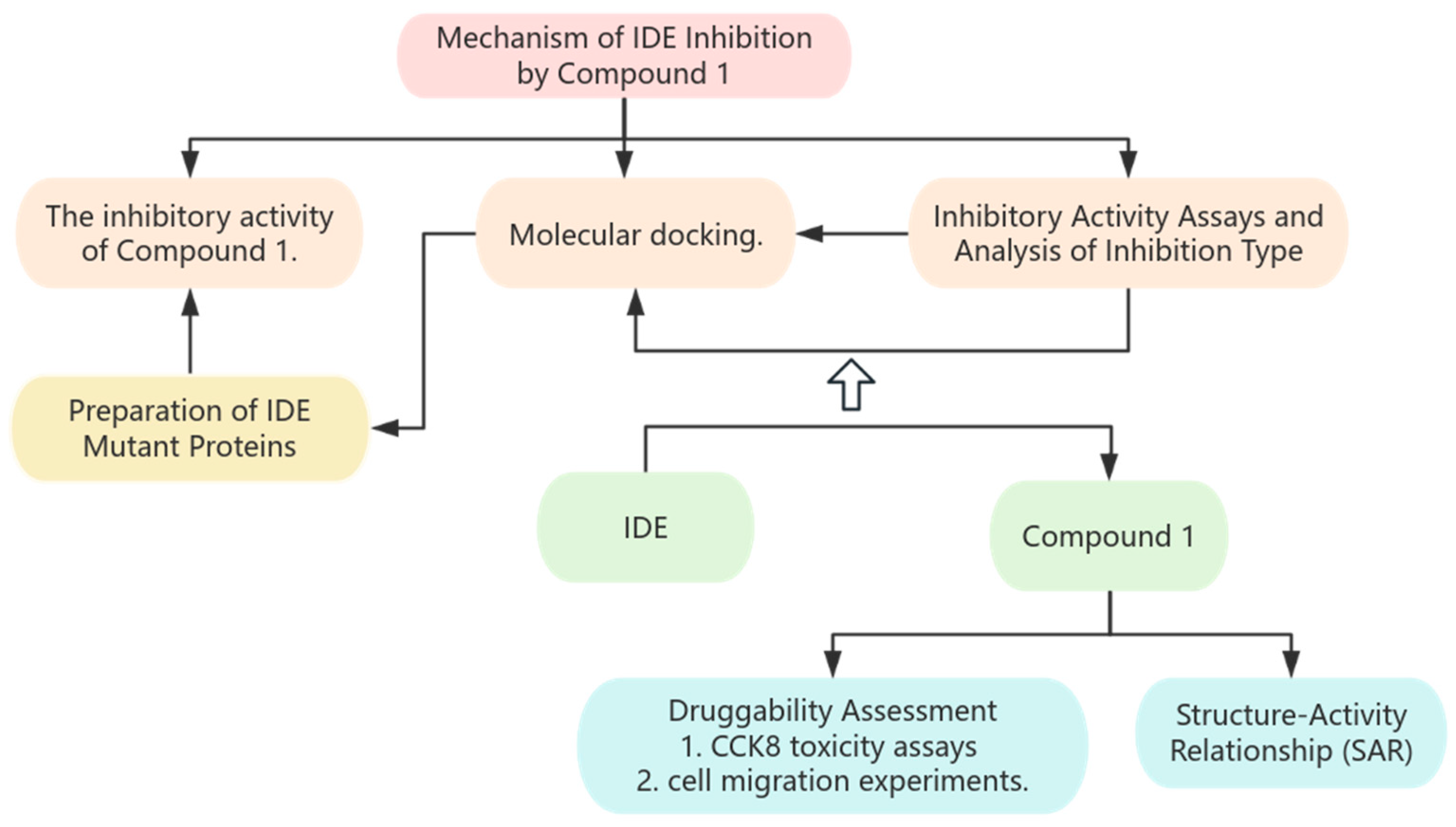
Figure 1. Experimental procedure flowchart.
Biochem 05 00027 g001
Figure 2. Compound 1 inhibits IDE in vitro. (a) Molecular structure and molecular weight of 1, TDZDs and TZD; (b) the inhibitory effect of 1 on IDE activity varies with concentration.
Biochem 05 00027 g002
Figure 3. Detection of 1 inhibition type on IDE. (a) The influence of 1 on the Michaelis–Menten curve of IDE enzyme activity reaction; (b) Lineweaver–Burk diagram of the effect of 1 on IDE enzyme activity.
Biochem 05 00027 g003
Figure 4. Docking of 1 with IDE. (a) Overall diagram of 1 docking with IDE molecule (PDB code: 2JG4); (b) amino acid residues in the 4 Å range around 1.
Biochem 05 00027 g004
Figure 5. Michaelis–Menten kinetics curves of wild-type IDE and mutant T142A. The Michaelis–Menten kinetics curve for T142A degrading the fluorescent substrate Mca-RPPGFSAFK(DNP)-OH was analyzed to assess its substrate-degrading activity.
Biochem 05 00027 g005
Figure 6. Evaluation of 1 for its cytotoxicity and selectivity. (a) The curve of cell viability of L929 cells with the concentration of 1. (b) The curve of cell viability of 4T1 cells with the concentration of 1; (c) effect of 10 μM 1 on cell viability of L929 cells and 4T1 cells (*** p < 0.001); (d) inhibitory effect of 10 μM 1 on the protein activity of IDE and MMP1 (** p < 0.01).
Biochem 05 00027 g006
Figure 7. Evaluation of 1 for its cell migration ability. (a) Cell migration ability of 4T1 cells. The left image shows 4T1 cells cultured in serum-free medium without the addition of an inhibitor, while the right image shows 4T1 cells cultured in serum-free medium with the addition of 3.6 μM of the inhibitor 1 (scale bar = 100 μm); (b) cell migration ability of L929 cells. The left image shows L929 cells cultured in serum-free medium without the addition of an inhibitor, while the right image shows L929 cells cultured in serum-free medium with the addition of 3.6 μM of the compound 1 (scale bar = 100 μm); (c) the changes in cell migration rate of 4T1 cells and L929 cells over time after interaction with 3.6 μM 1; the control group consisted of 4T1 and L929 cells cultured for 24 h without the addition of inhibitors (**** p < 0.0001).
Biochem 05 00027 g007aBiochem 05 00027 g007b
Table 1. The IC50 value of the inhibitor.
Comp.R1R2IC50p-Value
1Biochem 05 00027 i001Biochem 05 00027 i0023.602.09329 ± 0.41497
2Biochem 05 00027 i003Biochem 05 00027 i0043.231.24981 ± 0.52639
3Biochem 05 00027 i005Biochem 05 00027 i0067.596.55547 ± 0.25285
Table 2. Kinetic parameters and IC50 of IDE mutants.
IDEVmax (min−1)Km (μM)IC50 (μM)
WT2814.3211.933.5
T142A501.059.010.37
Disclaimer/Publisher’s Note: The statements, opinions and data contained in all publications are solely those of the individual author(s) and contributor(s) and not of MDPI and/or the editor(s). MDPI and/or the editor(s) disclaim responsibility for any injury to people or property resulting from any ideas, methods, instructions or products referred to in the content.