Review

Cancer Therapy-Related Left Ventricular Dysfunction: Are There New Gatekeepers?

by
Mariagrazia Piscione
1,*,
Maria Carmela Di Marcantonio
2,
Barbara Pala
3,* and
Gabriella Mincione
2
1
Fondazione Policlinico, Campus Bio-Medico University of Rome, Via Alvaro del Portillo, 200, 00128 Rome, Italy
2
Department of Innovative Technologies in Medicine & Dentistry, University “G. d’Annunzio” of Chieti-Pescara, Via dei Vestini 29, 66100 Chieti, Italy
3
School of Applied Medical-Surgical Sciences, University of Tor Vergata, Via Montpellier 1, 20, 00133 Rome, Italy
*
Authors to whom correspondence should be addressed.
BioChem 2025, 5(3), 25; https://doi.org/10.3390/biochem5030025
Submission received: 4 July 2025 / Revised: 8 August 2025 / Accepted: 11 August 2025 / Published: 12 August 2025
(This article belongs to the Special Issue Feature Papers in BioChem, 2nd Edition)

Abstract

The growing success of oncologic therapies has led to a significant improvement in patient survival; however, this has been accompanied by an increasing incidence of cardiovascular adverse events, particularly cancer therapy-related cardiac dysfunction (CTRCD). Among these, left ventricular impairment represents a major concern due to its potential to compromise both cardiac and oncologic outcomes. This review provides an in-depth overview of the cardiotoxic adverse events associated with several classes of anticancer agents. Particular focus is given to the molecular mechanisms involved in myocardial injury, such as oxidative stress, mitochondrial dysfunction, calcium dysregulation, endothelial reticulum stress, autophagy, and apoptosis. In parallel, established and emerging cardioprotective strategies, from conventional to newer therapeutic approaches, are explored. The role of advanced imaging modalities, as well as cardiac biomarkers, is discussed in the context of early detection and monitoring of subclinical cardiac injury. Finally, the integration of pharmacogenomics and epigenetics is considered as a promising avenue to personalize risk stratification and preventive therapy. By elucidating the complex interplay between cancer treatments and cardiovascular health, this review underscores the importance of a multidisciplinary, precision medicine approach to optimizing the care of patients undergoing potentially cardiotoxic therapies.

Graphical Abstract

1. Introduction

In recent years, cancer survival has markedly increased due to the plural progress in early diagnoses, improved surgical approaches, and new therapies [1]. As a matter of fact, updated detection techniques, precision oncology, and the emergence of new anticancer drugs have significantly improved the efficacy of cancer treatment protocols [1,2,3]. Nonetheless, these advancements have simultaneously increased the potential risks of cancer therapy-related cardiac dysfunction (CTRCD); treatment-induced hypertension; vasospastic and thromboembolic ischemia; and rhythm disorders, including life-threatening arrhythmias [4]. In addition, chest radiation is also considered responsible for pericardial and myocardial diseases, valve heart disease and prosthesis dysfunction, and myocardial fibrosis [4,5]. The prevalence of cardiovascular damage generated by cancer treatments significantly varies according to the type and the duration of therapy, and a patient’s preexisting comorbidities [4]. Surprisingly, according to a comprehensive review considering breast cancer survivors in the United States, treated women faced a markedly elevated risk of mortality due to cardiovascular disease, exceeding the probability of death from the primary cancer or its recurrence [6,7]. That is the reason why, over the decades, the acute and long-term cardiovascular consequences of cancer therapies have been deeply studied by a new discipline: cardio-oncology [4]. Nowadays, precision cardio-oncology plays a pivotal role, integrating molecular profiling and risk stratification to tailor surveillance and cardioprotective strategies [8]. Indeed, precision cardio-oncology, together with advanced imaging techniques—such as speckle-tracking echocardiography and cardiac magnetic resonance (CMR)—and the use of biomarkers have a proven efficacy for detecting subclinical myocardial dysfunction before irreversible damage occurs [9]. These are now considered essential to comprehensive cancer care, since they can guide cardiovascular assessments before, during, and after treatment, with the goal of integrating both oncologic efficacy and cardiovascular safety into standard clinical practice, emphasizing early intervention and continuous monitoring [4]. By contrast, a research field still in its early stages that deserves further development concerns the use of novel pharmacological strategies for both the prevention of chemotherapy-induced damage and the improvement of ventricular contractility at the end of therapeutic cycles [4].
This review aims to summarize the molecular mechanism of damage of the most dangerous drugs that cause left ventricular (LV) impairment. Furthermore, innovative approaches for evaluating and managing the risk factors associated with cardiovascular disease (CVD) in cancer-treated patients are described. Last, but not least, the role of new protective drugs for both asymptomatic and symptomatic CTRCD is discussed, with a focus on new technologies that provide individualized approaches to CTRCD prevention, such as genomics and gene therapy. According to the guidelines, this review emphasizes the need for a collaborative, multidisciplinary approach involving dedicated cardio-oncology teams, which are essential for identifying patients at risk early, tailoring monitoring strategies, and implementing preventive measures to reduce the burden of CVD.

2. Cancer Therapy and Cardiotoxicity: An Overview

Cardiotoxicity is a well-known adverse event of several cancer therapies (Table 1), and it represents a leading cause of morbidity among cancer survivors [4].
Anthracyclines, which are commonly used to treat solid tumors and hematologic malignancies, have cumulative, dose-dependent, and progressive cardiotoxic effects [10]. Doxorubicin (DOXO), daunorubicin, epirubicin, and idarubicin are derived from Streptomyces peucetius var. caesius [10]. Their clinical utility is hindered by the potential for toxicity, which can appear up to ten years after the cessation of chemotherapy [10]. Their acute side effects include hematological suppression, nausea, vomiting, and alopecia; however, the most feared one is cardiotoxicity. Acute toxicity is clinically characterized by a wide variety of signs, from asymptomatic electrocardiographic abnormalities to pericarditis and decompensated biventricular heart failure (HF) [11]. The likelihood of developing HF is predominantly dose-dependent, but cardiotoxicity may also manifest at low dosages and be due to individual vulnerabilities [11]. Age, the concurrent administration of additional cardiotoxic agents, mediastinal radiotherapy, and the cumulative dosage of DOXO are risk factors associated with an elevated risk of DOXO-induced HF [12,13,14,15]. Beyond these, gender differences have been identified as a risk factor for the harmful effects of DOXO [11]. Lipshultz, S.E. et al. showed that women experience more severe cardiotoxicity with greater contractility depression [12].
Even though the mechanisms of damage are not clearly and completely known, they can be grouped into genomic instability, oxidative stress, mitochondrial dysfunction, and activation of apoptotic pathways in cardiomyocytes [9], and are summarized in Figure 1.
First of all, anthracyclines inhibit topoisomerase 2β (Top2β), which prevents DNA repair [16,17]. Two Top2 enzymes exist: Top2α and Top2β [18]. Top2α, a recognized marker of cellular proliferation, is overexpressed in malignancies but is undetectable in quiescent tissues, and in the past it was thought to be responsible for anthracycline toxicity [18]. This notion remained accepted until Zhang et al., in 2012, identified topoisomerase 2β (Top2β) as a critical mediator of DOXO-related cardiac injury [17]. In particular, the researchers showed in murine models that the interaction between DOXO and Top2β in cardiomyocytes led to deoxyribonucleic acid (DNA) double-strand breaks [18]. Using a cardiomyocyte-specific deletion of Top2β, the authors demonstrated that the absence of this isoform completely removed the cardiotoxic effects of DOXO without altering its anticancer efficacy. These findings not only elucidated the molecular mechanism underlying cardiac harm, but also highlighted Top2β as a potential therapeutic target for selective cardioprotection during anthracycline therapy [17].
Another mechanism through which DOXO damages the myocardium is the stimulation of oxygen reactive species (ROS), considering that the heart is very susceptible to oxidative stress due to its high oxygen demand, dense mitochondrial content, limited antioxidant defenses, and vulnerability to injury during ischemia/reperfusion [18]. Mitochondria are severely and progressively involved since the cationic molecule of DOXO is sequestered in the mitochondrial inner membrane by establishing a nearly irreversible combination with cardiolipin, which is an essential phospholipid found mainly in the mitochondrial inner membrane, where it plays a critical role in oxidative phosphorylation [19]. The proteins of the electron transport chain need an optimal functioning of cardiolipin, but DOXO disrupts the cardiolipin–protein interface, leading to increased superoxide (O2) production [20]. It is highly probable that ROS production interferes with mitochondria and consequently cellular metabolism, as mitochondria generate over 90% of the adenosine tri-phosphate (ATP) consumed by cardiomyocytes [21]. This functional impairment results in ultrastructural pathological alterations, including mitochondrial swelling and the formation of myelin patterns inside these organelles [21]. However, it has to be considered that the doses used to treat malignancies are lower than those used in in vitro studies, so that DOXO-related side effects are generally less severe in vivo [22].
Moreover, several studies have highlighted the central role of endothelial nitric oxide synthase (eNOS) in DOXO-induced cardiotoxicity, which not only involves cardiomyocytes but also the cardiac vasculature [22]. Vasquez-Vivar et al. demonstrated that DOXO binds to the reductase domain of eNOS, leading to the generation of O2 via the formation of a semiquinone radical, which is an unstable and reactive molecule [22]. This reaction does not involve the oxygenase domain, which is commonly responsible for the production of nitric oxide (NO) that is protective of the endothelium [23]. By contrast, the redox activation of DOXO by eNOS is associated with apoptosis and endothelial dysfunction, shifting the enzyme function from NO production to ROS generation [24]. Murine models have shown that the genetic deletion of eNOS protects against DOXO-induced cardiac dysfunction, while eNOS overexpression worsens it [22,25].
Another pathogenic mechanism involves the dysregulation of intracellular calcium; actually, it has been demonstrated that DOXO-induced cardiotoxicity is associated with elevated intracellular calcium levels, while a calcium ion chelator can decrease DOXO-induced ROS production and apoptosis [26]. According to Saeki et al., DOXO can bind to the ryanodine channel in the sarcoplasmic reticulum, regardless of the channel’s state. DOXO inhibits the sodium–calcium exchanger channel in the sarcolemma, which expels calcium from cells and increases the activity of the L-type calcium channel, increasing intracellular calcium levels and promoting the development of DOXO-induced cardiomyopathy [27]. It is widely established that DOXO-induced oxidative stress stimulates apoptotic signaling, resulting in cardiomyocyte death, and that both extrinsic and intrinsic apoptotic mechanisms are involved [28,29].
Furthermore, DOXO has also been shown to trigger apoptosis by mechanisms other than ROS generation and oxidative stress, but this is confounded by the fact that apoptosis itself produces free radicals. Research studies have proved that oxidative stress stimulates heat shock factor 1 (HSF-1), which is a “sensor” protein that becomes activated when a cell is under stress [28]. Then, HSF-1 translocates to the nucleus and induces the expression of specific protective proteins known as heat shock proteins (HSPs), in particular Hsp25 (heat shock protein 25), which stabilizes protein 53 (p53), a key regulatory protein that monitors cellular damage and determines whether to initiate repair processes or to trigger cell elimination. When p53 is not degraded, it promotes the expression of pro-apoptotic proteins, which drive cell apoptosis [28]. The interplay of HSF-1, Hsp 25, and p53 provides an example of the difficulty in distinguishing between oxidative stress and apoptosis, since this cascade illustrates how a protective response can paradoxically lead to unjustified cell death and myocardial tissue loss. The close interconnection between oxidative stress and apoptosis complicates the distinction between cause and consequence, as each process can amplify the other [28].
Trastuzumab, a humanized monoclonal antibody which targets human epidermal growth factor receptor 2 (HER2), is approved for treating HER2-positive breast cancer and metastatic gastric cancer [30]. It suppresses tumor growth by binding to domain IV of the extracellular domain of HER2. It activates antibody-dependent cell-mediated cytotoxicity and inhibits HER2 extracellular domain cleavage [30]. The latter prevents cancer cells from releasing a truncated, active form of the HER2 receptor, which can promote tumor growth even in the absence of external signals. Trastuzumab blocks oncogenic cellular signaling, and downregulates angiogenesis and DNA repair pathways [30].
The specific molecular mechanisms of trastuzumab-induced cardiotoxicity are still unclear, despite extensive research [31]. In general, trastuzumab-induced cardiac dysfunction is considered milder and more reversible than anthracycline-related cardiomyopathy due to the absence of ultrastructural changes in the cardiomyocytes, since patients treated with trastuzumab do not experience primary myocyte injury [32]. Nonetheless, therapy with this monoclonal antibody has been linked to both short-term and long-term adverse events, highlighting the importance of prolonged cardiac follow-up [33]. In fact, its transient toxicity has been questioned by some in vivo studies, which have shown that it causes ultrastructural changes in mouse cardiac tissues, as observed by electron microscopy [34]. Indeed, treatment with it has altered the gene expression profiles related to cardiac and mitochondrial activities, as well as DNA repair [34]. The mechanism of myocardial injury consists of suppressed autophagy in cardiomyocytes, leading to the accumulation of toxic ROS [35]. Trastuzumab alters HER signaling by phosphorylating HER1 and HER2 at 845 and 1248 sites, respectively, and activating the autophagy-inhibitory signaling cascade constituted of extracellular signal-regulated kinase/mammalian target of rapamycin/Unc-51-like autophagy-activating kinase 1 (Erk/mTOR/Ulk 1). This compromises cardiomyocytes’ ability to eliminate toxic cellular substrates, resulting in cardiotoxicity [35]. Different from trastuzumab, pertuzumab, an anti-HER2 monoclonal antibody with a distinct epitope, has not affected the HER2 signaling cascade in in vitro models [36]. Actually, pertuzumab targets the dimerization domain (domain II) of the HER2 receptor, thereby preventing the heterodimerization of HER2 with other members of the endothelial growth factor receptor (EGFR) family, particularly HER3 [36]. This blockade effectively inhibits downstream signaling pathways, which are critical for tumor cell proliferation and survival. Pertuzumab is typically used in combination with trastuzumab and chemotherapy, taking advantage of their complementary mechanisms of action to enhance antitumor efficacy [36]. By contrast, the mechanisms of cardiac toxicity associated with the combination of anthracycline and trastuzumab are still unknown [35]. In a recent paper, it was demonstrated that Trastuzumab reduced Top2β, a key target in DOXO-induced cardiotoxicity, and that the combined use of this drug with DOXO appeared to exert a protective effect [35]. Conversely, trastuzumab and DOXO treatment significantly increased apoptosis, inhibited cell growth, and promoted production of ROS and nitrative species in human cardiomyocytes [35].
Proteasome inhibitors (PIs), bortezomib, carfilzomib, and the oral ixazomib, together with immunomodulatory drugs, monoclonal antibodies, and chemotherapy, are used to treat patients with plasma cell dyscrasias [37]. Proteostasis is essential for maintaining the integrity of the cellular proteome, ensuring cell function and viability [38]. The ubiquitin proteasome (UPP) is the primary proteolytic mechanism in mammalian cells, which degrading up to 80% to 90% of intracellular proteins. The UPP consists of three components: ubiquitin, ubiquitin-conjugating enzymes, and the proteasome [39]. The proteasome removes both short-lived ubiquitinated proteins and non-repairable polypeptides, and decreased proteasome activity leads to increased proteome instability, which can cause severe proteotoxic stress [39]. Inhibition of proteasomes resulted in both reduced angiogenesis and increased cancer cell death in multiple myeloma (MM) cells [40]. At the same time, cardiomyocytes, which are highly dependent on proteasomal activity for their contractile function, are particularly vulnerable to PIs [4]. A recent study demonstrated the molecular mechanism of heart toxicity due to carfilzomib [41]. In Drosophila, the genetic suppression of the proteasome altered heart function and decreased cell lifespan [41]. In addition, carfilzomib may cause LV dysfunction in mice via increasing phosphoprotein phosphatase 2A (PP2A) activity and disrupting autophagy by inhibiting AMP-activated protein kinase a (AMPKa) and its downstream pathways [41]. Furthermore, reduced UPP activity also depends on a patient’s fragility [42]. A study proved that PIs in old age lead to increased proteome instability [42]. Additionally, the elderly are more likely to have traditional cardiovascular risk factors, and patients with MM might have been exposed to potentially cardiotoxic therapies like anthracyclines or chest radiotherapy, which can increase the risk of developing HF [42]. Carfilzomib has been associated with a higher incidence of cardiovascular events compared to other PIs, likely due to its irreversible inhibition of proteasomal activity [43]. A systematic review and meta-analysis of 24 clinical trials reported an 18.1% incidence of cardiac adverse events and a 4.1% incidence of HF in patients treated with this drug [44]. Notably, in a prospective study, the patients receiving PIs developed signs and symptoms of HF despite having a normal or near-normal left ventricular ejection fraction (LVEF), suggesting a phenotype consistent with HF with preserved ejection fraction (HFpEF) [45]. Hypertension, a key driver in HFpEF pathogenesis, has been frequently observed during carfilzomib therapy and is considered one of the most common cardiovascular adverse events associated with its use [45]. Although the data remain limited, several risk factors for carfilzomib-related cardiotoxicity have been identified, including pre-existing cardiovascular disease (CVD), age over 75, obesity, use of a twice-weekly dosing schedule, concurrent administration of immunomodulatory drugs, doses exceeding 45 mg/m2, and the presence of chronic obstructive pulmonary disease [45]. The pathophysiological mechanisms appear to be multifactorial, with vascular dysfunction playing a central role [43]. Carfilzomib-induced endothelial damage leads to reduced NO bioavailability, increased vascular stiffness, and elevated systemic vascular resistance, ultimately resulting in persistent hypertension with LV remodeling and myocardial stiffening [43]. Additionally, this drug has been shown to promote pro-inflammatory signaling and oxidative stress, both of which contribute to the activation of fibrotic pathways and extracellular matrix deposition in the myocardium, which impairs diastolic relaxation [43,44]. Together, these mechanisms provide a plausible explanation for the HFpEF phenotype observed in patients treated with carfilzomib, highlighting the need for careful blood pressure control and the early recognition of subclinical diastolic dysfunction during therapy [43]. In contrast, bortezomib appears to have a more favorable cardiovascular safety profile [46]. A retrospective observational cohort study involving 1790 patients with MM found no significant increase in the risk of hospitalization for HF in bortezomib-treated patients compared to those receiving lenalidomide, following propensity score matching [46].
Small-molecule tyrosine kinase inhibitors (TKIs) targeting the Breakpoint Cluster Region–Abelson Leukemia (BCR-ABL) have been shown to effectively treat blood cancer and advanced solid tumors [47]. TKIs are thought to be safer than classic chemotherapy medications like DOXO [48]. However, since TKIs are often administered for long periods without a maximum dosage limit, some of them continue to cause significant cardiac side effects [48]. In a meta-analysis of 6935 patients, sunitinib was found to be related to 4.1% of congestive HF [49]. The mechanisms of LV dysfunction are many and interrelated. Recent findings, including those by Wang et al., have highlighted a common molecular mechanism underlying the cardiotoxic effects of multiple TKIs: the induction of ER stress [47]. In their study, human induced pluripotent stem cell-derived cardiomyocytes (hiPSC-CMs) were used to model the cardiac cellular response to eight different TKIs. All the compounds were found to activate the inositol-requiring enzyme 1 alpha (IRE1α) pathway, leading to the splicing of X-box binding protein 1 (XBP1), a key branch of the unfolded protein response [47]. This ER stress response was preceded by a transient but significant increase in ROS production, followed by lipid peroxidation—a typical manifestation of oxidative damage [47]. The downstream consequences of ER stress included the upregulation of pro-inflammatory genes and the reactivation of fetal gene programs, both of which are considered molecular signatures of pathological cardiac remodeling [47]. These findings provide mechanistic insight into how TKIs disrupt cardiac homeostasis and point to ER stress and oxidative imbalance as key contributors to their cardiotoxic potential [47].
Mitogen-activated protein kinase kinase (MEK) inhibitors target the RAS sarcoma–rapid accelerated fibrosarcoma–mitogen-activated protein kinase (Ras-Raf-MAP kinase) pathway and are used to treat metastatic melanoma [50]. They impede the downstream target of BRAF. The main cardiovascular adverse event is LV dysfunction, especially when combined with a BRAF inhibitor [50]. Interrupting or reducing the doses of this drug can lead to improved LV function [50]. The incidence of HF in patients treated with BRAF and MEK inhibitors in clinical trials, with a median follow-up of 9 to 16 months, has been reported to be 2% to 12% [51]. However, this could be an underestimate for a variety of reasons, one of the most important being that patients with severe underlying cardiovascular morbidity are often excluded from clinical studies [50].
Immune checkpoint inhibitors (ICIs) are monoclonal antibodies that prevent immune checkpoints from interacting with their ligands [52]. They have been evaluated in both preclinical and clinical trials [53]. Despite their efficacy against cancer, ICIs are associated with immune-related adverse events caused by off-target immune system hyperactivation and dysregulation, resulting in damage to various organ systems with clinical consequences, such as colitis, hepatitis, dermatitis, thyroiditis, myocarditis, or hypophysitis [53]. Although the rate of cardiac toxicity found in clinical trials following ICI treatments is rather rare, 3.1% in monotherapies and 5.8% in dual/combination therapies, it can be fatal [53]. Rubio-Infante et al. conducted a systematic analysis to elucidate the potential immune mechanisms contributing to cardiac toxicity associated with ICIs [54]. The study identified three primary pathways: T cell recruitment and activation, autoantibody-mediated cardiotoxicity, and inflammatory cytokine release [55]. ICIs alter peripheral immune tolerance, leading to the activation and infiltration of cluster of differentiation 4+ (CD4+) T cells and cluster of differentiation 8+ (CD8+) T cells into cardiac tissue [55]. This immune response can result in myocardial inflammation and damage [55]. Moreover, the pro-inflammatory environment induced by ICIs may activate autoreactive B cells, leading to the production of autoantibodies that target cardiac antigens [56]. These autoantibodies can mediate cardiotoxicity through mechanisms such as complement-dependent cytotoxicity and antibody-dependent cellular cytotoxicity. ICIs can also promote the release of pro-inflammatory cytokines, contributing to systemic inflammation and potential cardiac tissue damage [57]. In synthesis, myocardial injury is primarily driven by an aberrant activation of cytotoxic T lymphocytes, which can infiltrate cardiac tissue and induce direct cytotoxicity through the release of perforin, granzymes, and interferon-γ [57]. The heart naturally expresses Programmed Death Ligand-1 (PD-L1) as a protective mechanism to prevent autoimmunity; the blockade of this pathway renders cardiac tissue more susceptible to immune-mediated attack [57]. Additionally, antigenic cross-reactivity between tumor and myocardial epitopes (molecular mimicry) may trigger unintended autoimmune responses. The systemic release of pro-inflammatory cytokines, such as tumor necrosis-α (TNF-α) and interleukin-6 (IL-6), along with the activation of intracellular signaling pathways, contribute to endothelial dysfunction, myocardial fibrosis, and ventricular remodeling—key components in the pathogenesis of ICI-induced HF [57].
Table 1. Summary of the main antitumor treatments and their possible mechanisms of cardiotoxicity.
TreatmentsMechanism of CardiotoxicityCRTCD*/LV Systolic
Dysfunction
References
AnthracyclinesTop2β inhibition: DNA breaks; ROS production;
mitochondrial dysfunction;
calcium dysregulation;
apoptosis;
eNOS uncoupling.
Cumulative and dose-dependent;
occurs years after therapy.
[17,22,24]
HER2-Targeted TherapiesOxidative stress;
suppression of autophagy;
mitochondrial gene expression alteration.
Typically reversible;
increased risk when combined with anthracyclines.
[7,31,32]
Proteasome InhibitorsUPP inhibition: proteotoxic stress;
autophagy suppression;
vascular dysfunction;
ROS and inflammation.
HFpEF,
subclinical diastolic dysfunction;
higher risk in elderly or those with comorbidities.
[39,40,41]
Tyrosine Kinase InhibitorsER stress;
ROS burst and lipid peroxidation;
upregulation of pro-inflammatory and fetal gene programs.
LV dysfunction;
cardiac remodeling associated with long-term use.
[48,49,50]
MEK/BRAF InhibitorsMAPK pathway inhibition: LV dysfunction;
reversibility upon dose reduction.
LV dysfunction;
underreported due to patient selection.
[51,52]
Immune Checkpoint InhibitorsT CD4+ and CD8+ activation;
autoantibody generation;
cytokine storm;
PD-L1 pathway suppression; molecular mimicry: myocarditis, fibrosis, HF.
Myocarditis, HF;
rare but often severe or fatal.
[55,56]
* Abbreviations: CRTCD: cancer therapy-related cardiac dysfunction; CD4+: cluster of differentiation 4+; CD8+: cluster of differentiation 8+; DNA: deoxyribonucleic acid; ER: endoplasmic reticulum; eNOS: endothelial nitric oxide synthase; HER2: human epidermal growth factor receptor 2; HF: heart failure; HFpEF: heart failure with preserved ejection fraction; LV: left ventricle; MAPK: mitogen-activated protein kinase; MEK/BRAF: mitogen-activated protein kinase/v-Raf murine sarcoma viral oncogene homolog B1; PD-L1: Programmed Death Ligand-1; ROS: reactive oxygen species; Top2β: topoisomerase 2β; UPP: ubiquitin proteasome.

3. The “Old Gatekeepers”: Early Detection, Biomarkers, and Risk Stratification of CVDs

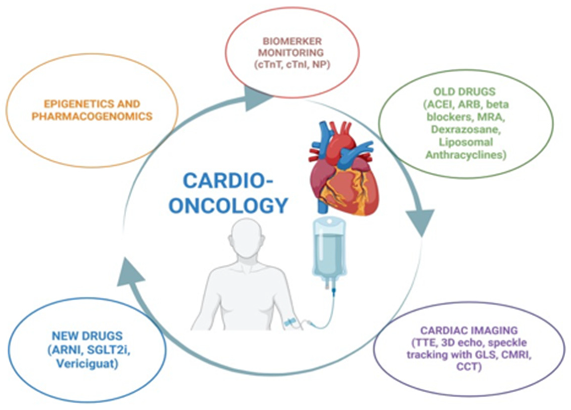
In a cardio-oncology setting, cardiovascular adverse effects can develop insidiously during or after cancer therapy, often progressing to irreversible myocardial damage before symptoms appear. The early detection of subclinical injury is therefore crucial to enable timely intervention and prevent long-term sequelae. Biomarkers and advanced cardiac imaging provide sensitive and specific means to identify these early changes, while validated risk stratification tools help individualize the surveillance intensity and therapeutic strategies. This combination forms the foundation for effective prevention, diagnosis, and management in contemporary cardio-oncology practice. Although this review primarily focuses on the molecular mechanisms underlying the cardiotoxic effects of major chemotherapeutic agents and new drugs, it is essential to briefly highlight the critical role of biomarkers, advanced imaging, and risk stratification tools in clinical practice [4] (Figure 2).
The 2022 ESC guidelines recommend measuring cardiac biomarkers (cardiac troponin I (cTnI) or cardiac troponin T (cTnT)) and natriuretic peptides (NPs), including B-type natriuretic peptide (BNP) and N-terminal pro-BNP (NT-proBNP), to assess the cardiovascular risk of patients receiving anthracyclines, HER2-targeted treatments, and vascular endothelial growth factor (VEGF) inhibitors [4,58]. Monitoring biomarkers during treatment can help identify patients who may benefit from cardioprotective therapies. Therefore, it is essential to establish the baseline levels of NPs and/or cTn for patients at high risk of CTRCD and to track their changes throughout treatment [58]. However, there is no universally agreed threshold value for these in cancer patients because of confounding factors, such as age, renal function, and comorbidities, so their alterations should be evaluated taking into account patients’ clinical profiles [58]. A 2020 meta-analysis found that elevated troponin levels following chemotherapy increased the likelihood of LV dysfunction by sevenfold, with a 93% negative predictive value [59]. In contrast, a BNP and NT-proBNP levels increase following cancer treatment had an inconsistent prognostic value [59].
In addition to biomarkers evaluation, cardiovascular imaging plays a crucial role in detecting the early signs of cardiac dysfunction, assessing pre-existing heart conditions, and monitoring changes during and after cancer treatment [4,60]. Transthoracic echocardiography (TTE) is essential for diagnosing LV failure, and the LV ejection fraction (EF) is the key measurement [60]. Three-dimensional echocardiography is now considered the best method to measure the LVEF and chamber volumes due to its higher accuracy and lower interobserver variability (5–6%) [60,61]. If 3D is not accessible, the global longitudinal strain (GLS), derived from speckle tracking, is a more sensitive and reproducible measure of LV systolic function, since it can detect subclinical dysfunction, corresponding with early molecular alterations, such as mitochondrial dysfunction, oxidative stress, and cardiomyocyte apoptosis [62]. A ≥15% decline in the GLS from baseline indicates subclinical cardiotoxicity and an increased risk of eventual LV dysfunction [62]. It is important to note that the GLS may exhibit slight variations depending on the equipment used and on chemotherapy-induced changes in the loading conditions [62]. Myocardial work (global work index, global constructive work, global wasted work, and global work efficiency) is a promising early cardiotoxicity marker, especially with anthracycline or anthracycline/trastuzumab therapy [63]. Although further research is needed, it is established that the GLS provides a non-invasive insight into molecular cardiotoxicity, reinforcing its role in risk stratification and the early monitoring of at-risk patients [4].
CMR represents an interesting alternative to TTE for the detection of CTRCD [64]. In addition to its superior spatial resolution, CMR offers unparalleled accuracy in quantifying both left and right ventricular function, an aspect of particular relevance in patients treated with HER2-targeted agents [64]. Crucially, CMR allows for myocardial tissue characterization via T1 and T2 mapping sequences, thereby enabling the detection of diffuse fibrosis, interstitial expansion, and myocardial edema—hallmarks of early molecular injury [65]. Among the most promising tools derived from CMR is the GLS obtained through feature tracking (FT). This parameter has been demonstrated to detect subclinical myocardial dysfunction well before an overt reduction in the LVEF, reflecting the early reduction in the EF linked to mitochondrial dysfunction, oxidative stress, and apoptosis of longitudinally oriented subendocardial fibers—structures especially vulnerable to cytotoxic insult [65]. In this regard, the retrospective analysis by Romano et al., considering 470 patients with reduced LVEF and CMR evaluation using both cine and late gadolinium enhancement (LGE), revealed that FT-derived GLS independently predicted all-cause mortality in both ischemic and non-ischemic cardiomyopathy. Remarkably, the GLS has added incremental prognostic value beyond that of established markers, such as the LVEF and LGE burden [65]. This finding underscores the vulnerability of the subendocardial longitudinal fibers, whose dysfunction likely represents a downstream consequence of cumulative molecular damage, including ROS generation, calcium dysregulation, and mitochondrial injury [65]. Moreover, CMR has proven capable of detecting early myocardial edema through T2-weighted imaging in patients receiving anthracyclines, suggesting its utility in recognizing the inflammatory and metabolic alterations that precede irreversible structural damage [66,67]. These features make CMR not only a diagnostic modality but also a non-invasive molecular “lens” through which cardiotoxicity can be visualized and understood in its earliest and most reversible stages [66].
Cardiac computed tomography (CCT) can examine heart chamber function by providing detailed volumetric and structural data for both ventricles using retrospective multiphase ECG-gated imaging [68]. CCT is a reliable alternative to CMR for functional and morphological analyses, and it is highly sensitive in excluding obstructive CAD in patients with a low-to-intermediate pre-test risk of CAD (high negative predictive value) [69]. Quantifying the coronary artery calcium score with a non-contrast cardiac CT has been proposed to estimate the cardiovascular risk in cancer patients and survivors [69]. Recent research suggests that the CCT-derived extracellular volume (ECV) could be a biomarker for monitoring anthracycline-induced cardiotoxicity in breast cancer patients [70,71]. A proactive, multimodal strategy combining biomarkers and imaging is essential to optimize cardio-oncologic care.

4. The “Old Gatekeepers”: Drugs Which Protect Against Ventricular Dysfunction

The 2022 ESC cardio-oncology guidelines recommend angiotensin-converting enzyme inhibitors (ACEIs) or angiotensin receptor blockers (ARBs), along with beta-blockers, for preventing cardiotoxicity in high- or very-high-risk patients undergoing anthracycline, anti-HER-2, or other targeted anticancer therapies that may cause LV systolic dysfunction, according to the Heart Failure Association–International Cardio-Oncology Society score (HFA-ICOS score) [4] (Table 2).

4.1. Angiotensin-Converting Enzyme Inhibitors and Angiotensin Receptor Blockers

ACEIs and ARBs reduce oxidative stress but enhance mitochondrial activity and cardiomyocyte metabolism. In a study, captopril and enalapril were tested for their antioxidant protection against DOXO (adriamycin)-induced cardiac and liver toxicity. Rats were given captopril (10 mg/kg) or enalapril (2 mg/kg) intragastrically (i.g.) daily for 7 days before receiving a single i.p. injection of DOXO (15 mg/kg). After 30 h, DOXO killed the animals [72]. Indeed, it increased thiobarbituric acid reactive substances (TBARS), an indicator of lipid peroxidation, and inhibited superoxide dismutase (SOD) in the heart and liver tissues, indicating acute cardiac toxicity [72]. Nonetheless, it was demonstrated how ACEI pretreatment significantly lowered heart and liver TBARS concentrations and improved cardiac and hepatic SOD activity inhibition [72]. These findings suggest that captopril and enalapril may protect the heart from DOXO-induced acute oxidative damage by limiting ROS production and reducing oxidative stress [72]. Furthermore, Vaynblant et al. evaluated the protective effects of enalapril against DOXO-induced HF in a canine model [73]. Seventeen dogs were treated with DOXO over four weeks, with one group receiving concurrent enalapril therapy [73]. The results showed a marked improvement in survival (100% vs. 36%), as well as significant improvement in the LVEF in the enalapril-treated group [73]. The experimental data from a rat model of daunorubicin-induced cardiotoxicity suggest that ACEIs may exert early cardioprotective effects, improving myocardial contractility and normalizing QT prolongation, even in the absence of a full recovery of the LVEF [74]. These findings support the potential for early neurohormonal modulation to prevent structural deterioration during anthracycline therapy [74]. Other preclinical studies have maintained that anthracyclines alter the renin–angiotensin–aldosterone system (RAAS), leading to increased levels of angiotensin II and overexpression of angiotensin type 1 receptor, thereby contributing to cardiotoxicity [75]. The clinical evidence supports these findings: both retrospective and prospective studies have demonstrated that ACEIs reduce the likelihood of LVEF decline in this setting [76,77]. Moreover, starting an ACEI medication within six months of chemotherapy improves the results [78]. Furthermore, HER-2 inhibition, as occurs with trastuzumab and other targeted therapies, can disrupt the myocardial signaling pathways that normally limit activation of the RAAS. This disruption has been associated with increased angiotensin II production and oxidative stress via NADPH oxidase activation [77]. These effects may contribute to myocardial injury, providing a mechanistic rationale for considering ACEIs to counteract neurohormonal activation and oxidative damage in this setting. So, according to recent studies, ACEIs may also help manage the cardiotoxicity of HER2 inhibitors [77].

4.2. Beta-Blockers

Beta-blockers have cardioprotective functions, including antioxidant and antiapoptotic properties [79]. β1-adrenergic receptors (β1ARs) are key regulators of cardiac function and are therapeutic targets in heart disease, mediating β-arrestin-dependent pathways [79]. Carvedilol, a β-arrestin-biased β-blocker, selectively activates these non-canonical signals [79]. To test the effects of carvedilol, Santos et al. extracted heart and liver mitochondria from rats treated for 7 weeks with DOXO (2 mg/kg subcutaneously per week), carvedilol (1 mg/kg intraperitoneally per week), or their combination [47]. The mitochondrial function was assessed by measuring state 3 respiration—the rate of oxygen consumption upon the addition of adenosine diphosphate (ADP)—which reflects active engagement in ATP synthesis and is a key indicator of cellular metabolic capacity [47]. Moreover, they investigated the respiratory control ratio (RCR), which is the ratio between state 3 respiration (ADP-stimulated, active respiration) and state 4 respiration (resting, basal respiration) and reflects how effectively mitochondria couple oxygen consumption to ATP production [47]. The heart mitochondria isolated from DOXO-treated rats had lower rates of state 3 respiration (336 +/− 26 versus 425 +/− 53 natom O2/min/mg protein) and a lower RCR (4.3 +/− 0.6 versus 5.8 +/− 0.4) than the cardiac mitochondria isolated from the saline-treated rats [47]. The DOXO treatment reduced the mitochondrial calcium-loading capacity and NADH-dehydrogenase activity. Moreover, it was observed that carvedilol coadministration reduced cellular vacuolization in cardiac myocytes and avoided DOXO’s inhibitory effect on mitochondrial respiration in both the heart and liver [47]. In conclusion, carvedilol gives protection against both structural and functional cardiac tissue damage and provides significant clinical benefit in reducing the dose-limiting mitochondrial dysfunction that accompanies long-term DOXO therapy in cancer patients [80]. In addition, in clinical investigations, such as the CarDHA study, it was demonstrated that carvedilol was necessary to minimize DOXO-induced cardiotoxicity and preserve LVEF. However, its oral intake does not necessarily result in a significant reduction in HF hospitalizations or cardiac fatalities [81].

4.3. Mineralcorticoid Receptor Antagonist

Spironolactone is a mineralcorticoid receptor antagonist (MRA) that inhibits the final phase of the RAAS pathway, which has shown antioxidative effects and is being studied for possible benefits; however, the research is limited [82].

4.4. Dexrazoxane and Liposomal Anthracyclines

The use of dexrazoxane and liposomal anthracyclines in individuals at high or very high risk of cardiotoxicity who require anthracycline therapy is strongly recommended [4].
Dexrazoxane is the only clinically approved cardioprotective agent for preventing anthracycline-induced cardiotoxicity, and its efficacy relies on a dual mechanism of action. First, it acts as an iron chelator, preventing the formation of anthracycline–iron complexes that drive the production of ROS. Second, it inhibits Top2β, a key mediator of DOXO-induced DNA damage in cardiac cells [83]. By competitively binding to Top2β, dexrazoxane prevents the formation of DOXO–Top2β complexes, avoiding double-strand DNA breaks and the subsequent activation of pro-apoptotic pathways [83]. These complementary mechanisms help preserve myocardial integrity without compromising the antitumor efficacy of anthracyclines [83].
Liposomal anthracyclines, particularly pegylated liposomal DOXO, have been developed to reduce the cardiotoxic effects of conventional anthracyclines [84]. Liposomal formulations of DOXO modify drug distribution, leading to lower peak plasma concentrations and reduced myocardial exposure. By preferentially targeting tumor tissue while limiting cardiac uptake, they have been associated with a lower incidence of anthracycline-induced cardiotoxicity [84,85]. As a result, cardiac exposure to DOXO is significantly decreased, leading to lower levels of ROS, reduced mitochondrial damage, and less induction of apoptosis in cardiomyocytes [85].

4.5. Statins

Even though statins are mainly considered for acute and chronic ischemic diseases, emerging evidence suggests that these drugs may offer cardioprotective effects against chemotherapy-induced cardiotoxicity, particularly in patients receiving anthracyclines and trastuzumab. Preclinical and early clinical studies first highlighted this potential, such as the pilot study by Acar et al., which demonstrated that atorvastatin significantly reduced the development of anthracycline-induced cardiomyopathy, possibly via anti-inflammatory and antioxidant mechanisms in addition to their ability to decrease cholesterol [86]. Moreover, Riad et al. demonstrated in an animal model that pretreatment with fluvastatin reduced the risk of chemotherapy-induced toxicity; actually, in fluvastatin-treated animals, they found lower oxidative stress, increased expression of the antioxidative enzyme SOD2, and decreased heart inflammation as evidenced by lower TNF-alpha expression [87]. More recently, a large propensity score-matched cohort study by Abdel-Qadir et al. confirmed that prior or concurrent statin use was associated with a lower incidence of HF in women undergoing anthracycline- or trastuzumab-based therapy for early breast cancer [88]. Supporting these findings, a meta-analysis by Shahid et al. concluded that statins significantly decreased the risk of LV dysfunction and clinical HF in cancer patients exposed to cardiotoxic agents [89]. Together, these findings support the hypothesis that statins could represent a promising strategy for cardioprotection in a cardio-oncology setting, warranting further validation through prospective randomized trials.

5. The “New Gatekeepers”: Drugs Which Protect Against Ventricular Dysfunction

In the evolving landscape of cardio-oncology, the term “new gatekeepers” refers to emerging cardioprotective agents that extend beyond the established classes traditionally used to mitigate cancer therapy-related cardiac dysfunction (CTRCD), such as ACE inhibitors, ARBs, and β-blockers. While these older agents primarily target neurohormonal activation and hemodynamic stress, the novel agents under this category act through additional or complementary mechanisms, often directly modulating oxidative stress, inflammation, and cardiomyocyte survival pathways.

5.1. Angiotensin Receptor-Neprilysin Inhibitors

Angiotensin receptor-neprilysin inhibitors (ARNIs) have prevented anthracycline-induced CTRCD by regulating the AMPKα–mechanistic target of rapamycin complex1 (mTORC1) signaling pathway in mice models, reducing myocardial oxidative stress and inflammation, and modulating dysregulated autophagy in cardiomyocytes [90,91]. The SARAH trial was presented in November 2024 at the American Heart Association meeting [92]. The study assessed the effectiveness of ARNIs at preventing anthracycline-induced CRTCD. From March 2022 to August 2024, 114 high-risk patients with raised high-sensitivity cTnI levels after any dose of anthracyclines were treated at a cancer hospital in Brazil. The majority of the patients were women with an average age of 52 [90]. The participants were randomly assigned to receive an ARNI or a placebo for 6 months, beginning with a low dose and gradually increasing it. The treatment with an ARNI resulted in a reduced percentage of patients experiencing a ≥15% drop in the GLS at 6 months compared to placebo (7.1% vs. 25.0%; HR, 0.23; 95% CI, 0.07–0.75; p = 0.015), lowering the risk of subclinical cardiotoxicity [90].

5.2. Sodium–Glucose Cotransporter Inhibitors-2

Also, sodium–glucose cotransporter inhibitors-2 (SGLT2is) have shown promise in cardio-oncology for preventing CTRCD. Preclinical research, including animal models, has demonstrated that SGLT2is can improve LV dysfunction in patients with CRTCD due to anthracyclines [93]. This protective impact is achieved through various mechanisms, including anti-inflammatory and antioxidant effects; a reduction in ER stress; and increases in ketogenesis, cell metabolism, autophagy, and ferroptosis [93]. Clinical research suggests that SGLT2is can benefit cancer patients undergoing cardiotoxic treatments, since they are associated with decreased cardiac event rates and better survival outcomes, according to retrospective observational studies [94]. The EMPACARD-PILOT trial, a prospective case–control study, assessed empagliflozin (10 mg/day) in high-cardiotoxic-risk breast cancer patients receiving anthracycline therapy. The trial demonstrated that empagliflozin significantly reduced the incidence of CTRCD (6.5% vs. 35.5%, p = 0.005) and improved the LVEF over six months, although there were no significant differences in biomarkers, such as NT-proBNP, or hospitalization rates [93,95].

5.3. Soluble Guanylate Cyclase Stimulators

Vericiguat stimulates soluble guanylate cyclase (sGC), an enzyme that synthesizes cyclic guanosine monophosphate (cGMP), which has vasodilating, antiproliferative, anti-inflammatory, and antifibrotic effects [96]. Vericiguat has been found to effectively prevent DOXO-induced apoptosis and inflammation in cellular models; moreover, its cardioprotective effects are attributed to its ability to lower inflammation by regulating critical pathways, such the NOD-like receptor protein 3 (NLRP3) inflammasome and cytokine production [96]. It also prevents mitochondrial dysfunction by balancing calcium levels and promoting mitochondrial biogenesis, essential for heart cell survival [96,97]. In addition, this drug increases cGMP levels, which improves vasodilation and lowers the afterload, resulting in improved LVEF [97].

6. Future Direction: Pharmacogenomics and Epigenetics

Pharmacogenomics can be defined as how an individual’s genetic makeup affects their responses to drugs, considering both the therapeutic efficacy and the risk of adverse reactions to treatments. It aims to optimize therapies based on genetic profiles, thus advancing the goals of precision medicine [98]. Genetic variations affecting drug metabolism and transport are key factors in susceptibility to chemotherapy-induced cardiotoxicity [98]. Variants in the ATP-binding cassette (ABC) genes, which control drug excretion across cell membranes, can vary the clearance and accumulation of drugs in myocardial cells, raising the risk of cardiac toxicity in some patients [98]. In Muckiene et al.’s study, a cohort of 71 individuals treated with DOXO-based chemotherapy had single-nucleotide polymorphisms (SNPs) in their ABC transporter genes evaluated for their association with cardiac dysfunction. The authors identified the ABCC1 rs4148350 TG genotype as significantly associated with an increased risk of anthracycline-induced cardiotoxicity (OR = 8.0; p = 0.019), as defined by a ≥10% reduction in the LVEF. These findings suggest that genetic variants in ABC transporters, particularly ABCC1 rs4148350, may serve as predictive biomarkers for cardiotoxicity risk stratification in breast cancer patients undergoing anthracycline therapy [99]. Conversely, variants in soluble carrier transporters (SLCs) can improve drug excretion and lower the risk of anthracycline-induced cardiotoxicity [100].
Moreover, chemotherapy-induced cardiac damage is heavily influenced by epigenetic factors [101]. Epigenetics refers to heritable changes in gene expression that occur without alterations to the DNA sequence itself, but are instead regulated by mechanisms such as DNA methylation, histone modification, and non-coding ribonucleic acids (RNAs) [101]. The methylated DNA of circulating and damaged myocytes may serve as a biomarker for cardiotoxicity. The overexpression of the Methyltransferase-Like 4 (METTL4) gene, resulting in increased mitochondrial DNA methylation, has been associated with mitochondrial malfunction and HF [101]. Integrating genetic and epigenetic profiling is a viable technique for stratifying oncology patients based on their cardiotoxicity risk. This technique could improve patient outcomes by detecting cardiac diseases early, guiding prompt therapies, and tailoring oncological therapy accordingly.

7. Future Direction: Gene Therapy

Gene therapy is a promising area in the fight against CTRCD since it may provide long-term and effective myocardial protection. Clinical investigations have focused on reducing the influence of autophagy, a key mechanism in DOXO-induced HF [97]. Excessive autophagy has been associated with cardiomyocyte death, with studies showing an upregulation of autophagic regulatory proteins, such as MAP1LC3 (Microtubule-Associated Protein 1 Light Chain 3), following DOXO therapy [102].
Xiaofan Sun et al. investigated a new gene therapy for autophagy [103]. The study showed that inhibiting autophagy-related protein 7 (Atg7), a key regulator of autophagy, could mitigate the course of DOXO-induced cardiotoxicity [103].
A Western blot analysis of cardiac tissue following tamoxifen injection confirmed the deletion of Atg7 in a mouse model. Both wild-type and Atg7-knockout mice received weekly DOXO injections for four weeks [103]. In the wild-type mice, the LVEF decreased to roughly 40%, while the Atg7-knockout mice maintained their systolic function at around 55% [103]. Moreover, a histological analysis confirmed that the Atg7-deleted animals had less autophagosome and vacuole production in their cardiomyocytes compared to the wild-type mice, highlighting the importance of autophagy in cardiotoxicity [103].
Targeting autophagy with gene therapy is a promising technique to reduce DOXO-induced cardiotoxicity. Further research into these approaches may lead to the development of new therapies that could improve cardiac outcomes in cancer patients undergoing chemotherapy.

8. Conclusions

Cancer therapy-related cardiac dysfunction represents a significant clinical challenge in the era of precision oncology. As survival rates increase, the burden of cardiovascular adverse events becomes more apparent. Consistent with international best practices, integrating cardio-oncology into routine cancer care demands a comprehensive, personalized approach that combines clinical evaluation, imaging, biomarkers, and cutting-edge technologies. This review highlights the complex interplay of molecular mechanisms—particularly oxidative stress, ER stress, mitochondrial dysfunction, and apoptosis—that underlie the cardiotoxicity of several modern oncologic therapies. Established cardioprotective agents, including ACEIs and beta-blockers, remain the cornerstone of prevention and treatment. However, newer pharmacological strategies, such as ARNIs, SGLT2 inhibitors, and vericiguat, offer additional tools with which to preserve cardiac function. In particular, the integration of biomarkers, advanced imaging techniques (such as GLS and cardiac MRI), and individualized risk stratification is transforming cardio-oncology into a precision-based discipline.
The emerging insights from pharmacogenomics and epigenetics further support a personalized approach, enabling the identification of patients at higher risk of cardiotoxicity and the tailoring of therapeutic regimens. Gene therapy targeting autophagy regulation also holds promise, although it remains at an early stage.
In conclusion, the field of cardio-oncology is rapidly evolving. Continued translational research, combined with multidisciplinary collaboration, is essential to mitigate cardiotoxic risks and ensure that the benefits of cancer therapy are not outweighed by preventable cardiovascular harm.

Author Contributions

Conceptualization, M.P. and B.P.; methodology, M.C.D.M.; investigation, G.M.; writing—original draft preparation, M.P.; writing—review and editing, M.P. and B.P.; supervision, G.M. All authors have read and agreed to the published version of the manuscript.

Funding

This research received no external funding.

Informed Consent Statement

Not applicable.

Conflicts of Interest

The authors declare no conflicts of interest.

Abbreviations

The following abbreviations are used in this manuscript:
ACBATP-binding cassette
ACEIsangiotensin-converting enzyme inhibitors
ADPadenosine diphosphate
AMPKaAMP-activated protein kinase a
ARBsangiotensin receptor blockers
ARNIangiotensin receptor-neprilysin inhibitor
Atg7autophagy-related protein 7
ATPadenosine tri-phosphate
β1ARsβ1-adrenergic receptors
BCR-ABLbreakpoint cluster region–Abelson leukemia
BNPB-type natriuretic peptide
BRAFv-Raf murine sarcoma viral oncogene homolog B1
CCTcardiac computed tomography
CD4+cluster of differentiation 4+
CD8+cluster of differentiation 8+
cGMPcyclic guanosine monophosphate
CMRcardiac magnetic resonance
cTnIcardiac troponin I
cTnTcardiac troponin T
CTRCDcancer therapy-related cardiac dysfunction
DNAdeoxyribonucleic acid
ECVextracellular volume
EFejection fraction
eNOSendothelial nitric oxide synthase
EGFRendothelial growth factor receptor
ERendoplasmic reticulum
ERKextracellular signal-regulated kinase
FTfeature tracking
GLSglobal longitudinal strain
HER2human epidermal growth factor receptor 2
HFheart failure
HFA-ICOSHeart Failure Association–International Cardio-Oncology Society
HFpEFheart failure with preserved ejection fraction
hiPSC-CMsinduced pluripotent stem cell-derived cardiomyocytes
HSF-1heat shock factor 1
Hsp 25heat shock protein 25
ICIsimmune checkpoint inhibitors
IL-6interleukin-6
IRE1α-inositol-requiring transmembrane kinase/endoribonuclease 1α-spliced
LVleft ventricle
MAP1LC3microtubule-associated protein 1 light chain 3
MAPKmitogen-activated protein kinase
MEKmitogen-activated protein kinase kinase
MMmultiple myeloma
MRAmineralcorticoid receptor antagonist
MRImagnetic resonance imaging
mTORmechanistic target of rapamycin
mTORC1mechanistic target of rapamycin complex1
NADPHnicotinamide adenine dinucleotide phosphate
NLP3NOD-like receptor protein 3
NOnitric oxide
NT-ptoBNPN-terminal pro-BNP
O2superoxide
p53protein 53
PDL-1programmed death ligand-1
PIsproteasome inhibitors
PP2Aphosphoprotein phosphatase 2A
PPRAperoxisome proliferator-activated receptor
RAASrenin–angiotensin aldosterone system
RAFrapid accelerated fibrosarcoma
RVright ventricle
RCRrespiratory control ratio
RNAribonucleic acid
ROSoxygen reactive species
sGCsoluble guanylate cyclase
SGLT2 inhibitorssodium–glucose cotransporter inhibitors-2
SLCsoluble carrier transporters
SODsuperoxide dismutase
TBARSthiobarbituric acid reactive substances
TKIstyrosine kinase inhibitors
TNFαtumor necrosis factor-α
Top2βtopoisomerase 2β
TTEtrans-thoracic echocardiogram
UlK1Unc-51-like autophagy-activating kinase 1
UPPubiquitin proteasome
VEGFvascular endothelial growth factor
XBP1X-box binding protein 1

References

  1. Haj-Yehia, E.; Michel, L.; Mincu, R.I.; Rassaf, T.; Totzeck, M. Prevention of cancer-therapy related cardiac dysfunction. Curr. Heart Fail. Rep. 2025, 22, 9. [Google Scholar] [CrossRef]
  2. Desai, A.; Reddy, N.K.; Subbiah, V. Top advances of the year: Precision oncology. Cancer 2023, 129, 1634–1642. [Google Scholar] [CrossRef] [PubMed]
  3. Fitzgerald, R.C.; Antoniou, A.C.; Fruk, L.; Rosenfeld, N. The future of early cancer detection. Nat. Med. 2022, 28, 666–677. [Google Scholar] [CrossRef]
  4. Lyon, A.R.; López-Fernández, T.; Couch, L.S.; Asteggiano, R.; Aznar, M.C.; Bergler-Klein, J.; Boriani, G.; Cardinale, D.; Cordoba, R.; Cosyns, B.; et al. 2022 ESC Guidelines on cardio-oncology developed in collaboration with the European Hematology Association (EHA), the European Society for Therapeutic Radiology and Oncology (ESTRO) and the International Cardio-Oncology Society (IC-OS). Eur. Heart J. Cardiovasc. Imaging 2022, 43, e333–e465, Erratum in Eur. Heart J. Cardiovasc. Imaging 2023, 44, e98.. [Google Scholar]
  5. Koene, R.J.; Prizment, A.E.; Blaes, A.; Konety, S.H. Shared Risk Factors in Cardiovascular Disease and Cancer. Circulation 2016, 133, 1104–1114. [Google Scholar] [CrossRef]
  6. Bodai, B.I.; Tuso, P. Breast cancer survivorship: A comprehensive review of long-term medical issues and lifestyle recommendations. Perm. J. 2015, 19, 48–79. [Google Scholar] [CrossRef]
  7. Lenneman, C.G.; Sawyer, D.B. Cardio-oncology: An update on cardiotoxicity of cancer-related treatment. Circ. Res. 2016, 118, 1008–1020. [Google Scholar] [CrossRef] [PubMed]
  8. Alvarez-Cardona, J.A.; Ray, J.; Carver, J.; Zaha, V.; Cheng, R.; Yang, E.; Mitchell, J.D.; Stockerl-Goldstein, K.; Kondapalli, L.; Dent, S.; et al. Cardio-Oncology Leadership Council. Cardio-Oncology Education and Training: JACC Council Perspectives. J. Am. Coll. Cardiol. 2020, 19, 2267–2281. [Google Scholar] [CrossRef] [PubMed] [PubMed Central]
  9. Contaldi, C.; D’Aniello, C.; Panico, D.; Zito, A.; Calabrò, P.; Di Lorenzo, E.; Golino, P.; Montesarchio, V. Cancer-Therapy-Related Cardiac Dysfunction: Latest Advances in Prevention and Treatment. Life 2025, 15, 471. [Google Scholar] [CrossRef]
  10. Octavia, Y.; Tocchetti, C.G.; Gabrielson, K.L.; Janssens, S.; Crijns, H.J.; Moens, A.L. Doxorubicin-induced cardiomyopathy: From molecular mechanisms to therapeutic strategies. J. Mol. Cell. Cardiol. 2012, 52, 1213–1225. [Google Scholar] [CrossRef]
  11. Jain, D. Cardiotoxicity of doxorubicin and other anthracycline derivatives. J. Nucl. Cardiol. 2000, 7, 53–62. [Google Scholar] [CrossRef]
  12. Lipshultz, S.E.; Lipsitz, S.R.; Mone, S.M.; Goorin, A.M.; Sallan, S.E.; Sanders, S.P.; Orav, E.J.; Gelber, R.D.; Colan, S.D. Female sex and higher drug dose as risk factors for late cardiotoxic effects of doxorubicin therapy for childhood cancer. N. Engl. J. Med. 1995, 332, 1738–1744. [Google Scholar] [CrossRef] [PubMed]
  13. Capranico, G.; Tinelli, S.; Austin, C.A.; Fisher, M.L.; Zunino, F. Different patterns of gene expression of topoisomerase II isoforms in differentiated tissues during murine development. Biochim. Biophys. Acta 1992, 1132, 43–48. [Google Scholar] [CrossRef]
  14. Biancaniello, T.; Meyer, R.A.; Wong, K.Y.; Sager, C.; Kaplan, S. Doxorubicin cardiotoxicity in children. J. Pediatr. 1980, 97, 45–50. [Google Scholar] [CrossRef]
  15. Hershman, D.L.; McBride, R.B.; Eisenberger, A.; Tsai, W.Y.; Grann, V.R.; Jacobson, J.S. Doxorubicin, cardiac risk factors, and cardiac toxicity in elderly patients with diffuse B-cell non-Hodgkin’s lymphoma. J. Clin. Oncol. 2008, 26, 3159–3165. [Google Scholar] [CrossRef] [PubMed]
  16. Martin, E.; Thougaard, A.V.; Grauslund, M.; Jensen, P.B.; Bjorkling, F.; Hasinoff, B.B.; Tjørnelund, J.; Sehested, M.; Jensen, L.H. Evaluation of the topoisomerase II-inactive bisdioxopiperazine ICRF-161 as a protectant against doxorubicin-induced cardiomyopathy. Toxicology 2008, 255, 72–79. [Google Scholar] [CrossRef]
  17. Zhang, S.; Liu, X.; Bawa-Khalfe, T.; Lu, L.-S.; Lyu, Y.L.; Liu, L.F.; Yeh, E.T.H. Identification of the molecular basis of doxorubicin-induced cardiotoxicity. Nat. Med. 2012, 18, 1639–1642. [Google Scholar] [CrossRef]
  18. Doroshow, J.H.; Locker, G.Y.; Myers, C.E. Enzymatic defenses of the mouse heart against reactive oxygen metabolites: Alterations produced by doxorubicin. J. Clin. Investig. 1980, 65, 128–135. [Google Scholar] [CrossRef]
  19. Schlame, M.; Rua, D.; Greenberg, M.L. The biosynthesis and functional role of cardiolipin. Prog. Lipid Res. 2000, 39, 257–288. [Google Scholar] [CrossRef] [PubMed]
  20. Ventura-Clapier, R.; Garnier, A.; Veksler, V. Energy metabolism in heart failure. J. Physiol. 2004, 555, 1–13. [Google Scholar] [CrossRef]
  21. Pecoraro, M.; Pala, B.; Di Marcantonio, M.C.; Muraro, R.; Marzocco, S.; Pinto, A.; Mincione, G.; Popolo, A. Doxorubicin-induced oxidative and nitrosative stress: Mitochondrial connexin 43 is at the crossroads. Int. J. Mol. Med. 2020, 46, 1197–1209. [Google Scholar] [CrossRef] [PubMed]
  22. Vásquez-Vivar, J.; Martasek, P.; Hogg, N.; Masters, B.S.; Pritchard, K.A., Jr.; Kalyanaraman, B. Endothelial nitric oxide synthase-dependent superoxide generation from adriamycin. Biochemistry 1997, 36, 11293–11297. [Google Scholar] [CrossRef] [PubMed]
  23. Kalivendi, S.V.; Kotamraju, S.; Zhao, H.; Joseph, J.; Kalyanaraman, B. Doxorubicin-induced apoptosis is associated with increased transcription of endothelial nitric-oxide synthase: Effect of antiapoptotic antioxidants and calcium. J. Biol. Chem. 2001, 276, 47266–47276. [Google Scholar] [CrossRef] [PubMed]
  24. Deng, S.; Kruger, A.; Kleschyov, A.; Kalinowski, L.; Daiber, A.; Wojnowski, L. Gp91phox-containing NAD(P)H oxidase increases superoxide formation by doxorubicin and NADPH. Free. Radic. Biol. Med. 2007, 42, 466–473. [Google Scholar] [CrossRef]
  25. Minotti, G.; Menna, P.; Salvatorelli, E.; Cairo, G.; Gianni, L. Anthracyclines: Molecular advances and pharmacologic developments in antitumor activity and cardiotoxicity. Pharmacol. Rev. 2004, 56, 185–229. [Google Scholar] [CrossRef]
  26. Boudoulas, K.D.; Triposkiadis, F.; Gumina, R.; Addison, D.; Iliescu, C.; Boudoulas, H. Cardiovascular Disease, Cancer, and Multimorbidity Interactions: Clinical Implications. Cardiology 2022, 147, 196–206. [Google Scholar] [CrossRef]
  27. Kalivendi, S.V.; Konorev, E.A.; Cunningham, S.; Vanamala, S.K.; Kaji, E.H.; Joseph, J.; Kalyanaraman, B. Doxorubicin activates nuclear factor of activated T-lymphocytes and Fas ligand transcription: Role of mitochondrial reactive oxygen species and calcium. Biochem. J. 2005, 389, 527–539. [Google Scholar] [CrossRef]
  28. Saeki, K.; Obi, I.; Ogiku, N.; Shigekawa, M.; Imagawa, T.; Matsumoto, T. Doxorubicin directly binds to the cardiac-type ryanodine receptor. Life Sci. 2002, 70, 2377–2389. [Google Scholar] [CrossRef]
  29. Vedam, K.; Nishijima, Y.; Druhan, L.J.; Khan, M.; Moldovan, N.I.; Zweier, J.L.; Ilangovan, G. Role of heat shock factor-1 activation in the doxorubicin-induced heart failure in mice. Am. J. Physiol. Heart Circ. Physiol. 2010, 298, H1832–H1841. [Google Scholar] [CrossRef]
  30. Kim, S.-C.; Stice, J.P.; Chen, L.; Jung, J.S.; Gupta, S.; Wang, Y.; Baumgarten, G.; Trial, J.; Knowlton, A.A. Extracellular heat shock protein 60, cardiac myocytes, and apoptosis. Circ. Res. 2009, 105, 1186–1195. [Google Scholar] [CrossRef]
  31. Bregni, G.; Galli, G.; Gevorgyan, A.; de Braud, F.; Di Cosimo, S. Trastuzumab cardiac toxicity: A problem we put our heart into. Tumori J. 2015, 102, 1–5. [Google Scholar] [CrossRef] [PubMed]
  32. Mohan, N.; Jiang, J.; Dokmanovic, M.; Wu, W.J. Trastuzumab-mediated cardiotoxicity: Current understanding, challenges, and frontiers. Antib. Ther. 2018, 1, 13–17. [Google Scholar] [CrossRef]
  33. Ewer, M.S.; Ewer, S.M. Cardiotoxicity of anticancer treatments. Nat. Rev. Cardiol. 2015, 12, 547–558, Erratum in Nat. Rev. Cardiol. 2015, 12, 620. https://doi.org/10.1038/nrcardio.2015.133. [Google Scholar] [CrossRef] [PubMed]
  34. Riccio, G.; Coppola, C.; Piscopo, G.; Capasso, I.; Maurea, C.; Esposito, E.; De Lorenzo, C.; Maurea, N. Trastuzumab and target-therapy side effects: Is still valid to differentiate anthracycline Type I from Type II cardiomyopathies? Hum. Vaccines Immunother. 2016, 12, 1124–1131. [Google Scholar] [CrossRef]
  35. ElZarrad, M.K.; Mukhopadhyay, P.; Mohan, N.; Hao, E.; Dokmanovic, M.; Hirsch, D.S.; Shen, Y.; Pacher, P.; Wu, W.J.; Peng, T. Trastuzumab alters the expression of genes essential for cardiac function and induces ultrastructural changes of cardiomyocytes in mice. PLoS ONE 2013, 8, e79543, Erratum in PLoS ONE 2014, 9. https://doi.org/10.1371/annotation/3ba18ef8-8c9c-45ab-9bc5-ad571a54a28c. [Google Scholar] [CrossRef]
  36. Mohan, N.; Shen, Y.; Endo, Y.; ElZarrad, M.K.; Wu, W.J. Trastuzumab, but Not Pertuzumab, Dysregulates HER2 Signaling to Mediate Inhibition of Autophagy and Increase in Reactive Oxygen Species Production in Human Cardiomyocytes. Mol. Cancer Ther. 2016, 15, 1321–1331. [Google Scholar] [CrossRef]
  37. Yu, A.F.; Singh, J.C.; Wang, R.; Liu, J.E.; Eaton, A.; Oeffinger, K.C.; Steingart, R.M.; Hudis, C.A.; Dang, C.T. Cardiac Safety of Dual Anti-HER2 Therapy in the Neoadjuvant Setting for Treatment of HER2-Positive Breast Cancer. Oncologist 2017, 22, 642–647. [Google Scholar] [CrossRef]
  38. Pohl, C.; Dikic, I. Cellular quality control by the ubiquitin-proteasome system and autophagy. Science 2019, 366, 818–822. [Google Scholar] [CrossRef]
  39. Manasanch, E.E.; Orlowski, R.Z. Proteasome inhibitors in cancer therapy. Nat. Rev. Clin. Oncol. 2017, 14, 417–433. [Google Scholar] [CrossRef]
  40. Sundaravel, S.H.; I Marar, R.; A Abbasi, M.; Baljevic, M.; Stone, J.R. Bortezomib-Induced Reversible Cardiomyopathy: Recovered With Guideline-Directed Medical Therapy. Cureus 2021, 13, e20295. [Google Scholar] [CrossRef] [PubMed]
  41. Papanagnou, E.-D.; Terpos, E.; Kastritis, E.; Papassideri, I.S.; Tsitsilonis, O.E.; Dimopoulos, M.A.; Trougakos, I.P. Molecular responses to therapeutic proteasome inhibitors in multiple myeloma patients are donor-, cell type- and drug-dependent. Oncotarget 2018, 9, 17797–17809. [Google Scholar] [CrossRef]
  42. Efentakis, P.; Kremastiotis, G.; Varela, A.; Nikolaou, P.-E.; Papanagnou, E.-D.; Davos, C.H.; Tsoumani, M.; Agrogiannis, G.; Konstantinidou, A.; Kastritis, E.; et al. Molecular mechanisms of carfilzomib-induced cardiotoxicity in mice and the emerging cardioprotective role of metformin. Blood 2019, 133, 710–723. [Google Scholar] [CrossRef]
  43. Lettino, M.; Mascherbauer, J.; Nordaby, M.; Ziegler, A.; Collet, J.P.; Derumeaux, G.; Hohnloser, S.H.; Leclercq, C.; E O’NEill, D.; Visseren, F.; et al. Cardiovascular disease in the elderly: Proceedings of the European Society of Cardiology-Cardiovascular Round Table. Eur. J. Prev. Cardiol. 2022, 29, 1412–1424. [Google Scholar] [CrossRef]
  44. Bringhen, S.; Milan, A.; D’AGostino, M.; Ferri, C.; Wäsch, R.; Gay, F.; Larocca, A.; Offidani, M.; Zweegman, S.; Terpos, E.; et al. Prevention, monitoring and treatment of cardiovascular adverse events in myeloma patients receiving carfilzomib A consensus paper by the European Myeloma Network and the Italian Society of Arterial Hypertension. J. Intern. Med. 2019, 286, 63–74. [Google Scholar] [CrossRef]
  45. Waxman, A.J.; Clasen, S.; Hwang, W.T.; Garfall, A.; Vogl, D.T.; Carver, J.; O’Quinn, R.; Cohen, A.D.; Stadtmauer, E.A.; Ky, B.; et al. Carfilzomib-Associated Cardiovascular Adverse Events: A Systematic Review and Meta-analysis. JAMA Oncol. 2018, 4, e174519. [Google Scholar] [CrossRef] [PubMed]
  46. Das, A.; Dasgupta, S.; Gong, Y.; Shah, U.A.; Fradley, M.G.; Cheng, R.K.; Roy, B.; Guha, A. Cardiotoxicity as an adverse effect of immunomodulatory drugs and proteasome inhibitors in multiple myeloma: A network meta-analysis of randomized clinical trials. Hematol. Oncol. 2022, 40, 233–242. [Google Scholar] [CrossRef]
  47. Georgiopoulos, G.; Makris, N.; Laina, A.; Theodorakakou, F.; Briasoulis, A.; Trougakos, I.P.; Dimopoulos, M.-A.; Kastritis, E.; Stamatelopoulos, K. Cardiovascular Toxicity of Proteasome Inhibitors: Underlying Mechanisms and Management Strategies: JACC CardioOncology State-of-the-Art Review. JACC CardioOncol. 2023, 5, 1. [Google Scholar] [CrossRef]
  48. Wang, H.; Wang, Y.; Li, J.; He, Z.; Boswell, S.A.; Chung, M.; You, F.; Han, S. Three tyrosine kinase inhibitors cause cardiotoxicity by inducing endoplasmic reticulum stress and inflammation in cardiomyocytes. BMC Med. 2023, 21, 147. [Google Scholar] [CrossRef]
  49. Wang, H.; Sheehan, R.P.; Palmer, A.C.; Everley, R.A.; Boswell, S.A.; Ron-Harel, N.; Ringel, A.E.; Holton, K.M.; Jacobson, C.A.; Erickson, A.R.; et al. Adaptation of Human iPSC-Derived Cardiomyocytes to Tyrosine Kinase Inhibitors Reduces Acute Cardiotoxicity via Metabolic Reprogramming. Cell Syst. 2019, 8, 412–426.e7. [Google Scholar] [CrossRef] [PubMed]
  50. Richards, C.J.; Je, Y.; Schutz, F.A.; Heng, D.Y.; Dallabrida, S.M.; Moslehi, J.J.; Choueiri, T.K. Incidence and risk of congestive heart failure in patients with renal and nonrenal cell carcinoma treated with sunitinib. J. Clin. Oncol. 2011, 29, 3450–3456. [Google Scholar] [CrossRef] [PubMed]
  51. Glen, C.; Adam, S.; McDowell, K.; Waterston, A.; Tan, Y.Y.; Petrie, M.C.; Coats, C.J.; Lang, N.N. Cardiotoxicity of BRAF/MEK Inhibitors: A Longitudinal Study Incorporating Contemporary Definitions and Risk Scores. JACC CardioOncol. 2023, 5, 628–637. [Google Scholar] [CrossRef]
  52. Banks, M.; Crowell, K.; Proctor, A.; Jensen, B.C. Cardiovascular Effects of the MEK Inhibitor, Trametinib: A Case Report, Literature Review, and Consideration of Mechanism. Cardiovasc. Toxicol. 2017, 17, 487–493. [Google Scholar] [CrossRef]
  53. Agostinetto, E.; Eiger, D.; Lambertini, M.; Ceppi, M.; Bruzzone, M.; Pondé, N.; Plummer, C.; Awada, A.H.; Santoro, A.; Piccart-Gebhart, M.; et al. Cardiotoxicity of immune checkpoint inhibitors: A systematic review and meta-analysis of randomised clinical trials. Eur. J. Cancer 2021, 148, 76–91. [Google Scholar] [CrossRef] [PubMed]
  54. Haanen, J.B.; Robert, C. Immune Checkpoint Inhibitors. Prog. Tumor Res. 2015, 42, 55–66. [Google Scholar] [CrossRef] [PubMed]
  55. Rubio-Infante, N.; Ramírez-Flores, Y.A.; Castillo, E.C.; Lozano, O.; García-Rivas, G.; Torre-Amione, G. A Systematic Review of the Mechanisms Involved in Immune Checkpoint Inhibitors Cardiotoxicity and Challenges to Improve Clinical Safety. Front. Cell Dev. Biol. 2022, 10, 851032. [Google Scholar] [CrossRef] [PubMed]
  56. Khan, S.; Gerber, D.E. Autoimmunity, checkpoint inhibitor therapy and immune-related adverse events: A review. Semin. Cancer Biol. 2020, 64, 93–101. [Google Scholar] [CrossRef]
  57. Grabie, N.; Lichtman, A.H.; Padera, R. T cell checkpoint regulators in the heart. Cardiovasc. Res. 2019, 115, 869–877. [Google Scholar] [CrossRef]
  58. Hanna, A.; Frangogiannis, N.G. Inflammatory Cytokines and Chemokines as Therapeutic Targets in Heart Failure. Cardiovasc. Drugs Ther. 2020, 34, 849–863. [Google Scholar] [CrossRef]
  59. Lyon, A.R.; Dent, S.; Stanway, S.; Earl, H.; Brezden-Masley, C.; Cohen-Solal, A.; Tocchetti, C.G.; Moslehi, J.J.; Groarke, J.D.; Bergler-Klein, J.; et al. Baseline cardiovascular risk assessment in cancer patients scheduled to receive cardiotoxic cancer therapies: A position statement and new risk assessment tools from the Cardio-Oncology Study Group of the Heart Failure Association of the European Society of Cardiology in collaboration with the International Cardio-Oncology Society. Eur. J. Heart Fail. 2020, 22, 1945–1960. [Google Scholar] [CrossRef]
  60. Michel, L.; Mincu, R.I.; Mahabadi, A.A.; Settelmeier, S.; Al-Rashid, F.; Rassaf, T.; Totzeck, M. Troponins and brain natriuretic peptides for the prediction of cardiotoxicity in cancer patients: A meta-analysis. Eur. J. Heart Fail. 2020, 22, 350–361. [Google Scholar] [CrossRef]
  61. Porter, T.R.; Mulvagh, S.L.; Abdelmoneim, S.S.; Becher, H.; Belcik, J.T.; Bierig, M.; Choy, J.; Gaibazzi, N.; Gillam, L.D.; Janardhanan, R.; et al. Clinical Applications of Ultrasonic Enhancing Agents in Echocardiography: 2018 American Society of Echocardiography Guidelines Update. J. Am. Soc. Echocardiogr. 2018, 31, 241–274. [Google Scholar] [CrossRef]
  62. Mousavi, N.; Tan, T.C.; Ali, M.; Halpern, E.F.; Wang, L.; Scherrer-Crosbie, M. Echocardiographic parameters of left ventricular size and function as predictors of symptomatic heart failure in patients with a left ventricular ejection fraction of 50-59% treated with anthracyclines. Eur. Heart J. Cardiovasc. Imaging. 2015, 16, 977–984. [Google Scholar] [CrossRef][Green Version]
  63. Ali, M.T.; Yucel, E.; Bouras, S.; Wang, L.; Fei, H.-W.; Halpern, E.F.; Scherrer-Crosbie, M. Myocardial Strain Is Associated with Adverse Clinical Cardiac Events in Patients Treated with Anthracyclines. J. Am. Soc. Echocardiogr. 2016, 29, 522–527.e3. [Google Scholar] [CrossRef]
  64. Manganaro, R.; Marchetta, S.; Dulgheru, R.; Ilardi, F.; Sugimoto, T.; Robinet, S.; Cimino, S.; Go, Y.Y.; Bernard, A.; Kacharava, G.; et al. Echocardiographic reference ranges for normal non-invasive myocardial work indices: Results from the EACVI NORRE study. Eur. Heart J. Cardiovasc. Imaging 2019, 20, 582–590. [Google Scholar] [CrossRef]
  65. Bellenger, N.G.; Burgess, M.I.; Ray, S.G.; Lahiri, A.; Coats, A.J.; Cleland, J.G.; Pennell, D.J. Comparison of left ventricular ejection fraction and volumes in heart failure by echocardiography, radionuclide ventriculography and cardiovascular magnetic resonance; are they interchangeable? Eur. Heart J. 2000, 21, 1387–1396. [Google Scholar] [CrossRef] [PubMed]
  66. Romano, S.; Judd, R.M.; Kim, R.J.; Kim, H.W.; Klem, I.; Heitner, J.; Shah, D.J.; Jue, J.; White, B.E.; Shenoy, C.; et al. Association of Feature-Tracking Cardiac Magnetic Resonance Imaging Left Ventricular Global Longitudinal Strain With All-Cause Mortality in Patients With Reduced Left Ventricular Ejection Fraction. Circulation 2017, 135, 2313–2315. [Google Scholar] [CrossRef] [PubMed]
  67. Thavendiranathan, P.; Shalmon, T.; Fan, C.-P.S.; Houbois, C.; Amir, E.; Thevakumaran, Y.; Somerset, E.; Malowany, J.M.; Urzua-Fresno, C.; Yip, P.; et al. Comprehensive Cardiovascular Magnetic Resonance Tissue Characterization and Cardiotoxicity in Women With Breast Cancer. JAMA Cardiol. 2023, 8, 524–534. [Google Scholar] [CrossRef]
  68. Farhad, H.; Staziaki, P.V.; Addison, D.; Coelho-Filho, O.R.; Shah, R.V.; Mitchell, R.N.; Szilveszter, B.; Abbasi, S.A.; Kwong, R.Y.; Scherrer-Crosbie, M.; et al. Characterization of the Changes in Cardiac Structure and Function in Mice Treated With Anthracyclines Using Serial Cardiac Magnetic Resonance Imaging. Circ. Cardiovasc. Imaging 2016, 9, 12. [Google Scholar] [CrossRef]
  69. Maffei, E.; Messalli, G.; Martini, C.; Nieman, K.; Catalano, O.; Rossi, A.; Seitun, S.; Guaricci, A.I.; Tedeschi, C.; Mollet, N.R.; et al. Left and right ventricle assessment with Cardiac CT: Validation study vs. Cardiac MR. Eur. Radiol. 2012, 22, 1041–1049. [Google Scholar] [CrossRef]
  70. Sharma, A.; Einstein, A.J.; Vallakati, A.; Arbab-Zadeh, A.; Mukherjee, D.; Lichstein, E. Meta-analysis of global left ventricular function comparing multidetector computed tomography with cardiac magnetic resonance imaging. Am. J. Cardiol. 2014, 113, 731–738. [Google Scholar] [CrossRef] [PubMed]
  71. Knuuti, J.; Wijns, W.; Saraste, A.; Capodanno, D.; Barbato, E.; Funck-Brentano, C.; Prescott, E.; Storey, R.F.; Deaton, C.; Cuisset, T.; et al. 2019 ESC Guidelines for the diagnosis and management of chronic coronary syndromes. Eur. Heart J. 2020, 41, 407–477, Erratum in Eur. Heart J. 2020, 41, 4242. https://doi.org/10.1093/eurheartj/ehz825. [Google Scholar] [CrossRef]
  72. Sueta, D.; Kidoh, M.; Oda, S.; Egashira, K.; Yamamoto, E.; Kaikita, K.; Matsushita, K.; Yamamoto, Y.; Hirai, T.; Tsujita, K. Usefulness of Cardiac Computed Tomography in the Diagnosis of Anti-Cancer Therapy-Related Cardiac Dysfunction-Consistency With Magnetic Resonance Imaging. Circ. J. 2021, 85, 393–396. [Google Scholar] [CrossRef] [PubMed]
  73. El-Aziz, M.A.A.; Othman, A.I.; Amer, M.; El-Missiry, M.A. Potential protective role of angiotensin-converting enzyme inhibitors captopril and enalapril against adriamycin-induced acute cardiac and hepatic toxicity in rats. J. Appl. Toxicol. 2001, 21, 469–473. [Google Scholar] [CrossRef] [PubMed]
  74. Vaynblat, M.; Shah, H.R.; Bhaskaran, D.; Ramdev, G.; Davis, W.J., 3rd; Cunningham, J.N., Jr.; Chiavarelli, M. Simultaneous angiotensin converting enzyme inhibition moderates ventricular dysfunction caused by doxorubicin. Eur. J. Heart Fail. 2002, 4, 583–586. [Google Scholar] [CrossRef]
  75. Cernecka, H.; Ochodnicka-Mackovicova, K.; Kucerova, D.; Kmecova, J.; Nemcekova, V.; Doka, G.; Kyselovic, J.; Krenek, P.; Ochodnicky, P.; Klimas, J. Enalaprilat increases PPARβ/δ expression, without influence on PPARα and PPARγ, and modulate cardiac function in sub-acute model of daunorubicin-induced cardiomyopathy. Eur. J. Pharmacol. 2013, 714, 472–477. [Google Scholar] [CrossRef]
  76. Boucek, R.J., Jr.; Steele, A.; Miracle, A.; Atkinson, J. Effects of angiotensin-converting enzyme inhibitor on delayed-onset doxorubicin-induced cardiotoxicity. Cardiovasc. Toxicol. 2003, 3, 319–330. [Google Scholar] [CrossRef]
  77. Blaes, A.H.; Gaillard, P.; Peterson, B.A.; Yee, D.; Virnig, B. Angiotensin converting enzyme inhibitors may be protective against cardiac complications following anthracycline chemotherapy. Breast Cancer Res. Treat. 2010, 122, 585–590. [Google Scholar] [CrossRef]
  78. Lee, S.; Alsamarrai, A.; Xiao, A.; Wang, T.K.M. Prevention of anthracycline and trastuzumab-induced decline in left ventricular ejection fraction with angiotensin-converting enzyme inhibitors or angiotensin receptor blocker: A narrative systematic review of randomised controlled trials. Intern. Med. J. 2024, 54, 1254–1263. [Google Scholar] [CrossRef]
  79. Cardinale, D.; Colombo, A.; Sandri, M.T.; Lamantia, G.; Colombo, N.; Civelli, M.; Martinelli, G.; Veglia, F.; Fiorentini, C.; Cipolla, C.M.; et al. Prevention of high-dose chemotherapy-induced cardiotoxicity in high-risk patients by angiotensin-converting enzyme inhibition. Circulation 2006, 114, 2474–2481. [Google Scholar] [CrossRef]
  80. Oliveira, P.J.; Bjork, J.A.; Santos, M.S.; Leino, R.L.; Froberg, M.; Moreno, A.J.; Wallace, K.B. Carvedilol-mediated antioxidant protection against doxorubicin-induced cardiac mitochondrial toxicity. Toxicol. Appl. Pharmacol. 2004, 2, 159–168. [Google Scholar] [CrossRef] [PubMed]
  81. Santos, D.L.; Moreno, A.J.; Leino, R.L.; Froberg, M.K.; Wallace, K.B. Carvedilol protects against doxorubicin-induced mitochondrial cardiomyopathy. Toxicol. Appl. Pharmacol. 2002, 185, 218–227. [Google Scholar] [CrossRef]
  82. Carrasco, R.; Ramirez, M.C.; Nes, K.; Schuster, A.; Aguayo, R.; Morales, M.; Ramos, C.; Hasson, D.; Sotomayor, C.G.; Henriquez, P.; et al. Prevention of doxorubicin-induced Cardiotoxicity by pharmacological non-hypoxic myocardial preconditioning based on Docosahexaenoic Acid (DHA) and carvedilol direct antioxidant effects: Study protocol for a pilot, randomized, double-blind, controlled trial (CarDHA trial). Trials 2020, 21, 137. [Google Scholar] [CrossRef] [PubMed]
  83. Wei, Y.; Whaley-Connell, A.T.; Habibi, J.; Rehmer, J.; Rehmer, N.; Patel, K.; Hayden, M.; DeMarco, V.; Ferrario, C.M.; Ibdah, J.A.; et al. Mineralocorticoid receptor antagonism attenuates vascular apoptosis and injury via rescuing protein kinase B activation. Hypertension 2009, 53, 158–165. [Google Scholar] [CrossRef] [PubMed]
  84. Varghese, S.S.; Eekhoudt, C.R.; Jassal, D.S. Mechanisms of anthracycline-mediated cardiotoxicity and preventative strategies in women with breast cancer. Mol. Cell Biochem. 2021, 476, 3099–3109. [Google Scholar] [CrossRef] [PubMed]
  85. Gabizon, A.; Shmeeda, H.; Barenholz, Y. Pharmacokinetics of pegylated liposomal Doxorubicin: Review of animal and human studies. Clin. Pharmacokinet. 2003, 42, 419–436. [Google Scholar] [CrossRef] [PubMed]
  86. Acar, Z.; Kale, A.; Turgut, M.; Demircan, S.; Durna, K.; Demir, S.; Meriç, M.; Ağaç, M.T. Efficiency of atorvastatin in the protection of anthracycline-induced cardiomyopathy. J. Am. Coll. Cardiol. 2011, 58, 988–989. [Google Scholar] [CrossRef] [PubMed]
  87. Riad, A.; Bien, S.; Westermann, D.; Becher, P.M.; Loya, K.; Landmesser, U.; Kroemer, H.K.; Schultheiss, H.P.; Tschöpe, C. Pretreatment with statin attenuates the cardiotoxicity of Doxorubicin in mice. Cancer Res. 2009, 69, 695–699. [Google Scholar] [CrossRef]
  88. Abdel-Qadir, H.; Bobrowski, D.; Zhou, L.; Austin, P.C.; Calvillo-Argüelles, O.; Amir, E.; Lee, D.S.; Thavendiranathan, P. Statin Exposure and Risk of Heart Failure After Anthracycline- or Trastuzumab-Based Chemotherapy for Early Breast Cancer: A Propensity Score–Matched Cohort Study. J. Am. Heart Assoc. 2021, 10, e018393. [Google Scholar] [CrossRef]
  89. Shahid, I.; Yamani, N.; Ali, A.; Kumar, P.; Figueredo, V.; Unzek, S.; Mookadam, F. Meta-analysis Evaluating the Use of Statins to attenuate Cardiotoxicity in Cancer Patients receiving Anthracyclines and Trastuzumab-based Chemotherapy. Am. J. Cardiol. 2021, 156, 142–145. [Google Scholar] [CrossRef]
  90. Sobiborowicz-Sadowska, A.M.; Kamińska, K.; Cudnoch-Jędrzejewska, A. Neprilysin Inhibition in the Prevention of Anthracycline-Induced Cardiotoxicity. Cancers 2023, 15, 312. [Google Scholar] [CrossRef]
  91. Hu, F.; Yan, S.; Lin, L.; Qiu, X.; Lin, X.; Wang, W. Sacubitril/valsartan attenuated myocardial inflammation, fibrosis, apoptosis and promoted autophagy in doxorubicin-induced cardiotoxicity mice via regulating the AMPKα-mTORC1 signaling pathway. Mol Cell Biochem. 2025, 480, 1891–1908, Erratum in Mol Cell Biochem. 2025, 480, 3217. https://doi.org/10.1007/s11010-024-05159-x. [Google Scholar] [CrossRef] [PubMed]
  92. Mecinaj, A.; Vinje-Jakobsen, V.; Ngo, D.T.M.; Sverdlov, A.L.; Myhre, P.L. The SARAH trial: More evidence on the role of neurohormonal blockers in prevention of anthracycline-induced cardiotoxicity. Heart Fail. Rev. 2025, 30, 627–631. [Google Scholar] [CrossRef]
  93. Dabour, M.S.; George, M.Y.; Daniel, M.R.; Blaes, A.H.; Zordoky, B.N. The Cardioprotective and Anticancer Effects of SGLT2 Inhibitors: JACC: CardioOncology State-of-the-Art Review. JACC CardioOncol. 2024, 6, 159–182. [Google Scholar] [CrossRef] [PubMed]
  94. Abdel-Qadir, H.; Carrasco, R.; Austin, P.C.; Chen, Y.; Zhou, L.; Fang, J.; Su, H.M.H.; Lega, I.C.; Kaul, P.; Neilan, T.G.; et al. The Association of Sodium-Glucose Cotransporter 2 Inhibitors With Cardiovascular Outcomes in Anthracycline-Treated Patients With Cancer. JACC CardioOncol. 2023, 5, 318–328. [Google Scholar] [CrossRef]
  95. Daniele, A.J.; Gregorietti, V.; Costa, D.; López-Fernández, T. Use of EMPAgliflozin in the prevention of CARDiotoxicity: The EMPACARD-PILOT trial. Cardiooncology 2024, 10, 58. [Google Scholar] [CrossRef]
  96. Quagliariello, V.; Berretta, M.; Bisceglia, I.; Giacobbe, I.; Iovine, M.; Giordano, V.; Arianna, R.; Barbato, M.; Izzo, F.; Maurea, C.; et al. The sGCa Vericiguat Exhibit Cardioprotective and Anti-Sarcopenic Effects through NLRP-3 Pathways: Potential Benefits for Anthracycline-Treated Cancer Patients. Cancers 2024, 16, 1487. [Google Scholar] [CrossRef]
  97. Mir, A.; Badi, Y.; Bugazia, S.; Nourelden, A.Z.; Fathallah, A.H.; Ragab, K.M.; Alsillak, M.; Elsayed, S.M.; Hagrass, A.I.; Bawek, S.; et al. Efficacy and safety of cardioprotective drugs in chemotherapy-induced cardiotoxicity: An updated systematic review & network meta-analysis. Cardiooncology 2023, 9, 10. [Google Scholar] [CrossRef] [PubMed]
  98. Fonoudi, H.; Jouni, M.; Cejas, R.B.; Magdy, T.; Blancard, M.; Ge, N.; Shah, D.A.; Lyra-Leite, D.M.; Neupane, A.; Gharib, M.; et al. Functional Validation of Doxorubicin-Induced Cardiotoxicity-Related Genes. JACC CardioOncol. 2024, 6, 38–50. [Google Scholar] [CrossRef]
  99. Muckiene, G.; Vaitiekus, D.; Zaliaduonyte, D.; Bartnykaite, A.; Plisiene, J.; Zabiela, V.; Juozaityte, E.; Jurkevicius, R. The Impact of Polymorphisms in ATP-Binding Cassette Transporter Genes on Anthracycline-Induced Early Cardiotoxicity in Patients with Breast Cancer. J. Cardiovasc. Dev. Dis. 2023, 10, 232. [Google Scholar] [CrossRef]
  100. Visscher, H.; Ross, C.J.; Rassekh, S.R.; Sandor, G.S.; Caron, H.N.; Van Dalen, E.C.; Kremer, L.C.; Van Der Pal, H.J.; Rogers, P.C.; Rieder, M.J.; et al. Validation of variants in SLC28A3 and UGT1A6 as genetic markers predictive of anthracycline-induced cardiotoxicity in children. Pediatr. Blood Cancer. 2013, 60, 1375–1381. [Google Scholar] [CrossRef]
  101. Zemmour, H.; Planer, D.; Magenheim, J.; Moss, J.; Neiman, D.; Gilon, D.; Korach, A.; Glaser, B.; Shemer, R.; Landesberg, G.; et al. Non-invasive detection of human cardiomyocyte death using methylation patterns of circulating DNA. Nat. Commun. 2018, 9, 1443. [Google Scholar] [CrossRef] [PubMed]
  102. Gu, J.; Hu, W.; Song, Z.-P.; Chen, Y.-G.; Zhang, D.-D.; Wang, C.-Q. Resveratrol-induced autophagy promotes survival and attenuates doxorubicin-induced cardiotoxicity. Int. Immunopharmacol. 2016, 32, 1–7. [Google Scholar] [CrossRef] [PubMed]
  103. Sun, X.; Du, J.; Meng, H.; Liu, F.; Yang, N.; Deng, S.; Wan, H.; Ye, D.; Song, E.; Zeng, H. Targeting autophagy with SAR405 alleviates doxorubicin-induced cardiotoxicity. Cell Biol. Toxicol. 2023, 39, 3255–3267. [Google Scholar] [CrossRef] [PubMed]
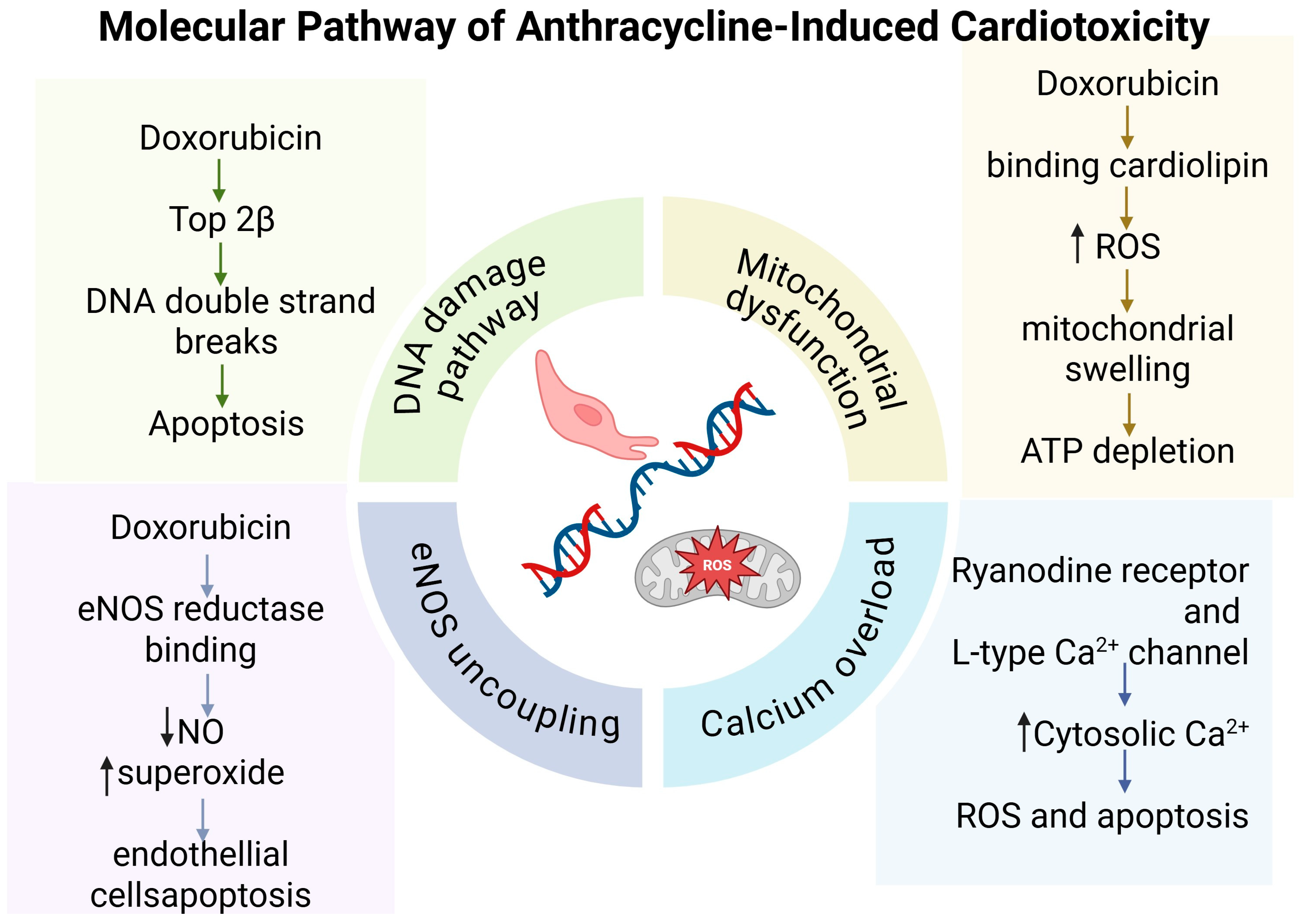
Figure 1. Summarized molecular mechanisms of anthracycline-induced cardiotoxicity (created in BioRender.com on 30 May 2025). Abbreviations: ATP: adenosine tri-phosphate; Ca2+: calcium ion; DNA: deoxyribonucleic acid; eNOS: endothelial nitric oxide synthase; HSF-1: heat shock factor 1; Hsp 25: heat shock protein 25; p53: protein 53; Top2β: topoisomerase 2β; ROS: oxygen reactive species.
Biochem 05 00025 g001
Figure 2. Schematic illustration of the phases of risk stratification, primary prevention, monitoring, and treatment of CTRCD (created in BioRender.com on 30 May 2025). Abbreviations: ACEIs: angiotensin-converting enzyme inhibitors; ARBs: angiotensin receptor blockers; ARNI: angiotensin receptor-neprilysin inhibitor; CCT: cardiac coronary CT; CMRI: cardiac magnetic resonance imaging; cTnI cardiac troponin I; cTnT: cardiac troponin T; GLS: global longitudinal strain; MRA: mineralcorticoid receptor antagonist; SGLT2i: sodium–glucose cotransporter inhibitor; TTE: trans-thoracic echocardiogram.
Biochem 05 00025 g002
Table 2. This tables summarizes the main drugs used to treat or prevent left ventricular dysfunction.
Drug/ClassMechanism of ProtectionKey Evidence
ACE Inhibitors↓ ROS, ↑ SOD,
mitochondrial support, PPARα modulation,
renin–angiotensin system balance.
[4,72,73,74,75,76,77,78]
Angiotensin Receptor BlockersAntioxidant effects.[4]
Beta-Blockers (Carvedilol)β-arrestin-biased signaling,
↓ apoptosis,
mitochondrial respiration preservation.
[79,80,81]
Mineralcorticoid ReceptoragonistsAldosterone receptor blockade,
antioxidant effects.
[82]
DexrazoxaneIron chelation,
Top2β inhibition,
prevention of DNA damage, and ROS generation.
[83]
Liposomal Anthracyclines↓ cardiac exposure to anthracycline,
prolonged circulation,
↓ ROS, and apoptosis.
[84,85]
Statins↓ oxidative stress,
↑ SOD2,
↓ heart inflammation as evidenced by ↓ TNF-α expression.
[86,87,88,89]
Angiotensin Receptor-Neprilysin InhibitorsAMPKα–mTORC1 modulation,
↓ oxidative stress and inflammation,
↓ autophagy.
[86]
Sodium–Glucose Cotransporter Inhibitors↓ ER stress,
↑ autophagy/ketogenesis,
antioxidant/anti-inflammatory effects,
↑ survival.
[87,88,89,90]
Soluble Guanylate Cyclase Stimulators↑ cGMP,
↓ inflammation,
mitochondrial support,
vasodilation.
[91,92]
Abbreviations: ↓: reduction, ↑: increase, ACE: angiotensin-converting enzyme; AMPKα–mTORC1: AMP-activated protein kinaseα–mechanistic target of rapamycin complex1; cGMP: cyclic guanosine monophosphate; ER: endoplasmic reticulum; PPARα: Peroxisome Proliferator-Activated Receptor; ROS: oxygen reactive species; SOD: superoxide dismutase; TNF-alpha: tumor necrosis factor-alpha.
Disclaimer/Publisher’s Note: The statements, opinions and data contained in all publications are solely those of the individual author(s) and contributor(s) and not of MDPI and/or the editor(s). MDPI and/or the editor(s) disclaim responsibility for any injury to people or property resulting from any ideas, methods, instructions or products referred to in the content.