Review

Advances in Structural Biology for Anesthetic Drug Mechanisms: Insights into General and Local Anesthesia

1
School of Medical Imaging, Xuzhou Medical University, Xuzhou 221004, China
2
Department of Pathology, University of Texas MD Anderson Cancer Center, Houston, TX 77030, USA
3
Department of Internal Medicine, Montefiore Medical Center Wakefield Campus, Bronx, NY 10466, USA
4
Department of Radiology, Ohio State University, Columbus, OH 43210, USA
5
Department of Surgery, University of Pittsburgh, Pittsburgh, PA 15213, USA
6
Department of Internal Medicine, Greenwich Hospital, Yale New Haven Health, Greenwich, CT 06830, USA
7
Department of Clinical Medicine, The First Clinical Medical College of Norman Bethune University of Medical Sciences, Changchun 130021, China
8
Department of Integrative Biology and Pharmacology, McGovern Medical School, University of Texas Health Science Center at Houston (UTHealth), Houston, TX 77030, USA
9
Department of Neuroscience, Baylor College of Medicine, Houston, TX 77030, USA
*
Authors to whom correspondence should be addressed.
These authors contributed equally to this work.
BioChem 2025, 5(2), 18; https://doi.org/10.3390/biochem5020018
Submission received: 8 February 2025 / Revised: 24 May 2025 / Accepted: 10 June 2025 / Published: 12 June 2025

Abstract

Anesthesia is a cornerstone of modern medicine, enabling surgery, pain management, and critical care. Despite its widespread use, the precise molecular mechanisms of anesthetic action remain incompletely understood. Recent advancements in structural biology, including cryo-electron microscopy (Cryo-EM), X-ray crystallography, and computational modeling, have provided high-resolution insights into anesthetic–target interactions. This review examines key molecular targets, including GABA_A receptors, NMDA receptors, two-pore-domain potassium (K2P) channels (e.g., TREK-1), and voltage-gated sodium (Nav) channels. General anesthetics modulate GABA_A and NMDA receptors, affecting inhibitory and excitatory neurotransmission, while local anesthetics primarily block Nav channels, preventing action potential propagation. Structural studies have elucidated anesthetic binding sites and gating mechanisms, providing a foundation for drug optimization. Advances in computational drug design and AI-assisted modeling have accelerated the development of safer, more selective anesthetics, paving the way for precision anesthesia. Future research aims to develop receptor-subtype-specific anesthetics, Nav1.7-selective local anesthetics, and investigate the neural mechanisms of anesthesia-induced unconsciousness and postoperative cognitive dysfunction (POCD). By integrating structural biology, AI-driven drug discovery, and neuroscience, anesthesia research is evolving toward safer, more effective, and personalized strategies, enhancing clinical outcomes and patient safety.

1. Introduction

Anesthesia is an essential component of modern medicine, playing a pivotal role in surgery, pain relief, emergency treatment, intensive care, and diagnostic procedures. Since the first use of ether anesthesia in 1846, the field of anesthesiology has undergone significant advancements. The application of anesthetic agents has not only transformed surgical procedures but has also greatly reduced the pain associated with surgery and improved patient survival rates. However, despite the widespread clinical use of various general and local anesthetics, the precise molecular mechanisms underlying their effects remain incompletely understood. In particular, how anesthetic drugs selectively regulate the central nervous system, leading to loss of consciousness, analgesia, and muscle relaxation, is still an area of ongoing research [1,2].
Recent advances in structural biology have provided new perspectives on anesthetic drug mechanisms. Utilizing high-resolution imaging techniques such as cryo-electron microscopy (Cryo-EM), X-ray crystallography, and nuclear magnetic resonance (NMR) spectroscopy, researchers have elucidated the three-dimensional structures of key anesthetic targets and investigated their interactions with anesthetic agents [1,3]. These breakthroughs not only enhance our understanding of anesthetic drug actions but also provide a theoretical foundation for the development of new, safer anesthetic agents. For example, resolving the structures of targets such as the γ-aminobutyric acid type A (GABA_A) receptor [4], N-methyl-D-aspartate (NMDA) receptor, two-pore-domain potassium channels (e.g., TREK-1) [5], and voltage-gated sodium (Nav) channels aids in the design of more selective and efficient anesthetics, thereby reducing side effects and enhancing patient safety.
In general anesthesia, the GABA_A receptor, NMDA receptor, and two-pore-domain potassium channels (K2P, such as TREK-1) are considered primary molecular targets. These ion channels and receptors play crucial roles in modulating synaptic transmission and neuronal excitability, mediating the effects of general anesthesia [6]. The GABA_A receptor is the primary inhibitory receptor in the central nervous system, while the NMDA receptor is a key excitatory glutamate receptor involved in learning, memory, synaptic plasticity, and pain perception. Various types of general anesthetics can enhance or inhibit these receptors, leading to sedation, analgesia, and unconsciousness. Additionally, studies on two-pore-domain potassium channels, such as TREK-1, have demonstrated their role in regulating neuronal excitability and anesthetic sensitivity [7].
Compared with general anesthesia, the mechanism of local anesthesia primarily relies on the blockade of Nav channels. Local anesthetics, such as lidocaine and bupivacaine, bind to Nav channels, preventing the initiation and propagation of action potentials, thereby achieving localized nerve blockade. This mechanism is widely applied in surgical procedures, dental treatments, and chronic pain management [8,9]. However, despite the well-established clinical use of local anesthetics, the interaction mechanisms between these drugs and Nav channels still require further investigation. Recent advancements in structural biology have enabled the resolution of Nav channel subtype-specific structures, facilitating research into optimizing local anesthetic selectivity and affinity, thereby reducing side effects such as cardiotoxicity and central nervous system toxicity.
With the emergence of precision medicine, anesthetic drug research is increasingly shifting toward personalized and targeted approaches. By analyzing individual patient genomic, transcriptomic, and proteomic data, researchers can predict the sensitivity to various anesthetic agents, achieving personalized anesthesia [10]. For example, specific mutations in Nav channels have been linked to local anesthetic resistance, while variations in GABA_A receptor subtypes may influence the potency of general anesthetics. Additionally, artificial intelligence (AI) and computational drug design (CADD) are revolutionizing the structural optimization of anesthetic drugs, accelerating the development of novel anesthetic agents with enhanced safety and efficacy [11].
This review summarizes recent advancements in the structural biology of anesthetic targets, with a particular focus on the high-resolution structural analysis of the GABA_A receptor, NMDA receptor, and Nav channels. It also discusses future prospects for precision drug design, personalized anesthesia, and the development of novel targeted anesthetics. By deepening our understanding of anesthetic drug mechanisms, we can develop safer and more effective anesthetic strategies, providing robust support for modern medicine (Figure 1).

2. Materials and Methods

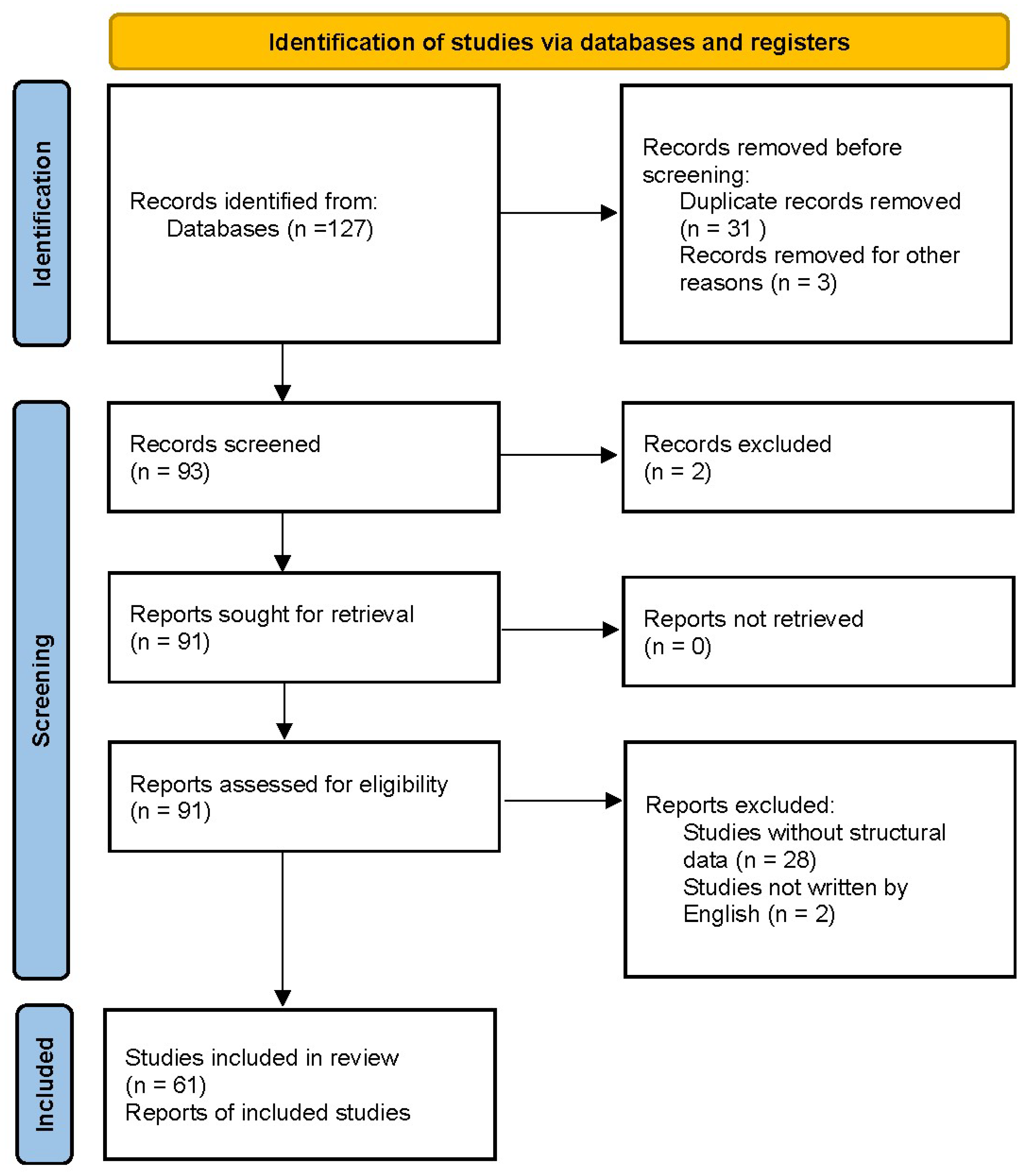
We performed a systematic literature review of structural studies relevant to anesthesia, following the PRISMA guidelines. A comprehensive search was conducted in PubMed, EMBASE, and Web of Science for articles published between 2000 and 2024, using combinations of keywords such as “general anesthesia,” “local anesthesia,” “GABA_A receptor structure,” “cryogenic electron microscopy,” “voltage-gated sodium channel,” and specific anesthetic agents (e.g., “propofol,” “lidocaine”). We included original research articles and recent reviews that reported X-ray crystallography or cryo-EM structures of anesthetic targets, as well as studies employing photoaffinity labeling or computational modeling to identify anesthetic binding sites. Only English-language articles were considered. Studies without structural data—such as purely pharmacological investigations and case reports—were excluded. The number of records identified, screened, and included is summarized in a PRISMA flow diagram. (Figure 2) The selected publications from 2000 to 2024 form the basis of this descriptive structural overview.

3. Molecular Targets of General Anesthesia

General anesthesia is a reversible state of unconsciousness accompanied by analgesia, muscle relaxation, and autonomic regulation. Although general anesthetics have been widely used in clinical practice, their precise mechanisms of action have long been a crucial research topic in neuroscience and anesthesiology. In recent years, interdisciplinary research involving molecular biology, neuroscience, and structural biology has provided deeper insights into the molecular targets of general anesthesia [12].
Currently, the main molecular targets of general anesthesia include:
The GABA_A receptor, which primarily mediates inhibitory synaptic transmission and serves as the main target for intravenous anesthetics such as propofol and thiopental sodium [13].
The NMDA receptor, which mediates excitatory synaptic transmission and is a key target for certain inhaled anesthetics such as ketamine.
Amphipathic ion channel proteins, including two-pore-domain potassium channels (e.g., TREK-1 and TASK), which regulate neuronal excitability and contribute to the action mechanism of volatile anesthetics such as isoflurane and desflurane.
Hyperpolarization-activated cyclic nucleotide-gated (HCN) channels and ethanol-sensitive channels which are associated with the sleep-like state induced by anesthesia [14].
Neurotransmitter release mechanisms, including the SNARE protein complex and synaptic vesicle release, which may be modulated by certain anesthetics.
Among these targets, the GABA_A receptor, NMDA receptor, and potassium channels have been studied most extensively. The following section focuses on the structure of the GABA_A receptor and its interaction with anesthetics.

3.1. Structure and Function of the GABA_A Receptor

3.1.1. Basic Structure of the GABA_A Receptor

The GABA_A receptor is a pentameric ligand-gated ion channel belonging to the Cys-loop receptor family, which also includes nicotinic acetylcholine receptors (nAChRs), serotonin type 3 (5-HT3) receptors, and glycine receptors. Among these, the GABA_A receptor is the primary inhibitory neurotransmitter receptor in the central nervous system (CNS) and serves as a key molecular target of general anesthesia [15].
Structurally, the GABA_A receptor is composed of five subunits, which can be arranged in various combinations. The most common subunit classes include α, β, γ, δ, and ε, each containing four transmembrane helices (TM1-TM4). These helices are arranged to form a central chloride ion (Cl) channel, allowing the receptor to modulate neuronal excitability. The binding of GABA (γ-aminobutyric acid) to the extracellular ligand binding site induces a conformational change, leading to chloride influx. This influx causes neuronal hyperpolarization, decreasing the likelihood of action potential firing, thereby producing sedative, anxiolytic, muscle relaxant, hypnotic, and anesthetic effects [16].
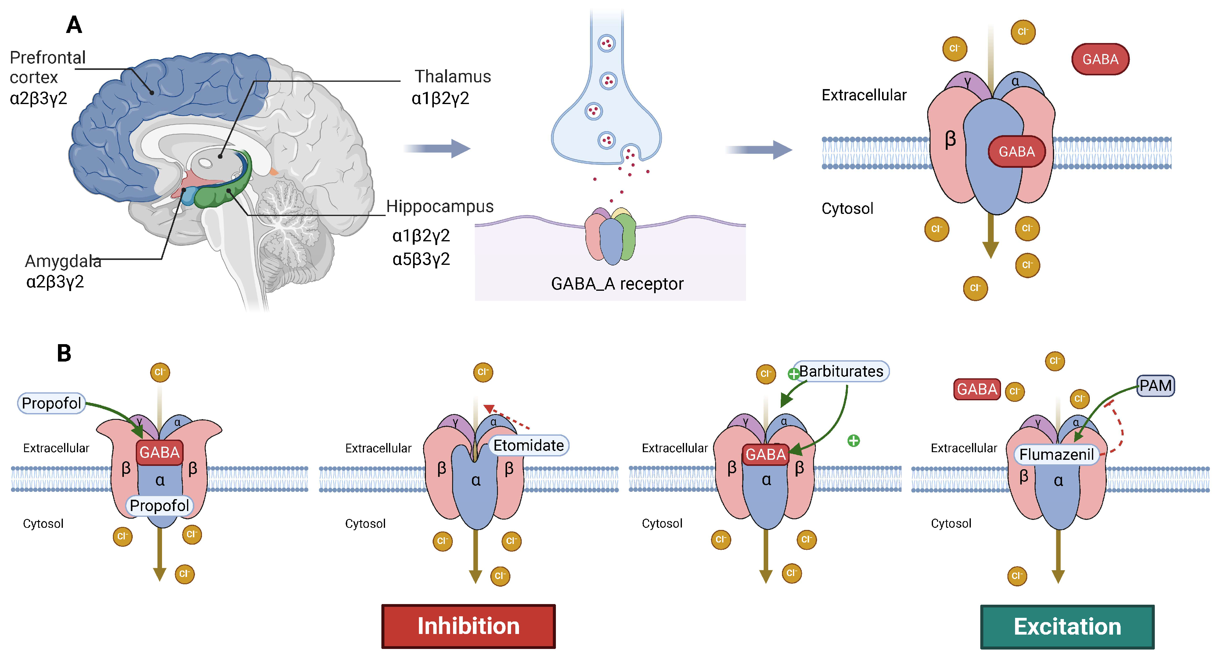
The subunit composition of the GABA_A receptor plays a crucial role in determining its pharmacological properties, localization in the brain, and response to anesthetics and other modulators. Different subunit combinations lead to the formation of distinct receptor subtypes, which are expressed in various regions of the brain and perform specialized functions [17]. The most common and well-studied subtype is the α1β2γ2 configuration, which is highly expressed in the cerebral cortex, hippocampus, and thalamus. This subtype is responsible for mediating the sedative and hypnotic effects of many general anesthetics, such as benzodiazepines, barbiturates, propofol, and volatile anesthetics [6].
Another significant subtype is the α2β3γ2 configuration, which is primarily associated with anxiolytic and muscle relaxant effects. This subtype is expressed in brain regions involved in emotional regulation, such as the amygdala and prefrontal cortex, making it an important target for anxiolytics and anesthetic adjuncts [18]. Anesthetic drugs that act on this receptor subtype may help to reduce anxiety and muscle tone during anesthesia induction.
The α5β3γ2 subtype is particularly enriched in the hippocampus, especially the CA1 region, which is critically involved in learning and memory. Studies suggest that the modulation of this subtype by anesthetics may contribute to postoperative cognitive dysfunction (POCD) [19], a condition in which some patients experience temporary or long-lasting cognitive impairment after surgery. Understanding the structure and function of this receptor subtype could lead to the development of targeted anesthetics that minimize cognitive side effects.
While γ-containing GABA_A receptors are the most well characterized, δ-containing GABA_A receptors also play an important role in anesthesia [17]. These receptors are often found extrasynaptically and mediate tonic inhibition, a form of sustained neuronal inhibition that regulates overall network excitability. Anesthetic drugs such as etomidate and neurosteroids have been shown to preferentially target δ-containing GABA_A receptors, enhancing their tonic inhibitory effects and contributing to long-lasting sedation and hypnosis [20].
In addition to their normal physiological function, mutations or genetic variations in GABA_A receptor subunits can significantly influence anesthetic sensitivity. Some individuals may have genetic polymorphisms in α or β subunits, which alter receptor function and lead to variability in anesthetic responses [21]. Understanding these genetic factors could pave the way for personalized anesthesia strategies, where anesthetic choice and dosage are tailored based on a patient’s genetic profile.
GABA_A receptor structures in complex with anesthetics have been elucidated through recent advancements in Cryo-EM and X-ray crystallography [22]. These studies have identified specific anesthetic binding pockets within the receptor, revealing how different drugs stabilize the receptor in an open or closed conformation [23]. For instance, propofol and etomidate bind at inter-subunit pockets between β and α subunits, while volatile anesthetics such as isoflurane interact within transmembrane regions. These findings not only enhance our understanding of GABA_A receptor modulation but also aid in the rational design of next-generation anesthetics with improved selectivity and fewer side effects [24].
Recent advancements in Cryo-EM have provided unprecedented structural insights into the GABA_A receptor and its interactions with anesthetics. Miller et al. resolved the GABA_A receptor–propofol complex at a resolution of ~3.3 Å. This study revealed that propofol binds to a highly specific pocket located at the β+ and α− subunit interface, leading to an enhancement of GABA-induced chloride ion influx. By stabilizing the open state of the channel, propofol effectively increases inhibitory synaptic transmission, producing its sedative and hypnotic effects [25]. Laverty et al. expanded upon these findings by analyzing the GABA_A receptor bound to isoflurane, a commonly used volatile anesthetic, and further identified hydrophobic pockets in the transmembrane domain as critical sites for isoflurane binding. Unlike propofol, which interacts at the β+ and α− subunit interface, isoflurane binds within the lipid-exposed transmembrane regions, stabilizing the receptor in an open conformation [26]. Further advancing the field, Masiulis et al. investigated GABA_A receptor conformational transitions under different physiological conditions, with and without anesthetic agents [25]. This work provided new insights into state-dependent drug binding, which could lead to more selective modulators with improved therapeutic profiles [27]. Certain GABA_A receptor isoforms, such as those containing α5 or δ subunits, exhibit altered anesthetic binding affinities, which may contribute to variability in clinical responses and side effects such as POCD. Ongoing structural studies are now focusing on receptor subunit heterogeneity, which could lead to subtype-selective anesthetics with fewer off-target effects [28].
Overall, the GABA_A receptor is a highly complex and functionally diverse target in anesthesia. Its structural and pharmacological diversity allows for the precise modulation of neuronal inhibition, enabling the fine-tuning of sedative, anxiolytic, and anesthetic effects. Future research into its subunit-specific functions, genetic variations, and structural interactions with anesthetics will continue to refine anesthesia protocols, minimize cognitive side effects, and improve patient safety in clinical settings (Figure 3).

3.1.2. Mechanism of Action of Anesthetics on the GABA_A Receptor

The GABA_A receptor plays a central role in general anesthesia by mediating inhibitory synaptic transmission in the CNS. Different general anesthetics modulate GABA_A receptor function through distinct mechanisms, primarily by acting as positive allosteric modulators (PAMs), direct agonists, or negative allosteric modulators (NAMs) [29]. These mechanisms influence chloride ion influx, neuronal excitability, and synaptic inhibition, leading to sedation, hypnosis, muscle relaxation, and anesthesia induction.
A major category of anesthetics acting on the GABA_A receptor are PAMs, which do not directly activate the receptor but enhance the effect of endogenous GABA, leading to increased chloride conductance and prolonged neuronal inhibition. Propofol, a widely used intravenous anesthetic, is a potent PAM that binds at the β+ and α− subunit interface, increasing the receptor’s sensitivity to GABA. This results in prolonged channel opening, sustained neuronal hyperpolarization, and enhanced CNS inhibition, producing deep sedation and hypnosis [30].
Barbiturates, such as thiopental sodium, also act as PAMs but bind to a distinct site on the GABA_A receptor. They enhance GABAergic transmission through both positive allosteric modulation and direct channel gating, making them effective as induction agents and anti-seizure medications. However, due to their longer half-life and potential for severe respiratory depression, their use in modern anesthesia has declined in favor of safer alternatives like propofol.
Volatile anesthetics, including isoflurane and sevoflurane, exert their effects by interacting with the transmembrane domains of the GABA_A receptor, increasing the probability of channel opening. These inhaled anesthetics modulate lipid-exposed hydrophobic pockets within the receptor, stabilizing its open conformation and thereby potentiating inhibitory neurotransmission [31]. Unlike propofol and barbiturates, which primarily enhance ligand binding and gating properties, volatile anesthetics modulate channel conductance and receptor conformation, contributing to their dose-dependent effects on anesthesia depth and immobility.
In addition to PAMs, some anesthetics act as direct agonists of the GABA_A receptor, meaning that they can activate the receptor independently of GABA binding. Etomidate, for example, is an effective intravenous anesthetic induction agent that can bind directly to β subunits, inducing chloride channel opening even in the absence of GABA [20]. This property makes etomidate particularly useful in patients with unstable cardiovascular function, as it provides reliable anesthesia induction with minimal hemodynamic suppression. However, adrenal suppression caused by prolonged etomidate use has limited its long-term application [32].
Conversely, some drugs act as NAMs, reducing GABA_A receptor activity and thereby decreasing inhibitory neurotransmission. While not typically used as anesthetics, certain anticonvulsant and proconvulsant drugs target the GABA_A receptor to modulate neuronal excitability. For example, flumazenil, a benzodiazepine antagonist, can reverse the sedative effects of PAMs by competitively inhibiting benzodiazepine binding sites on the GABA_A receptor [33]. Similarly, some neurosteroids and GABA_A receptor modulators are being investigated for their ability to fine-tune inhibitory neurotransmission in clinical conditions such as epilepsy, anxiety disorders, and anesthesia reversal.
These distinct mechanisms of anesthetic action on the GABA_A receptor help explain variations in drug onset, duration of action, potency, and side effect profiles. Drugs such as propofol and etomidate produce rapid induction of anesthesia due to their high affinity and direct receptor modulation, whereas volatile anesthetics like isoflurane exhibit slower equilibration but provide prolonged anesthesia maintenance. Furthermore, subunit-specific drug interactions contribute to individual variability in anesthetic response, with receptor subtypes such as α5-containing GABA_A receptors being implicated in POCD and δ-containing receptors influencing long-lasting sedation and neurosteroid modulation [19].
Overall, the GABA_A receptor remains a fundamental target for general anesthetics, with its modulation playing a crucial role in inducing and maintaining anesthesia. Understanding the molecular interactions between anesthetics and the receptor provides valuable insights for optimizing clinical anesthesia protocols, developing safer anesthetic agents, and potentially designing personalized anesthesia strategies based on individual receptor subunit expression and genetic variation. Future research in this area will likely continue to refine our knowledge of anesthetic mechanisms, leading to more effective and targeted interventions in perioperative care.

3.1.3. Future Research Directions

Despite significant advances in understanding the GABA_A receptor’s role in anesthesia, many fundamental questions remain unanswered. The complexity of GABA_A receptor subunit diversity, genetic variability, and interactions with other anesthetic targets presents opportunities for future research that could enhance anesthetic drug efficacy, safety, and individualization [34].
One of the most promising areas for future exploration is personalized anesthesia. Individual variability in GABA_A receptor-mediated anesthetic responses is increasingly recognized, with genetic polymorphisms in receptor subunits potentially influencing drug sensitivity, metabolism, and side effects. For example, variations in α, β, or γ subunits could alter the receptor binding affinity for anesthetics like propofol, etomidate, or volatile agents, leading to differences in induction time, depth of sedation, and recovery profiles [6]. Understanding these genetic factors through genomic screening and pharmacogenomic studies could allow for personalized anesthesia strategies, where drug selection and dosing are tailored to an individual’s genetic makeup, reducing the risk of adverse effects such as excessive sedation, prolonged recovery, or POCD.
The development of novel anesthetics represents another key research direction. While existing anesthetics effectively modulate GABA_A receptor function, they often exhibit off-target effects and side effects such as respiratory depression, cardiovascular instability, or prolonged cognitive impairment [6]. By elucidating the structures of different GABA_A receptor subtypes through techniques such as Cryo-EM and molecular docking, researchers can design more selective anesthetics that target specific receptor configurations with fewer unwanted effects. For instance, drugs that preferentially modulate α1-containing receptors could enhance sedation while minimizing memory impairment, while those targeting α2-containing receptors might offer anxiolysis without excessive hypnosis [33]. Similarly, the study of δ-containing extrasynaptic GABA_A receptors, which mediate tonic inhibition, could lead to longer-lasting sedative agents with distinct pharmacokinetics [35].
POCD remains a significant clinical concern, particularly in elderly patients and those undergoing major surgery under general anesthesia. Some anesthetics have been implicated in transient or long-term cognitive impairment, possibly due to their effects on the GABA_A receptor subtypes involved in synaptic plasticity and memory formation. Notably, α5-containing GABA_A receptors in the hippocampus are thought to play a role in learning and memory, and modulation of these receptors by certain anesthetics may contribute to POCD [36]. Future research should focus on developing anesthetics that minimize α5 receptor interactions or identifying neuroprotective strategies that mitigate cognitive impairment, such as the co-administration of adjunct drugs or preoperative conditioning protocols.
A more integrated understanding of general anesthesia mechanisms may also arise from studying the interactions between the GABA_A receptor and other anesthetic targets, such as the NMDA receptor, and TREK-1 channels. While GABA_A receptor modulation is the primary mechanism of action for many anesthetics, cross-talk between inhibitory and excitatory neurotransmission systems plays a significant role in anesthesia depth, emergence, and side effects [37]. For example, ketamine’s blockade of NMDA receptors produces a distinct anesthetic state characterized by dissociative effects and strong analgesia, while volatile anesthetics modulate both GABA_A and potassium channels (TREK-1), contributing to their unique pharmacodynamic profiles. Understanding these multi-receptor interactions could lead to the development of combination anesthetic strategies that achieve optimal effects with lower drug doses, reducing toxicity and side effects.
Advancements in structural biology, molecular pharmacology, and computational modeling are expected to further refine anesthetic drug design. With the integration of AI and high-throughput screening, researchers can identify anesthetic candidates with improved receptor selectivity, metabolic stability, and reduced off-target effects. AI-driven drug discovery may also enable precise predictions of anesthetic–receptor interactions, accelerating the development of next-generation anesthetics with enhanced safety and efficacy [11].

3.2. Structure and Function of the NMDA Receptor

3.2.1. Basic Structure and Function of the NMDA Receptor

The NMDA receptor is a major subtype of ionotropic glutamate receptors (iGluRs) and plays a fundamental role in excitatory synaptic transmission in the CNS. It is widely distributed throughout the cortex, hippocampus, and spinal cord, where it regulates key neurophysiological functions such as synaptic plasticity, learning, memory, and pain processing. As a voltage-dependent, ligand-gated cation channel, the NMDA receptor allows the influx of sodium (Na+) and calcium (Ca2+) ions upon activation, leading to membrane depolarization and signal amplification within neuronal circuits [38].
Compared with other glutamate receptors, such as AMPA and kainate receptors, the NMDA receptor exhibits unique biophysical and pharmacological properties that allow for more complex regulatory mechanisms in neurotransmission. One of its defining characteristics is the magnesium block (Mg2+ block), where at resting membrane potential, the ion channel remains blocked by Mg2+ ions, preventing ion flux. For the receptor to function, prior depolarization is required to remove this blockade, allowing glutamate binding to trigger channel opening. This feature makes the NMDA receptor highly activity-dependent, meaning it plays a pivotal role in experience-driven neural plasticity and cognitive functions such as long-term potentiation (LTP), learning, and memory formation [39].
Another distinctive property of the NMDA receptor is its high calcium permeability, which is significantly greater than that of AMPA receptors. Calcium influx through NMDA receptors serves as a critical intracellular signal for synaptic strengthening, making this receptor essential for neuronal adaptation, plasticity, and long-term memory storage [9]. However, the excessive activation of NMDA receptors, leading to excessive calcium influx, can result in excitotoxicity, a process implicated in neurodegenerative diseases such as Alzheimer’s disease, Parkinson’s disease, and stroke-induced brain damage. Thus, NMDA receptor activity must be tightly regulated to balance normal excitatory transmission and neuroprotection.
A further distinguishing feature is the dual ligand regulation required for receptor activation. Unlike AMPA or kainate receptors, which are activated by a single ligand (glutamate), the NMDA receptor requires the binding of both glutamate and glycine (or D-serine) at distinct sites for full activation. This dual-ligand dependency introduces an additional level of synaptic regulation, preventing excessive receptor activation under basal conditions. Additionally, the NMDA receptor undergoes desensitization and inactivation via phosphorylation, allosteric modulation, and interactions with intracellular scaffolding proteins, allowing neurons to fine-tune synaptic transmission based on changing physiological demands [40].
Structurally, the NMDA receptor is a tetrameric complex composed of four subunits, with the most common subunit arrangement being GluN1-GluN2A/B/C/D. Each subunit contributes to different aspects of receptor function, kinetics, and drug sensitivity. The GluN1 subunit is present in all functional NMDA receptors and contains the glycine binding site, making it essential for receptor activation. The GluN2 subunits (GluN2A–GluN2D) define kinetic properties, receptor desensitization rates, and Mg2+ block characteristics, with GluN2B being particularly abundant in the cortex and hippocampus, where it supports prolonged synaptic signaling [41]. The GluN3 subunits (GluN3A and GluN3B) have received less research attention but are believed to modulate receptor function under pathological conditions, such as neurodevelopmental disorders and neurodegenerative diseases.
Because NMDA receptors mediate excitatory neurotransmission and synaptic plasticity, their dysregulation is associated with a broad spectrum of neurological and psychiatric disorders. Excessive NMDA receptor activity is implicated in epilepsy, neurotoxicity, chronic pain syndromes, and neurodegenerative diseases, whereas reduced NMDA receptor function is linked to schizophrenia, cognitive impairment, and depression. As a result, NMDA receptors are critical pharmacological targets for anesthetics, analgesics, and neuropsychiatric drugs.
Anesthetic drugs such as ketamine, nitrous oxide (N2O), and xenon exert their effects by blocking NMDA receptor activity, leading to diminished excitatory neurotransmission and altered consciousness. These NMDA receptor antagonists induce sedation, dissociative anesthesia, and analgesia by preventing glutamate-mediated synaptic transmission, making them effective agents for surgical anesthesia, pain management, and psychiatric interventions [42]. Additionally, ketamine has gained significant interest as a rapid-acting antidepressant, as its blockade of NMDA receptors has been shown to increase synaptic plasticity and promote neurogenesis, offering therapeutic potential for treatment-resistant depression and mood disorders.
Given the NMDA receptor’s dual role in both normal cognition and pathological conditions, ongoing research seeks to develop more selective NMDA receptor modulators that can preserve physiological function while minimizing adverse effects [43]. Structural biology techniques such as Cryo-EM and molecular docking studies have provided new insights into subunit-specific binding sites, drug interactions, and allosteric modulation, paving the way for next-generation anesthetics and neuroprotective agents [44,45]. In 2016, the Gouaux laboratory elucidated the crystal structure of the NMDA receptor–ketamine complex, demonstrating ketamine binding within the GluN2B subunit’s channel pore at the M2 transmembrane region. This binding mechanism inhibits ion channel opening by blocking calcium and sodium influx, providing structural evidence for ketamine’s NMDA receptor antagonism and suggesting potential avenues for developing more selective antagonists with reduced psychotropic effects [46].
Subsequent cryo-EM analysis revealed that ketamine physically occludes ion permeation and induces M3 helix conformational changes, stabilizing the receptor in a closed configuration. This structural modification correlates with ketamine’s extended pharmacodynamic profile and its reported neuroprotective properties [47]. Furthermore, the ability of ketamine to induce long-lasting receptor inhibition provides an explanation for its prolonged analgesic and antidepressant effects, as well as its potential for neuroprotection in conditions such as stroke, traumatic brain injury, and neurodegenerative disorders [48].
The NMDA receptor–nitrous oxide interaction has been characterized, identifying binding at the M2/M3 transmembrane domain with a comparatively lower affinity than ketamine. This reduced binding affinity necessitates higher concentrations for anesthetic efficacy and results in partial NMDA receptor inhibition, properties that are advantageous in clinical contexts requiring rapid recovery [49]. It was identified that cyclopropyl ketone binds within the GluN2A/2B subunits’ transmembrane domain, resulting in receptor inactivation and synaptic plasticity inhibition. These findings expand the potential therapeutic applications of NMDA receptor modulation beyond anesthesia to include neuropsychiatric and neurodegenerative disorders [50].
Future studies aim to refine NMDA receptor-targeting drugs, optimizing their selectivity, efficacy, and safety while expanding their applications beyond anesthesia into neurological and psychiatric therapeutics (Figure 4).

3.2.2. NMDA Receptor and General Anesthesia

The NMDA receptor plays a crucial role in excitatory synaptic transmission, and its blockade is a key mechanism underlying the effects of certain general anesthetics. Unlike GABA_A receptor-positive modulators, which enhance inhibitory neurotransmission, NMDA receptor antagonists work by inhibiting excitatory neurotransmission, leading to analgesia, sedation, and dissociative anesthesia [51]. This mechanism is particularly relevant for drugs such as ketamine, nitrous oxide (N2O), and cyclopropyl ketone, which act as non-competitive NMDA receptor antagonists. By preventing glutamate-mediated depolarization and calcium influx, these drugs effectively reduce neuronal excitability and produce their anesthetic effects.
One of the most well-known NMDA receptor antagonists in anesthesia is ketamine, which induces a unique form of anesthesia known as dissociative anesthesia. Unlike traditional anesthetics, which cause global CNS depression, ketamine functionally disconnects the thalamocortical and limbic systems, resulting in a state where patients appear conscious yet unresponsive to external stimuli [52]. This dissociative state allows for the preservation of certain reflex functions, such as eye opening and spontaneous respiration, making ketamine an attractive anesthetic choice for patients with respiratory compromise. In addition to its strong analgesic properties, ketamine is particularly useful in trauma and burn patients, as well as in individuals at high risk of hypotension, due to its sympathomimetic effects that help maintain cardiovascular stability.
Beyond its anesthetic use, ketamine has also gained attention for its rapid-acting antidepressant effects, making it a promising treatment for treatment-resistant depression (TRD) and major depressive disorder (MDD). Unlike conventional antidepressants, which take weeks to exert their effects, ketamine produces rapid mood elevation within hours, likely due to its ability to enhance synaptic plasticity and promote neurogenesis through NMDA receptor modulation [53,54]. This discovery has led to an expansion of ketamine’s clinical applications beyond anesthesia into psychiatric and pain medicine.
Another important NMDA receptor antagonist is nitrous oxide (N2O), which is commonly used for analgesia and adjunct anesthesia. Nitrous oxide acts as a weak NMDA receptor antagonist, but it also interacts with GABA_A receptors, contributing to its sedative effects. Due to its rapid onset and metabolism, nitrous oxide is widely employed in short surgical procedures, dental interventions, and outpatient surgeries, where fast recovery times are desirable [55]. Additionally, N2O is frequently used as an analgesic in obstetric care and emergency medicine, providing effective pain relief with minimal systemic effects. Its low potency as an NMDA receptor antagonist makes it less effective as a sole anesthetic agent, but when combined with other anesthetics, it enhances analgesia and reduces the required dose of more potent anesthetics, thereby minimizing their side effects [56].
Although dexmedetomidine primarily acts as an α2-adrenergic receptor agonist, emerging evidence suggests that it may also indirectly influence NMDA receptor signaling, thereby contributing to its sedative, analgesic, and neuroprotective effects. Unlike traditional anesthetics, dexmedetomidine induces a state of sedation that closely resembles natural sleep, preserving respiratory function while providing adequate sedation and pain relief. This makes it an ideal choice for pediatric anesthesia, ICU sedation, and procedural sedation in high-risk patients.
A clinical advantage of NMDA receptor antagonists is their relatively low risk of respiratory depression compared with other anesthetics. Because NMDA receptors are primarily involved in excitatory neurotransmission, blocking them does not significantly suppress brainstem respiratory centers, unlike opioids or GABAergic anesthetics, which can lead to profound respiratory suppression [57]. This makes NMDA antagonists particularly valuable for critically ill patients, pediatric anesthesia, and field or battlefield medicine, where maintaining spontaneous ventilation is crucial.
Despite their benefits, NMDA receptor antagonists also have certain limitations and side effects. Ketamine, for instance, is associated with emergence delirium, hallucinations, and psychotropic effects, which can be distressing for patients upon awakening [53]. These effects are believed to be due to dysregulated glutamatergic signaling and altered cortical processing. Strategies such as the co-administration of benzodiazepines or using lower sub-anesthetic doses have been explored to mitigate these adverse reactions [58]. Similarly, the prolonged use of N2O has been linked to the inhibition of methionine synthase, potentially leading to vitamin B12 deficiency and neurological complications, especially with chronic exposure.
Given the growing clinical applications of NMDA receptor antagonists, research efforts continue to refine their pharmacological properties. Structural biology techniques, including Cryo-EM and computational modeling, have provided detailed insights into anesthetic binding sites within the NMDA receptor, aiding in the development of more selective NMDA modulators. Future anesthetic agents may seek to enhance NMDA receptor blockade with fewer dissociative or neuropsychiatric side effects, potentially leading to the next generation of safer and more effective anesthetics.

3.2.3. Future Research Directions

Although significant progress has been made in understanding NMDA receptor function in general anesthesia, several fundamental questions remain. Ongoing research is focused on refining our understanding of NMDA receptor subtypes, optimizing anesthetic selectivity, and exploring novel therapeutic applications. Future studies integrating structural biology, pharmacology, and neuroscience will be crucial in addressing these challenges, ultimately leading to more effective and safer NMDA receptor-targeted anesthetic strategies [59].
One key area of future investigation is the high-resolution structural analysis of NMDA receptor–anesthetic complexes. While current studies have provided atomic-level insights into how ketamine, nitrous oxide (N2O), and cyclopropyl ketone interact with NMDA receptors, there is still limited knowledge about how different receptor subtypes (e.g., GluN2A vs. GluN2B) exhibit distinct drug-binding patterns. Given that GluN2A and GluN2B are differentially expressed in various brain regions and have unique physiological roles, understanding their specific interactions with anesthetics could lead to the design of subtype-selective drugs [60]. Such precision would allow for the targeted modulation of NMDA receptor activity, reducing unwanted side effects while maintaining effective anesthesia, neuroprotection, and pain relief.
Another important direction is the development of novel NMDA receptor-targeted anesthetics that minimize cognitive side effects. Existing NMDA antagonists, such as ketamine, while effective, often produce dissociative symptoms, hallucinations, and cognitive impairment. These adverse effects are thought to result from broad NMDA receptor inhibition across multiple brain regions, disrupting normal neural network function. Future drug development efforts will aim to design more precise NMDA receptor modulators that can selectively inhibit excitatory transmission in pain and anesthesia-relevant circuits while sparing cognitive and mood-related pathways. Advances in medicinal chemistry, AI-driven drug design, and computational modeling will facilitate the development of next-generation NMDA receptor antagonists with improved pharmacokinetic and pharmacodynamic properties [61].
The regional specificity of NMDA receptor function is another critical factor in optimizing anesthetic efficacy and safety. NMDA receptors are not uniformly expressed throughout the brain, and their function varies significantly across different neural circuits. For example, NMDA receptors in the hippocampus are highly involved in learning and memory, while those in the thalamus and spinal cord are more relevant to sensory processing and pain perception. This raises the possibility that certain anesthetics may have region-specific effects, selectively modulating NMDA receptors in pain processing circuits while sparing cognitive function. High-resolution imaging techniques such as functional MRI (fMRI) and optogenetics could help elucidate how NMDA receptor antagonists exert differential effects across brain regions, paving the way for spatially selective anesthetic approaches.
Another fascinating area of research is the intersection between NMDA receptor-mediated anesthesia and psychiatric treatment. The discovery that ketamine exhibits rapid antidepressant effects has significantly influenced neuropsychiatric drug development, suggesting that NMDA receptor antagonists play a key role in mood regulation. This has led to the increasing interest in developing dual-function anesthetics that not only induce anesthesia but also provide neuropsychiatric benefits. Given that major depressive disorder (MDD) and chronic pain share overlapping neural circuits, future drugs could be designed to simultaneously alleviate pain, provide anesthesia, and exert rapid antidepressant effects. Ongoing clinical research on esketamine (S-ketamine) for depression highlights the potential for NMDA-targeted therapies that bridge the gap between anesthesia and mental health treatment [62].
In addition to clinical applications, NMDA receptor research has important implications for neuroprotection and neurodegenerative diseases [63]. The overactivation of NMDA receptors is implicated in conditions such as stroke, traumatic brain injury (TBI), and Alzheimer’s disease, where excessive calcium influx leads to excitotoxicity and neuronal damage. Developing anesthetic agents that selectively modulate NMDA receptor activity in a way that protects against excitotoxic injury while preserving cognitive function could have profound implications for perioperative neuroprotection and long-term brain health. Studies exploring the role of NMDA receptor antagonists in preventing neurodegeneration could lead to new therapeutic strategies for aging-related cognitive decline.
From a drug development perspective, integrating structural biology with pharmacology and computational chemistry will be essential in designing safer and more effective NMDA receptor modulators. Advances in Cryo-EM, X-ray crystallography, and molecular dynamics simulations have already provided detailed insights into NMDA receptor structure, but further research is needed to map allosteric regulatory sites, identify novel binding pockets, and optimize drug interactions. Combining machine learning-based virtual screening with AI-driven molecular design will accelerate the discovery of new anesthetic agents, allowing for the development of NMDA receptor modulators tailored to specific clinical needs [64].

4. Molecular Mechanisms of Local Anesthesia

Local anesthesia involves the selective blockade of nerve impulse conduction in a specific area, providing effective analgesia without affecting consciousness. Unlike general anesthetics, which act on the CNS to induce sedation, amnesia, and muscle relaxation, local anesthetics target peripheral nerves, preventing pain signal transmission while allowing the patient to remain fully alert [65]. These drugs, including lidocaine, bupivacaine, and ropivacaine, are widely used in various clinical settings, such as surgical procedures, dental interventions, labor analgesia, regional nerve blocks, and chronic pain management. Their ability to provide rapid and reversible pain relief with minimal systemic effects has made them indispensable in modern anesthesia practice.
The primary molecular target of local anesthetics is the Nav channel. By inhibiting sodium ion (Na+) influx through these channels, local anesthetics prevent depolarization and the subsequent transmission of pain signals along sensory neurons [66]. This mechanism of action ensures effective pain relief while minimizing interference with other neural functions, making local anesthesia a safer alternative for many procedures compared with systemic analgesics or general anesthesia.
One of the key advantages of local anesthetics is their clinical versatility and safety profile. These drugs provide rapid onset and effective nerve blockade while avoiding the major systemic risks associated with general anesthesia, such as respiratory depression, hemodynamic instability, and POCD. Additionally, local anesthetics offer a high degree of controllability, as different agents exhibit varied onset times, durations of action, and diffusion characteristics, allowing anesthesiologists to tailor anesthesia based on surgical requirements [67]. For example, lidocaine, with its fast onset and moderate duration, is well suited for short procedures, whereas bupivacaine, with its longer duration of action, is preferred for prolonged surgical interventions or continuous nerve blocks for postoperative pain management [68].
Recent advancements in structural biology and computational modeling have significantly enhanced our understanding of how local anesthetics interact with Nav channels at the molecular level. Cryo-EM, X-ray crystallography, and molecular docking studies have provided high-resolution structural data, revealing precise drug binding sites, conformational changes, and state-dependent inhibition mechanisms [69]. These insights have led to the development of novel anesthetics with improved efficacy, reduced toxicity, and greater selectivity for pain-specific Nav channel subtypes. For instance, studies have shown that certain Nav channel subtypes, such as Nav1.7, play a crucial role in pain perception, prompting researchers to explore Nav1.7-selective blockers as a potential breakthrough in local anesthetic design. Such drugs could maximize pain relief while minimizing the unwanted effects on other sodium channel isoforms, such as Nav1.5, which is critical for cardiac function [70].
The growing knowledge of local anesthetic pharmacodynamics and receptor interactions has also enabled the development of advanced drug formulations and delivery systems. For example, liposomal-encapsulated local anesthetics have been designed to provide prolonged nerve blockade with reduced systemic toxicity, offering promising applications in postoperative pain management. Similarly, adjunctive agents such as vasoconstrictors (e.g., epinephrine) or adjuvants like dexmedetomidine are being used to enhance anesthetic efficacy, prolong duration, and minimize systemic absorption [9].

4.1. Nav Channels and the Mechanism of Local Anesthetics

4.1.1. Basic Structure and Function of Nav Channels

Nav channels are integral membrane proteins that play a fundamental role in the generation and propagation of action potentials in excitable cells, including neurons, cardiac myocytes, and skeletal muscle fibers. These channels are essential for rapid signal transmission in the nervous system, making them critical targets for local anesthetics. By selectively blocking Nav channels in peripheral sensory neurons, local anesthetics inhibit nerve conduction, preventing pain perception without affecting consciousness [71].
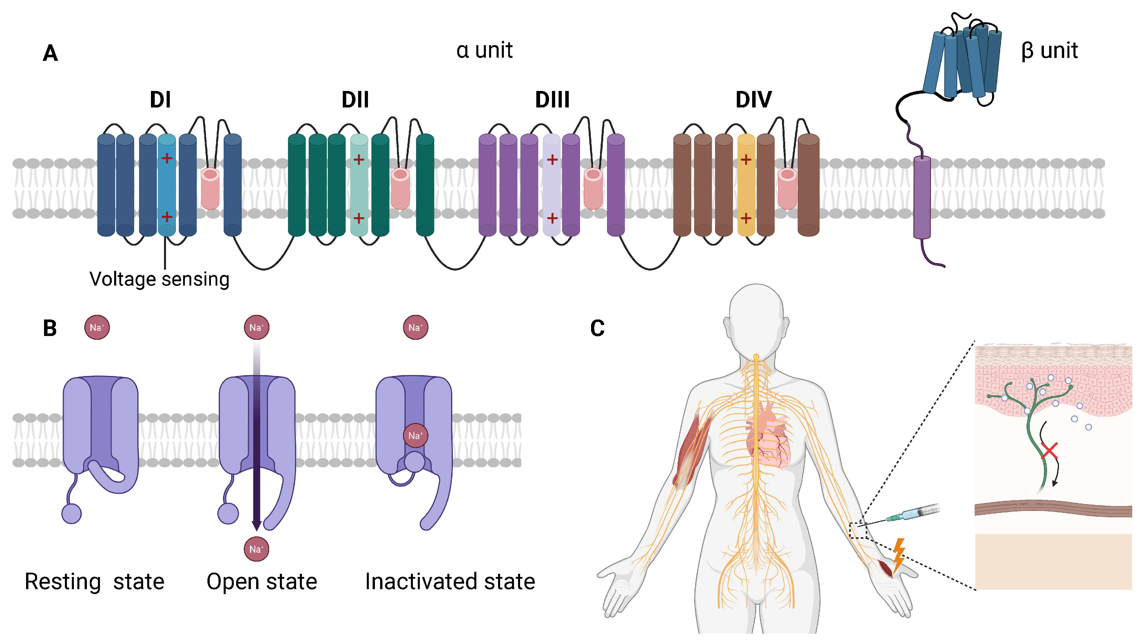
The Nav channel structure consists of a pore-forming α-subunit and auxiliary β-subunits, which together regulate the channel’s gating properties and pharmacological interactions. The α-subunit, the primary functional component, is composed of four homologous domains (DI–DIV), each containing six transmembrane helices (S1–S6) [72]. The S4 helix functions as the voltage-sensing domain, detecting changes in membrane potential, while the S5–S6 helices form the ion pore, controlling sodium ion flow across the membrane. The β-subunits, while not directly involved in ion conduction, modulate channel kinetics, stability, and drug sensitivity, fine-tuning neuronal excitability and the response to local anesthetics [72].
Functionally, Nav channels cycle through three primary states: Resting state—in the absence of membrane depolarization, the channel remains closed, preventing sodium ion influx and keeping the neuron in a non-excited state. Open state—upon membrane depolarization, the voltage-sensing S4 helix moves outward, triggering conformational changes that open the channel, allowing sodium ions (Na+) to enter the cell and generate an action potential. Inactivated state—immediately after depolarization, the channel transitions into an inactivated, non-conducting conformation, preventing excessive excitation and allowing the neuron to reset before the next action potential [73]. Because Nav channels regulate the excitability of neurons, their selective inhibition by local anesthetics effectively blocks pain signal transmission while maintaining other neural functions.
Nav channels exist in multiple isoforms (Nav1.1–Nav1.9), each with distinct tissue distributions and physiological functions. The diversity of Nav subtypes allows for the precise regulation of electrical signaling in different cell types. Among these, certain Nav channels are particularly relevant to anesthesia and pain management: 1. Nav1.7: Highly expressed in peripheral sensory neurons, Nav1.7 is crucial for pain perception and transmission. Genetic mutations in this channel are associated with extreme pain disorders or congenital insensitivity to pain, highlighting its central role in nociception [74]. Given its exclusive expression in sensory neurons, Nav1.7 is a promising target for highly selective local anesthetics that block pain without affecting motor or autonomic function [75]. 2. Nav1.4: Predominantly found in skeletal muscle, Nav1.4 is essential for muscle contraction. Its inhibition can cause muscle weakness or paralysis, which is why muscle-related side effects are sometimes observed with certain local anesthetics [76]. 3. Nav1.5: Expressed in cardiac muscle, Nav1.5 plays a key role in heart rhythm regulation. Local anesthetics that inadvertently affect Nav1.5 may lead to cardiac conduction disturbances, arrhythmias, or cardiotoxicity, a major concern with high doses or prolonged exposure to certain anesthetics (e.g., bupivacaine) [77].
Advancements in structural biology and Cryo-EM have enhanced our understanding of local anesthetic interactions with Nav channels [78]. In 2017, the Yan laboratory resolved the Nav1.7–lidocaine complex structure, revealing lidocaine binding at the domain III-IV interface near the S6 helix, physically obstructing the sodium ion pore. This provided the first structural basis for lidocaine’s mechanism and created opportunities for developing Nav1.7-selective anesthetics that could provide effective pain relief with minimal cardiac or motor effects [79].
Bupivacaine’s higher lipophilicity enables more efficient membrane penetration and stronger binding pocket interactions compared with lidocaine, as shown in studies of the Nav1.4–bupivacaine complex. Bupivacaine preferentially stabilizes the channel’s inactivated state, explaining its longer duration of action and higher cardiotoxicity risk [80,81]. The Catterall group resolved the Nav1.5 cardiac channel’s anesthetic binding sites, providing critical insights into cardiotoxicity mechanisms. This research informed strategies to modify anesthetic molecules to improve selectivity for sensory neuron Nav channels while reducing the affinity for cardiac Nav1.5 channels, potentially yielding safer clinical formulations [30].
Because different Nav subtypes are distributed across various tissues, an ideal local anesthetic would selectively block Nav1.7-mediated pain signals without affecting Nav1.5 in the heart or Nav1.4 in skeletal muscle. Ongoing research focuses on developing subtype-specific Nav inhibitors to enhance anesthetic selectivity and safety [82] (Figure 5).

4.1.2. Mechanism of Action of Local Anesthetics

The primary mechanism by which local anesthetics exert their effect is through the inhibition of Nav channels, preventing action potential propagation along peripheral nerves. By blocking sodium ion influx, local anesthetics effectively disrupt neuronal depolarization, halting the transmission of pain signals from the periphery to the central nervous system [9]. This action is highly selective, affecting primarily small-diameter, unmyelinated C fibers, which are responsible for pain perception, while larger-diameter, myelinated Aβ fibers, which mediate touch and motor function, are relatively less sensitive. This selectivity allows for effective analgesia without the significant impairment of motor or sensory functions, making local anesthetics ideal for surgical procedures, nerve blocks, and pain management [83].
In addition to direct channel blockade, local anesthetics further stabilize the inactivated state of Nav channels, prolonging their non-conductive conformation. This prevents neurons from repeatedly firing high-frequency action potentials, which are common in pain signaling pathways [84]. By extending the inactive phase of sodium channels, local anesthetics enhance nerve blockade effectiveness, reducing the likelihood of pain signal transmission even under conditions of heightened nerve activity, such as inflammation or nerve injury. This state-dependent inhibition explains why local anesthetics preferentially block actively firing neurons, making them particularly useful for suppressing acute and chronic pain conditions [85].
The lipid solubility of local anesthetics plays a crucial role in determining their efficacy, onset time, and duration of action. More lipophilic anesthetics, such as bupivacaine, are able to rapidly diffuse across nerve membranes, resulting in a faster onset and prolonged anesthetic effect. Their higher affinity for lipid-rich nerve sheaths allows for longer retention within nerve tissues, providing extended-duration pain relief [86]. In contrast, less lipophilic anesthetics, such as procaine, exhibit slower penetration through nerve membranes, leading to a delayed onset and shorter duration of action. This property is particularly relevant when selecting anesthetics for different clinical applications, where the desired balance between onset speed and duration must be considered [87].
Another key factor influencing local anesthetic activity is pH-dependent ionization. Local anesthetics are weak bases, meaning their ionization state varies with the pH of the surrounding tissue. In normal physiological conditions, local anesthetics exist in both ionized and non-ionized forms, allowing them to cross the nerve membrane (in non-ionized form) and bind to the intracellular side of the Nav channel (in ionized form) [88]. However, in acidic environments, such as those found in infected or inflamed tissues, the proportion of ionized drug molecules increases, reducing membrane permeability and leading to decreased anesthetic efficacy. This explains why local anesthetics may be less effective in inflamed or infected tissues, often requiring higher doses or buffering agents (e.g., bicarbonate) to enhance penetration and efficacy [88].

4.1.3. Future Research Directions

Despite significant advancements in understanding how local anesthetics inhibit Nav channels, several critical challenges remain in optimizing their efficacy, selectivity, and safety. Current local anesthetics, such as lidocaine and bupivacaine, lack specificity for individual Nav channel subtypes, leading to off-target effects, including potential cardiotoxicity and motor impairment. Future research must focus on developing more selective Nav channel blockers, particularly targeting Nav1.7, which is a key regulator of pain transmission [89]. Since Nav1.7 is predominantly expressed in peripheral sensory neurons, designing highly selective Nav1.7 inhibitors could provide powerful analgesia while minimizing the effects on cardiac and motor function, thereby reducing systemic side effects and improving patient safety [90]. Advances in structural biology, cryo-EM imaging, and molecular docking will be instrumental in designing next-generation local anesthetics with enhanced subtype selectivity.
Another crucial area of research is reducing cardiotoxicity, particularly for potent local anesthetics such as bupivacaine, which can bind to Nav1.5 (cardiac sodium channels) and disrupt heart rhythm. Cardiotoxicity is a significant limitation in clinical practice, as bupivacaine-induced cardiac arrest is a rare but serious complication [91]. Future studies should explore structural modifications of anesthetic molecules to develop drugs that preferentially bind to Nav1.7 without significantly affecting Nav1.5. A better understanding of Nav1.5’s drug-binding dynamics could also lead to the development of safer formulations or reversal agents to mitigate the risk of cardiotoxic events [88].
In addition to molecular optimization, innovative drug delivery technologies are emerging as promising strategies for enhancing local anesthetic efficacy while reducing systemic toxicity. The liposomal encapsulation of local anesthetics has shown potential in prolonging nerve blockade, reducing the need for repeated dosing while minimizing peak plasma concentrations, thereby decreasing the toxicity risks [92]. Other novel delivery systems, such as polymeric nanoparticles and controlled-release formulations, could offer targeted delivery to nerve tissues, further improving drug localization and the duration of action [93]. These technologies are particularly beneficial for postoperative pain management and chronic pain conditions, where longer-lasting nerve blocks are desirable.
Another exciting area of research is the personalized application of local anesthetics. Individual genetic variability can influence anesthetic response, with certain polymorphisms in sodium channel genes affecting drug sensitivity [2]. Advances in genomic screening could allow clinicians to predict a patient’s response to specific local anesthetics, enabling precision anesthesia strategies tailored to each individual. Personalized approaches could enhance analgesic effectiveness, reduce adverse effects, and optimize dosage selection based on genetic predisposition, metabolic rate, and receptor sensitivity [10].

5. Limitations and Future Directions

While structural biology offers powerful tools, several hurdles remain before the insights can inform clinical anesthesia. First, Cryo-EM and crystallography capture static structures that may not reflect all physiological states. Ion channels are dynamic, with multiple open, closed, and inactivated conformations. For example, Goldstein et al. highlight that anesthetic binding is “highly dynamic”, complicating structural interpretation [94]. Additionally, endogenous lipids or modulators may co-purify with channels, meaning the structure represents one specific biochemical context [22].
The path from structure to drug is lengthy. A binding pocket seen in Cryo-EM must still be validated and then exploited by medicinal chemistry. The case of Nav1.7 illustrates this: even with new structural pockets identified, creating a safe, potent antagonist remains unsolved, as evidenced by repeated clinical setbacks [95]. Other challenges include the throughput and expense of Cryo-EM: obtaining high-resolution structures is slower than traditional crystallography and requires specialized equipment and expertise.
For local anesthetics, similar issues apply. Structures of Nav channels bound to classic local anesthetics have begun to appear, but capturing all relevant states (e.g., different voltage-dependent conformations) is difficult. We note these limitations to emphasize a realistic outlook: structural data provide hypotheses for how drugs might work, but extensive functional validation is needed before clinical translation.

6. Conclusions

As anesthesia research continues to advance, several key areas are emerging as crucial for the development of safer, more effective, and personalized anesthetic strategies. One of the primary focuses is the design of highly selective anesthetic drugs. Traditional general anesthetics often interact with multiple receptor subtypes, which can lead to non-specific effects, including excessive sedation, postoperative delirium, and POCD. By leveraging high-resolution structural studies of GABA_A and NMDA receptor subtypes—such as α1, α2, and α5 GABA_A receptor subtypes and GluN2A, GluN2B NMDA receptor subtypes—it may be possible to develop highly selective anesthetics that specifically target these receptor subtypes, thereby reducing adverse effects and improving overall patient safety [3,17,25]. Furthermore, studying the interactions between anesthetic drugs and amphipathic ion channels, such as TREK-1 and TASK, could provide further optimization for volatile anesthetic drug targeting, potentially enhancing their specificity and minimizing unwanted side effects [6].
Another critical area for innovation is the development of novel local anesthetics. While the mechanism of local anesthetics, such as lidocaine and bupivacaine, is well understood, these drugs often lack specificity for different Nav channel subtypes. For instance, blocking Nav1.5 channels, which are essential for cardiac function, can lead to serious cardiac toxicity, a major concern with potent anesthetics like bupivacaine [96]. Future research, leveraging high-resolution structural analysis of Nav channel–local anesthetic complexes, may enable the design of new local anesthetics that selectively target Nav1.7—a key regulator of pain transmission—to provide more effective pain relief while minimizing cardiotoxicity [97]. Additionally, optimizing the lipophilicity and pKa of local anesthetic molecules could further extend the duration of nerve blockade, leading to improved clinical outcomes in both surgical anesthesia and chronic pain management.
The integration of computational modeling and AI-assisted anesthetic drug screening is also revolutionizing the field. Traditional drug development relies on extensive experimental screening and structural optimization, which can be time-consuming and costly. However, with the advent of CADD, molecular dynamics (MD) simulations, and AI-driven screening, researchers can now accelerate the discovery and optimization of anesthetic drugs [11]. AI-based virtual screening allows for the rapid prediction of novel, high-efficacy anesthetics by analyzing existing drug–receptor complex structures. Additionally, computational binding predictions using molecular docking and free energy calculations can help assess the stability and efficiency of new anesthetic compounds with key targets such as GABA_A, NMDA, and Nav channels. Furthermore, personalized anesthesia approaches, driven by genomics and proteomics data, could allow clinicians to predict individual patient responses to different anesthetics, thereby reducing adverse reactions and optimizing drug selection for each patient [10].
Finally, the intersection of general anesthesia and neuroscience remains a highly promising area of research. While the primary molecular targets of general anesthetics have been identified, the precise neural mechanisms underlying anesthesia-induced loss of consciousness, memory suppression, and POCD remain incompletely understood [1]. Future studies integrating functional magnetic resonance imaging (fMRI), single-cell sequencing, and optogenetics will enable researchers to investigate how anesthesia alters neural network activity, identify the specific neural circuits responsible for the induction and recovery of consciousness, and develop more targeted anesthesia management strategies [98]. These insights could not only improve perioperative anesthesia safety but also enhance postoperative cognitive function recovery, reducing the incidence of long-term neurocognitive complications in surgical patients [1].
In summary, the future of anesthesia research lies in the development of more selective, personalized, and mechanistically refined anesthetics. Through advancements in structural biology, computational modeling, AI-driven drug discovery, and neuroscience, researchers are paving the way for the next generation of anesthetic agents. These innovations will not only enhance the safety and precision of anesthesia but also contribute to broader applications in pain management, neuroprotection, and psychiatric disorders. As the field continues to evolve, a more individualized, safer, and mechanistically sophisticated approach to anesthesia will transform clinical practice, leading to improved patient outcomes and surgical care.

Author Contributions

Conceptualization, S.S., J.Z. and H.L.; methodology, S.S. and H.L.; software, H.L.; validation, S.S., J.Z., H.L., Z.L., H.Z., R.Y., Y.L., X.Z., L.B. and Y.Y.; formal analysis, H.L.; investigation, H.L.; resources, S.S. and J.Z.; data curation, H.L.; writing—original draft preparation, H.L.; writing—review and editing, S.S., J.Z., H.L., Z.L., H.Z., R.Y., Y.L., X.Z., L.B. and Y.Y.; visualization, H.L.; supervision, S.S. and J.Z.; project administration, S.S. All authors have read and agreed to the published version of the manuscript.

Funding

This research received no external funding.

Institutional Review Board Statement

Not applicable.

Data Availability Statement

No new data were created or analyzed in this study. Data sharing is not applicable to this article.

Conflicts of Interest

The authors declare no conflicts of interest.

References

  1. Hu, Y.; Wang, Y.; Zhang, L.; Luo, M.; Wang, Y. Neural Network Mechanisms Underlying General Anesthesia: Cortical and Subcortical Nuclei. Neurosci. Bull. 2024, 40, 1995–2011. [Google Scholar] [CrossRef] [PubMed]
  2. Lirk, P.; Hollmann, M.W.; Strichartz, G. The Science of Local Anesthesia: Basic Research, Clinical Application, and Future Directions. Anesth. Analg. 2018, 126, 1381–1392. [Google Scholar] [CrossRef] [PubMed]
  3. Scott, S.; Aricescu, A.R. A structural perspective on GABAA receptor pharmacology. Curr. Opin. Struct. Biol. 2019, 54, 189–197. [Google Scholar] [CrossRef] [PubMed]
  4. Sente, A. Gatekeepers of the brain: Identifying hidden mechanisms of type A GABA receptor signaling and assembly. Science 2024, 386, 738–739. [Google Scholar] [CrossRef]
  5. Wiedmann, F.; Rinne, S.; Donner, B.; Decher, N.; Katus, H.A.; Schmidt, C. Mechanosensitive TREK-1 two-pore-domain potassium (K2P) channels in the cardiovascular system. Prog. Biophys. Mol. Biol. 2021, 159, 126–135. [Google Scholar] [CrossRef]
  6. Philip, A.B.; Brohan, J.; Goudra, B. The Role of GABA Receptors in Anesthesia and Sedation: An Updated Review. CNS Drugs 2025, 39, 39–54. [Google Scholar] [CrossRef]
  7. Catterall, W.A.; Lenaeus, M.J.; Gamal El-Din, T.M. Structure and Pharmacology of Voltage-Gated Sodium and Calcium Channels. Annu. Rev. Pharmacol. Toxicol. 2020, 60, 133–154. [Google Scholar] [CrossRef]
  8. Garmon, E.H.; Huecker, M.R. Topical, Local, and Regional Anesthesia and Anesthetics. In StatPearls; StatPearls Publishing: Treasure Island, FL, USA, 2025. [Google Scholar]
  9. Zhao, M.; Zhou, M.; Lu, P.; Wang, Y.; Zeng, R.; Liu, L.; Zhu, S.; Kong, L.; Zhang, J. Local anesthetic delivery systems for the management of postoperative pain. Acta Biomater. 2024, 181, 1–18. [Google Scholar] [CrossRef]
  10. Zeng, S.; Qing, Q.; Xu, W.; Yu, S.; Zheng, M.; Tan, H.; Peng, J.; Huang, J. Personalized anesthesia and precision medicine: A comprehensive review of genetic factors, artificial intelligence, and patient-specific factors. Front. Med. 2024, 11, 1365524. [Google Scholar] [CrossRef]
  11. Liu, X.; Xue, Z.; Luo, M.; Ke, B.; Lv, J. Anesthetic drug discovery with computer-aided drug design and machine learning. Anesthesiol. Perioper. Sci. 2024, 2, 7. [Google Scholar] [CrossRef]
  12. Franks, N.P. General anaesthesia: From molecular targets to neuronal pathways of sleep and arousal. Nat. Rev. Neurosci. 2008, 9, 370–386. [Google Scholar] [CrossRef] [PubMed]
  13. Olsen, R.W.; Li, G.D. GABAA receptors as molecular targets of general anesthetics: Identification of binding sites provides clues to allosteric modulation. Can. J. Anaesth. 2011, 58, 206–215. [Google Scholar] [CrossRef] [PubMed]
  14. Weng, X.C.; Liu, S.J. Role of HCN channels in the nervous system: Membrane excitability and various modulations. Zhongguo Ying Yong Sheng Li Xue Za Zhi 2014, 30, 506–510. [Google Scholar] [PubMed]
  15. Kim, J.J.; Hibbs, R.E. Direct Structural Insights into GABAA Receptor Pharmacology. Trends Biochem. Sci. 2021, 46, 502–517. [Google Scholar] [CrossRef]
  16. Thompson, A.J.; Lester, H.A.; Lummis, S.C. The structural basis of function in Cys-loop receptors. Q. Rev. Biophys. 2010, 43, 449–499. [Google Scholar] [CrossRef]
  17. Ghit, A.; Assal, D.; Al-Shami, A.S.; Hussein, D.E.E. GABAA receptors: Structure, function, pharmacology, and related disorders. J. Genet. Eng. Biotechnol. 2021, 19, 123. [Google Scholar] [CrossRef]
  18. Engin, E.; Liu, J.; Rudolph, U. α2-containing GABAA receptors: A target for the development of novel treatment strategies for CNS disorders. Pharmacol. Ther. 2012, 136, 142–152. [Google Scholar] [CrossRef]
  19. Mohamad, F.H.; Has, A.T.C. The alpha5-Containing GABAA Receptors-a Brief Summary. J. Mol. Neurosci. 2019, 67, 343–351. [Google Scholar] [CrossRef]
  20. Zhang, H.; Wu, A.; Nan, X.; Yang, L.; Zhang, D.; Zhang, Z.; Liu, H. The Application and Pharmaceutical Development of Etomidate: Challenges and Strategies. Mol. Pharm. 2024, 21, 5989–6006. [Google Scholar] [CrossRef]
  21. Hung, C.C.; Chen, P.L.; Huang, W.M.; Tai, J.J.; Hsieh, T.J.; Ding, S.T.; Hsieh, Y.W.; Liou, H.H. Gene-wide tagging study of the effects of common genetic polymorphisms in the α subunits of the GABAA receptor on epilepsy treatment response. Pharmacogenomics 2013, 14, 1849–1856. [Google Scholar] [CrossRef]
  22. Sun, C.; Zhu, H.; Clark, S.; Gouaux, E. Cryo-EM structures reveal native GABAA receptor assemblies and pharmacology. Nature 2023, 622, 195–201. [Google Scholar] [CrossRef] [PubMed]
  23. Olsen, R.W.; Lindemeyer, A.K.; Wallner, M.; Li, X.; Huynh, K.W.; Zhou, Z.H. Cryo-electron microscopy reveals informative details of GABAA receptor structural pharmacology: Implications for drug discovery. Ann. Transl. Med. 2019, 7, S144. [Google Scholar] [CrossRef] [PubMed]
  24. Mishra, L.; Rajkumar, N.; Singh, S.; Dubey, R.; Yadav, G. A Comparative Study of Propofol and Isoflurane Anaesthesia using Butorphanol in Neurosurgery. Indian J. Anaesth. 2009, 53, 324–329. [Google Scholar] [PubMed]
  25. Masiulis, S.; Desai, R.; Uchański, T.; Serna Martin, I.; Laverty, D.; Karia, D.; Malinauskas, T.; Zivanov, J.; Pardon, E.; Kotecha, A.; et al. GABAA receptor signalling mechanisms revealed by structural pharmacology. Nature 2019, 565, 454–459. [Google Scholar] [CrossRef]
  26. Laverty, C.; Oliver, C.; Moss, J.; Nelson, L.; Richards, C. Persistence and predictors of self-injurious behaviour in autism: A ten-year prospective cohort study. Mol. Autism 2020, 11, 8. [Google Scholar] [CrossRef]
  27. Uchanski, T.; Masiulis, S.; Fischer, B.; Kalichuk, V.; Lopez-Sanchez, U.; Zarkadas, E.; Weckener, M.; Sente, A.; Ward, P.; Wohlkonig, A.; et al. Megabodies expand the nanobody toolkit for protein structure determination by single-particle cryo-EM. Nat. Methods 2021, 18, 60–68. [Google Scholar] [CrossRef]
  28. Maitra, R.; Reynolds, J.N. Subunit dependent modulation of GABAA receptor function by neuroactive steroids. Brain Res. 1999, 819, 75–82. [Google Scholar] [CrossRef]
  29. Bonin, R.P.; Orser, B.A. GABAA receptor subtypes underlying general anesthesia. Pharmacol. Biochem. Behav. 2008, 90, 105–112. [Google Scholar] [CrossRef]
  30. Li, Z.; Jin, X.; Wu, T.; Zhao, X.; Wang, W.; Lei, J.; Pan, X.; Yan, N. Structure of human Nav1.5 reveals the fast inactivation-related segments as a mutational hotspot for the long QT syndrome. Proc. Natl. Acad. Sci. USA 2021, 118, e2100069118. [Google Scholar] [CrossRef]
  31. Pavel, M.A.; Petersen, E.N.; Wang, H.; Lerner, R.A.; Hansen, S.B. Studies on the mechanism of general anesthesia. Proc. Natl. Acad. Sci. USA 2020, 117, 13757–13766. [Google Scholar] [CrossRef]
  32. Forman, S.A. Clinical and molecular pharmacology of etomidate. Anesthesiology 2011, 114, 695–707. [Google Scholar] [CrossRef] [PubMed]
  33. Zhu, S.; Sridhar, A.; Teng, J.; Howard, R.J.; Lindahl, E.; Hibbs, R.E. Structural and dynamic mechanisms of GABAA receptor modulators with opposing activities. Nat. Commun. 2022, 13, 4582. [Google Scholar] [CrossRef] [PubMed]
  34. Zhu, X.; Li, P. GABAA Receptor Subunit (γ2, δ, β1-3) Variants in Genetic Epilepsy: A Comprehensive Summary of 206 Clinical Cases. J. Child. Neurol. 2024, 39, 354–370. [Google Scholar] [CrossRef] [PubMed]
  35. Arslan, A. Extrasynaptic δ-subunit containing GABAA receptors. J. Integr. Neurosci. 2021, 20, 173–184. [Google Scholar] [CrossRef]
  36. Wang, W.; Wang, Y.; Wu, H.; Lei, L.; Xu, S.; Shen, X.; Guo, X.; Shen, R.; Xia, X.; Liu, Y.; et al. Postoperative cognitive dysfunction: Current developments in mechanism and prevention. Med. Sci. Monit. 2014, 20, 1908–1912. [Google Scholar] [CrossRef]
  37. Heurteaux, C.; Guy, N.; Laigle, C.; Blondeau, N.; Duprat, F.; Mazzuca, M.; Lang-Lazdunski, L.; Widmann, C.; Zanzouri, M.; Romey, G.; et al. TREK-1, a K+ channel involved in neuroprotection and general anesthesia. EMBO J. 2004, 23, 2684–2695. [Google Scholar] [CrossRef]
  38. Nuno-Perez, A.; Mondoloni, S.; Tchenio, A.; Lecca, S.; Mameli, M. Biophysical and synaptic properties of NMDA receptors in the lateral habenula. Neuropharmacology 2021, 196, 108718. [Google Scholar] [CrossRef]
  39. Luscher, C.; Malenka, R.C. NMDA receptor-dependent long-term potentiation and long-term depression (LTP/LTD). Cold Spring Harb. Perspect. Biol. 2012, 4, a005710. [Google Scholar] [CrossRef]
  40. Bender, P.A.; Chakraborty, S.; Durham, R.J.; Berka, V.; Carrillo, E.; Jayaraman, V. Bi-directional allosteric pathway in NMDA receptor activation and modulation. Nat. Commun. 2024, 15, 8841. [Google Scholar] [CrossRef]
  41. Ferreira, J.S.; Schmidt, J.; Rio, P.; Aguas, R.; Rooyakkers, A.; Li, K.W.; Smit, A.B.; Craig, A.M.; Carvalho, A.L. GluN2B-Containing NMDA Receptors Regulate AMPA Receptor Traffic through Anchoring of the Synaptic Proteasome. J. Neurosci. 2015, 35, 8462–8479. [Google Scholar] [CrossRef]
  42. Pochwat, B.; Nowak, G.; Szewczyk, B. An update on NMDA antagonists in depression. Expert Rev. Neurother. 2019, 19, 1055–1067. [Google Scholar] [CrossRef] [PubMed]
  43. Dupuis, J.P.; Nicole, O.; Groc, L. NMDA receptor functions in health and disease: Old actor, new dimensions. Neuron 2023, 111, 2312–2328. [Google Scholar] [CrossRef] [PubMed]
  44. Meng, X.Y.; Zhang, H.X.; Mezei, M.; Cui, M. Molecular docking: A powerful approach for structure-based drug discovery. Curr. Comput. Aided Drug Des. 2011, 7, 146–157. [Google Scholar] [CrossRef]
  45. Murata, K.; Wolf, M. Cryo-electron microscopy for structural analysis of dynamic biological macromolecules. Biochim. Biophys. Acta Gen. Subj. 2018, 1862, 324–334. [Google Scholar] [CrossRef]
  46. Zhu, S.; Stein, R.A.; Yoshioka, C.; Lee, C.H.; Goehring, A.; McHaourab, H.S.; Gouaux, E. Mechanism of NMDA Receptor Inhibition and Activation. Cell 2016, 165, 704–714. [Google Scholar] [CrossRef]
  47. Zhang, Y.; Ye, F.; Zhang, T.; Lv, S.; Zhou, L.; Du, D.; Lin, H.; Guo, F.; Luo, C.; Zhu, S. Structural basis of ketamine action on human NMDA receptors. Nature 2021, 596, 301–305. [Google Scholar] [CrossRef]
  48. Hashimoto, K. Molecular mechanisms of the rapid-acting and long-lasting antidepressant actions of (R)-ketamine. Biochem. Pharmacol. 2020, 177, 113935. [Google Scholar] [CrossRef]
  49. Chou, T.H.; Tajima, N.; Romero-Hernandez, A.; Furukawa, H. Structural Basis of Functional Transitions in Mammalian NMDA Receptors. Cell 2020, 182, 357–371.e13. [Google Scholar] [CrossRef]
  50. Zhang, M.; Chen, Y.; Liu, J.; Yang, Y.; Wang, R.; Zhang, D.; Zhu, T. Development of NMDA receptors contributes to the enhancement of electroencephalogram oscillations under volatile anesthetics in rats. Front. Neural Circuits 2022, 16, 1065374. [Google Scholar] [CrossRef]
  51. Olsen, R.W. GABAA receptor: Positive and negative allosteric modulators. Neuropharmacology 2018, 136, 10–22. [Google Scholar] [CrossRef]
  52. Brenna, C.T.A.; Goldstein, B.I.; Zarate, C.A., Jr.; Orser, B.A. Repurposing General Anesthetic Drugs to Treat Depression: A New Frontier for Anesthesiologists in Neuropsychiatric Care. Anesthesiology 2024, 141, 222–237. [Google Scholar] [CrossRef] [PubMed]
  53. Gould, T.D.; Zarate, C.A., Jr.; Thompson, S.M. Molecular Pharmacology and Neurobiology of Rapid-Acting Antidepressants. Annu. Rev. Pharmacol. Toxicol. 2019, 59, 213–236. [Google Scholar] [CrossRef] [PubMed]
  54. Kim, J.W.; Suzuki, K.; Kavalali, E.T.; Monteggia, L.M. Bridging rapid and sustained antidepressant effects of ketamine. Trends Mol. Med. 2023, 29, 364–375. [Google Scholar] [CrossRef]
  55. Autry, A.E.; Adachi, M.; Nosyreva, E.; Na, E.S.; Los, M.F.; Cheng, P.F.; Kavalali, E.T.; Monteggia, L.M. NMDA receptor blockade at rest triggers rapid behavioural antidepressant responses. Nature 2011, 475, 91–95. [Google Scholar] [CrossRef]
  56. Zafirova, Z.; Sheehan, C.; Hosseinian, L. Update on nitrous oxide and its use in anesthesia practice. Best Pract. Res. Clin. Anaesthesiol. 2018, 32, 113–123. [Google Scholar] [CrossRef]
  57. Stroebel, D.; Paoletti, P. Architecture and function of NMDA receptors: An evolutionary perspective. J. Physiol. 2021, 599, 2615–2638. [Google Scholar] [CrossRef]
  58. Miladinovic, T.; Nashed, M.G.; Singh, G. Overview of Glutamatergic Dysregulation in Central Pathologies. Biomolecules 2015, 5, 3112–3141. [Google Scholar] [CrossRef]
  59. Abbott, J.A.; Wen, H.; Liu, B.; Gupta, S.S.; Iacobucci, G.J.; Zheng, W.; Popescu, G.K. Allosteric inhibition of NMDA receptors by low dose ketamine. Mol. Psychiatry 2024, 30, 1009–1018. [Google Scholar] [CrossRef]
  60. Chou, T.H.; Epstein, M.; Michalski, K.; Fine, E.; Biggin, P.C.; Furukawa, H. Structural insights into binding of therapeutic channel blockers in NMDA receptors. Nat. Struct. Mol. Biol. 2022, 29, 507–518. [Google Scholar] [CrossRef]
  61. Takahashi, H.; Xia, P.; Cui, J.; Talantova, M.; Bodhinathan, K.; Li, W.; Saleem, S.; Holland, E.A.; Tong, G.; Pina-Crespo, J.; et al. Pharmacologically targeted NMDA receptor antagonism by NitroMemantine for cerebrovascular disease. Sci. Rep. 2015, 5, 14781. [Google Scholar] [CrossRef]
  62. Apkarian, V.A.; Hashmi, J.A.; Baliki, M.N. Pain and the brain: Specificity and plasticity of the brain in clinical chronic pain. Pain 2011, 152, S49–S64. [Google Scholar] [CrossRef] [PubMed]
  63. Moore, T.J.; Alami, A.; Alexander, G.C.; Mattison, D.R. Safety and effectiveness of NMDA receptor antagonists for depression: A multidisciplinary review. Pharmacother. J. Hum. Pharmacol. Drug Ther. 2022, 42, 567–579. [Google Scholar] [CrossRef] [PubMed]
  64. Murphy, N.; Lijffijt, M.; Ramakrishnan, N.; Vo-Le, B.; Vo-Le, B.; Iqbal, S.; Iqbal, T.; O’Brien, B.; Smith, M.A.; Swann, A.C.; et al. Does mismatch negativity have utility for NMDA receptor drug development in depression? Braz. J. Psychiatry 2022, 44, 61–73. [Google Scholar] [CrossRef] [PubMed]
  65. Piacherski, V.; Marachkou, A. Pharmacological and clinical implications of local anaesthetic mixtures: A narrative review. Anaesthesia 2022, 77, 939. [Google Scholar] [CrossRef]
  66. Meretsky, C.R.; Plitt, V.E.; Friday, B.L.; Schiuma, A.T.; Ajebli, M. A Comparative Analysis of the Efficacy of Local Anesthetics and Systemic Anesthetics in the Red-Headed Versus Non-Red-Headed Patient Population: A Comprehensive Review. Cureus 2024, 16, e61797. [Google Scholar] [CrossRef]
  67. Jain, E.; Bubanale, S.C. Comparative study to assess the effect of ropivacaine and a mixture of lidocaine and bupivacaine on intraocular pressure after peribulbar anesthesia for cataract surgery. Indian J. Ophthalmol. 2022, 70, 3844–3848. [Google Scholar] [CrossRef]
  68. Stewart, Z.E. Safety of local anesthetics in cervical nerve root injections: A narrative review. Skelet. Radiol. 2023, 52, 1893–1900. [Google Scholar] [CrossRef]
  69. Payandeh, J.; Gamal El-Din, T.M.; Scheuer, T.; Zheng, N.; Catterall, W.A. Crystal structure of a voltage-gated sodium channel in two potentially inactivated states. Nature 2012, 486, 135–139. [Google Scholar] [CrossRef]
  70. Beyder, A.; Strege, P.R.; Bernard, C.; Farrugia, G. Membrane permeable local anesthetics modulate NaV1.5 mechanosensitivity. Channels 2012, 6, 308–316. [Google Scholar] [CrossRef]
  71. Cortada, E.; Brugada, R.; Verges, M. Role of protein domains in trafficking and localization of the voltage-gated sodium channel β2 subunit. J. Biol. Chem. 2024, 300, 107833. [Google Scholar] [CrossRef]
  72. Li, Z.; Wu, Q.; Huang, G.; Jin, X.; Li, J.; Pan, X.; Yan, N. Dissection of the structure-function relationship of NaV channels. Proc. Natl. Acad. Sci. USA 2024, 121, e2322899121. [Google Scholar] [CrossRef] [PubMed]
  73. de Lera Ruiz, M.; Kraus, R.L. Voltage-Gated Sodium Channels: Structure, Function, Pharmacology, and Clinical Indications. J. Med. Chem. 2015, 58, 7093–7118. [Google Scholar] [CrossRef] [PubMed]
  74. Dib-Hajj, S.D.; Yang, Y.; Black, J.A.; Waxman, S.G. The NaV1.7 sodium channel: From molecule to man. Nat. Rev. Neurosci. 2013, 14, 49–62. [Google Scholar] [CrossRef] [PubMed]
  75. Goodwin, G.; McMahon, S.B. The physiological function of different voltage-gated sodium channels in pain. Nat. Rev. Neurosci. 2021, 22, 263–274. [Google Scholar] [CrossRef]
  76. Simkin, D.; Bendahhou, S. Skeletal muscle Na+ channel disorders. Front. Pharmacol. 2011, 2, 63. [Google Scholar] [CrossRef]
  77. Zhang, Z.; Zhang, Z.; Liu, P.; Xue, X.; Zhang, C.; Peng, L.; Shen, W.; Yang, S.; Wang, F. The Role of Photobiomodulation to Modulate Ion Channels in the Nervous System: A Systematic Review. Cell. Mol. Neurobiol. 2024, 44, 79. [Google Scholar] [CrossRef]
  78. Wang, T.; Huang, W.; Xu, K.; Sun, Y.; Zhang, Q.C.; Yan, C.; Li, Z.; Yan, N. CryoSeek II: Cryo-EM analysis of glycofibrils from freshwater reveals well-structured glycans coating linear tetrapeptide repeats. Proc. Natl. Acad. Sci. USA 2025, 122, e2423943122. [Google Scholar] [CrossRef]
  79. Shen, H.; Liu, D.; Wu, K.; Lei, J.; Yan, N. Structures of human NaV1.7 channel in complex with auxiliary subunits and animal toxins. Science 2019, 363, 1303–1308. [Google Scholar] [CrossRef]
  80. Shen, A.; Chen, D.; Kaur, M.; Bartels, P.; Xu, B.; Shi, Q.; Martinez, J.M.; Man, K.M.; Nieves-Cintron, M.; Hell, J.W.; et al. β-blockers augment L-type Ca2+ channel activity by targeting spatially restricted β2AR signaling in neurons. eLife 2019, 8, e49464. [Google Scholar] [CrossRef]
  81. Zhang, J.; Shi, Y.; Huang, Z.; Li, Y.; Yang, B.; Gong, J.; Jiang, D. Structural basis for NaV1.7 inhibition by pore blockers. Nat. Struct. Mol. Biol. 2022, 29, 1208–1216. [Google Scholar] [CrossRef]
  82. Kwong, K.; Carr, M.J. Voltage-gated sodium channels. Curr. Opin. Pharmacol. 2015, 22, 131–139. [Google Scholar] [CrossRef] [PubMed]
  83. Getachew, M.; Tesfaye, H.; Yihunie, W.; Ayenew, T.; Alemu, S.; Dagnew, E.M.; Biyazin, Y.; Abebe, D.; Degefu, N.; Abebaw, A. Sustained release local anesthetics for pain management: Relevance and formulation approaches. Front. Pain Res. 2024, 5, 1383461. [Google Scholar] [CrossRef] [PubMed]
  84. Scholz, A. Mechanisms of (local) anaesthetics on voltage-gated sodium and other ion channels. Br. J. Anaesth. 2002, 89, 52–61. [Google Scholar] [CrossRef] [PubMed]
  85. Wang, G.K.; Strichartz, G.R. State-Dependent Inhibition of Sodium Channels by Local Anesthetics: A 40-Year Evolution. Biochem. (Mosc.) Suppl. Ser. A Membr. Cell Biol. 2012, 6, 120–127. [Google Scholar] [CrossRef]
  86. Kılınç, G. Management of local anesthetic toxicity and importance of lipid infusion. J. Surg. Med. 2019, 3, 406–410. [Google Scholar] [CrossRef]
  87. Tsuchiya, H.; Mizogami, M. Interaction of local anesthetics with biomembranes consisting of phospholipids and cholesterol: Mechanistic and clinical implications for anesthetic and cardiotoxic effects. Anesthesiol. Res. Pract. 2013, 2013, 297141. [Google Scholar] [CrossRef]
  88. Aulestia-Viera, P.V.; Braga, M.M.; Borsatti, M.A. The effect of adjusting the pH of local anaesthetics in dentistry: A systematic review and meta-analysis. Int. Endod. J. 2018, 51, 862–876. [Google Scholar] [CrossRef]
  89. Kim, J.S.; Meeker, S.; Ru, F.; Tran, M.; Zabka, T.S.; Hackos, D.; Undem, B.J. Role of NaV1.7 in postganglionic sympathetic nerve function in human and guinea-pig arteries. J. Physiol. 2024, 602, 3505–3518. [Google Scholar] [CrossRef]
  90. Klint, J.K.; Smith, J.J.; Vetter, I.; Rupasinghe, D.B.; Er, S.Y.; Senff, S.; Herzig, V.; Mobli, M.; Lewis, R.J.; Bosmans, F.; et al. Seven novel modulators of the analgesic target NaV 1.7 uncovered using a high-throughput venom-based discovery approach. Br. J. Pharmacol. 2015, 172, 2445–2458. [Google Scholar] [CrossRef]
  91. Neal, J.M.; Bernards, C.M.; Butterworth, J.F.t.; Di Gregorio, G.; Drasner, K.; Hejtmanek, M.R.; Mulroy, M.F.; Rosenquist, R.W.; Weinberg, G.L. ASRA practice advisory on local anesthetic systemic toxicity. Reg. Anesth. Pain Med. 2010, 35, 152–161. [Google Scholar] [CrossRef]
  92. Syed, O.; Nemr, C.; Knezevic, N.N. Exparel and Beyond: Novel Local Anesthetics, Where We are and What is Coming up. Curr. Anesthesiol. Rep. 2024, 14, 490–496. [Google Scholar] [CrossRef]
  93. Wang, B.; Wang, S.; Zhang, Q.; Deng, Y.; Li, X.; Peng, L.; Zuo, X.; Piao, M.; Kuang, X.; Sheng, S.; et al. Recent advances in polymer-based drug delivery systems for local anesthetics. Acta Biomater. 2019, 96, 55–67. [Google Scholar] [CrossRef] [PubMed]
  94. Hemmings, H.C., Jr.; Riegelhaupt, P.M.; Kelz, M.B.; Solt, K.; Eckenhoff, R.G.; Orser, B.A.; Goldstein, P.A. Towards a Comprehensive Understanding of Anesthetic Mechanisms of Action: A Decade of Discovery. Trends Pharmacol. Sci. 2019, 40, 464–481. [Google Scholar] [CrossRef] [PubMed]
  95. Mulcahy, J.V.; Pajouhesh, H.; Beckley, J.T.; Delwig, A.; Du Bois, J.; Hunter, J.C. Challenges and Opportunities for Therapeutics Targeting the Voltage-Gated Sodium Channel Isoform NaV1.7. J. Med. Chem. 2019, 62, 8695–8710. [Google Scholar] [CrossRef]
  96. Butterworth, J.F.t. Models and mechanisms of local anesthetic cardiac toxicity: A review. Reg. Anesth. Pain Med. 2010, 35, 167–176. [Google Scholar] [CrossRef]
  97. Plakhotnik, J.; Zhang, L.; Estrada, M.; Coles, J.G.; Lonnqvist, P.A.; Maynes, J.T. Local Anesthetic Cardiac Toxicity Is Mediated by Cardiomyocyte Calcium Dynamics. Anesthesiology 2022, 137, 687–703. [Google Scholar] [CrossRef]
  98. Yang, Y.; Liu, T.; Li, J.; Yan, D.; Hu, Y.; Wu, P.; Fang, F.; McQuillan, P.M.; Hang, W.; Leng, J.; et al. General anesthetic agents induce neurotoxicity through astrocytes. Neural Regen. Res. 2024, 19, 1299–1307. [Google Scholar] [CrossRef]
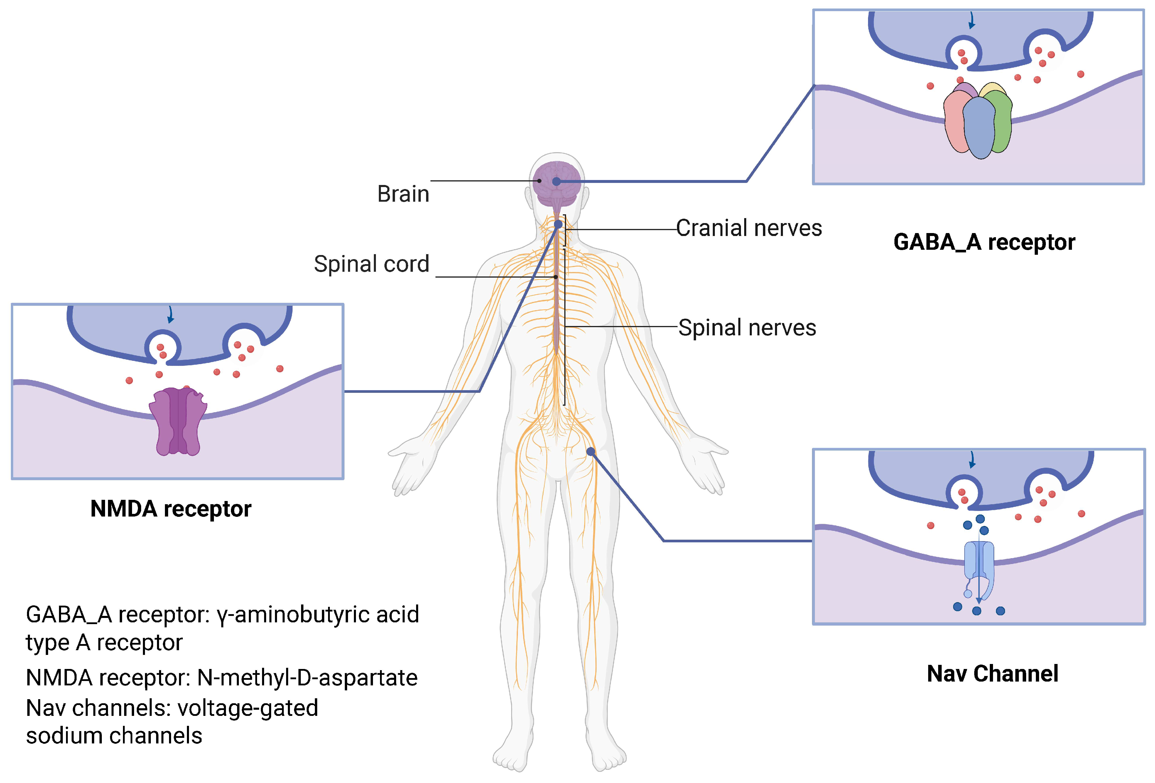
Figure 1. General anesthesia receptors in human body. In the human nervous system, numerous anesthetic drug target receptors are distributed throughout both the central nervous system (CNS) and peripheral nervous system (PNS). This article primarily focuses on GABA_A receptors (γ-aminobutyric acid type A receptor) and NMDA receptors (N-methyl-D-aspartate) predominantly distributed in the CNS, as well as Nav channels mainly found in the PNS. These receptors play crucial roles in general anesthesia. The arrow in the figure indicates the direction of neurotransmitter release from the presynaptic terminal into the synaptic cleft.
Biochem 05 00018 g001
Figure 2. Study selection process using the PRISMA 2020 flow diagram.
Biochem 05 00018 g002
Figure 3. GABA_A receptor distribution and pharmacological modulation in the central nervous system. (A) The GABA_A receptor consists of five subunits in various combinations. The most common subunit classes include α, β, γ, δ, and ε, each containing four transmembrane helices (TM1-TM4). These helices form a central chloride ion (Cl) channel that modulates neuronal excitability. GABA binding to the extracellular ligand binding site triggers a conformational change, facilitating chloride influx. The α1β2γ2 subtype is predominantly found in the cerebral cortex, hippocampus, and thalamus; α2β3γ2 configuration in the amygdala and prefrontal cortex; and α5β3γ2 in the hippocampus. (B) Pharmacological modulation of GABA_A receptors by general anesthetics occurs through distinct mechanisms. Three major anesthetic agents induce neuronal inhibition through different pathways: propofol acts as a positive allosteric modulator (PAM) by binding at the β+/α− interface, etomidate serves as a direct agonist through β subunit binding, and barbiturates function as PAMs at their distinct binding sites. Additionally, flumazenil acts as a competitive antagonist at benzodiazepine binding sites, potentially reversing the inhibitory effects. These diverse mechanisms ultimately modulate chloride conductance, determining the balance between neuronal inhibition and excitation.
Biochem 05 00018 g003
Figure 4. NMDA receptor structure and pharmacological modulation. (A) The N-methyl-D-aspartate (NMDA) receptor functions as a voltage-dependent, ligand-gated cation channel. It features a unique dual-regulation mechanism: At resting membrane potential, magnesium ions (Mg2+) block the channel, preventing ion flux. Upon activation, the receptor requires both glutamate and glycine binding, which leads to the removal of the magnesium block, allowing sodium (Na+) and calcium (Ca2+) ions to flow into the cell. (B) Key pharmacological modulators of the NMDA receptor include ketamine and nitrous oxide (N2O). Ketamine binds deeply within the channel pore of NMDA receptors, functionally disconnecting the thalamocortical and limbic systems, resulting in dissociative anesthesia. It also shows antidepressant properties. N2O binds to the M2/M3 transmembrane domain, acting as a weak NMDA receptor antagonist. It stabilizes the receptor’s closed conformation, suppressing excitatory synaptic transmission, leading to analgesia and sedation. This makes it particularly valuable in pediatric care and emergency medicine. Dexmedetomidine primarily acts as an α2-adrenergic receptor agonist; emerging evidence suggests that it may also indirectly influence NMDA receptor signaling, thereby contributing to its sedative, analgesic, and neuroprotective effects.
Biochem 05 00018 g004
Figure 5. Voltage-gated sodium (Nav) channel structure and function. (A) Voltage-gated sodium (Nav) channels are integral membrane proteins essential for action potential generation and propagation. The channel’s core structure consists of a pore-forming α-subunit containing four homologous domains (DI–DIV), each with six transmembrane helices (S1–S6). The S4 helix functions as the voltage-sensing domain, while S5–S6 helices form the ion pore regulating sodium flow. Auxiliary β-subunits modulate channel kinetics, stability, and drug sensitivity. (B) Nav channels dynamically transition through three functional states. At rest, the channel maintains a closed conformation, preventing ion flow. Upon membrane depolarization, the channel briefly adopts an open state, allowing sodium ions to enter the cell. Following this activation, the channel transitions to an inactivated state, a crucial mechanism that prevents excessive neuronal excitation by temporarily preventing further sodium influx. (C) The Nav channel family includes multiple isoforms (Nav1.1–Nav1.9), each with specific tissue distributions and physiological roles. Specifically, Nav1.7 primarily mediates pain perception, Nav1.4 controls muscle contraction, and Nav1.5 regulates cardiac rhythm.
Biochem 05 00018 g005
Disclaimer/Publisher’s Note: The statements, opinions and data contained in all publications are solely those of the individual author(s) and contributor(s) and not of MDPI and/or the editor(s). MDPI and/or the editor(s) disclaim responsibility for any injury to people or property resulting from any ideas, methods, instructions or products referred to in the content.