Article

Biochemical Changes in Salivary pH and Its Correlation to Hemoglobin Levels, Calcium and Phosphate Ion Concentrations among Pregnant Women, Tanzania: A Cross-Sectional Study

by
Oscar Mbembela
1,2,*,
Davis Ngarashi
2 and
Kasusu Klint Nyamuryekung’e
3
1
Department of Physiology, Mwanza University, Mwanza P.O. Box 3068, Tanzania
2
Department of Physiology, Muhimbili University of Health and Allied Sciences, Dar es Salaam P.O. Box 65001, Tanzania
3
School of Dentistry, Muhimbili University of Health and Allied Sciences, Dar es Salaam P.O. Box 65014, Tanzania
*
Author to whom correspondence should be addressed.
Oral 2023, 3(3), 325-336; https://doi.org/10.3390/oral3030027
Submission received: 9 January 2023 / Revised: 16 April 2023 / Accepted: 9 May 2023 / Published: 14 July 2023

Abstract

:
Background: Surveys in Tanzania show that pregnant women have a significant burden of oral disease, although there is still little literature on the variation of their intraoral electrolytes. The present study investigated changes in salivary calcium (Ca2+), phosphate (PO43−), and hydrogen (H) ions during pregnancy and the correlation between salivary pH and hemoglobin levels. Methodology: A cross-sectional study involved sixty (60) pregnant women stratified by trimester and twenty (20) non-pregnant women attending an antenatal clinic at Mnazi Mmoja Hospital. Consecutive sampling was used, saliva was collected, and electrolyte levels were measured. Gestation age was the independent variable, whereas saliva pH and calcium and phosphate ion concentrations and hemoglobin levels were the dependent variables. Results: A difference in calcium concentration (z = −3.145, p = 0.001) and salivary pH (t = −2.49, p = 0.014) was observed between pregnant and non-pregnant women. Kruskal–Wallis tests for saliva Ca2+ and PO43− and ANOVA for saliva pH revealed differences in concentrations of saliva Ca2+, PO43−, and pH levels between trimester groups (saliva Ca2+ (H = 9.91, df = 3, p = 0.019), saliva PO43− (H = 12.36, df = 3, p = 0.006), saliva pH (F (3.76) = (16.42); p < 0.001)). Pearson’s correlation tests showed no association between salivary pH and hemoglobin levels. Conclusions: Pregnancy is associated with a reduction in saliva pH and saliva calcium levels with a progressive increase in the magnitude of reduction from the first trimester to the third trimester. Saliva phosphate reduction was noticed during the third trimester only and saliva pH levels were independent of hemoglobin levels.

1. Background

Saliva is the fluid synthesized and secreted into the oral cavity by the salivary glands [1]. The primary constituent of saliva is water, which forms about 99% of the whole saliva’s composition. It is also enriched with inorganic and organic constituents [2]. The components of saliva differ from one type of gland to another and are also influenced by various physiological and pathological changes, for instance, pregnancy, anemia, and cardiovascular and circulatory diseases [3]. Each component of saliva serves a specific function, especially in maintaining oral health and gastrointestinal digestive functions [4].
The pH of the oral cavity is maintained by saliva primarily through the action of bicarbonate ions, phosphate ions, and histidine-rich short peptides [5]. These saliva components also help neutralize acids in the plaque caused by microbial fermentation of carbohydrate remnants. Urea from saliva also gets broken down into ammonia and carbon dioxide; ammonia is alkaline and hence it is used to neutralize the acid in the plaque [3,6,7]. Saliva pH has also been extensively studied concerning how its change is associated with an increased risk of some oral lesions like gingivitis and dental caries [8,9,10]. It has been proposed that the saliva pH causes dental caries in different patterns depending on the levels of saliva pH. Weak acids with pH levels of 4.5–6 are the primary culprits for enamel subsurface dissolution. In collaboration with bacteria activities in the plaque, they precipitate the formation of carious lesions [11]. It has been shown that children with dental caries, aside from presenting with low saliva pH [9], also have anemia [10]. Furthermore, Deshpande et al. observed concurrent low hemoglobin and saliva pH levels among children with severe dental caries in comparison to caries-free children [12]. Studies on the correlation between saliva pH and hemoglobin levels among pregnant women are scarce.
Pregnancy modifies the concentration of calcium and phosphates in the saliva. A study by Salvolini et al. [13] indicated a significant decrease in saliva calcium and phosphate concentrations during the second and third trimester of pregnancy. Rio et al. [14] showed that salivary calcium levels were lower in the first and third trimesters compared to non-pregnant women. Salivary phosphate levels were statistically significantly increased only during the first trimester. On the other hand, Rockenbatch et al. [15] did not find any significant difference in the concentration of total calcium and phosphates between pregnant and non-pregnant women. Furthermore, some studies that have been conducted to elucidate the relationship between saliva concentrations of calcium and phosphate ions in pregnant and non-pregnant women have shown contradicting results [13,14,15]. Research has shown that the fall in calcium and phosphate ion concentrations and salivary pH levels are among the risk factor for dental caries and gingivitis [8,16].
Globally, pregnancy has been associated with a higher prevalence of oral health maladies such as dental caries, gingivitis, tooth erosion, and periodontitis [17,18]. It has also been shown that pregnancy changes the concentration of hydrogen, calcium, and phosphate ions in the saliva to different extents from one population group to another [13,14]. Studies conducted in Tanzania have shown that pregnant women have a significant burden of oral diseases [19]. However, no published studies have attempted to investigate the association between pregnancy and saliva calcium, phosphate, and hydrogen ions. Therefore, this study aimed to examine how saliva pH during pregnancy correlates to hemoglobin levels, and calcium and phosphate ion concentrations among Tanzanian pregnant women attending prenatal care at Mnazi Mmoja Hospital.

2. Methods

2.1. Study Design

A stratified, comparative, cross-sectional, health facility-based study was conducted at Mnazi Mmoja Hospital located in Dar es Salaam, Tanzania. The hospital’s reproductive health clinic serves about 1600–1800 pregnant women monthly. Stratification was applied during enrolment to ensure equal representation of the participants by pregnancy trimester.

2.2. Sample Size and Study Participant Selection Procedure

The sample size was estimated based on a previous study [11], which showed that the saliva pH was about 6.56 (SD 0.42) and 6.97 (SD 0.48) in pregnant and non-pregnant women, respectively. When these parameters were substituted into the online statistical software OpenEpi Version 3.01 and based on the formula published by Soe and Sullivan [20], they gave a sample size of 80 participants with a ratio of 1:3 (20:60) between the non-pregnant and pregnant groups and with a 90% power to detect the difference at a 95% confidence level.
In both pregnant and non-pregnant women, participants were selected using a consecutive sampling technique. The inclusion criteria were Tanzanian pregnant and non-pregnant women aged between 23 and 26 years and in their first parity. Pregnant women in the first parity were selected because of the increased possibility of obtaining appropriate controls (non-pregnant women of the same parity) from the same facility. Thus, non-pregnant women of the same age range and parity were consecutively selected as controls among women attending the Reproductive and Child Health (RCH) unit for consultation at Mnazi Mmoja Hospital. Age matching was introduced to control for possible variation in saliva electrolytes due to age [21]. Participants’ enrolment was divided into four equal groups, comprising twenty women. These groups contained women in their first, second, and third trimesters and the non-pregnant controls. Therefore, twenty pregnant women for each of the three trimesters and non-pregnant women were enrolled, making a total of eighty participants.
Women who reported having any oral wounds, a history of tooth extraction within the last three days, any known systemic, hypertensive, or diabetic illness, xerostomia, hyperemesis gravidarum, regular smokers, and non-pregnant women in menstruation were excluded from the study.

2.3. Saliva Collection

Saliva was collected from 8.00 a.m. to 11.00 a.m. to control for circadian rhythm variations [17,18,22,23]. To ensure the collection of debris-free saliva, the collection was performed at least 2 h after a meal. Additionally, each participant was requested to rinse their mouth with 15 mL of distilled water to remove exfoliative cells and other remnants that might be present before collection [13,18,24]. Participants were asked to stay quiet and position their heads with a slight forward tilt to guarantee the collection of unstimulated whole saliva. Then, participants were asked to expectorate saliva that had accumulated on the floor of the mouth into a sterile disposable plastic container 5 min after rinsing the mouth in a quiet room. Saliva pH was measured immediately after collecting saliva to avoid the effect of microorganisms that can initiate biochemical reactions that may alter the pH of collected saliva [24]. The collected saliva was labeled and then stored in a cool box at 4 °C. Saliva calcium and phosphate ion concentrations were measured at the Muhimbili University of Health and Allied Sciences (MUHAS) clinical research laboratory on the same day as the data collection.

2.4. Study Variables

Salivary pH was measured using a digital pH meter (Ohaus ST, New York, NY, USA). The pH meter had a scale that ranged from 0 to 14. Saliva with a pH level of 7.0 was classified as neutral, below 7.0 was classified as an acidic pH, and above 7.0 as an alkaline pH.
Calcium and phosphate ions were measured and recorded in mmol/L using a Cobas biochemistry analyzer machine (Roche Diagnostics, GmbH, Mannheim, Germany) in the MUHAS clinical research laboratory.
Gestational age was extracted from the participants’ RCH cards and recorded as completed pregnancy weeks. About 40 weeks, which are commonly divided into three trimesters, constitute the typical gestation period. The first trimester is the duration from the end of the last menstrual period until 13 weeks, the second trimester comprises weeks 14 to 27, and the third trimester is from 28 weeks until delivery.
Hemoglobin levels are routinely measured using a hemoglobin analyzer machine HemoCue® 201+ (Ängelholm, Sweden) during each visit of pregnant women to the facility. The hemoglobin levels of pregnant women routinely measured on the day of the clinic visit were recorded from RCH cards in g/dL. According to WHO guidelines, a hemoglobin concentration of less than 7 g/dL was considered severe anemia, from 7 to 9.9 g/dL as moderate anemia, from 10 to 10.9 g/dL as mild anemia, and 11 and above was considered non-anemic [25].

2.5. Data Management and Analysis

The recorded data were entered into a Microsoft Excel sheet. Consistency and verification of missing information were cross-checked before exporting the data to the STATA software version 16 for analysis. Shapiro–Wilk tests were used to test for normality of the data whereby saliva pH and hemoglobin levels showed parametric distributions and therefore were summarized as the mean and standard deviation. However, saliva calcium and phosphate ion concentrations showed non-parametric distributions and were therefore summarized as the median and interquartile range. An unpaired t-test was conducted to test for differences in mean saliva pH between pregnant and non-pregnant women while the Mann–Whitney U test was conducted to test for differences in saliva calcium and phosphate ion concentrations. Kruskal–Wallis rank test and ANOVA were used to test for differences in medians and means of saliva electrolytes in non-pregnant women and the three groups of pregnant women. Pearson’s’ correlation test was conducted to determine the association between salivary pH and hemoglobin levels of pregnant women.

3. Results

3.1. Study Participant’s Demographic and Clinical Characteristics

During selection, matching was applied whereby all participants were in their first parity and had an age range of 23–26 years. The mean weight, height, and body mass index (BMI) of the participants are shown in (Table 1).

3.2. Description of Saliva Electrolytes Values
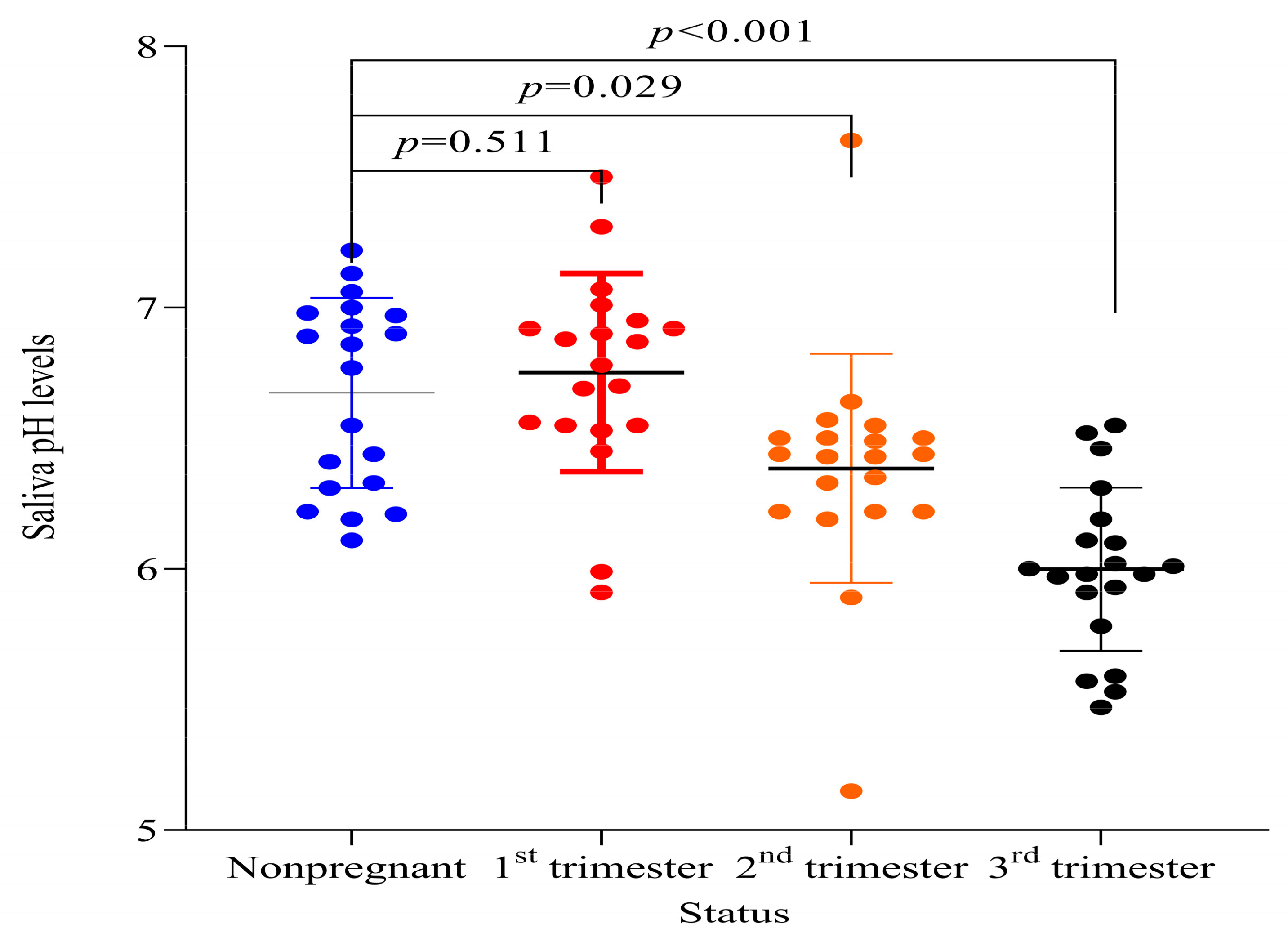
Saliva pH (p = 0.015) and calcium ion concentrations (p = 0.001) from pregnant women were different from non-pregnant women. No difference in saliva phosphate concentration between pregnant and non-pregnant women was observed (p = 0.084). A Spearman’s correlation test between calcium to phosphate ratios and saliva pH revealed a statistically significant negative relationship (Table 2). Comparisons of the electrolyte values between the four groups revealed statistically significant differences in means and medians using ANOVA and Kruskal–Wallis tests (saliva pH < 0.001, saliva Ca2+ p = 0.019, saliva PO43− p = 0.006) (Table 3).

3.3. Saliva pH

Pregnant women in the third trimester showed the biggest difference in saliva pH levels (p < 0.001) according to the pairwise comparison between non-pregnant women and pregnant women in the first, second, and third trimester as shown in (Figure 1).

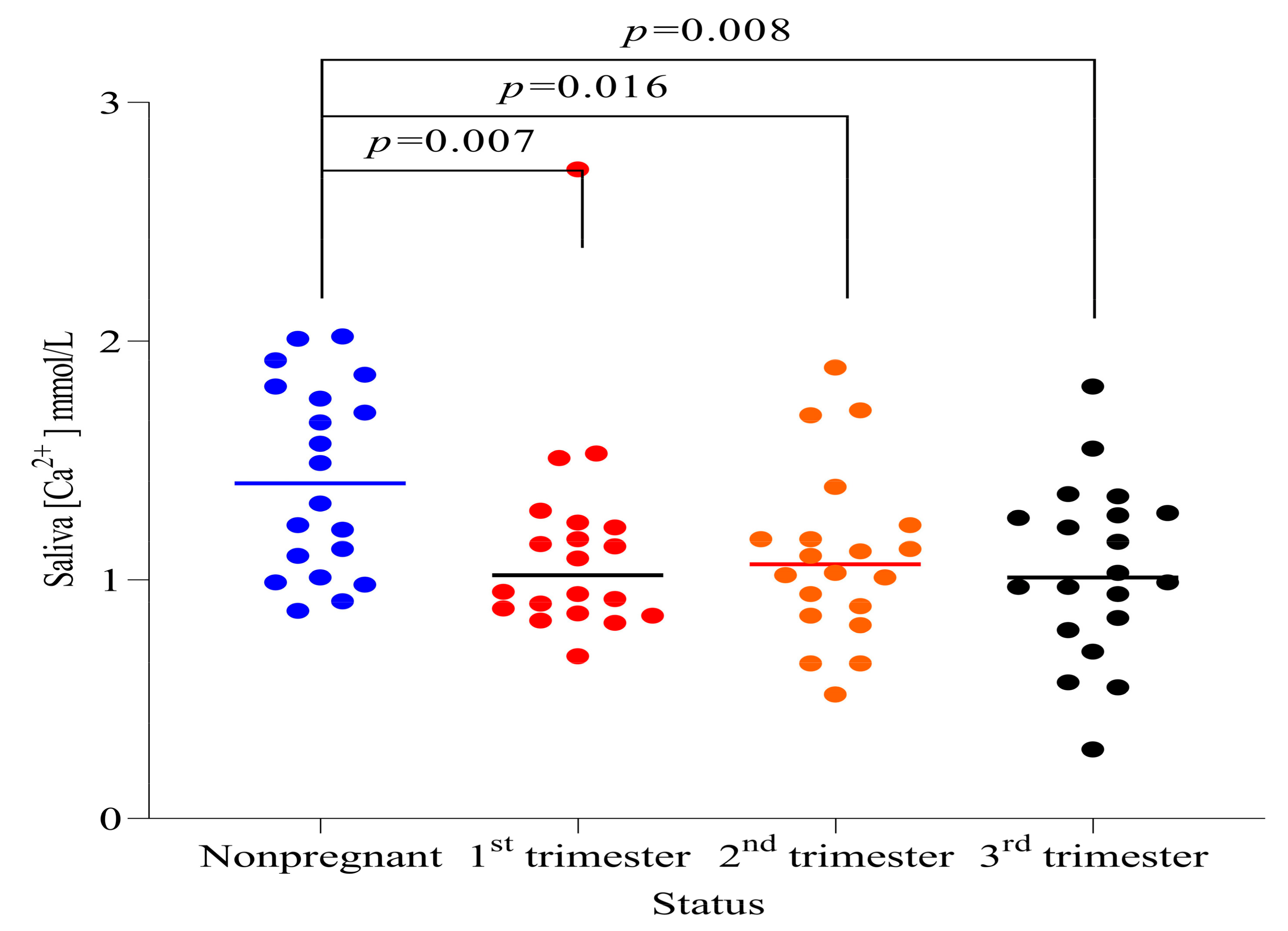
3.4. Saliva Calcium Ion Concentration

Salivary calcium ions levels decreased considerably across all trimesters. There was a statistically significant difference in each group compared to non-pregnant women as shown in (Figure 2).

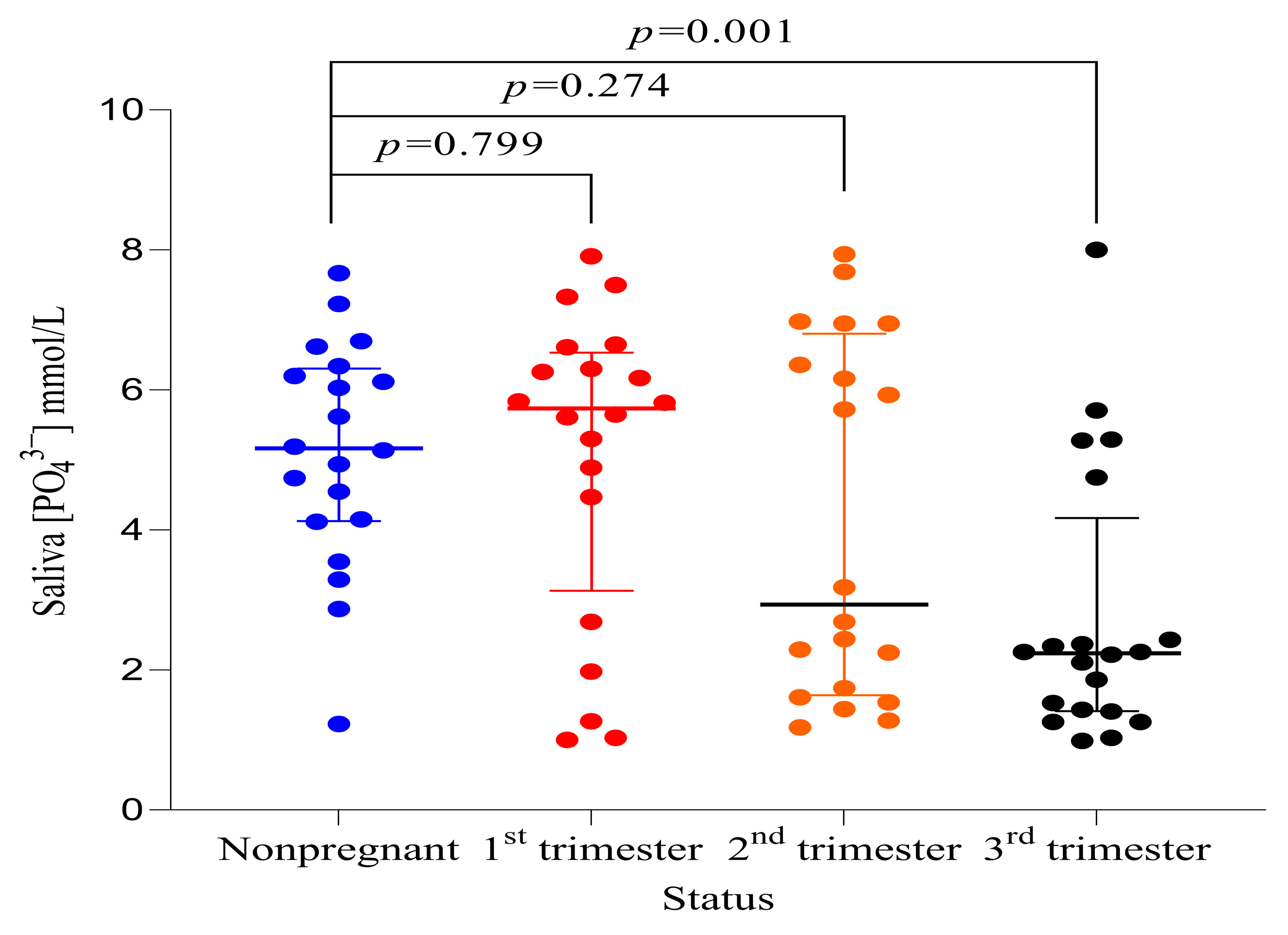
3.5. Saliva Phosphate Ion Concentration

Overall, no differences in saliva phosphate ion concentrations between pregnant and non-pregnant women were observed (p = 0.084) (Table 2). However, pregnant women in their third trimester had saliva phosphate concentrations that were significantly lower compared to non-pregnant women (p = 0.001) (Figure 3).

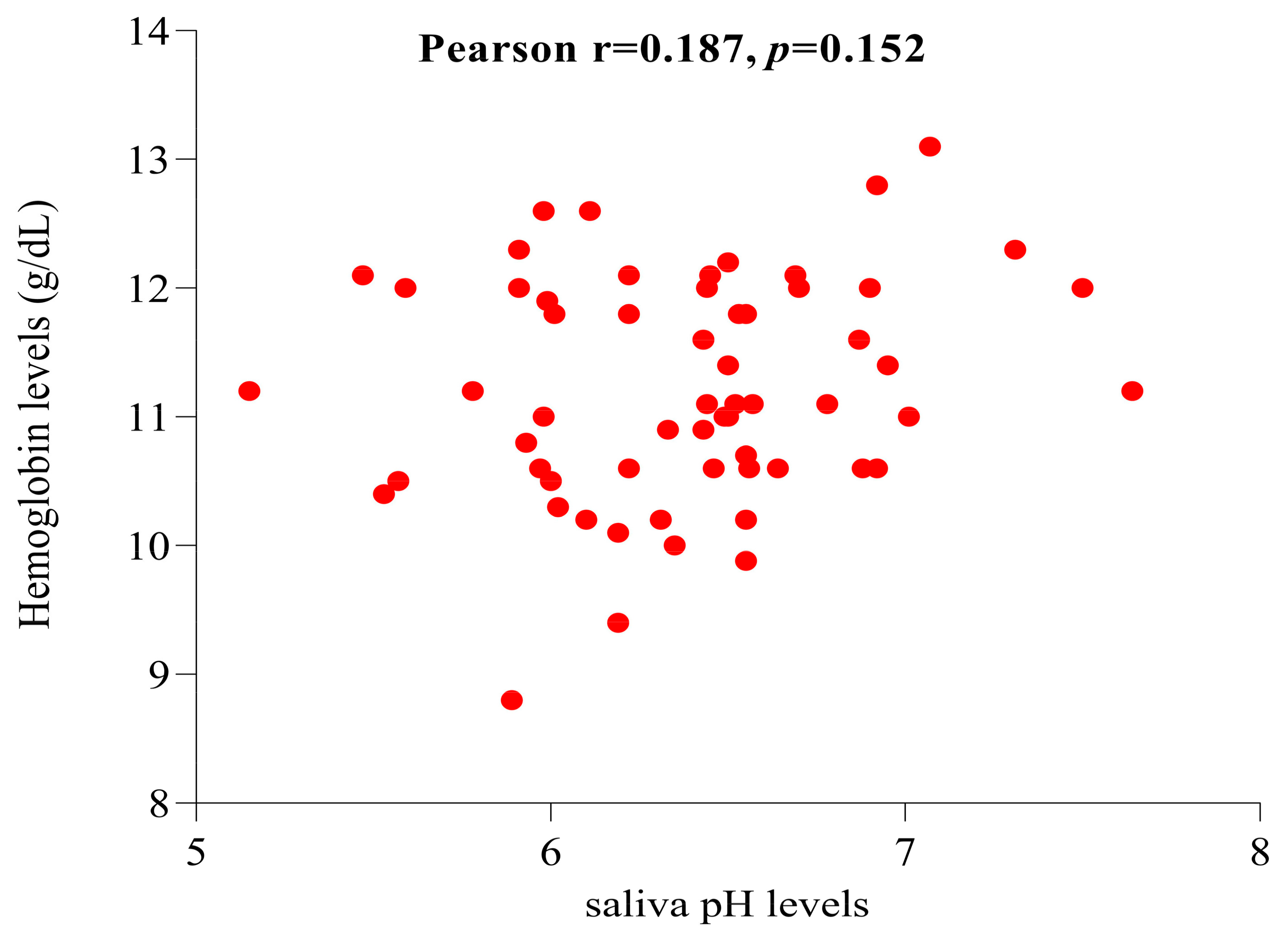
3.6. Correlation of Saliva pH Levels and Hemoglobin Levels among Pregnant Women

Hemoglobin levels showed a parametric distribution according to a Shapiro–Wilk test for normality (p = 0.55). The mean and SD were 11.22 ± 0.87 g/dL and the minimum and maximum values were 8.8 g/dL and 13.1 g/dL, respectively. A Pearson correlation test between pregnant women’s hemoglobin levels and saliva pH was performed. There was no statistically significant correlation between saliva pH and hemoglobin levels (p = 0.152) as shown in Figure 4.

4. Discussion

The findings from this study have revealed differences in saliva pH and calcium ion concentrations in unstimulated whole saliva between pregnant and non-pregnant women. On the other hand, saliva phosphate ion concentrations were similar between pregnant and non-pregnant women.
The current findings corroborate those of previous studies showing that pregnancy is associated with a reduction in salivary pH [13,14,15,26,27]. It has also been shown that the reduction in saliva pH during pregnancy is among the risk factors for developing dental caries and periodontal diseases during pregnancy [13,28]. The lowest saliva pH was observed during the third trimester, indicating a progressive reduction with increasing gestational age. Low saliva pH in pregnant women has been postulated to be due to progesterone hormone action in reducing levels of plasma bicarbonate ions [26]. Other studies have hypothesized that the saliva pH reduction during pregnancy is caused by changes in dietary habits and cravings that occur during pregnancy [11]. However, this has yet to be clearly explained and this study could not determine the possible reasons for the observed variation.
Studies have indicated that a decrease in saliva buffering capacity during pregnancy may cause a reduction in saliva pH [22,29]. The low levels of saliva pH observed in this study can be a risk factor for increased demineralization of dental hard tissues and the development of dental caries [23]. However, research suggests there is a critical saliva pH below which the teeth demineralization process can start [24]. Contrary to this, a prospective clinical investigation presented a weak association between saliva factors and the development of dental caries. It suggested that dental caries have a multifactorial causation with consistent interaction between factors, including disequilibrium between mineralization and demineralization processes [16]. Studies have also shown an association between low saliva pH and a shift in acid-tolerant and acid-producing bacteria that play a significant role in caries development [30]. Low saliva pH and periodontitis are associated with low birth weight among neonates and affect their quality of life [31,32].
Hemoglobin is one of the intracellular buffers in the circulation that play a key role in the defense against changes in hydrogens ion concentrations [33]. Several studies conducted on children have indicated a high prevalence of dental caries in children with anemia [10,34,35,36]. Additionally, Mahantesha et al. [37] found that after the correction of anemia in children, a positive relationship between hemoglobin levels and salivary pH was observed. Therefore, it was hypothesized that there could be a correlation between saliva pH and hemoglobin levels in pregnant women as hemoglobin is the most potent regulator of blood pH [33]. A possible explanation for the failure to observe a correlation between hemoglobin levels and saliva pH could be due to differences in the strength and types of buffer systems that are used in the saliva in comparison to those used in the blood [38]. Under resting conditions, the phosphate buffer system is the most important in the oral cavity while in the plasma, the bicarbonate buffer system is the major buffer system. Similarly, Pradeep et al. [39] did not observe any significant baseline blood pH changes despite significant changes in saliva pH from acidic to neutral pH among patients pre and post non-surgical periodontal therapy. Additionally, Easwaran et al. [40] found no relationship between dental caries and iron deficiency anemia among children. Our study corroborates previous findings showing a lack of correlation between salivary pH and blood hemoglobin levels.
Consistent with previous findings, saliva calcium and phosphate ion concentrations were shown to decrease with the progression of pregnancy [13,41]. The reasons behind a fall in saliva calcium and phosphate ion concentrations during pregnancy may include the increased demand for these minerals for the fetal bone ossification process that requires a large amount of calcium and phosphate ions from the plasma [13]. Some epidemiological studies have reported a correlation between saliva calcium levels and plasma estrogen levels. It has been shown that as plasma levels of estrogen hormone increase, there is a concurrent decrease in saliva calcium levels [42]. This tendency could explain why saliva calcium levels decrease during pregnancy as there is potentially an increase in plasma estrogen hormone levels produced by the placenta during pregnancy [43]. Low levels of saliva calcium and phosphates increase the risk for the development of caries and teeth erosion. This is because of disturbance to the demineralization and mineralization equilibrium of the oral environment. The teeth mineralization process performs best when the saliva is supersaturated with calcium and phosphate, and is at a suitable pH (8). In addition to supersaturation, teeth remineralization is favored when there is a low calcium-to-phosphate ratio [44]. The results from this study support the observation that non-pregnant women and pregnant women who were in the first trimester of pregnancy had the lowest calcium-to-phosphate ratios. Furthermore, a negative relationship was observed suggesting that a decline in calcium-to-phosphate ratios corresponds to an increase in saliva pH which is favorable for teeth mineralization. Therefore, pregnancy was associated with biochemical salivary changes favoring demineralization of calcified dental tissues.
This study applied solid matching criteria for parity and age in comparison to other studies investigating the alteration of salivary electrolytes during pregnancy. Participants within a small age range were sampled, unlike previous studies that used participants with a large age interval and lacked parity matching. Nevertheless, these stringent criteria for participation could have influenced our results because of the observed variation of salivary electrolyte values by age (21). Despite this, our results showed consistent electrolyte variation across the studied groups and largely reflected previous findings. However, no clinical assessment of oral diseases was undertaken in our study, eliminating the possibility of corroboration of oral disease burden in pregnant women and the effect of such pathologies in salivary electrolyte variations. Regarding the overall procedure for the assessment of electrolytes, some limitations need attention. There is a possibility that a few participants’ saliva may have been collected while they consumed food in less than the specified time of two hours as recommended. This is because the only proof of when the last food was consumed was by asking a question to the participants and given the fact that there is a difference in ability in recalling among individuals. However, this did not affect the reliability and validity of our results due to the larger sample size and power of the study as compared to previous studies. Additionally, the salivary flow rate was not recorded; however, this study applied solid age-matching criteria which eliminated the effect of age on the saliva flow rate [32]. The spitting method used to collect saliva also has the disadvantage of the possibility of conferring some stimulatory effects. However, this is one of the recommended methods for collecting unstimulated saliva. Furthermore, participants were made aware to allow saliva to flow without undue stimulation [42,45].
Although the study design has been able to establish the effect of pregnancy on the aforementioned saliva electrolytes, it does not allow extrapolation of causal relationships compared to other study designs [46]. Nevertheless, the results from this study are valuable and corroborate similar previous primary studies performed in different ethnic groups and geographical locations.

5. Conclusions

Pregnancy is associated with a reduction in saliva pH and saliva calcium levels with a progressive increase in the magnitude of reduction from the first trimester to the third trimester. No correlation between saliva pH levels and hemoglobin levels among pregnant women was observed. Salivary phosphate levels were shown to be markedly reduced during the third trimester. In summary, our findings suggest that pregnant women are at a higher risk of developing dental caries and periodontal diseases as compared to non-pregnant women due to alterations in key electrolytes for the maintenance of optimum intraoral equilibrium.

6. Recommendations

Due to the increased risk of dental diseases from salivary electrolyte variations at this critical time, pregnant women must maintain proper oral hygiene practices. Prompt visits to dental facilities for clinical assessment and other preventive measures are strongly advised during pregnancy. Further research is required to better understand the prevalence of oral illnesses and their potential association with altered levels of salivary electrolyte concentrations and hemoglobin levels in pregnant women. Additionally, it would be beneficial to investigate when any alterations in electrolyte levels return to normal during the postpartum period.

Author Contributions

O.M. performed the data collection, data analysis, and wrote the manuscript. D.N. and K.K.N. participated in designing the study, data analysis, interpretation of findings, and critically reviewing the manuscript at each step of preparation. The final version of the manuscript was read and approved by all authors. All authors have read and agreed to the published version of the manuscript.

Funding

The design and data gathering were financed by Mwanza University, and the authors paid for the remaining costs. The funder did not influence, in any way, the conduct of this research, the development of the manuscript, and the conclusions provided.

Institutional Review Board Statement

Ethical clearance with reference number DA.282/298/01.C/ was obtained from the MUHAS Institutional Review Board (IRB) on behalf of the National Research Ethics Committee. All methods and procedures were carried out in compliance with the Helsinki Declaration. Informed consent was obtained from the participants before participation in the study. Permission to conduct the study at Mnazi Mmoja Hospital was sought from the regional, district, municipal, and hospital administrative offices in succession.

Informed Consent Statement

Not applicable.

Data Availability Statement

The dataset is available from the corresponding author and can be provided upon reasonable request.

Acknowledgments

We would like to thank the Department of Physiology at the Muhimbili University of Health and Allied Sciences for providing a conducive environment for undertaking this research work. We would also like to thank the management of Mnazi Mmoja Health Center with extended appreciation to the Reproductive and Child Health Unit workers for their corporation and for allowing the study to be conducted within their vicinity. Critical thanks go out to all the women who voluntarily agreed to be study participants. Lastly, we would like to extend our profound thanks to the management of Mwanza University for funding this study.

Conflicts of Interest

The authors declare that they have no competing interest.

References

  1. Proctor, G.B. The physiology of salivary secretion. Periodontol. 2000 2016, 70, 11–25. [Google Scholar] [CrossRef] [PubMed] [Green Version]
  2. Spielman, A.I.; D’Abundo, S.; Field, R.B.; Schmale, H. Protein Analysis of Human von Ebner Saliva and a Method for its Collection from the Foliate Papillae. J. Dent. Res. 2016, 72, 1331–1335. [Google Scholar] [CrossRef] [PubMed]
  3. De Almeida, P.D.V.; Grégio, A.M.T.; Machado, M.Â.N.; De Lima, A.A.S.; Azevedo, L.R. Saliva composition and functions: A comprehensive review. J. Contemp. Dent. Pract. 2008, 9, 72–80. [Google Scholar] [PubMed]
  4. Saher, F.; Khurshid, Z.; Zafar, M.S.; Mohammed, F.; Khurram, S.A. Salivary Glands. In An Illustrated Guide to Oral Histology; Wiley Online: Hoboken, NJ, USA, 2021; pp. 147–166. [Google Scholar]
  5. Maddu, N.; Maddu, N. Functions of Saliva. Saliva and Salivary Diagnostics. 2019. Available online: https://www.intechopen.com/state.item.id (accessed on 29 November 2022).
  6. Kumar, B.; Kashyap, N.; Avinash, A.; Chevvuri, R.; Sagar, M.K.; Shrikant, K. The composition, function and role of saliva in maintaining oral health: A review. Int. J. Contemp. Dent. Med. Rev. 2017, 220, 011217. [Google Scholar]
  7. Humphrey, S.P.; Williamson, R.T. A review of saliva: Normal composition, flow, and function. J. Prosthet. Dent. 2001, 85, 162–169. [Google Scholar] [CrossRef]
  8. Abou Neel, E.A.; Aljabo, A.; Strange, A.; Ibrahim, S.; Coathup, M.; Young, A.M.; Bozec, L.; Mudera, V. Demineralization–remineralization dynamics in teeth and bone. Int. J. Nanomed. 2016, 11, 4743. [Google Scholar] [CrossRef] [PubMed]
  9. Preethi, B.P.; Pyati, A.; Dodawad, R. Evaluation of flow rate, ph, buffering capacity, calcium, total protein and total antioxidant levels of saliva in caries free and caries active children—An in vivo study. Biomed. Res. 2010, 25, 425–428. [Google Scholar] [CrossRef] [Green Version]
  10. Mohamed, W.E.; Abou El Fadl, R.K.; Thabet, R.A.; Helmi, M.; Kamal, S.H. Iron deficiency anaemia and early childhood caries: A cross-sectional study. Aust. Dent. J. 2021, 66, S27–S36. [Google Scholar] [CrossRef]
  11. Joel, A.M.M.; Zurisadai, N.A.; Alejandra, L.A.; Hector, M.H.N.; Jany, A.J.D.V.; Alma, N.C.P.; Sosa-Martinez, L.R.; De La Garza-Ramos, M.A. The importance of pH, salivary flow and different dental caries risk factors in pregnant women. J. Dent. Oral Hyg. 2018, 10, 18–22. [Google Scholar] [CrossRef]
  12. Deshpande, A.N.; Sudani, U.; Wadhwa, M.; Joshi, N.; Patel, K.S.; Jain, A. Association of Anthropometric Measurements, Hemoglobin Level and Salivary Parameters among Caries-free and S-ECC Children. Int. J. Clin. Pediatr. Dent. 2022, 15 (Suppl. S2), S164. [Google Scholar]
  13. Salvolini, E.; Di Giorgio, R.; Curatola, A.; Mazzanti, L.; Fratto, G. Biochemical modifications of human whole saliva induced by pregnancy. BJOG 1998, 105, 656–660. [Google Scholar] [CrossRef]
  14. Rio, R.; Azevedo, Á.; Simões-Silva, L.; Marinho, J.; Silva, M.J.; Sampaio-Maia, B. The biochemistry of saliva throughout pregnancy. Med. Express 2015, 2, M150506. Available online: http://www.scielo.br/scielo.php?script=sci_arttext&pid=S2358-04292015000500006&lng=en&nrm=iso&tlng=en (accessed on 20 April 2021). [CrossRef] [Green Version]
  15. Rockenbach, M.I.; Marinho, S.A.; Veeck, E.B.; Lindemann, L.; Shinkai, R.S. Salivary flow rate, pH, and concentrations of calcium, phosphate, and sIgA in Brazilian pregnant and non-pregnant women. Head Face Med. 2006, 2, 44. [Google Scholar] [CrossRef] [PubMed] [Green Version]
  16. Khan, A.; Qureshi, B.; Qureshi, A.; Imtiaz, Y.; Qadeer, S. Correlation of salivary characteristics with high risk of dental caries; A clinical investigation. Future Dent. J. 2017, 4, 72–75. [Google Scholar] [CrossRef]
  17. Naseem, M.; Khurshid, Z.; Khan, H.A.; Niazi, F.; Zohaib, S.; Zafar, M.S. Oral health challenges in pregnant women: Recommendations for dental care professionals. Saudi J. Dent. Res. 2016, 7, 138–146. [Google Scholar] [CrossRef] [Green Version]
  18. Hemalatha, V.T.; Manigandan, T.; Sarumathi, T.; Aarthi Nisha, V.; Amudhan, A. Dental Considerations in Pregnancy-A Critical Review on the Oral Care. J. Clin. Diagn. Res. 2013, 7, 948. [Google Scholar]
  19. Mwangosi, I.E.A.T.; Kiango, M.M. Oral health experience during pregnancy and dental service utilization in Bariadi District, Tanzania. Tanzan J. Health Res. 2012, 14, 1–7. [Google Scholar] [CrossRef] [PubMed] [Green Version]
  20. Soe, M.M.; Sullivan, K.M. Documentation of Sample Size for Comparing Two Means. 2005. Available online: https://www.openepi.com/PDFDocs/SSMeanDoc.pdf (accessed on 20 April 2021).
  21. Sevón, L.; Laine, M.A.; Karjalainen, S.; Doroguinskaia, A.; Helenius, H.; Kiss, E.; Lehtonen-Veromaa, M. Effect of Age on Flow-Rate, Protein and Electrolyte Composition of Stimulated Whole Saliva in Healthy, Non-Smoking Women. Open Dent. J. 2008, 2, 89. [Google Scholar] [CrossRef] [Green Version]
  22. Yousefi, M.; Parvaie, P.; Riahi, S.M. Systematic Review Salivary Factors Related to Caries in Pregnancy A Systematic Review and Meta-Analysis. J. Am. Dent. Assoc. 2020, 151, 576–588. [Google Scholar] [CrossRef]
  23. Nieuw Amerongen, A.V.; Veerman, E.C.I. Saliva—The defender of the oral cavity. Oral Dis. 2002, 8, 12–22. [Google Scholar] [CrossRef] [Green Version]
  24. Dawes, C. What is the critical pH and why does a tooth dissolve in acid? J. Can. Dent. Assoc. 2003, 69, 722–724. [Google Scholar]
  25. Chan, M.; WHO. Haemoglobin Concentrations for the Diagnosis of Anaemia and Assessment of Severity; World Health Organization: Geneva, Switzerland, 2011.
  26. Naveen, S.; Asha, M.L.; Shubha, G.; Bajoria, A.A.; Jose, A.A. Salivary Flow Rate, pH and Buffering Capacity in Pregnant and Non Pregnant Women-A Comparative Study. JMED Res. 2014, 2014, 1–8. [Google Scholar] [CrossRef] [Green Version]
  27. Migliario, M.; Bindi, D.; Surico, A.; DE Deprini, S.; Minsenti, F.; Pezzotti, B.; Melle, P.L.F.B. Changes in salivary flow rate and pH in pregnancy. Eur. Rev. Med. Pharmacol. Sci. 2021, 25, 1804–1810. [Google Scholar] [PubMed]
  28. Rakchanok, N.; Amporn, D.; Yoshida, Y.; Harun-Or-Rashid, M.D.; Sakamoto, J. Dental caries and gingivitis among pregnant and non-pregnant women in Chiang Mai, Thailand. Nagoya J. Med. Sci. 2010, 72, 43–50. [Google Scholar] [PubMed]
  29. Lasisi, T.J.; Ugwuadu, P.N. Pregnancy related changes in human salivary secretion and composition in a Nigerian population. Afr. J. Med. Med. Sci. 2014, 43, 347–351. [Google Scholar] [PubMed]
  30. Zhou, J.; Jiang, N.; Wang, Z.; Li, L.; Zhang, J.; Ma, R.; Nie, H.; Li, Z. Influences of pH and iron concentration on the salivary microbiome in individual humans with and without caries. Appl. Environ. Microbiol. 2017, 83, e02412-16. [Google Scholar] [CrossRef] [PubMed] [Green Version]
  31. Guimares, A.N.; Silva-Mato, A.; Siqueira, F.M.; Cyrino, R.M.; Cota, L.O.M.; Costa, F.O. Very low and low birth weight associated with maternal periodontitis. J. Clin. Periodontol. 2012, 39, 1024–1031. [Google Scholar] [CrossRef] [PubMed]
  32. Ahmad, M.S.; Bhayat, A.; Zafar, M.S.; Al-Samadani, K.H. The Impact of Hyposalivation on Quality of Life (QoL) and Oral Health in the Aging Population of Al Madinah Al Munawarrah. Int. J. Environ. Res. Public Health 2017, 14, 445. [Google Scholar] [CrossRef] [Green Version]
  33. Rubana, I.E.; Aulik, I.V. Buffer function of hemoglobin. Res. Sport. Med. Int. J. 2009, 1, 125–126. [Google Scholar] [CrossRef]
  34. Schroth, R.J.; Levi, J.; Kliewer, E.; Friel, J.; Moffatt, M.E.K. Association between iron status, iron deficiency anaemia, and severe early childhood caries: A case-control study. BMC Pediatr. 2013, 13, 22. [Google Scholar] [CrossRef] [Green Version]
  35. Abdallah, M.A.; Abed, H.H.; Hamza, G.; Alsahafi, E.N. The association between dmft index and haemoglobin levels in 3-6 year-old Saudi children with anaemia: A cross sectional study. J. Taibah Univ. Med. Sci. 2016, 11, 72–76. [Google Scholar] [CrossRef] [Green Version]
  36. Hashemi, A.; Bahrololoomi, Z.; Pediatric SSIJ. Undefined Relationship between Early Childhood Caries and Anemia: A Systematic Review. 2018. Available online: https://www.semanticscholar.org/paper/Relationship-Between-Early-Childhood-Caries-and-A-Hashemi-Bahrololoomi/404e665e2fd99db8077fa94452a09e06e3992a97 (accessed on 21 April 2023).
  37. Mahantesha, T.; Parveen Reddy, K.M.; Ellore, V.P.K.; Ramagoni, N.K.; Iitagi, V.; Anitha, K.S. Evaluation and association of iron deficiency anaemia with salivary pH and buffering capacity. Natl. J. Physiol. Pharm. Pharmacol. 2014, 4, 229. [Google Scholar] [CrossRef]
  38. Lilienthal, B. Buffering systems in the mouth. Oral Surg. Oral Med. Oral Pathol. 1955, 8, 828–841. [Google Scholar] [CrossRef] [PubMed]
  39. Koppolu, P.; Sirisha, S.; Penala, S.; Reddy, P.K.; Alotaibi, D.H.; Abusalim, G.S.; Lingam, A.S.; Mukhtar, A.H.; Barakat, A.; AlMokhatieb, A.A. Correlation of Blood and Salivary pH Levels in Healthy, Gingivitis, and Periodontitis Patients before and after Non-Surgical Periodontal Therapy. Diagnostics 2022, 12, 97. [Google Scholar] [CrossRef]
  40. Easwaran, H.N.; Annadurai, A.; Muthu, M.S.; Sharma, A.; Patil, S.S.; Jayakumar, P.; Jagadeesan, A.; Nagarajan, U.; Pasupathy, U.; Wadgave, U. Early Childhood Caries and Iron Deficiency Anaemia: A Systematic Review and Meta-Analysis. Caries Res. 2022, 56, 36–46. [Google Scholar] [CrossRef] [PubMed]
  41. Bakhshi, M.; Sabet, M.S.; Hashemi, E.S.; Bakhtiari, S.; Tofangchiha, M.; Marhabi, S.A.; Alirezaei, S. Evaluation of biochemical changes in unstimulated salivary, calcium, phosphorous and total protein during pregnancy. Afr. J. Biotechnol. 2012, 11, 2078–2082. [Google Scholar]
  42. Bhattarai, K.R.; Kim, H.R.; Chae, H.J. Compliance with Saliva Collection Protocol in Healthy Volunteers: Strategies for Managing Risk and Errors. Int. J. Med. Sci. 2018, 15, 823. [Google Scholar] [CrossRef] [Green Version]
  43. Aps, J.K.M.; Martens, L.C. Review: The physiology of saliva and transfer of drugs into saliva. Forensic. Sci. Int. 2005, 150, 119–131. [Google Scholar] [CrossRef]
  44. Hara, A.T.; Karlinsey, R.L.; Zero, D.T. Dentine remineralisation by simulated saliva formulations with different Ca and Pi contents. Caries Res. 2008, 42, 51–56. [Google Scholar] [CrossRef]
  45. Navazesh, M. Methods for collecting saliva. Ann. N. Y. Acad. Sci. 1993, 694, 72–77. [Google Scholar] [CrossRef]
  46. Greenhalgh, T. How to read a paper. Getting Your Bearings (Deciding What the Paper Is About). Br. Med. J. 1997, 315, 243. [Google Scholar] [CrossRef] [Green Version]
Figure 1. Individual value scatter plots of saliva pH levels in non-pregnant women (n = 20) and pregnant women (n = 20 in each trimester).
Oral 03 00027 g001
Figure 2. Individual value scatter plots of saliva calcium ion concentrations in non-pregnant women (n = 20) and pregnant women (n = 20 in each trimester).
Oral 03 00027 g002
Figure 3. Individual value scatter plots of saliva phosphate ion concentrations in non-pregnant women (n = 20) and pregnant women (n = 20 in each trimester).
Oral 03 00027 g003
Figure 4. Scatter plot of saliva pH levels vs. hemoglobin levels.
Oral 03 00027 g004
Table 1. Characteristics of participants (n = 80).
Non-Pregnant (n = 20)Pregnant (n = 60)p-Value
Mean ± SD
Weight (kg)63.6 ± 12.1366.18 ± 9.940.344
Height (cm)157.95 ± 9.93157.26 ± 7.330.743
Frequency (%)Frequency (%)
BMIUnderweight (<18.5)6 (30) 1 (1.67)
Normal weight (18.5–24.9)12 (60) 17 (28.33)
Overweight (25–29.9)2 (10) 34 (56.67)
Obese (30–39.9)0 (0) 8 (13.33)
Hb levelsSevere anemia (<7 g/dL) 0 (0)
Moderate (7–9.9 g/dL) 3 (5)
Mild (10–10.9 g/dL) 20 (33.33)
Non-anemic (≥11 g/dL) 37 (61.67)
p-value for unpaired t-test for height and weight differences between pregnant and non-pregnant women.
Table 2. Variation in saliva pH, and calcium and phosphate ion concentrations between pregnant and non-pregnant women (n = 60 and n = 20, respectively) and calcium-to-phosphate ratios and its correlation to saliva pH.
Non-Pregnant (n = 20)(Pregnant n = 60)
Median (IQR)Median (IQR)z-ValueExact p-Value
Saliva [Ca2+] mmol/L1.41 (1.06–1.79)1.03 (0.86–1.25)−3.1450.001
Saliva [PO43−] mmol/L5.17 (4.14–6.27)2.94 (1.68–6.17)−1.7330.084
Mean ± (SD)t-valuep-value
Saliva pH6.67 (0.36)6.38 (0.49)−2.490.015
Non-pregnantFirst trimesterSecond trimesterThird trimester
[Ca2+]/[PO43−] ratios (Mean ± SD)0.35 ± 0.310.33 ± 0.330.40 ± 0.320.53 ± 0.36
Correlation between saliva pH and [Ca2+]/[PO43−] ratios Spearman’s r95% Confidence Interval of rp-value
−0.274−0.471 to −0.0510.014
Mann–Whitney U test for saliva [Ca2+] and [PO43−] and unpaired t-test for saliva pH.
Table 3. Comparison of means of saliva pH and medians of saliva calcium and phosphate ion concentrations across the groups.
Variable(H)Degree of Freedomp-Value
Saliva [Ca2+] mmol/L9.9130.019 **
Saliva [PO43−] mmol/L12.3630.006 **
R2F
Saliva pH0.393216.423.76<0.001 *
Note. The four groups are non-pregnant women, and first, second, and third-trimester pregnant women (n = 20 per group). ** p-value for Kruskal–Wallis test, * p-value for ANOVA, H Kruskal–Wallis value, R2 value for ANOVA, F value for ANOVA.
Disclaimer/Publisher’s Note: The statements, opinions and data contained in all publications are solely those of the individual author(s) and contributor(s) and not of MDPI and/or the editor(s). MDPI and/or the editor(s) disclaim responsibility for any injury to people or property resulting from any ideas, methods, instructions or products referred to in the content.

