
Effect of Enamel Bleaching on the Bond Strength of Ceramic—A Systematic Review


Abstract
:1. Introduction
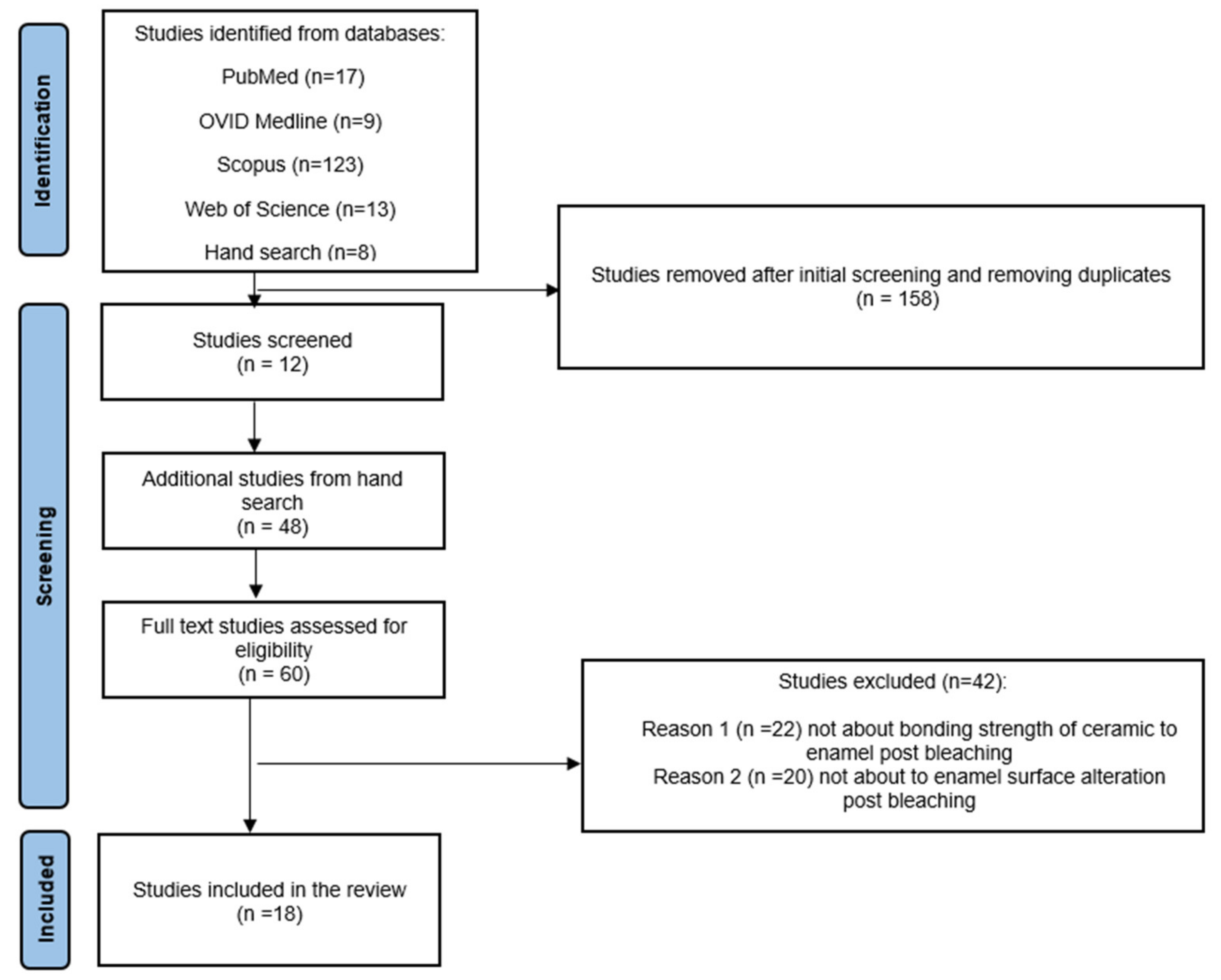
2. Materials and Methods
2.1. Search Strategy
2.2. Selection of Studies
3. Results
3.1. Effect on Bond Strength of Ceramic to Enamel after Bleaching Protocol
3.2. Changes in Surface Roughness of Enamel Post Bleaching Protocol
3.3. Risk of Bias
4. Discussion
5. Conclusions
- Bleaching treatments alter the surface roughness of enamel and, thus, the shear bond strength between ceramic and enamel.
- Bleaching treatments with a higher concentration of hydrogen peroxide (more than 35%) reduce the bond strength between ceramic and enamel.
- Delaying bonding after bleaching for up to 7 days increases the bond strength between ceramic and enamel.
Author Contributions
Funding
Institutional Review Board Statement
Informed Consent Statement
Data Availability Statement
Conflicts of Interest
References
- Carey, C.M. Tooth whitening: What we now know. J. Evid. Based Dent. Pract. 2014, 14, 70–76. [Google Scholar] [CrossRef] [PubMed] [Green Version]
- Kwon, S.R.; Wertz, P.W. Review of the mechanism of tooth whitening. J. Esthet. Restor. Dent. 2015, 27, 240–257. [Google Scholar] [CrossRef] [PubMed]
- Kang, T.-Y.; Kim, J.-H.; Kwon, J.-S. In Vitro Effects of Cyclic Dislodgement on Retentive Properties of Various Titanium-Based Dental Implant Overdentures Attachment System. Materials 2019, 12, 3770. [Google Scholar] [CrossRef] [Green Version]
- Giannini, M.; Silva, A.P.; Cavalli, V.; Leme, A.F.P. Effect of carbamide peroxide-based bleaching agents containing fluoride or calcium on tensile strength of human enamel. J. Appl. Oral Sci. 2006, 14, 82–87. [Google Scholar] [CrossRef] [PubMed]
- Haywood, V.B.; O Heymann, H. Nightguard vital bleaching. Quintessence Int. 1989, 20, 173–176. [Google Scholar] [PubMed]
- Kihn, P.W.; Barnes, D.M.; Romberg, E.; Peterson, K.A. Clinical evaluation of 10 percent vs. 15 percent carbamide peroxide tooth-whitening agents. J. Am. Dent. Assoc. 2000, 131, 1478–1484. [Google Scholar] [CrossRef] [PubMed]
- Christensen, G.J. Bleaching teeth: Practitioner trends. J. Am. Dent. Assoc. 1997, 128, 16S–18S. [Google Scholar] [CrossRef]
- Joiner, A. The bleaching of teeth: A review of the literature. J. Dent. 2006, 34, 412–419. [Google Scholar] [CrossRef]
- Türkün, M.; Kaya, A.D. Effect of 10% sodium ascorbate on the shear bond strength of composite resin to bleached bovine enamel. J. Oral Rehabil. 2004, 31, 1184–1191. [Google Scholar] [CrossRef]
- Titley, K.; Torneck, C.; Smith, D.; Chernecky, R.; Adibfar, A. Scanning electron microscopy observations on the penetration and structure of resin tags in bleached and unbleached bovine enamel. J. Endod. 1991, 17, 72–75. [Google Scholar] [CrossRef]
- García-Godoy, F.; Dodge, W.W.; Donohue, M.A.; O’Quinn, J. Composite resin bond strength after enamel bleaching. Oper. Dent. 1993, 18, 144. [Google Scholar]
- Silva, F.B.D.; Chisini, L.A.; Demarco, F.F.; Horta, B.L.; Correa, M.B. Desire for tooth bleaching and treatment performed in Brazilian adults: Findings from a birth cohort. Braz. Oral Res. 2018, 32, e12. [Google Scholar] [CrossRef] [Green Version]
- Josey, A.; Meyers, I.; Romaniuk, K.; Symons, A. The effect of a vital bleaching technique on enamel surface morphology and the bonding of composite resin to enamel. J. Oral Rehabil. 1996, 23, 244–250. [Google Scholar] [CrossRef]
- Ruse, N.D.; Smith, D.; Torneck, C.; Titley, K. Preliminary Surface Analysis of Etched, Bleached, and Normal Bovine Enamel. J. Dent. Res. 1990, 69, 1610–1613. [Google Scholar] [CrossRef]
- Rosa, E.; Soares, P. Effect of fluoride therapies on the surface roughness of human enamel exposed to bleaching agents. Quintessence Int. 2010, 41, 71–78. [Google Scholar]
- Al-Salehi, S.; Wood, D.; Hatton, P. The effect of 24h non-stop hydrogen peroxide concentration on bovine enamel and dentine mineral content and microhardness. J. Dent. 2007, 35, 845–850. [Google Scholar] [CrossRef]
- Grazioli, G.; Valente, L.L.; Isolan, C.P.; Pinheiro, H.A.; Duarte, C.G.; Münchow, E.A. Bleaching and enamel surface interactions resulting from the use of highly-concentrated bleaching gels. Arch. Oral Biol. 2018, 87, 157–162. [Google Scholar] [CrossRef]
- Attin, T.; Schmidlin, P.R.; Wegehaupt, F.; Wiegand, A. Influence of study design on the impact of bleaching agents on dental enamel microhardness: A review. Dent. Mater. 2009, 25, 143–157. [Google Scholar] [CrossRef] [Green Version]
- Attin, T.; Kocabiyik, M.; Buchalla, W.; Hannig, C.; Becker, K. Susceptibility of Enamel Surfaces to Demineralization after Application of Fluoridated Carbamide Peroxide Gels. Caries Res. 2003, 37, 93–99. [Google Scholar] [CrossRef]
- Chen, H.-P.; Chang, C.-H.; Liu, J.-K.; Chuang, S.-F.; Yang, J.-Y. Effect of fluoride containing bleaching agents on enamel surface properties. J. Dent. 2008, 36, 718–725. [Google Scholar] [CrossRef]
- Silva, N.R.; Coelho, P.G.; Valverde, G.B.; Becker, K.; Ihrke, R.; Quade, A.; Thompson, V.P. Surface characterization of Ti and Y-TZP following non-thermal plasma exposure. J. Biomed. Mater. Res. Part B Appl. Biomater. 2011, 99, 199–206. [Google Scholar] [CrossRef] [PubMed]
- Leonard, R.H., Jr.; Eagle, J.C.; Garland, G.E.; Matthews, K.P.; Rudd, A.L.; Phillips, C. Nightguard vital bleaching and its effect on enamel surface morphology. J. Esthet. Restor. Dent. 2001, 13, 132–139. [Google Scholar] [CrossRef] [PubMed]
- Titley, K.; Torneck, C.; Ruse, N.; Krmec, D. Adhesion of a resin composite to bleached and unbleached human enamel. J. Endod. 1993, 19, 112–115. [Google Scholar] [CrossRef]
- El-Din, A.N.; Miller, B.H.; Griggs, J.A.; Wakefield, C. Immediate bonding to bleached enamel. Oper. Dent. 2006, 31, 106–114. [Google Scholar] [CrossRef]
- Schemehorn, B.; González-Cabezas, C.; Joiner, A. A SEM evaluation of a 6% hydrogen peroxide tooth whitening gel on dental materials in vitro. J. Dent. 2004, 32, 35–39. [Google Scholar] [CrossRef]
- Turker, Ş.B.; Biskin, T. Effect of three bleaching agents on the surface properties of three different esthetic restorative materials. J. Prosthet. Dent. 2003, 89, 466–473. [Google Scholar] [CrossRef]
- Torneck, C.; Titley, K.; Smith, D.; Adibfar, A. The influence of time of hydrogen peroxide exposure on the adhesion of composite resin to bleached bovine enamel. J. Endod. 1990, 16, 123–128. [Google Scholar] [CrossRef]
- Swift, E.J., Jr. Reversal of Compromised Bonding in Bleached Enamel. J. Esthet. Restor. Dent. 2012, 24, 357–358. [Google Scholar] [CrossRef]
- Scherer, W.; Cooper, H.; Ziegler, B.; Vijayaraghavan, T. At-Home Bleaching System: Effects on Enamel and Cementum. J. Esthet. Restor. Dent. 1991, 3, 54–56. [Google Scholar] [CrossRef]
- Komine, F.; Tomic, M.; Gerds, T.; Strub, J.R. Influence of different adhesive resin cements on the fracture strength of aluminum oxide ceramic posterior crowns. J. Prosthet. Dent. 2004, 92, 359–364. [Google Scholar] [CrossRef]
- Raptis, N.V.; Michalakis, K.X.; Hirayama, H. Optical behavior of current ceramic systems. Int. J. Periodontics Restor. Dent. 2006, 26, 31–40. [Google Scholar]
- Li, Q.; Yu, H.; Wang, Y. Colour and surface analysis of carbamide peroxide bleaching effects on the dental restorative materials in situ. J. Dent. 2009, 37, 348–356. [Google Scholar] [CrossRef]
- Cavalli, V.; Reis, A.F.; Giannini, M.; Ambrosano, G.M. The effect of elapsed time following bleaching on enamel bond strength of resin composite. Oper. Dent. 2001, 26, 597–602. [Google Scholar]
- Basting, R.T.; Rodrigues, A.L., Jr.; Serra, M.C. The effects of seven carbamide peroxide bleaching agents on enamel microhardness over time. J. Am. Dent. Assoc. 2003, 134, 1335–1342. [Google Scholar] [CrossRef] [Green Version]
- Rahul, M.; Kumar, P.A.; Nair, A.S.; Mathew, S.; Amaladas, A.S.; Ommen, A. Effects of at-home and in-office bleaching agents on the shear bond strength of metal, ceramic, and composite brackets to enamel. Indian. J. Dent. Res. 2017, 28, 566–573. [Google Scholar]
- Ozturk, A.N.; Oztürk, B.; Malkoc, M.A.; Buyukozer, T. Bond strengths of two ceramic laminate systems to bleached enamel. Mater. Res. Innov. 2011, 15, 63–65. [Google Scholar] [CrossRef]
- Öztaş, E.; Bağdelen, G.; Kılıçoğlu, H.; Ulukapı, H.; Aydin, I. The effect of enamel bleaching on the shear bond strengths of metal and ceramic brackets. Eur. J. Orthod. 2011, 34, 232–237. [Google Scholar] [CrossRef] [Green Version]
- Firoozmand, L.M.; Brandão, J.V.P.; Fialho, M.P.N. Influence of microhybrid resin and etching times on bleached enamel for the bonding of ceramic brackets. Braz. Oral Res. 2013, 27, 142–148. [Google Scholar] [CrossRef] [Green Version]
- Abouassi, T.; Wolkewitz, M.; Hahn, P. Effect of carbamide peroxide and hydrogen peroxide on enamel surface: An in vitro study. Clin. Oral Investig. 2010, 15, 673–680. [Google Scholar] [CrossRef]
- Britto, F.A.R.; Lucato, A.S.; Valdrighi, H.C.; Vedovello, S.A.S. Influence of bleaching and desensitizing gel on bond strength of orthodontic brackets. Dent. Press J. Orthod. 2015, 20, 49–54. [Google Scholar] [CrossRef] [Green Version]
- Cooper, J.R.; Young, N.B.; Haywood, V.B.; Mettenburg, D.; Callan, R.S.; Rueggeberg, F.A. Effect of Short-Duration, Localized Carbamide Peroxide Application to Remove Enamel Staining on Bond Strength of Resin Cement to Enamel. J. Esthet. Restor. Dent. 2016, 28, 190–196. [Google Scholar] [CrossRef] [PubMed]
- Miles, P.G.; Pontier, J.-P.; Bahiraei, D.; Close, J. The effect of carbamide peroxide bleach on the tensile bond strength of ceramic brackets: An in vitro study. Am. J. Orthod. Dentofac. Orthop. 1994, 106, 371–375. [Google Scholar] [CrossRef]
- Dastjerdi, E.V.; Khaloo, N.; Mojahedi, S.M.; Azarsina, M. Shear Bond Strength of Orthodontic Brackets to Tooth Enamel After Treatment with Different Tooth Bleaching Methods. Iran. Red Crescent Med. J. 2015, 17, e20618. [Google Scholar] [CrossRef] [PubMed] [Green Version]
- Gökçe, B.; Çömlekoğlu, E.; Özpinar, B.; Türkün, M.; Kaya, A.D. Effect of antioxidant treatment on bond strength of a luting resin to bleached enamel. J. Dent. 2008, 36, 780–785. [Google Scholar] [CrossRef] [PubMed]
- McGuckin, R.S.; Babin, J.; Meyer, B. Alterations in human enamel surface morphology following vital bleaching. J. Prosthet. Dent. 1992, 68, 754–760. [Google Scholar] [CrossRef]
- Hegedüs, C.; Bistey, T.; Flóra-Nagy, E.; Keszthelyi, G.; Jenei, A. An atomic force microscopy study on the effect of bleaching agents on enamel surface. J. Dent. 1999, 27, 509–515. [Google Scholar] [CrossRef]
- Titley, K.; Torneck, C.D.; Smith, D. The effect of concentrated hydrogen peroxide solutions on the surface morphology of human tooth enamel. J. Endod. 1988, 14, 69–74. [Google Scholar] [CrossRef]
- Pinto, C.F.; De Oliveira, R.; Cavalli, V.; Giannini, M. Peroxide bleaching agent effects on enamel surface microhardness, roughness and morphology. Braz. Oral Res. 2004, 18, 306–311. [Google Scholar] [CrossRef]
- Zuryati, A.-G.; Qian, O.Q.; Dasmawati, M. Effects of home bleaching on surface hardness and surface roughness of an experimental nanocomposite. J. Conserv. Dent. 2013, 16, 356–361. [Google Scholar] [CrossRef] [Green Version]
- Moraes, R.R.; Marimon, J.L.M.; Schneider, L.F.J.; Sobrinho, L.C.; Camacho, G.B.; Bueno, M. Carbamide peroxide bleaching agents: Effects on surface roughness of enamel, composite and porcelain. Clin. Oral Investig. 2006, 10, 23–28. [Google Scholar] [CrossRef]


| Focus Questions | 1. Does Bleaching Cause the Surface Roughness of Enamel? 2. Does Bleaching Affect the Bonding Strength of Ceramic? |
| PICO Criteria | |
| Population | Extracted human teeth or bovine teeth or enamel slab |
| Intervention | Tooth bleaching using hydrogen peroxide or carbamide peroxide with different concentrations |
| Comparison | Unbleached enamel |
| Outcome | Shear bonding strength, tensile bond strength, change in surface roughness |
| Inclusion Criteria | Exclusion Criteria |
|---|---|
| English language only Full text only In vitro studies In vivo studies Case–control studies Commercially available products Randomised control trial | Studies in a language other than English Abstract only Studies irrelevant to the focus questions Review articles Case reports Conference paper Technique article Studies with a sample size of <10 extracted teeth |
| Author | Specimen | Bleaching Materials | Intervention | Testing Method | Restorative Material | Qualitative Analysis | Summary of Results |
|---|---|---|---|---|---|---|---|
| Rahul et al., 2017 [35] | Human lower premolars teeth (n = 96) | Opalescence Boost PF gel (40% hydrogen peroxide) (Ultradent, South Jordan, UT, USA) (at-home) and Opalescence non-PF (10% carbamide peroxide) (Ultradent, South Jordan, UT, USA) (in-office) | Group 1 (n = 30): at-home bleach was applied for 8 h daily for 14 days. Group 2 (n = 30): in-office bleaching gel was applied for 20 min and then washed away, with repeated procedure the next day. Between intervals and after completion of bleaching protocol, all specimens were stored in distilled water. Control (n = 30)—no treatment n = 6 specimen used for SEM | SBS | Metal, ceramic, and composite bracket | Yes | No significant change in shear bond strength was found in at-home testing groups against all restorative materials after 24 h (14.68 ± 1.67 MPa). Significant decrease was observed in-office groups against all tested materials (10.95 ± 2.61 MPa), while shear bond strength of ceramic brackets in the control group was 16.03 ± 0.87 MPa. |
| Cooper et al., 2016 [41] | Bovine teeth (n = 40) | 10% carbamide peroxide gel (OpalescenceVR, Ultradent Products, Inc., South Jordan, UT, USA) | Flattened and polished specimens were used. (n = 10)—unbleached (control) (n = 10) bleached with 10% Opalescence for 10 s (n = 10) bleached with 10% Opalescence for 20 s (n = 10) bleached with 10% Opalescence for 30 s | SBS | Resin veneering cement | No | No significant difference in bond strength was found after bleaching for 10 s (16.1± 3.7 MPa), 20 s (15.0 ± 2.8 MPa), or 30 s (15.2 ± 2.1 MPa) and control (15.9 ± 3.7 MPa) groups. |
| Britto et al., 2015 [40] | Bovine teeth (n = 100) | 35% hydrogen peroxide agent (Whiteness HP Maxx 35%, FGM, Joinville, Santa Catarina, Brazil) | Group 1 (n = 20): no bleaching (control) Group 2 (n = 20): bleached for 3 × 15 min. Group 3 (n = 20): bleaching protocol same as group 2, prior to specimens being washed and dried followed by application of desensitising agent. Group 4 (n = 20): bleached for 40 min, then specimens were washed and dried, followed by polishing with a felt disc. Group 5 (n = 20): bleached for 40 min, then specimens were washed and dried, followed by application of desensitising agent for 10 min, before being washed and polished with a felt disc. Bonding was done after 7 days. | SBS | Orthodontic ceramic bracket | No | Shear bond strength: Group 1 control: 19.19 ± 6.12 MPa, Group 2 HP Maxx 20.59 ± 7.17 MPa, Group 3 (HP Maxx + KF 2%) 24.33 ± 5.45 MPa, Group 4 (HP blue) 23.25 ± 6.85 MPa, Group 5 (HP blue + KF a 2%) 29.33 ± 6.03 MPa. Groups 2 and 5, with application of desensitising agent, increased the shear bond strength. No significant impact was observed in the shear strength bond in other groups. |
| Dastijerdi et al., 2015 [43] | Human premolars teeth (n = 48) | 20% and 45% of carbamide peroxide gel (Opalescence; Ultradent Products, South Jordan, UT, USA) 40% aqueous hydrogen peroxide gel (Opalescence; Ultradent Products, South Jordan, UT, USA) | Each group had n = 12 specimens. Group 1: the group was stored artificial saliva and etched with 37% phosphoric acid, then steamed and dried. Group 2: 45% carbamide peroxide applied for 30 min, then rinsed, dried, and stored in artificial saliva for 2 weeks. Group 3: 20% carbamide peroxide applied for two weeks for 4 h daily. Other steps were repeated, as in Group 1. Group 4: A 40% aqueous hydrogen peroxide gel was applied for 60 s at an 810 nm wavelength. Specimens were maintained in artificial saliva for two weeks before going through the same protocols as Group 1. The metallic brackets were then bonded to enamel surfaces, which were subsequently thermocycled for 500 cycles between 5 and 55 °C. | SBS | Metallic bracket | No | Groups 2 (6.37 ± 0.92 MPa), 3 (7.67 ± 1.01 MPa) and 4 (7.49 ± 1.19 MPa) had statistically significant lower bond shear strength in comparison to Group 1 (10.54 ± 1.51 MPa), p < 0.001. Group 4 had no statistically significant difference between Groups 2 and 3. |
| Firoozmand et al., 2013 [38] | Bovine incisors teeth (n = 144) | 35% hydrogen peroxide whiteness HP Maxx gel (FGM, Joinvile, Brazil) | Control group (n = 72)—none of the specimens were stored in artificial saliva at 37 °C for 14 days. Treated group (n = 72)—bleached for 15 min, then enamel surface rinsed with water and procedure repeated three times (45 min of bleaching in total). Specimens were stored in artificial saliva at 37 °C for one week before repeating the bleaching protocol and re-stored in artificial saliva for 14 days before bonding. Both groups were divided into two groups each (n = 36) (Transbond Xt group and Filtek Z250 group), which were further divided into three groups (n = 12), and each group was etched for either 15, 30 or 60 s before bonding. | SBS | Polycrystalline ceramic central incisor brackets, (Orthometric Inceram, Maanshan, China) | No | Significant interaction was found between bleaching and etching times within specimens. Highest shear bond strength was found in non-bleached specimens (10.67 MPa) compared to bleached specimens (9.65 MPa). Etching time of 30 s increased bond strength among groups by 14.5% or 11.05 MPa, while 14 and 60 s had lower results, 9.8 MPa and 9.63 MPa. Highest bond strength was found in samples that were not bleached but etched for 30 s − 12.41 ± 1.07 MPa. |
| Öztas et al., 2011 [37] | Human first premolar teeth (n = 120) | 20% of carbamide peroxide—bleaching—gel, CP, at-home (Opalescence, Ultradent Products, South Jordan, UT, USA) | Bleached group (n = 80). Not bleached group (n = 40). Prior to bleaching, enamel was polished with pumice (oil- and fluoride-free). Bleaching agent was applied for 6 h a day for 14 days. Bleached Group 1: (n = 40) was bonded 24 h after bleaching. Bleached Group 2 (n = 40) was bonded after 14 days of bleaching. 12 subgroups were further made (n = 10) and metal and ceramic brackets were bonded to each group with either self- (Unite 3 MUnitek) or light-cured (Transbond XT) composite adhesives. | SBS | Ceramics and metal bracket | Yes | No statistically significant difference in shear bond strength was found between experimental and control groups when bonding occurred within 24 h or 14 days. Group 1 ceramic brackets Self-cured adhesive resin—13.794 ± 2.157 MPa. Light-cured adhesive resin—15.367 ± 3.513 MPa. Group 2 metal brackets Self-cured adhesive resin—12.131 ± 5.422 MPa. Light-cured adhesive resin—14.735 ± 4.556 MPa. |
| Nilgun Ozturk et al., 2011 [36] | Human maxillary central incisors teeth (n = 96) | 38% hydrogen peroxide in-office gel (Whiteness HP Maxx 35%, FGM, Joinville, Santa Catarina, Brazil) | Bleached group (n = 48) and non-bleached groups (n = 48). Bleaching procedure was repeated twice. Prior to attaching ceramic systems (Cerec 3 and IPS Empress2), they were divided into two groups (n = 12), where luting agents were applied (Variolink 2 and Rely-X). Specimens were subjected to tensile forces. After luting, specimens underwent thermocycling of 1000 cycles at 5 to 55 °C | TBS | Ceramic systems | Stereomicroscope | Non-bleached groups had significantly higher shear bond strength than bleached groups. Ceramic systems had no statistically significant difference from each other. |
| Gökce et al., 2008 [44] | Human molar teeth (n = 40) | 10% carbamide peroxide bleaching gel (Opalescence, Ultradent, South Jordan, UT, USA) | Group 1 (n = 10): ceramic specimens bonded after bleaching. Group 2 (n = 10): sodium ascorbate applied after bleaching. Group 3 (n = 10): stored in artificial saliva for 7 days prior to bonding after bleaching. Group 4 (n = 10): control—not bleached but immersed in artificial saliva for 7 days prior to bonding. | SBS | Ceramics (Empress 2) | Yes | No statistically significant difference was found between delayed bond Group 3 (37.5 ± 8.3 MPa) and control Group 4 (38.8 ± 4.1 MPa). Samples with immediate bonding after bleaching in Group 1 (31.6 ± 9.3 MPa) and Group 2 (41.7 ± 1.9 MPa) demonstrated significantly higher shear bond strength. |
| Miles et al., 1994 [42] | Human first, second upper and lower premolars teeth (n = 60) | 10% carbamide (urea) peroxide gel (Rembrandt Lighten, Den-Mat Products, Santa Maria, CA, USA) | Group 1 (n = 20): control (unbleached group) Group 2 (n = 20): bleached and directly bonded to the ceramics Group 3 (n = 20): bleached and stored in distilled water for 7 days before bonding. | TBS | Ceramics | No | Group 2 showed statistically significant lower tensile bond strength than Group 1 and Group 3. No statistically significant difference in tensile bond strength was observed between control Group 1 and Group 3. |
| Author | Analysis | Specimen | Bleaching Material | Intervention | Outcome |
|---|---|---|---|---|---|
| Rahul et al., 2017 [35] | SEM | Human lower premolar teeth (n = 96) | Opalescence Boost PF in-office (40% hydrogen peroxide) (Ultradent, South Jordan, UT, USA) and Opalesence non-PF at-home (10% carbamide peroxide) (Ultradent, South Jordan, UT, USA) | Group 1 (n = 30): at-home gel was applied for 8 h daily for 14 days. Group 2 (n = 30): in-office gel was applied for 20 min and then washed away, with repeated procedure the next day. Control (n = 30)—no bleaching treatment. Between intervals and after completion of bleaching protocol, all specimens were stored in distilled water. n = 6 specimen used for SEM. | 40% hydrogen peroxide samples The surface alterations were greatly significant compared to other testing groups with numerous surface deposits. Morphologic surface alterations became significantly noticeable after in-office bleaching. Present irregular depressions: craters and shallow erosions were visible. 10% carbamide peroxide samples Minor changes to enamel surface were observed, including alterations in surface smoothness. |
| Schemehorn et al., 2004 [25] | SEM | Admixed high-copper amalgam (Dispersalloy, Dentsply, York, PA, USA) and hybrid resin composite (Herculite, Kerr, Orange, CA, USA) (n = 10) | 6% hydrogen peroxide (Xtra White, Unilever Oral care) | Specimens were immersed in 6% hydrogen peroxide at 37 °C for 20 min, rinsed, brushed to remove residual gel and placed back in saliva for 1 h at 37 °C. Bleaching treatment repeated after 1 h immersion in saliva and specimen later immersed overnight in saliva. 1st day—4 treatments. Next four days—6 treatments each; 28 treatments in total. | There were no visual differences at magnifications of 200× or 2000× magnifications between the control group and any other bleach gel treated groups. No significant visual effects of the 6% hydrogen peroxide gel on surface alterations were observed on the tested dental materials. |
| Turker & Biskin., 2003 [26] | SEM | 3 bleaching products and 3 restorative materials (feldspathic porcelain, glass ionomer, microfilled composite) (n = 30) | Nite White (G1) (Discus Dental Inc., Beverly Hills, CA, USA), Opalescence (G2) (Ultradent Products Inc., South Jordan, Utah), Rembrandt (G3) (Den-Mat Corp, Santa Maria, CA, USA) | In total, 3 groups of n = 10 specimens were prepared; 2 specimens from each group were used as control. Each group was bleached with one of the three bleaching agents for 8 h per day for 30 days. Surface roughness was examined at intervals of 1 day, 2 days, 1, 2, 3, and 4 weeks (30 days). | Significant increase in surface roughness was found from using G3 gel on modified glass ionomer cement. In the first 2 weeks, surface roughness dramatically increased for all bleaching groups and each restorative material. All the modified glass ionomer specimens showed cracking areas; microfilled composite samples showed significantly increased surface porosity and cracking in comparison to control specimens. Surface spectral analysis results indicated a decrease in the SiO2 content in the feldspathic porcelain and microfilled composite groups for all bleaching materials. |
| Leonard et al., 2001 [22] | SEM in vivo study | Human subjects (n = 24) (The study teeth were the four maxillary central and lateral incisors.) untreated extracted tooth- used as control | Nite White classic (Discus Dental, Inc., Culver city, CA, USA) | Participants were exposed to bleaching protocol for 8–10 h per day for 2 weeks by wearing an active whitening solution filled guard. Impressions of the study teeth were taken at baseline (after 2 weeks) and at 6 months after treatment and epoxy casts made. Casts were examined using the SEM with magnification taken at 200× times and 2000×. | After 2 weeks of treatment, no or minimal effect on the surface morphology of enamel was observed; 6 months post treatment comparisons also showed no long-term effect or minimal alterations in enamel surface morphology. |
| Hegedus et al., 1999 [46] | Atomic force microscopy | Extracted incisors, non-carious human teeth (ten maxillary and five mandibular) (n = 15) | G1: Opalescence (Ultradent Product Inc., South Jordan, UT, USA), G2: Nite White (Discus Dental Inc., Los Angeles, CA, USA) G3: 30% hydrogen peroxide (Sigma Chemical Co., Ltd., USA) | Three groups of (n = 5) teeth were treated with either Opalescence, Nite White or 30% hydrogen peroxide solution, respectively, for 28 h (4 individual) treatments and examined 28 h after the final treatment. | On comparing the AFM images of untreated and treated enamel. G1:
G2
G3
|
| Josey et al., 1996 [13] | SEM | Non-carious human teeth (n = 32) | Rembrandt lighten (carbamide peroxide) (Den-Mat Corporation, Santa Maria, CA, USA) | (n = 8)—bleached and stored in artificial saliva for 24 h. (n = 8) bleached and stored in artificial saliva for 12 weeks. (n = 8) not bleached and stored in artificial saliva for 12 weeks. (n = 8) not bleached and stored in artificial saliva for 24 h. | Macroscopic observation
Light microscopy observation Sections of specimens that underwent bleaching and were stored for 24 h or 12 weeks in artificial saliva showed demineralisation in the tested region and exhibited an indistinct structure of the enamel. SEM observation
The storage intervals did not show any significant differences between specimens. |
| McGuckin et al. 1992 [45] | SEM | Extracted intact human teeth (n = 14) | G1: Superoxol, 30% hydrogen peroxide (Union Broach, York, PA, USA), G2: Proxigel—10% carbamide peroxide and Carbapol, (Reed & Carn-rick, Piscataway, NJ, USA), G3: White & Brite—10% carbamide peroxide (Omni Products Inter- National, Gravette, AR, USA) | Home 1 group (Proxigel bleach) (n = 4) Treatment time was 8 h per day for 30 days. Specimen rinsed in tap water every day before storage while waiting for next treatment. Home 2 group (White & Brite) (n = 4) Treatment time was 24 h daily. After exposure every day, specimens were rinsed in tap water for 30 s, then placed in 4% fluoride gel, and re-rinsed in tap water prior to being exposed to bleaching protocol again. Office bleaching group (30% hydrogen peroxide) (n = 4) Treatment time was 30 min, repeated after 7 days. After exposure, specimens were rinsed in tap water prior to storage in isotonic saline with 0.2% sodium azide until next treatment. Control (n = 2) no bleaching treatment specimen stored in isotonic saline with 0.2% sodium azide | Mean surface roughness control (1.9 µm), Proxigel (1.06 µm), White & Brite (0.9 µm), office (0.6 µm). SEM analysis Control
|
| Titley et al., 1991 [10] | SEM | Bovine teeth (n = 128) | 35% hydrogen peroxide (Drug Trading Co. Ltd., Toronto, ON, Canada) | Group 1: specimens were immersed in 35% hydrogen peroxide for 5 or 30 min, washed with tap water for 1 min, dried with air, then etched with 37% phosphoric acid, washed, and dried again. Group 2: bleaching protocol same as Group 1, but specimens were immersed in saline instead of hydrogen peroxide. Group 3: was treated with E for 60 s, washed before immersion in hydrogen peroxide, then rinsed, etched, washed and dried again. Group 4: was treated the same as Group 3, except it was immersed in saline. | It was suggested that there could be an interaction between the resin and the residual hydrogen peroxide at or near the enamel surface. |
| Scherer et al., 1991 [29] | SEM | Extracted non-carious human anterior teeth in 10% buffered formalin solution. (n = 15) | Vital home bleaching (10%) (manufacturer not stated) | Specimens were divided into five groups: Control (n = 3)—no treatment Vital bleach Group 1 (n = 3), bleached for 5 days Vital bleach Group 2 (n = 3) bleached for 15 days. Vital bleach Group 3 (n = 3) bleached for 30 days. Etched group (n = 3)—specimen etched with 37% phosphoric acid gel for 20 s. | Photomicrographs showed that the etching pattern on enamel surfaces was exhibited and visible only in specimens that were treated for 30 days. SEM analysis showed no significant morphological differences among the 5-day, 15-day, and 30-day treatment specimen groups. |
| Titley et al., 1988 [47] | SEM | Paired human tooth sections (n = 36 sections) | 35% hydrogen peroxide (Drug Trading Co. Ltd., Toronto, ON, Canada) | Group 1—bleached with 35% hydrogen peroxide or normal saline. Group 2—bleached with 35% hydrogen peroxide for durations of 1,3, 5, 10, 20, 30, and 60 min prior to 60 s etching with 37% phosphoric acid. Control—stored in normal saline for durations of 1, 3, 5, 10, 20, 30, and 60 min prior to etching with 37% phosphoric acid for 60 s. Group 3—60 s etching with 37% phosphoric acid prior to being kept in 35% hydrogen peroxide for durations of 1,3, 5, 10, 20, 30, and 60 min. Control—60 s etching with 37% phosphoric acid prior to being kept in normal saline for durations of 1,3, 5, 10, 20, 30, and 60 min. | Hydrogen peroxide immersion only
Surface etching, followed by immersion in hydrogen peroxide.
|
| Rahul et al., 2017 [35] | Cooper et al., 2016 [41] | Britto et al., 2015 [40] | Dastjerdi et al., 2015 [43] | Firoozmand et al., 2013 [38] | Nilgun Ozturk et al., 2011 [36] | Öztas et al., 2011 [37] | Gökce et al., 2008 [44] | Schemehorn et al., 2004 [25] | Turker & Biskin, 2003 [26] | Leonard et al., 2001 [22] | Hegedus et al., 1999 [46] | Titley et al., 1988 [47] | Josey et al., 1996 [13] | Miles et al., 1994 [42] | McGuckin et al., 1992 [45] | Titley et al., 1991 [10] | Scherer et al., 1991 [29] | ||
|---|---|---|---|---|---|---|---|---|---|---|---|---|---|---|---|---|---|---|---|
| Abstract | Abstract 1 1 | Yes | Yes | Yes | Yes | Yes | Yes | Yes | Yes | Yes | Yes | Yes | Yes | Yes | Yes | Yes | Yes | Yes | No |
| Introduction | Background and objectives 2 a 2 | Yes | Yes | Yes | Yes | Yes | Yes | Yes | Yes | Yes | Yes | Yes | Yes | Yes | Yes | Yes | Yes | Yes | Yes |
| Specific objectives 2 b 2 | Yes | Yes | No | No | Yes | Yes | Yes | Yes | No | Yes | Yes | Yes | No | No | Yes | No | No | No | |
| Method | Intervention 3 | No | Yes | Yes | Yes | Yes | Yes | Yes | Yes | Yes | Yes | Yes | Yes | Yes | Yes | Yes | Yes | Yes | Yes |
| Outcome 4 | Yes | Yes | Yes | Yes | Yes | Yes | Yes | Yes | Yes | Yes | Yes | Yes | Yes | Yes | Yes | Yes | Yes | Yes | |
| Sample size 5 | No | Yes | Yes | Yes | Yes | Yes | Yes | Yes | Yes | Yes | Yes | Yes | No | Yes | Yes | Yes | No | Yes | |
| Randomisation: sequence generation 6 3 | No | No | No | No | No | No | No | No | No | No | No | No | No | No | No | No | No | No | |
| Allocation concealment mechanism 7 3 | No | No | No | No | No | No | No | No | No | No | No | No | No | No | No | No | No | No | |
| Implementation 8 3 | No | No | No | No | No | No | No | No | No | No | No | No | No | No | No | No | No | No | |
| Blinding 9 3 | No | No | No | No | No | No | No | No | No | No | No | No | No | No | No | No | No | No | |
| Statistical methods 10 | Yes | Yes | Yes | Yes | Yes | Yes | Yes | Yes | No | Yes | No | Yes | Yes | Yes | Yes | Yes | No | No | |
| Results | Outcome and estimation 11 | Yes | Yes | Yes | Yes | Yes | Yes | Yes | Yes | Yes | Yes | Yes | Yes | No | Yes | Yes | Yes | Yes | No |
| Discussion | Limitations 12 | Yes | Yes | No | No | Yes | No | No | Yes | No | Yes | No | No | No | No | No | No | No | No |
| Other Information | Funding 13 4 | No | No | No | No | Yes | No | Yes | No | No | No | Yes | No | Yes | No | No | No | Yes | No |
| Trial protocol 14 4 | No | No | No | No | No | No | No | No | No | No | No | No | No | No | No | No | No | No | |
| Risk of bias | Low | Low | Low | Low | Low | Low | Low | Low | Low | Low | Low | Low | Low | Low | Low | Low | Low | Low |
Publisher’s Note: MDPI stays neutral with regard to jurisdictional claims in published maps and institutional affiliations. |
© 2022 by the authors. Licensee MDPI, Basel, Switzerland. This article is an open access article distributed under the terms and conditions of the Creative Commons Attribution (CC BY) license (https://creativecommons.org/licenses/by/4.0/).
Share and Cite
Seto, T.H.; Grymak, A.; Mudliar, V.; Choi, J.J.E. Effect of Enamel Bleaching on the Bond Strength of Ceramic—A Systematic Review. Oral 2022, 2, 182-197. https://doi.org/10.3390/oral2020018
Seto TH, Grymak A, Mudliar V, Choi JJE. Effect of Enamel Bleaching on the Bond Strength of Ceramic—A Systematic Review. Oral. 2022; 2(2):182-197. https://doi.org/10.3390/oral2020018
Chicago/Turabian StyleSeto, Tsz Ho, Anastasiia Grymak, Vidya Mudliar, and Joanne Jung Eun Choi. 2022. "Effect of Enamel Bleaching on the Bond Strength of Ceramic—A Systematic Review" Oral 2, no. 2: 182-197. https://doi.org/10.3390/oral2020018
APA StyleSeto, T. H., Grymak, A., Mudliar, V., & Choi, J. J. E. (2022). Effect of Enamel Bleaching on the Bond Strength of Ceramic—A Systematic Review. Oral, 2(2), 182-197. https://doi.org/10.3390/oral2020018