Review

Imatinib in Targeted Therapy: Advances in Biomedical Applications and Drug Delivery Systems

1
Department of Pharmaceutical Technology and Biopharmacy, Faculty of Pharmacy, Medical University of Plovdiv, 4002 Plovdiv, Bulgaria
2
Research Institute at Medical University of Plovdiv (RIMU), 4002 Plovdiv, Bulgaria
3
Center for Competence “PERIMED-2”, Medical University of Plovdiv, 4002 Plovdiv, Bulgaria
*
Author to whom correspondence should be addressed.
Hemato 2025, 6(4), 40; https://doi.org/10.3390/hemato6040040
Submission received: 29 August 2025 / Revised: 31 October 2025 / Accepted: 11 November 2025 / Published: 12 November 2025
(This article belongs to the Section Chronic Myeloid Disease)

Abstract

Imatinib (IMT) is a small-molecule tyrosine kinase inhibitor that primarily targets platelet-derived growth factor receptor-β and related kinases. Beyond its established efficacy in chronic myeloid leukemia, IMT has also demonstrated therapeutic benefits in gastrointestinal stromal tumors, dermatofibrosarcoma, acute lymphoblastic leukemia, and as a second-line treatment for aggressive systemic mastocytosis or as an anti-Mycobacterium agent. From a physicochemical perspective, IMT exhibits poor aqueous solubility but high membrane permeability, classifying it as a Biopharmaceutics Classification System Class II compound. Pharmacokinetically, IMT shows variable oral absorption and a prolonged terminal half-life, resulting in dose-dependent systemic exposure. Despite relatively high oral bioavailability, its clinical use requires large doses to achieve therapeutic efficacy, underscoring the need for advanced drug delivery strategies. Nano- and microscale delivery systems offer promising approaches to enhance tumor-specific accumulation through the enhanced permeability and retention effect while mitigating resistance mechanisms. However, achieving high drug loading introduces formulation challenges, such as controlling particle size distribution, polydispersity, and scalability. Moreover, designing carriers capable of controlled release without premature leakage remains crucial for maintaining systemic bioavailability and therapeutic performance. Emerging delivery platforms—including polymeric, lipid-based, carbon-derived, and stimuli-responsive nanocarriers—have shown significant potential in overcoming these limitations. Such systems can enhance IMT’s bioavailability, improve selective tumor targeting, and minimize systemic toxicity, thereby advancing its translational potential. This review aims to highlight the different biomedical applications of IMT and off-label uses, and to discuss current advances in drug delivery to optimize its clinical efficacy and safety profile.

1. Introduction

Cancer arises from the accumulation of genetic and epigenetic alterations that disrupt normal cellular homeostasis and drive uncontrolled proliferation [1]. Over the past two and a half centuries, significant advances have transformed the landscape of cancer management, leading to the development of diverse therapeutic modalities [2]. Contemporary treatment strategies—including chemotherapy, radiotherapy, surgical resection, targeted combination regimens, and emerging physical approaches such as laser therapy—have improved patient outcomes but remain limited by their lack of selectivity, often damaging healthy tissues and eliciting severe systemic toxicities [3]. These limitations underscore the urgent need for innovative therapeutic approaches that can selectively eradicate malignant cells while minimizing collateral damage to normal tissues [4,5].
Protein tyrosine kinases (PTKs), a family of about 90 enzymes, are essential regulators of numerous biological processes, playing key roles in cell division, differentiation, survival, morphogenesis, and control of the cell cycle [6]. PTKs are divided into two groups: receptor tyrosine kinases and non-receptor tyrosine kinases. Receptor tyrosine kinases are cell surface receptors that transmit signals upon ligand binding (Figure 1). As transmembrane proteins, they span the cell membrane and possess extracellular domains where ligands attach. In contrast, non-receptor tyrosine kinases are located in the cytosol; they become activated through interaction with an already activated receptor tyrosine kinase and, in turn, mediate receptor activation via phosphorylation without the direct involvement of a ligand [7]. Aberrations in the PTKs activity are closely associated with signaling pathways that drive tumorigenesis [8]. In recent years, PTKs modulators have attracted considerable attention for their therapeutic potential, especially in the treatment of cancer and other diseases. These modulators have the capacity to either suppress or enhance kinase activity, providing valuable means for managing diseases associated with dysregulated PTK function [9].
Tyrosine kinase inhibitors (TKIs) are small molecules specifically developed to disrupt signaling pathways that support the survival and proliferation of cancer cells. By binding to tyrosine kinase receptors within the cell membrane, they block pro-tumor signaling [10]. Introduced into clinical practice in the early 2000s, TKIs represented a groundbreaking shift toward “targeted” cancer therapy, in contrast to traditional cytotoxic drugs that indiscriminately affect both healthy and cancerous cells [11]. Imatinib (IMT), the first tyrosine kinase inhibitor (TKI) approved for cancer therapy, was deve- loped by a European pharmaceutical company. Designed to block the BCR-ABL protein driving the oncogenic pathway in chronic myeloid leukemia (CML), the drug demonstrated remarkable safety and efficacy, leading to rapid approval in both the US and Europe [12] and to be called it a “magical bullet” in the treatment of CML [13]. It gained FDA approval for the treatment of CML in 2001 and was later authorized for use in several other cancer indications [14]. The clinical advancement of IMT in CML was spearheaded by American oncologist and cancer researcher Brian Druker [15,16]. In general, the therapeutic action of IMT has been linked to its direct effects on tumor cells harboring oncogenic kinases or through inhibition of cellular kinases involved in pathogenic processes. More recent findings, however, indicate that IMT also modulates the immune system by suppressing T-cell signaling in vitro and, at higher doses, can lead to immunosuppression and even neutropenia in certain patients [17].
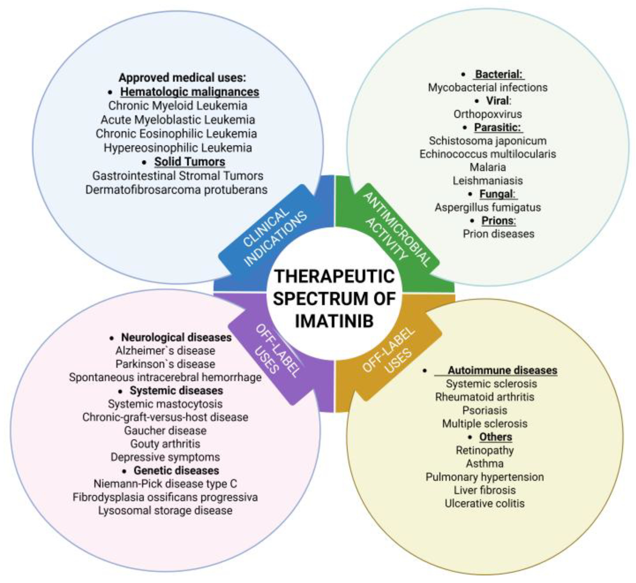
IMT has received U.S. Food and Drug Administration (FDA) approval as a first-line therapy for Philadelphia chromosome-positive CML, gastrointestinal stromal tumors (GIST), dermatofibrosarcoma protuberans, acute lymphoblastic leukemia, chronic eosinophilic leukemia, hypereosinophilic syndrome, and as a second-line option for aggressive systemic mastocytosis. Beyond these indications, it is also employed off-label in the management of CML post-allogeneic stem cell transplantation, advanced KIT-mutant melanomas, chordomas, and desmoid tumors [18]. In addition to its established anti-neoplastic effects, IMT has shown therapeutic potential across a spectrum of non-malignant disorders, including pulmonary arterial hypertension, autoimmune encephalomyelitis, bronchial asthma, ischemic stroke, intracerebral hemorrhage, multiple sclerosis, Parkinson’s disease, Alzheimer’s disease, Huntington’s disease, and spinal cord injury [4]. Collectively, these applications underscore IMT’s role as a broad-spectrum tyrosine kinase inhibitor.
IMT is administered orally, typically as 100 mg or 400 mg tablets as imatinib mesylate (IMTM) [19], which is highly soluble in acidic solutions but exhibits reduced solubility under neutral or alkaline conditions [20]. This solubility limitation can impact its absorption and therapeutic efficacy. Given the broad spectrum of diseases in which IMT has demonstrated efficacy, there is a growing need for advanced drug delivery systems to optimize its pharmacological properties. Innovative delivery strategies, such as micro- and nanocarriers, are being developed to improve targeted transport of IMT to cancer cells while minimizing adverse effects on healthy tissues [21,22]. The emergence of IMT resistance in certain cancer cells further highlights the need for such advanced systems [22,23]. Current research has presented polymeric and lipid-based nanoparticles (NPs), which can enhance solubility, stability, and site-specific delivery. Stimuli-responsive NPs, for example, are engineered to release IMT in response to specific cues like pH shifts or enzymatic activity, thereby improving tumor selectivity [23]. Galactose-based hydrogels, sometimes combined with nanohydroxyapatite, are also under investigation for targeted delivery of IMT in bone tumors [19]. Together, these approaches demonstrate the potential of advanced carriers to enable precise and effective dosing of IMT at the intended site. This review aims to comprehensively summarize both the biomedical applications of IMT and the drug delivery systems developed to optimize its therapeutic performance.

2. Methods

This review is based on articles from PubMed, Web of Science, ScienceDirect, and Google Scholar. The literature search spans a period from the first articles reporting the synthesis and application of IMT to the new IMT delivery systems in 2025, with 194 references selected based on their relevance to the research topic. Keywords such as “imatinib”, “imatinib mesylate”, “targeted delivery”, “tyrosine kinase inhibitor”, “chronic myeloid leukemia”, “gastrointestinal stromal tumors”, “biomedical application”, and “drug delivery systems” were used. We conducted a Google search to identify all included clinical trials.

3. Chemical Properties and Pharmacological Profile of IMT

3.1. Mechanism of Action

IMT has the following IUPAC name: 4-[(4-methylpiperazin-1-yl)methyl]-N- (4-methyl-3-{[4-(pyridin-3-yl)pyrimidin-2-yl]amino}phenyl)benzamide [24]. It is commercially available as an oral tyrosine kinase inhibitor under the brand names Gleevec® or Glivec® (both marketed worldwide by Novartis, East Hanover, NJ, USA), typically in the form of its monomesylate salt [20]. The chemical structures of IMT and IMTM are presented in Figure 2.
IMTM is a 2-phenylamino-pyrimidine derivative that acts as a protein and tyrosine kinase inhibitor. It inhibits several structurally related PTKs in a dose-dependent manner. IMTM was originally developed to target the platelet-derived growth factor receptor (PDGFR). Later studies revealed that it also inhibits other tyrosine kinases, including c-Kit (important in GIST) and the BCR-ABL fusion protein (associated with Philadelphia chromosome-positive CML) [25]. At nanomolar concentrations, it blocks c-Abl1, c-Abl2, PDGFRα, PDGFRβ, and the stem cell factor receptor (c-Kit). At micromolar concentrations, it also inhibits the macrophage colony-stimulating factor receptor (m-CSFR or c-Fms) [17]. Typical trough plasma concentrations of imatinib in patients with CML are generally maintained at or above 1000 ng/mL to ensure optimal therapeutic effectiveness [26]. These tyrosine kinases normally phosphorylate specific amino acids on substrate proteins, triggering signal transduction pathways that regulate key cellular processes such as cell migration, morphogenesis, responses to stress, growth, differentiation, cytoskeletal organization, and apoptosis. When these kinases become abnormal or dysregulated, due to mutations or other alterations, they can drive malignant transformation [14].

3.2. Pharmacokinetics and Pharmacodynamics

IMT is administered orally and primarily eliminated through the liver, with only minimal renal clearance (<15%) [27]. It is a low hepatic extraction drug, about 95% bound to plasma proteins, and acts at an intracellular site. Its elimination occurs through both biliary and urinary excretion. Oral bioavailability is very high (95–100%), and peak plasma concentrations occur roughly 2 h after dosing. Food intake does not influence the bioavailability of IMT [26]. Its volume of distribution ranges between 170 and 430 L, with a total clearance of approximately 9–14 L/h [28,29]. The terminal half-life of IMT is around 18 h, whereas its primary active metabolite exhibits a longer half-life of about 40 h [30]. IMT is predominantly metabolized in the liver through N-demethylation, yielding N-desmethyl IMT, a major metabolite with markedly reduced activity (IC50 values 3- to >10-fold higher than those of IMT). The active metabolite N-desmethyl IMT exhibits lower cytotoxicity than the parent compound and represents roughly 20% of plasma concentrations. Additionally, two N-oxide metabolites have been identified in urine shortly after administration [31]. Variability in plasma protein binding and intrinsic hepatic clearance (via metabolism and transport) influences steady-state plasma concentrations [28].

3.3. Clinical Indications

In 2001, IMT was introduced as the first BCR-ABL1 tyrosine kinase inhibitor and received approval for treating CML after showing remarkable efficacy in phase 2 clinical trials [32]. CML is a myeloproliferative neoplasm driven by the Philadelphia chromosome, which arises from the fusion of the ABL1 proto-oncogene with the constitutively active BCR gene, resulting in the BCR-ABL1 oncogene [33]. This fusion produces the BCR-ABL protein, which disrupts the normal regulatory function of the Abl exon, causing persistent tyrosine kinase activation and resistance to natural inhibitory mechanisms [34]. Leukemia is characterized by abnormal and uncontrolled proliferation of blood cell lineages, a process known as neoplastic proliferation. CML usually progresses in two phases. The early chronic phase is defined by excessive expansion of granulocytic cells. With disease progression, CML may advance to the blast phase (CML-BP), which involves a rapid accumulation of immature myeloid cells and is frequently associated with additional genetic alterations [34]. Today, IMT remains a standard first-line therapy for patients with Philadelphia chromosome-positive CML, as well as for GIST [35]. After its remarkable success in adults, IMT was also approved for use in children with CML. However, because pediatric CML is extremely rare, large-scale clinical trials are difficult to perform. As a result, IMT remains the only TKI currently licensed for treating CML in the pediatric population [36].
GISTs are mesenchymal tumors, with approximately 80–90% driven by activating mutations in the tyrosine kinase receptors KIT or PDGFRA. Most cases arise from gain-of-function mutations in the proto-oncogene c-KIT, which encodes the KIT receptor, or in the PDGFRA gene [37]. Treatment with IMT has markedly improved outcomes, extending median overall survival to about 57 months in patients with advanced, unresectable, or metastatic disease [38]. Additionally, three years of adjuvant IMT therapy has been shown to significantly increase recurrence-free survival in patients with high-risk, KIT-positive GISTs [37].

3.4. Possible Interactions

IMT is primarily metabolized by cytochrome P450 enzymes, particularly CYP3A4 and CYP2C8. Because of this, it has the potential for clinically significant drug interactions with substances that inhibit or induce these enzymes [14,39]. CYP3A4 inhibitors—including protease inhibitors (e.g., indinavir, lopinavir/ritonavir), azole antifungals (ketoconazole, itraconazole, voriconazole), and macrolide antibiotics (erythromycin, clarithromycin)—can elevate plasma levels of IMT, thereby increasing the risk of toxicity [14,40,41]. Amlodipine may also raise IMT concentrations [40]. In contrast, CYP3A4 inducers such as dexamethasone, carbamazepine, and rifampicin can lower plasma concentrations of IMT, potentially reducing its therapeutic effectiveness [41]. Additionally, IMT itself can increase plasma levels of other drugs metabolized by CYP3A4 [14]. IMT could change the concentration levels of some agents such as simvastatin, fentanyl, bortezomib, and docetaxel [41]. The concomitant use of IMT and clozapine may increase the risk of agranulocytosis. Since IMT can alter plasma concentrations of drugs, caution is warranted when co-administering agents with a narrow therapeutic index, such as ciclosporin [42]. Enzyme-inducing antiepileptics may lower IMT plasma levels, potentially reducing efficacy. Co-administration of IMT and L-asparaginase has been associated with an increased risk of hepatotoxicity [43]. In addition, levothyroxine exposure may be reduced when given alongside IMT, necessitating regular monitoring of thyroid function tests. Care should also be taken when using high-dose IMT with paracetamol, due to potential adverse interactions [44]. IMT has been reported to increase metoprolol exposure, with studies showing a 23% rise in metoprolol AUC when co-administered. This interaction is attributed to IMT’s inhibitory effect on the CYP2D6 enzyme [40]. The findings suggest that caution is needed when co-administering IMT with grapefruit products or St. John’s wort. Grapefruit juice may elevate plasma concentrations of IMT, increasing the risk of adverse effects [14]. In contrast, St. John’s wort can reduce absorption and accelerate elimination of IMT, thereby lowering its therapeutic effectiveness [45].
Escudero-Vilaplana et al. (2021) described a case in which a female patient, treated with IMT for CML and gefitinib for lung adenocarcinoma, developed acute pancreatitis [46]. Following the initiation of IMT, her liver function tests and pancreatic enzyme levels progressively worsened. The likely mechanism was a drug–drug interaction, as IMT inhibits CYP2D6, thereby reducing the metabolism of gefitinib (a CYP2D6 substrate) and raising its serum concentration. This case highlights the importance of closely monitoring hepatic and pancreatic function in patients receiving agents that act as CYP2D6 or CYP3A4 inhibitors/inducers, such as IMT and gefitinib [46].
Because IMT is a local irritant, it should be taken while sitting upright with a large glass (at least 100 mL) of water or apple juice. Tablets may also be dispersed before intake—using 50 mL of liquid for a 100 mg tablet or 200 mL for a 400 mg tablet—stirred until fully dissolved, and consumed immediately. In children under 3 years of age, it is recommended to administer IMT with at least 120 mL of water or soft food (such as yogurt or apple puree) to minimize the risk of esophageal irritation [47].

4. Biomedical Applications Beyond Cancer

Beyond its primary target, the BCR-ABL fusion protein in CML, IMT also modulates other signaling pathways and has been effectively employed off-label in a variety of adult and pediatric conditions (Figure 3).

4.1. Neurological Diseases

4.1.1. Alzheimer’s Disease

Alzheimer’s is a chronic neurodegenerative disorder marked by progressive cognitive decline, particularly impairing memory, along with functional limitations and behavioral changes [35]. It is the most common form of dementia, accounting for 60–70% of cases worldwide [48]. Several studies have reported the potential of IMT in the prevention and treatment of Alzheimer [49,50,51]. In vivo studies showed that administering IMT (25 mg/kg, i.p.) every other day for two weeks in mice lowered circulating amyloid-β oligomers in plasma and reduced their accumulation in the brain [50]. Moreover, IMT treatment improved Alzheimer’s disease-related pathology by reducing neuroinflammation, cognitive impairments, tau hyperphosphorylation, and elevated levels of the amyloid-β C-terminal fragment (β-CTF) [48].

4.1.2. Parkinson’s Disease

Parkinson’s is the second most prevalent neurodegenerative disorder, affecting about 1% of individuals younger than 65 and up to 5% of those older than 85. It is characterized by the progressive loss of dopaminergic neurons in the substantia nigra, leading to motor impairments [2,52]. Studies using MPTP-induced mouse models of Parkinson’s have highlighted the neuroprotective potential of IMT. Treatment with 30 mg/kg of IMT was shown to significantly reduce c-Abl tyrosine phosphorylation and safeguard dopaminergic neurons against degeneration [53].

4.1.3. Ischemic Stroke

IMT is under investigation for its potential benefits in acute ischemic stroke. Research indicates that it may limit brain injury, enhance functional recovery, and extend the therapeutic window for thrombolytic therapies such as tissue plasminogen activator [54]. Its effects are thought to arise from restoration of blood–brain barrier integrity, which is often compromised during stroke, as well as modulation of inflammatory and fibrotic responses in the brain. A Phase II clinical trial has shown that IMT is well-tolerated and may help reduce neurological disability in patients with acute ischemic stroke [55].

4.1.4. Spontaneous Intracerebral Hemorrhage (ICH)

ICH is a severe condition with high mortality, occurring at a rate of up to 37 per 100,000 people in developed countries and accounting for approximately 15% of all strokes [56]. The potential of IMT to reduce ICH-induced blood–brain barrier disruption was first reported by Ma et al. (2011) [57]. Subsequent research has demonstrated that IMT can also mitigate cerebral vasospasm and help maintain BBB integrity following subarachnoid hemorrhage [56].

4.1.5. Spinal Cord Injury

Spinal cord injury triggers secondary damage, including inflammation and apoptotic cell death, which ultimately contribute to permanent neurological deficits. Park et al. (2025) examined the therapeutic potential of IMT (Gleevec®, Novartis), in an experimental spinal cord injury model. In this study, rats with contusion injury were given oral IMT for five days, beginning 30 min after injury [58]. Treatment significantly improved blood–spinal cord barrier integrity, hindlimb locomotion, sensorimotor coordination, and bladder function. IMT also reduced astrogliosis and chondroitin sulfate proteoglycan deposition, promoted tissue preservation, and was linked to enhanced vascular stability and reduced inflammation. Overall, these findings suggest that IMT promotes functional recovery after spinal cord injury [58].

4.2. Genetic Diseases

4.2.1. Niemann–Pick Type C (NPC)

NPC disease is a lethal autosomal recessive disorder marked by the buildup of free cholesterol and glycosphingolipids within the endosomal–lysosomal system. A hallmark of NPC is progressive neuronal degeneration, particularly involving cerebellar Purkinje cells [59]. Interestingly, recent studies have identified important parallels between Niemann–Pick disease type C (NPC) and Alzheimer’s disease. In NPC mouse models, IMT treatment was shown to counteract weight loss, improve neurological function, reduce cerebellar apoptosis, enhance Purkinje cell survival, and extend lifespan. Alvarez et al. (2008) further reported that the c-Abl/p73 signaling pathway plays a role in NPC-related neurodegeneration, and that c-Abl inhibition—including by IMT—may slow disease progression, supporting its potential as a therapeutic strategy for NPC patients [60].

4.2.2. Fibrodysplasia Ossificans Progressiva

It is a rare, progressive genetic disorder characterized by heterotopic ossification, leading to the formation of an additional skeleton outside the normal one. In the study of Kaplan et al. (2017), IMT was administered off-label to seven children experiencing persistent Fibrodysplasia ossificans progressiva flares, mainly affecting the axial skeleton, who had not responded to standard therapies [61]. The treatment was generally well-tolerated, and six of the children showed a reduction in flare severity. However, these observations come from an off-trial study, and definitive evidence from controlled clinical trials is still lacking—though such trials may be feasible in the future [61].

4.3. Autoimmune Diseases

4.3.1. Chronic Graft-Versus-Host Disease (cGvHD)

cGvHD is a complex and challenging syndrome with diverse clinical manifestations that often resemble autoimmune and other immunologic disorders. Its features may be predominantly inflammatory, fibrotic, or a combination of both [62]. IMT, a PDGFR inhibitor, has been proposed as a novel therapeutic approach for cGvHD by targeting fibrosis. In preclinical studies using a bleomycin-induced fibrosis model, IMT suppressed dermal fibrosis by reducing the transcription of COL1A1, COL1A2, and fibronectin 1. Beyond the skin, IMT has also shown the potential to reduce fibrosis in the kidney and liver, key target organs in cGvHD. In mouse models, prophylactic IMT administration prevented the development of sclerodermatous GvHD, inhibited dermal fibrosis, and reduced dermal thickness. Moreover, treatment initiated after disease onset also significantly attenuated GvHD severity [63].

4.3.2. Multiple Sclerosis (MS)

MS is an autoimmune disorder of the central nervous system, defined by chronic neuroinflammation and demyelination. In a mouse model of MS, Crespo et al. (2011) demonstrated that IMT could both prevent disease onset and ameliorate established disease [64]. Evidence suggests that IMT reduces central nervous system (CNS) inflammation by limiting the infiltration of macrophages and T lymphocytes into perivascular and meningeal regions. In addition, IMT inhibits the growth and proliferation of several immune cell types, including dendritic cells, T lymphocytes, and macrophages [65]. By doing so, it interferes with multiple signaling pathways believed to contribute to MS pathogenesis [66]. Azizi et al. (2014) reported that daily oral administration of IMT at 60 mg/kg reduced both the number of inflammatory cells and inflammatory foci in the central nervous system of mice with experimental autoimmune encephalomyelitis, a widely used model of MS [67]. These findings suggest that IMT, through its immunomodulatory and therapeutic properties, holds potential as a treatment option for MS.

4.4. Others

4.4.1. Pulmonary Arterial Hypertension

It is a progressive and life-threatening condition defined by persistently elevated pulmonary arterial pressure. While current pharmacological treatments have reduced mortality, long-term outcomes for patients remain unsatisfactory [59]. In a study by Speich et al. (2015), 15 patients with Pulmonary arterial hypertension received off-label IMT at a dose of 400 mg daily [68]. After six months of therapy, significant improvements were noted in hemodynamics (p < 0.01), functional class (p = 0.035), and quality of life (p = 0.005). Based on these findings, the authors suggested that long-term IMT therapy may enhance both functional status and quality of life, with rare cases even achieving hemodynamic remission [68].

4.4.2. Asthma

IMT has shown encouraging results in alleviating severe asthma symptoms. In clinical studies, it was found to reduce airway hyperresponsiveness, lower mast-cell counts, and decrease tryptase release. These findings highlight the role of KIT-dependent pathways and mast cells in the underlying mechanisms of severe asthma [69]. Although early, small-scale studies suggest potential benefits, IMT is not currently a standard therapy for asthma. More research is needed to validate its effectiveness and to develop safer, asthma-targeted alternatives [70].

4.4.3. Ulcerative Colitis (UC)

Ulcerative colitis is a chronic, immune-mediated disorder marked by recurrent inflammation, tissue damage, and remodeling of the mucosal and submucosal layers of the large intestine [71]. In a recent study, Shalaby et al. (2023) demonstrated that IMT alleviates intestinal injury in an acetic acid-induced colitis model [72]. Its protective effects are attributed to the modulation of inflammation, apoptosis, and oxidative stress. Interestingly, a lower dose (10 mg/kg) was more effective than a higher dose (20 mg/kg), suggesting that exceeding the optimal dose may reduce bioavailability and therapeutic efficacy [72]. These findings position IMT as a promising candidate that could open new avenues for ulcerative colitis treatment.

4.5. Skin Disorders

The topical application of IMT may provide therapeutic value in treating several skin disorders. In a study by Hayakawa et al. (2023), the effects of topical IMTM were examined in the context of psoriasis, a chronic skin disorder marked by scaly, inflamed patches [73]. When applied to mice with imiquimod-induced psoriasis, IMT significantly improved skin symptoms resembling those seen in human psoriasis. Histological analysis revealed that IMT suppressed keratinocyte hyperproliferation, hyperkeratosis, inflammatory cell infiltration, and increased vascularity. Mechanistically, it inhibited PDGFR activation in fibroblasts and reduced the production of angiogenic factors, thereby alleviating disease features [73]. These findings suggest that topical IMT, through its antiproliferative and anti-angiogenic effects, may represent a potential therapeutic strategy for psoriasis and other dermatologic conditions.
In addition to its potential in psoriasis, IMT may also be beneficial in the treatment of systemic sclerosis (SSc) and established fibrosis. Akhmetshina et al. (2009) demonstrated that IMT not only halted the progression of fibrosis but also reversed pre-existing dermal fibrosis, leading to a reduction in dermal thickness below pretreatment levels in a model of established dermal fibrosis [74]. These findings suggest that IMT could be particularly effective in the later, less inflammatory stages of SSc and in managing established fibrotic disease. Immunohistochemical analysis by Horton et al. (2012) showed that IMT treatment led to a marked reduction in PDGFR-β phosphorylation, along with decreased expression of transforming growth factor-β in irradiated mouse skin [75]. These findings suggest that IMT may be used as an antifibrotic agent in SSc. Consequently, IMT emerges as a promising candidate for clinical trials in patients with longstanding SSc and pre-existing tissue fibrosis [76].
Juvenile scleroderma is a rare group of chronic fibrosing disorders in childhood, marked by progressive collagen deposition that leads to skin hardening and tissue fibrosis [77]. In 2013, Inamo and Ochiai described a 3-year-old patient with progressive juvenile scleroderma who was treated with a combination of IMT, systemic corticosteroids, and methotrexate. This regimen successfully halted skin thickening and prevented further deformities of the hands and finger joints, suggesting that IMT may be a useful adjunct to corticosteroids and immunosuppressants in pediatric juvenile scleroderma [78]. Additionally, clinical trials evaluating IMT in active diffuse scleroderma had advanced to phase 2 by 2012 [79]. Banks et al. (2013) described a seven-year-old girl with pansclerotic morphea who showed little response to several immunosuppressants, including corticosteroids, methotrexate, and cyclophosphamide [80]. The introduction of IMT mesylate led to substantial and lasting improvement, ultimately resolving her condition. This case highlights the successful use of IMT mesylate as part of combination therapy for treating pediatric pansclerotic morphea [80].

5. Antimicrobial and Antiparasitic Activity

In addition to its therapeutic benefits in various severe diseases, IMT also exhibits activity against certain bacterial and viral pathogens by interfering with pathogen entry (e.g., polyomaviruses), intracellular trafficking (e.g., Mycobacteria), and egress (e.g., poxviruses and filoviruses) [17]. Several studies have shown that IMT can influence mycobacterial infections in both humans and animals by strengthening the host immune response and stimulating anti-mycobacterial mechanisms within immune cells [17]. It has been reported to limit bacterial dissemination and reduce granuloma-associated pathology. IMT enhances macrophage activation, promotes TNFα production, and improves the ability of macrophages to recognize and combat mycobacterial infection. Taken together, these findings position IMT as a promising host-directed therapy for mycobacterial diseases such as tuberculosis, acting both by boosting innate immunity and by mitigating tissue damage [81]. IMT may hold potential in the treatment of prion diseases, a group of fatal neurodegenerative disorders for which no effective therapies currently exist. In prion-infected cell culture models, IMT mesylate promoted the clearance of abnormal prion protein isoforms by selectively inhibiting c-Abl [82]. In scrapie-infected neuroblastoma cells, treatment with IMT reduced prion infectivity to nearly undetectable levels. Moreover, in mice, early administration of the drug delayed the accumulation of abnormal prion proteins in the central nervous system and postponed the onset of clinical disease [83]. Leishmaniasis is a neglected tropical disease caused by protozoan parasites of the Leishmania genus [84]. In a study by Moslehi et al. (2019), IMT demonstrated anti-Leishmania activity in a concentration- and time-dependent manner [85]. At a concentration of 100 μg, IMT produced effects comparable to 25 μg of amphotericin B against both L. major promastigotes and amastigotes [85]. These findings highlight the need for further in vivo studies to explore the potential therapeutic role of IMT in leishmaniasis. Malaria, a leading cause of mortality worldwide, results in about two million deaths annually and is transmitted by five human-infecting Plasmodium species. Ahmed et al. (2025) reported that combining IMT with standard antimalarial therapy significantly accelerated both fever resolution and parasite clearance [86]. Rapid therapeutic response is critical in malaria management, as it reduces complications and shortens illness duration, particularly in chloroquine-resistant cases [86]. Additionally, faster parasite clearance may help lower transmission rates, especially in areas affected by drug-resistant malaria. Moreover, by hastening parasite clearance, this approach could potentially decrease transmission rates within communities, especially in regions burdened by drug resistance. The opportunistic fungus Aspergillus fumigatus can cause a spectrum of diseases, ranging from allergic reactions to fatal invasive mycoses, largely through the formation of biofilms within the airways [87]. In a study by Seegers et al. (2023), both actinomycin D and IMT, at a concentration of 100 µM, significantly reduced biofilm biomass without inhibiting fungal growth [88]. Furthermore, IMT was shown to decrease the virulence of A. fumigatus in a Galleria mellonella infection model in a galactosaminogalactan deacetylase (Agd3)-dependent manner [88]. Additionally, IMTM has shown anti-orthopoxviral activity in vitro, meaning it could be used as a drug against monkeypox [89]. Alveolar echinococcosis is a severe liver disease caused by the larval stage (metacestode) of Echinococcus multilocularis. Standard management relies on benzimidazole chemotherapy, often combined with surgery when feasible [90]. In a study by Hemer et al. (2012), IMT demonstrated strong in vitro activity against Echinococcus stem cells, metacestode vesicles, and protoscoleces at a concentration of 25 μM [91]. Even at 10 μM, IMT significantly inhibited the formation of metacestode vesicles from parasite stem cells, inactivated 50% of vesicles within 7 days, and caused notable morphological changes in the metacestode after short-term exposure [91].

6. New Indications for IMT Application

6.1. Systemic Diseases

Mastocytosis is a myeloproliferative disorder marked by abnormal mast-cell accumulation in multiple organs [92]. IMT has shown efficacy in patients with non-kinase domain (non-D816V) mutations and was the first FDA-approved therapy for adults with systemic mastocytosis who either lack the c-KIT D816V mutation or have an unknown c-KIT mutational status, administered at a daily dose of 400 mg [93].
Central giant cell granuloma of the jaw (CGCJ) is a potentially aggressive lesion that can cause facial and dental deformities. In one pediatric case, treatment with IMT proved effective, with computed tomography revealing marked ossification after 2 months and continued improvement over the following 8 months. This report highlights the potential of tyrosine kinase inhibitors as a low-toxicity therapeutic option for CGCJ [94].
Gouty arthritis results from the deposition of monosodium urate crystals within the joints. In a mouse model of monosodium urate crystal-induced acute arthritis, Reber et al. (2017) demonstrated that IMT effectively reduced ankle swelling and synovial inflammation [95]. These findings suggest that both systemic and local administration of IMT may help suppress acute gouty arthritis in mice [95].
IMT may be considered for malignant melanoma cases with KIT aberrations. Recent studies have demonstrated its efficacy in patients with advanced melanoma harboring KIT mutations or amplifications. In obese mice, IMT reduced TNFα gene expression in peritoneal and liver macrophages and lowered systemic lipid levels within one month, leading to decreased hepatic steatosis, reduced systemic and adipose tissue inflammation, and improved insulin sensitivity after three months [96,97]. Regarding metastatic melanoma, ten clinical trials have been registered to evaluate the safety and efficacy of IMT, alone or in combination with other agents: one trial’s status is unknown, one was withdrawn, one is recruiting for stage III and IV disease, two were terminated, and five have been completed. Among these, three trials reported that patients with c-KIT mutations derived clinical benefit from IMT treatment [98].
Accumulating evidence indicates that TKIs possess antidiabetic effects in patients with type 1 diabetes (T1D) [99]. In one case, a patient with CML and T1D experienced a reduced need for insulin and significant improvement in blood glucose levels while on IMT [100]. This antidiabetic effect persisted beyond the treatment session, whereas discontinuation of other TKIs led to recurrence of diabetes. Similarly, in patients with CML and type 2 diabetes (T2D), IMT treatment normalized blood glucose and HbA1c levels, and in some cases allowed discontinuation of insulin therapy [101]. Vascular endothelial dysfunction plays a key role in T2D pathophysiology, and IMT’s positive effect on insulin-mediated vasodilation may further contribute to its antidiabetic benefits [102].
Dyslipidemia, a major cardiovascular risk factor for coronary artery disease and atherosclerosis, is characterized by elevated serum cholesterol and lipid levels [103]. Limited clinical evidence suggests that daily IMT at 400 mg may lower total cholesterol. Sucharski et al. (2022) showed in vitro that IMT enhances HDL binding and increases SR-BI expression [104]. In mice fed a high-fat, high-cholesterol diet, IMT (50 mg/kg) reduced plasma total cholesterol, HDL-C, and triglyceride levels while boosting hepatic SR-BI expression. These results indicate that IMT may have potential as a therapeutic option for dyslipidemia [104].
Chronic progressive mesangioproliferative nephropathy is a leading cause of end-stage renal disease worldwide. IMT therapy has been shown to slow the progression of chronic glomerulosclerosis toward tubulointerstitial fibrosis and renal failure [105]. Some studies in mouse models of diabetic nephropathy have demonstrated renoprotective effects of IMT, linked to increased renal PDGF-B expression [106]. In kidney diseases, PDGFR-β is typically upregulated, and blocking its signaling—either with specific antibodies or non-specific tyrosine kinase inhibitors like IMT—has been found to reduce kidney injury across various renal disease models [107].

6.2. Genetic Diseases

Lysosomal storage diseases are a group of inherited neurovisceral disorders caused by mutations in genes encoding lysosomal hydrolases, their activators, or transporters. The kinase RIPK3 plays a role in Gaucher disease (GD) pathogenesis, and its deficiency has been shown to alleviate both neurological and visceral symptoms in a murine model. In this context, IMT was found to inhibit the increase in RIPK3 phosphorylation across different GD models, representing a promising therapeutic strategy not only for GD but potentially for other lysosomal storage disorders as well [108].
Schistosoma japonicum is a digenetic blood fluke linked to the development of several human cancers, particularly liver and colorectal malignancies [109]. Treatment of the worms with IMT significantly impaired their motility and pairing stability, ultimately killing adult worms within 3–5 days in culture. The drug also induced severe morphological changes, reduced egg production, caused the formation of abnormal eggs, and disrupted digestive organ development [110]. These findings suggest that IMT may represent a promising therapeutic option against S. japonicum.
To encompass all potential applications, confirmed therapeutic benefits, and emerging treatments involving IMT, Table 1 has been compiled to include data from clinical trials and in vivo studies, presenting all diseases with reported indications or effects in alphabetical order.

7. Therapeutic Combinations of IMT

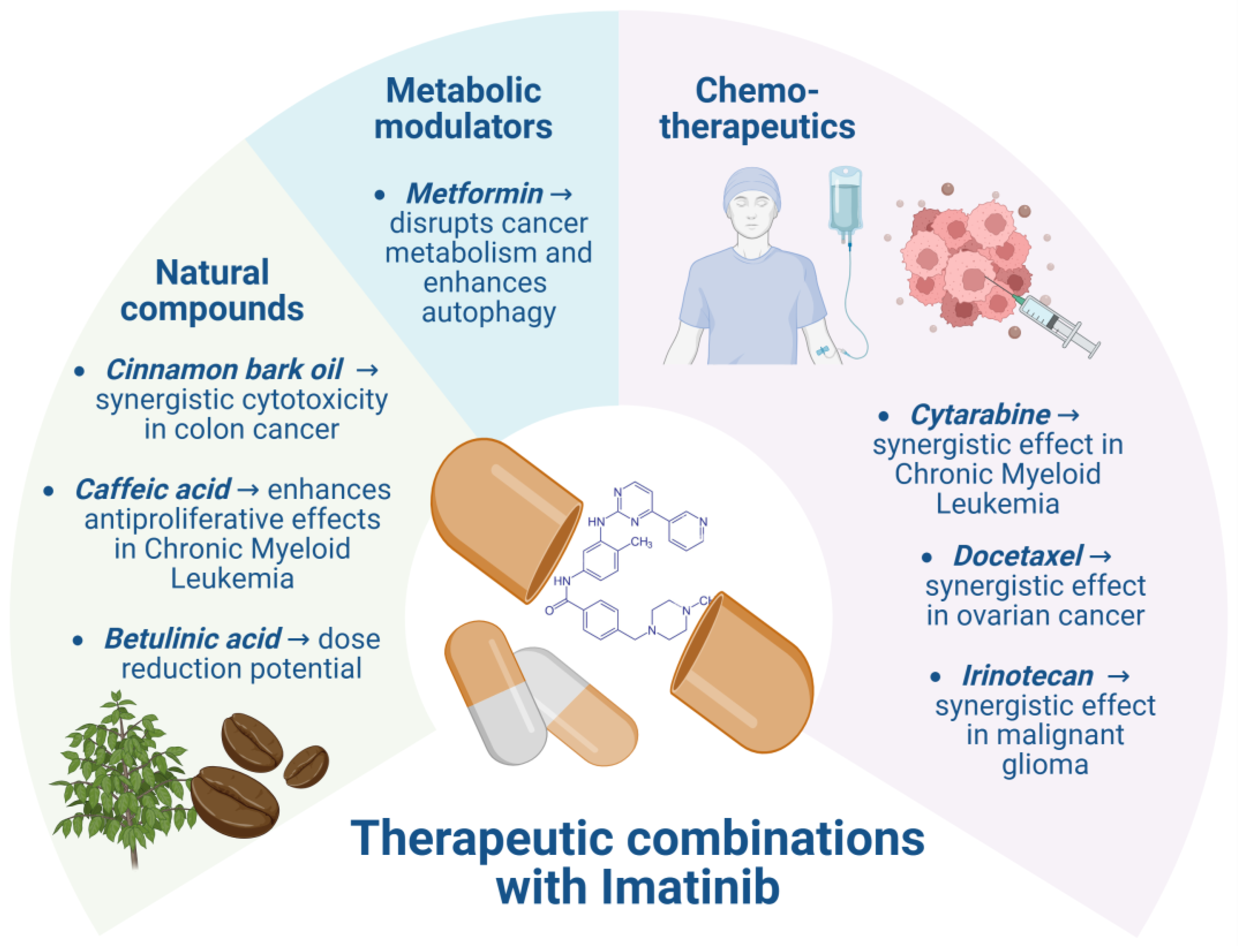
Because most cancers are genetically diverse and highly adaptable, using a single targeted therapy is unlikely to produce lasting remission or cure. Achieving durable antitumor effects in advanced cancers will likely require carefully designed combination therapies (Figure 4) [142]. In leukemia therapy, combinations of multiple drugs are often employed to achieve synergistic effects and enhance treatment outcomes. Studies have demonstrated that IMT combined with cytarabine showed significant efficacy in CML. Furthermore, research indicates that integrating antitumor agents with immunomodulators can not only improve therapeutic responses but also extend patient survival [143]. The biguanide metformin is approved by the FDA for the treatment of T2D. It has also demonstrated antiproliferative and pro-apoptotic effects in various cancer cell types, both as a single agent and in combination with other therapies. These findings suggest that metformin may have potential as a therapeutic option or combination candidate for multiple cancers, including lung, breast, colorectal, and ovarian malignancies [144]. Lee et al. (2017) investigated the effects of IMT alone and in combination with metformin on HCT15 colorectal cancer cells, focusing on cell viability, cell cycle, and autophagy [145]. They found that both IMT and the combination treatment induced autophagy in these cancer cells [145]. Metformin exerts anticancer effects through two main mechanisms: insulin-dependent and insulin-independent. By disrupting cancer cell metabolism and mimicking glucose deprivation, metformin induces the accumulation of misfolded proteins and endoplasmic reticulum stress, ultimately reducing the survival of leukemia cells [146]. Kim et al. (2016) proved that co-treatment of CML cells with deferasirox and IMT induced a synergistic dose-dependent inhibition of proliferation of two different CML cell lines to overcome the IMT resistance of CML cells [147]. IMT has also shown activity when combined with docetaxel. Matei et al. demonstrated that the IMt-docetaxel combination is both effective and well-tolerated in patients with advanced, platinum-resistant ovarian cancer [148].
IMT can also be combined with biologically active natural compounds, which also show a synergistic effect. Cinnamon bark oil (CO) has demonstrated anticancer activity across several cancer cell types and may serve as an adjunct to conventional anticancer drugs. CO showed strong cytotoxicity against HCT116 cells, while IMT exhibited potent activity against HT29 cells. When combined, CO and IMT produced a marked synergistic effect (CI < 1) in HT29 cells. In HCT116 cells, the combination of CO with IMT also demonstrated strong synergy (CI = 0.52) [149]. Feriotto et al. (2021) demonstrated that combining IMT with suboptimal concentrations of caffeic acid produced a synergistic effect, enhancing antiproliferative activity and inducing apoptosis [150]. These results suggest that caffeic acid can strengthen IMT’s anti-leukemic effects in CML [150]. Similarly, Willig et al. (2023) showed that betulinic acid and brosimine B synergize with IMT in K-562 cells, allowing for dose reduction while maintaining or enhancing therapeutic efficacy [151]. This strategy could help minimize drug resistance and improve treatment adherence by reducing IMT-related side effects [151]. Similar synergistic effects with IMT have also been reported for several flavonoids, including apigenin, luteolin, and 5-desmethyl sinensetin, in K-562 cells [152]. Malignant glioma is an aggressive and often treatment-resistant cancer with a distinct tumor microenvironment [153]. Lu et al. (2020) reported that combining IMT with irinotecan or its active metabolite SN-38 produced synergistic anticancer effects both in vitro and in vivo, offering a promising therapeutic opportunity for glioma patients [154].

8. Limitations and Challenges

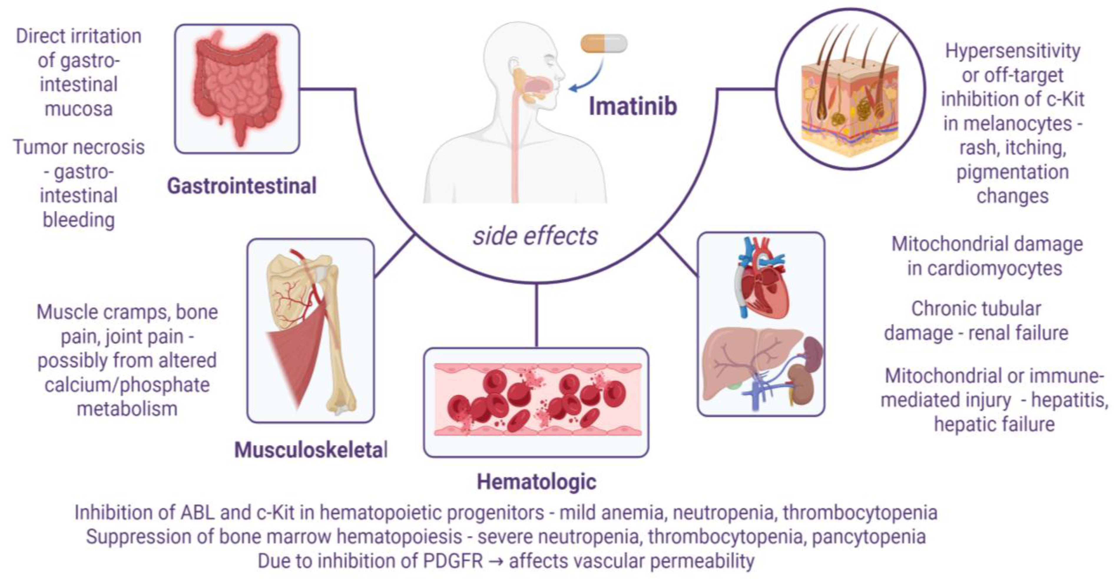
The clinical use of IMT is limited by several adverse effects and safety concerns. IMT therapy has been linked to various toxicities, including cardiac, renal, hematologic, and hepatic toxicity [155], hypereosinophilic cardiac toxicity in patients with hypereosinophilic syndrome, renal impairment, and dermatologic reactions such as erythema multiforme and Stevens–Johnson syndrome [14]. Furthermore, potential interactions between IMT and certain foods or nutritional supplements can influence its pharmacokinetics and safety profile, necessitating careful patient monitoring. Special caution is advised in patients with pre-existing cardiac conditions, as they may be at heightened risk for cardiotoxicity during IMT therapy. In addition to these possible interactions, the development of drug resistance—either primary or secondary—remains a significant obstacle to sustained therapeutic efficacy.
Primary resistance (also referred to as refractoriness) occurs when patients fail to respond to IMT from the outset, unable to reach specific therapeutic milestones. In contrast, secondary or acquired resistance develops in patients who initially benefit from IMT but subsequently lose their therapeutic response over time [156]. Upregulation of multidrug resistance (MDR) genes can effectively reduce intracellular concentrations of IMT to sub-therapeutic levels, thereby contributing to treatment failure. Among the MDR gene family, MDR1 is particularly associated with resistance to chemotherapeutic agents. The MDR1 gene encodes P-glycoprotein, which actively pumps cytotoxic compounds out of cells. This efflux mechanism lowers the intracellular accumulation of IMT and other drugs, ultimately leading to multidrug resistance and diminished therapeutic efficacy [157].
However, most patients with GISTs eventually show disease progression within 2–3 years of IMT treatment, primarily as a result of acquiring secondary KIT mutations [38]. Mutations in the BCR-ABL1 protein are responsible for 40–90% of cases of resistance to TKI therapy in CML [158]. These mutations can render certain drugs ineffective while leaving others still active. Although IMT remains the most widely used therapy, numerous mutations can compromise its efficacy. Without careful monitoring, patients may progress to the accelerated phase of the disease despite ongoing TKI treatment. In such cases, second- or third-line therapies become necessary, which may include alternative TKIs, conventional chemotherapies, or combination regimens such as IFN-α-based therapy [159].
Beyond the risk of developing resistance, IMT therapy is also associated with numerous side effects, necessitating important clinical warnings and precautions (Figure 5) [14].
Table 2 highlights several of the most clinically significant examples for adverse effects of IMT therapy.

9. Advances in Drug Delivery Systems

When IMT was first approved by the FDA, it was available in both tablet and capsule forms. However, according to the FDA, the capsule formulation was later discontinued for safety- or efficacy-related reasons [160]. IMTM exists in two crystalline forms, a and b [161]. The b form is preferred for solid oral dosage formulations. However, manufacturing IMT tablets poses certain challenges, including high friability and low abrasion resistance. In addition, the substantial drug loading often restricts the flexibility in selecting and optimizing excipients [162]. In older patients with newly diagnosed CML, IMT demonstrates efficacy comparable to that in younger patients; however, it is generally associated with greater toxicity, resulting in higher rates of dose reductions and treatment discontinuation [163]. To address this issue, alternative dosing strategies and enhanced formulations are currently under investigation. Currently, the recommended adult dosage for patients with Philadelphia chromosome-positive CML or metastatic GISTs is 400 or 600 mg once daily with meals, administered as monotherapy in accordance with the IMT drug label [30]. Several studies have indicated that a daily dose of 300 mg of IMT can be effective for certain patients, offering improved tolerability while maintaining therapeutic efficacy [164].
Fortunately, strategies have been developed to mitigate many of IMT’s adverse effects, reduce toxicity, and prevent resistance, particularly through novel targeted drug carriers. Advances in biomedical science have driven interest in delivery systems that selectively target the cells responsible for disease initiation and progression. This approach is especially important for life-threatening conditions, where conventional therapies often cause significant side effects, making precise tissue targeting critical to limit systemic exposure. Modern drug delivery systems (DDSs), engineered with advanced technologies, improve the delivery of therapeutic agents directly to the intended site, maximizing efficacy while minimizing off-target accumulation. These next-generation DDSs offer notable advantages over traditional methods, including greater precision, automation, higher throughput, and improved therapeutic outcomes [165]. Typically constructed from nanomaterials or miniaturized, multifunctional, biocompatible, and biodegradable components, they play an increasingly vital role in the management and treatment of complex diseases.
Nanomedicine has become an essential component of personalized cancer therapy. Many of the drawbacks of conventional treatments—such as poor drug solubility, chemoresistance, systemic toxicity, a narrow therapeutic index, and low oral bioavailability—can be addressed using NP-based drug delivery systems [166]. Recent advances include “smart” delivery platforms and modified-release systems, which offer new ways to overcome these challenges [167]. The latest direction in the field focuses on combination strategies, where two or more approaches are integrated to achieve optimal bioavailability and an improved safety profile of the drug.
Nanocarriers, typically measuring less than 100 nm in at least one dimension and designed to deliver active agents to specific target sites, are made from a variety of materials. Nanocarriers can be grouped into three main categories: self-assembling structures (e.g., liposomes, micelles), processed structures (e.g., NPs, emulsions), and chemically bound structures (e.g., dendrimers, silica-based carriers, carbon nanotubes) [168]. Typically, nanocarrier-based systems rely on physically entrapping the drug, which poses two main challenges: (1) the drug’s hydrophilic or hydrophobic nature must be compatible with the carrier to ensure sufficient loading, and (2) the release profile often shows an initial burst followed by a sustained, first-order release. Usually, only the drug released during the sustained phase effectively reaches the tumor site and exerts its therapeutic effect [169]. Thanks to prolonged circulation, nanomedicines can preferentially accumulate in tumors through the enhanced permeability and retention (EPR) effect [170].
Numerous studies have demonstrated that incorporating IMT into advanced drug delivery systems can enhance therapeutic efficacy and cytotoxicity while reducing side effects [171]. Research in this area has been ongoing since 2010, reflecting a sustained effort by scientists to improve cancer treatment outcomes and minimize the adverse effects of therapy. The literature reports a wide range of nanocarrier systems, including liposomes, polymeric NPs, dendrimers, solid lipid NPs, stimuli-responsive systems (pH-, temperature-, or enzyme-sensitive), targeted delivery platforms such as ligand-conjugated NPs, as well as hydrogels and implants designed for localized and sustained release. Figure 6 shows advanced strategies for IMT targeted delivery with improved characteristics.

9.1. Polymeric NPs

Bhattacharya (2020) developed chitosan (CS)-based polymeric NPs of IMT for colorectal cancer targeting [172]. Hasandoost et al. (2017) prepared IMTM-loaded polybutylcyanoacrylate NPs and evaluated their efficacy on leukemia cell line K562 [173]. In the study by Li et al. (2019), N-oleoyl-D-galactosamine was synthesized to create biomimetic galactose-modified NPs, aiming to enhance the oral bioavailability of IMT and overcome its poor intestinal solubility and associated adverse effects [174]. Bhattacharya et al. (2025) developed IMT-loaded Poly Lactic-co-Glycolic Acid-D-α-tocopheryl polyethylene glycol succinate—Polyethylene glycol hybrid NPs with optimized physicochemical properties for targeted delivery to glioblastoma multiforme [175].

9.2. Lipid-Based Nanocarriers

El-Mezayen et al. (2017) presented IMT loading in liposomes coupled with vitamin A, which represents a successful approach for active HSCs (hepatic stellate cells) targeting intended for liver fibrosis [176]. Molaahmadi et al. (2019) investigated the potential benefits of IMT-loaded lipid nanocapsules as a novel drug delivery vehicle against the melanoma cell line [177]. Siram et al. (2022) prepared solid lipid NPs of IMTM and coated them with hyaluronic acid to enhance their specificity towards human breast cancer MCF-7 cells [178].

9.3. Inorganic NPs

Lababa et al. (2015) developed layer-by-layer polymer-coated gold NPs for topical iontophoretic delivery of IMTM to treat melanoma [179]. Naeimipour et al. (2022) synthesized IMTM-loaded β-cyclodextrin/magnetic iron oxide NPs via an eco-friendly green method using aqueous extracts of Mentha longifolia (wild mint) [180].

9.4. “Smart” Systems

Shoaib et al. (2018) developed a polyurethane–amino acids–pH-responsive drug delivery system of IMT with 94% loading efficiency for use as a smart anticancer carrier [181]. Maral Mashreghi et al. (2023) developed an IMT drug delivery system using a magnetite–graphene oxide and bovine serum albumin conjugate as the nanocarrier [182]. Aslehashemi et al. (2025) synthesized a magnetic mesoporous nanocomposite using Fe3O4-SiO2 NPs grafted with pH-sensitive biopolymers, carboxymethyl cellulose, and hyaluronic acid as a drug delivery system of IMTM [183].

9.5. Hydrogels

In 2022, Cong-Yu Wang and colleagues developed an intestine enzyme-responsive hydrogel for IMT delivery, utilizing crosslinked methacrylic anhydride-modified carboxymethyl CS as the polysaccharide-based matrix [184]. In 2023, Paulina Sobierajska and co-workers designed galactose-based hydrogels functionalized with IMT and incorporated with nanohydroxyapatite to serve as a drug delivery platform for osteosarcoma treatment [19]. In 2024, Pang and colleagues developed a pH-sensitive hydrogel capable of sustained IMTM release for the therapeutic management of tendon adhesion [185].
Below, we highlight several notable studies that showcase promising strategies and may serve as a foundation for developing innovative IMT drug delivery systems.
In the study by Ye et al. (2014), IMT was remote-loaded into folate receptor (FR)-targeted liposomes prepared via thin-film hydration and polycarbonate membrane extrusion [186]. These FR-targeted liposomes significantly enhanced apoptosis in HeLa cervical cancer cells in vitro compared to non-targeted IMT liposomes and also demonstrated prolonged circulation in mice [186].
Shandiz et al. (2016) developed IMT-loaded silver NPs through phyto-synthesis using leaf extracts from Eucalyptus procera [187]. The release of IMT was in 2 stages—an initial burst release (1–40 h) followed by a sustained, constant release phase (40–80 h), which is advantageous for enhancing therapeutic efficacy. Cell viability assays showed that the prepared NPs significantly reduced cell survival across different concentrations, with effects comparable to silver NPs and IMT alone. Moreover, cytotoxicity studies confirmed that the prepared IMT-loaded NPs induced apoptotic cell death in the MCF-7 cell line, rather than necrosis [187].
Varshosaz et al. (2020) reported the development of nanostructured lipid carriers (NLCs) containing both curcumin and IMT and conjugated with rituximab to specifically target CD20 receptors on lymphoma cell lines [188]. The study demonstrated that the targeted NLCs exhibited significantly greater cytotoxicity against Ramos cells compared to free drugs or non-targeted NLCs. In particular, co-administration of curcumin (15 μg/mL) and IMT (5 μg/mL) reduced the IC50 of free curcumin from 8.3 ± 0.9 μg/mL to 1.9 ± 0.2 μg/mL. These findings suggest that rituximab-conjugated NLCs carrying curcumin and IMT may enhance the cytotoxic effects of IMT in the treatment of non-Hodgkin lymphoma [188].
To address tumor cell resistance to TRAIL and the adverse effects associated with IMT, Fu et al. (2024) developed multifunctional liposomes, in which IMT was encapsulated in the aqueous core while TRAIL was incorporated into the liposomal membrane [189]. These liposomes demonstrated strong accumulation in TRAIL-resistant cells and in an HT-29 tumor-bearing mouse model. In vitro cytotoxicity assays revealed that the prepared liposomes increased the killing activity against HT-29 cells by 50%, with apoptosis confirmed as the underlying mechanism [189].
Vosoughifar et al. (2024) developed a topical delivery system for melanoma using CS nanofibers coated with covalent triazine-based framework NPs loaded with IMT [190]. The CS nanofiber sheets were fabricated via electrospinning. Results showed that the prepared nanosheets effectively inhibited melanoma cell growth while exhibiting no toxicity toward normal fibroblasts. Moreover, melanoma cells cultured on these nanosheets displayed increased expression of pro-apoptotic genes and decreased expression of anti-apoptotic genes. These findings suggest that the prepared composite dressings could serve as a promising targeted therapy for melanoma, enhancing treatment efficacy while reducing the adverse effects of IMT on healthy cells [190].
Another study explored the potential of IMT for inhibiting corneal neovascularization, though its poor aqueous solubility has limited its use in ocular therapies. To overcome this, Wang et al. (2024) developed IMT-loaded glycymicelles using glycyrrhizin as a nanocarrier and further encapsulated them in a hydroxypropyl methylcellulose–sodium hyaluronate hydrogel to extend retention on the ocular surface [191]. This formulation markedly improved the solubility of IMT. Ocular tolerance tests in rabbits confirmed its safety, while studies in a murine alkaline corneal burn model demonstrated that topical application of IMT-hydrogel effectively promoted corneal wound healing, restored corneal sensitivity, and reduced corneal opacity and proved the potential of IMT-hydrogel as a therapeutic option for eye injuries [191].
Ozgenc et al. (2025) developed freeze-dried kit formulations of 177Lu-labeled IMT and assessed their potential efficacy through in vitro studies [192]. Targeted radiopharmaceuticals represent a promising strategy for both cancer diagnosis and therapy, and this study achieved noteworthy results. Under optimal conditions (45 mg IMT-chelator, pH 5, 60 min incubation), radiolabeling efficiency exceeded 90%, with stability maintained for up to 7 days. The optimized formulation demonstrated high labeling efficiency, excellent stability, and selective in vitro cytotoxicity against breast cancer cells, supporting its potential use as a targeted radiopharmaceutical [192].

10. Future Perspectives

Future advancements in IMT drug delivery should focus on designing multifunctional nanocarrier systems capable of achieving enhanced therapeutic effects, overcoming resistance mechanisms, and minimizing systemic toxicity. The development of advanced IMT carriers is a key aspect of personalized medicine, which integrates genomic data to identify patient-specific mutations and biomarkers, alongside imaging data to assess tumor heterogeneity and aggressiveness. This information enables the design of personalized nanocarriers with tailored dosing and targeted delivery, improving therapeutic precision and outcomes [193]. Integration of multimodal targeting approaches—combining passive accumulation with active ligand-mediated recognition—could further refine tumor selectivity and drug bioavailability. Incorporating dual or multiple ligands, such as folic acid, hyaluronic acid, and monoclonal antibodies, may allow simultaneous targeting of different tumor-specific receptors, thereby mitigating the limitations of receptor heterogeneity. Another promising direction involves the development of stimuli-responsive hybrid nanocarriers that respond to both internal (e.g., pH, enzyme activity, redox potential) and external (e.g., magnetic field, light, ultrasound) cues for controlled and site-specific release. Such systems can ensure precise drug delivery, improving therapeutic efficacy while reducing adverse effects on normal tissues. The incorporation of biodegradable and biocompatible polymers or lipid–polymer hybrid nanostructures may also enhance stability, circulation time, and controlled release kinetics. Future studies should prioritize translational research, focusing on scalable synthesis, reproducibility, and regulatory compliance to facilitate the clinical application of these findings. Overall, the convergence of nanotechnology, molecular targeting, and personalized medicine offers a transformative path toward improving IMT’s therapeutic index and overcoming its pharmacological limitations in cancer therapy.
Another promising future research lies in exploring new therapeutic applications of IMT beyond its currently approved indications. Ongoing studies have already revealed several off-label uses, yet the potential for further repurposing remains substantial. Interestingly, some of IMT’s adverse effects may be harnessed for therapeutic benefit through targeted formulation and delivery strategies. For instance, while oral administration has been associated with dermatologic side effects, topical application has demonstrated efficacy in treating certain skin conditions, including atopic dermatitis. Similarly, although IMT may induce hepatotoxicity when administered orally, its incorporation into pH-responsive delivery systems offers potential in the targeted management of liver fibrosis [194]. These findings underscore the importance of optimizing both the dose and drug delivery system to improve IMT’s therapeutic profile.

11. Conclusions

The tyrosine kinase inhibitor IMT has transformed the treatment of malignancies driven by its targets, c-ABL, c-KIT, and PDGFR, and is now the standard of care for CML and GIST, dramatically improving patient outcomes in adults and children. Beyond these indications, studies suggest IMT may have therapeutic potential for a wide range of conditions, including inflammatory bowel diseases, ocular injuries, and certain skin disorders. Developing advanced drug delivery systems for IMT could help minimize systemic toxicity, overcome resistance, and expand its use to additional diseases. Future research should focus on sustained-release formulations, stimuli-responsive nanocarriers, and tumor-targeted delivery platforms to maximize efficacy and safety.

Author Contributions

Conceptualization, Y.G. and P.G.; methodology, Y.G.; investigation, Y.G.; resources, Y.G. and P.G.; writing—original draft preparation, Y.G.; writing—review and editing, Y.G. and P.K.; visualization, P.K. and P.G.; formal analysis, P.K.; project administration, P.K.; Software, P.K.; supervision, Y.G. and P.G. All authors have read and agreed to the published version of the manuscript.

Funding

This research received no external funding.

Acknowledgments

We would like to express our gratitude to the program “Research, Innovation and Digitalisation for Smart Transformation” 2021–2027, funded by the European Union, Project BG16RFPR002-1.014-0007, Center for Competence “PERIMED-2” for supporting this article.

Conflicts of Interest

The authors declare no conflicts of interest.

Abbreviations

The following abbreviations are used in this manuscript:
FDAU.S. Food and Drug Administration
IMTImatinib
IMTMImatinib mesylate
PTKsProtein tyrosine kinases
RTKsReceptor tyrosine kinases
TKIsTyrosine kinase inhibitors
CMLChronic myeloid leukemia
PDGFRPlatelet-derived growth factor receptor
GISTGastrointestinal stromal tumors
NPCNiemann–Pick type C
ICHIntracerebral hemorrhage
cGvHDChronic graft-versus-host disease
MSMultiple sclerosis
CNSCentral nervous system
SScSystemic sclerosis
CGCJCentral giant cell granuloma of the jaw
GDGaucher disease
T1DType 1 Diabetes
T2DType 2 Diabetes
COCinnamon bark oil
DDSsDrug delivery systems
FRFolate receptor
NLCsNanostructured lipid carriers
CSChitosan
NPsNanoparticles
OSOverall survival
PFSProgression-free survival
MDRMultidrug resistance

References

  1. Colonna, G. Overcoming Barriers in Cancer Biology Research: Current Limitations and Solutions. Cancers 2025, 17, 2102. [Google Scholar] [CrossRef]
  2. Ardasheva, R.; Prissadova, N.; Turiyski, V.; Tolekova, A.; Krastev, A.; Pencheva, M.; Popov, V. Effects of Electron Radiation on Serotonin Signaling and Reactivity of Rat Gastric Smooth Muscle. Toxics 2023, 11, 603. [Google Scholar] [CrossRef]
  3. Debela, D.T.; Muzazu, S.G.; Heraro, K.D.; Ndalama, M.T.; Mesele, B.W.; Haile, D.C.; Kitui, S.K.; Manyazewal, T. New Approaches and Procedures for Cancer Treatment: Current Perspectives. SAGE Open Med. 2021, 9, 20503121211034366. [Google Scholar] [CrossRef]
  4. Mongre, R.K.; Mishra, C.B.; Shukla, A.K.; Prakash, A.; Jung, S.; Ashraf-Uz-Zaman, M.; Lee, M.-S. Emerging Importance of Tyrosine Kinase Inhibitors against Cancer: Quo Vadis to Cure? Int. J. Mol. Sci. 2021, 22, 11659. [Google Scholar] [CrossRef] [PubMed]
  5. Apostolova, E.G.; Kokova, V.; Peychev, Z.; Apostolov, A. Effect of Fucoidan, Haberlea Rhodopensis and Propolis on Mobilization of The CD34+ Stem Cells in Rats. Farmacia 2017, 65, 567–570. [Google Scholar]
  6. Kim, M.; Baek, M.; Kim, D.J. Protein Tyrosine Signaling and Its Potential Therapeutic Implications in Carcinogenesis. Curr. Pharm. Des. 2017, 23, 4226–4246. [Google Scholar] [CrossRef] [PubMed]
  7. Metibemu, D.S.; Akinloye, O.A.; Akamo, A.J.; Ojo, D.A.; Okeowo, O.T.; Omotuyi, I.O. Exploring Receptor Tyrosine Kinases-Inhibitors in Cancer Treatments. Egypt. J. Med. Hum. Genet. 2019, 20, 35. [Google Scholar] [CrossRef]
  8. Hussain, S.; Mursal, M.; Verma, G.; Hasan, S.M.; Khan, M.F. Targeting Oncogenic Kinases: Insights on FDA Approved Tyrosine Kinase Inhibitors. Eur. J. Pharmacol. 2024, 970, 176484. [Google Scholar] [CrossRef]
  9. Das, R.; Tambe, G.; Shard, A. Sulfonamides as Tyrosine Kinase Modulators—A Promising Class of Anticancer Agents. Results Chem. 2023, 5, 100950. [Google Scholar] [CrossRef]
  10. Orrico, K.B. Basic Concepts of Cancer Genetics and Receptor Tyrosine Kinase Inhibition for Pharmacists. A Narrative Review. J. Oncol. Pharm. Pract. 2023, 29, 1187–1195. [Google Scholar] [CrossRef]
  11. Mhaibes, R.M. The Impact of Tyrosine Kinase Inhibitors on Cancer Therapy: A Comprehensive Review. Med. Med. Chem. 2024, 1, 36–42. [Google Scholar]
  12. Braun, T.P.; Eide, C.A.; Druker, B.J. Response and Resistance to BCR-ABL1-Targeted Therapies. Cancer Cell 2020, 37, 530–542. [Google Scholar] [CrossRef]
  13. Iqbal, N.; Iqbal, N. Imatinib: A Breakthrough of Targeted Therapy in Cancer. Chemother. Res. Pract. 2014, 2014, 357027. [Google Scholar] [CrossRef] [PubMed]
  14. Flynn, J.P.; Gerriets, V. Imatinib. In StatPearls [Internet]; StatPearls Publishing: Treasure Island, FL, USA, 2025. Available online: https://www.ncbi.nlm.nih.gov/books/NBK551676/ (accessed on 28 August 2025).
  15. Druker, B.J.; Talpaz, M.; Resta, D.J.; Peng, B.; Buchdunger, E.; Ford, J.M.; Lydon, N.B.; Kantarjian, H.; Capdeville, R.; Ohno-Jones, S.; et al. Efficacy and Safety of a Specific Inhibitor of the BCR-ABL Tyrosine Kinase in Chronic Myeloid Leukemia. N. Engl. J. Med. 2001, 344, 1031–1037. [Google Scholar] [CrossRef] [PubMed]
  16. Druker, B.J.; Tamura, S.; Buchdunger, E.; Ohno, S.; Segal, G.M.; Fanning, S.; Zimmermann, J.; Lydon, N.B. Effects of a Selective Inhibitor of the Abl Tyrosine Kinase on the Growth of Bcr–Abl Positive Cells. Nat. Med. 1996, 2, 561–566. [Google Scholar] [CrossRef] [PubMed]
  17. Napier, R.J.; Norris, B.A.; Swimm, A.; Giver, C.R.; Harris, W.A.C.; Laval, J.; Napier, B.A.; Patel, G.; Crump, R.; Peng, Z.; et al. Low Doses of Imatinib Induce Myelopoiesis and Enhance Host Anti-Microbial Immunity. PLoS Pathog. 2015, 11, e1004770. [Google Scholar] [CrossRef]
  18. Roskoski, R. Properties of FDA-Approved Small Molecule Protein Kinase Inhibitors: A 2023 Update. Pharmacol. Res. 2023, 187, 106552. [Google Scholar] [CrossRef]
  19. Sobierajska, P.; Wiatrak, B.; Jawien, P.; Janeczek, M.; Wiglusz, K.; Szeląg, A.; Wiglusz, R.J. Imatinib-Functionalized Galactose Hydrogels Loaded with Nanohydroxyapatite as a Drug Delivery System for Osteosarcoma: In Vitro Studies. ACS Omega 2023, 8, 17891–17900. [Google Scholar] [CrossRef]
  20. National Center for Biotechnology Information. “PubChem Compound Summary for CID 123596, Imatinib Mesylate” PubChem. Available online: https://pubchem.ncbi.nlm.nih.gov/compound/imatinib-mesylate (accessed on 28 August 2025).
  21. Akagi, S.; Nakamura, K.; Miura, D.; Saito, Y.; Matsubara, H.; Ogawa, A.; Matoba, T.; Egashira, K.; Ito, H. Delivery of Imatinib-Incorporated Nanoparticles into Lungs Suppresses the Development of Monocrotaline-Induced Pulmonary Arterial Hypertension. Int. Heart J. 2015, 56, 354–359. [Google Scholar] [CrossRef]
  22. Bhullar, S.; Goyal, N.; Gupta, S. In-Vitro pH-Responsive Release of Imatinib from Iron-Supplement Coated Anatase TiO2 Nanoparticles. Sci. Rep. 2022, 12, 4600. [Google Scholar] [CrossRef]
  23. Ghadami, A.; Fathi-karkan, S.; Siddiqui, B.; Gondal, S.A.; Rahdar, A.; Garousi, N.A.; Kharaba, Z.; Ghotekar, S. Nanotechnology in Imatinib Delivery: Advancing Cancer Treatment through Innovative Nanoparticles. Med. Oncol. 2025, 42, 116. [Google Scholar] [CrossRef] [PubMed]
  24. National Center for Biotechnology Information. “PubChem Compound Summary for CID 5291, Imatinib” PubChem. Available online: https://pubchem.ncbi.nlm.nih.gov/compound/imatinib (accessed on 24 August 2025).
  25. Vandyke, K.; Fitter, S.; Dewar, A.L.; Hughes, T.P.; Zannettino, A.C.W. Dysregulation of Bone Remodeling by Imatinib Mesylate. Blood 2010, 115, 766–774. [Google Scholar] [CrossRef] [PubMed]
  26. Arora, B.; Gota, V.; Menon, H.; Sengar, M.; Nair, R.; Patial, P.; Banavali, S.D. Therapeutic Drug Monitoring for Imatinib: Current Status and Indian Experience. Indian J. Med. Paediatr. Oncol. 2013, 34, 224–228. [Google Scholar] [CrossRef] [PubMed]
  27. Peng, B.; Dutreix, C.; Mehring, G.; Hayes, M.J.; Ben-Am, M.; Seiberling, M.; Pokorny, R.; Capdeville, R.; Lloyd, P. Absolute Bioavailability of Imatinib (Glivec®) Orally versus Intravenous Infusion. J. Clin. Pharmacol. 2004, 44, 158–162. [Google Scholar] [CrossRef]
  28. Barratt, D.T.; Somogyi, A.A. Role of Pharmacogenetics in Personalised Imatinib Dosing. Transl. Cancer Res. 2017, 6, S1541–S1557. [Google Scholar] [CrossRef]
  29. Di Gion, P.; Kanefendt, F.; Lindauer, A.; Scheffler, M.; Doroshyenko, O.; Fuhr, U.; Wolf, J.; Jaehde, U. Clinical Pharmacokinetics of Tyrosine Kinase Inhibitors: Focus on Pyrimidines, Pyridines and Pyrroles. Clin. Pharmacokinet. 2011, 50, 551–603. [Google Scholar] [CrossRef]
  30. Chen, Y.; Dong, X.; Wang, Q.; Liu, Z.; Dong, X.; Shi, S.; Xiao, H. Factors Influencing the Steady-State Plasma Concentration of Imatinib Mesylate in Patients With Gastrointestinal Stromal Tumors and Chronic Myeloid Leukemia. Front. Pharmacol. 2020, 11, 569843. [Google Scholar] [CrossRef]
  31. Omran, M.M.; Ibrahim, A.B.; Abdelfattah, R.; Shouman, S.A.; Hamza, M.S. Imatinib Pharmacokinetics and Creatine Kinase Levels in Chronic Myeloid Leukemia Patients: Implications for Therapeutic Response and Monitoring. Eur. J. Clin. Pharmacol. 2024, 80, 1061–1068. [Google Scholar] [CrossRef]
  32. Hochhaus, A.; Larson, R.A.; Guilhot, F.; Radich, J.P.; Branford, S.; Hughes, T.P.; Baccarani, M.; Deininger, M.W.; Cervantes, F.; Fujihara, S.; et al. Long-Term Outcomes of Imatinib Treatment for Chronic Myeloid Leukemia. N. Engl. J. Med. 2017, 376, 917–927. [Google Scholar] [CrossRef]
  33. Jabbour, E.; Kantarjian, H. Chronic Myeloid Leukemia: A Review. JAMA 2025, 333, 1618. [Google Scholar] [CrossRef]
  34. El-Tanani, M.; Nsairat, H.; Matalka, I.I.; Lee, Y.F.; Rizzo, M.; Aljabali, A.A.; Mishra, V.; Mishra, Y.; Hromić-Jahjefendić, A.; Tambuwala, M.M. The Impact of the BCR-ABL Oncogene in the Pathology and Treatment of Chronic Myeloid Leukemia. Pathol.-Res. Pract. 2024, 254, 155161. [Google Scholar] [CrossRef] [PubMed]
  35. Kumar, M.; Kulshrestha, R.; Singh, N.; Jaggi, A.S. Expanding Spectrum of Anticancer Drug, Imatinib, in the Disorders Affecting Brain and Spinal Cord. Pharmacol. Res. 2019, 143, 86–96. [Google Scholar] [CrossRef] [PubMed]
  36. Suttorp, M.; Bornhäuser, M.; Metzler, M.; Millot, F.; Schleyer, E. Pharmacology and Pharmacokinetics of Imatinib in Pediatric Patients. Expert Rev. Clin. Pharmacol. 2018, 11, 219–231. [Google Scholar] [CrossRef]
  37. Di Vito, A.; Ravegnini, G.; Gorini, F.; Aasen, T.; Serrano, C.; Benuzzi, E.; Coschina, E.; Monesmith, S.; Morroni, F.; Angelini, S.; et al. The Multifaceted Landscape behind Imatinib Resistance in Gastrointestinal Stromal Tumors (GISTs): A Lesson from Ripretinib. Pharmacol. Ther. 2023, 248, 108475. [Google Scholar] [CrossRef]
  38. Masucci, M.T.; Motti, M.L.; Minopoli, M.; Di Carluccio, G.; Carriero, M.V. Emerging Targeted Therapeutic Strategies to Overcome Imatinib Resistance of Gastrointestinal Stromal Tumors. Int. J. Mol. Sci. 2023, 24, 6026. [Google Scholar] [CrossRef]
  39. Adiwidjaja, J.; Boddy, A.V.; McLachlan, A.J. Implementation of a Physiologically Based Pharmacokinetic Modeling Approach to Guide Optimal Dosing Regimens for Imatinib and Potential Drug Interactions in Paediatrics. Front. Pharmacol. 2020, 10, 1672. [Google Scholar] [CrossRef]
  40. Teo, Y.L.; Ho, H.K.; Chan, A. Metabolism-related Pharmacokinetic Drug−drug Interactions with Tyrosine Kinase Inhibitors: Current Understanding, Challenges and Recommendations. Br. J. Clin. Pharmacol. 2015, 79, 241–253. [Google Scholar] [CrossRef]
  41. Reichardt, P. The Story of Imatinib in GIST—A Journey through the Development of a Targeted Therapy. Oncol. Res. Treat. 2018, 41, 472–477. [Google Scholar] [CrossRef]
  42. Copyright Northumbria Healthcare NHS Trust & North of England Cancer Network Systemic Anticancer Therapy Drug Interactions Table. 2012. Available online: https://northerncanceralliance.nhs.uk/wp-content/uploads/2018/10/SACT-Interactions-Table1.pdf (accessed on 24 August 2025).
  43. Accord Healthcare Limited. ANNEX I SUMMARY OF PRODUCT CHARACTERISTICS Imatinib Accord 100 Mg Film-Coated Tablets. Available online: https://www.accord-healthcare.com/ie/system/files/spc/ema-spc-h-2681-en-var%2039.G.pdf (accessed on 24 August 2025).
  44. De Diego García, P.; Trincado Aznar, P.; Playán Usón, J.; Albero Gamboa, R. Tratamiento con levotiroxina e imatinib. Endocrinol. Nutr. 2008, 55, 304–307. [Google Scholar] [CrossRef]
  45. Chan, W.-J.J.; Adiwidjaja, J.; McLachlan, A.J.; Boddy, A.V.; Harnett, J.E. Interactions between Natural Products and Cancer Treatments: Underlying Mechanisms and Clinical Importance. Cancer Chemother. Pharmacol. 2023, 91, 103–119. [Google Scholar] [CrossRef]
  46. Escudero-Vilaplana, V.; Collado-Borrell, R.; Villanueva-Bueno, C.; Álvarez, R.; Herranz, A.; Sanjurjo, M. Acute Pancreatitis in a Patient Treated with Imatinib and Gefitinib. J. Oncol. Pharm. Pract. 2021, 27, 980–983. [Google Scholar] [CrossRef] [PubMed]
  47. De La Fuente, J.; Baruchel, A.; Biondi, A.; De Bont, E.; Dresse, M.; Suttorp, M.; Millot, F.; International BFM Group (iBFM) Study Group Chronic Myeloid Leukaemia Committee. Managing Children with Chronic Myeloid Leukaemia (CML): Recommendations for the Management of CML in Children and Young People up to the Age of 18 Years. Br. J. Haematol. 2014, 167, 33–47. [Google Scholar] [CrossRef] [PubMed]
  48. León, R.; Gutiérrez, D.A.; Pinto, C.; Morales, C.; De La Fuente, C.; Riquelme, C.; Cortés, B.I.; González-Martin, A.; Chamorro, D.; Espinosa, N.; et al. C-Abl Tyrosine Kinase down-Regulation as Target for Memory Improvement in Alzheimer’s Disease. Front. Aging Neurosci. 2023, 15, 1180987. [Google Scholar] [CrossRef] [PubMed]
  49. Estrada, L.D.; Chamorro, D.; Yañez, M.J.; Gonzalez, M.; Leal, N.; Von Bernhardi, R.; Dulcey, A.E.; Marugan, J.; Ferrer, M.; Soto, C.; et al. Reduction of Blood Amyloid-β Oligomers in Alzheimer’s Disease Transgenic Mice by c-Abl Kinase Inhibition. J. Alzheimers Dis. 2016, 54, 1193–1205. [Google Scholar] [CrossRef]
  50. Cancino, G.I.; Perez De Arce, K.; Castro, P.U.; Toledo, E.M.; Von Bernhardi, R.; Alvarez, A.R. C-Abl Tyrosine Kinase Modulates Tau Pathology and Cdk5 Phosphorylation in AD Transgenic Mice. Neurobiol. Aging 2011, 32, 1249–1261. [Google Scholar] [CrossRef]
  51. Cancino, G.I.; Toledo, E.M.; Leal, N.R.; Hernandez, D.E.; Yévenes, L.F.; Inestrosa, N.C.; Alvarez, A.R. STI571 Prevents Apoptosis, Tau Phosphorylation and Behavioural Impairments Induced by Alzheimer’s β-Amyloid Deposits. Brain 2008, 131, 2425–2442. [Google Scholar] [CrossRef]
  52. Marín, T.; Valls, C.; Jerez, C.; Huerta, T.; Elgueta, D.; Vidal, R.L.; Alvarez, A.R.; Cancino, G.I. The C-Abl/P73 Pathway Induces Neurodegeneration in a Parkinson’s Disease Model. IBRO Neurosci. Rep. 2022, 13, 378–387. [Google Scholar] [CrossRef]
  53. Lastovetskyi, I.; Cytlau, B.; Marczyk, Ł.; Zdrojewska, K.; Łach, A.; Krupa, J.; Lorkowska-Zawicka, B.; Giżycka, B.B. Modern Pharmacological Treatment of Parkinson’s Disease: Reviving Known Drugs and New Perspectives. Int. J. Pharm. Res. Allied Sci. 2024, 13, 29–39. [Google Scholar] [CrossRef]
  54. Zeitelhofer, M.; Stefanitsch, C.; Protzmann, J.; Torrente, D.; Adzemovic, M.Z.; Lewandowski, S.A.; Muhl, L.; Eriksson, U.; Nilsson, I.; Su, E.; et al. Reduced Myofibroblast Transdifferentiation and Fibrotic Scarring in Ischemic Stroke after Imatinib Treatment. bioRxiv 2021. [Google Scholar] [CrossRef]
  55. Wahlgren, N.; Thorén, M.; Höjeberg, B.; Käll, T.-B.; Laska, A.-C.; Sjöstrand, C.; Höijer, J.; Almqvist, H.; Holmin, S.; Lilja, A.; et al. Randomized Assessment of Imatinib in Patients with Acute Ischaemic Stroke Treated with Intravenous Thrombolysis. J. Intern. Med. 2017, 281, 273–283. [Google Scholar] [CrossRef]
  56. Pearce, W.J.; Doan, C.; Carreon, D.; Kim, D.; Durrant, L.M.; Manaenko, A.; McCoy, L.; Obenaus, A.; Zhang, J.H.; Tang, J. Imatinib Attenuates Cerebrovascular Injury and Phenotypic Transformation after Intracerebral Hemorrhage in Rats. Am. J. Physiol.-Regul. Integr. Comp. Physiol. 2016, 311, R1093–R1104. [Google Scholar] [CrossRef]
  57. Ma, Q.; Huang, B.; Khatibi, N.; Rolland, W.; Suzuki, H.; Zhang, J.H.; Tang, J. PDGFR-α Inhibition Preserves Blood-brain Barrier after Intracerebral Hemorrhage. Ann. Neurol. 2011, 70, 920–931. [Google Scholar] [CrossRef]
  58. Park, C.S.; Lee, J.Y.; Yune, T.Y. Imatinib Prevents Blood-Spinal Cord Barrier Disruption by Inhibiting PDGFR-Mediated JMJD3 Expression and Activation after Spinal Cord Injury. Fluids Barriers CNS 2025, 22, 76. [Google Scholar] [CrossRef]
  59. Yáñez, M.J.; Belbin, O.; Estrada, L.D.; Leal, N.; Contreras, P.S.; Lleó, A.; Burgos, P.V.; Zanlungo, S.; Alvarez, A.R. C-Abl Links APP-BACE1 Interaction Promoting APP Amyloidogenic Processing in Niemann-Pick Type C Disease. Biochim. Biophys. Acta BBA-Mol. Basis Dis. 2016, 1862, 2158–2167. [Google Scholar] [CrossRef]
  60. Alvarez, A.R.; Klein, A.; Castro, J.; Cancino, G.I.; Amigo, J.; Mosqueira, M.; Vargas, L.M.; Yevenes, L.F.; Bronfman, F.C.; Zanlungo, S. Imatinib Therapy Blocks Cerebellar Apoptosis and Improves Neurological Symptoms in a Mouse Model of Niemann-Pick Type C Disease. FASEB J. 2008, 22, 3617–3627. [Google Scholar] [CrossRef] [PubMed]
  61. Kaplan, F.S.; Andolina, J.R.; Adamson, P.C.; Teachey, D.T.; Finklestein, J.Z.; Ebb, D.H.; Whitehead, B.; Jacobs, B.; Siegel, D.M.; Keen, R.; et al. Early Clinical Observations on the Use of Imatinib Mesylate in FOP: A Report of Seven Cases. Bone 2018, 109, 276–280. [Google Scholar] [CrossRef] [PubMed]
  62. Malard, F.; Mohty, M. Updates in Chronic Graft-versus-host Disease Management. Am. J. Hematol. 2023, 98, 1637–1644. [Google Scholar] [CrossRef] [PubMed]
  63. Braun, L.M.; Zeiser, R. Kinase Inhibition as Treatment for Acute and Chronic Graft-Versus-Host Disease. Front. Immunol. 2021, 12, 760199. [Google Scholar] [CrossRef]
  64. Crespo, O.; Kang, S.C.; Daneman, R.; Lindstrom, T.M.; Ho, P.P.; Sobel, R.A.; Steinman, L.; Robinson, W.H. Tyrosine Kinase Inhibitors Ameliorate Autoimmune Encephalomyelitis in a Mouse Model of Multiple Sclerosis. J. Clin. Immunol. 2011, 31, 1010–1020. [Google Scholar] [CrossRef]
  65. Fawzy, M.G.; Selim, M.S.; Khorshid, O.A.; Rashed, L.A.; Elkordy, M.A.; Abdel Latif, N.S. Comparative Study on the Possible Ameliorative Effects of Imatinib and Ibudilast in a Rat Model of Multiple Sclerosis. Future J. Pharm. Sci. 2025, 11, 47. [Google Scholar] [CrossRef]
  66. Yotov, V.; Ardasheva, R.; Mihaylova, A.; Doncheva, N.; Kostadinov, I.; Turiyski, V. Simulated Microgravity Affects Carrageenan-Induced Inflammation Process in Rats. Pharmacia 2023, 70, 1531–1538. [Google Scholar] [CrossRef]
  67. Azizi, G.; Haidari, M.R.; Khorramizadeh, M.; Naddafi, F.; Sadria, R.; Javanbakht, M.H.; Sedaghat, R.; Tofighi Zavareh, F.; Mirshafiey, A. Effects of Imatinib Mesylate in Mouse Models of Multiple Sclerosis and in Vitro Determinants. Iran J. Allergy Asthma Immunol. 2014, 13, 198–206. [Google Scholar]
  68. Speich, R.; Ulrich, S.; Domenighetti, G.; Huber, L.C.; Fischler, M.; Treder, U.; Breitenstein, A. Efficacy and Safety of Long-Term Imatinib Therapy for Pulmonary Arterial Hypertension. Respiration 2015, 89, 515–524. [Google Scholar] [CrossRef] [PubMed]
  69. Cahill, K.N.; Katz, H.R.; Cui, J.; Lai, J.; Kazani, S.; Crosby-Thompson, A.; Garofalo, D.; Castro, M.; Jarjour, N.; DiMango, E.; et al. KIT Inhibition by Imatinib in Patients with Severe Refractory Asthma. N. Engl. J. Med. 2017, 376, 1911–1920. [Google Scholar] [CrossRef] [PubMed]
  70. Baek, S.H.; Foer, D.; Cahill, K.N.; Israel, E.; Maiorino, E.; Röhl, A.; Boyce, J.A.; Weiss, S.T. Systems Approaches to Treatment Response to Imatinib in Severe Asthma: A Pilot Study. J. Pers. Med. 2021, 11, 240. [Google Scholar] [CrossRef]
  71. Khodir, A.E.; Said, E.; Atif, H.; ElKashef, H.A.; Salem, H.A. Targeting Nrf2/HO-1 Signaling by Crocin: Role in Attenuation of AA-Induced Ulcerative Colitis in Rats. Biomed. Pharmacother. 2019, 110, 389–399. [Google Scholar] [CrossRef]
  72. Shalaby, M.; Abdelaziz, R.R.; Ghoneim, H.A.; Suddek, G.M. Imatinib Mitigates Experimentally-Induced Ulcerative Colitis: Possible Contribution of NF-kB/JAK2/STAT3/COX2 Signaling Pathway. Life Sci. 2023, 321, 121596. [Google Scholar] [CrossRef]
  73. Hayakawa, K.; Maeda, T.; Egusa, C.; Okubo, Y.; Harada, K. Topical Application of Imatinib Mesylate Ameliorated Psoriasis-like Skin Lesions in Imiquimod-induced Murine Model via Angiogenesis Inhibition. Exp. Dermatol. 2023, 32, 878–888. [Google Scholar] [CrossRef]
  74. Akhmetshina, A.; Venalis, P.; Dees, C.; Busch, N.; Zwerina, J.; Schett, G.; Distler, O.; Distler, J.H.W. Treatment with Imatinib Prevents Fibrosis in Different Preclinical Models of Systemic Sclerosis and Induces Regression of Established Fibrosis. Arthritis Rheum. 2009, 60, 219–224. [Google Scholar] [CrossRef]
  75. Horton, J.A.; Chung, E.J.; Hudak, K.E.; Sowers, A.; Thetford, A.; White, A.O.; Mitchell, J.B.; Citrin, D.E. Inhibition of Radiation-Induced Skin Fibrosis with Imatinib. Int. J. Radiat. Biol. 2013, 89, 162–170. [Google Scholar] [CrossRef]
  76. Waheed, A.; Buckingham, C.; Brar, G. AB0626 Imatinib for the Treatment of Systemic Sclerosis: Rationale, Clinical Evidence and Future Development. Ann. Rheum. Dis. 2020, 79, 1608–1609. [Google Scholar] [CrossRef]
  77. Sassetti, C.; Borrelli, C.; Mazuy, M.; Guerriero, C.; Rigante, D.; Esposito, S. New Challenging Systemic Therapies for Juvenile Scleroderma: A Comprehensive Review. Pharmaceuticals 2025, 18, 643. [Google Scholar] [CrossRef]
  78. Inamo, Y.; Ochiai, T. Successful Combination Treatment of a Patient with Progressive Juvenile Localized Scleroderma (Morphea) Using Imatinib, Corticosteroids, and Methotrexate. Pediatr. Dermatol. 2013, 30, e191–e193. [Google Scholar] [CrossRef]
  79. Spiera, R.F.; Gordon, J.K.; Mersten, J.N.; Magro, C.M.; Mehta, M.; Wildman, H.F.; Kloiber, S.; Kirou, K.A.; Lyman, S.; Crow, M.K. Imatinib Mesylate (Gleevec) in the Treatment of Diffuse Cutaneous Systemic Sclerosis: Results of a 1-Year, Phase IIa, Single-Arm, Open-Label Clinical Trial. Ann. Rheum. Dis. 2011, 70, 1003–1009. [Google Scholar] [CrossRef] [PubMed]
  80. Banks, T.; Steen, V.; Katona, I.; Jones, O. Successful Treatment of Pansclerotic Morphea with Imatinib Mesylate in a Pediatric Patient. Ann. Paediatr. Rheumatol. 2013, 2, 43. [Google Scholar] [CrossRef]
  81. Cleverley, T.L.; Peddineni, S.; Guarner, J.; Cingolani, F.; Garcia, P.K.; Koehler, H.; Mocarski, E.S.; Kalman, D. The Host-Directed Therapeutic Imatinib Mesylate Accelerates Immune Responses to Mycobacterium Marinum Infection and Limits Pathology Associated with Granulomas. PLoS Pathog. 2023, 19, e1011387. [Google Scholar] [CrossRef] [PubMed]
  82. Udayakumar, S.; Girigoswami, A.; Girigoswami, K. A Review on Current Theories and Potential Therapies for Prion Diseases. Mol. Biol. Rep. 2025, 52, 674. [Google Scholar] [CrossRef]
  83. Yun, S.-W.; Ertmer, A.; Flechsig, E.; Gilch, S.; Riederer, P.; Gerlach, M.; Schätzl, H.M.; Klein, M.A. The Tyrosine Kinase Inhibitor Imatinib Mesylate Delays Prion Neuroinvasion by Inhibiting Prion Propagation in the Periphery. J. Neurovirol. 2007, 13, 328–337. [Google Scholar] [CrossRef]
  84. Kato, H. Epidemiology of Leishmaniasis: Risk Factors for Its Pathology and Infection. Parasitol. Int. 2025, 105, 102999. [Google Scholar] [CrossRef]
  85. Moslehi, M.; Namdar, F.; Esmaeilifallah, M.; Hejazi, S.; Sokhanvari, F.; Siadat, A.; Hosseini, S.; Iraji, F. Evaluation of Different Concentrations of Imatinib on the Viability of Leishmania Major: An In Vitro Study. Adv. Biomed. Res. 2019, 8, 61. [Google Scholar] [CrossRef]
  86. Zubair Ahmed, F.; Shaifulla, P. Assessment of Efficacy and Safety of Imatinib as an Adjunct Host-Targeted Therapy for Parasite Clearance in Chloroquine-Resistant Malaria: A Prospective Case Control Study. Egypt. J. Intern. Med. 2025, 37, 11. [Google Scholar] [CrossRef]
  87. Subroto, E.; Van Neer, J.; Valdes, I.; De Cock, H. Growth of Aspergillus Fumigatus in Biofilms in Comparison to Candida Albicans. J. Fungi 2022, 8, 48. [Google Scholar] [CrossRef]
  88. Seegers, C.I.I.; Lee, D.J.; Zarnovican, P.; Kirsch, S.H.; Müller, R.; Haselhorst, T.; Routier, F.H. Identification of Compounds Preventing A. Fumigatus Biofilm Formation by Inhibition of the Galactosaminogalactan Deacetylase Agd3. Int. J. Mol. Sci. 2023, 24, 1851. [Google Scholar] [CrossRef] [PubMed]
  89. Lu, J.; Xing, H.; Wang, C.; Tang, M.; Wu, C.; Ye, F.; Yin, L.; Yang, Y.; Tan, W.; Shen, L. Mpox (Formerly Monkeypox): Pathogenesis, Prevention and Treatment. Signal Transduct. Target. Ther. 2023, 8, 458. [Google Scholar] [CrossRef] [PubMed]
  90. Autier, B.; Robert-Gangneux, F.; Dion, S. Chemotherapy for the Treatment of Alveolar Echinococcosis: Where Are We? Parasite 2024, 31, 56. [Google Scholar] [CrossRef] [PubMed]
  91. Hemer, S.; Brehm, K. In Vitro Efficacy of the Anticancer Drug Imatinib on Echinococcus Multilocularis Larvae. Int. J. Antimicrob. Agents 2012, 40, 458–462. [Google Scholar] [CrossRef]
  92. Giannetti, M.P. Treatment of Systemic Mastocytosis. Ann. Allergy Asthma Immunol. 2021, 127, 412–419. [Google Scholar] [CrossRef]
  93. Buonomo, A.; Nucera, E.; Criscuolo, M. Treatment of Indolent and Advanced Systemic Mastocytosis: Systemic Mastocytosis Treatment. Mediterr. J. Hematol. Infect. Dis. 2022, 14, e2022040. [Google Scholar] [CrossRef]
  94. Tallent, B.; Padilla, R.J.; McKay, C.; Foreman, A.K.M.; Fan, Z.; Blatt, J. Response of Central Giant Cell Granuloma of the Jaw to Imatinib. J. Pediatr. Hematol. Oncol. 2023, 45, 278–280. [Google Scholar] [CrossRef]
  95. Reber, L.L.; Starkl, P.; Balbino, B.; Sibilano, R.; Gaudenzio, N.; Rogalla, S.; Sensarn, S.; Kang, D.; Raghu, H.; Sokolove, J.; et al. The Tyrosine Kinase Inhibitor Imatinib Mesylate Suppresses Uric Acid Crystal-Induced Acute Gouty Arthritis in Mice. PLoS ONE 2017, 12, e0185704. [Google Scholar] [CrossRef]
  96. Carvajal, R.D. KIT as a Therapeutic Target in Metastatic Melanoma. JAMA 2011, 305, 2327. [Google Scholar] [CrossRef]
  97. Wei, X.; Mao, L.; Chi, Z.; Sheng, X.; Cui, C.; Kong, Y.; Dai, J.; Wang, X.; Li, S.; Tang, B.; et al. Efficacy Evaluation of Imatinib for the Treatment of Melanoma: Evidence From a Retrospective Study. Oncol. Res. Featur. Preclin. Clin. Cancer Ther. 2019, 27, 495–501. [Google Scholar] [CrossRef] [PubMed]
  98. National Library of Medicine. Available online: https://clinicaltrials.gov/search?cond=metastatic%20melanoma&intr=imatinib (accessed on 29 October 2025).
  99. Zhou, M.; Shen, Q.; Li, B. JAK Inhibitors: A New Choice for Diabetes Mellitus? Diabetol. Metab. Syndr. 2025, 17, 33. [Google Scholar] [CrossRef] [PubMed]
  100. Salaroli, A.; Loglisci, G.; Serrao, A.; Alimena, G.; Breccia, M. Fasting Glucose Level Reduction Induced by Imatinib in Chronic Myeloproliferative Disease with TEL-PDGFRβ Rearrangement and Type 1 Diabetes. Ann. Hematol. 2012, 91, 1823–1824. [Google Scholar] [CrossRef] [PubMed]
  101. Althubiti, M. Tyrosine Kinase Targeting: A Potential Therapeutic Strategy for Diabetes. Saudi J. Med. Med. Sci. 2022, 10, 183–191. [Google Scholar] [CrossRef]
  102. Box, C.V.J.; Sandhu, A.K.; Turaihi, A.H.; Xiaoké, P.; Dallinga-Thie, G.; Aman, J.; Eringa, E.C. Effects of Imatinib on Vascular Insulin Sensitivity and Free Fatty Acid Transport in Early Weight Gain. PLoS ONE 2021, 16, e0250442. [Google Scholar] [CrossRef]
  103. Hedayatnia, M.; Asadi, Z.; Zare-Feyzabadi, R.; Yaghooti-Khorasani, M.; Ghazizadeh, H.; Ghaffarian-Zirak, R.; Nosrati-Tirkani, A.; Mohammadi-Bajgiran, M.; Rohban, M.; Sadabadi, F.; et al. Dyslipidemia and Cardiovascular Disease Risk among the MASHAD Study Population. Lipids Health Dis. 2020, 19, 42. [Google Scholar] [CrossRef]
  104. Sucharski, H.C.; Dudley, E.K.; Williams, J.; Dewal, R.; Stanford, K.I.; Mohler, P.J.; Koenig, S.N. Abstract 423: Imatinib Promotes Reverse Cholesterol Transport And Elevates Sr-Bi. Arterioscler. Thromb. Vasc. Biol. 2022, 42, A423. [Google Scholar] [CrossRef]
  105. Wang-Rosenke, Y.; Khadzhynov, D.; Loof, T.; Mika, A.; Kawachi, H.; Neumayer, H.-H.; Peters, H. Tyrosine Kinases Inhibition by Imatinib Slows Progression in Chronic Anti-Thy1 Glomerulosclerosis of the Rat. BMC Nephrol. 2013, 14, 223. [Google Scholar] [CrossRef]
  106. Zoja, C.; Corna, D.; Rottoli, D.; Zanchi, C.; Abbate, M.; Remuzzi, G. Imatinib Ameliorates Renal Disease and Survival in Murine Lupus Autoimmune Disease. Kidney Int. 2006, 70, 97–103. [Google Scholar] [CrossRef]
  107. Buhl, E.M.; Djudjaj, S.; Klinkhammer, B.M.; Ermert, K.; Puelles, V.G.; Lindenmeyer, M.T.; Cohen, C.D.; He, C.; Borkham-Kamphorst, E.; Weiskirchen, R.; et al. Dysregulated Mesenchymal PDGFR-β Drives Kidney Fibrosis. EMBO Mol. Med. 2020, 12, e11021. [Google Scholar] [CrossRef] [PubMed]
  108. Yañez, M.J.; Campos, F.; Marín, T.; Klein, A.D.; Futerman, A.H.; Alvarez, A.R.; Zanlungo, S. C-Abl Activates RIPK3 Signaling in Gaucher Disease. Biochim. Biophys. Acta BBA-Mol. Basis Dis. 2021, 1867, 166089. [Google Scholar] [CrossRef] [PubMed]
  109. Hamid, H.K.S. Schistosoma Japonicum–Associated Colorectal Cancer: A Review. Am. J. Trop. Med. Hyg. 2019, 100, 501–505. [Google Scholar] [CrossRef] [PubMed]
  110. Wu, K.; Zhai, X.; Huang, S.; Jiang, L.; Yu, Z.; Huang, J. Protein Kinases: Potential Drug Targets Against Schistosoma Japonicum. Front. Cell. Infect. Microbiol. 2021, 11, 691757. [Google Scholar] [CrossRef]
  111. Weintraub, M.K.; Bisson, C.M.; Nouri, J.N.; Vinson, B.T.; Eimerbrink, M.J.; Kranjac, D.; Boehm, G.W.; Chumley, M.J. Imatinib Methanesulfonate Reduces Hippocampal Amyloid-Beta and Restores Cognitive Function Following Repeated Endotoxin Exposure. Brain. Behav. Immun. 2013, 33, 24–28. [Google Scholar] [CrossRef]
  112. Merali, Z.; Leung, J.; Mikulis, D.; Silver, F.; Kassner, A. Longitudinal Assessment of Imatinib’s Effect on the Blood–Brain Barrier After Ischemia/Reperfusion Injury with Permeability MRI. Transl. Stroke Res. 2015, 6, 39–49. [Google Scholar] [CrossRef]
  113. Magro, F.; Costa, C. Long-Standing Remission of Crohn’s Disease under Imatinib Therapy in a Patient with Crohn’s Disease. Inflamm. Bowel Dis. 2006, 12, 1087–1089. [Google Scholar] [CrossRef]
  114. Boctor, A.; Hugot, J.-P.; Leblanc, T.; Martinez-Vinson, C.; Allez, M.; Bellaïche, M. Imatinib in Refractory Crohn Disease: A Series of 6 Cases. Crohns Colitis 360 2019, 1, otz034. [Google Scholar] [CrossRef]
  115. Samei, L.; Yaling, P.; Lihua, Y.; Yan, Z.; Shuyan, J. Effects and Mechanism of Imatinib in Inhibiting Colon Cancer Cell Proliferation. Med. Sci. Monit. 2016, 22, 4126–4131. [Google Scholar] [CrossRef]
  116. Olivieri, A.; Locatelli, F.; Zecca, M.; Sanna, A.; Cimminiello, M.; Raimondi, R.; Gini, G.; Mordini, N.; Balduzzi, A.; Leoni, P.; et al. Imatinib for Refractory Chronic Graft-versus-Host Disease with Fibrotic Features. Blood 2009, 114, 709–718. [Google Scholar] [CrossRef]
  117. Baird, K.; Comis, L.E.; Joe, G.O.; Steinberg, S.M.; Hakim, F.T.; Rose, J.J.; Mitchell, S.A.; Pavletic, S.Z.; Figg, W.D.; Yao, L.; et al. Imatinib Mesylate for the Treatment of Steroid-Refractory Sclerotic-Type Cutaneous Chronic Graft-versus-Host Disease. Biol. Blood Marrow Transplant. 2015, 21, 1083–1090. [Google Scholar] [CrossRef] [PubMed]
  118. Magro, L.; Mohty, M.; Catteau, B.; Coiteux, V.; Chevallier, P.; Terriou, L.; Jouet, J.-P.; Yakoub-Agha, I. Imatinib Mesylate as Salvage Therapy for Refractory Sclerotic Chronic Graft-versus-Host Disease. Blood 2009, 114, 719–722. [Google Scholar] [CrossRef] [PubMed]
  119. Belle, L.; Fransolet, G.; Somja, J.; Binsfeld, M.; Delvenne, P.; Drion, P.; Hannon, M.; Beguin, Y.; Ehx, G.; Baron, F. Limited Impact of Imatinib in a Murine Model of Sclerodermatous Chronic Graft-versus-Host Disease. PLoS ONE 2016, 11, e0167997. [Google Scholar] [CrossRef] [PubMed]
  120. Samaha, M.M.; Said, E.; Salem, H.A. Modulatory Role of Imatinib Mesylate on Pancreatic β-Cells’ Secretory Functions in an STZ Rat Model of Diabetes Mellitus. Chem. Biol. Interact. 2020, 328, 109197. [Google Scholar] [CrossRef]
  121. Gitelman, S.E.; Bundy, B.N.; Ferrannini, E.; Lim, N.; Blanchfield, J.L.; DiMeglio, L.A.; Felner, E.I.; Gaglia, J.L.; Gottlieb, P.A.; Long, S.A.; et al. Imatinib Therapy for Patients with Recent-Onset Type 1 Diabetes: A Multicentre, Randomised, Double-Blind, Placebo-Controlled, Phase 2 Trial. Lancet Diabetes Endocrinol. 2021, 9, 502–514. [Google Scholar] [CrossRef]
  122. Lassila, M.; Jandeleit-Dahm, K.; Seah, K.K.; Smith, C.M.; Calkin, A.C.; Allen, T.J.; Cooper, M.E. Imatinib Attenuates Diabetic Nephropathy in Apolipoprotein E-Knockout Mice. J. Am. Soc. Nephrol. 2005, 16, 363–373. [Google Scholar] [CrossRef]
  123. Savikko, J.; Taskinen, E.; Von Willebrand, E. Chronic Allograft Nephropathy Is Prevented by Inhibition of Platelet-Derived Growth Factor Receptor: Tyrosine Kinase Inhibitors as a Potential Therapy. Transplantation 2003, 75, 1147–1153. [Google Scholar] [CrossRef]
  124. Schellings, M.W.M.; Baumann, M.; Van Leeuwen, R.E.W.; Duisters, R.F.J.J.; Janssen, S.H.P.; Schroen, B.; Peutz-Kootstra, C.J.; Heymans, S.; Pinto, Y.M. Imatinib Attenuates End-Organ Damage in Hypertensive Homozygous TGR(mRen2)27 Rats. Hypertension 2006, 47, 467–474. [Google Scholar] [CrossRef]
  125. Zhan, Y.; Krafft, P.R.; Lekic, T.; Ma, Q.; Souvenir, R.; Zhang, J.H.; Tang, J. Imatinib Preserves Blood–Brain Barrier Integrity Following Experimental Subarachnoid Hemorrhage in Rats. J. Neurosci. Res. 2015, 93, 94–103. [Google Scholar] [CrossRef]
  126. Chien, H.D.; Pantaleo, A.; Kesely, K.R.; Noomuna, P.; Putt, K.S.; Tuan, T.A.; Low, P.S.; Turrini, F.M. Imatinib Augments Standard Malaria Combination Therapy without Added Toxicity. J. Exp. Med. 2021, 218, e20210724. [Google Scholar] [CrossRef]
  127. Guo, J.; Si, L.; Kong, Y.; Flaherty, K.T.; Xu, X.; Zhu, Y.; Corless, C.L.; Li, L.; Li, H.; Sheng, X.; et al. Phase II, Open-Label, Single-Arm Trial of Imatinib Mesylate in Patients With Metastatic Melanoma Harboring c-Kit Mutation or Amplification. J. Clin. Oncol. 2011, 29, 2904–2909. [Google Scholar] [CrossRef]
  128. Jung, S.; Armstrong, E.; Wei, A.Z.; Ye, F.; Lee, A.; Carlino, M.S.; Sullivan, R.J.; Carvajal, R.D.; Shoushtari, A.N.; Johnson, D.B. Clinical and Genomic Correlates of Imatinib Response in Melanomas with KIT Alterations. Br. J. Cancer 2022, 127, 1726–1732. [Google Scholar] [CrossRef]
  129. Coelho-Macias, V.; Mendes-Bastos, P.; Assis-Pacheco, F.; Cardoso, J. Imatinib: A Novel Treatment Approach for Generalized Morphea. Int. J. Dermatol. 2014, 53, 1299–1302. [Google Scholar] [CrossRef]
  130. Alcántara-Reifs, C.M.; Garnacho-Saucedo, G.M.; Salido-Vallejo, R.; De La Corte-Sánchez, S.; García-Nieto, A.V. Imatinib Treatment of Therapy Resistant Generalized Deep Morphea: Imatinib in Resistant Generalized Deep Morphea. Dermatol. Ther. 2015, 28, 271–273. [Google Scholar] [CrossRef] [PubMed]
  131. Adzemovic, M.Z.; Zeitelhofer, M.; Eriksson, U.; Olsson, T.; Nilsson, I. Imatinib Ameliorates Neuroinflammation in a Rat Model of Multiple Sclerosis by Enhancing Blood-Brain Barrier Integrity and by Modulating the Peripheral Immune Response. PLoS ONE 2013, 8, e56586. [Google Scholar] [CrossRef]
  132. Jain, A. Imatinib Induced Complete Remission of Psoriasis in a Patient with Chronic Myeloid Leukemia. Indian J. Hematol. Blood Transfus. 2020, 36, 198–199. [Google Scholar] [CrossRef]
  133. Rothman, A.; Villar, S.; Middleton, J.; Law, M.; Varian, F.; Zafar, H.; Kiely, D.G.; Roussakis, A.; Howard, L.; Toshner, M.; et al. Positioning Imatinib for Pulmonary Arterial Hypertension (PIPAH) Study. Eur. Heart J. 2024, 45, ehae666.2182. [Google Scholar] [CrossRef]
  134. Wang, S.; Wilkes, M.C.; Leof, E.B.; Hirschberg, R. Imatinib Mesylate Blocks a non-Smad TGF-β Pathway and Reduces Renal Fibrogenesis in Vivo. FASEB J. 2005, 19, 1–11. [Google Scholar] [CrossRef]
  135. Khanna, D.; Saggar, R.; Mayes, M.D.; Abtin, F.; Clements, P.J.; Maranian, P.; Assassi, S.; Saggar, R.; Singh, R.R.; Furst, D.E. A One-year, Phase I/IIa, Open-label Pilot Trial of Imatinib Mesylate in the Treatment of Systemic Sclerosis–Associated Active Interstitial Lung Disease. Arthritis Rheum. 2011, 63, 3540–3546. [Google Scholar] [CrossRef]
  136. Sfikakis, P.P.; Gorgoulis, V.G.; Katsiari, C.G.; Evangelou, K.; Kostopoulos, C.; Black, C.M. Imatinib for the Treatment of Refractory, Diffuse Systemic Sclerosis. Rheumatology 2008, 47, 735–737. [Google Scholar] [CrossRef]
  137. Vega-Ruiz, A.; Cortes, J.E.; Sever, M.; Manshouri, T.; Quintás-Cardama, A.; Luthra, R.; Kantarjian, H.M.; Verstovsek, S. Phase II Study of Imatinib Mesylate as Therapy for Patients with Systemic Mastocytosis. Leuk. Res. 2009, 33, 1481–1484. [Google Scholar] [CrossRef] [PubMed]
  138. Droogendijk, H.J.; Kluin-Nelemans, H.J.C.; Van Doormaal, J.J.; Oranje, A.P.; Van De Loosdrecht, A.A.; Van Daele, P.L.A. Imatinib Mesylate in the Treatment of Systemic Mastocytosis: A Phase II Trial. Cancer 2006, 107, 345–351. [Google Scholar] [CrossRef] [PubMed]
  139. Lim, K.H.; Pardanani, A.; Butterfield, J.H.; Li, C.; Tefferi, A. Cytoreductive Therapy in 108 Adults with Systemic Mastocytosis: Outcome Analysis and Response Prediction during Treatment with Interferon-alpha, Hydroxyurea, Imatinib Mesylate or 2-chlorodeoxyadenosine. Am. J. Hematol. 2009, 84, 790–794. [Google Scholar] [CrossRef]
  140. Alvarez-Twose, I.; Matito, A.; Morgado, J.M.; Sánchez-Muñoz, L.; Jara-Acevedo, M.; García-Montero, A.; Mayado, A.; Caldas, C.; Teodósio, C.; Muñoz-González, J.I.; et al. Imatinib in Systemic Mastocytosis: A Phase IV Clinical Trial in Patients Lacking Exon 17 KIT Mutations and Review of the Literature. Oncotarget 2017, 8, 68950–68963. [Google Scholar] [CrossRef] [PubMed]
  141. Sadanaga, A.; Nakashima, H.; Masutani, K.; Miyake, K.; Shimizu, S.; Igawa, T.; Sugiyama, N.; Niiro, H.; Hirakata, H.; Harada, M. Amelioration of Autoimmune Nephritis by Imatinib in MRL/Lpr Mice. Arthritis Rheum. 2005, 52, 3987–3996. [Google Scholar] [CrossRef]
  142. Stegmeier, F.; Warmuth, M.; Sellers, W.R.; Dorsch, M. Targeted Cancer Therapies in the Twenty-First Century: Lessons from Imatinib. Clin. Pharmacol. Ther. 2010, 87, 543–552. [Google Scholar] [CrossRef]
  143. Qian, S.; Zheng, C.; Wu, Y.; Huang, H.; Wu, G.; Zhang, J. Targeted Therapy for Leukemia Based on Nanomaterials. Heliyon 2024, 10, e34951. [Google Scholar] [CrossRef]
  144. Paz, G.S.; Fernandes, J. Metformin as a Therapeutic Tool for Resistant Hematological Malignancies: A Literature Review. Chin. Clin. Oncol. 2024, 13, 39. [Google Scholar] [CrossRef]
  145. Lee, J.; Park, D.; Lee, Y. Metformin Synergistically Potentiates the Antitumor Effects of Imatinib in Colorectal Cancer Cells. Dev. Reprod. 2017, 21, 139–150. [Google Scholar] [CrossRef]
  146. Glamoclija, U.; Mahmutovic, L.; Bilajac, E.; Soljic, V.; Vukojevic, K.; Suljagic, M. Metformin and Thymoquinone Synergistically Inhibit Proliferation of Imatinib-Resistant Human Leukemic Cells. Front. Pharmacol. 2022, 13, 867133. [Google Scholar] [CrossRef]
  147. Kim, D.S.; Na, Y.J.; Kang, M.H.; Yoon, S.-Y.; Choi, C.W. Use of Deferasirox, an Iron Chelator, to Overcome Imatinib Resistance of Chronic Myeloid Leukemia Cells. Korean J. Intern. Med. 2016, 31, 357–366. [Google Scholar] [CrossRef]
  148. Matei, D.; Emerson, R.E.; Schilder, J.; Menning, N.; Baldridge, L.A.; Johnson, C.S.; Breen, T.; McClean, J.; Stephens, D.; Whalen, C.; et al. Imatinib Mesylate in Combination with Docetaxel for the Treatment of Patients with Advanced, Platinum-resistant Ovarian Cancer and Primary Peritoneal Carcinomatosis: A Hoosier Oncology Group Trial. Cancer 2008, 113, 723–732. [Google Scholar] [CrossRef] [PubMed]
  149. Khaing, E.M.; Phaechamud, T.; Intaraphairot, T. Synergistic Anticancer Activity of Cinnamon Bark Oil and Imatinib Mesylate Combination on Colorectal Cancer Cell Lines. Key Eng. Mater. 2022, 914, 93–98. [Google Scholar] [CrossRef]
  150. Feriotto, G.; Tagliati, F.; Giriolo, R.; Casciano, F.; Tabolacci, C.; Beninati, S.; Khan, M.T.H.; Mischiati, C. Caffeic Acid Enhances the Anti-Leukemic Effect of Imatinib on Chronic Myeloid Leukemia Cells and Triggers Apoptosis in Cells Sensitive and Resistant to Imatinib. Int. J. Mol. Sci. 2021, 22, 1644. [Google Scholar] [CrossRef] [PubMed]
  151. Willig, J.B.; De Couto, N.M.G.; Vianna, D.R.B.; Mariot, C.D.S.; Gnoatto, S.C.B.; Buffon, A.; Pilger, D.A. Betulinic Acid-Brosimine B Hybrid Compound Has a Synergistic Effect with Imatinib in Chronic Myeloid Leukemia Cell Line, Modulating Apoptosis and Autophagy. Pharmaceuticals 2023, 16, 586. [Google Scholar] [CrossRef]
  152. Danışman Kalındemirtaş, F.; Birman, H.; Candöken, E.; Bilgiş Gazioğlu, S.; Melikoğlu, G.; Kuruca, S. Cytotoxic Effects of Some Flavonoids and Imatinib on the K562 Chronic Myeloid Leukemia Cell Line: Data Analysis Using the Combination Index Method. Balk. Med. J. 2019, 36, 96–105. [Google Scholar] [CrossRef]
  153. Kampers, L.F.C.; Metselaar, D.S.; Vinci, M.; Scirocchi, F.; Veldhuijzen Van Zanten, S.; Eyrich, M.; Biassoni, V.; Hulleman, E.; Karremann, M.; Stücker, W.; et al. The Complexity of Malignant Glioma Treatment. Cancers 2025, 17, 879. [Google Scholar] [CrossRef]
  154. Lu, J.; Hu, Y.; Qian, R.; Zhang, Y.; Yang, X.; Luo, P. Enhanced Proliferation Inhibition and Apoptosis in Glioma Cells Elicited by Combination of Irinotecan and Imatinib. Eur. J. Pharmacol. 2020, 874, 173022. [Google Scholar] [CrossRef]
  155. Stavrakeva, K.; Popova, M.; Esad, M.; Apostolova, E.; Kokova, V.; Bacelova, M.; Alakidi, A.; Bivolarska, A. Drug-Induced Liver Toxicity. Acta Medica Bulg. 2024, 51, 77–85. [Google Scholar] [CrossRef]
  156. Cortes, J.; Goldman, J.M.; Hughes, T. Current Issues in Chronic Myeloid Leukemia: Monitoring, Resistance, and Functional Cure. J. Natl. Compr. Canc. Netw. 2012, 10, S-1–S-13. [Google Scholar] [CrossRef]
  157. Knight, G.W.A.; Mclellan, D. Use and Limitations of Imatinib Mesylate (Glivec), a Selective Inhibitor of the Tyrosine kinaseAbl Transcript in the Treatment of Chronic Myeloid Leukaemia. Br. J. Biomed. Sci. 2004, 61, 103–111. [Google Scholar] [CrossRef]
  158. Amir, M.; Javed, S. A Review on the Therapeutic Role of TKIs in Case of CML in Combination With Epigenetic Drugs. Front. Genet. 2021, 12, 742802. [Google Scholar] [CrossRef] [PubMed]
  159. Shen, L. A Discussion of Chronic Myeloid Leukemia. J. Innov. Med. Res. 2023, 2, 11–22. [Google Scholar] [CrossRef]
  160. Cohen, M.H.; Johnson, J.R.; Pazdur, R.U.S. Food and Drug Administration Drug Approval Summary: Conversion of Imatinib Mesylate (STI571; Gleevec) Tablets from Accelerated Approval to Full Approval. Clin. Cancer Res. 2005, 11, 12–19. [Google Scholar] [CrossRef] [PubMed]
  161. Zimmermann, J.; Sutter, B.; BÜRGER, H.M. Crystal Modification of a N-Phenyl-2-Pyrimidineamine Derivative, Processes for Its Manufacture and Its Use. WO1999003854A1, 28 January 1999. [Google Scholar]
  162. Musumeci, F.; Schenone, S.; Grossi, G.; Brullo, C.; Sanna, M. Analogs, Formulations and Derivatives of Imatinib: A Patent Review. Expert Opin. Ther. Pat. 2015, 25, 1411–1421. [Google Scholar] [CrossRef]
  163. Latagliata, R.; Breccia, M.; Carmosino, I.; Cannella, L.; De Cuia, R.; Diverio, D.; Frustaci, A.; Loglisci, G.; Mancini, M.; Santopietro, M.; et al. “Real-Life” Results of Front-Line Treatment with Imatinib in Older Patients (≥65 Years) with Newly Diagnosed Chronic Myelogenous Leukemia. Leuk. Res. 2010, 34, 1472–1475. [Google Scholar] [CrossRef]
  164. Cervantes, F.; Correa, J.-G.; Pérez, I.; García-Gutiérrez, V.; Redondo, S.; Colomer, D.; Jiménez-Velasco, A.; Steegmann, J.-L.; Sánchez-Guijo, F.; Ferrer-Marín, F.; et al. Imatinib Dose Reduction in Patients with Chronic Myeloid Leukemia in Sustained Deep Molecular Response. Ann. Hematol. 2017, 96, 81–85. [Google Scholar] [CrossRef]
  165. Ezike, T.C.; Okpala, U.S.; Onoja, U.L.; Nwike, C.P.; Ezeako, E.C.; Okpara, O.J.; Okoroafor, C.C.; Eze, S.C.; Kalu, O.L.; Odoh, E.C.; et al. Advances in Drug Delivery Systems, Challenges and Future Directions. Heliyon 2023, 9, e17488. [Google Scholar] [CrossRef]
  166. Russo, E.; Spallarossa, A.; Tasso, B.; Villa, C.; Brullo, C. Nanotechnology of Tyrosine Kinase Inhibitors in Cancer Therapy: A Perspective. Int. J. Mol. Sci. 2021, 22, 6538. [Google Scholar] [CrossRef]
  167. Kalaydina, R.-V.; Bajwa, K.; Qorri, B.; DeCarlo, A.; Szewczuk, M.R. Recent Advances in “Smart” Delivery Systems for Extended Drug Release in Cancer Therapy. Int. J. Nanomed. 2018, 13, 4727–4745. [Google Scholar] [CrossRef]
  168. Svenson, S. What Nanomedicine in the Clinic Right Now Really Forms Nanoparticles? WIREs Nanomed. Nanobiotechnology 2014, 6, 125–135. [Google Scholar] [CrossRef]
  169. Hawthorne, D.; Pannala, A.; Sandeman, S.; Lloyd, A. Sustained and Targeted Delivery of Hydrophilic Drug Compounds: A Review of Existing and Novel Technologies from Bench to Bedside. J. Drug Deliv. Sci. Technol. 2022, 78, 103936. [Google Scholar] [CrossRef]
  170. Golombek, S.K.; May, J.-N.; Theek, B.; Appold, L.; Drude, N.; Kiessling, F.; Lammers, T. Tumor Targeting via EPR: Strategies to Enhance Patient Responses. Adv. Drug Deliv. Rev. 2018, 130, 17–38. [Google Scholar] [CrossRef] [PubMed]
  171. Varshosaz, J.; Fardshouraki, S.; Mirian, M.; Safaeian, L.; Jandaghian, S.; Taymouri, S. Encapsulation of Imatinib in Targeted KIT-5 Nanoparticles for Reducing Its Cardiotoxicity and Hepatotoxicity. Anticancer Agents Med. Chem. 2020, 20, 1966–1980. [Google Scholar] [CrossRef] [PubMed]
  172. Bhattacharya, S. Fabrication and Characterization of Chitosan-Based Polymeric Nanoparticles of Imatinib for Colorectal Cancer Targeting Application. Int. J. Biol. Macromol. 2020, 151, 104–115. [Google Scholar] [CrossRef]
  173. Hasandoost, L.; Akbarzadeh, A.; Attar, H.; Heydarinasab, A. In Vitro Effect of Imatinib Mesylate Loaded on Polybutylcyanoacrylate Nanoparticles on Leukemia Cell Line K562. Artif. Cells Nanomed. Biotechnol. 2017, 45, 665–669. [Google Scholar] [CrossRef]
  174. Li, Y.; Yang, B.; Zhang, X. Oral Delivery of Imatinib through Galactosylated Polymeric Nanoparticles to Explore the Contribution of a Saccharide Ligand to Absorption. Int. J. Pharm. 2019, 568, 118508. [Google Scholar] [CrossRef]
  175. Bhattacharya, S.; Shinde, P.; Page, A.; Prajapati, B.G. Poly Lactic Co-Glycolic Acid d-α-Tocopheryl Polyethylene Glycol 1000 Succinate Fabricated Polyethylene Glycol Hybrid Nanoparticles of Imatinib Mesylate for the Treatment of Glioblastoma Multiforme. Curr. Med. Chem. 2025, 32, 8350–8370. [Google Scholar] [CrossRef]
  176. El-Mezayen, N.S.; El-Hadidy, W.F.; El-Refaie, W.M.; Shalaby, T.I.; Khattab, M.M.; El-Khatib, A.S. Hepatic Stellate Cell-Targeted Imatinib Nanomedicine versus Conventional Imatinib: A Novel Strategy with Potent Efficacy in Experimental Liver Fibrosis. J. Control. Release 2017, 266, 226–237. [Google Scholar] [CrossRef]
  177. Molaahmadi, M.R.; Varshosaz, J.; Taymouri, S.; Akbari, V. Lipid Nanocapsules for Imatinib Delivery: Design, Optimization and Evaluation of Anticancer Activity Against Melanoma Cell Line. Iran. J. Pharm. Res. 2019, 18, 1676. [Google Scholar] [CrossRef]
  178. Siram, K.; Karuppaiah, A.; Gautam, M.; Sankar, V. Fabrication of Hyaluronic Acid Surface Modified Solid Lipid Nanoparticles Loaded with Imatinib Mesylate for Targeting Human Breast Cancer MCF-7 Cells. J. Clust. Sci. 2023, 34, 921–931. [Google Scholar] [CrossRef]
  179. Labala, S.; Mandapalli, P.K.; Kurumaddali, A.; Venuganti, V.V.K. Layer-by-Layer Polymer Coated Gold Nanoparticles for Topical Delivery of Imatinib Mesylate To Treat Melanoma. Mol. Pharm. 2015, 12, 878–888. [Google Scholar] [CrossRef]
  180. Naeimipour, B.; Moniri, E.; Vaziri Yazdi, A.; Safaeijavan, R.; Faraji, H. Green Biosynthesis of Magnetic Iron Oxide Nanoparticles Using Mentha Longifolia for Imatinib Mesylate Delivery. IET Nanobiotechnol. 2022, 16, 225–237. [Google Scholar] [CrossRef]
  181. Shoaib, M.; Bahadur, A.; Saeed, A.; Rahman, M.S.U.; Naseer, M.M. Biocompatible, pH-Responsive, and Biodegradable Polyurethanes as Smart Anti-Cancer Drug Delivery Carriers. React. Funct. Polym. 2018, 127, 153–160. [Google Scholar] [CrossRef]
  182. Mashreghi, M.; Sabeti, B.; Chekin, F. Magnetite Graphene Oxide-Albumin Conjugate: Carrier for the Imatinib Anticancer Drug. J. Mater. Sci. Mater. Med. 2023, 34, 32. [Google Scholar] [CrossRef]
  183. Aslehashemi, A.; Miralinaghi, M.; Heydarinasab, A. A Magnetic/pH Dual-Sensitive Nanocarrier Based on Biopolymer-Grafted Mesoporous Fe3O4@SiO2 for Imatinib Delivery: Fabrication, Characterization, and Study of the in-Vitro Release Kinetics. Inorg. Chem. Commun. 2025, 174, 113970. [Google Scholar] [CrossRef]
  184. Wang, C.-Y.; Sun, M.; Fan, Z.; Du, J.-Z. Intestine Enzyme-Responsive Polysaccharide-Based Hydrogel to Open Epithelial Tight Junctions for Oral Delivery of Imatinib against Colon Cancer. Chin. J. Polym. Sci. 2022, 40, 1154–1164. [Google Scholar] [CrossRef]
  185. Pang, S.; Wu, R.; Lv, W.; Zou, J.; Li, Y.; Li, Y.; Zhang, P.; Ma, X.; Wang, Y.; Liu, S. Use of a pH-Responsive Imatinib Mesylate Sustained-Release Hydrogel for the Treatment of Tendon Adhesion by Inhibiting PDGFRβ/CLDN1 Pathway. Bioact. Mater. 2024, 38, 124–136. [Google Scholar] [CrossRef] [PubMed]
  186. Ye, P.; Zhang, W.; Yang, T.; Lu, Y.; Lu, M.; Gai, Y.; Ma, X.; Xiang, G. Folate Receptor-Targeted Liposomes Enhanced the Antitumor Potency of Imatinib through the Combination of Active Targeting and Molecular Targeting. Int. J. Nanomed. 2014, 9, 2167. [Google Scholar] [CrossRef]
  187. Sadat Shandiz, S.A.; Shafiee Ardestani, M.; Shahbazzadeh, D.; Assadi, A.; Ahangari Cohan, R.; Asgary, V.; Salehi, S. Novel Imatinib-Loaded Silver Nanoparticles for Enhanced Apoptosis of Human Breast Cancer MCF-7 Cells. Artif. Cells Nanomed. Biotechnol. 2017, 45, 1082–1091. [Google Scholar] [CrossRef]
  188. Varshosaz, J.; Jandaghian, S.; Mirian, M.; Sajjadi, S.E. Co-Delivery of Rituximab Targeted Curcumin and Imatinib Nanostructured Lipid Carriers in Non-Hodgkin Lymphoma Cells. J. Liposome Res. 2021, 31, 64–78. [Google Scholar] [CrossRef] [PubMed]
  189. Fu, R.; Chang, R.; Peng, A.; Feng, C.; Zhu, W.; Chen, Y.; Tian, X.; Wang, R.; Yan, H.; Jia, D.; et al. Efficient Treatment of Colon Cancer with Codelivery of TRAIL and Imatinib by Liposomes. Pharm. Dev. Technol. 2024, 29, 52–61. [Google Scholar] [CrossRef]
  190. Vosoughifar, M.; Torabi, Z.; Jalali, H.; Dinari, M. Chitosan-Coated Covalent Triazine Framework-Imatinib Composite Nanofiber Skin Dressing for Melanoma Cancer Treatment. BioNanoScience 2025, 15, 10. [Google Scholar] [CrossRef]
  191. Wang, Y.; Shi, S.; Zhang, L.; Wang, S.; Qin, H.; Wei, Y.; Wu, X.; Zhang, M. Imatinib@glycymicelles Entrapped in Hydrogel: Preparation, Characterization, and Therapeutic Effect on Corneal Alkali Burn in Mice. Drug Deliv. Transl. Res. 2025, 15, 171–184. [Google Scholar] [CrossRef]
  192. Ozgenc, E.; Karpuz, M.; Guler, G.; Burak, Z.; Başpınar, Y.; Gundogdu, E.A. Development and Evaluation of 177Lu-Imatinib: Radiolabeling and Cell Culture Studies. Radiochim. Acta 2025, 113, 899–911. [Google Scholar] [CrossRef]
  193. Shirzad, M.; Salahvarzi, A.; Razzaq, S.; Javid-Naderi, M.J.; Rahdar, A.; Fathi-karkan, S.; Ghadami, A.; Kharaba, Z.; Romanholo Ferreira, L.F. Revolutionizing Prostate Cancer Therapy: Artificial Intelligence—Based Nanocarriers for Precision Diagnosis and Treatment. Crit. Rev. Oncol. Hematol. 2025, 208, 104653. [Google Scholar] [CrossRef]
  194. Siapoush, S.; Mousazadeh, H.; Rezaei, R.; Hatami, B.; Mazhari, S.; Hashemi, N.; Reza Zali, M.; Baghaei, K. Oral Targeted Delivery of Imatinib by pH Responsive Copolymer Modulates Liver Fibrosis in the Mice Model. Int. J. Pharm. 2023, 641, 123068. [Google Scholar] [CrossRef] [PubMed]
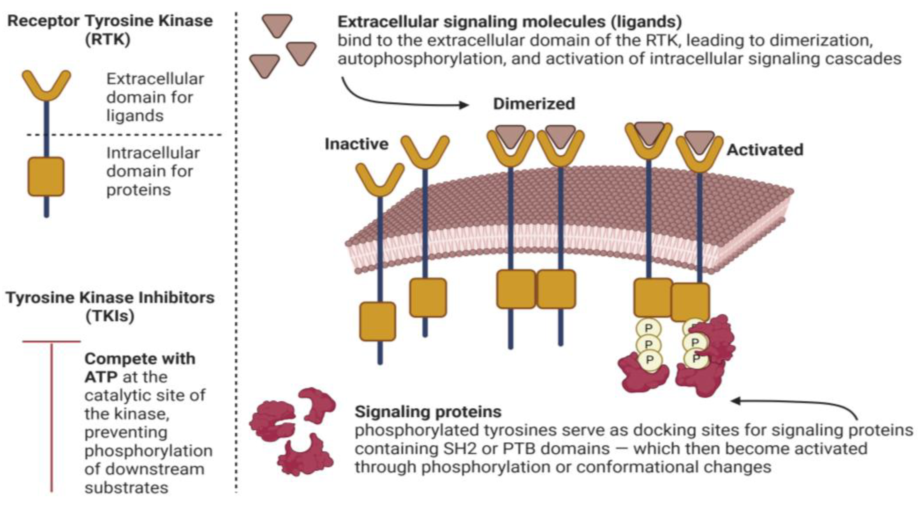
Figure 1. Activation/Inhibition of Receptor Tyrosine Kinases (RTKs) (created in BioRender, https://BioRender.com/veaygpg on 30 October 2025).
Hemato 06 00040 g001
Figure 2. Chemical structure of IMT (a) and IMTM (b), created by https://www.chemspider.com/, accessed on 24 August 2025.
Hemato 06 00040 g002
Figure 3. Applications of IMT (created in BioRender, https://BioRender.com/2y98kit on 29 October 2025).
Hemato 06 00040 g003
Figure 4. Therapeutic combinations with imatinib (created in BioRender, https://BioRender.com/oscfeyk on 30 October 2025).
Hemato 06 00040 g004
Figure 5. Side effects of IMT (created in BioRender, https://BioRender.com/yv0d1s2 on 31 October 2025).
Hemato 06 00040 g005
Figure 6. Advanced drug delivery systems of IMT (created in BioRender, https://BioRender.com/79zasag on 29 October 2025).
Hemato 06 00040 g006
Table 1. Table of disease models, IMT dosages, clinical outcomes, and evidence levels.
DiseaseModelIMT DoseEvidence LevelOutcome
Alzheimer’s diseaseMouse model20 mg/kg/day i.p.In vivoReduced amyloid-ß and improved
cognitive performance [111]
Alzheimer’s disease(1) Mouse model
(2) Neuronal cells
30 mg/kg/day i.p.(1) In vivo
(2) In vitro
Reduced neuronal apoptosis [51]
Acute
ischaemic stroke
Rat model100 mg/kgIn vivo study1. Reduced BBB permeability;
2. Reduced cerebral edema;
3. Increased expression of the tight-junction protein ZO-1;
4. Reduced activation of NF-kB [112].
Acute
ischaemic stroke
60 patients
18–85 years old
400, 600, 800 mgPhase II clinical studyRedtored BBB, reduced
inflammation and edema [55]
Asthma62 patients
aged 18–65
200 mg/day for 2 weeks, then 400 mg/dayA randomized double-blind placebo- controlled trialDecreased airway
Hyperresponsiveness [69]
Crohn’s diseaseCase report600 mg/dayCase reportLong-lasting remission [113]
Crohn’s diseaseHuman case series300–400 mgCase seriesRemission in 4/6 patients [114]
Colon cancerSW480 cell linesn/aCell cultureInhibited proliferation
of colon cancer cells [115]
Early weight gain/insulin resistance(1) Mouse model;
(2) Human endothelial cells
(1) In vivo
(2) In vitro
Improved insulin-mediated vasodilatation and reduced free fatty acid transport [102]
Fibrodysplasia ossificans
progressiva
6 children340 mg/m2/
day
In vivo studyDecrease intensity and
frequency of flare-ups [61]
cGVHDHuman
clinical pilot study: 19 patients
Starting dose 100 mg/
day
Case series7 complete and 8 partial remissions among 19 patients
18-month overall survival~84% [116]
Steroid-
Refractory cGVHD
Open-label Pilot II trial in children and adults100–400 mg daily in adults
65–260 mg/m2 in children
Phase II single-arm trialPartial response in ~36% and overall clinical benefit in ~86% of patients [117]
Refractory sclerotic
cGVHD
Retrospective study with 14 patients100–400 mg dailyRetrospective study50% response rate; Steroid dose reduced;
Some patients discontinued treatment [118]
Acute and chronic GVHDHuman patients100–400 mg/
day/orally
Clinical studyImproved skin/mucosal lesions [63]
Sclerodermatous cGVHDMouse model150 mg/kg/
daily by oral
In vivoIMT decreased the proliferation of total T cells and of regulatory T cells [119]
Diabetes
mellitus
Rat model10 mg/kg
20 mg/kg
In vivoEnhanced serum insulin levels
Reduced serum glucagon levels [120]
Diabetes
Type 1
Randomized, double-blind, placebo-controlled, phase II trial
In adults (aged 18–45)
400 mg/day (4 × 100 mg) for 26 weeksRandomized, double-blind, placebo-controlled, phase II trialEnhanced ß-cell function and
ß-cell glucose responsiveness [121]
Diabetic
nephropathy
ApoE-knockout mouse model10 mg/kg/dayIn vivoReduced diabetic nephropathy in apolipoprotein E-knockout mice [122]
Radiation-
induced skin
fibrosis
Mouse model0.5 mg/gPreclinical
(in vivo)
Reduced radiation-induced skin fibrosis and collagen deposition [75]
Gouty ArthritisMouse model30/100 mg/kg i.p.In vivo studyReduced joint swelling and inflammation in MSU-induced gout [95]
Gaucher
disease
Mouse model
Human fibroblasts from patients
n/aIn vivo study
In vitro study
IMT inhibition of c-Abl reduces RIPK3 activation, suggesting therapeutic
potential [108]
Chronic
glomerulo-
sclerosis
Rat model10 mg/kg/dayIn vivoIMT slowed the progression of
Glomerulosclerosis [105]
Chronic
allograft
nephopathy
Rat model10 mg/kg/dayIn vivoIMT prevented the progression of
chronic allograft [123]
Hypertension-
induced end-
organ damage
Rat model30 mg/kg/dayIn vivo studyIMT reduced cardiac dysfunction and protected renal microvasculature [124]
Spontaneous Intracerebral HemorrhageRat model60 mg/kg i.p.In vivo study1. IMT mitigates cerebral vasospasm;
2. IMT helps maintain BBB integrity [56].
Spontaneous Intracerebral HemorrhageMouse model30,60,120 mg/kg i.p.In vivo study1. Reduced brain edema;
2. Better neurobehavioral outcomes;
3. Maintained BBB integrity [57].
Subarachnoid hemorrhageRat model40 mg/kg
120 mg/kg
In vivo studyMaintained BBB integrity and
enhanced neurological function [125]
LeishmaniasisIn vitro
parasite model
Various
concentrations tested
In vitro comparative studyDose-dependent reduction in the viability of Leishmania major, though amphotericin B showed stronger activity [85]
MalariaClinical trial in humans 400 mg/day for 3 daysPhase II clinical trialAccelerated parasite clearance
and fever resolution
with no increase in adverse events [126]
Chloroquine-
resistant
malaria
Open-label,
prospective case–control study in male patients
400 mg for 3 daysClinical-prospective, case–control studyFaster fever reduction;
More rapid parasite clearance [86]
MelanomaRetrospective control study of 78 patients 400 mg/dayRetrospective studyMedian overall survival (OS) 13.1 months
Progression-free survival (PFS) 4.2 months [97]
Metastatic
melanoma
Phase II study involving 43 patients with c-KIT mutations400 mg/dayPhase II open-label, single-arm studyOverall response rate of 23.3%; [127]
Melanoma with KIT
alternations
Retrospective study of 38 KIT-altered melanoma patients400 mg/dayClinical-
multicenter
retrospective study
PFS and OS were longer in patients
with exon 11/13 mutations
compared to exon 17 mutations [128]
Metastatic
melanoma
A phase II study of patients with metastatic melanoma with KIT mutation400 mg twice dailyA single-group, open-label,
phase 2 trial
Clinical responses observed in
subset of KIT-altered metastatic
melanoma patients [96]
MorpheaHuman case report200 mg/dayn/aClinical improvement with
reduced skin thickening [129]
Human therapy-resistant
generalized deep morphea
Human case report400 mg/dayCase report
2015
Marked clinical improvement
No new lesions during 11-month follow-up [130]
Multiple
sclerosis
Mouse model and
U-87 MG, C6 and WEHI-164 cell lines
60 mg/kg/dayPreclinical
(mouse model and in vitro cell lines)
In vivo reduced disease severity and delayed symptom onset
In vitro: decreased cell proliferation,
lower pro-inflammatory cytokines [67]
Multiple
sclerosis
Rat modelMourine dosingPreclinicalReduced blood–brain barrier integrity and reduced neuroinflammation [131]
Niemann–Pick Type CMouse model5 mg/kg in NaClIn vitro study1. Counteract weight loss;
2. Improve neurological function;
3. Enhance Purkinje cells’ survival;
4. Extend lifespan [60].
PsoriasisImiquimod-induced psoriasis-like skin in miceMourine dosingPreclinical
(mouse model)
Topical IMTM ameliorated psoriasis-
like skin lesions by
inhibiting angiogenesis [73]
PsoriasisCase study400 mg/dailyn/aImproved skin lesions and
complete hematologic remission [132]
Pulmonary arterial
hypertension
17 patients200 mg starting doseA phase III studyLower pulmonary pressure,
higher cardiac output,
reduced vascular resistance [133]
Pulmonary arterial
hypertension
15 patients400 mgObservational study
In vivo
Improvement in hemodynamics,
quality of life and echocardiographic parameters of right ventricular function [68]
Renal fibrosisRat modelDays 1 and 2: 50 mg/kg; days 3 and 4: 100 mg/kg; days 5–7: 150 mg/kg)In vivoIMT reduced renal fibrogenesis and blocked TGF-ß [134]
Spinal cord injuryRat study100 mg/kg i.p.In vivoImproved functional recovery and reduced secondary spinal cord damage [58]
Systemic sclerosis-assosiated
interstitial lung disease
Phase I/IIa one-year, open-label pilot trialUp to 600 mg/dayClinical trial
Phase I/IIa single-arm
12/20 patients completed the treatment;
7 withdrew due to adverse effects
1 lost to follow-up [135]
Systemic
sclerosis
Mouse models of SSc150 mg/kg/
day
Preclinical
(in vivo)
Prevented fibrosis and induced
regression of established fibrosis [74]
Human-
refractory diffuse systemic sclerosis
Case report400 mg/dayCase reportModest improvement in skin scores;
Partial clinical responses [136]
Systemic
mastocytosis
Clinical study on 20 patients 400 mg/dayPhase II clinical study1 patient with complete remission
6 patients with symptomatic improvement [137]
Systemic
mastocytosis
Phase II study400 mg/dayPhase II clinical studyIMT was effective in treatment, including those who had the D816V mutation [138]
Systemic
mastocytosis
Adult patients100–400 mg/dayn/aPartial/complete improvement. Response depends on KIT mutation status [139]
Systemic
mastocytosis
(indolent and advanced forms)
Adult patientsDose depending on disease severity and KIT mutation Case seriesHematologic and symptomatic
improvement observed mainly in patients without KIT D816V mutation [93]
Systemic
mastocytosis
Adult patients without an exon 17 KIT mutation300–400 mg/dayPhase IV clinical studyWell-tolerated safety profile;
Partial/complete hematologic and symptomatic improvement in patients without KIT D816V mutation [140]
Systemic lupus erythematosus with lupus nephritisMouse model10 mg/kg
50 mg/kg
In vivo studyIMT (50 mg/kg) prevented glomerular
cell proliferation, crescent formation,
and reduced mesengial matrix [141]
Ulcerative
colitis
Rat model10 mg/kg/day
20 mg/kg/day (oral pretreatment for 1 week)
Preclinical animal studyPretreatment with IMT significantly reduced macroscopic and histologic damage, decreased oxidative and inflammatory markers and suppressed COX-2 signaling in the colon [72]
Table 2. Clinically significant adverse effects of IMT therapy.
System Organ Class DisordersAdverse Effects
Cardiovascular
  • Fluid retention, Hypereosinophilic cardiac toxicity
Dermatologic
  • Alopecia, Erythema multiforme, Pruritus, Rash, Stevens–Johnson syndrome
Gastrointestinal
  • Abdominal pain, Anorexia, Constipation, Diarrhea, Distention, Dyspepsia, Flatulence, Nausea, Vomiting
General
  • Asthenia, Fatigue, Night sweats, Pain, Peripheral edema, Periorbital edema, Pyrexia, Rigors, Weight gain
Hematologic
  • Anemia, Hemoglobin decrease, Hemorrhage, Leukopenia, Neutropenia, Thrombocytopenia
Hepatic
  • ASL and ALT elevation
Infectious
  • Pneumonia, Upper respiratory tract infections
Metabolic/nutritional
  • Hypoalbuminemia, Hypokalemia, Hypoproteinemia
Musculoskeletal
  • Arthralgia, Myalgia, Muscle cramps
Neurological
  • Dizziness, Headache
Ocular
  • Blurred vision, Ischemic maculopathy, Optic neuritis, Optic nerve edema, Retinal hemorrhage
Psychiatric
  • Depression, Insomnia
Renal and urinary
  • Serum creatinine increase
Respiratory
  • Cough, Dyspnea, Pharyngolaryngeal pain
Disclaimer/Publisher’s Note: The statements, opinions and data contained in all publications are solely those of the individual author(s) and contributor(s) and not of MDPI and/or the editor(s). MDPI and/or the editor(s) disclaim responsibility for any injury to people or property resulting from any ideas, methods, instructions or products referred to in the content.