Article

Immuno-Hematological Complications of Transfusion in Thalassemia Patients: First Report in the Marrakech Region (Morocco)

1
High Institute of Nursing Professions and Health Techniques, Marrakech 40000, Morocco
2
Laboratory of Anthropogenetics, Biotechnologies and Health, Faculty of Sciences of El Jadida, Chouaïb Doukkali University, El Jadida 24000, Morocco
3
Laboratory of Microbial Biotechnology, Agrosciences, and Environment (BioMAgE), Labeled Research Unit-CNRST N°4, Faculty of Sciences Semlalia, Cadi Ayyad University, Marrakech 40000, Morocco
4
Physical Chemistry Laboratory of Materials and Environment, Faculty of Sciences Semlalia, Cadi Ayyad University, Marrakech 40000, Morocco
5
Moroccan Blood Agency and Its Derivatives of the Marrakech-Safi Region, Marrakech 40000, Morocco
*
Author to whom correspondence should be addressed.
Hemato 2025, 6(4), 35; https://doi.org/10.3390/hemato6040035
Submission received: 21 August 2025 / Revised: 21 September 2025 / Accepted: 25 September 2025 / Published: 30 September 2025

Abstract

Background/Objectives: Erythrocyte alloimmunization is a critical complication impacting the efficacy of transfusion therapy in patients with thalassemia. This study seeks to evaluate the prevalence, characterization, and determinants of erythrocyte alloimmunization in multi-transfused thalassemia patients in south of Morocco. Methods: A retrospective study was conducted at the Moroccan Blood and Blood Derivatives Agency in Marrakech (Morocco) over 2 years, from June 2022 to June 2024, including 89 patients with beta-thalassemia receiving regular transfusions. The clinical, demographic, and transfusion characteristics of patients who developed alloimmunization were compared with those of non-alloimmunized patients. Results: Analysis of 89 β-thalassemia patients in the Marrakech region, mostly young and suffering from major form (67%), shows a significant male predominance (p = 0.004) and a high frequency of blood group O+ (49.4%). Alloimmunization mainly affects major forms and males and is associated with frequent annual transfusions (over 12 per year), usually resulting in the use of 24 to 60 packed red blood cell units annually. Alloimmunized patients mostly present anti-K and anti-E antibodies, indicating the involvement of the Kell and Rh systems. The direct Coombs test was more often positive in these patients (21.4% vs. 7.9%, p < 0.01). Conclusions: The high prevalence of alloimmunization in thalassemia patients in the Marrakech region highlights the need for a rigorous and personalized transfusion strategy, including molecular genotyping and alternative therapies.

1. Introduction

Thalassaemia is one of the most common genetic diseases worldwide. It is an autosomal recessive disorder characterized by decreased hemoglobin synthesis due to mutations in the genes encoding the alpha, beta, and delta chains of globin. These mutations result in ineffective erythropoiesis [1]. As a result, there is an overall deficit of hemoglobin tetramers in the red blood cells (RBC), and the mean corpuscular volume (MCV) and mean corpuscular hemoglobin (MCH) are reduced [2].
Thalassemias are classified into two main forms: alpha-thalassemia and beta-thalassemia, which are the most common, as well as rarer forms such as delta-beta-thalassemia and gamma-delta-beta-thalassemia [3].
Clinical manifestations of the disease typically occur during the first year of life, during which fetal hemoglobin (HbF) production declines. Pallor is often the first observable symptom, accompanied by splenomegaly of varying severity, fever, and growth restriction. Affected children exhibit abnormal physical development and growth. Standard treatment for beta-thalassemia major is regular blood transfusions initiated early in life, leading to improvement in anemia and reduction in skeletal abnormalities caused by increased erythropoiesis [4,5]. Although this transfusion is essential for these patients, it can lead to complications such as iron overload and alloimmunization of red blood cells and platelets [6,7]. Repeated transfusions activate the patient’s immune response, leading to the formation of antibodies directed against red blood cells (alloantibodies and/or autoantibodies). Although autoantibodies are less common, they can cause clinical hemolysis and complicate blood compatibility. Alloimmunization against erythrocyte antigens increases transfusion requirements and makes treatment more complex. Some alloantibodies are hemolytic, which can trigger hemolytic transfusion reactions, thus reducing the safety of future transfusions. Others, however, are clinically insignificant [8,9].
Alloimmunization rates among multi-transfused thalassemia patients vary worldwide, ranging from 2.9% to 42.5%. The most frequently involved antibodies target antigens of the Rhesus and Kell blood group systems. Within the Rh system, alloimmunization is particularly high, accounting for 52.4% of reported instances, with the E antigen being the most commonly implicated (22%), followed by D (6.9%), C (5.5%), c (5.5%), and e (1.3%). Additional alloantibodies have been identified against antigens of the Kell, Kidd, and MNS systems [10]. However, available data on the frequency of anti-red blood cell alloimmunization in Moroccan transfusion-dependent thalassemia patients are limited.
Hemoglobinopathies, including thalassemia, contribute to 3.4% of global mortality in children under the age of five, with a proportion of 6.4% in Africa [11]. In Morocco, thalassemia represents a real public health problem due to its frequency, social cost, complications, and difficulties related to its management. The average frequency of thalassemia trait is estimated at 6.5%, which could correspond to approximately 30,000 cases of major forms of thalassemia in the country [12].
This situation highlights the need to develop coherent national programs to prevent and treat hemoglobinopathies, particularly thalassemias.
This study aims to determine the frequency, types, and factors influencing erythrocyte alloimmunization in multi-transfused thalassemia patients in southern Morocco, as part of a broader approach aimed at contributing to international efforts, particularly in Morocco, to improve blood compatibility in β-thalassemia patients exposed to an increased risk of alloimmunization due to repeated transfusions.

2. Materials and Methods

2.1. Patients

This study was conducted at the Moroccan Blood and Blood Derivatives Agency in Marrakech over 2 years, from June 2022 to June 2024, including 89 patients with beta-thalassemia. The inclusion criteria were patients with thalassemia who received blood transfusions, managed by the Marrakech Blood and Blood Derivatives Agency, and had complete and usable medical records. All patients were diagnosed with β-thalassemia following clinical and laboratory tests. Confirmation of the diagnosis was then based on standard hemoglobin electrophoresis. All patients received ABO and Rh(D)-compatible blood, subjected to cross-matching and leukocyte depletion.
Data were collected from patient transfusion records. The variables analyzed included:
-
Sociodemographic data: age, sex.
-
Clinical data: types of thalassemia.
-
Transfusion data: frequency of transfusions per year, number of bags of packed red blood cells per year, average interval between transfusions per week.
-
Immunohematological data: ABO/Rh group, RAI status, TCD, and alloantibody types.

2.2. Methods

Hemoglobin electrophoresis was performed in an alkaline medium using two complementary techniques: capillary electrophoresis (Capillarys® system) and agarose gel electrophoresis (Hydrasys® system). Red cell antibody screening (irregular agglutinins) is routinely performed for any request for labile blood products (LBP). Before the first transfusion, antibody screening is carried out in saline at 22 °C to detect naturally occurring antibodies and at 37 °C to detect clinically significant alloantibodies. If the screening is positive, antibody identification is performed. Both screening and identification are conducted using the indirect antiglobulin test (IAT) by gel hemagglutination technique on microcolumn gel cards (BIO-RAD, Cressier, Switzerland) at low temperature and at 37 °C, with an ID-Incubator (BIO-RAD, Cressier, Switzerland) for heat-reactive alloantibodies. Positive plasma samples were identified using a commercial extended 11-cell identification panel (ID-DiaPanel, BIO-RAD, Cressier, Switzerland) in combination with LISS/Coombs gel cards (BIO-RAD, Cressier, Switzerland). The search for autoantibodies was performed using a direct Coombs test in gel cards containing polyspecific (anti-IgG/C3d) and specific antihuman globulin (BIO-RAD, Cressier, Switzerland).

2.3. Statistical Analysis

Differences between subgroups were examined for the following variables: sex (categorical, binary), age (ordinal), ABO/Rh blood group (categorical, multinomial), and thalassemia types (categorical, multinomial). The transfusion parameters studied, proportion of patients transfused (categorical), number of annual transfusions (continuous variable), number of packed red blood cells units per year (continuous variable), and mean transfusion interval per week (continuous variable) were compared between the different thalassemia subgroups. In addition, patients with and without alloantibodies were compared to identify significant differences in clinical and demographic characteristics, presence of autoantibodies (categorical, binary), number of transfusions per year (categorical, binary), and number of packed red blood cells per year (continuous variable). Categorical variables were expressed as numbers and percentages, while continuous variables were summarized as ranges (minimum–maximum). Statistical analysis was performed using IBM SPSS Statistics version 23 (IBM Corp., Armonk, NY, USA). For categorical variables with multiple categories, a Chi-square goodness-of-fit test was applied to determine whether the observed distribution of subgroups differed significantly from an expected uniform distribution. To compare alloimmunized and non-alloimmunized patients, categorical variables were compared using the Chi-square test (or Fisher’s exact test when the expected cell number was less than 5). For comparisons involving two independent groups whose continuous variables did not follow a normal distribution, the Mann–Whitney U test was applied, while for more than two independent groups, the Kruskal–Wallis test was used. A p-value < 0.05 was considered statistically significant.

3. Results

3.1. Demographic and Clinical Characteristics of Patients

The sample included 89 patients with various forms of thalassemia or those associated with sickle cell disease. Characteristics analyzed included sex, age, ABO/Rh blood group, and thalassemia type. p values indicate the statistical significance of the differences observed between subgroups: male vs. female, different age groups, ABO/Rh blood types and different types of thalassemia (Table 1).
Analysis of the demographic and clinical characteristics of 89 patients revealed a slight male predominance (51.7%), which was statistically significant (p = 0.004).
The study population was predominantly young, with 52.8% children and adolescents (0–18 years) and 41.6% young adults (19–30 years), while only 5.6% were over 30 years old (p = 0.001). Concerning the immuno-hematological level, the distribution of ABO/Rh groups is statistically significant (p = 0.001) and the O blood group is the most frequent (49.4%), followed by A+ (21.3%), while negative groups (Rh-) are relatively rare (approximately 12% in total). Regarding the type of thalassemia, the major form is largely predominant (67%), followed by the intermediate form (8.3%), the minor form (2.8%) and the thalassemia-sickle cell association (3.7%), with a highly significant difference (p = 0.001), reflecting the clinical severity of the cases followed.

3.2. Characteristics of Transfusion in the Study Population

The results in Table 2 show a wide variability in transfusion requirements depending on the type of thalassemia. Patients with β-thalassemia major represent the majority of transfused cases (67%) and require intensive treatment, with 12 to 26 transfusions per year, or up to 60 blood bags and intervals of 0.5 to 4 weeks between sessions. In comparison, patients with β-thalassemia intermedia (8.3%) and the minor form (2.8%) receive transfusions much more spaced out and in small quantities, often occasionally. Cases associated with sickle cell disease (3.7%) have moderate needs, with 6 to 12 transfusions per year. These differences across thalassemia subgroups were statistically significant for all transfusion parameters, including the percentage of transfused cases (p = 0.001), the number of transfusions per year (p = 0.031), the number of packed red blood cell units per year (p = 0.001), and the average interval between transfusions (p = 0.047).

3.3. Comparison Between Alloimmunized and Non-Alloimmunized Thalassemia Patients

Table 3 presents a detailed comparison between alloimmunized (n = 42; 47.2%) and non-alloimmunized (n = 47; 52.8%) thalassemia patients, based on several clinical and transfusion parameters. A significant difference was observed by gender (p < 0.01); alloimmunized patients were predominantly male (26.9%), while non-alloimmunized patients were more often female (28.1%). Regarding age, no statistically significant difference was noted between the groups (p = 0.50): the majority of patients in both groups were under 30 years of age. The number of transfusions per year and the number of bags of packed red blood cells received were significantly higher in alloimmunized patients. Indeed, 49.9% of them received ≥12 transfusions per year, compared to 0% in the non-alloimmunized group (p < 0.01). Similarly, alloimmunized patients received between 24 and 60 blood bags per year, compared to 12 to 24 in the non-alloimmunized group (p < 0.01). The distribution of thalassemia types also differed significantly between the two groups (p < 0.01): β-thalassemia major was predominant in the alloimmunized group (46%), while intermediate and minor forms, and those associated with sickle cell disease were more common in the non-alloimmunized group. Finally, the direct antiglobulin test (DAT) was more often positive in alloimmunized patients (21.4%) than in non-alloimmunized patients (7.9%) (p ˂ 0.01).

3.4. Alloantibodies in Alloimmunized Thalassemic Patients

Figure 1 represents the different alloantibodies identified in alloimmunized patients. A predominance of antibodies directed against the Kell and Rh system antigens was observed. The most frequently found antibody was anti-K (34%), followed by anti-E (19%). Associations of alloantibodies were also common, including anti-E + anti-K (14%), anti-D + anti-C (13%), and anti-E + anti-D (10%). Other antibodies were identified in smaller proportions, such as anti-JKa (4%), anti-JKb (2%), anti-K + anti-Fya (2%), as well as anti-Fya + anti-Fyb + anti-S (2%). These results confirm the major involvement of the Kell, Rh, Kidd and Duffy systems in alloimmune reactions in multi-transfused thalassemia patients.

4. Discussion

The data collected during our study reveal a slight dominance of male patients with thalassemia (51.7%). These findings are consistent with observations reported in previous studies, which also indicate a male predominance. For example, Bejaoui & Guirat [13], highlighted a significantly higher proportion of male patients (65.66%) compared to female patients (34.33%). Similar findings were reported in studies conducted in Banu and Pakistan, where the male predominance was 56.95% and 59.49%, respectively [12,14]. In North Africa, several studies corroborate this trend, notably those conducted at the Ibn Sina University Hospital in Rabat (Morocco) and the Farhat-Hached University Hospital in Sousse (Tunisia), where the observed sex ratios were 1.4 and 1.36, respectively [15,16]. Several hypotheses have been put forward to explain this discordance. These include increased susceptibility of men to iron overload, the possible interaction between thalassemia genes and genes located on the sex chromosomes, and the potential impact of sex hormones on disease severity or patient survival [17]. Finally, although the autosomal recessive mode of inheritance of thalassemia suggests an equal distribution between the sexes, we emphasize that differences in access to care or the size of the sample studied could influence this difference. Moreover, the study population was composed of children or adolescents 52.8% followed by young adults 41.6%, while people over 30 years old represented only 5.6%. These results are comparable with the study of Hamani & Oribi [18] who found a predominance of the age group between [5 and 11 years]. On the other hand, Zahir et al. [19] found that the age group between [18 and 60 years] was predominant with a percentage of 48%. This age distribution reflects the chronic and early nature of thalassemia major, the progress made but also the current limitations in terms of long-term survival, as well as the organization of medical follow-up focused on young patients.
In Morocco, the natural course of thalassemia major is characterized by early onset of anemia and regular transfusion requirements [20,21]. Without appropriate management, patients often develop complications such as iron overload, leading to cardiac, hepatic, and endocrine dysfunctions [15]. A study conducted in Kenitra (Morocco) reported that all patients studied with Beta thalassemia major were transfused, but only 14% received iron chelation therapy, highlighting a gap in optimal management [22]. Regarding life expectancy, data in Morocco are limited. However, it is known that without regular transfusions and chelation therapy, survival is significantly reduced. Conversely, with appropriate treatment, patients can achieve substantial improvements in survival and quality of life.
The distribution of blood groups among thalassemia patients in our study showed that O+ was the most frequent group (49.44%), followed by A+ (21.35%) and B+ (12.36%). These findings are consistent with previous studies in thalassemia populations, which also report a predominance of O+ and A+ blood groups [23,24]. When compared to the general Moroccan population, where O+ and A+ groups are similarly the most common [25,26], our results indicate that the blood group distribution in thalassemia patients largely reflects that of the broader population. However, this distribution can vary significantly depending on the region and population, thus influencing the results of epidemiological studies. Although blood group does not directly affect transfusion frequency, it can influence transfusion management due to the risk of alloimmunization. The presence of antibodies against minor antigens can complicate the search for compatible blood, making transfusion management more complex.
Regarding the study of the different types of thalassemia, the results of our study highlight a clear predominance of thalassemia major with a frequency of 67%, which is consistent with the data reported by Ben Salah et al. in their study in Tunisia [27], which also indicates a dominance of this form with a rate of 63%. Globally, approximately 60,000 newborns are born with β-thalassemia major per year, with the majority living in developing countries and constituting about 1.5% of the total population [11]. This concordance underlines the clinical and epidemiological importance of this severe form in at-risk populations.
Evaluation of the therapeutic management of thalassemia patients showed a significant difference in the number of transfusions, the number of packed red blood cell units, and the average interval between transfusions, depending on the type of beta-thalassemia. Patients with thalassemia major benefited most from transfusions, phenotyped red blood cell concentrates, and compatible with very short transfusion intervals of 0.5 to 4 weeks. These patients followed a regular transfusion schedule, compared to other types who received occasional transfusions. Previous studies conducted in Morocco reported that patients with thalassemia major received regular transfusions with different average intervals; 4 to 6 weeks and 2 to 6 weeks, respectively [21,28]. These data are confirmed by Cao and Galanello [29] in their guidelines on transfusion management of thalassemia. This difference can be explained by the clinical complication of patients.
In our study, the first on erythrocyte alloimmunization in patients with thalassemia in Marrakech (Morocco), the overall prevalence of erythrocyte alloimmunization in regularly transfused patients with thalassemia was 47.2%, a rate significantly higher than those reported in the international literature where estimates generally vary between 2.5% and 37% [30,31,32,33]. This high prevalence could be explained by the high frequency of transfusions, the low degree of broad compatibility practiced and the predominance of severe clinical forms.
Analysis of associated factors shows that gender is significantly linked to alloimmunization (p < 0.01), with a higher proportion of alloimmunized men. Although some studies report a more pronounced predisposition in women, attributed to antigenic exposure linked to pregnancy [32], others, such as Wilson et al. [34], did not find a significant difference, which is partially consistent with our observation of disparities but without a universal trend. Age did not show a significant association with alloimmunization (p = 0.50), which is consistent with the conclusions of several studies that do not identify age as a major independent factor [32,34]. Regarding thalassemia type, β-thalassemia major was the predominant form in alloimmunized patients (46% vs. 36%, p < 0.01). This trend is well documented, as thalassemia major requires regular lifelong transfusions, which increases exposure to erythrocyte antigens and therefore the risk of alloimmunization [32,35]. However, a systematic review of the literature [10], including 41 cohort studies with a total of 9256 patients, has reported that alloimmunization occurs more frequently in thalassemia intermedia, as these patients are transfused less often and are therefore at higher risk when extended antigen matching is not performed. In our cohort, however, no alloimmunization was observed among thalassemia intermedia patients. Alloantibodies were detected exclusively in regularly transfused thalassemia major patients. This pattern can be attributed to the transfusion practices at our center, which primarily rely on ABO and RhD compatibility, with selective extended typing for Rh (C, c, E, e) and Kell antigens. Comprehensive red blood cell phenotyping, including Duffy, Kidd, and MNS systems, is not routinely performed due to technical and financial limitations. This limited antigen-matching strategy likely contributes to the persistence of alloimmunization in our transfused population.
Transfusion frequency is a key determinant: in our population, 49.9% of alloimmunized patients received ≥12 transfusions/year (p < 0.01), and the number of bags transfused per year was significantly higher (24–60 bags vs. 12–24, p < 0.01). These results confirm that the cumulative number of units transfused is one of the main risk factors [32,36]. Finally, for the direct antiglobulin test (DAT), we observed a lower rate of positive DAT in alloimmunized patients (7.9% vs. 21.4%, p < 0.01), which contrasts with some studies that have found a positive correlation between alloimmunization and the presence of autoantibodies [9,37]. This difference could reflect immunological variations specific to our population or be related to clinical factors such as splenectomy or disease duration. The presence of both is an important indicator of immunohematological complications in chronically transfused patients such as beta-thalassemia patients. It requires careful monitoring to adapt the transfusion strategy (extended phenotyping, early antibody detection, compatible transfusion).
In our cohort, alloimmunization was predominantly directed against the Kell and Rh systems, with anti-K being the most frequent antibody (34%), followed by anti-E (19%). This pattern is consistent with several studies conducted in polytransfused thalassemia patients, which also report a predominance of anti-K antibodies followed by anti-E [33,38,39]. These findings suggest that the Kell system is often highly immunogenic in such populations. However, other studies have reported different patterns, with anti-E antibodies being more frequent than anti-K, reflecting population-specific and regional variations in alloimmunization [40,41].
Alloantibody associations, including anti-E + anti-K (14%), anti-D + anti-C (13%), and anti-E + anti-D (10%), demonstrate multiple immunizations. Associations were also observed in studies in Casablanca and Rabat (Morocco) [33,42], highlighting the high frequency of combined alloimmunizations in the Rh and Kell systems in polytransfused patients with β-thalassemia. Antibodies directed against the Kidd and Duffy systems (anti-Jka: 4%, anti-Jkb: 2%, anti-Fya: 2%) were much rarer in our series, which is consistent with the data of El Kababi et al. and Yadav et al. [32,33] who generally report frequencies below 6% for these specificities in populations where Rh and Kell antigens are predominant.
This study has several limitations. First, its retrospective design and the relatively small number of patients may limit the generalizability of the findings. Second, transfusion practices were not fully standardized across all cases. Third, molecular typing and systematic evaluation of all minor blood group antigens could not be performed due to resource constraints, which may have underestimated the true prevalence of alloimmunization. These limitations highlight the need for optimizing transfusion strategies to reduce alloimmunization. In our center, Rh (C, c, E, e) and Kell matching is routinely performed for all patients. Ideally, full extended red blood cell phenotyping—including Duffy (Fya, Fyb), Kidd (Jka, Jkb), and MNS (M, N, S) antigens—should be applied to further minimize alloimmunization. Although implementing comprehensive antigen matching requires additional technical and financial resources, it may prove cost-effective in the long term by preventing alloimmunization-related complications, facilitating transfusion management, and reducing delays in providing compatible blood. Furthermore, the adoption of molecular typing platforms could offer a sustainable solution for improving transfusion safety and personalized care in our setting.

5. Conclusions

The prevalence of red blood cell alloimmunization among thalassemia patients in the Marrakech region was found to be significantly high, highlighting the importance of strengthening preventive and control measures for both alloimmunization and autoimmunization.
To effectively reduce the risk of alloimmunization and autoimmunization in thalassemic patients at the Moroccan blood agency and its derivatives of the Marrakech-Safi region, it is essential to adopt a rigorous and personalized transfusion strategy. This includes, first and foremost, the implementation of regular immunohematological monitoring to enable early detection of antibody formation. Patients who develop autoantibodies require specialized care, including the strict selection of compatible red blood cell concentrates.
Additionally, it is recommended to enhance the training and awareness of medical and paramedical teams regarding the immunological risks associated with repeated transfusions, to ensure better coordination between immunohematology laboratories and clinical departments.
Furthermore, the establishment of local and regional blood banks with detailed antigen profiles can improve the availability of compatible units, particularly for patients with multiple or complex alloantibodies.
Patient phenotyping prior to the first transfusion should be considered, including for red cell concentrates, in order to ensure better matching and minimize the risk of alloantibody development. Moreover, the integration of advanced technologies, such as molecular red blood cell genotyping, together with alternative strategies to reduce transfusion burden including pharmacological approaches and, when feasible, hematopoietic stem cell transplantation may further improve long-term outcomes.

Author Contributions

All authors confirm that they meet the current authorship criteria of the International Committee of Medical Journal Editors (ICMJE). Study framework development, H.A.H. and T.E.D.; Data analysis, W.S. and N.E.; Manuscript writing, H.A.H. and M.O.; Overall supervision and coordination of research activities, M.S.E. and S.F. All authors have read and agreed to the published version of the manuscript.

Funding

This research received no external funding.

Institutional Review Board Statement

The study was conducted in accordance with the Declaration of Helsinki, and approved by Regional Directorate of the Ministry of Health and Social Protection for Marrakech-Safi region (protocol code 975 and date of approval 13 February 2025).

Informed Consent Statement

Consent was obtained from each subject or their legal guardians prior to study enrollment and from the local committee. The authors declare that this report does not contain any personal information that could lead to the identification of patients.

Data Availability Statement

The data that support the findings of this study are available.

Conflicts of Interest

The authors declare no conflicts of interest.

References

  1. Kattamis, A.; Kwiatkowski, J.L.; Aydinok, Y. Thalassaemia. Lancet 2022, 18, 2310–2324. [Google Scholar] [CrossRef] [PubMed]
  2. Elghetany, M.T.; Banki, K. Erythrocytic disorders. In Henry’s Clinical Diagnosis and Management by Laboratory Methods, 21st ed.; McPherson, R.A., Pincus, M.R., Eds.; Elsevier Saunders: Philadelphia, PA, USA, 2007; pp. 528–529. [Google Scholar]
  3. Baird, D.C.; Batten, S.H.; Sparks, S.K. Alpha- and Beta-thalassemia: Rapid Evidence Review. Am. Fam. Physician 2022, 105, 272–280. [Google Scholar] [PubMed]
  4. Borgna-Pignatti, C.; Galanello, R. Thalassemias and related disorders: Quantitative disorders of hemoglobin synthesis. In Wintrobe’s Clinical Hematology, 11th ed.; Greer, J.P., Rodgers, G.M., Paraskevas, F., Foerster, J., Lukens, J.N., Glader, B., Eds.; Lippincott Williams and Wilkins: Philadelphia, PA, USA, 2004; pp. 1332–1335. [Google Scholar]
  5. Rebulla, P. Blood transfusion in beta thalassaemia major. Transfus. Med. 1995, 5, 247–258. [Google Scholar] [CrossRef] [PubMed]
  6. Prati, D. Benefits and complications of regular blood transfusion in patients with beta-thalassemia major. Vox Sang. 2000, 79, 129–137. [Google Scholar] [CrossRef]
  7. Bhatti, F.A.; Salamat, N.; Nadeem, A.; Shabbir, N. Red cell immunization in beta thalassemia major. J. Coll. Physicians Surg. Pak. 2004, 14, 657–660. [Google Scholar]
  8. Salama, M.A.; Sadek, N.A.; Hassab, H.M.; Abadeer, A.F.; Mikhael, I.L. Erythrocyte autoantibodies and expression of CD59 on the surface of red blood cells of polytransfused patients with beta-thalassemia major. Br. J. Biomed. Sci. 2004, 61, 88–92. [Google Scholar] [CrossRef]
  9. Singer, S.T.; Wu, V.; Mignacca, R.; Kuypers, F.A.; Morel, P.; Vichinsky, E.P. Alloimmunization and erythrocyte autoimmunization in transfusion-dependent thalassemia patients of predominantly Asian descent. Blood 2000, 96, 3369–3373. [Google Scholar] [CrossRef]
  10. Franchini, M.; Forni, G.L.; Marano, G.; Cruciani, M.; Mengoli, C.; Pinto, V.; Liumbruno, G.M. Red blood cell alloimmunisation in transfusion-dependent thalassaemia: A systematic review. Blood Transfus. 2019, 17, 4. [Google Scholar]
  11. Modell, B.; Darlison, M. Global epidemiology of haemoglobin disorders and derived service indicators. Bull. World Health Organ. 2008, 86, 480–487. [Google Scholar] [CrossRef]
  12. Khan, M.S. Consanguinity ratio in β-thalassemia major patients in District Bannu. J. Pak. Med. Assoc. 2015, 65, 1161–1163. [Google Scholar]
  13. Bejaoui, M.; Guirat, N. Beta thalassemia major in a developing country: Epidemiological, clinical and evolutionary aspects. Mediterr. J. Hematol. Infect. Dis. 2013, 5, e2013002. [Google Scholar] [CrossRef]
  14. Laghari, Z.; Baig, N.; Charan, T.; Lashari, K.; Suhag, R. Distribution of ABO Blood Groups and Rhesus Factor in ß-Thalassemia Patients at Thalassemia Care Center NawabShah, Pakistan. Sindh Univ. Res. J. 2018, 50, 123–128. [Google Scholar] [CrossRef]
  15. Agouzal, M.; Quyou, A.; Benchekroune, K.; Khattab, M. Aspects épidémiologiques et économiques des traitements chélateurs au centre thérapeutique de la thalassémie au Maroc. Rev. Méd. Bruxelles 2010, 31, 73–144. [Google Scholar]
  16. Romdhane, H.; Amara, H.; Abdelkefi, S.; Souyeh, N.; Chakroun, T.; Jarrey, I.; Yacoub, S.J. Profil clinico-biologique et immunohématologique des patients atteints de β-thalassémie en Tunisie: À propos de 26 cas. Transfus. Clin. Biol. 2014, 21, 309–313. [Google Scholar] [CrossRef] [PubMed]
  17. Marsella, M.; Borgna-Pignatti, C.; Meloni, A.; Caldarelli, V.; Dell’Amico, M.C.; Spasiano, A.; Pitrolo, L.; Cracolici, E.; Valeri, G.; Positano, V. Cardiac iron and cardiac disease in males and females with transfusion-dependent thalassemia major: A T2* magnetic resonance imaging study. Haematologica 2011, 96, 515–520. [Google Scholar] [CrossRef]
  18. Hamani, F.; Oribi, C. La prévalence de la bêta-thalassémie au niveau de l’EPH Ain Tadless. DSpace 2018, 2, 181. [Google Scholar]
  19. Zahir, H.; Chakour, M.; Mouhib, H.; Yahyaoui, H.; Ameur, M.A. Aspect épidémiologique, clinico-biologique, thérapeutique et évolutif de la ß-thalassémie au Maroc. Ann. Biol. Clin. 2019, 77, 169–173. [Google Scholar]
  20. Ouadghiri, S.; Morabit, K.E.; Elansari, N.; Atouf, O.; Elkababri, M.; Hessissen, L.; Essakalli, M. Human leukocyte antigen immunization in transfusiondependent Moroccan patients with beta-thalassemia major: Prevalence and risk factors. Hemato. Trans. & Cell Th. 2024, 46, 360–365. [Google Scholar]
  21. Laghmami, R. Les thalassémies en région de Marrakech, Haouz et Sud du Maroc. Ph.D. Dissertation, Cadi Ayyad University, Marrakech, Morocco, 2018. [Google Scholar]
  22. Agouzal, M.; Arfaoui, A.; Quyou, A.; Khattab, M. Beta thalassemia major: The Moroccan experience. J. Public Health Epidemiol. 2010, 2, 25–28. [Google Scholar]
  23. Almorish, M.A.; Al-Absi, B.; Elkhalifa, A.M.; Alhamidi, A.H.; Abdelrahman, M. Red blood cell alloimmunization in blood transfusion-dependent β thalassemia major patients in Sana’a City-Yemen. Sci. Rep. 2024, 14, 1005. [Google Scholar] [CrossRef]
  24. Debele, G.J.; Fita, F.U.; Tibebu, M. Prevalence of ABO and Rh blood group among volunteer blood donors at the blood and tissue bank service in Addis Ababa, Ethiopia. J. Blood Med. 2023, 14, 19–24. [Google Scholar] [CrossRef]
  25. Benahadi, A.; Alami, R.; Boulahdid, S.; Adouani, B.; Laouina, A.; Mokhtari, A.; Benajiba, M. Distribution of ABO and Rhesus D blood antigens in Morocco. Internet J. Biol. Anthropol. 2013, 6, 1–6. [Google Scholar]
  26. Benalla, A.; Trougouty, N.; Sidqi, Z.; Mekhfi, H.; Benajiba, M. Distribution of ABO and Rh blood groups in the oriental region of Morocco. Mintage J. Pharm. Med. Sci. 2017, 6, 5–7. [Google Scholar]
  27. Ben Salah, N.; El Borgi, W.; Lakhal, F.B.; Mansour, M.B.; Gouider, E.; Gorgi, Y.; Hafsia, R. Immunisation anti-érythrocytaire et anti-HLA au cours des hémoglobinopathies. Transfus. Clin. Biol. 2014, 21, 314–319. [Google Scholar] [CrossRef] [PubMed]
  28. Dahmani, F.; Benkirane, S.; Kouzih, J.; Woumki, A.; Mamad, H.; Masrar, A. Profil épidémiologique des hémoglobinopathies: Étude transversale descriptive autour du cas index. Pan Afr. Med. J. 2017, 27, 1. [Google Scholar] [CrossRef]
  29. Cao, A.; Galanello, R. Beta-thalassemia. Genet. Med. 2010, 12, 61–76. [Google Scholar] [CrossRef]
  30. Ameen, R.; Al-Shemmari, S.; Al-Humood, S.; Chowdhury, R.I.; Al-Eyaadi, O.; Al-Bashir, A. RBC alloimmunization and autoimmunization among transfusion-dependent Arab thalassemia patients. Transfusion 2003, 43, 1604–1610. [Google Scholar] [CrossRef]
  31. Azarkeivan, A.; Ansari, S.; Ahmadi, M.H.; Hajibeigy, B.; Maghsudlu, M.; Nasizadeh, S.; Shaigan, M.; Toolabi, A.; Salahmand, M. Blood Transfusion and Alloimmunization in Patients with Thalassemia: Multicenter Study. Pediatr. Hematol. Oncol. 2011, 28, 479–485. [Google Scholar] [CrossRef]
  32. Yadav, B.K.; Chaudhary, R.K.; Elhence, P.; Phadke, S.R.; Mandal, K.; Saxena, D.; Moirangthem, A. Red cell alloimmunization and associated risk factors in multiply transfused thalassemia patients: A prospective cohort study conducted at a tertiary care center in Northern India. Asian J. Transfus. Sci. 2023, 17, 145–150. [Google Scholar] [CrossRef]
  33. El Kababi, S.; Benajiba, M.; El Khalfi, B.; Hachim, J.; Soukri, A. Red blood cell alloimmunizations in beta-thalassemia patients in Casablanca/Morocco: Prevalence and risk factors. Transfus. Clin. Biol. 2019, 26, 240–248. [Google Scholar] [CrossRef]
  34. Wilson, M.M.; El Masry, M.M.; El-Ghamrawy, M.K.; El-Hadi, N.A.; Abou-Elalla, A.A. Study of the frequency and specificity of red cell antibodies in patients with hemoglobinopathies. Indian J. Hematol. Blood Transfus. 2023, 39, 579–585. [Google Scholar] [CrossRef]
  35. Al-Riyami, A.Z.; Daar, S. Red cell alloimmunization in transfusion-dependent and transfusion-independent beta thalassemia: A review from the Eastern Mediterranean Region (EMRO). Transfus. Apher. Sci. 2019, 58, 102678. [Google Scholar] [CrossRef]
  36. Romphruk, A.V.; Simtong, P.; Butryojantho, C.; Pimphumee, R.; Junta, N.; Srichai, S.; Puapairoj, C. The prevalence, alloimmunization risk factors, antigenic exposure, and evaluation of antigen-matched red blood cells for thalassemia transfusions: A 10-year experience at a tertiary care hospital. Transfusion 2019, 59, 177–184. [Google Scholar] [CrossRef] [PubMed]
  37. Valle Neto, O.G.D.; Alves, V.M.; Pereira, G.D.A.; Moraes-Souza, H.; Martins, P.R.J. Clinical and epidemiological profile of alloimmunized and autoimmunized multi-transfused patients against red blood cell antigens in a blood center of Minas Gerais. Hematol. Transfus. Cell Ther. 2018, 40, 107–111. [Google Scholar] [CrossRef] [PubMed]
  38. Hoeltge, G.A.; Domen, R.E.; Rybicki, L.A.; Schaffer, P.A. Multiple red cell transfusions and alloimmunization. Experience with 6996 antibodies detected in a total of 159,262 patients from 1985 to 1993. Arch. Pathol. Lab. Med. 1995, 119, 42–45. [Google Scholar] [PubMed]
  39. Davari, K.; Soltanpour, M.S. Study of alloimmunization and autoimmunization in Iranian β-thalassemia major patients. Asian J. Transfus. Sci. 2016, 10, 88–92. [Google Scholar] [CrossRef]
  40. Vichinsky, E.; Neumayr, L.; Trimble, S.; Giardina, P.J.; Cohen, A.R.; Coates, T.; Boudreaux, J.; Neufeld, E.J.; Kenney, K.; Grant, A.; et al. Transfusion complications in thalassemia patients: A report from the Centers for Disease Control and Prevention (CME): Transfusion Complications in Thalassemia. Transfusion 2014, 54, 972–981. [Google Scholar] [CrossRef]
  41. Indriani, V.; Mulyono, B.; Triyono, T.; Handayaningsih, A.E.; Chandra, L.A. Prevalence of alloimmunization events in thalassemia patients with repeated transfusions in the Rhesus blood group system: A systematic review and meta-analysis. J. Clin. Med. Res. 2025, 17, 106. [Google Scholar] [CrossRef]
  42. Achargui, S.; Zidouh, A.; Abirou, S.; Merhfour, F.Z.; Monsif, S.; Amahrouch, S.; El Ghobre, A.; El Halhali, M.; Temmara, H.; El Hryfy, A. Identification des allo-anticorps seuls et associés: Bilan de trois années au centre régional de transfusion sanguine de Rabat/Maroc et difficultés de prise en charge transfusionnelle. Transfus. Clin. Biol. 2017, 24, 422–430. [Google Scholar] [CrossRef]
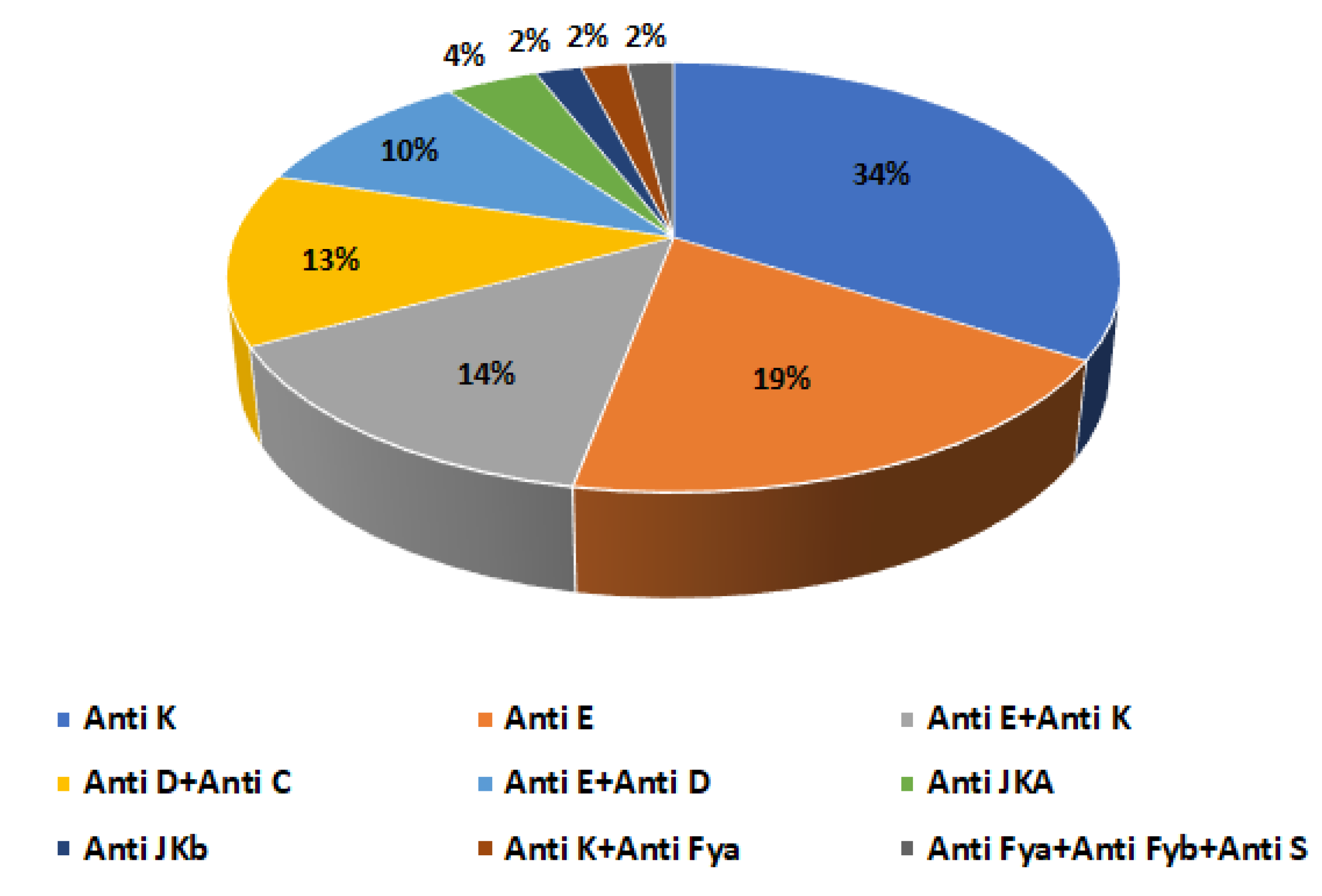
Figure 1. Frequency of alloantibodies in alloimmunized thalassemic patients.
Hemato 06 00035 g001
Table 1. Demographic and clinical characteristics of patients (n = 89).
CharacteristicsNumber of Patients n = 89 (%)p Value
Sex 0.004
Male46 (51.7%)
Female43 (48.3%)
Age (years) 0.001
0–1847 (52.8%)
19–3037 (41.6%)
31–705 (5.6%)
ABO/Rh blood type 0.001
A-1 (1.12%)
A+19 (21.34%)
AB-1 (1.12%)
AB+5 (5.6%)
B-3 (3.4%)
B+10 (11.23%)
O-6 (6.79%)
O+44 (49.4%)
Types of Thalassemia 0.001
β-Th Major73 (67%)
β-Th Intermediate9 (8.3%)
β-Th Minor3 (2.8%)
Th-Sickle Cell Disease Association4 (3.7%)
Table 2. Characteristics of transfusion in the study population.
Parameterβ-Thalassemia Majorβ-Thalassemia Intermediateβ-Thalassemia MinorThalassemia-Sickle Cell
Association
p Value
Percentage of transfused cases (%)678.32.83.70.001
Number of transfusions/year12–263–616–120.031
Number of packed red blood cell units/year12–602–121–24–360.001
Average interval between transfusions (week)0.5–48–16502–60.047
Table 3. Comparison of alloimmunized and non alloimmunized thalassemia patients.
CharacteristicsAlloimmunized
Patients
n = 42 (47.2%)
Non-Alloimmunized Patients
n = 47 (52.8%)
p Value
Sex ˂0.01
Males26.9%24.7%
Females20.3%28.1%
Age (years) 0.50
0–1828%24.7%
19–3016.8%24.7%
31–702.3%3.5%
Number of transfusions/year ˂0.01
˂122.3%52.8%
≥1249.9%0%
Number of bags of packed red blood cells/year (min–max)24–6012–24˂0.01
Types of Thalassemia ˂0.01
β-Thalassemia Major46%36%
β-Thalassemia Intermediate0%10.1%
β-Thalassemia Minor0%3.4%
Thalassemia-Sickle Cell Association1.1%3.4%
Direct Antiglobulin Test (DAT) ˂0.01
DAT negative25.8%44.9%
DAT positive21.4%7.9%
Disclaimer/Publisher’s Note: The statements, opinions and data contained in all publications are solely those of the individual author(s) and contributor(s) and not of MDPI and/or the editor(s). MDPI and/or the editor(s) disclaim responsibility for any injury to people or property resulting from any ideas, methods, instructions or products referred to in the content.