Case Report

Rare Coexistence of Atypical Chronic Lymphocytic Leukemia and B-Acute Lymphoblastic Leukemia in a Patient Followed Up for Monoclonal B-Cell Lymphocytosis

by
Fabio Bertani
1,2,*,
Francesco Faiella
1,
Claudia Di Franco
1,
Raffaella Milani
3,
Antonella Gualdoni
1,
Cinzia Fulceri
1 and
Elena Costa
1
1
Laboratorio Analisi Clinico Generale Con Aree Specialistiche, IRCCS Policlinico San Donato, Piazza Malan, 2 San Donato Milanese, 20097 Milan, Italy
2
Residency Program in Clinical Biochemistry and Clinical Pathology, Università degli Studi di Milano, Via Luigi Mangiagalli 31, 20133 Milan, Italy
3
Immunohematology and Transfusion Medicine Unit, IRCCS San Raffaele Scientific Institute, Via Olgettina, 60, 20132 Milan, Italy
*
Author to whom correspondence should be addressed.
Hemato 2025, 6(3), 27; https://doi.org/10.3390/hemato6030027
Submission received: 20 June 2025 / Revised: 21 July 2025 / Accepted: 4 August 2025 / Published: 7 August 2025

Abstract

Monoclonal B Lymphocytosis (MBL) is considered the pre-malignant state of chronic lymphocytic leukemia (CLL) and atypical chronic lymphocytic leukemia (aCLL). Both entities are rarely found in association with other hematological tumors; still, they naturally tend to progress to more aggressive lymphomas. This manuscript reports the case of an 81-year-old Caucasian male under follow up for MBL who presented to the Emergency Department with severe anemia and thrombocytopenia. A complete diagnostic workup revealed the simultaneous presence of atypical CLL and B-acute lymphoblastic leukemia, with minimal blast presence in peripheral blood.

1. Introduction

Monoclonal B Lymphocytosis (MBL) is defined as the presence of a monotonous population of B-cells of less than 5 × 10⁹/L and no constitutional symptoms or signs of leukemia [1,2]. MBL is phenotypically identified as chronic lymphocytic leukemia (CLL)-like, non-CLL-like, or atypical-CLL-like, depending on the expression of CDs [3]. As such, it is considered the pre-malignant form of both typical and atypical CLL [4].
Atypical CLL (aCLL) can be distinguished from typical CLL both morphologically and immunophenotypically. Morphologically atypical CLL cells have been defined mainly as large, atypical forms, prolymphocytes, or cleaved cells [5,6]. However, current aCLL diagnostics rely more on immunophenotypic characteristics rather than atypical morphology. Immunophenotypically, atypical CLL differs from classic CLL in the lack of expression of one or fewer surface antigens, most commonly CD5 and CD23, and the patient does not meet the criteria for a diagnosis of any other B-cell lymphoid malignancy [7,8]. Clinically, aCLL has a more aggressive behavior and poorer prognosis compared to typical CLL. Nonetheless, atypical CLL still lacks common standard classification features to be distinguished from typical CLL [6].
Progression to acute lymphoblastic leukemia (ALL) from CLL, call, or MBL is extremely rare, as is the concurrent presence of two distinct pathological populations (MBL/CLL and ALL) [9]. On the contrary, some aCLL and CLL patients may develop diffuse large B-cell lymphoma (DLBCL) or Hodgkin lymphoma (HL) in what is known as Richter’s Syndrome [10,11,12].
Despite being the most common form of leukemia affecting children, ALL is also commonly diagnosed in adults as well. The disease has a completely different course in young patients compared to adults, and this could be explained by the diverse genetic signatures of ALL in younger and older patients [13]. The precise etiology of the disease is still unknown, but a strong correlation between genotoxic chemicals and ionizing radiation exposure has been extensively characterized, especially in the context of adult B-ALL [13].

2. Case Report

An 81-year-old Caucasian male presented to our Emergency Department on a Friday afternoon in May 2025 with complaints of exertional dyspnea and generalized asthenia persisting for several days.
Upon arrival, the patient was eupneic and afebrile. Physical examination of the thorax and abdomen revealed no pathological findings. His past medical history included a diagnosis of throat cancer in 2008, treated with radiotherapy.
The patient, a former smoker, had been under surveillance for ten years without evidence of disease recurrence. He was also being followed at another facility for monoclonal B-lymphocytosis (MBL), which recently worsened into severe lymphocytosis at the latest hematological check-up in March 2024 (lymphocytes 12 × 109/L). Additional medical history included chronic hepatitis B virus (HBV) infection (anti-HBc positive), a renal cyst, and benign prostatic hyperplasia (BPH).
Initial laboratory investigations revealed severe normocytic anemia (hemoglobin 66 g/L) and thrombocytopenia (20 × 109/L). The white blood cell count was elevated at 41 × 109/L, with a differential showing 98% lymphocytes (40 × 109/L). Analysis on the ADVIA 2120 hematology platform detected a suspicious population characterized by medium-large cell volume and absence of myeloperoxidase (MPO) activity (Figure 1A,B), suggestive of atypical or reactive lymphocytes. As a consequence of this finding, a peripheral blood (PB) smear was prepared and reviewed by the on-call clinical pathologist (Figure 1C,D). Importantly, no automatic flags for blasts presence were reported by ADVIA software. Indeed, the cytograms did not demonstrate the presence of blasts since these cells are characterized by large nuclear volume and low lobularity, characteristically localizing near the origin of the axes in the BASO channel dot plot (white events gate—Figure 1B) and in the upper part of the large unstained cells (LUC) gate in the PEROX channel dot plot (light blue events gate—Figure 1A).
Renal function was mildly impaired, with serum creatinine at 1.19 mg/dL and an estimated glomerular filtration rate (eGFR) of 59 mL/min/1.73 m2 (MDRD equation). Inflammatory markers were modestly elevated: C-reactive protein (CRP) at 14 mg/L and lactate dehydrogenase (LDH) at 365 U/L.
Liver function tests were within normal limits (ALT 13 U/L, total bilirubin 0.79 mg/dL), as were pancreatic enzymes (lipase 24 U/L). However, serum glucose suggested poor glycemic control, confirmed by an elevated glycated hemoglobin (HbA1c) of 55 mmol/mol. Coagulation parameters were within the reference range (INR 1.19, aPTT ratio 0.99).
Microscopic examination of the PB smear confirmed the marked thrombocytopenia and revealed a predominant polymorphic population of medium-large-sized lymphocytes with atypical appearances, such as chromatin clumping, nuclear indentations, and cytoplasmic polarization (Figure 2). One non-granulated blast-like cell was found on 200 counted leukocytes (0.5%) (Figure 3). Moderate platelet anisocytosis was observed, along with red blood cell anisopoikilocytosis and hypochromia.
The patient was admitted to the Internal Medicine ward, and a comprehensive diagnostic workup was initiated. Considering the hematologic history, targeted flow cytometry analysis for lymphoid neoplasms was performed.
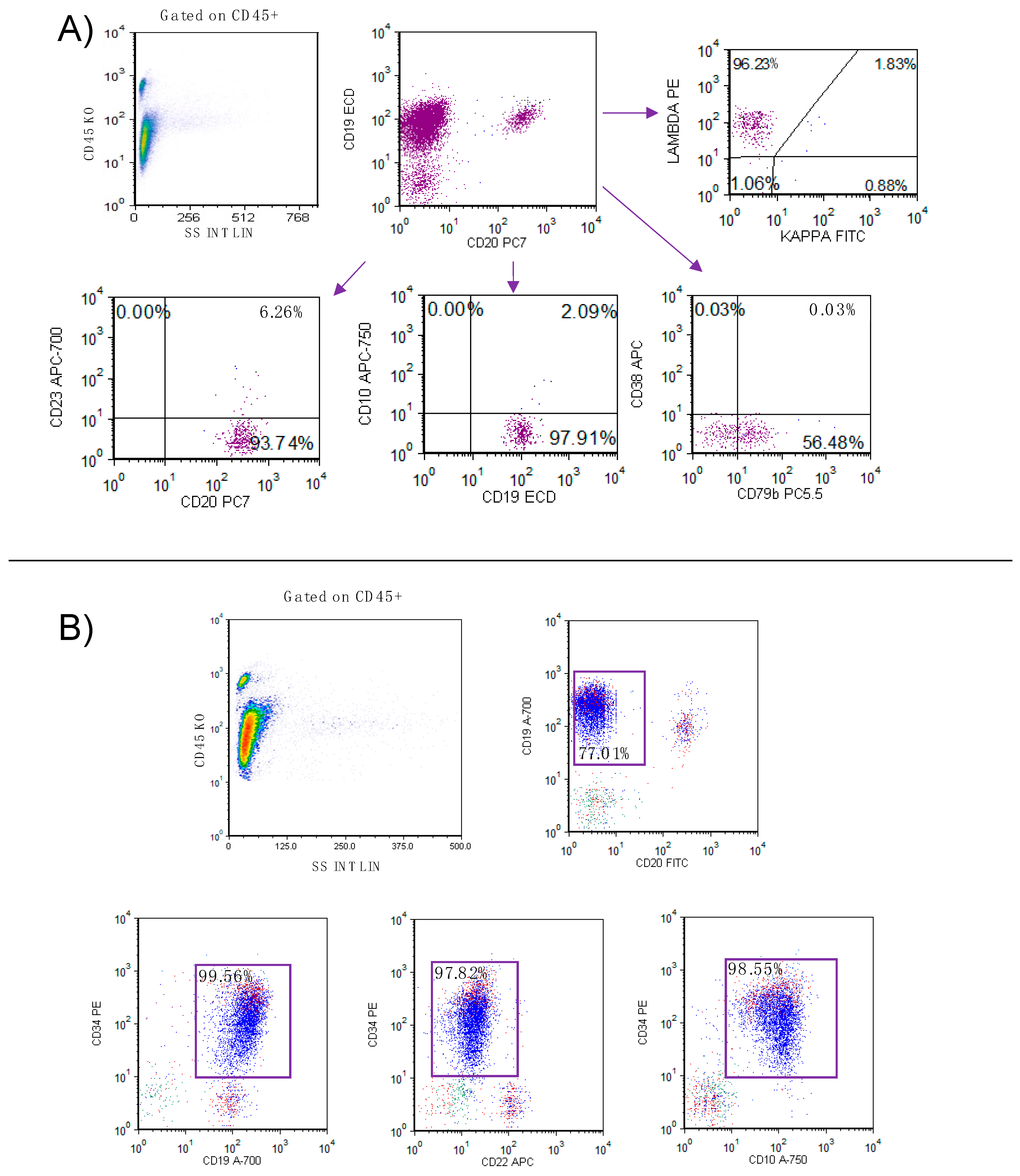
Flow cytometry analysis of peripheral blood, with gating on CD45+ bright/SSC low, identified a circulating B-cell population (83% of total lymphocytes and 97% of total leukocytes) characterized by CD19+, CD20 bright expression with surface lambda light chain restriction and negativity for CD5, CD23, and CD79b (Figure 2A). Given the suspicion of bone marrow (BM) involvement, further evaluation was undertaken, including BM aspiration and trephine biopsy for morphological assessment, flow cytometry, and cytogenetic analysis.
Microscopic examination of the BM aspirate revealed hypercellular fragments composed of 4% myeloid, 9% erythroid, and 8% lymphoid precursors, alongside a predominant population (78%) of medium to large blasts. These blasts exhibited agranular, moderately basophilic cytoplasm, with condensed chromatin and one or more prominent nucleoli. Plasma cells accounted for 1% of nucleated cells, and megakaryocytes were markedly reduced in number. BM flow cytometry was subsequently performed to complete the diagnostic workup (Supplementary Materials). Gating on CD45+ bright/SSC low confirmed the presence of a clonal B-cell population (CD19+ 77.01% of lymphocytes), with lambda light chain restriction, CD5 and CD23 negativity, and co-expression of CD20 bright, FMC7, CD81, and CD79b (61% on CD19 gated lymphocytes), compatible with the clonal population observed in the peripheral blood. However, the most significant finding was a large population of immature cells (CD45+ dim/SSClow) representing 87.69% of total leukocytes, expressing CD34, CD19, CD10, and CD22, an immunophenotypic profile consistent with B-cell acute lymphoblastic leukemia (B-ALL) (Figure 2B).
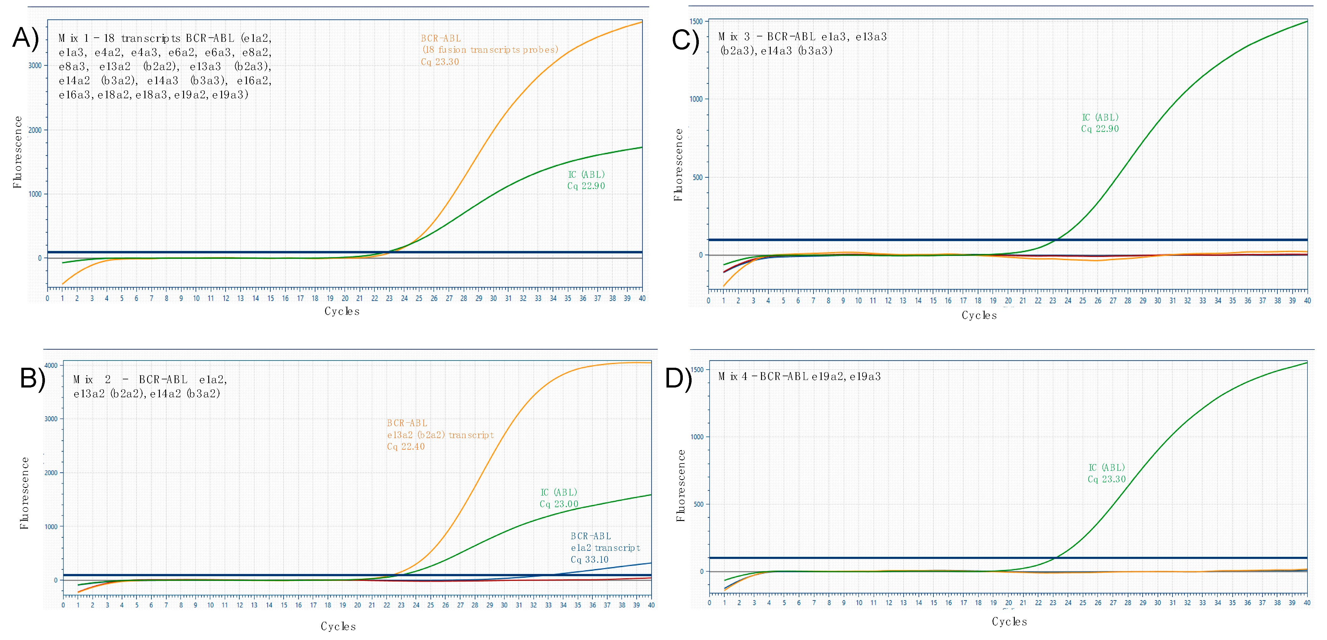
Qualitative molecular analysis (Supplementary Materials) was further required, identifying the presence of p210 e13a2 (b2a2) BCR-ABL fusion transcript and lower expression of p190 e1a2, confirming the diagnosis of BCR-ABL–positive B-ALL (Figure 3).
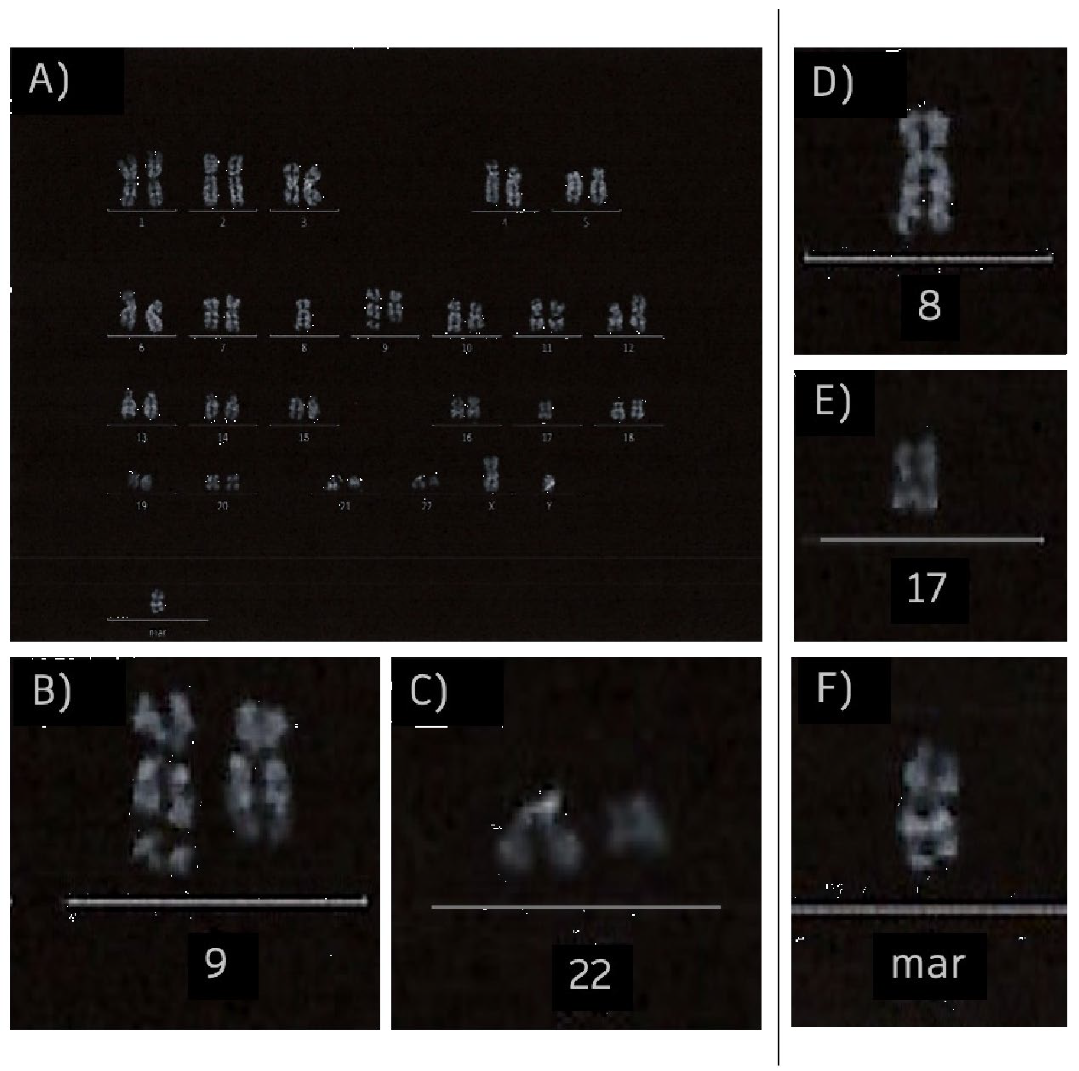
Karyotype analysis conducted on 10 metaphases from lymphocytes cultured for 72 h revealed an asset of 44–46 chromosomes and confirmed the t(9;22)(q34;q11) translocation and additionally identified a small marker chromosome (mar) (Figure 4).

3. Discussion

This case report examines the rare coexistence of atypical CD5-, CD23- chronic lymphocytic leukemia (CLL), possibly evolved from a monoclonal B-lymphocytosis, and acute lymphoblastic leukemia (ALL) in a single patient. To date, only a few similar cases have been reported in the literature. Other reports have documented the coexistence of CLL and B-ALL [14,15] or AML [16] or CLL with various solid tumors [17,18,19].
Most recently, in 2025, a Chinese group reported the 2021 case of a 77-year-old male unexpectedly found with ALL, having been previously diagnosed with typical CLL (CD19, dim CD20, CD5, CD23, CD200, and CD38, negative for CD10 and FMC7) that was never treated. The patient had a worsening mild thrombocytopenia (nadir platelets 120 × 109/L) that raised concern, ultimately leading to bone marrow investigations [20]. Unlike a few previously reported cases, our patient exhibited a marked lymphocytosis consistent with atypical CLL, characterized by the absence of both CD5 and CD23 expression. Nevertheless, despite being immunophenotypically distinct, typical and atypical CLL are commonly considered as the same pathology. As a matter of fact, atypical CLL is not differently classified by WHO HAEM5 nor the ICC classification [6].
In our patient, only a small number of circulating blasts was highlighted by peripheral blood microscopic examination and flow cytometry analysis, unlike what has been reported in other cases. As an example, Huang et al. reported a case of coexistence of CLL and B-ALL, characterized by a marked leukocytosis and the co-circulation of both blasts and mature-appearing leukemic lymphocyte populations, further revealed by flow cytometric analysis and cytogenetic studies confirming the presence of two separate karyotypes [21].
Our immunophenotypic findings posed a diagnostic challenge: the absence of CD5 and CD23 in the context of marked lymphocytosis with atypical morphology necessitated the consideration of other B-cell lymphoproliferative disorders such as mantle cell lymphoma (MCL), splenic marginal zone lymphoma (SMZL), or hairy cell leukemia (HCL), all of which can infiltrate the bone marrow and exert a mass effect, which may have explained the patient’s severe anemia and thrombocytopenia upon presentation to the emergency department. Moreover, microscopic examination of the peripheral blood smear revealed a polymorphic population of lymphocytes sharing features commonly associated with MCL and SMZL (nuclear indentations, cytoplasmic polarization). On the other hand, despite the small number of blast-like cells in peripheral blood (0.5%), the possibility of an acute leukemia had to be considered in differential diagnosis, further highlighting the importance of accurate microscopic examination of blood smear in CBC validation of outpatients.
Bone marrow examination was eventually essential to investigate the possibility of a CD5 and CD23 negative neoplasm, surprisingly leading to the diagnosis of acute lymphoblastic leukemia (ALL), rather than a B-cell lymphoma as initially suspected.
To the best of our knowledge, this represents the third reported case [19] in the literature of a chronic lymphocytic leukemia patient developing B-acute lymphoblastic leukemia and the first ever reported case involving a CD5- and CD23-negative, atypical chronic lymphocytic leukemia.
This case underscores the critical role of laboratory medicine in patient care. The “brain-to-brain loop” concept highlights the importance of interpreting laboratory requests in a clinical context, transforming test results into actionable information for physicians. Laboratory professionals are responsible not only for performing diagnostic tests but also for initiating second-level investigations when clinically indicated. The goal is to provide comprehensive and timely diagnostic insights, minimizing unnecessary procedures and accelerating the diagnostic process.

4. Conclusions

In conclusion, although initial assessments pointed toward a B-cell lymphoma, laboratory workup (severe anemia and thrombocytopenia), microscopic blood smear examination, and bone marrow analysis ultimately revealed the presence of acute lymphoblastic leukemia, highlighting the complexity of such presentations, the need to keep an open-minded approach in both physicians and clinical pathologists, and the indispensable value of first-line laboratory as well as second-level laboratory diagnostic studies. Therefore, even rare or atypical disease combinations must not be overlooked, as early recognition is essential for accurate diagnosis and optimal patient care management.

Supplementary Materials

The supporting information can be downloaded at: https://www.mdpi.com/article/10.3390/hemato6030027/s1.

Author Contributions

Conceptualization, F.B. and R.M.; methodology, F.B. and A.G.; resources, E.C.; data curation, C.D.F.; writing—original draft preparation, F.B.; writing—review and editing, F.F. and C.D.F.; visualization, F.F.; supervision, C.F. All authors have read and agreed to the published version of the manuscript.

Funding

This research received no external funding.

Institutional Review Board Statement

All procedures followed were in accordance with the ethical standards of the committee in charge of human experimentation (institutional and national) and the Declaration of Helsinki. This study did not require approval by the Institutional Review Board as a case report.

Informed Consent Statement

Informed consent was obtained from all subjects involved in the study.

Data Availability Statement

All data concerning the case can be accessed by contacting the corresponding author.

Conflicts of Interest

The authors declare no conflicts of interest.

References

  1. Marti, G.E.; Rawstron, A.C.; Ghia, P.; Hillmen, P.; Houlston, R.S.; Kay, N.; Schleinitz, T.A.; Caporaso, N.; The International Familial CLL Consortium. Diagnostic criteria for monoclonal B-cell lymphocytosis. Br. J. Haematol. 2005, 130, 325–332. [Google Scholar] [CrossRef]
  2. Caporaso, N.E.; Marti, G.E.; Landgren, O.; Azzato, E.; Weinberg, J.B.; Goldin, L.; Shanafelt, T. Monoclonal B cell lymphocytosis: Clinical and population perspectives. Cytom. Part B Clin. Cytom. 2010, 78B, S115–S119. [Google Scholar] [CrossRef]
  3. Rawstron, A.C.; Bennett, F.L.; O’COnnor, S.J.; Kwok, M.; Fenton, J.A.; Plummer, M.; de Tute, R.; Owen, R.G.; Richards, S.J.; Jack, A.S.; et al. Monoclonal B-Cell Lymphocytosis and Chronic Lymphocytic Leukemia. N. Engl. J. Med. 2008, 359, 575–583. [Google Scholar] [CrossRef]
  4. Criado, I.; Rodríguez-Caballero, A.; Gutiérrez, M.L.; Pedreira, C.E.; Alcoceba, M.; Nieto, W.; Teodosio, C.; Bárcena, P.; Romero, A.; Fernández-Navarro, P.; et al. Low-count monoclonal B-cell lymphocytosis persists after seven years of follow up and is associated with a poorer outcome. Haematologica 2018, 103, 1198–1208. [Google Scholar] [CrossRef]
  5. Urbaniak, M.; Iskierka-Jażdżewska, E.; Majchrzak, A.; Robak, T. Atypical immunophenotype of chronic lymphocytic leukemia. Acta Haematol. Pol. 2022, 53, 48–52. [Google Scholar] [CrossRef]
  6. Robak, T.; Krawczyńska, A.; Cebula-Obrzut, B.; Urbaniak, M.; Iskierka-Jażdżewska, E.; Robak, P. Atypical Chronic Lymphocytic Leukemia—The Current Status. Cancers 2023, 15, 4427. [Google Scholar] [CrossRef]
  7. Frater, J.L.; McCarron, K.F.; Hammel, J.P.; Shapiro, J.L.; Miller, M.L.; Tubbs, R.R.; Pettay, J.; Hsi, E.D. Typical and atypical chronic lymphocytic leukemia differ clinically and immunophenotypically. Am. J. Clin. Pathol. 2001, 116, 655–664. [Google Scholar] [CrossRef]
  8. D’arena, G.; Vitale, C.; Pietrantuono, G.; Villani, O.; Mansueto, G.; D’auria, F.; Statuto, T.; D’agostino, S.; Sabetta, R.; Tarasco, A.; et al. What Does Atypical Chronic Lymphocytic Leukemia Really Mean? A Retrospective Morphological and Immunophenotypic Study. Cancers 2024, 16, 469. [Google Scholar] [CrossRef] [PubMed]
  9. Dalivand, M.M.; Tizro, P.; Partain, J.; Aggarwal, A.; Lichy, J.; Ara, F.M.; Nava, V.E.; Tauro, S. Concurrent B Cell Acute Lymphoblastic Lymphoma/Leukemia and Monoclonal B Cell Lymphocytosis: A Case Report with Extensive Molecular Analysis. Case Rep. Hematol. 2022, 2022, 1132544. [Google Scholar] [CrossRef] [PubMed]
  10. Condoluci, A.; Rossi, D. Richter Syndrome. Richter Syndrome. Curr. Oncol. Rep. 2021, 23, 26. [Google Scholar] [CrossRef] [PubMed]
  11. Mahmoud, A.M.; Gaidano, G.; Mouhssine, S. Immunological Aspects of Richter Syndrome: From Immune Dysfunction to Immunotherapy. Cancers 2023, 15, 1015. [Google Scholar] [CrossRef]
  12. Favini, C.; Talotta, D.; Almasri, M.; Andorno, A.; Rasi, S.; Adhinaveni, R.; Kogila, S.; Awikeh, B.; Schipani, M.; Boggione, P.; et al. Clonally unrelated Richter syndrome are truly de novo diffuse large B-cell lymphomas with a mutational profile reminiscent of clonally related Richter syndrome. Br. J. Haematol. 2022, 198, 1016–1022. [Google Scholar] [CrossRef] [PubMed]
  13. Roberts, K.G. Genetics and prognosis of ALL in children vs adults. Hematol. Am. Soc. Hematol. Educ. Program 2018, 2018, 137–145. [Google Scholar] [CrossRef]
  14. Preudhomme, C.; Lepelley, P.; Lovi, V.; Zandecki, M.; Cosson, A.; Fenaux, P. T-cell acute lymphoblastic leukemia occurring in the course of b cell chronic lymphocytic leukemia: A case report. Leuk. Lymphoma 1995, 18, 361–364. [Google Scholar] [CrossRef] [PubMed]
  15. Haznedaroğlu, İ.C.; Savaş, M.C.; Benekli, M.; Sayinalp, N.M.; Özcebe, O.I.; Dündar, S. Acute lymphoblastic leukemia occurring in a case of chronic lymphocytic leukemia. Hematology 1997, 2, 87–89. [Google Scholar] [CrossRef]
  16. Kajtár, B.; Rajnics, P.; Egyed, M.; Alizadeh, H. Case report: Concomitant chronic lymphocytic leukaemia and cytogenetically normal de novo acute leukaemia in a patient. Ann. Clin. Lab. Sci. 2015, 45, 602–606. [Google Scholar]
  17. Maurer, C.; Langerbeins, P.; Bahlo, J.; Cramer, P.; Fink, A.M.; Pflug, N.; Engelke, A.; von Tresckow, J.; Kovacs, G.; Stilgenbauer, S.; et al. Effect of first-line treatment on second primary malignancies and Richter’s transformation in patients with CLL. Leukemia. 2016, 10, 2019–2025. [Google Scholar] [CrossRef]
  18. Raza, S.; Hampel, P.J.; Firwana, B.; Wang, Z.; Shad, Y.; Aluri, L.M.; Naidzionak, U.; Doll, D.C. Chronic lymphocytic leukemia-associated chromosomal abnormalities and risk of secondary malignancies. Blood 2014, 124, 5636. [Google Scholar] [CrossRef]
  19. Mellemgaard, A.; Geisler, C.H.; Storm, H.H. Risk of kidney cancer and other second solid malignancies in patients with chronic lymphocytic leukemia. Eur. J. Haematol. 1994, 53, 218–222. [Google Scholar] [CrossRef]
  20. Song, A.; Li, J.Y. Unexpected concurrent B-lymphoblastic leukaemia and untreated chronic lymphocytic leukaemia presenting as worsening thrombocytopenia: A rare case report. Pathology 2025, 57, 123–126. [Google Scholar] [CrossRef] [PubMed]
  21. Huang, Q. Cocirculating precursor B acute lymphoblastic leukemia and chronic lymphocytic leukemia. Blood 2013, 122, 2148. [Google Scholar] [CrossRef] [PubMed]
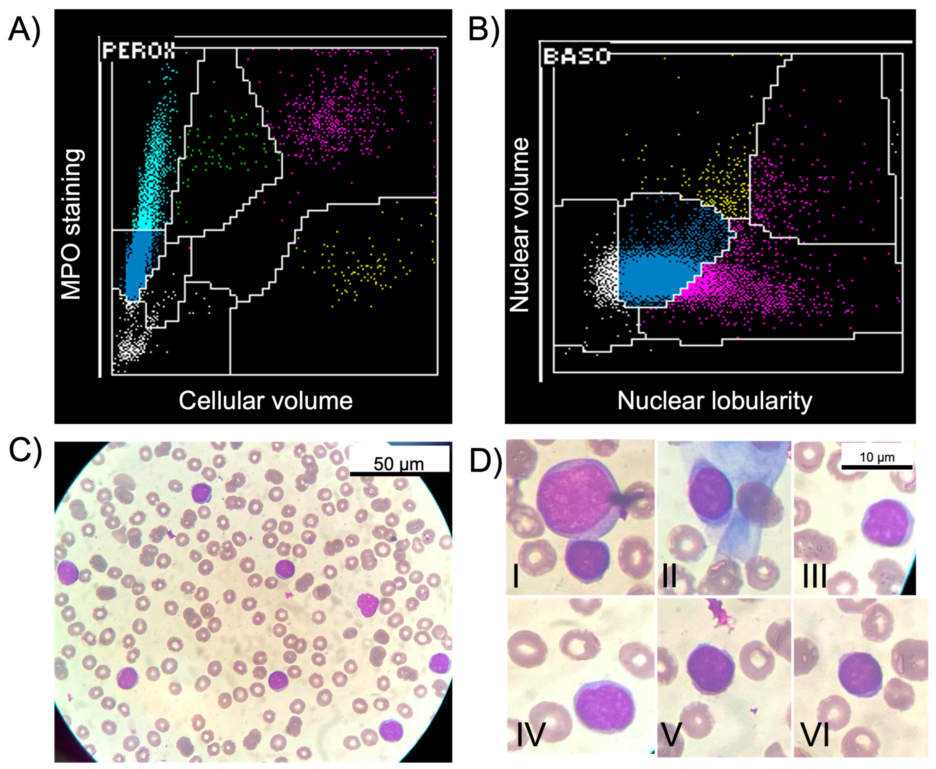
Figure 1. (A,B) Cytograms obtained for complete blood count analysis with differentials on ADVIA 2120 (Siemens) performed on admission at the Emergency Department. In panel (A), an MPO-negative population of medium-large cells is visible in the Perox channel (light-blue events), suggestive of atypical/reactive lymphocytes. In panel (B), a predominant population of mononuclear cells is represented in the MN gate (blue events). In both analytical channels, there was no evidence of circulating blasts, and no automatic flags were reported. (C,D) Microscopic analysis (May-Grünwald Giemsa—1000×) of peripheral blood smear demonstrated marked lymphocytosis and morphological features suggestive of atypical lymphocytes, such as chromatin clumping (D-II,D-V,D-VI), nuclear grooves and indentations (D-III,D-IV) and cytoplasm polarization (D-II). Only one, non-granulated, blast-looking, abnormal cell was detected on 200 cells examined (D-I), confirming the extremely low number of circulating blasts that was already suggested by cytogram analysis on PEROX and BASO channels of ADVIA2120.
Hemato 06 00027 g001
Figure 2. (A) Flow cytometry on peripheral blood: gating on CD45+ bright/SSC low events demonstrated the presence of a circulating mature B-cell population (83% of total lymphocytes) characterized by CD19+, CD20 bright with surface lambda light chain restriction and negativity for CD5, CD23, CD10, and CD79b. (B) Bone marrow cytometric analysis: gating on CD45+ dim/SSC low events discovered a large population of immature cells, representing 87.69% of total leukocytes (CD45+), expressing CD34, CD19, CD10, and CD22, an immunophenotypic profile consistent with B-cell acute lymphoblastic leukemia (B-ALL).
Hemato 06 00027 g002
Figure 3. BCR-ABL qualitative polymerase chain reaction amplification curves. The qualitative assay is composed of four different PCR reaction mixes. Mix 1 allows the amplification of 18 BCR-ABL fusion transcripts, without the possibility of further characterization; mix 2 amplifies and discriminates BCR-ABL e1a2, e13a2 (b2a2), and e14a2 (b3a2); mix 3 amplifies and discriminates BCR-ABL e1a3, e13a3 (b2a3), and e14a3 (b3a3); and mix 4 amplifies and discriminates BCR-ABL e19a2, and e19a3. Amplification curves were observed in mixes 1 and 2, demonstrating the presence of p210 e13a2 (b2a2) BCR-ABL fusion transcript and lower expression of the p190 e1a2 (A,B). No transcript amplification was observed in mix 3 and mix 4 (C,D).
Hemato 06 00027 g003
Figure 4. Q-banding (QFQ) conventional cytogenetic analysis on bone marrow cultured lymphocytes (A) resulting in a 44, XY karyotype. At 300 bands resolution, the analysis demonstrated the presence of t(9;22)(q34;q11) translocation (B,C) consistent with BCR-ABL rearrangement. It was also reported the presence of additional morphological and numerical anomalies (DF).
Hemato 06 00027 g004
Disclaimer/Publisher’s Note: The statements, opinions and data contained in all publications are solely those of the individual author(s) and contributor(s) and not of MDPI and/or the editor(s). MDPI and/or the editor(s) disclaim responsibility for any injury to people or property resulting from any ideas, methods, instructions or products referred to in the content.