Case Report

A Rare Case of Primary Bone Follicular Lymphoma with Multiple Osteolytic Lesions: A Case Report and Review of the Literature

1
Hematology, Department of Translational and Precision Medicine, Sapienza University, 00185 Rome, Italy
2
Hematology Unit, Department of Clinical and Molecular Medicine, Sant’Andrea University Hospital, Sapienza University, 00189 Rome, Italy
3
Pathology Unit, Department of Clinical and Molecular Medicine, Sant’Andrea University Hospital, Sapienza University, 00189 Rome, Italy
4
Nuclear Medicine Unit, Department of Medical-Surgical Sciences and Translational Medicine, Sant’Andrea University Hospital, Sapienza University, 00189 Rome, Italy
5
Radiology Unit, Department of Medical-Surgical Sciences and Translational Medicine, Sant’Andrea University Hospital, 00189 Rome, Italy
*
Authors to whom correspondence should be addressed.
Hemato 2024, 5(4), 388-395; https://doi.org/10.3390/hemato5040028
Submission received: 25 June 2024 / Revised: 28 August 2024 / Accepted: 23 September 2024 / Published: 29 September 2024
(This article belongs to the Section Lymphomas)

Abstract

:
Introduction: Primary bone lymphoma (PBL) is a rare clinical entity, accounting for less than 5% of all extranodal non-Hodgkin lymphomas and approximately 5% of primary bone tumors. Diffuse large B-cell lymphoma (DLBCL) is the most common histotype, accounting for about 80% of all PBL cases. Conversely, the incidence of indolent primary bone lymphomas (iPBL) represents less than 1% of all reported PBL cases, and data on these rarer lymphomas are scarce. Drawing on diagnostic criteria developed by the World Health Organization (WHO) and the International Extranodal Lymphoma Study Group (IELSG), we report a rare case of primary bone follicular lymphoma, focusing specifically on the clinical presentation and treatment. Discussion: Additionally, we provide a systematic review of the literature data on this very rare lymphoproliferative entity.

1. Introduction

According to the WHO Classification of Tumours of Soft Tissue and Bone (2013), primary bone lymphoma (PBL) is defined as a “neoplasm composed of malignant lymphoid cells confined to the skeletal tissue, without the involvement of supra-regional lymph nodes or concurrent extranodal lesions” [1,2]. The most common histological subtype is diffuse large B-cell lymphoma (DLBCL) accounting for about 80%, with the other subtypes being follicular lymphomas, marginal zone lymphomas, anaplastic large cell lymphomas, Hodgkin lymphomas, and T-cell lymphomas, respectively, at extremely rare incidences [3]. However, the new WHO Classification of Tumours of Soft Tissue and Bone (2020) lacks well-defined criteria. The general consensus relies on the definition provided by the International Extranodal Lymphoma Study Group (IELSG), in their retrospective study (IELSG 14) [4,5,6]. Thus, the presence of a single bone lesion, with or without regional lymph node involvement (“monostotic lymphoma”) or, more rarely, the presence of multiple bone lesions without nodal or visceral disease (“multifocal bone lymphoma”) can be classified as primary bone lymphomas. Needless to say, that this entity should be distinguished from the more common condition characterized by secondary bone involvement of systemic lymphomas. The more frequent sites for PBL are the femurs and pelvis (50%) as well as the long bones of the upper limb. Less common sites include ribs, scapulae, or jaw [7,8]. Due to their rarity, the literature shows few data regarding indolent primary bone lymphomas (iPBL) [9].

2. Case Report

We present the case of a 58-year-old man with a personal history of occipital glioma, for which he had undergone surgical removal in 2006 and anti-epileptic prophylaxis until the 2015. In October of 2021, the patient experienced a first episode of seizures, followed by a traumatic fall. At the time of the seizure, he did not complain of fever, general weakness, night sweats and weight loss. Hence, he underwent both a whole-brain and spine magnetic resonance imaging (MRI). The whole-brain magnetic resonance imaging was negative, whereas the spine magnetic resonance imaging revealed signal alterations in the thoracolumbar vertebral segments. The seizure was defined as an isolated case and, therefore, after a neurologic consult, the patient was restarted on anti-epileptic prophylaxis. One month later, the patient, still asymptomatic, underwent a second whole-body MRI, which confirmed the focal signal alterations in multiple thoracolumbar segments, accompanied by pathological vertebral collapses at T6, T7, and T8. Moreover, additional hyperintense lesions on T2 and iso-intense lesions on T1-weighted images were found at the level of the thoracic cage (clavicles, sternum, ribs), pelvis, and left femur as shown in Figure 1.
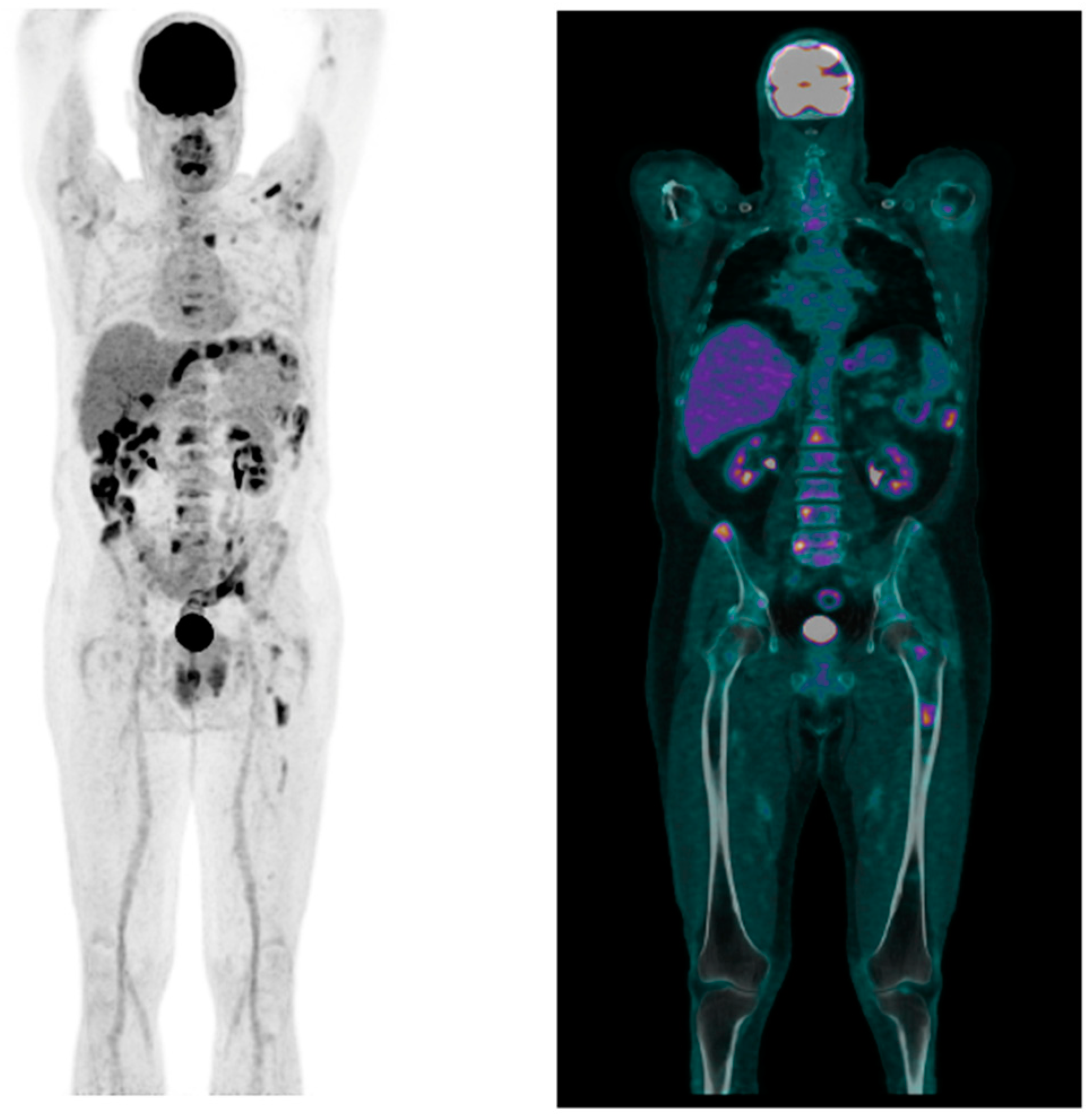
On computed tomography (CT), T5, T6, T7, and T8 vertebral body fractures were described, with Grade 2 fractures at T6 and T7. A positron emission tomography-CT (PET/CT) scan confirmed the presence of high-metabolism lesions (SUVmax 10) in the left clavicle, ipsilateral humeral head, right scapula, bilateral iliac bones, left femoral diaphysis, and multiple vertebral segments (T5–T7 and T9, L1–L3) as shown in Figure 2. On physical examination, no abnormal findings were detected, and the patient’s laboratory tests, including complete blood count, liver and renal function, alkaline phosphatase (ALP) and coagulation, were all within normal ranges, except for a moderately increased LDH level (270 U/L, range: 125–220 U/L). There was no hypercalcemia or evidence of pathological monoclonal components in serum and urine protein electrophoresis and immunofixation. The serum kappa and lambda free light chain ratio was also within normal limits. His ESR and CRP values were 2 mm/h (range: 2–25 mm/h) and 0.08 mg/dL (reference range: 0.00–0.50 mg/dL), respectively. In addition, major tumor markers were negative.
Therefore, based on the radiological findings and in the absence of clinical and laboratory abnormalities, a CT-guided biopsy on the left anterior iliac crest was performed.
Histopathological examination revealed an involvement by a lymphoproliferative disease with a predominantly para-trabecular distribution, consisting of small and large cells expressing CD20+, CD79a+, PAX5+, CD10− BCL6+, BCL2+, LMO2+, HGAL+ (low), MUM1−, CD5−, CD23+, CyclinD1−, MYC+, p53+/− CD25−, AnnexinA1−, CD30−/+, CD15−, LMP1−, ALK1−, and TdT−. The analysis of B-lymphocyte clonality also revealed a monoclonal rearrangement of both the IGH and IGK genes on a polyclonal background. Conversely, the analysis of T-lymphocyte clonality showed a polyclonal rearrangement of the TRG gene. The morphological, immunophenotypic and molecular characteristics, in relation to the absence of disease in extraosseous sites, therefore, appeared suggestive of bone-marrow infiltration by a B-cell lymphoma of follicular center origin. Based on the clinical, radiological, and histological findings, a diagnosis of primary bone follicular lymphoma was established. The disease was staged as polyostotic lymphoma (IVE), according to the PBL-modified Ann Arbor staging system [10], and classified as an intermediate risk through the Follicular International Prognostic Index (FLIPI). Therefore, the patient underwent chemoimmunotherapy with a combined regimen of cyclophosphamide-doxorubicin-vincristine-prednisone (CHOP) and Obinutuzumab (G) for VI cycles at 21 day intervals (G-CHOP). G was administered according to the schedule at day 1, 8, and 15 for the first cycle and on day 1 from cycles 2–6. Two consolidation cycles with only G were also administered. The end-of-therapy PET/CT and MRI restaging, respectively, showed complete remission (CR) of the disease and a reduction in focal intensity of the bone alterations. The bone-marrow biopsy did not show lymphoproliferative infiltration. Therefore, the patient has started G maintenance therapy at 2 month intervals, in combination with periodic infusions of Zoledronate (4 mg IV). Currently, the patient shows excellent general condition and is completing the maintenance treatment under active surveillance, without disease recurrence.

3. Discussion

Primary Bone Lymphoma (PBL) is an extremely rare clinical entity, with diffuse large B-cell lymphoma (DLBCL) being the most common histotype [1,2,7]. Indolent PBLs represent less than 1% of reported PBL cases and, due to their rarity, data are exceptionally scarce [4,9]. To our knowledge, only a few case reports and small case series have collected data on PBLs. Among these, follicular lymphomas represent an even rarer subclass, requiring careful differential diagnosis necessary to guide optimal therapeutic choices [11].
Follicular lymphoma (FL) is the most common subtype among indolent B-cell lymphomas. The t(14;18) (q32;q21) translocation, found in over 85% of FL cases, is considered the primary genetic event. This translocation juxtaposes the BCL2 oncogene next to the immunoglobulin heavy chain enhancer, leading to BCL2 overexpression. Although FL is generally considered an indolent disease with a good prognosis, its transformation to aggressive lymphoma (DLBCL) can occur in approximately 10–20% of cases [12,13].
Regarding indolent PBLs, one of the few significant studies is the retrospective study conducted by IELSG, known as the “IELSG #14 study”. In this study, subjects with primary and secondary indolent PBL were selected from an international database of 499 patients with non-Hodgkin lymphoma. Over a 15 year period, only 26 indolent PBL patients were identified, 10 of whom had the follicular variant. The 5 year progression-free survival (PFS) and overall survival (OS) rates were 25 ± 15% and 23 ± 14%, respectively. These data, however, should be interpreted with caution due to the small sample size and, additionally, it is important to consider that these results were obtained in an era where current immunochemotherapy treatments were not yet available [9].
However, the prognosis of PBL is quite favorable and largely depends on the stage of the disease: 5 year OS rates vary from 88% for localized disease (stage I) to 74 ± 8% for advanced stages (stage IV) [10].
In terms of age, as demonstrated also in our case, the incidence peaks ranged between 50 and 60 years and is more frequent in males, with a very low incidence in pediatric populations, where Ewing’s sarcoma, osteosarcoma, and osteomyelitis should be considered in the initial differential diagnosis [14,15,16,17].
As previously mentioned, PBL can develop in any part of the skeleton, but the most common sites are the femur, humerus, tibia, spine, and pelvis. Less frequent sites of onset include the skull, forearm, scapula, clavicle, patella, hands, and feet. This was partially consistent in our case, where the patient also presented with involvement of less frequent regions (clavicles and scapula) [7,8].
In our case, disease staging was performed based on the adapted Ann Arbor system, considering that traditional Ann Arbor staging presents numerous limitations for this rare entity. This modified classification allows for the distinction, as presented in our patient, between stage IV–E (bone disease in the absence of nodal or visceral involvement) and stage IV (disseminated lymphoma with at least one bone lesion). This distinction, integrated with histological, clinical, and radiological data, is crucial for diagnosing primary bone disease [10].
The clinical presentation of PBL is highly variable but primarily includes localized pain, which is the most common symptom, soft tissue swelling, palpable mass, and, less frequently, pathological fractures. Pain has been described in most cases as persistent or intermittent, progressively worsening, and not relieved by rest [18].
Contrary to such data, our patient did not present any specific symptoms, and the diagnostic process was initiated following an accidental event. This might be partly explained by the indolent nature of the lymphoma and, as described in the literature, the extremely variable presentation of this disease.
Despite our limited experience, however, we suggest that PBL is considered as a possible diagnosis even in the absence of specific initial symptoms.
B symptoms, including fever, sweating, or weight loss, may occur in 16% of cases, as described by Govi et al., but are absent in many instances, which can significantly delay diagnosis. In line with these findings, our patient did not present any specific symptoms [19].
Radiological imaging of PBL does not exhibit pathognomonic features, but common characteristics may include lytic destruction, periosteal reactions, cortical destruction, pathological fractures, and soft tissue masses in over 70% of cases. In some lymphoma cases, only mild changes are observed in bone imaging [20,21,22,23,24].
Given that the imaging appearance of PBL is highly variable and nonspecific, surgical biopsy of bone lesions for histological and immunohistochemical examination remains the gold standard for diagnosis. MRI and PET-CT are the most accurate techniques for highlighting pathological osseous lesions, determining the local extent of disease, and performing accurate staging of the disease [25,26,27]. A study by L.J. Wang demonstrated that PBL is highly FDG-avid, with PET’s diagnostic sensitivity being much higher than that of CT (98.9% vs. 43.2%, p = 0.000) [28]. However, nuclear bone scans can lead to false-negative results due to the predominantly osteolytic nature of the lesions. Similarly, post-therapeutic osteonecrosis can be a potential source of false-positive results on F-FDG PET/CT re-evaluation [29].
The treatment of primary bone lymphomas may vary depending on histological subtype, disease extent, and initial response to treatment. Specifically, the literature on the management of indolent PBLs is scarce, with the few studies addressing this issue focusing almost exclusively on the management of PBL-DLBCL. Given the rarity of indolent PBL, there is no clear indication for first-line treatment; however, overall, the treatment strategies for indolent PBL should be similar to the established approach for indolent nodal NHL. Based on the limited experiences in the literature, radiotherapy covering the entire affected bone appears to be the treatment of choice in localized disease or in cases of risk of pathological fracture. As in our case, in the presence of disseminated disease or constitutional symptoms, chemo-immunotherapy (with consolidative radiotherapy if necessary), seems to be the preferred approach [11,30].
It is also worth noting that indolent PBL has the same risk of transformation into aggressive lymphoproliferative diseases as the non-osseous counterparts and, therefore, it is important to restudy rare, but nonetheless possible, relapsed PBLs. Management of aggressive PBLs, despite not being a topic of this case, has been described in different studies and will be discussed further to improve the topic [11].
Historically, the treatment of PBL included surgery (often radical) or local irradiation, which resulted in low survival rates with a 5 year OS of 45% and high relapse rates [31]. More recently, systemic chemotherapy has shown better results compared to radiotherapy alone, which remains the best treatment for local disease control, or in cases of secondary complications such as neurological compression, impending fracture, or pathological fractures [24,32]. In the IELSG-14 study, patients with localized PBL were treated with primary chemotherapy, with or without radiotherapy, achieving significantly better OS rates compared to those treated with primary radiotherapy alone [21]. Similar results were obtained by K.M. Ramadan et al. in a study involving a cohort of 131 patients (79% with PBL-DLBCL). In this study, 120 out of 131 patients treated with CHOP or CHOP-like regimens achieved 10 year OS and PFS rates of 41% and 40%, respectively [32]. The combined use of radiotherapy and systemic chemotherapy remains controversial, with no clear consensus on their joint application and timing. In advanced stages, as proposed by Mendenhall et al. and IELSG, radiotherapy should be used as consolidation therapy on macroscopically involved sites, if feasible, after the completion of chemotherapy to reduce the amount of radiation [4,33,34]. Currently, there are no guidelines regarding CNS (central nervous system) prophylaxis. However, according to IELSG, patients with multifocal PBL-DLBCL should receive the same treatment as disseminated DLBCL [4].

4. Conclusions

Given the low incidence of primary bone lymphoma (PBL), particularly for indolent variants, precise guidelines for the optimal management of this rare condition are still lacking. As a result, treatment is often tailored to the specific presentation characteristics of the disease [4,11,20,33].
Here, we report a rare case of advanced-stage (IVE) primary bone follicular lymphoma, which was successfully treated with combined immunochemotherapy alone. Its rarity and unusual onset made the diagnosis challenging; however, the patient achieved a complete remission with full clinical recovery and is currently undergoing maintenance therapy.
Based on our experience, we suggest including PBL in the differential diagnosis when encountering characteristic lytic lesions as described in our case, even in the absence of typical symptoms. Furthermore, the chemoimmunotherapy regimen administered, although representing a single case, demonstrated efficacy consistent with the data reported in other studies. However, given the rarity of this disease, more research is required to standardize diagnostic and therapeutic management of indolent primary bone lymphoma.

Author Contributions

C.T. conceptualized and wrote the manuscript, M.P.B., G.L.V., A.D.N., T.L. and M.Z. researched data; G.M. and A.T. (Ambra Taglietti). wrote and edited the manuscript, S.P., A.L. and A.T. (Agostino Tafuri). reviewed the manuscript. All authors have read and agreed to the published version of the manuscript.

Funding

This research received no external funding.

Institutional Review Board Statement

Not applicable.

Informed Consent Statement

Informed consent was obtained from all subjects involved in the study and written informed consent has been obtained from the patient to publish this paper.

Data Availability Statement

Due to patient privacy, all data pertaining to the patient in this case report cannot be shared or distributed.

Acknowledgments

The authors would like to thank all those who contributed to the treatment of the patient. We would also like to thank the patient for giving us consent to write this case report.

Conflicts of Interest

The authors declare no conflicts of interest.

References

  1. Jain, A.; Alam, K.; Maheshwari, V.; Khan, R.; Nobin, H.; Narula, V. Primary bone lymphomas-Clinical cases and review of literature. J. Bone Oncol. 2013, 2, 132–136. [Google Scholar] [CrossRef] [PubMed] [PubMed Central]
  2. Hogendoorn, P.C.W.; Bovée, J.V.M.G.; Nielsen GPFletcher, C.D.; Bridges, A.J.; Hogendoorn, C.W.; Mertens, F. Chondrosarcoma (grade I-III), including primary and secondary variants and periosteal chondrosarcoma. In WHO Classification of Tumours of Soft Tissue and Bone, 4th ed.; IARC Press: Lyon, France, 2013; pp. 264–268. [Google Scholar]
  3. Fletcher, C.D.M.; Unni, K.K.; Mertens, F. (Eds.) World Health Organization Classification of Tumours. In Pathology and Genetics of Tumours of Soft Tissue and Bone; IARC Press: Lyon, France, 2002. [Google Scholar]
  4. Messina, C.; Ferreri, A.J.; Govi, S.; Bruno-Ventre, M.; Gracia Medina, E.A.; Porter, D.; Radford, J.; Heo, D.S.; Park, H.Y.; Pro, B.; et al. Clinical features, management and prognosis of multifocal primary bone lymphoma: A retrospective study of the international extranodal lymphoma study group (the IELSG 14 study). Br. J. Haematol. 2014, 164, 834–840. [Google Scholar] [CrossRef] [PubMed]
  5. Flanagan, A.M.; Blay, J.Y.; Bovée, J.V.M.G.; Bredella, A.; Cool, P.; Nielsen, G.P.; Yoshida, A. Bone tumours: Introduction. In WHO Classification of Tumours Soft Tissue and Bone Tumours, 5th ed.; The WHO Classification of Tumours Editorial Board, Ed.; IARC Press: Lyon, France, 2020; pp. 340–344. [Google Scholar]
  6. Maruyama, D.; Watanabe, T.; Beppu, Y.; Kobayashi, Y.; Kim, S.W.; Tanimoto, K.; Makimoto, A.; Kagami, Y.; Terauchi, T.; Matsuno, Y.; et al. Primary bone lymphoma: A new and detailed characterization of 28 patients in a single-institution study. Jpn. J. Clin. Oncol. 2007, 37, 216–223. [Google Scholar] [CrossRef] [PubMed]
  7. Beal, K.; Allen, L.; Yahalom, J. Primary bone lymphoma: Treatment results and prognostic factors with long-term follow-up of 82 patients. Cancer 2006, 106, 2652–2656. [Google Scholar] [CrossRef] [PubMed]
  8. Desai, S.; Jambhekar, N.A.; Soman, C.S.; Advani, S.H. Primary lymphoma of bone: A clinicopathologic study of 25 cases reported over 10 years. J. Surg. Oncol. 1991, 46, 265–269. [Google Scholar] [CrossRef] [PubMed]
  9. Govi, S.; Christie, D.; Mappa, S.; Marturano, E.; Bruno-Ventre, M.; Messina, C.; Medina, E.A.; Porter, D.; Radford, J.; Heo, D.S.; et al. The clinical features, management and prognosis of primary and secondary indolent lymphoma of the bone: A retrospective study of the International Extranodal Lymphoma Study Group (IELSG #14 study). Leuk. Lymphoma 2014, 55, 1796–1799. [Google Scholar] [CrossRef] [PubMed]
  10. Vannata, B.; Zucca, E. Primary extranodal B-cell lymphoma: Current concepts and treatment strategies. Chin. Clin. Oncol. 2015, 4, 10. [Google Scholar] [CrossRef] [PubMed]
  11. Sarro, R.; Bisig, B.; Guey, B.; Missiaglia, E.; Cairoli, A.; Omoumi, P.; Letovanec, I.; Judith, A.F.; Robert, P.H.; de Leval, L. Follicular Lymphoma Presenting with Symptomatic Bone Involvement: A Clinicopathologic and Molecular Analysis of 16 Cases. Mod. Pathol. 2024, 37, 100440, ISSN 0893-3952. [Google Scholar] [CrossRef]
  12. López, C.; Mozas, P.; López-Guillermo, A.; Beà, S. Molecular Pathogenesis of Follicular Lymphoma: From Genetics to Clinical Practice. Hemato 2022, 3, 595–614. [Google Scholar] [CrossRef]
  13. Jaffe, E.S.; Carbone, A. Evolution in the Definition of Follicular Lymphoma and Diffuse Large B-Cell Lymphoma: A Model for the Future of Personalized Medicine. Hemato 2022, 3, 466–474. [Google Scholar] [CrossRef]
  14. Jacobs, A.J.; Michels, R.; Stein, J.; Levin, A.S. Socioeconomic and demographic factors contributing to outcomes in patients with primary lymphoma of bone. J. Bone Oncol. 2014, 4, 32–36. [Google Scholar] [CrossRef] [PubMed] [PubMed Central]
  15. Huan, Y.; Qi, Y.; Zhang, W.; Chu, J. Primary bone lymphoma of radius and tibia: A case report and review of literature. Medicine 2017, 96, e6603. [Google Scholar] [CrossRef] [PubMed] [PubMed Central]
  16. Coppes, M.J.; Patte, C.; Couanet, D.; Caillaud, J.M.; Salloum, E.; Brugières, L.; Hartmann, O.; Kalifa, C.; Bernard, A.; Lemerle, J. Childhood malignant lymphoma of bone. Med. Pediatr. Oncol. 1991, 19, 22–27. [Google Scholar] [CrossRef] [PubMed]
  17. Jones, D.; Kraus, M.D.; Dorfman, D.M. Lymphoma presenting as a solitary bone lesion. Am. J. Clin. Pathol. 1999, 111, 171–178. [Google Scholar] [CrossRef] [PubMed]
  18. Cunha, G.; Alçada, M.; Mestre, A.; Duarte, M.B.; Roque, F. Primary Bone Lymphoma: A Rare Cause of Chronic Back Pain. Cureus 2022, 14, e21147. [Google Scholar] [CrossRef]
  19. Govi, S.; Christie, D.; Messina, C.; Bruno Ventre, M.; Gracia Medina, E.A.; Porter, D.; Radford, J.; Seog Heo, D.; Park, Y.; Martinelli, G.; et al. The clinical features, management and prognostic effects of pathological fractures in a multicenter series of 373 patients with diffuse large B-cell lymphoma of the bone. Ann. Oncol. 2014, 25, 176–181. [Google Scholar] [CrossRef] [PubMed]
  20. Gianelli, U.; Patriarca, C.; Moro, A.; Ponzoni, M.; Giardini, R.; Massimino, M.; Alfano, R.M.; Armiraglio, E.; Nuciforo, P.; Bosari, S.; et al. Lymphomas of the bone: A pathological and clinical study of 54 cases. Int. J. Surg. Pathol. 2002, 10, 257–266. [Google Scholar] [CrossRef] [PubMed]
  21. Bruno Ventre, M.; Ferreri, A.J.; Gospodarowicz, M.; Govi, S.; Messina, C.; Porter, D.; Radford, J.; Heo, D.S.; Park, Y.; Martinelli, G.; et al. Clinical features, management, and prognosis of an international series of 161 patients with limited-stage diffuse large B-cell lymphoma of the bone (the IELSG-14 study). Oncologist 2014, 19, 291–298. [Google Scholar] [CrossRef] [PubMed] [PubMed Central]
  22. Krishnan, A.; Shirkhoda, A.; Tehranzadeh, J.; Armin, A.R.; Irwin, R.; Les, K. Primary bone lymphoma: Radiographic-MR imaging correlation. Radiographics 2003, 23, 1371–1383, discussion 1384–1387. [Google Scholar] [CrossRef] [PubMed]
  23. Dürr, H.R.; Müller, P.E.; Hiller, E.; Maier, M.; Baur, A.; Jansson, V.; Refior, H.J. Malignant lymphoma of bone. Arch. Orthop. Trauma. Surg. 2002, 122, 10–16. [Google Scholar] [CrossRef] [PubMed]
  24. Heyning, F.H.; Kroon, H.M.; Hogendoorn, P.C.; Taminiau, A.H.; van der Woude, H.J. MR imaging characteristics in primary lymphoma of bone with emphasis on non-aggressive appearance. Skelet. Radiol. 2007, 36, 937–944. [Google Scholar] [CrossRef] [PubMed]
  25. Stiglbauer, R.; Augustin, I.; Kramer, J.; Schurawitzki, H.; Imhof, H.; Radaszkiewicz, T. MRI in the diagnosis of primary lymphoma of bone: Correlation with histopathology. J. Comput. Assist. Tomogr. 1992, 16, 248–253. [Google Scholar] [CrossRef] [PubMed]
  26. Paes, F.M.; Kalkanis, D.G.; Sideras, P.A.; Serafini, A.N. FDG PET/CT of extranodal involvement in non-Hodgkin lymphoma and Hodgkin disease. Radiographics 2010, 30, 269–291. [Google Scholar] [CrossRef] [PubMed]
  27. Seam, P.; Juweid, M.E.; Cheson, B.D. The role of FDG-PET scans in patients with lymphoma. Blood 2007, 110, 3507–3516. [Google Scholar] [CrossRef] [PubMed]
  28. Wang, L.J.; Wu, H.B.; Wang, M.; Han, Y.J.; Li, H.S.; Zhou, W.L.; Wang, Q.S. Utility of F-18 FDG PET/CT on the evaluation of primary bone lymphoma. Eur. J. Radiol. 2015, 84, 2275–2279. [Google Scholar] [CrossRef] [PubMed]
  29. Liu, Y. The role of 18F-FDG PET/CT in staging and restaging primary bone lymphoma. Nucl. Med. Commun. 2017, 38, 319–324. [Google Scholar] [CrossRef] [PubMed]
  30. Misgeld, E.; Wehmeier, A.; Krömeke, O.; Gattermann, N. Primary non-Hodgkin’s lymphoma of bone: Three cases and a short review of the literature. Ann. Hematol. 2003, 82, 440–443. [Google Scholar] [CrossRef]
  31. Scoccianti, G.; Rigacci, L.; Puccini, B.; Campanacci, D.A.; Simontacchi, G.; Bosi, A.; Capanna, R. Primary lymphoma of bone: Outcome and role of surgery. Int. Orthop. 2013, 37, 2437–2442. [Google Scholar] [CrossRef] [PubMed] [PubMed Central]
  32. Ramadan, K.M.; Shenkier, T.; Sehn, L.H.; Gascoyne, R.D.; Connors, J.M. A clinicopathological retrospective study of 131 patients with primary bone lymphoma: A population-based study of successively treated cohorts from the British Columbia Cancer Agency. Ann. Oncol. 2007, 18, 129–135. [Google Scholar] [CrossRef] [PubMed]
  33. Mendenhall, N.P.; Jones, J.J.; Kramer, B.S.; Hudson, T.M.; Carter, R.L.; Enneking, W.F.; Marcus, R.B., Jr.; Million, R.R. The management of primary lymphoma of bone. Radiother. Oncol. 1987, 9, 137–145. [Google Scholar] [CrossRef] [PubMed]
  34. Held, G.; Zeynalova, S.; Murawski, N.; Ziepert, M.; Kempf, B.; Viardot, A.; Dreyling, M.; Hallek, M.; Witzens-Harig, M.; Fleckenstein, J.; et al. Impact of rituximab and radiotherapy on outcome of patients with aggressive B-cell lymphoma and skeletal involvement. J. Clin. Oncol. 2013, 31, 4115–4122. [Google Scholar] [CrossRef] [PubMed]
Figure 1. Figure shows the most representative sequence of the 1.5 T whole-body MRI. (A) A coronal reconstruction of diffusion-weighted imaging (DWI) with restricted diffusion of the bone lesion (blue arrows) at high b-values; (B) Image represents a high b-value DWI with inverted grey-scale PET-like reconstruction of the bone lesions. (C) A coronal short tau inversion recovery (STIR) that enhances bone substitution with a hyperintense signal compared to the adjacent fatty bone marrow. (D) Image represents a sagittal turbo-spin-echo (TSE) sequence where the bone lesions appear as focal hypointense alterations (asterisks).
Hemato 05 00028 g001
Figure 2. 18-FDG PET/CT showing the presence of high-metabolism lesions in the left clavicle, ipsilateral humeral head, right scapula, bilateral iliac bones, left femoral diaphysis, and multiple vertebral segments.
Hemato 05 00028 g002
Disclaimer/Publisher’s Note: The statements, opinions and data contained in all publications are solely those of the individual author(s) and contributor(s) and not of MDPI and/or the editor(s). MDPI and/or the editor(s) disclaim responsibility for any injury to people or property resulting from any ideas, methods, instructions or products referred to in the content.