Opinion

Tumour Microenvironment Contribution to Checkpoint Inhibitor Therapy in Classic Hodgkin Lymphoma

by
Annunziata Gloghini
1,* and
Antonino Carbone
2,*
1
Department of Advanced Pathology, Fondazione IRCCS Istituto Nazionale dei Tumori, 20133 Milan, Italy
2
Centro di Riferimento Oncologico, Istituto di Ricovero e Cura a Carattere Scientifico, National Cancer Institute, 33081 Aviano, Italy
*
Authors to whom correspondence should be addressed.
Hemato 2024, 5(2), 199-207; https://doi.org/10.3390/hemato5020016
Submission received: 29 January 2024 / Revised: 11 April 2024 / Accepted: 27 May 2024 / Published: 3 June 2024
(This article belongs to the Section Lymphomas)

Abstract

:
Classic Hodgkin lymphoma (cHL) is a B-cell lymphoma in which tumour cells, the so-called Hodgkin Reed–Sternberg (HRS) cells, are admixed with non-malignant cell types that are a functional part of the disease. Immune cells, fibroblasts, specialised mesenchymal cells, and microvasculature together make up the tumour microenvironment and have functional interactions with tumour cells. HRS cells are surrounded by T and B cells admixed with plasma cells, macrophages, eosinophils, and mast cells. A cross-talk occurs between HRS cells and immune cells of the TME. This cross-talk is mediated either by a large network of cytokines and chemokines expressed by HRS cells or molecules produced by different cell types of the TME, i.e., CD30/CD30L, CD40/CD40L, OX40L/OX40, Il- 3/Il-3R, CCR5/CCL5, CD74 macrophage migration inhibitory factor/macrophages, and PD-L1/PD-1. The over-expression of CD30 and CD40, members of the TNF receptor family, is a hallmark of HRS cells. This review highlights the current development of newer therapeutic strategies as a means of immune checkpoint blockade and suggests that further research should explore innovative molecules aimed at targeting components of HL that are involved in cancer cell growth and/or immune escape. Hopefully, this will influence sensitivity or resistance to checkpoint inhibitor therapy in an individual patient.

1. Introduction

Classic Hodgkin lymphoma (cHL) is a B-cell lymphoma [1,2] characterised by typical tumour cells, the so-called Hodgkin Reed–Sternberg (HRS) cells that might be associated with Epstein–Barr virus (EBV) infection [3] and are consistently surrounded by large numbers of immune and inflammatory cells. The tumour microenvironment (TME) in cHL demonstrates variable cellularity, which is different in each subtype [4].
HRS cells grow and survive through cross-talk with immune and inflammatory cells of the TME [5]. HRS cells with surrounding T cells participate in the programmed cell death ligand 1 (PD-L1)–programmed cell death (PD)-1 pathway, which functions as a checkpoint that inactivates tumour-specific T cells [6].
A variety of immune checkpoint inhibitors (ICIs) able to enhance anti-tumour immune responses through targeting specific immune checkpoints present on both immune cells and tumour cells are currently available in the clinic. ICIs have been recognised as a means of immune checkpoint blockade (ICB), which could relieve the immune cells from suppression and enable them to identify and eliminate tumour cells. Assessments of the response to immunotherapy may be complicated by the occurrence of the flare/pseudoprogression phenomenon. Due to their mechanism of action on the immune system, treatment with ICIs may cause a transient increase in tumour burden due to inflammation, called pseudoprogression, and this has led to the implementation of new criteria, such as the lymphoma-specific immune-related criteria (LYRIC), for the assessment of response to immunotherapy [7].
Promising progress has been made in the application and research of currently available ICIs, i.e., Nivolumab and Pembrolizumab, independently and in combination with other drugs in the treatment of relapsed or refractory cHL (r/r cHL) [8,9,10,11]. Early-phase clinical trials have demonstrated the remarkable efficacy of anti–PD-1 in cHL [9,12]. However, a proportion of these cHL patients failed to show a positive response to ICI therapy or responded briefly before developing resistance. Recent clinical trials using checkpoint-inhibiting antibodies showed promising results in r/r cHL patients. A phase 2 trial evaluated PET-adapted nivolumab alone or in combination with ifosfamide, carboplatin, and etoposide (NICE) as the first salvage therapy and bridge to autologous hematopoietic cell transplantation (AHCT) in r/r cHL. PET-adapted sequential salvage therapy with nivolumab/nivolumab+NICE was well tolerated and effective, resulting in a high two-year progression-free survival (72%) and overall survival (95%) in all treated patients, bridging most patients to AHCT without chemotherapy [13]. In a randomised, open-label, phase 3 study evaluating pembrolizumab versus brentuximab vedotin for r/r cHL, pembrolizumab showed a statistically significant and clinically meaningful improvement in progression-free survival compared with brentuximab vedotin, with a safety consistent with previous reports. These data support pembrolizumab as the preferred treatment option for patients with r/r cHL who have relapsed post-autologous HSCT or are ineligible for autologous HSCT [14]. It is clear that for those patients who progress after ASCT, ICIs represent a paradigm-changing therapy, although the median event-free interval is still between 1 and 2 years [15].
The approaches to overcome drug resistance include research on other checkpoint molecules, enhancing T-cell exposure to antigens, or combining ICIs with other therapeutic modalities such as cytokine therapies, oncolytic viruses, bispecific antibodies, and tumour vaccines [16,17,18].

2. Hodgkin Lymphoma Microenvironment and Promotion of Tumour Growth and Immune Escape

2.1. Microenvironmental Cell Types

cHL is composed of mononuclear Hodgkin cells and multinucleated HRS cells residing in an abundant cellular microenvironment. Microenvironmental cell types include non-neoplastic B and T small lymphocytes, plasma cells, eosinophils, mast cells, and histiocytes/macrophages [1,2,4,19]. In addition, a great number of fibroblast-like cells are detectable, often in association with HRS cells. An abnormal network of cytokines and chemokines and/or their receptors in HRS cells is involved in the attraction of many of the microenvironmental cells into the lymphoma background [20].
In mixed-cellularity cHL (MCcHL), the TME consists of a polymorphous reactive infiltrate of B and T cells, neutrophils, histiocytes, plasma cells, and mast cells. In nodular sclerosis cHL (NScHL), the TME is specifically characterised by fibroblast-like cells and sclerosis [1,4]. The TME in lymphocyte depletion cHL (LDcHL) is characterised by many macrophages [2,4,21], whereas in lymphocyte-rich cHL (LRcHL), it is rich in B cells and histiocytes [5].

2.2. Molecules Capable of Promoting Proliferation, Survival, and Anti-Apoptotic Mechanisms in HRS Cells

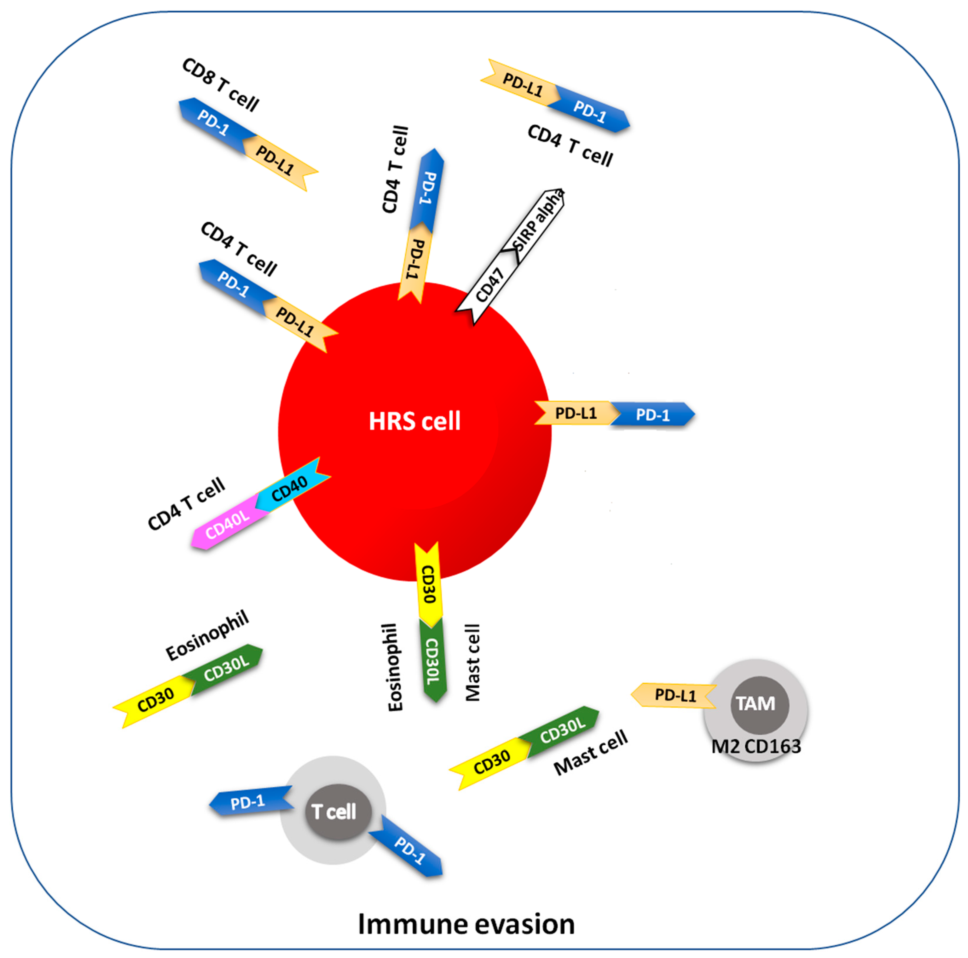
HRS cells are dependent on survival signals received from other cells, including CD40L-expressing T cells and CD30L+ mast cells and eosinophils [19,20]. A considerable fraction of infiltrating CD4+ T cells are regulatory T (Treg) cells. Treg cells and programmed death 1 (PD-1)+ T cells also interact with HRS cells, which produce the Treg attractant galectin-1 and the PD-1 ligand (PDL-1). The expression of PD-L1 points to the functional mechanism of immune escape using the PD-1 triggering of intratumoural T cells [19,20].
Therefore, cellular components of the cHL microenvironment express molecules involved in cancer cell growth and survival, such as CD30L or CD40L, or in immune escape, such as PD-1. For example, CD30L+ eosinophils and mast cells and proliferation-inducing ligand (APRIL)+ neutrophils consistently interact with HRS cells, whereas CD40L-expressing CD4+ T lymphocytes, that surround “rosetting” HRS cells, interact negatively with HRS cells. PD-1+ T cells also interact with HRS cells, which produce PD-L1 [19,20].

2.3. Expression of Functional CD40 in HRS Cells and HL Cell Lines

HRS cells and their morphologic variants express CD40, a member of the tumour necrosis factor/nerve growth factor receptor superfamily. The pattern of CD40 expression is highly distinctive—i.e., very intense, membranous, plus intracytoplasmic, and with a strong dot-like pattern in the paranuclear area [22]. Therefore, the immunohistochemical detection of CD40 appears of high value in the identification of HRS cells on tissue sections. The CD40 ligand is expressed on the microenvironmental reactive T cells of Hodgkin lymphoma (Figure 1). In all cHL subtypes, CD40L-expressing CD3+/CD4+ T lymphocytes are numerous in the HL-involved areas and are mainly located in close proximity to the HRS cells. These data provide morphological evidence that CD40L may play an important role in the cell-contact-dependent interaction of HRS cells (CD40+) in HL-involved areas and the microenvironmental CD3+/CD4+/CD40L+ T lymphocytes [23].
CD40 shows a significant homology to the HL-associated antigen CD30 and it is capable of transducing growth signals in a number of cell types. The relative cell density of CD40 on HL cell lines (L-428, KM-H2, HDLM-2) as assessed by flow-cytometry was significantly higher than on all other lymphoma cells analysed [22]. The engagement of CD40 by its soluble ligand (CD40L) enhances both the clonogenic capacity and colony cell survival of HL cell lines, stimulates proliferation, reduces apoptosis, and mediates the in vitro rosetting of activated CD4+ T cells to HL cells. CD40 engagement may play a role in the regulation of HRS cell expansion and the contact-dependent interactions of these cells with cytokine-producing T lymphocytes [22].

2.4. Interactions between PD-1 and Its Ligand PD-L1 in the Classic Hodgkin lymphoma Microenvironment

HRS cells escape immune surveillance and thereby immune destruction through activation of the PD-1 signal transduction pathway, which inactivates tumour-specific T cells (Figure 2). The PD-L1—PD-1 pathway therefore functions as a checkpoint that regulates T-cell-mediated immune responses. Checkpoint-inhibiting antibodies showed promising results in r/r cHL patients, but their tumours developed resistance to therapy [6,19,24,25].

3. Microenvironmental Impact of EBV Infection

The demonstration of monoclonal EBV genomes in HRS cells indicates that EBV infection occurred prior to clonal expansion and indicates a pathogenic role for this herpes virus in EBV-positive cases, probably as an early event in cHL development [27]. EBV-positive HRS cells express a relatively restricted set of viral genes, the so-called type II latency pattern, including EBNA-1, LMP-1, and LMP-2 latent proteins. The immunophenotypic features of HRS cells are identical in the different histologic subtypes of cHL. Conversely, the association with EBV shows marked differences. Notably, the virologic characteristics of cHL vary according to the immunocompetence status of the host and cHL subtype (Table 1) [27].
EBV is found in HRS cells in about 40% of cHL in the Western world, mostly in cases of MCcHL and LDcHL, and less frequently in NScHL and LRcHL. HL in patients living with HIV (PLWHs) show EBV-infected HRS cells in nearly all cases [21,27,28]. Regarding the PLWH setting, unfortunately, there are no data on ICIs in HIV-associated cHL, because PLWHs have been excluded from most trials in the general population. The results of two clinical trials support the safety of ICIs in PLWHs with advanced cancers, with no adverse effects on viral suppression and CD4 cell counts [29,30].
EBV infection might also affect the microenvironment composition by increasing the production of molecules involved in immune escape and T cell recruitment, such as IL-10 and CCL5. In EBV-positive cHL, HRS cells invariably express high levels of LMP-1 expression and are endowed with a highly efficient interaction with the cells of the tumour microenvironment, being able to attract a variety of cellular populations and respond to several autocrine and paracrine signals. The survival and low proliferation capacity of HRS cells are critically dependent on these interactions.

Do EBV-Related and EBV-Unrelated Hodgkin lymphomas Differ with Regard to Susceptibility to Checkpoint Blockade?

The tumour microenvironment of EBV-related cHLs contains higher numbers of macrophages and higher expression levels of PD-L1 than that of EBV-unrelated cHLs. Moreover, viral oncoprotein LMP1 may sustain an immunosuppressive microenvironment by inducing/enhancing the production of immunosuppressive cytokines and the expression of PD-1. The presence of enhanced immunosuppressive features in EBV-related cHL should make EBV-related cHL patients more susceptible to checkpoint blockade [27,31].

4. The Hodgkin lymphoma Microenvironment as a Therapeutic Target

Although cHL is a curable tumour, current treatments are unable to eradicate disease in about 30–35% of patients. Current active, yet non-curative, drugs such as BV, nivolumab, histone deacetylase inhibitors (HDACis), and immunomodulatory agents have allowed high rates of objective responses in r/r patients who fail autologous stem cell transplantation (SCT) [8,15,32]. Alternative treatment strategies for cHL patients have been envisioned not only to address the therapeutic challenge of primary refractory and early-relapsed cHL patients but also to reduce the long-term toxicities of chemo-radiotherapy in chemosensitive patients.
Current clinical results have recently been discussed and reviewed in ref. [33]. The Keynote-087 study reported a median duration of response of 16.6 months and a PFS of 13.7 months for patients receiving single-agent pembrolizumab [34]. Because of this study and others, combination ICI therapies have also been investigated, and studies such as SWOG S1826 have shown promising results of nivolumab in combination with chemotherapy [35]. Of note, several patients in a very recent reported cohort did receive combination doxorubicin, vinblastine, dacarbazine, and nivolumab. None of these patients relapsed [33]. However, despite these advances, most patients with relapsed or refractory disease do progress after ICIs, and additional therapeutic options are urgently needed. In fact, several clinical trials are ongoing, as reviewed in ref. [36].
Ipilimumab is included among the ICIs that are approved by the FDA, together with pembrolizumab, nivolumab, atezolizumab, avelumab, durvalumab, and cemiplimab. In a phase 1/2 trial, patients with r/r cHL were treated with either the combination of BV and nivolumab or ipilimumab (CTLA-4 antibody) or both. This study demonstrated improved responses with BV+ nivolumab over BV+ ipilimumab or BV+ both groups. About half of patients in the BV +ipilimumab group suffered grade 3–4 adverse events. The phase 2 study comparing BV+ nivolumab with or without ipilimumab is ongoing (NCT01896999) [11]. Treatment for r/r cHL after ASCT includes allogeneic stem cell transplant, brentuximab vedotin and other CD30 antibodies, immune checkpoint inhibitors (ICIs) targeting programmed death signalling, and investigational agents such as histone deacetylase inhibitors, Janus kinase 2 inhibitors, and chimeric antigen receptor (CAR) T-cell therapy [37]. Of these, ICIs have shown promising results, and for relapsed and refractory disease, the addition of nivolumab results in durable disease control for some patients, with a median progression-free survival (PFS) of 14.7 months in the CheckMate 205 trial [15].
Evidence to date suggests that CD40 activation is a critical mechanism to convert so-called “not-inflammed” tumours to inflamed ones, i.e., with a prominent tumour infiltration of T cells, sensitising them to checkpoint inhibition [26]. The clinical use of anti-CD40 agonist monoclonal antibodies (mAbs) is aimed at recruiting the immune system to fight the tumour cells, as it has been well demonstrated in various preclinical models. Realising the effective application of CD40 mAbs in clinical practice requires novel approaches designed to prevent or mitigate the systemic toxicity associated with CD40 agonism [38,39,40].

5. Conclusions

In a previous work [26], we reported how little the results of checkpoint blockade in diffuse large B-cell lymphomas and other B-cell lymphomas are similar to those obtained in Hodgkin lymphoma, probably due to the different composition of the tumour microenvironment.
In the current work, we hypothesise how to increase the efficacy of checkpoint inhibitors in patients with classic Hodgkin lymphoma. In these patients, it is discussed how checkpoint inhibition should be integrated with other types of immunotherapy. In particular, how to inhibit the growth pathways of the Reed–Sternberg cell is discussed, for example, with the use of CD40 antagonists.
Recent years have witnessed an increasing focus on immune checkpoints encompassing biomarkers, innovative therapies combining immune checkpoint inhibitors, and the comprehensive exploration of resistance and toxicity. A major challenge is how to overcome drug resistance in cHL through rationale combinations of checkpoint inhibitors with other therapies.
When employed as a single therapy, the efficacy of ICIs encounters limitations due to a low response rate and associated immune-related side-effects. The evolving landscape of combination therapies provide new opportunities for applying ICIs in cHL therapy.
Furthermore, numerous unsatisfactory clinical issues still need to be addressed in the application of ICIs due to the nascent state of technology and factors, such as resistance and adverse effects [41]. Several systems may encounter complications linked to ICI. These complications span the skin, digestive, respiratory, endocrine, cardiovascular, urinary, and central nervous systems. While skin and gastrointestinal toxicities are more frequent, they tend to be less severe. Conversely, lung-, cardiac-, neurological-, and nephrotoxicity are infrequent but more severe, occasionally leading to fatal outcomes [18,41,42].
In the future, immunotherapy will not be limited to monotherapy with immune checkpoints alone. Recent research has focused on the application of multiple ICIs in combination with other therapies including antiviral therapies in EBV-positive cHL, preventing and eliminating their adverse effects through monitoring and early intervention. Therefore, this strategy could increase the efficacy of ICI, decrease or mitigate adverse effects, and ultimately expand the fraction of patients who can benefit from ICIs.

Funding

This research received no external funding.

Institutional Review Board Statement

Not applicable.

Informed Consent Statement

Not applicable.

Data Availability Statement

No new data were created or analyzed in this study. Data sharing is not applicable to this article.

Conflicts of Interest

The authors declare no conflict of interest.

References

  1. Connors, J.M.; Cozen, W.; Steidl, C.; Carbone, A.; Hoppe, R.T.; Flechtner, H.H.; Bartlett, N.L. Hodgkin lymphoma. Nat. Rev. Dis. Primers 2020, 6, 61. [Google Scholar] [CrossRef] [PubMed]
  2. Campo, E.; Jaffe, E.S.; Cook, J.R.; Quintanilla-Martinez, L.; Swerdlow, S.H.; Anderson, K.C.; Brousset, P.; Cerroni, L.; de Leval, L.; Dirnhofer, S.; et al. The International Consensus Classification of Mature Lymphoid Neoplasms: A Report from the Clinical Advisory Committee. Blood 2022, 140, 1229–1253. [Google Scholar] [CrossRef] [PubMed]
  3. Bouvard, V.; Baan, R.; Straif, K.; Grosse, Y.; Secretan, B.; El Ghissassi, F.; Benbrahim-Tallaa, L.; Guha, N.; Freeman, C.; Galichet, L.; et al. A Review of Human Carcinogens. Part B: Biological Agents; IARC: Lyon, France, 2012. [Google Scholar]
  4. Younes, A.; Carbone, A.; Johnson, P.; Dabaja, B.; Ansell, S.; Kuruvilla, J. Hodgkin’s Lymphoma; De Vita, V., Lawrence, T., Rosemberg, S., Eds.; Wolters Kluwer Health/Lippincott Williams and Wilkins: Philadelphia, PA, USA, 2014. [Google Scholar]
  5. Anagnostopoulos, I.; Piris, M.; Isaacson, P.; Jaffe, E.S.; Stein, H. Lymphocyte-rich classic Hodgkin lymphoma. In WHO Classification of Tumours of Haematopoietic and Lymphoid Tissues, 4th ed.; Swerdlow, S.H., Campo, E., Harris, N.L., Jaffe, E.S., Pileri, S.A., Stein, H., Thiele, J., Vardiman, J.W., Eds.; IARC Press: Lyon, France, 2017; pp. 438–440. [Google Scholar]
  6. Armand, P. Immune checkpoint blockade in hematologic malignancies. Blood 2015, 125, 3393–3400. [Google Scholar] [CrossRef] [PubMed]
  7. Tutino, F.; Giovannini, E.; Chiola, S.; Giovacchini, G.; Ciarmiello, A. Assessment of Response to Immunotherapy in Patients with Hodgkin Lymphoma: Towards Quantifying Changes in Tumor Burden Using FDG-PET/CT. J. Clin. Med. 2023, 12, 3498. [Google Scholar] [CrossRef] [PubMed] [PubMed Central]
  8. Advani, R.H.; Moskowitz, A.J.; Bartlett, N.L.; Vose, J.M.; Ramchandren, R.; Feldman, T.A.; LaCasce, A.S.; Christian, B.A.; Ansell, S.M.; Moskowitz, C.H.; et al. Brentuximab vedotin in combination with nivolumab in relapsed or refractory Hodgkin lymphoma: 3-year study results. Blood 2021, 138, 427–438. [Google Scholar] [CrossRef] [PubMed]
  9. Ansell, S.M.; Lesokhin, A.M.; Borrello, I.; Halwani, A.; Scott, E.C.; Gutierrez, M.; Schuster, S.J.; Millenson, M.M.; Cattry, D.; Freeman, G.J.; et al. PD-1 blockade with nivolumab in relapsed or refractory Hodgkin’s lymphoma. N. Engl. J. Med. 2015, 372, 311–319. [Google Scholar] [CrossRef] [PubMed] [PubMed Central]
  10. Armand, P.; Chen, Y.B.; Redd, R.A.; Joyce, R.M.; Bsat, J.; Jeter, E.; Merryman, R.W.; Coleman, K.C.; Dahi, P.B.; Nieto, Y.; et al. PD-1 blockade with pembrolizumab for classical Hodgkin lymphoma after autologous stem cell transplantation. Blood 2019, 134, 22–29. [Google Scholar] [CrossRef] [PubMed] [PubMed Central]
  11. Diefenbach, C.S.; Hong, F.; Ambinder, R.F.; Cohen, J.B.; Robertson, M.J.; David, K.A.; Advani, R.H.; Fenske, T.S.; Barta, S.K.; Palmisiano, N.D.; et al. Ipilimumab, nivolumab, and brentuximab vedotin combination therapies in patients with relapsed or refractory Hodgkin lymphoma: Phase 1 results of an open-label, multicentre, phase 1/2 trial. Lancet Haematol. 2020, 7, e660–e670. [Google Scholar] [CrossRef] [PubMed] [PubMed Central]
  12. Armand, P.; Shipp, M.A.; Ribrag, V.; Michot, J.M.; Zinzani, P.L.; Kuruvilla, J.; Snyder, E.S.; Ricart, A.D.; Balakumaran, A.; Rose, S.; et al. Programmed Death-1 Blockade with Pembrolizumab in Patients with Classical Hodgkin Lymphoma after Brentuximab Vedotin Failure. J. Clin. Oncol. 2016, 34, 3733–3739. [Google Scholar] [CrossRef] [PubMed] [PubMed Central]
  13. Mei, M.G.; Lee, H.J.; Palmer, J.M.; Chen, R.; Tsai, N.C.; Chen, L.; McBride, K.; Smith, D.L.; Melgar, I.; Song, J.Y.; et al. Response-adapted anti-PD-1-based salvage therapy for Hodgkin lymphoma with nivolumab alone or in combination with ICE. Blood 2022, 139, 3605–3616. [Google Scholar] [CrossRef] [PubMed] [PubMed Central]
  14. Kuruvilla, J.; Ramchandren, R.; Santoro, A.; Paszkiewicz-Kozik, E.; Gasiorowski, R.; Johnson, N.A.; Fogliatto, L.M.; Goncalves, I.; de Oliveira, J.S.; Buccheri, V.; et al. Pembrolizumab versus brentuximab vedotin in relapsed or refractory classical Hodgkin lymphoma (KEYNOTE-204): An interim analysis of a multicentre, randomised, open-label, phase 3 study. Lancet Oncol. 2021, 22, 512–524. [Google Scholar] [CrossRef] [PubMed]
  15. Armand, P.; Engert, A.; Younes, A.; Fanale, M.; Santoro, A.; Zinzani, P.L.; Timmerman, J.M.; Collins, G.P.; Ramchandren, R.; Cohen, J.B.; et al. Nivolumab for Relapsed/Refractory Classic Hodgkin Lymphoma after Failure of Autologous Hematopoietic Cell Transplantation: Extended Follow-Up of the Multicohort Single-Arm Phase II CheckMate 205 Trial. J. Clin. Oncol. 2018, 36, 1428–1439. [Google Scholar] [CrossRef] [PubMed] [PubMed Central]
  16. Veldman, J.; Visser, L.; Berg, A.V.D.; Diepstra, A. Primary and acquired resistance mechanisms to immune checkpoint inhibition in Hodgkin lymphoma. Cancer Treat. Rev. 2020, 82, 101931. [Google Scholar] [CrossRef] [PubMed]
  17. Wenthe, J.; Naseri, S.; Labani-Motlagh, A.; Enblad, G.; Wikström, K.I.; Eriksson, E.; Loskog, A.; Lövgren, T. Boosting CAR T-cell responses in lymphoma by simultaneous targeting of CD40/4-1BB using oncolytic viral gene therapy. Cancer Immunol. Immunother. 2021, 70, 2851–2865. [Google Scholar] [CrossRef] [PubMed] [PubMed Central]
  18. Wang, D.Y.; Salem, J.E.; Cohen, J.V.; Chandra, S.; Menzer, C.; Ye, F.; Zhao, S.; Das, S.; Beckermann, K.E.; Ha, L.; et al. Fatal Toxic Effects Associated with Immune Checkpoint Inhibitors: A Systematic Review and Meta-analysis. JAMA Oncol. 2018, 4, 1721–1728. [Google Scholar] [CrossRef] [PubMed] [PubMed Central]
  19. Liu, Y.; Sattarzadeh, A.; Diepstra, A.; Visser, L.; van den Berg, A. The microenvironment in classical Hodgkin lymphoma: An actively shaped and essential tumor component. Semin. Cancer Biol. 2014, 24, 15–22. [Google Scholar] [CrossRef] [PubMed]
  20. Aldinucci, D.; Gloghini, A.; Pinto, A.; De Filippi, R.; Carbone, A. The classical Hodgkin’s lymphoma microenvironment and its role in promoting tumour growth and immune escape. J. Pathol. 2010, 221, 248–263. [Google Scholar] [CrossRef]
  21. Vaccher, E.; Gloghini, A.; Volpi, C.C.; Carbone, A. Lymphomas in People Living with HIV. Hemato 2022, 3, 527–542. [Google Scholar] [CrossRef]
  22. Carbone, A.; Gloghini, A.; Gattei, V.; Aldinucci, D.; Degan, M.; De Paoli, P.; Zagonel, V.; Pinto, A. Expression of functional CD40 antigen on Reed-Sternberg cells and Hodgkin’s disease cell lines. Blood 1995, 85, 780–789. [Google Scholar] [CrossRef] [PubMed]
  23. Carbone, A.; Gloghini, A.; Gruss, H.-J.; Pinto, A. CD40 ligand is constitutively expressed in a subset of T cell lymphomas and on the microenvironmental reactive T cells of follicular lymphomas and Hodgkin’s disease. Am. J. Pathol. 1995, 147, 912–922. [Google Scholar]
  24. Carbone, A.; Gloghini, A. Checkpoint blockade therapy resistance in Hodgkin’s lymphoma. Lancet 2018, 392, 1194–1196. [Google Scholar] [CrossRef] [PubMed]
  25. Yi, M.; Zheng, X.; Niu, M.; Zhu, S.; Ge, H.; Wu, K. Combination strategies with PD-1/PD-L1 blockade: Current advances and future directions. Mol. Cancer 2022, 21, 28. [Google Scholar] [CrossRef] [PubMed] [PubMed Central]
  26. Carbone, A.; Gloghini, A.; Carlo-Stella, C. Tumor microenvironment contribution to checkpoint blockade therapy: Lessons learned from Hodgkin lymphoma. Blood 2023, 141, 2187–2193. [Google Scholar] [CrossRef] [PubMed] [PubMed Central]
  27. Carbone, A.; Gloghini, A.; Caruso, A.; De Paoli, P.; Dolcetti, R. The impact of EBV and HIV infection on the microenvironmental niche underlying Hodgkin lymphoma pathogenesis. Int. J. Cancer 2017, 140, 1233–1245. [Google Scholar] [CrossRef] [PubMed]
  28. Chadburn, A.; Gloghini, A.; Carbone, A. Classification of B-Cell Lymphomas and Immunodeficiency-Related Lymphoproliferations: What’s New? Hemato 2023, 4, 26–41. [Google Scholar] [CrossRef]
  29. Uldrick, T.S.; Gonçalves, P.H.; Abdul-Hay, M.; Claeys, A.J.; Emu, B.; Ernstoff, M.S.; Fling, S.P.; Fong, L.; Kaiser, J.C.; Lacroix, A.M.; et al. Assessment of the Safety of Pembrolizumab in Patients with HIV and Advanced Cancer-A Phase 1 Study. JAMA Oncol. 2019, 5, 1332–1339. [Google Scholar] [CrossRef] [PubMed] [PubMed Central]
  30. Gonzalez-Cao, M.; Morán, T.; Dalmau, J.; Garcia-Corbacho, J.; Bracht, J.W.; Bernabe, R.; Juan, O.; de Castro, J.; Blanco, R.; Drozdowskyj, A.; et al. Assessment of the Feasibility and Safety of Durvalumab for Treatment of Solid Tumors in Patients with HIV-1 Infection: The Phase 2 DURVAST Study. JAMA Oncol. 2020, 6, 1063–1067. [Google Scholar] [CrossRef] [PubMed] [PubMed Central]
  31. Carbone, A.; Gloghini, A.; Carlo-Stella, C. Are EBV-related and EBV-unrelated Hodgkin lymphomas different with regard to susceptibility to checkpoint blockade? Blood 2018, 132, 17–22. [Google Scholar] [CrossRef] [PubMed]
  32. Carbone, A.; Gloghini, A.; Castagna, L.; Santoro, A.; Carlo-Stella, C. Primary refractory and early-relapsed Hodgkin’s lymphoma: Strategies for therapeutic targeting based on the tumour microenvironment. J. Pathol. 2015, 237, 4–13. [Google Scholar] [CrossRef] [PubMed]
  33. Burlile, J.F.; Frechette, K.M.; Breen, W.G.; Hwang, S.R.; Higgins, A.S.; Nedved, A.N.; Harmsen, W.S.; Pulsipher, S.D.; Witzig, T.E.; Micallef, I.N.; et al. Patterns of progression after immune checkpoint inhibitors for Hodgkin lymphoma: Implications for radiation therapy. Blood Adv. 2024, 8, 1250–1257. [Google Scholar] [CrossRef] [PubMed] [PubMed Central]
  34. Armand, P.; Zinzani, P.L.L.; Lee, H.J.; Johnson, N.; Brice, P.; Radford, J.; Ribrag, V.; Molin, D.; Vassilakopoulos, T.P.; Tomita, A.; et al. Five-year follow-up of KEYNOTE-087: Pembrolizumab monotherapy for relapsed/refractory classical Hodgkin lymphoma. Blood 2023, 142, 878–886. [Google Scholar] [CrossRef] [PubMed] [PubMed Central]
  35. Herrera, A.F.; LeBlanc, M.L.; Castellino, S.M.; Li, H.; Rutherford, S.C.; Evens, A.M.; Davison, K.; Punnett, A.; Hodgson, D.C.; Parsons, S.K.; et al. SWOG S1826, a randomized study of nivolumab(N)-AVD versus brentuximab vedotin(BV)-AVD in advanced stage (AS) classic Hodgkin lymphoma (HL). J. Clin. Oncol. 2023, 41 (Suppl. S17), LBA4. [Google Scholar] [CrossRef]
  36. Pophali, P.; Varela, J.C.; Rosenblatt, J. Immune checkpoint blockade in hematological malignancies: Current state and future potential. Front. Oncol. 2024, 14, 1323914. [Google Scholar] [CrossRef] [PubMed] [PubMed Central]
  37. Domingo-Domènech, E.; Sureda, A. Treatment of Hodgkin Lymphoma Relapsed after Autologous Stem Cell Transplantation. J. Clin. Med. 2020, 9, 1384. [Google Scholar] [CrossRef] [PubMed] [PubMed Central]
  38. Salomon, R.; Dahan, R. Next Generation CD40 Agonistic Antibodies for Cancer Immunotherapy. Front. Immunol. 2022, 13, 940674. [Google Scholar] [CrossRef] [PubMed] [PubMed Central]
  39. Van Hooren, L.; Vaccaro, A.; Ramachandran, M.; Vazaios, K.; Libard, S.; van de Walle, T.; Georganaki, M.; Huang, H.; Pietilä, I.; Lau, J.; et al. Agonistic CD40 therapy induces tertiary lymphoid structures but impairs responses to checkpoint blockade in glioma. Nat. Commun. 2021, 12, 4127. [Google Scholar] [CrossRef] [PubMed] [PubMed Central]
  40. Vonderheide, R.H. CD40 Agonist Antibodies in Cancer Immunotherapy. Annu. Rev. Med. 2020, 71, 47–58. [Google Scholar] [CrossRef] [PubMed]
  41. Morad, G.; Helmink, B.A.; Sharma, P.; Wargo, J.A. Hallmarks of response, resistance, and toxicity to immune checkpoint blockade. Cell 2021, 184, 5309–5337. [Google Scholar] [CrossRef] [PubMed] [PubMed Central]
  42. Hradska, K.; Hajek, R.; Jelinek, T. Toxicity of Immune-Checkpoint Inhibitors in Hematological Malignancies. Front. Pharmacol. 2021, 12, 733890. [Google Scholar] [CrossRef] [PubMed] [PubMed Central]
Figure 1. (A) A multinucleated Hodgkin Reed–Sternberg cell is surrounded by a “rosette” of small lymphocytes. (B) mRNA in situ hybridisation shows a distinct brown signal for CD40 in a Hodgkin Reed–Sternberg cell.
Hemato 05 00016 g001
Figure 2. In classic Hodgkin lymphoma (cHL), the expression of PD-L1 can be found in Hodgkin Reed–Sternberg (HRS) tumour cells, and the expression of PD-1 is found on microenvironmental CD4+ T cells. PD-1, which acts as an immunomodulatory molecule, is a negative regulator of activated T cells, B cells, and myeloid cells. PD-L1, a ligand of PD-1, is expressed on T cells, B cells, dendritic cells, and macrophages. PD-L1 binds to PD-1 on T cells and regulates their activity. The binding of PD-1 to T cells with PD-L1 to HRS cells inactivates tumour-specific T cells, functioning as a checkpoint used by HRS cells to escape immune surveillance and evade immune destruction. The engagement of CD40 by its ligand (CD40L) may play a role in the regulation of HRS cell expansion (modified and adapted from ref. [26]).
Hemato 05 00016 g002
Table 1. Morphologic and virologic characteristic of classic Hodgkin lymphoma according to the immunocompetence status of the host.
HostHodgkin Lymphoma SubtypeEBV Infection
cHL of the general population
cHL, nodular sclerosis Usually − *
cHL, mixed cellularity Usually + *
Rare types
cHL, lymphocyte-rich Variably +
cHL, lymphocyte depleted Variably +
Immunodeficiency-associated cHL
HIV-associated cHL
cHL, nodular sclerosis+
cHL, lymphocyte depleted +
cHL, mixed cellularity +
Less frequent
cHL, lymphohistiocyoid +
Post-transplant (cHL-type PTLD)
Similar to other cHL+
Other iatrogenic immune-deficiency-associated cHL
cHL, mixed cellularityVariably + (usually +)
Abbreviations. cHL, classic Hodgkin lymphoma; PTLD, post-transplant lymphoproliferative disorder; −, negative; +, positive. * Association with EBV is less frequent in NS (10–40%) than in MC cHL (approximately 75% of cases).
Disclaimer/Publisher’s Note: The statements, opinions and data contained in all publications are solely those of the individual author(s) and contributor(s) and not of MDPI and/or the editor(s). MDPI and/or the editor(s) disclaim responsibility for any injury to people or property resulting from any ideas, methods, instructions or products referred to in the content.