Abstract
This study investigates the cytotoxic properties of metal complexes incorporating thio-uracil derivatives, specifically 2,4-dithiouracil and 6-propyl-2-thiouracil. The research focuses on the cytotoxic effects of Cu(II) and Pd(II) complexes with 6-propyl-2-thiouracil, as well as mixed-ligand transition metal Cu(II) and Au(III) complexes of 2,4-dithiouracil with 2-thiouracil and uracil. Cytotoxic activity was assessed against human cervical carcinoma cells (HeLa) and normal kidney cells from the African green monkey. The results demonstrated that incorporating Cu(II) and Au(III) into the compound structures significantly enhanced their cytotoxic effects. Notably, all tested complexes exhibited a stronger inhibitory effect on cancer cell proliferation compared to normal cells, with the palladium(II) complex of 6-propyl-2-thiouracil showing the lowest CD50 value against the tumor cell line (0.00064 mM), which were 149 times lower than that of the ligand (0.0955 mM). These findings suggest that thio-uracil-based metal complexes, particularly those containing palladium (II) and gold(III), hold significant potential for further development as anticancer agents.
Keywords:
6-propyl-2-thiouracil; 2,4-dithiouracil; copper(II) complexes; palladium(II) complexes; gold(III) complexes; cytotoxic properties Key Contribution:
All tested compounds exhibited a stronger inhibitory effect on cancer cell proliferation compared to normal cells, with the Pd(II) complex of 6-propyl-2-thiouracil showing the lowest CD50 value against the tumor cell line (0.00064 mM), which were 149 times lower than that of the ligand (0.0955 mM).
1. Introduction
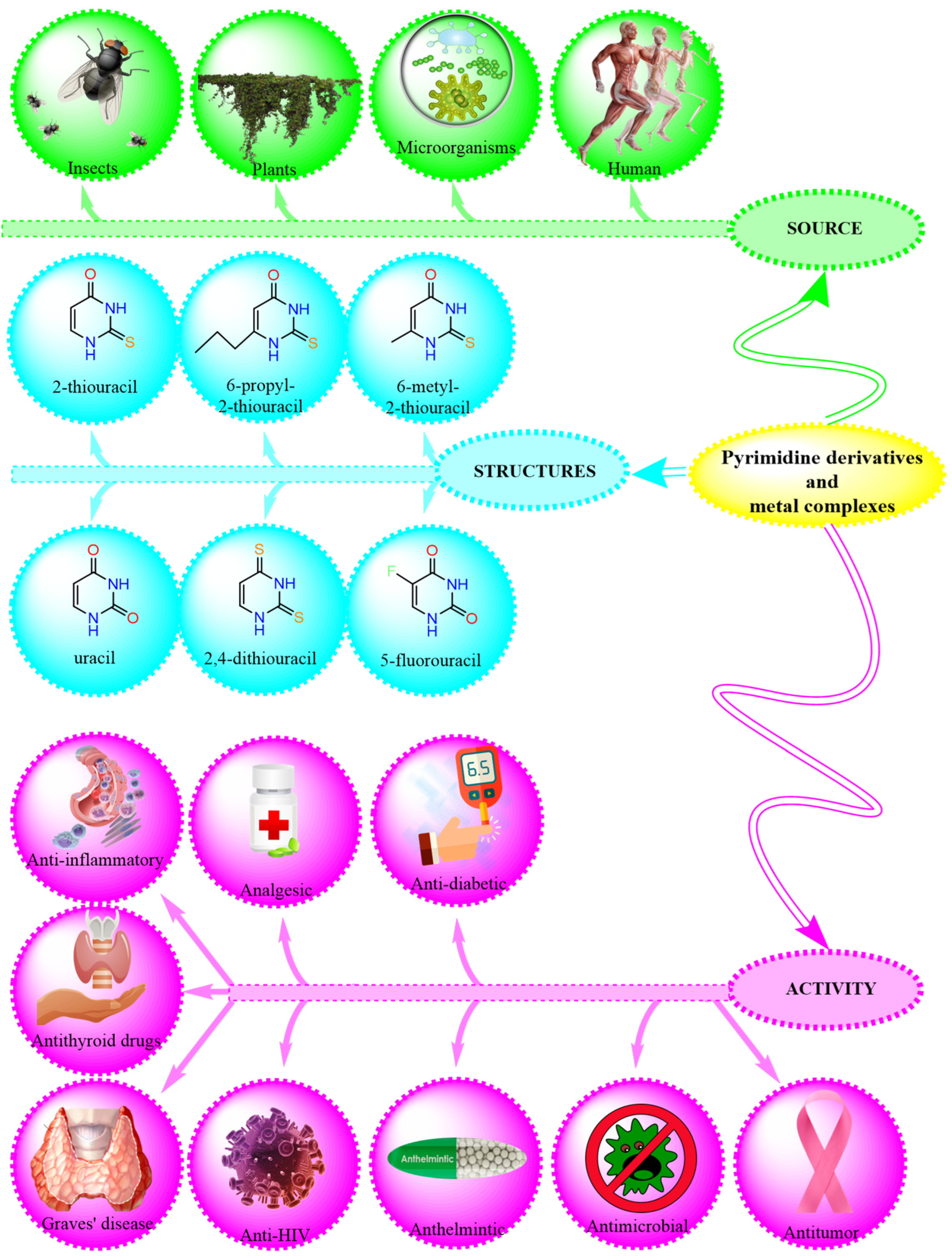
Thionamides, a category of relatively straightforward molecules, function as antithyroid medications, incorporating a sulfhydryl group and a thiourea unit within a heterocyclic structure [1,2,3,4]. The interest in platinum and palladium compounds arises from their notable cytostatic properties [5]. In Scheme 1, the different applications of bioactive pyrimidines, such as uracil, 2,4-dithiouracil, 6-propyl-2-thiouracil, and other pyrimidines and their metal complexes, is given [6,7,8,9,10]. These compounds are modifications of the pyrimidine ring, which are often used in medicinal chemistry and drug design.
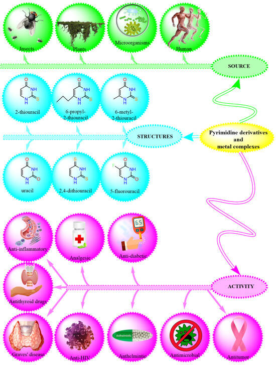
Scheme 1.
Different applications and structures of pyrimidine derivatives and their metal complexes.
Recently, cis-dihalogenated complexes of Pd(II) and Pt(II) were investigated in combination with 6-tert-butyl-2-thiouracil [11]. Vetter et al. described the synthesis, characterization, and in vitro study of the cytotoxic potential of platinum(IV) complexes with thiouracil-based ligands [12]. Ru(II)-centered complexes incorporating antimicrobal 2-thiouracil derivatives—specifically 6-methyl-2-thiouracil (6m2TU) and 2-thiouracil (2TU)—were tested against HepG2 (human hepatocellular carcinoma), K-562 (human chronic myeloid leukemia), HL-60 (human promyelocytic leukemia), and B16-F10 (mouse melanoma) cancer cells, as well as non-cancerous cells (PBMCs) (human peripheral blood mononuclear cells activated with concanavalin A—human lymphoblasts) [13]. The biological evaluation indicated that a complex with 6-methyl-2-thiouracil demonstrated greater potential than a complex with 2-thiouracil. Ultimately, this investigation suggests that complexes with 2-thiouracil and 6-methyl-2-thiouracil induce apoptosis, significantly increasing the proportion of apoptotic HL-60 cells by disrupting the cell cycle and decreasing the presence of cells in the G1/G0, G2/M, and S phases [13]. Correa et al. presented the chemical and cytotoxic investigations of four novel ruthenium(II) complexes incorporating uracil derivatives [4]. In vitro studies tested their activity against HL-60, HepG2 (human liver cancer), B16-F10, and K562 cells, along with non-cancerous PBMCs [14]. Bomfim et al. synthesized Ru(II) complexes incorporating 6-methyl-2-thiouracil, demonstrating their potential as innovative antileukemic drug candidates [15]. Oladipo and Isola carried out a comprehensive review of uracil’s coordination chemistry and the practical uses of several of its complexes [16]. 5-substituted uracils are recognized as key structural components in various therapeutics [17]. Prachayasittikul et al. synthesized novel mixed-ligand transition metal (Cu, Ni, and Mn) complexes of 5-iodouracil (5Iu) with 8-hydroxyquinoline (8HQ) and 5-nitrouracil (5Nu) with 8HQ. These metal complexes showed notable cytotoxic effect against HuCCA-1, A-549, HepG2, and MOLT-3 cell lines [7]. Al-Halbosy et al. presented the cytotoxic effect of the N-Phenylmorpholine-4-carbothioamide (HPMCT) and PdCl2(HPMCT)2] and [PtCl2(HPMCT)2] complexes. The compounds were assessed on breast cancer cell lines (MCF-7), and a complex with a palladium ion revealed the most promising activity with an IC50 value 12.72 ± 0.4 μM [18]. Copper complexes exhibit cytotoxic activity through mechanisms distinct from those employed by cisplatin, which is a platinum-based chemotherapeutic agent currently in clinical use [19]. The biological efficacy of these copper compounds varies significantly depending on the nature of the coordinated ligands. Investigations into their cytotoxic potential are often grounded in the hypothesis that endogenous copper may exert lower toxicity on normal cells compared to malignant ones [20]. The involvement of copper in angiogenesis remains a matter of scientific debate, and the broader role of transition metals in this process continues to be actively explored [20]. Notably, copper complexes have been shown to mimic the activity of superoxide dismutase (SOD) [21], which is a key antioxidant enzyme that catalyzes the dismutation of superoxide radicals into less reactive molecular species.
Recently, a complex with the general formulae [Cu(C20H22NO3)2]·H2O was obtained and its cytotoxic properties were assessed [22]. The in vitro cytotoxic investigation was conducted using an MTT assay, and the result revealed that a metal complex exhibited enhanced cytotoxicity, high selectivity, and dose-dependent cytotoxicity [22]. Zou et al. described gold complexes in detail in a recent review [23]. Many Au(III) complexes were synthesized, and the anticancer effect was assisted against different cancer cell lines. In most cases, the ligands contained a donor atom, either Cl, Br, S, or P. da Silva Maia et al. have also synthesized gold(III) complexes, and their cytotoxic effects were studied [24]. Most of the cited gold(III) complexes had a profound effect on cisplatin-resistant cell lines. The cytotoxic properties of a complex with the general formulae [(η-C5H5)2Ti{OC(O)CH2PPh2AuCl}2] was investigated in vitro against prostate and renal cell lines as potential chemotherapeutics drugs [25]. The result showed that the complex acts synergistically because the resulting cytotoxic effect is more pronounced when compared to [{HOC(O)RPPh2}AuCl] (R = −CH2−6, −4-C6H4−7) in renal cancer cell lines [25]. Metal complexes tend to associate with specific residues, many of which play key roles in the enzyme’s catalytic function. This interaction can disrupt cellular activities, ultimately leading to programmed cell death (apoptosis) [26]. Rana et al. investigated a gold(III) complex known to bind to TrxR [26]. In this enzyme, the catalytic residues were positioned between two subunits, each contributing to the gold(III) complex attachment. To date, numerous metal complexes have been developed utilizing uracil and thiouracil derivatives, incorporating various metals such as Cu, Fe, Co, Ni, Zn, Mn, Cd, and V [27,28,29], along with Pd, Pt, and Au, with analyses conducted on their composition and structural characteristics [30]. Novel thiolate gold(I) complexes, incorporating P(NMe2)3 (HMPT) as a phosphine ligand, were successfully synthesized [31], with two of these gold(I) thiolate complexes exhibiting potential as promising chemotherapy agents. Additionally, the cytotoxic effect of various metal complexes derived from thiouracil have been investigated against different cancer cell lines [31,32,33,34,35]. Recently, Marinova et al. successfully synthesized copper(II), palladium(II), and gold(III) complexes with 2-thiouracil [36], along with novel palladium(II) and copper(II) complexes containing 6-propyl-2-thiouracil and 6-methyl-2-thiouracil [37]. Additionally, Au(III) and Cu(II) complexes incorporating 2,4-dithiouracil (2,4-DTu) were developed [38], as well as an Au(III) complex with 6-methyl-2-thioxo-2,3-dihydropyrimidin-4(1H)-one [39]. Furthermore, the biological effect and synthesis of several metal complexes derived from 2-thiouracil and its derivatives were explored [40]. Adhikari et al. carried out a comprehensive review of Pt, Ru, Au, Cu, Ir, and Os coordination compounds and its anti-cancer activities [41]. Numerous Au(I) and Au(III) complexes have been obtained and investigated as possible antitumor therapies in recent years [42,43,44,45].
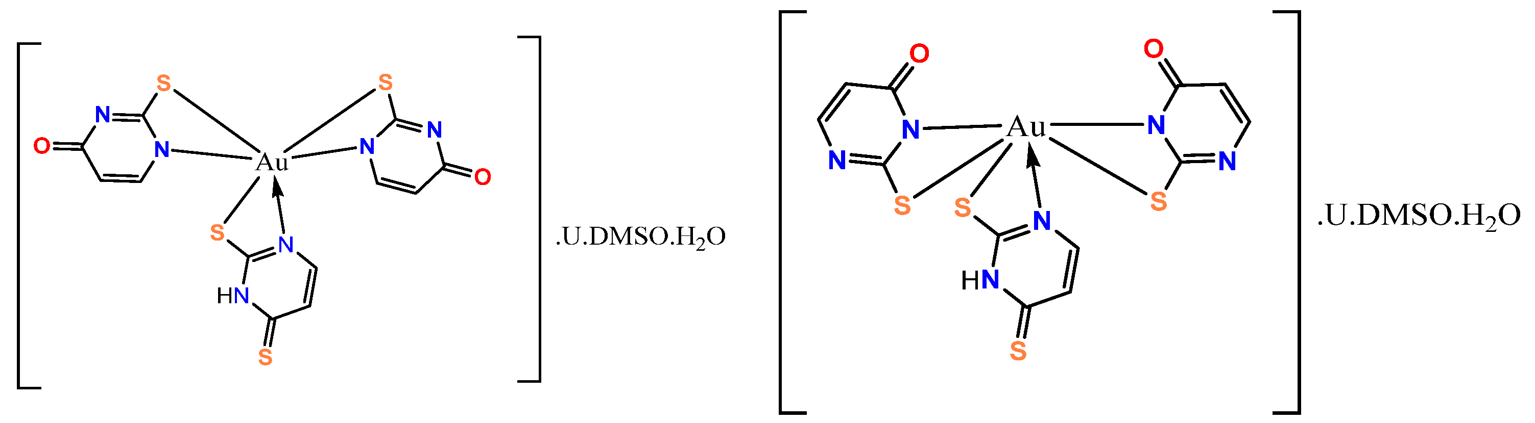
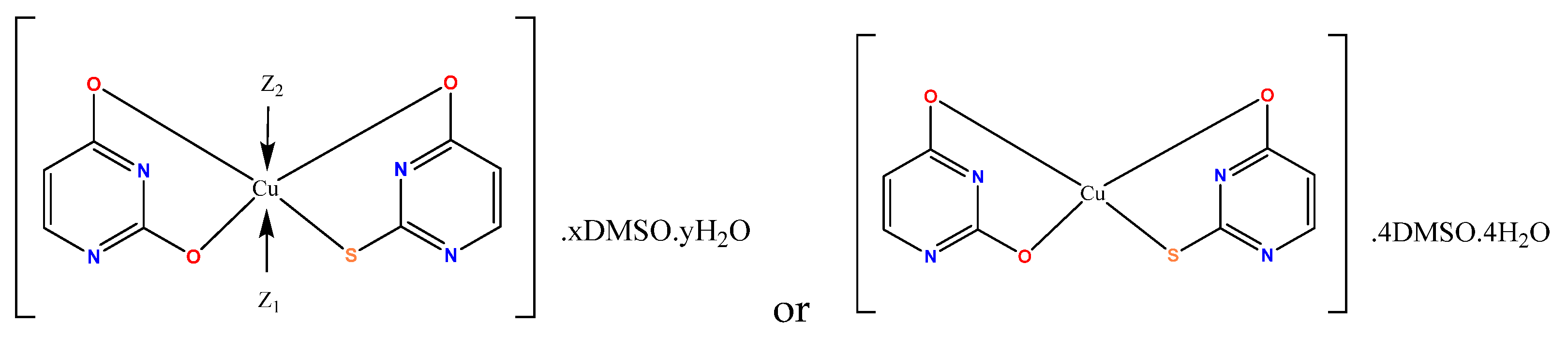
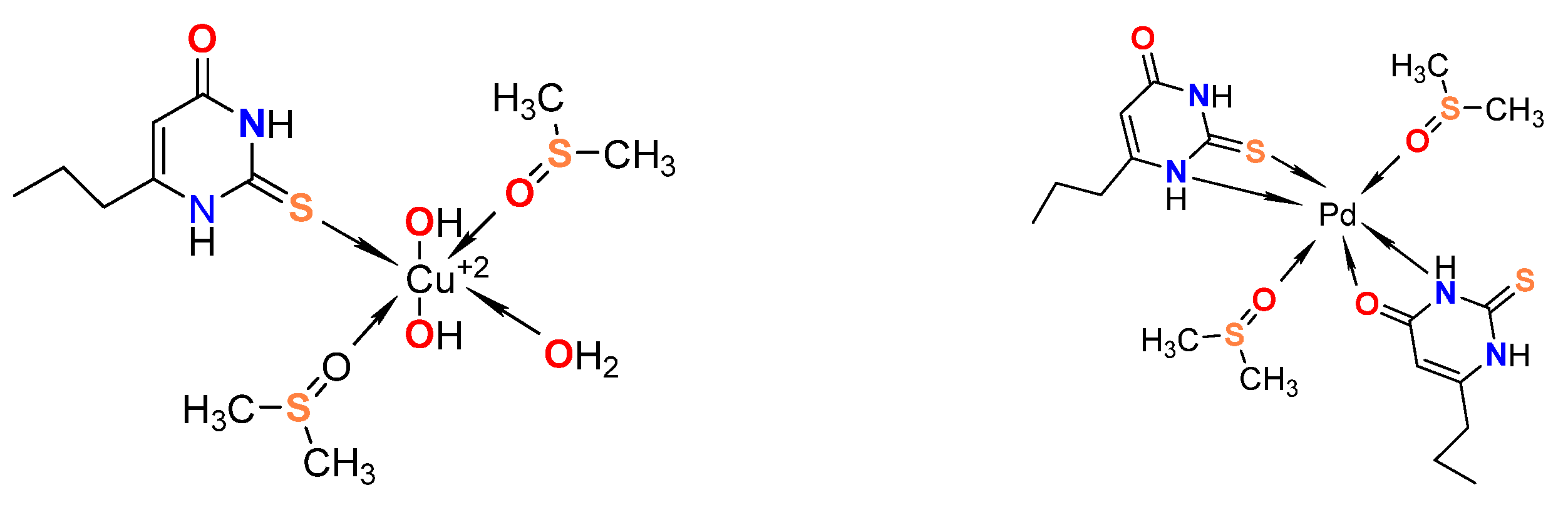
This article describes the cytotoxic properties of metal compounds with 2,4-dithiouracil and 6-propyl-2-thiouracil. In this study, we provide details regarding the cytotoxic effect of Pd(II) and Cu(II) complexes with 6-propyl-2-thiouracil and Au(III) and Cu(II) complexes with 2,4-dithiouracil. The possible structures of the gold and copper complexes with 2,4-dithiouracil are shown in Figure 1 and Figure 2, respectively, and Cu(II) and Pd(II) complexes with 6-propyl-2-thiouracil are shown in Figure 3. All complexes that are the subject of this study have been previously published by us in two articles [37,38]. In our previous study, it was proposed that 2,4-dithiouracil might undergo desulfurization due to the action of the NaOH employed during the synthesis of the gold complex. This process could lead to the substitution of one or both sulfur atoms with oxygen, thereby yielding 2-thiouracil and uracil as potential products within the reaction mixture [38]. The structures of the present complexes are discussed based on melting point analysis, microwave plasma atomic emission spectrometry (MP-AES) for Cu and Pd, inductively coupled plasma optical emission spectrometry (ICP-OES) for S (complexes with 2,4-dithiouracil), solution and solid-state NMR, attenuated total reflection (ATR), and Raman spectroscopy [37,38].
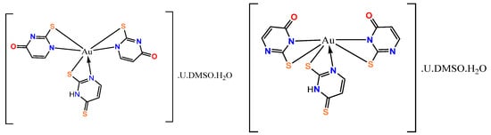
Figure 1.
The probable structure of the gold complex with 2,4-dithiouracil, 2-Tu, and U [38].
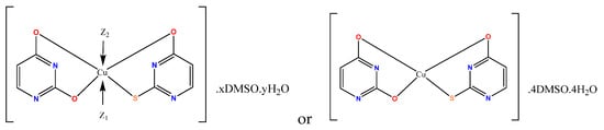
Figure 2.
A proposed structural arrangement and the corresponding coordination binding sites in the copper complex. If Z1 = H2O or DMSO-h6 then Z2 = H2O or DMSO-h6 [38].
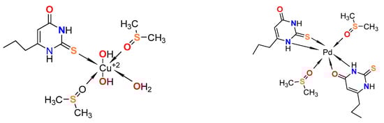
Figure 3.
The illustration of the suggested coordination binding sites for 6-propyl-2-thiouracil with copper and palladium [37].
2. Materials and Methods
2.1. Cytotoxicity Assay
To assess the in vitro biocompatibility of the tested compounds, a series of cell viability assays were conducted on human cervical carcinoma cell lines (HeLa), as well as on normal Vero cells (kidney cells from the African green monkey). All cell lines were obtained from the German Collection of Microorganisms and Cell Cultures (DSMZ GmbH, Braunschweig, Germany). The cell cultures were maintained in DMEM (Dulbecco’s Modified Eagle Medium) (Gibco Thermofisher Scientific, 168 Third Avenue, Waltham, MA, USA, 02451) growth medium, supplemented with 10% fetal bovine serum (FBS, Sigma-Aldrich, Darmstadt, Germany) and 5% L-glutamine (Sigma-Aldrich, Darmstadt, Germany) and incubated under controlled conditions of 37 °C in a humidified atmosphere with 5% CO2.
2.2. Cell Viability Assessment
The experimental approach included a series of cytotoxicity assays to determine the extent of cell proliferation inhibition by the synthesized complexes and their free ligands. Cell viability was quantified using a standard MTT-based colorimetric assay. Exponentially growing cells were harvested and seeded (100 μL per well) into 96-well plates at an appropriate density—3 × 10⁵ for HeLa and Vero cells. The cells were exposed to various concentrations of the tested compounds, ranging from 0.0001 to 10 mg/mL, and incubated for 72 h.
Following the incubation period, a filter-sterilized MTT substrate solution (5 mg/mL in PBS (Sigma-Aldrich, Darmstadt, Germany) was introduced into each well. A subsequent incubation of 1–4 h facilitated the formation of insoluble purple formazan crystals, which were then dissolved in an isopropanol solution containing 5% formic acid. Absorbance measurements were taken at 550 nm using a microplate reader (Labexim LMR-1). The absorbance values were corrected against MTT and isopropanol controls and normalized to the mean value of the untreated control (100% cell viability). Semi-logarithmic dose–response curves were generated, and the cytotoxic dose CD50 (causing a 50% reduction in cell viability) for each tested compound against the respective cell lines was determined. Statistical significance was established at p ≤ 0.05.
3. Results and Discussion
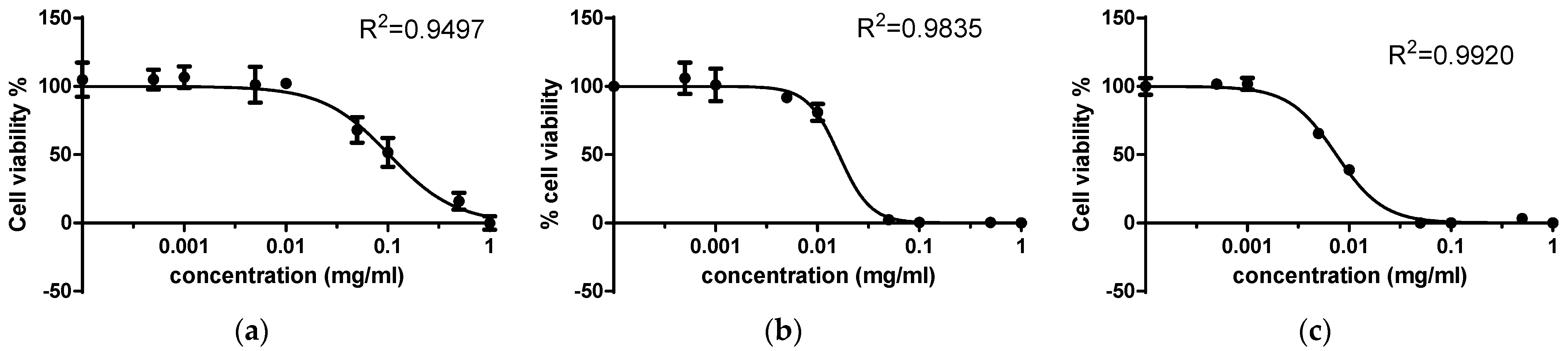
Two thiouracil derivatives and their complexes with Au(III), Pd(II), and Cu(II) were tested for cytotoxicity against two cell lines—a normal cell line of kidney cells from African green monkey and a tumor cell line of human cervical carcinoma. The concentration–response curves for 2,4-dithiouracil and its metal complexes against the normal cell line are presented on Figure 4. The results show that the tested compounds reduced the proliferation of the treated cells in a concentration-dependent manner, but the addition of Cu(II) and Au(III) improved cell viability at the higher tested concentrations. A similar tendency was observed in the treatment of the tumor cell line (human cervical carcinoma) (Figure 5) with the same compounds.

Figure 4.
Concentration–response curves of (a) 2,4-dithiouracil and its (b) Cu(II) and (c) Au(III) complex against the Vero cell line (kidney cells from African green monkey).
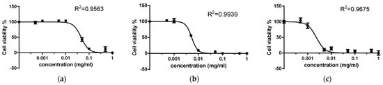
Figure 5.
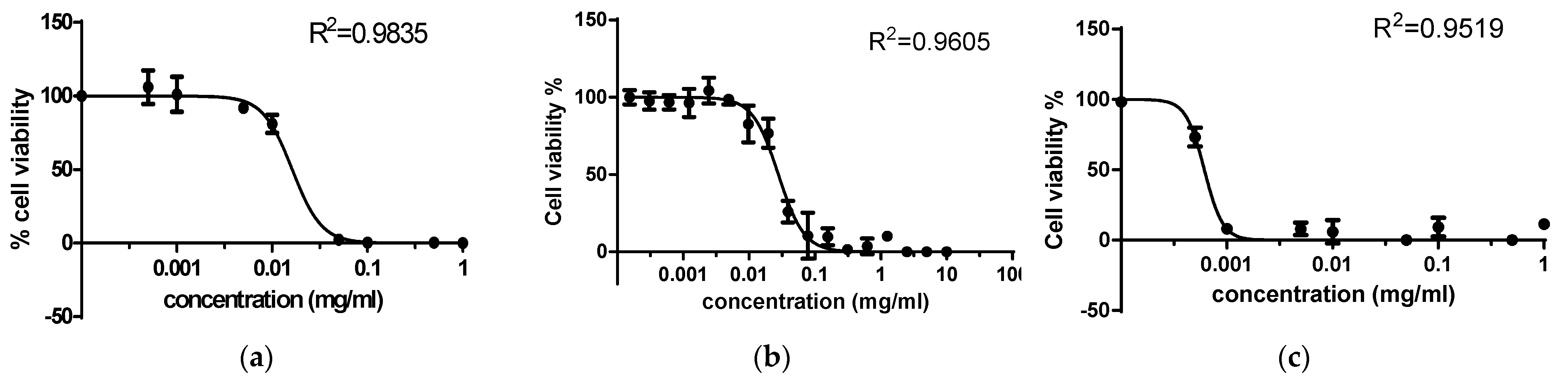
Concentration–response curves of (a) 2,4-dithiouracil and its (b) Cu(II) and (c) Au(III) complex against the HeLa cell line (human cervical carcinoma).The CD50 values of the 2,4-dithiouracil ligand and its metal complexes were determined through MTT testing and are presented in Table 1. The hierarchal order of cytotoxicity against the normal cell line revealed that the addition of Cu(II) increased the cytotoxic activity of the ligand and the addition of Au(III) to the complex led to an even lower CD50. The increase in activity was 37-hold for the Au(III) complex and 36-fold for the Cu(II) complex in comparison with the ligand. The tumor cell line was also less sensitive to 2,4-dithiouracil in comparison with its complexes, with the Au(III) complex being the stronger cytotoxic agent. The cytotoxic effect against the tumor line of the Cu(II) complex was 41 times stronger than that of the ligand, and the addition of Au(III) to the complex resulted in almost 162 times higher activity. Other authors also reported an improvement of cytotoxicity after the addition of copper and gold ions to ligands [20,23,41]. The comparison between the normal and tumor line sensitivity towards 2,4-dithiouracil and its metal complexes show much higher cytotoxicity against human cervical carcinoma cells, which is a good prerequisite for possible chemotherapy applications.
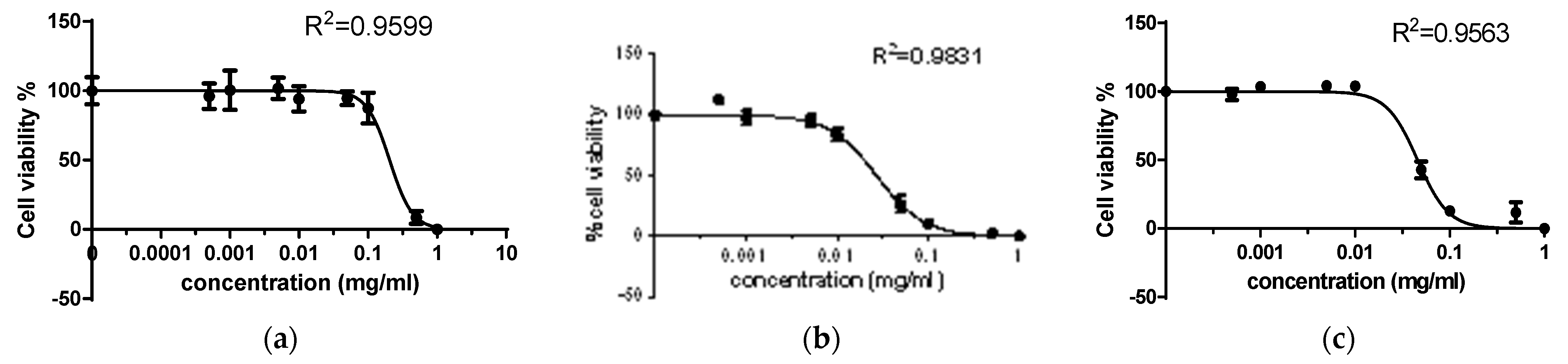
Similar results were obtained in the investigation of the cytotoxic effect of 6-propyl-2-thiouracil and its complexes with copper(II) and palladium(II). The concentration–response curves depicted in Figure 6 reveal that the cell viability of the normal cell line is also concentration-dependent and the metal complexes are cytotoxic in lower concentration in comparison with the ligand. In the experiments with tumor cells and the same compounds, the cell viability was affected by even lower test concentrations (Figure 7).
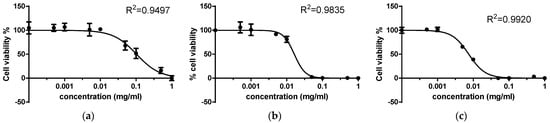
Figure 6.
Concentration–response curves of (a) 6-propyl-2-thiouracil and its (b) Cu (II) and (c) Pd (II) complex against the Vero cell line (kidney cells from African green monkey).

Figure 7.
Concentration–response curves of (a) 6-propyl-2-thiouracil and its (b) Cu(II) and (c) Pd(II) complex against the HeLa cell line (human cervical carcinoma).
The determined CD50 values of 6-propyl-2-thiouracil and its complexes were lower than those of 2,4-DTu and its Pd(II) and Cu(II) compounds. The highest cytotoxic activity against the normal cell line was detected for Pd(II) complex, followed by its Cu(II) and 6-Pro-2Tu (Table 2). The addition of Cu(II) to the ligand increased its activity 17 times, while the inclusion of Pd(II) contributed to an almost 78 times increase. Similarly, the most potent compound preventing the proliferation of the tumor cells was the Pd(II) complex of 6-propyl-2-thiouracil, followed by the Cu(II) complex and the ligand. The Cu(II) complex exhibited a 9-fold higher cytotoxic activity against the tumor cell line, and the Pd(II) complex exhibited a more than 149-fold higher cytotoxic activity. This second set of compounds also showed a better effect on tumor cells in comparison with the normal cells.

Table 2.
Hierarchical order of 6-Pro-2Tu and its complexes according to their cytotoxicity against the Vero cell line (kidney cells from African green monkey).
The cytotoxic activity against the normal cell line of kidney cells from African green monkey of the 6-propyl-2-thiouracil was two times stronger than that of the 2,4-dithiouracil and three times against the HeLa tumor cell line. When the copper complex of 6-propyl-2-thiouracil is compared to that with 2,4-DTu, it can be noted that the cytotoxic effect of the Cu(II) complex with 6-Pro-2-Tu against the normal cell line and the human cervical cell line was almost the same compared to the Cu(II) complex with 2,4-DTu. It should be noted that the Pd(II) complex with 6-propyl-2-thiouracil exhibited a 149-fold higher cytotoxic activity against the tumor cell line HeLa. The proposed mechanism of the cytotoxic action of the Pd(II) complex is given in Scheme 2.
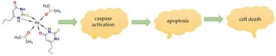
Scheme 2.
Proposed mechanism of the cytotoxic action of the Pd(II) complex.
Cisplatin is a widely used chemotherapeutic agent, and its cytotoxic effects on HeLa cell lines have been extensively studied [46,47,48,49,50,51]. On average, a concentration of 0.003 mg/mL is reported to lead to a significant loss of cell viability in HeLa cell lines. Becit et al. reported that for complete inhibition of the viability of a human cervical carcinoma cell line, a concentration of cisplatin higher than 1.5 mg/mL was needed [52]. Another study reported that 0.15 mg/mL of cisplatin caused about 10% reduction in cell viability in the same cell line after 24 h [53]. In comparison, the compound included in this study completely inhibited the proliferation of the tumor line at concentrations between 0.001 and 0.1 mg/mL. Another platinum-based chemotherapeutic agent that exhibits cytotoxic effects on HeLa cells is carboplatin. Various potency levels have been reported across different studies. Aborehab and Osama [54] investigated the effect of combining gallic acid with carboplatin on HeLa cells, employing concentrations of carboplatin ranging from 0.00781 mg/mL to 1 mg/mL. The application of 1 mg/mL yielded complete inhibition of cell viability, which is a significantly higher concentration than the ones reported in this study.
4. Conclusions
The conducted study on 2-thiouracil derivatives and their metal complexes showed that the inclusion of Cu(II), Pd(II), and Au(III) in the compounds structure significantly improved their cytotoxicity. All tested compounds exhibited a higher capability to reduce the proliferation of human cervical carcinoma cells in comparison with normal kidney cells from African green monkey. The cytotoxic activity against the tumor cell line of human cervical carcinoma of the Cu(II) complex was 41 times stronger than that of the 2,4-dithiouracil, and the addition of Au(III) to the complex resulted in an almost 162 times higher effect. The Cu(II) complex with 6-propyl-2-thiouracil exhibited a 9-fold higher cytotoxic activity against the tumor cell line HeLa, and the Pd(II) complex was more than 149-fold. The lowest CD50 detected was for the complex of 6-propyl-2-thiouracil with Pd(II) against the tumor cell line—0.00064 mM. These findings suggest that the coordination of metal ions, especially Cu(II), Au(III), and Pd(II) to 2-thiouracil derivatives, plays a significant role in the improvement of their cytotoxicity towards tumor cells while maintaining lower toxicity towards normal cells. This makes them promising candidates for further investigation as anticancer agents.
Author Contributions
Conceptualization, P.M. and D.B.; methodology, P.M. and P.G.-K.; formal analysis, P.G.-K.; investigation, P.M., D.B., P.G.-K. and A.S.; resources, P.G.-K.; data curation, P.M.; writing—original draft preparation, P.M., D.B. and A.S.; writing—review and editing, P.M., D.B. and A.S.; supervision, P.M.; project administration and funding acquisition, P.M. All authors have read and agreed to the published version of the manuscript.
Funding
This research was funded by [the Fund for Scientific Research of the Plovdiv University] grant number [project CП 23-XΦ-006].
Institutional Review Board Statement
Not applicable.
Informed Consent Statement
Not applicable.
Data Availability Statement
The original contributions presented in this study are included in this article. Further inquiries can be directed to the corresponding author.
Conflicts of Interest
The authors declare no conflicts of interest.
References
- Undare, S.S.; Valekar, N.J.; Patravale, A.A.; Jamale, D.K.; Vibhute, S.S.; Walekar, L.S.; Kolekar, G.B.; Deshmukh, M.B.; Anbhule, P.V. One-pot synthesis and in vivo biological evaluation of new pyrimidine privileged scaffolds as potent anti-inflammatory agents. Res. Chem. Intermed. 2016, 42, 4373–4386. [Google Scholar] [CrossRef]
- Mohamed, M.S.; Fatahala, S.S.; El-Hameed, R.H.A.; El Saeed Mohamed, M. Pyrimidine as a naturally occurring bioactive ring and its importance in different arenas. Egypt. J. Chem. 2025, 68, 71–98. [Google Scholar] [CrossRef]
- Shi, W.-X.; Lv, J.-Y.; Xiao, W.-J.; Xie, H.-D.; Li, J.-J.; Jiang, Y.-L.; Su, X.-C.; Xue, J.-Y.; Li, C.-X.; Zou, Y.; et al. Pyrimidines to Pyridines: Two Atom Swap Skeletal Editing. CCS Chem. 2025. ahead of print. [Google Scholar] [CrossRef]
- Tantawya, E.S.; Nafie, M.S.; Morsyd, H.A.; El-Sayed, H.A.; Moustafaa, A.H.; Mohammed, S.M. Synthesis of novel bioactive pyrido[2,3-d]pyrimidine derivatives with potent cytotoxicity through apoptosis as PIM-1 kinase inhibitors. RSC Adv. 2024, 14, 11098–11111. [Google Scholar] [CrossRef] [PubMed]
- Mohite, M.; Sheokand, S.; Maravanji, B. Synthesis of Pyrimidines and Quinazolines via Acceptorless Dehydrogenative Coupling Catalyzed by PNN-Pd Complex. Asian J. Org. Chem. 2025, 14, e202500165. [Google Scholar] [CrossRef]
- Sakhare, D.T. Green Synthesis, Characterization and Biological Evaluation of Divalent Transition Metal Complexes of Substituted Aminopyrimidine Novel Schiff Base Ligand. Sci. J. Chem. 2025, 13, 1–10. [Google Scholar]
- Zhang, W.; Chen, J.; Du, X. Design, Synthesis of Novel Pyrimidine Derivatives Containing Alkenyl Moieties With Herbicidal Activities. J. Heterocycl. Chem. 2025, 62, 5–12. [Google Scholar] [CrossRef]
- Pant, S.; Kumar K, R.; Rana, P.; Anthwal, T.; Ali, S.M.; Gupta, M.; Chauhan, M.; Nain, S. Novel Substituted Pyrimidine Derivatives as Potential Anti-Alzheimer’s Agents: Synthesis, Biological, and Molecular Docking Studies. ACS Chem. Neurosci. 2024, 15, 783–797. [Google Scholar] [CrossRef]
- Abolibda, T.Z.; El-Sayed, A.-A.A.A.; Farag, B.; Zaki, M.E.A.; Alrehaily, A.; Elbadawy, H.M.; Al-Shahri, A.A.; Alsenani, S.R.; Gomha, S.M. Novel thiazolyl-pyrimidine derivatives as potential anticancer agents: Synthesis, biological evaluation, and molecular docking studies. Results Chem. 2025, 13, 102008. [Google Scholar] [CrossRef]
- Abdelrahman, A.H.; Azab, M.E.; Hegazy, M.A.; Labena, A.; Ramadan, S.K. Design, Synthesis, Antiproliferative Screening, and In Silico Studies of Some Pyridinyl-Pyrimidine Candidates. J. Heterocycl. Chem. 2025, 62, 303–315. [Google Scholar] [CrossRef]
- Golubyatnikova, L.G.; Khisamutdinov, R.А.; Grabovskii, S.А.; Kabal’nova, N.N.; Murinov, Y.I. Complexes of Palladium(II) and Platinum(II) with 6-tert-Butyl-2-thiouracil. Russ. J. Gen. Chem. 2017, 87, 117–121. [Google Scholar] [CrossRef]
- Vetter, C.; Kaluđerović, G.N.; Paschke, R.; Kluge, R.; Schmidt, J.; Steinborn, D. Synthesis, characterization and in vitro cytotoxicity studies of platinum(IV) complexes with thiouracil ligands. Inorg. Chim. Acta 2010, 363, 2452–2460. [Google Scholar] [CrossRef]
- Carvalho, D.E.L.; Oliveira, K.M.; Bomfim, L.M.; Soares, M.B.P.; Bezerra, D.P.; Batista, A.A.; Correa, R.S. Nucleobase Derivatives as Building Blocks to Form Ru(II)-BasedComplexes with High Cytotoxicity. ACS Omega 2020, 5, 122–130. [Google Scholar] [CrossRef] [PubMed]
- Correa, R.S.; Bomfim, L.M.; Oliveira, K.M.; Moreira, D.R.M.; Soares, M.B.P.; Ellena, J.; Bezerra, D.P.; Batista, A.A. Ru(II) complexes containing uracil nucleobase analogs with cytotoxicity against tumor cells. J. Inorg. Biochem. 2019, 198, 110751. [Google Scholar] [CrossRef]
- Bomfim, L.M.; de Araujo, F.A.; Dias, R.B.; Sales, C.B.S.; Gurgel Rocha, C.A.; Correa, R.S.; Soares, M.B.P.; Batista, A.A.; Bezerra, D.P. Ruthenium(II) complexes with 6-methyl-2-thiouracil selectively reduce cell proliferation, cause DNA double-strand break and trigger caspase-mediated apoptosis through JNK/p38 pathways in human acute promyelocytic leukemia cells. Sci. Rep. 2019, 9, 11483. [Google Scholar] [CrossRef]
- Oladipo, M.A.; Isola, K.T. Coordination Possibility of Uracil and Applications of Some of Its Complexes: A Review. Res. J. Pharm. Biol. Chem. Sci. 2013, 4, 386–394. [Google Scholar] [CrossRef]
- Prachayasittikul, S.; Worachartcheewan, A.; Pingaew, R.; Suksrichavalit, T.; Isarankura-Na-Ayudhya, C.; Ruchirawat, S.; Prachayasittikul, V. Metal Complexes of Uracil Derivatives with Cytotoxicity and Superoxide Scavenging Activity. Lett. Drug Des. Discov. 2012, 9, 282–287. [Google Scholar] [CrossRef]
- Al-Halbosy, A.T.F.; Hamada, A.A.; Faihan, A.S.; Saleh, A.M.; Yousef, T.A.; Abou-Krisha, M.M.; Alhalafi, M.H.; Al-Janabi, A.S.M. Thiourea Derivative Metal Complexes: Spectroscopic, Anti-Microbial Evaluation, ADMET, Toxicity, and Molecular Docking Studies. Inorganics 2023, 11, 390. [Google Scholar] [CrossRef]
- Palma, G.; D’Aiuto, M.; Rea, D.; Bimonte, S.; Lappano, R.; Sinicropi, M.S.; Maggiolini, M.; Longo, P.; Arra, C.; Saturnino1, C. Organo-metallic compounds: Novel molecules in cancer therapy. Biochem. Pharmacol. Open Access 2014, 13, 1603–1615. [Google Scholar] [CrossRef]
- Santini, C.; Pellei, M.; Gandin, V.; Porchia, M.; Tisato, F.; Marzano, C. Advances in copper complexes as anticancer agents. Chem. Rev. 2014, 114, 815–862. [Google Scholar] [CrossRef]
- Khalid, H.; Hanif, M.; Hashmi, M.A.; Mahmood, T.; Ayub, K.; Monim-Ul-Mehboob, M. Copper complexes of bioactive ligands with superoxide dismutase activity. Mini Rev. Med. Chem. 2013, 13, 1944–1956. [Google Scholar] [CrossRef]
- Shokohi-Pour, Z.; Chiniforoshan, H.; Momtazi-Borojeni, A.A.; Notash, B. A novel Schiff base derived from the gabapentin drug and copper (II) complex: Synthesis, characterization, interaction with DNA/protein and cytotoxic activity. J. Photochem. Photobiol. B 2015, 162, 34–44. [Google Scholar] [CrossRef]
- Zou, T.; Ching, A.; Lum, T.; Lok, C.-N.; Zhang, J.-J.; Che, C.-M. Chemical biology of anticancer gold(III) and gold(I) complexes. Chem. Soc. Rev. 2015, 44, 8786–8801. [Google Scholar] [CrossRef]
- da Silva Maia, P.I.; Deflon, V.M.; Abram, U. Gold(III) complexes in medicinal chemistry. Future Med. Chem. 2014, 6, 1515–1536. [Google Scholar] [CrossRef]
- Ferna, J.; Elie, B.T.; Sulzmaier, F.J.; Sanau, M.; Ramos, J.W.; Contel, M. Organometallic titanocene-gold compounds as potential chemotherapeutics in renal cancer. Study of their protein kinase inhibitory properties. Organometallics 2014, 33, 6669–6681. [Google Scholar] [CrossRef]
- Rana, B.K.; Nandy, A.; Bertolasi, V.; Bielawski, C.W.; Saha, K.D.; Dinda, J. Novel gold(I)– and gold(III)–N-heterocyclic carbene complexes: Synthesis and evaluation of their anticancer properties. Organometallics 2014, 33, 2544–2548. [Google Scholar] [CrossRef]
- Abou-Melha, K.S. Elaborated studies for the ligitional behavior of thiouracil derivative towards Ni(II), Pd(II), Pt(IV), Cu(II) and UO22 2 ions. Spectrochim. Acta Part A Mol. Biomol. Spectrosc. 2012, 97, 6–16. [Google Scholar] [CrossRef]
- Masoud, M.S.; Amira, M.F.; Ramadan, A.M.; El-Ashry, G.M. Synthesis and characterization of some pyrimidine, purine, amino acid and mixed ligand complexes. Spectrochim. Acta Part A 2008, 69, 230–238. [Google Scholar] [CrossRef]
- Singh, U.P.; Ghose, R.; Ghose, A.K.; Sodhi, A.; Singh, S.M.; Singh, R.K. The effect of histidine on the structure and antitumoractivity of metal-5-halouracil complexes. J. Inorg. Biochem. 1989, 37, 325–329. [Google Scholar] [CrossRef]
- El-Morsy, F.A.; Jean-Claude, B.J.; Butler, I.S.; El- Sayed, S.A.; Mostafa, S.I. Synthesis, characterization and anticancer activity of new zinc(II), molybdate(II), palladium(II), silver(I), rhodium(III), ruthenium(II) and platinum(II) complexes of 5,6-diamino-4-hydroxy2-mercaptopyrimidine. Inorg. Chim. Acta 2014, 423, 144–155. [Google Scholar] [CrossRef]
- Abás, E.; Pena-Martínez, R.; Aguirre-Ramírez, D.; Rodríguez-Diéguez, A.; Laguna, M.; Grasa, L. New selective thiolate gold(I) complexes inhibit proliferation of different human cancer cells and induce apoptosis in primary cultures of mouse colon tumors. Dalton Trans. 2020, 49, 1915–1927. [Google Scholar] [CrossRef]
- Singh, U.P.; Singh, S.; Singh, S.M. Synthesis, characterization and antitumour activity of metal complexes of 5-carboxy-2-thiouraci. Met.-Based Drugs 1998, 5, 35–39. [Google Scholar] [CrossRef]
- Papazoglou, I.; Cox, P.J.; Hatzidimitriou, A.G.; Kokotidou, C.; Choli-Papadopoulou, T.; Aslanidis, P. Copper(I) halide complexes of 5-carbethoxy-2-thiouracil: Synthesis, structure and in vitro cytotoxicity. Eur. J. Med. Chem. 2014, 78, 383–391. [Google Scholar] [CrossRef]
- Hoeschele, J.D.; Piscataway, N.J. Ethylenediamineplatinum(II) 2,4-Dioxopyrimidine Complexes. U.S. Patent 4,207,416, 10 June 1980. [Google Scholar]
- Illán-Cabeza, N.A.; García-García, A.R.; Moreno-Carretero, M.N.; Martínez-Martos, J.M.; Ramírez-Expósito, M.J. Synthesis, characterization and antiproliferative behavior of tricarbonyl complexes of rhenium(I) with some 6-amino-5-nitrosouracil derivatives: Crystal structure of fac-[ReCl(CO)3(DANU-N5,O4)] (DANU = 6-amino-1,3-dimethyl-5-nitrosouracil). J. Inorg. Biochem. 2005, 99, 1637–1645. [Google Scholar] [CrossRef]
- Marinova, P.; Tsoneva, S.; Frenkeva, M.; Blazheva, D.; Slavchev, A.; Penchev, P. New Cu(II), Pd(II) and Au(III) complexes with 2-thiouracil: Synthesis, Characteration and Antibacterial Studies. Russ. J. Gen. Chem. 2022, 92, 1578–1584. [Google Scholar] [CrossRef]
- Marinova, P.; Hristov, M.; Tsoneva, S.; Burdzhiev, N.; Blazheva, D.; Slavchev, A.; Varbanova, E.; Penchev, P. Synthesis, Characterization, and Antibacterial Studies of New Cu(II) and Pd(II) Complexes with 6-Methyl-2-Thiouracil and 6-Propyl-2-Thiouracil. Appl. Sci. 2023, 13, 13150. [Google Scholar] [CrossRef]
- Marinova, P.; Stoitsov, D.; Burdzhiev, N.; Tsoneva, S.; Blazheva, D.; Slavchev, A.; Varbanova, E.; Penchev, P. Investigation of the Complexation Activity of 2,4-Dithiouracil with Au(III) and Cu(II) and Biological Activity of the Newly Formed Complexes. Appl. Sci. 2024, 14, 6601. [Google Scholar] [CrossRef]
- Marinova, P.; Burdzhiev, N.; Blazheva, D.; Slavchev, A. Synthesis and Antibacterial Studies of a New Au(III) Complex with 6-Methyl-2-Thioxo-2,3-Dihydropyrimidin-4(1H)-One. Molbank 2024, 2024, M1827. [Google Scholar] [CrossRef]
- Marinova, P.E.; Tamahkyarova, K.D. Synthesis and Biological Activities of Some Metal Complexes of 2-Thiouracil and Its Derivatives: A Review. Compounds 2024, 4, 186–213. [Google Scholar] [CrossRef]
- Adhikari, S.; Nath, P.; Das, A.; Datta, A.; Baildya, N.; Duttaroy, A.K.; Pathak, S. A review on metal complexes and its anti-cancer activities: Recent updates from in vivo studies. Biomed. Pharmacother. 2024, 171, 116211. [Google Scholar] [CrossRef]
- Lu, Y.; Ma, X.; Chang, X.; Liang, Z.; Lv, L.; Shan, M.; Lu, Q.; Wen, Z.; Gust, R.; Liu, W. Recent development of gold(I) and gold(III)complexes as therapeutic agents for cancer diseases. Chem. Soc. Rev. 2022, 51, 5518–5556. [Google Scholar] [CrossRef]
- Kim, J.H.; Ofori, S.; Parkin, S.; Vekaria, H.; Sullivan, P.G.; Awuah, S.G. Anticancer gold(III)-bisphosphine complex alters the mitochondrial electron transport chain to induce in vivo tumor inhibition. Chem. Sci. 2021, 12, 7467–7479. [Google Scholar] [CrossRef]
- Arojojoye, A.S.; Kim, J.H.; Olelewe, C.; Parkin, S.; Awuah, S.G. Chiral gold(III) complexes: Speciation, in vitro, and in vivo anticancer profile. Chem. Commun. 2022, 58, 10237–10240. [Google Scholar] [CrossRef]
- Mirzadeh, N.; Telukutla, S.R.; Luwor, R.; Privér, S.; Velma, G.R.; Jakku, R.K.; Andrew N, S.; Plebanski, M.; Christian, H.; Bhargava, S. Dinuclear orthometallatedgold(I)-gold(III) anticancer complexes with potent in vivo activity through an ROS-dependent mechanism. Metallomics 2021, 13, mfab039h. [Google Scholar] [CrossRef]
- Qi, Y.Y.; Gan, Q.; Liu, Y.X.; Xiong, Y.H.; Mao, Z.W.; Le, X.Y. Two new Cu(II) dipeptide complexes based on 5-methyl-2-(2’-pyridyl)benzimidazole as potential antimicrobial and anticancer drugs: Special exploration of their possible anticancer mechanism. Eur. J. Med. Chem. 2018, 154, 220–232. [Google Scholar] [CrossRef]
- Reddy, T.S.; Privér, S.H.; Mirzadeh, N.; Bhargava, S.K. Synthesis of gold(I) phosphine complexes containing the 2-BrC6F4PPh2 ligand: Evaluation of anticancer activity in 2D and 3D spheroidal models of HeLa cancer cells. Eur. J. Med. Chem. 2018, 145, 291–301. [Google Scholar] [CrossRef]
- Fei, B.L.; Tu, S.; Wei, Z.; Wang, P.; Qiao, C.; Chen, Z.F. Optically pure chiral copper(II) complexes of rosin derivative as attractive anticancer agents with potential anti-metastatic and anti-angiogenic activities. Eur. J. Med. Chem. 2019, 176, 175–186. [Google Scholar] [CrossRef]
- Khan, T.M.; Gul, N.S.; Lu, X.; Wei, J.H.; Liu, Y.C.; Sun, H.; Liang, H.; Orvig, C.; Chen, Z.F. In vitro and in vivo anti-tumor activity of two gold(III) complexes with isoquinoline derivatives as ligands. Eur. J. Med. Chem. 2019, 163, 333–343. [Google Scholar] [CrossRef]
- Pérez-Villanueva, J.; Matadamas-Martínez, F.; Yépez-Mulia, L.; Pérez-Koldenkova, V.; Leyte-Lugo, M.; Rodríguez-Villar, K.; Cortés-Benítez, F.; Macías-Jiménez, A.P.; González-Sánchez, I.; Romero-Velásquez, A.; et al. Synthesis and Cytotoxic Activity of Combretastatin A-4 and 2,3-Diphenyl-2H-indazole Hybrids. Pharmaceuticals 2021, 14, 815. [Google Scholar] [CrossRef]
- Zeng, Z.-F.; Huang, Q.-P.; Cai, J.-H.; Zheng, G.-J.; Huang, Q.-C.; Liu, Z.-L.; Chen, Z.-L.; Wei, Y.-H. Synthesis, Characterization, DNA/HSA Interactions, and Anticancer Activity of Two Novel Copper(II) Complexes with 4-Chloro-3-Nitrobenzoic Acid Ligand. Molecules 2021, 26, 4028. [Google Scholar] [CrossRef]
- Becit, M.; Aydın Dilsiz, S.; Başaran, N. Interaction of curcumin on cisplatin cytotoxicity in HeLa and HepG2 carcinoma cells. Istanb. J. Pharm. 2020, 50, 202–210. [Google Scholar] [CrossRef]
- Ganesan, B.S.; Prabhakaran, P. Effect of HeLa Cell Density Towards Cisplatin Treatment. Proc. Sci. Math. 2022, 12, 58–65. Available online: https://science.utm.my/procscimath/wp-content/uploads/sites/605/2022/11/07_Barthi-S-Ganesen_58-65new-1.pdf (accessed on 8 November 2024).
- Aborehab, N.M.; Osama, N. Effect of Gallic acid in potentiating chemotherapeutic effect of Paclitaxel in HeLa cervical cancer cells. Cancer Cell Int. 2019, 19, 154. [Google Scholar] [CrossRef]


Disclaimer/Publisher’s Note: The statements, opinions and data contained in all publications are solely those of the individual author(s) and contributor(s) and not of MDPI and/or the editor(s). MDPI and/or the editor(s) disclaim responsibility for any injury to people or property resulting from any ideas, methods, instructions or products referred to in the content. |
© 2025 by the authors. Licensee MDPI, Basel, Switzerland. This article is an open access article distributed under the terms and conditions of the Creative Commons Attribution (CC BY) license (https://creativecommons.org/licenses/by/4.0/).