Perspective

Integrin–RGD Peptide Interaction Revisited: Bringing Integrin αvβ6 into Focus for Implications in Cancer Targeting


1
School of Pharmacy and Medical Science, Griffith University, Southport, QLD 4215, Australia
2
Institute for Biomedicine and Glycomics, Griffith University, Southport, QLD 4215, Australia
3
School of Environment and Science, Griffith University, Nathan, QLD 4111, Australia
*
Authors to whom correspondence should be addressed.
Macromol 2026, 6(3), 76; https://doi.org/10.3390/macromol6030076
Submission received: 10 August 2026 / Revised: 2 September 2026 / Accepted: 5 September 2026 / Published: 11 September 2026

Abstract

The RGD motif (Arg-Gly-Asp) mediates interaction with integrins, governing adhesion and signaling. Integrin αvβ6, an RGD-binding integrin, has gained attention as a therapeutic and diagnostic target because its expression is restricted in normal tissues yet markedly upregulated in pancreatic and colorectal cancer, where it is linked to poor prognosis. Although sequence identities across both subunits of examined integrin subtypes range from approximately 40% to 55%, structural analyses show that all integrins are highly similar, with an RMSD of less than 1.3 Å. This similarity underlies a conserved mechanism in which arginine and aspartate interact with the α-subunit and the β-subunit metal ion–dependent adhesion site (MIDAS), respectively, stabilizing ligand engagement. In line with this, αvβ6 follows the same recognition principle; however, its unique β6-specificity-determining loop provides an extra binding interface, enhancing affinity toward particular ligands. Herein, this perspective highlights the structural features of RGD-binding integrins, their interaction with linear and cyclic RGD-peptides, and approaches for improving specificity to αvβ6. Beyond the RGD motif, linker design, including spacer length, bioconjugation chemistry, and stimuli-responsive cleavable motifs with self-immolative spacers, balances plasma stability with tumor-specific, traceless payload release, determining the pharmacological behavior of cyclic RGD–nanoparticle conjugates and their adaptation to future delivery and radiotracer platforms.

1. Introduction

Integrins are transmembrane receptors that serve a crucial role in mediating cellular adhesion to the extracellular matrix (ECM) and other cells, which is essential for maintaining tissue structure and functionality [1,2]. These heterodimeric proteins are formed from different combinations of α and β subunits that are assembled into 24 distinct integrin receptors in mammals [3]. Aside from adhesion, integrins integrate mechanical signals from the ECM, initiating intracellular signaling cascades that regulate proliferation, survival, and migration, thereby playing a role in physiological processes such as embryogenesis and wound healing [4,5].
At the heart of integrin functions is their ability to recognize specific motifs in ECM proteins, including the Arg-Gly-Asp (RGD) peptide, which is found in fibronectin and vitronectin [6]. This tripeptide sequence is recognized by several integrins, such as αvβ3, αvβ6, αvβ8, α5β1, and αIIbβ3, which mediate cell adhesion by binding to the RGD sequence [7]. RGD motifs interact with integrins in a manner that is crucial for cellular processes, which is particularly important in pathological conditions such as cancer [8]. This interaction is facilitated by the metal ion-dependent adhesion site (MIDAS), which is a shared structural feature within the integrins’ extracellular head domain [9,10]. At MIDAS, divalent metal ions, such as magnesium ions (Mg2+) and manganese ions (Mn2+), are coordinated to activate integrins’ binding to their ligands [11,12]. Following ligand binding, integrins undergo conformational changes that initiate intracellular signaling pathways that modulate cellular activity [13,14].
The integrin-mediated signaling pathway plays a vital role in cell survival, determining cellular shape, and controlling gene expression, all of which are crucial in specific pathological conditions like cancer [15,16]. Integrins such as αvβ3, αvβ6 and α5β1 are frequently overexpressed in tumors, facilitating crucial processes like cell migration, invasion, and metastasis by mediating tumor interactions with the surrounding ECM [15,17]. Furthermore, tumor vasculature is also enriched with integrins (αvβ3), promoting angiogenesis and, consequently, contributing to tumor growth and metastatic spread [18,19].
Among the integrin family, αvβ6 has gained popularity as an attractive therapeutic and diagnostic target due to its limited expression pattern in healthy tissues, while it is highly upregulated in epithelial-derived cancers [17,20,21]. Trivehexin, a trimeric cyclic RGD nonapeptide, has been introduced as a gallium-68-labeled radiotracer designed specifically for αvβ6 to allow real-time tumor visualization and assessment of treatment response [22,23]. In addition to its success as an imaging contrast agent, conjugating RGD peptides to an active agent or delivery system can augment receptor-mediated endocytosis and promote their uptake precisely into integrin-overexpressing tumors [24,25,26,27].
As our understanding of integrin biology evolves, the targeted binding of RGD peptides to integrins is poised to unlock a new era in cancer therapy, enabling precise diagnostic tools and delivery systems. Herein, we focus on five representative RGD-binding integrins out of the eight, with suitable high-resolution ligand-bound structures selected for structural analysis in this study (Table 1). Special attention is dedicated to integrin αvβ6, considering its peculiar binding affinities, especially to the selectivity of this integrin toward different RGD ligands and the implications for designing finely tuned targeted ligands.

2. Structural and Sequence Identity of Integrin Subtypes

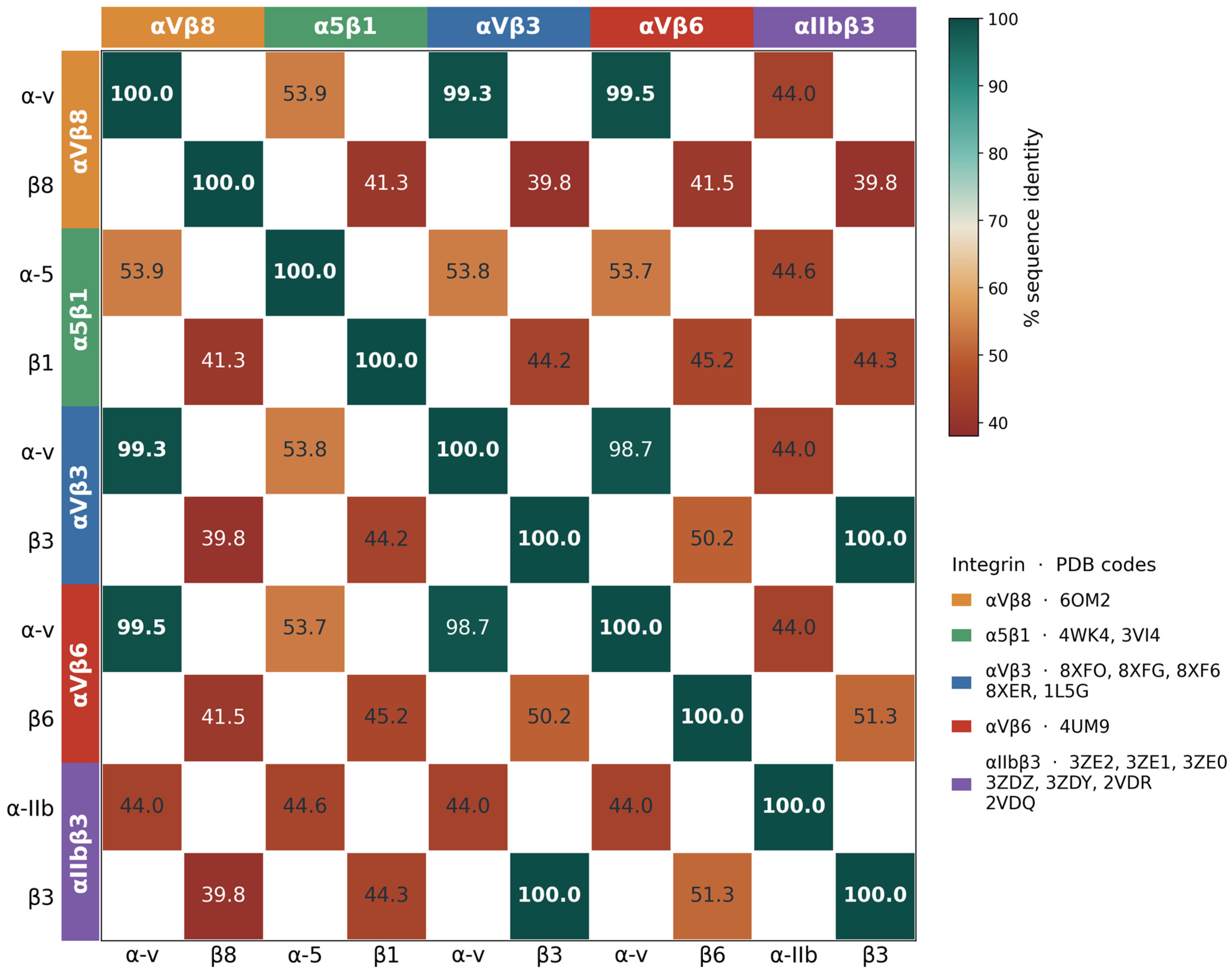
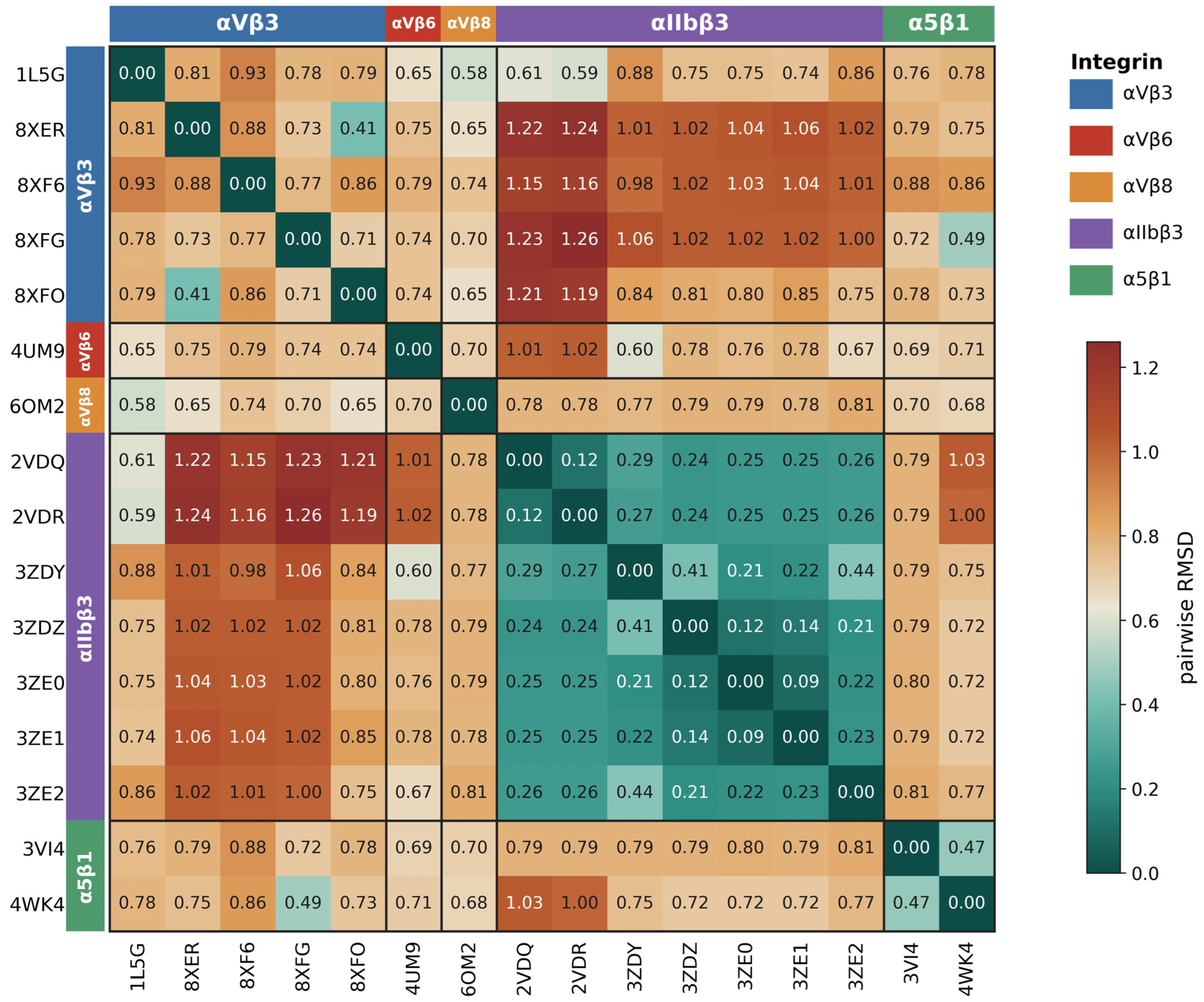
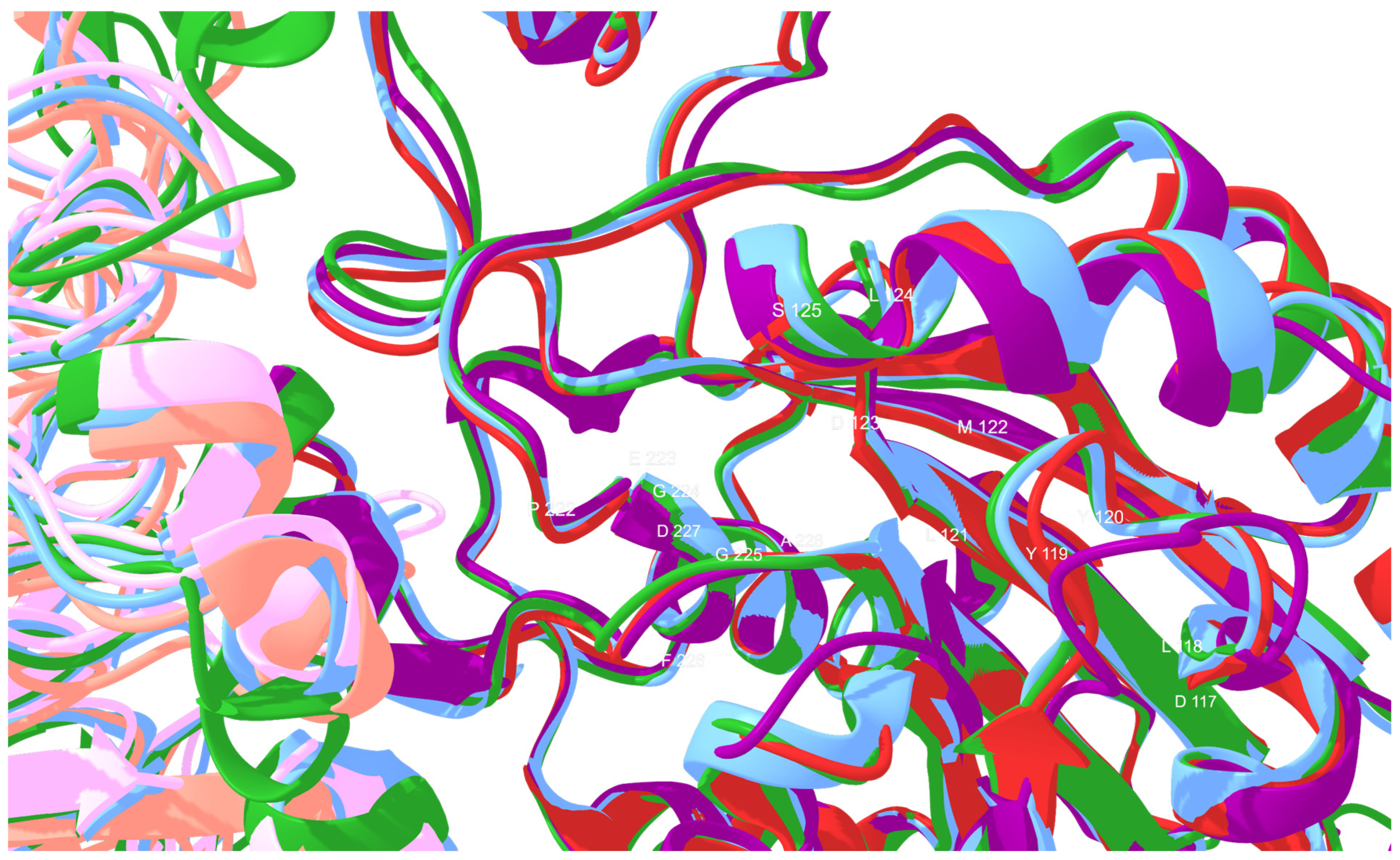
Sixteen integrin-peptide complexes (Table 1) have been identified in the Protein Data Bank and selected for further study of the structural and sequence identity of integrins and the relationship between these and peptide interactions in the binding pocket. The RGD-binding integrins exhibit only moderate sequence identity among their paralogous α- and β-subunits, ranging from approximately 40% to 55% (Figure 1). Despite this, they maintain a high degree of structural similarity, as evidenced by low backbone RMSDs ranging from 0.09 Å between closely related αIIbβ3 structures to 1.26 Å across the most divergent subtypes (Figure 2). This indicates that structural conservation within this family is significantly greater than sequence conservation. As a result, the five integrins, irrespective of their sequence differences, adopt a highly similar 3D fold, as represented by their four βA domains (Figure 3).
A fundamental principle underlying this conservation is that the strong preservation of tertiary protein folding compared with primary sequences is driven by evolutionary pressures to maintain essential biochemical functions, such as binding to substrates, catalysis, and molecular recognition. Generally, residues forming protein cores and functional sites are typically conserved, whereas those on the surface or within flexible loops exhibit more variability. This unique similarity can be an outcome of divergent evolution, where homologous chains accumulate substitutions without disrupting their folds, or convergent evolution, where divergent sequences in each chain adopt similar folding to fulfill analogous structural or functional roles.
This exceptionally high structural identity has significant implications for their interaction with RGD-based peptides. These minimal structural deviations, particularly within the ligand-binding pocket, imply that the spatial arrangement of key residues that recognize the RGD motif is highly conserved across these receptors. It is therefore likely that peptides containing RGDs interact with MIDAS similarly, potentially leading to comparable binding affinity and cross-reactivity between integrin subtypes. In practical terms, structural conservation is thought to explain why RGD-based ligands bind promiscuously to integrin subtypes. This structural analysis reveals nearly identical binding environments, helping guide the rational design of peptide therapeutics intended to modulate these interactions.

3. Sequence Alignment and Structural Similarity of Integrin Ligand-Binding Pocket

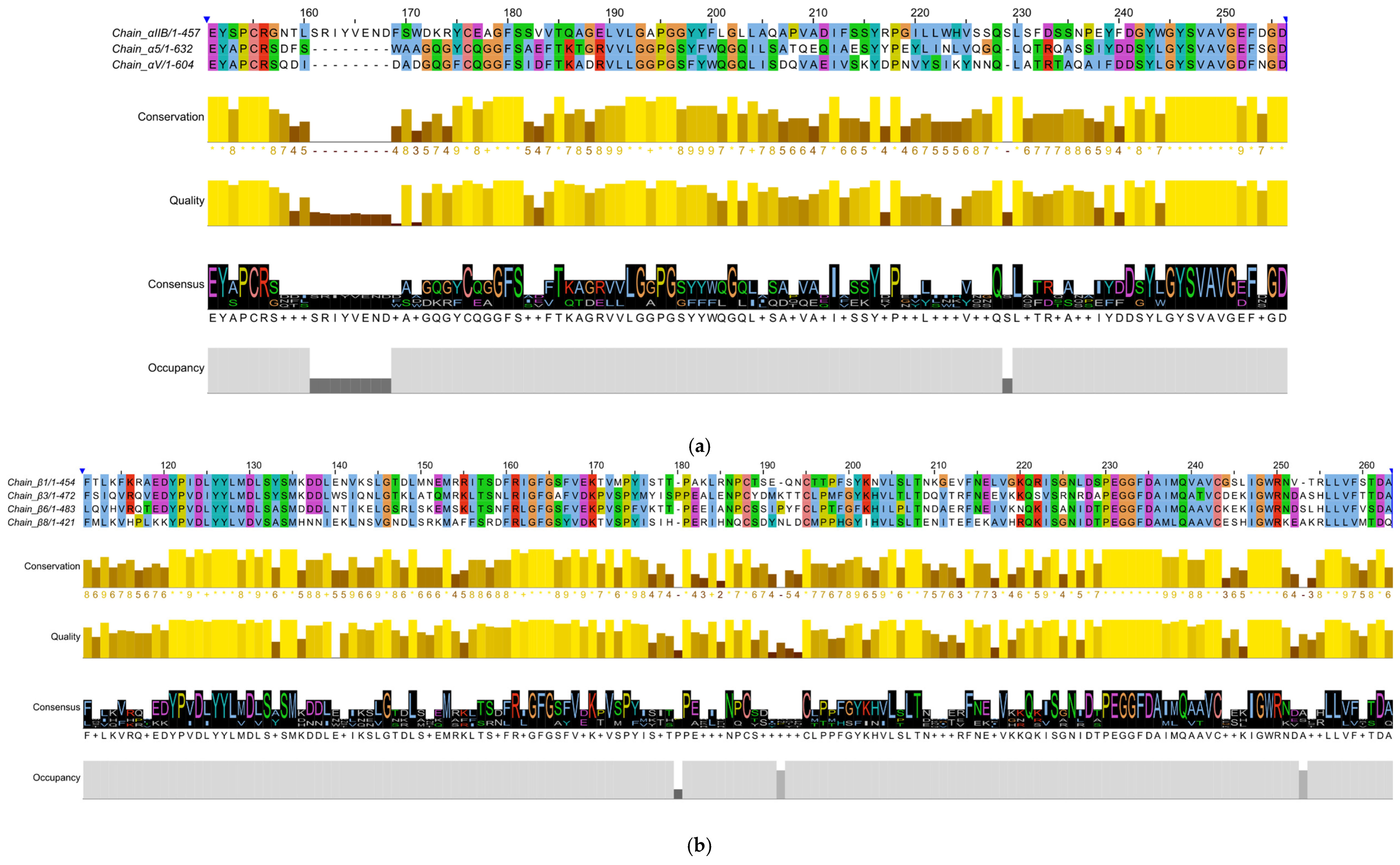
The two alignments collectively comprise the integrin head, holding the bipartite ligand-binding pocket. The β-chain alignment extends over the βA domain, a von Willebrand factor type A (vWA)-like fold. Within this domain lies the metal ion-dependent adhesion site (MIDAS), a conserved cation-binding region that coordinates divalent metal ions, such as Mg2+ or Mn2+. These ions are critical for ligand recognition because they facilitate interactions between integrins and adhesive motifs, including the RGD sequence found in many extracellular matrix proteins. Conformational changes within the βA/vWA-like domain can alter the accessibility and activity of the MIDAS region, thereby regulating integrin affinity and downstream cellular responses. Meanwhile, the α-chain alignment spans the blades of the β-propeller domain that caps the βA domain in the assembled heterodimer. Therefore, both chains provide information pertaining directly to the structural conservation surroundings of the pocket rather than peripheral areas.
On the β-chain, conservation patterns are evident. There is almost complete identity in sequences corresponding to residues constituting the pocket’s metal-binding chemistry. These sequences include the DLYYLMDLS vWA signature scaffolding around the fold, the Cys invariant of the NPCS/NPCT block that staples the loop tightly to the metal face, the PEGGFDAIM block that supplies the conserved MIDAS Glu, and the IGWRNDA block that provides the canonical MIDAS aspartate. The intervening variable columns map exclusively to the specificity-determining loops on the upper face of the βA domain, confirming that divergence across β-chains is restricted to ligand-selection elements while the MIDAS itself remains structurally invariant.
Where the multiple sequence alignment can only suggest βA-domain conservation, pairwise structural superposition proves it directly: the four headpieces (PDBs 3ZDY, 3VI4, 4UM9, 6OM2) superpose with Cα-RMSDs of 0.83–1.09 Å (Table 2), establishing same-fold equivalence across β1, β3, β6 and β8. Restricting the analysis to MIDAS-lining residues provides a clearer picture since the pocket Cα-RMSDs fall to 0.43–0.57 Å for every pair. All four structures share the same active two-metal-occupied state: Mg2+ at MIDAS with the canonical DXSXS + E coordination shell, Ca2+ at SyMBS with the canonical N + D + P (backbone) + E shell, both bridged by the conserved Glu within 3.3 Å. The divergence between subfamilies therefore reflects loop-level alteration rather than rearrangement of the catalytic center, demonstrating that MIDAS is structurally invariant across all four.
The α-chain alignment delivers a precisely complementary pattern. Consensus blocks visible in the alignment (EYAPCRS, GQGYCQGGFS, FTKAGRVVLGPGSYYWQGQ and IYDDSYLGYSVAV), most strongly matched by α5, delineate the β-strand cores of the seven-bladed propeller (with Cys–Cys closures in the cysteine-bearing blocks). Meanwhile, the intervening variable residues localize to conformationally plastic inter-blade loops on the upper face. This conformational plasticity is functionally significant: it underpins induced-fit recognition of subunit-specific ligands. As αIIb, α5 and αV are all RGD-binding subunits, the conserved residues define the architectural core of an RGD-binding propeller, while the divergent rim loops encode their distinct ligand specificities (fibrinogen for αIIbβ3; fibronectin for α5β1; vitronectin, fibronectin or latent TGF-β for αvβ3/β5/β6). Together, the two analyses resolve the heterodimeric pocket of αIIbβ3, α5β1, αvβ6 and αvβ8 as a single conserved pharmacophore, with invariant metal chemistry on β and a conserved scaffold with tuned rims on α, directly accounting for the cross-subfamily activity of RGD-mimetic drugs and antibodies.

4. Analysis of RGD Interaction with Integrins

Most of the existing RGD peptides exhibit poor subtype selectivity, binding promiscuously to multiple integrins, which results in reduced therapeutic precision and increased off-target effects [32]. As far as discovering new peptides goes, computational and high-throughput approaches are constrained by limited available datasets, as well as by a few studies exploring multifunctional or dual-integrin targeting peptides. Hence, of the sixteen complexes in Table 1, seven ligand-bound structures spanning five integrin subtypes were selected for detailed interaction analysis (Table 3). The structural analysis is conducted to pinpoint both shared and unique binding characteristics among these subtypes. This examination also considers the implications for ligand selectivity and the design of therapeutic agents. The research examines whether the conserved nature of the binding pocket can account for the broad specificity of RGD ligands and whether the variations in specificity-determining loops explain the remaining subtype preferences.
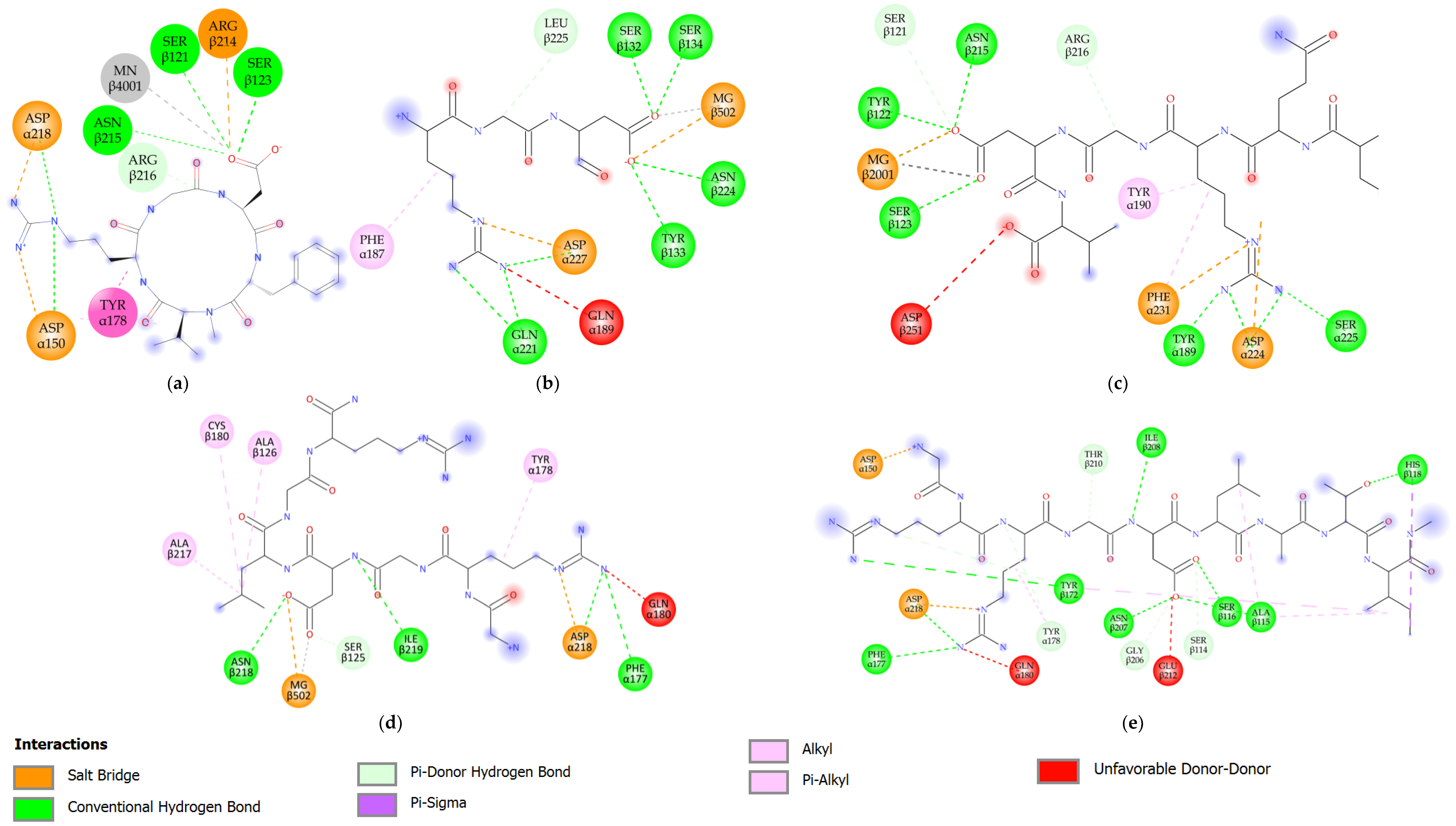
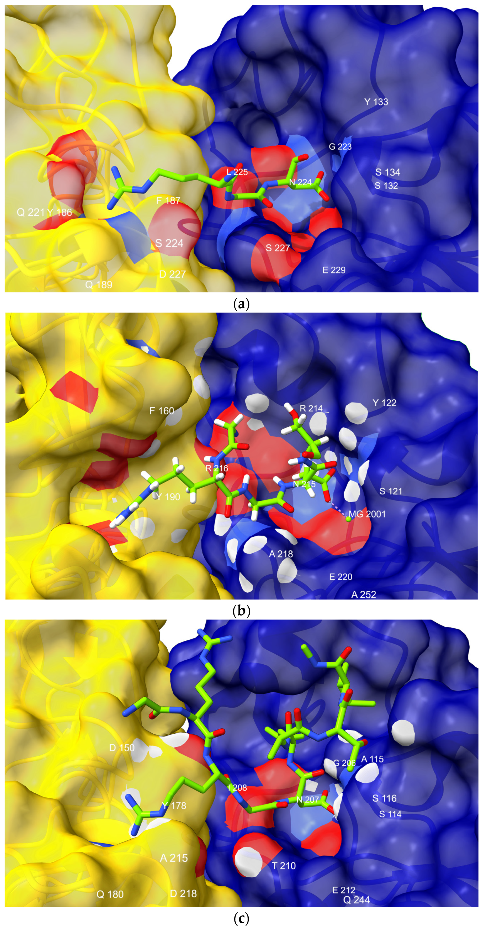
An in-depth analysis of these RGD-integrin complexes revealed a highly conserved binding mechanism across a variety of integrins (Table 3). For each peptide, the Arg consistently interacts with a diverse set of residues, engaging acidic residues of the α subunit: Asp α218 in the αv-containing integrins αvβ3, αvβ6 and αvβ8; Asp α224 in αIIbβ3; and Asp α227 in α5β1 (Figure 4b and Figure 5). These aspartates occupy the same position in their respective β-propellers, each immediately preceded by an aromatic residue: Phe α217 in αv, Phe α223 in αIIb, and Tyr α226 in α5, in the loop between blades 3 and 4 resolved in the alignment (Figure 4b). This pattern highlights the propensity for the Arg-guanidinium to serve as charge-based anchoring with acidic residues of the α-chains of integrins. Though this is not exclusive, as Arg also forms a π-alkyl contact with aromatic residues such as Tyr α178 (αvβ6). Arg’s side chain provides binding flexibility, suggesting the motif has the affinity to adapt to a range of integrin pocket residue types, forming stable configurations with stable binding.
It is apparent that the vicinity of the Asp residue is rich in hydrogen-bonding networks, as Asn exists in all tested integrins, and Ser dominates the binding pocket, which provides evidence of the structural integrity of MIDAS (Figure 5). Furthermore, additional binding to Tyr β122 of β3 was detected in several complexes (3ZDY, 2VDQ, 2VDR) which contributes to the overall structure. Aside from its carboxylate-mediated interactions, the Asp backbone amide donates a hydrogen bond to the backbone carbonyl of Ile β219 on β6 (2.74 Å), Ile β208 on β8 (3.05 Å) and Leu β225 on β1 (3.74 Å). Additionally, these hydrophobic residues offer structural support, stabilizing the RGD–integrin complex.
Each complex features a divalent cation at the MIDAS, directly coordinated by the ligand Asp carboxylate: Mg2+ in α5β1, αIIbβ3, αvβ6 and αvβ8 with bond lengths between 1.91 and 2.17 Å and Mn2+ in αvβ3 (2.65 Å), providing the electrostatic force for RGD recognition (Figure 5 and Figure 6b). The alanine residues contacting the motif in the αvβ6 and αvβ8 complexes likely provide steric accommodation alongside their contribution to binding.
Glycine, lacking a side chain, does not engage in side-chain-mediated interactions. Instead, its proximity to the pocket is facilitated by the backbone, with the nearest approach being Thr β210 in αvβ8 at a distance of 2.93 Å. This accentuates its primary role in the motif as a structural hinge, imparting the requisite flexibility to orient Arg and Asp in a manner that permits optimal binding. Taking complex binding interactions into account, future research will be able to optimize RGD-based therapeutics and better understand how integrins mediate cell signaling.

5. Bringing Integrin αvβ6 into Focus

Aside from the shared role played by integrins in cell adhesion, migration, and survival, integrin αvβ6 plays a distinct role in carcinogenesis [17]. Its presence activates transforming growth factor-β, driving epithelial-mesenchymal transition, a key process in tumor progression and metastasis [33,34,35,36]. Typical adult epithelial tissues lack this integrin; however, it is upregulated in a set of tumors. Moreover, tumors overexpressing αvβ6, such as those in skin, pancreatic, and breast cancers, are linked with poor prognosis [17,35,37,38,39,40]. Targeting integrin αvβ6 with antibodies, cyclic peptides, and other peptidomimetics appears to be a promising option for cancer treatment, whereas its potential as a diagnostic marker is currently being investigated [41,42,43,44,45,46]. Ongoing research into RGD peptides, which specifically bind to integrin αvβ6, holds great promise for developing targeted therapies and diagnostic tools [41,42,43]. Here, we discuss the distinctive affinity of the αvβ6 integrin for certain RGD ligands and how peptide modifications further enhance binding.

5.1. Interaction of Integrin αvβ6 with Linear Peptides

While the majority of integrin-binding ligands are proteins of the ECM, a small number of linear peptides have been identified as having significant affinity for αvβ6. The peptides were typically derived from natural ligands, such as the LAP of TGF-β1 or the GH loop of the VP1 coat proteins of foot-and-mouth disease viruses. Unlike cyclic RGD peptides, linear RGD peptides are defined by their secondary structure. RGD tripeptides are capable of mediating strong binding with the αvβ6 integrin; however, these interactions are often insufficient to confer high specificity or potency. Structural features of high-affinity αvβ6 linear peptide ligands demonstrate a shared, conserved architecture with the alpha helix. On the helix surface, the Leu/Ile residues are exposed at the Asp+1 and Asp+4 positions, permitting additional contacts with the β6 subunit beyond the canonical RGD–metal coordination [47].
In certain peptide sequences with a stable post-RGD helix, such as A20FMDV2 (Table 4), the binding affinity of the peptide can reach nanomolar levels, whereas sequences with reduced or lacking this helical propensity demonstrate significantly compromised affinity. Mutants lacking the helix, such as A20DV1217, exhibit a 40-fold reduction in inhibitory potency, illustrating how structural rigidity and residue positioning are crucial to activity. In essence, the binding mechanism of αvβ6-targeting linear peptides is further dictated by the structural presentation of flanking residues designed to establish a framework for selective recognition and inhibition.

5.2. Interaction of Integrin αvβ6 with Cyclic Peptides

Cyclic RGD-peptides are recognized for their high affinity and selectivity for integrins. Cyclization imposes a conformational constraint on the peptide structure, protecting it from being degraded by enzymes, and also enables the RGD motif to be presented in a geometric configuration, allowing it to be efficiently identified by receptors (Figure 7). They also display superior in vivo stability compared to their linear counterparts, which is a key feature for imaging probes and targeted drug delivery. The cyclic RGD scaffolds can be adapted to exploit structural features of αvβ6 by altering ring size, residue composition, and flanking amino acids to enhance their receptor specificity. Additionally, in this section, we will discuss how other modifications, including aromatic substitutions and multimerization strategies, can promote tumor uptake and biodistribution profiles.

5.2.1. Cyclic Pentapeptides

In 2017, Civera et al. [51] tested three established RGD-based cyclic pentapeptides and evaluated them for their inhibitory activity toward αvβ6 and αvβ3 integrins. The peptides included two c(-RGDfV-) derivatives and the c(-RGDfK-) variant. Interestingly, cRGDfK exhibited a twofold increase in potency, indicating that replacing Val with Lys in the pentapeptide scaffold may improve integrin binding. These findings indicate that small variations in the chemical nature, size, or stereochemistry of residues surrounding the RGD motif can affect αvβ6 binding. Their research has also primarily focused on synthesizing peptidomimetics and examining the influence of substitutions and the stereochemistry of post-RGD residues, which yielded valuable insights into the binding mechanisms of both integrins. Although the prepared peptidomimetics exhibited greater selectivity toward αvβ3 over αvβ6, these findings were nevertheless instrumental in shaping the overall concept.
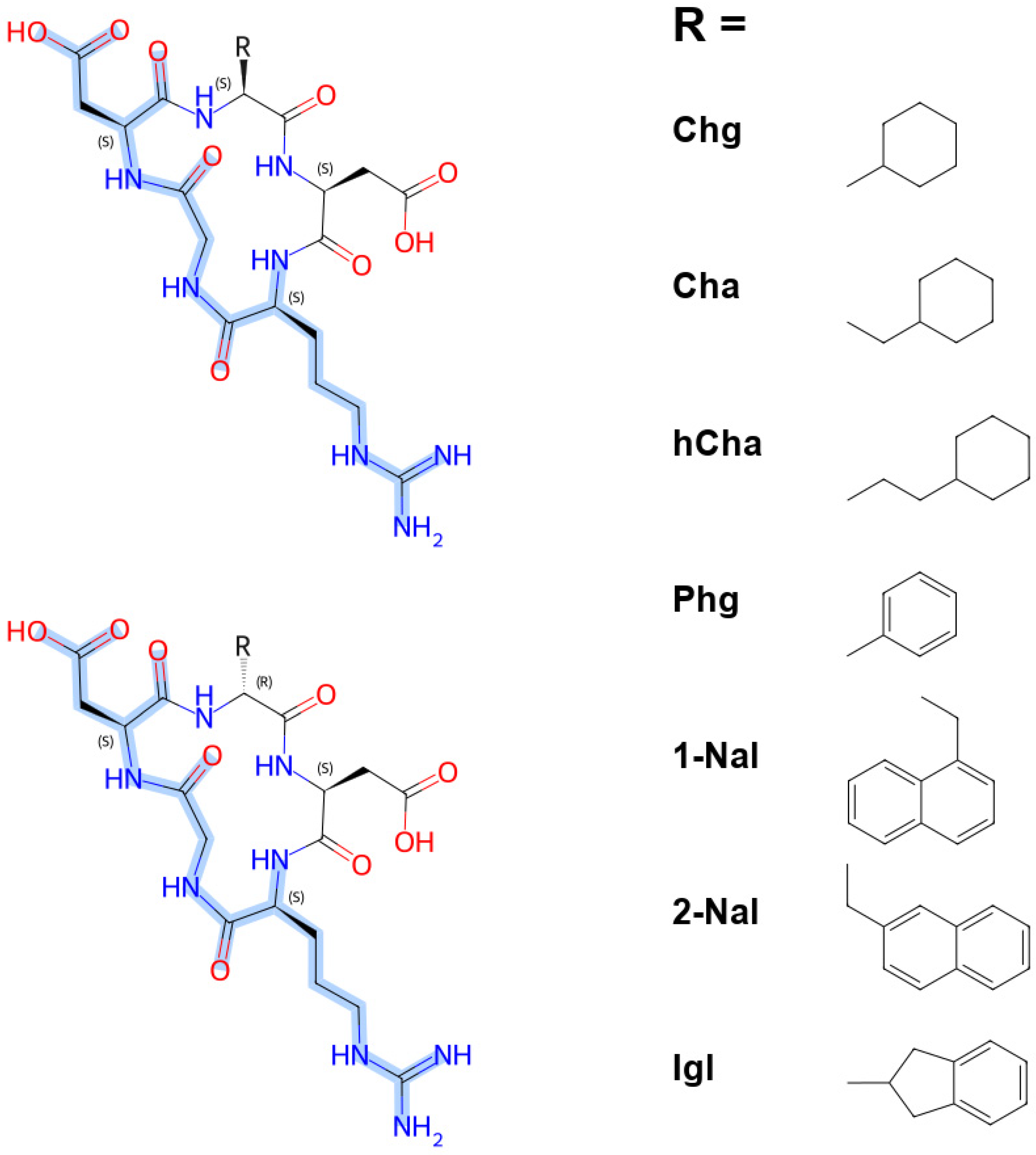
Building upon previous findings and to understand how modifications at the Asp+1 position influence integrin αvβ6 recognition, researchers at Marinelli’s labs [50] synthesized a series of cyclic pentapeptide analogues with a modified flanking residue and a C-terminal amide, c(-RGD–X–E-)–CONH2 (C1) (Figure 8). This series revealed that αvβ6 binding is exquisitely contingent on both stereochemistry and the chemistry of the flanking residue. It is clear that L-isomers consistently outperformed their D-counterparts, albeit with varying penalties. There is a dramatic difference in potency between c(-RGD–Chg–E-)–CONH2, where the L-form binds at 1.3 nM, while the D-form requires 608 nM, indicating an approximately 500-fold difference in potency. This consistently superior affinity of L-isomers confirms the rigid stereochemical constraints of the integrin binding pocket.
Analyzing the side-chain identities of these cyclic pentapeptides, compact hydrophobic residues provide the greatest potential. For instance, the compact cyclohexyl group of Chg (cyclohexylglycine) achieves optimal van der Waals packing within the hydrophobic pocket, which results in the highest potency (1.3 nM). Introducing a slight modification in ring size or flexibility, CHA (cyclohexylalanine) and Hcha (homocyclohexylalanine) are accommodated, but less optimally (23 nM and 33 nM, respectively), implying that steric compactness and tight packing in hydrophobic complementarity can be essential for activity. As a result of aromatic substitution, both bulk and orientation are stressed. Although 1-Nal (1-naphthylalanine) and 2-Nal are of similar bulk, 1-Nal is nearly twice as potent (15 nM vs. 28 nM), demonstrating the importance of side-chain vectors and hydrophobic cleft alignment. The rigid bicyclic Igl (indanylglycine, 57 nM) was tolerated but far less potent than 1-Nal, indicating that excessive rigidity impairs hydrophobic contact.

5.2.2. Cyclic Nonapeptide and Decapeptides

Internalizing RGD or iRGD, c(-CRGDKGPDC-) is a well-established cyclic nonapeptide that binds to integrins and is overexpressed in tumor vessels and cancer cells [52,53]. A multistep process underlies iRGD function, as it binds to integrins that recognize RGD (previously known as αvβ3 and αvβ5, and newly confirmed as αvβ6). This ligand differs from conventional RGD ligands in that it undergoes proteolytic activation post-lysine, revealing a cryptic CendR sequence (CRGDK) that binds to neuropilin-1 (NRP-1) [25,54]. Thanks to this unique binding mechanism, iRGD homes to tumors, triggers active internalization, and has thus been applied to targeted tumor delivery. This has led to iRGD being applied as a cancer-penetrating peptide for enhanced imaging [26,55], more effective chemotherapy [24], biologics [56], and nanoparticle-based systems to improve delivery [27], and it has already demonstrated potential in aggressive cancers like PDAC [24,57].
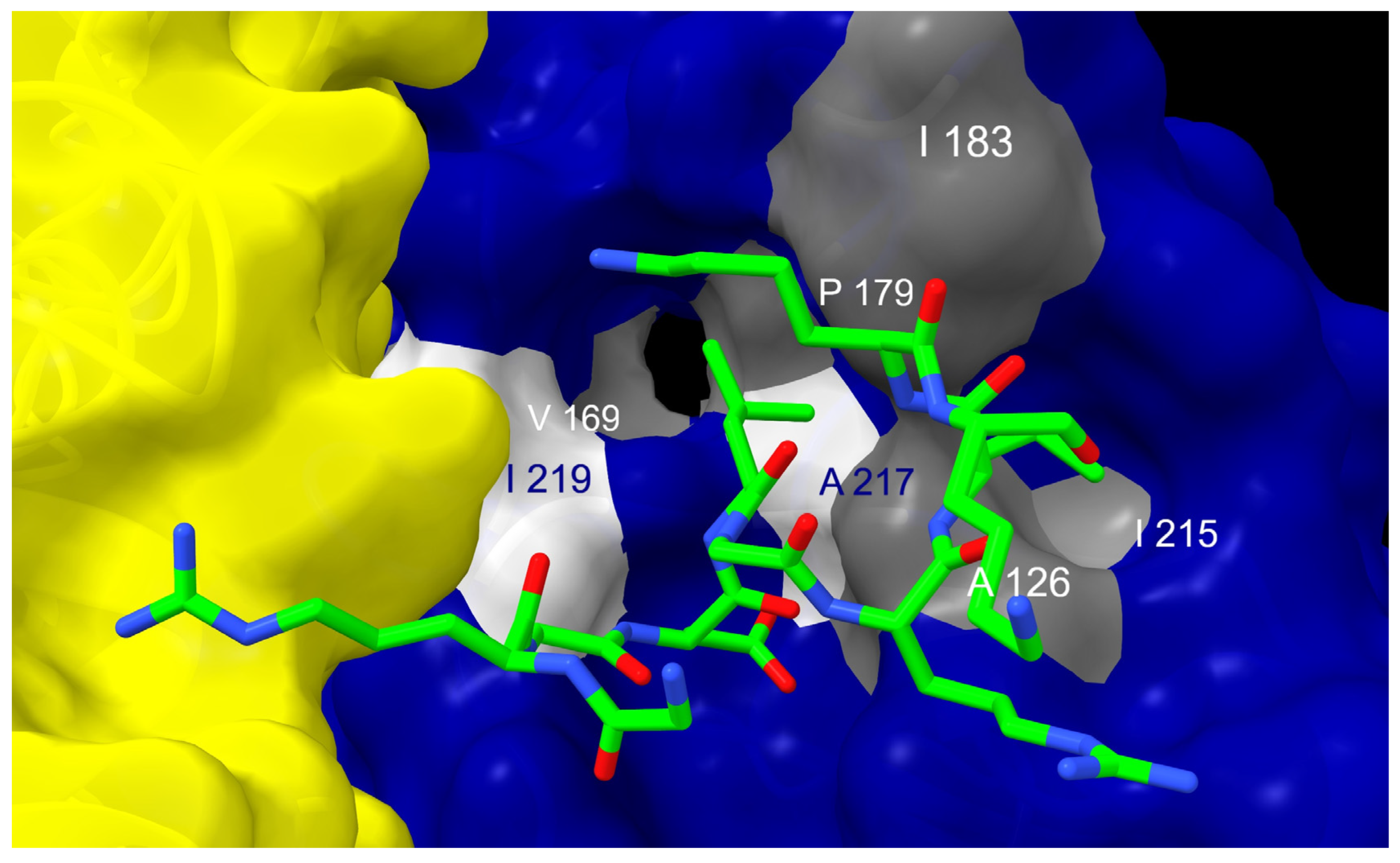
The insights into the ways in which iRGD may be modified to enhance selectivity and potency for αvβ6 are equally important. D’Amore et al. [49] have revealed that the specificity-determining loop (SDL) of β6 consists of a wider, more lipophilic pocket than β3/β5 (viz., residues I218 and K169 shift to expand the pocket) (Figure 7). This topology penalizes iRGD, causing it to rearrange partially and lose its ideal RGD contacts, which explains its lower αvβ6 potency. According to their structural analysis, integrin binding is not disrupted by residues such as Lys5, Gly6, or Cys9 that are solvent-exposed, which facilitates proteolytic cleavage. In this way, iRGD can be safely bioconjugated to drugs, nanoparticles, or other carriers while retaining its activity. Modifying the Lys residue presents challenges. Despite being solvent-exposed and not essential for integrin engagement, it becomes the C-terminal residue of the CendR fragment after proteolytic cleavage. Consequently, its modification eliminates NRP-1 binding and the associated tumor-penetrating activity. Based on this, they adopted a selectivity-boosting strategy for αvβ6 by inserting a fairly bulky, lipophilic side chain into the SDL without compromising the canonical RGD-MIDAS contacts. They considered mutating Gly at the Asp+2 position to a more lipophobic residue, including natural (e.g., Val, Leu, Ile) and non-natural amino acids (e.g., cyclohexylglycine, cyclohexylalanine, allylglycine). The Leu variant appears to orient iRGD in a pose consistent with SDL of β6, enhancing affinity and selectivity for the new side chain [49]. With such design strategies, it is possible not only to expand the therapeutic specificity and versatile applications of iRGDs, but also to design next-generation integrin-specific tumor-penetrating peptides.
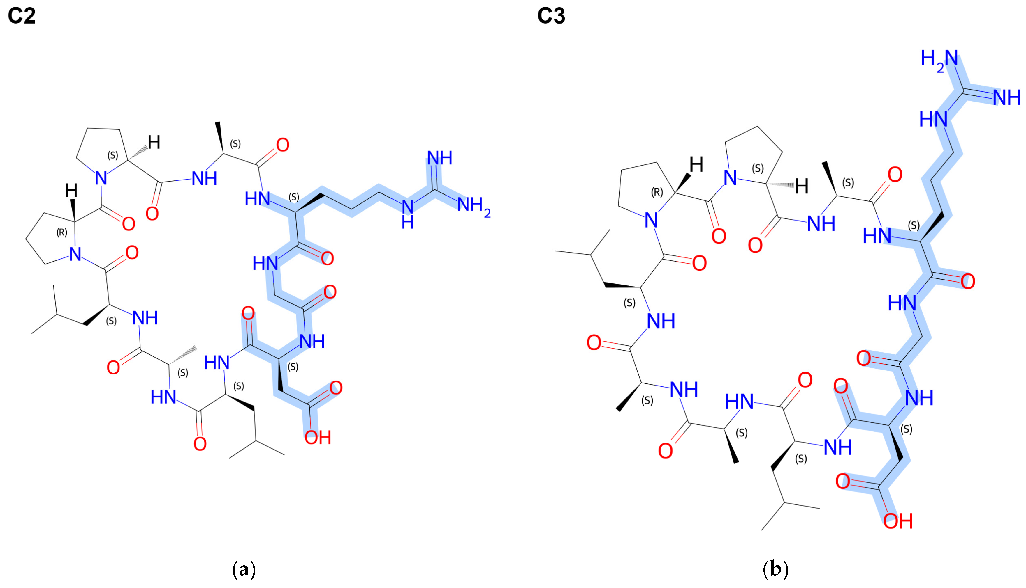
Kessler and colleagues [48] designed the parent cyclic decapeptide, c(-RGDLAALApP-), which exhibited good affinity for integrin αvβ6 with an IC50 of 14.5 nM. Relocating the alanine residue from Asp+5 to the N-terminus, as in c(-ARGDLAALpP-) (C2) (Figure 9), improved potency to 8.3 nM, implying that the promoted ligand–receptor interaction is the result of reduced steric hindrance at the N-terminus. Nevertheless, relocating alanine and leucine at Asp+4 and Asp+5 to the N-terminus c(-LARGDLAApP-) did not improve affinity, suggesting that side chain properties and peptide conformation play a critical role in determining optimal binding affinity. In C2, the replacement of leucine with an alanine residue at Asp+1 led to a dramatic drop in potency by ~39-fold, whereas a similar change at Asp+4 only reduced it by around twofold, underscoring the importance of side-chain identity specifically at Asp+1, where the hydrophobic contact is essential, whereas Asp+4 tolerates substitution. Therefore, increasing peptide flexibility or disrupting essential conformational constraints of the peptide may compromise productive contacts with integrins.
Truncation of the parent cyclic decapeptide sequence and introducing aromatic residues close to the C-terminus yields even greater enhancements in affinity. Reducing the peptide sequence by removing the alanine residue at position 8 (Figure 9), yielding the 9-mer parent c(-ARGDLALpP-) (C3), led to a 2.7-fold increase in potency compared to C2. Furthermore, substituting the alanine of C3 with an aromatic residue at the N-terminus improved its potency by over twofold, whereas analogous substitution at the C-terminus yielded a more potent analogue with IC50 = 0.67 nM. Most importantly, the C-terminal modifications can further optimize integrin targeting. The N-methylation of proline and substitution of C-terminal proline with lysine, as seen in c(-FRGDLAFp(NMe)K-), results in the highest observed affinity (IC50 = 0.26 nM). Meanwhile, N-terminal acetylation in c(-FRGDLA-Fp(NMe)K(Ac)-) maintained comparable low IC50 values, implying that this capping strategy has no impact on receptor binding, which would be beneficial for improving peptide stability.
Subtype selectivity challenges of RGD peptide-based imaging due to integrin cross-reactivity prompted Yang et al. [58] to tune macrocycle geometry instead of sequence, creating a 25-member library by cyclizing the bis-cysteine scaffold CRGDX1X2RGDC with varied carbon-spacer cross-linkers and turn-inducing motifs. Biolayer interferometry demonstrated that the recognition of αvβ6 was contingent upon a rigid D-Pro-L-Pro β-turn in conjunction with a short spacer. The three most effective ligands exhibited nanomolar dissociation constants (Kd), differing only in spacer: two-carbon maleimide (8.9 nM), four-carbon ortho-xylyl (19.3 nM) and one-carbon acetonyl (49.2 nM). These tuned cyclic decapeptides demonstrated a preference of at least 220-fold over other integrin subtypes, compared with the 20-mer reference αvβ6-BP peptide (32.8 nM), which was attributed to linker-dependent β-sheet stabilization. The fluorescein-labeled analogues containing one and two carbon atoms demonstrated binding to αvβ6-high BxPC3 cell membranes, whereas the four-carbon analogue did not exhibit such binding. In BxPC3 xenografts, the 68Ga-labeled one-carbon probe demonstrated superior performance compared to the similarly labeled 20-mer over a period of 1–4 h in vivo, even though the tumor signal was reduced due to an excess of unlabeled peptide and renal clearance.

5.2.3. Interaction with Cyclic Nonapeptide Trimers

Recently, Quigley et al. [59] investigated trimeric targeting peptides to improve binding avidity to αvβ6 integrin, while promoting peptide stability and in vivo performance compared to their monomeric counterparts. They applied triazacyclononane–triphosphinate (TRAP) cores, which allow simultaneous coupling of three peptide arms to mimic interactions between integrin clusters on tumor cells. Multimerization of the peptides not only recovers potency lost during conjugation or radiolabeling, but also enhances tumor uptake and contrast in imaging. Furthermore, they investigated how to adapt this trimeric platform by introducing aromatic substitutions across multiple arms without compromising RGD-mediated integrin recognition.
At the two positions of the parent peptide (C3), tyrosine residues were substituted with phenylalanine, leading to variant trimers with varying Tyr contents (Y0 to Y6). The findings revealed that even a single tyrosine substitution per trimer arm (e.g., Y1) efficiently suppressed non-specific uptake in organs such as the liver, heart, and pancreas, while promoting faster blood clearance. Increasing Tyr content improved biodistribution profiles, culminating in the Y6 variant, composed solely of double-tyrosine peptide arms, now known as [68Ga]Trivehexin [22]. This construct exhibited the most favorable tumor-to-background ratios in PET imaging, becoming the first αvβ6-selective RGD radiotracer evaluated clinically in pancreatic cancer [23].

6. Integrin-Targeting Peptide-Based Delivery Systems

6.1. Challenges in Tumor Delivery

Biological tumor microenvironments are hostile to therapeutic cargos, which impose formidable hurdles for mainstream therapeutic delivery approaches. Several potent small-molecule cargos experience precipitation under physiological conditions, resulting in their inactivation prior to reaching target sites [60,61]. In contrast, peptide and protein therapeutics encounter a different set of bioavailability barriers due to their large molecular size, imposing difficulties in traversing cellular membrane, while being prone to proteolytic degradation. Moreover, among the candidates that have achieved remarkable success are small-interfering RNA (siRNA) and messenger RNA (mRNA), which have high potential as cancer therapeutics. These RNA macromolecules are merely futile by themselves and require sophisticated delivery systems to override biological barriers [62]. This is due to the fact that these macromolecules are hydrophilic and negatively charged; hence, they are unable to cross cell membranes and rapidly degraded by nucleases in biological fluids [63]. In serum, naked siRNA has a fleeting half-life of approximately 15 min due to RNase activity [64]. Oftentimes, encapsulating them into nanoparticles is the solution to provide physical protection against degradation by nucleases [65]. These nanoparticles have additional roles in shielding immunostimulatory motifs from pattern recognition receptors and helping cellular uptake through endocytosis [66,67]. The delivery of nucleic acids is subject to multiple challenges, as it necessitates controlled electrostatic interactions dictated by lipid pKa and/or N/P ratio optimizations [67]. After uptake, the majority of RNA cargo is eliminated by lysosomes before reaching the cytoplasm or ribosomes, leaving behind only 2% of the total nucleic acids that egress through endosomes [68]. Aside from this, non-specific uptake reduces tumor selectivity; these limitations point to the need for more advanced carriers that protect nucleic acids, facilitate endosomal escapes, and enable receptor-mediated delivery to the target cells [69,70].

6.2. Nanoparticles as Delivery Vehicles

Nanoscale drug delivery systems have been reshaped by the understanding that therapeutic success is dictated not merely by payload potency, but also by how active agents are delivered into complex biological environments [62,71]. Several nanoparticulate carriers have been shown to modulate drug pharmacokinetics, including mesoporous structured materials, polymeric micelles, lipid assemblies, and metallic constructs. The enhanced permeability and retention (EPR) effect of these nanoplatforms, despite seeming promising, yielded clinically inconsistent passive accumulation, prompting a paradigm shift toward active targeting [72,73].
In this context, the surface functionalization of nanoparticles with peptides stands out as an efficient targeting strategy intended to address the shortcomings of passive delivery in terms of poor specificity [74]. These targeting peptides are typically composed of 5 to 20 residues, and their use is on the rise due to their low immunogenicity, tissue penetration depth, and synthetic feasibility compared to antibodies and small molecules [75,76]. Systemically administered RGD peptide-decorated nanosystems circulate until they engage their target integrin through specific noncovalent forces, triggering an internalization cascade via receptor-mediated, mostly clathrin-driven endocytosis [77,78]. Kemker et al. observed that unconjugated small monomeric RGD peptides entered cells mainly via fluid-phase endocytosis, whereas PEGylated RGD peptides were internalized via clathrin-dependent endocytosis [79]. Consequently, this integrin-induced uptake helps orient the therapeutic cargo within the target rather than relying on conventional passive EPR. Additionally, some peptide conjugation strategies may also address instability by forming a corona around nanoparticle surfaces that shields against nonspecific protein adsorption, ultimately reducing the early opsonization by mononuclear phagocytes [80,81].

6.2.1. Lipid-Based Nanoparticles

Lipid-based nanoplatforms are the most clinically promising approach to targeted therapeutic delivery owing to their biocompatibility, biodegradability, and structural versatility [62,82]. The phospholipid bilayers of liposomes, encasing an aqueous core, can trap hydrophilic cargos in their interiors, while hydrophobic agents can be trapped in their membranes. RGD-decorated liposomes loaded with anticancer agents like 5-Fluorouracil, Doxorubicin, and Paclitaxel exhibited improved tumor accumulation and higher therapeutic efficacy than their nontargeted counterparts [83,84,85]. Endosomes acidify during endocytosis (pH 5–6), destabilizing the nanocarrier and promoting payload release within the cytoplasm. Tumor-penetrating peptides, iRGD, undergo proteolytic cleavage at the lysine/gylcine junction, exposing a CendR motif (CRGDK) to NRP-1, thereby facilitating tumor penetration [49]. Thus, iRGD-targeted liposomes and other lipid-based nanoparticles have been reported to enhance tumor accumulation and chemotherapeutic efficacy [86,87,88].
A typical strategy for targeting integrins is to conjugate RGD peptides to polyethylene glycol (PEG) terminals anchored to the liposome surface [82,89,90]. In this process, PEGylated lipids like DSPE-PEG (1,2-distearoyl-sn-glycero-3-phosphoethanolamine) conjugated with maleimide undergo thioether bonding with free cysteine-containing RGD peptides. Multimerization of RGD peptides further enhances binding affinity and internalization through avidity effects, facilitating tumor uptake and improving cytotoxicity even at lower therapeutic doses [59,85,91].
Furthermore, ionizable lipid-based nanoparticles have gained increasing attention due to their composition, which entails helper phospholipids (e.g., DSPC), cholesterol, and PEG-lipids [62,92]. The FDA has approved a number of clinically validated ionizable lipid formulations, such as DLin-MC3-DMA (patisiran, an siRNA therapeutic) and ALC-0315 and SM-102 (COVID-19 mRNA vaccines) [93,94]. Their ionization, influenced by pH levels, maintains a near-neutral charge during circulation and becomes cationic within the acidifying endosome. This property, along with their ability to form stable lipid assemblies, enables effective surface functionalization with peptides or other ligands. Similarly, there are a variety of other lipid-based nanoplatforms exhibiting distinct features like improved stability, higher loading capacity, and controlled cargo release behavior. For instance, SLNs comprise solid lipids, whereas NLCs contain solid–liquid lipid mixtures, both stabilized by surfactants to enhance physical stability and control over drug release compared with liposomes [62].

6.2.2. Polymeric Nanoparticles

Polymeric nanoparticles can be precisely controlled with respect to size, surface charge, degradation kinetics, and release profiles [95]. Poly(lactic-co-glycolic acid) (PLGA), an FDA-approved biodegradable polymer, is extensively studied for nanoparticle drug delivery [96] and can be functionalized directly using 1-ethyl-3-(3-dimethylaminopropyl) carbodiimide and NHS (N-hydroxysuccinimide) to interact with the lysine residues of peptides [97]. PLGA copolymers, like PLGA-PEG-maleimide copolymers, are alternatively used to form thioether bonds, which allow RGD peptides to be conjugated with cysteine termini [98].
Natural cationic polysaccharides, such as chitosan, are capable of complexing with negatively charged nucleic acids via electrostatic interactions [99]. The primary amine groups of chitosan provide sites for RGD peptide attachment through carbodiimide coupling [100,101]. Similarly, RGD ligands combined with cationic polymers, such as polyethyleneimine (PEI), provide dual functionality by condensing nucleic acids to prevent degradation and by inducing internalization through integrins. In this strategy, PEI is first functionalized with PEG–vinyl sulfone, after which the thiol of the RGDC peptide reacts with the vinyl sulfone groups, thereby conferring enhanced stability over conventional maleimide–thiol linkages [102]. Furthermore, PAMAM dendrimers are hyperbranched polymers with well-defined architectures; a generation five (G5) PAMAM dendrimer contains 128 surface amine groups, allowing multivalent display of RGD peptides for enhanced binding affinity with integrins [103,104,105].

6.2.3. Inorganic Nanoparticles

Theranostic applications can be made possible by several forms of inorganic nanoparticles exhibiting unique characteristics, like gold, iron oxide, mesoporous silica nanoparticles and other inorganic nanoplatforms. Cysteine-containing RGD peptides can simply be conjugated to gold nanoparticles (AuNPs) without additional coupling agents owing to their strong affinity for thiol groups [106,107]. To confer colloidal stability, mixed monolayers containing both RGD-PEG-thiol and methoxy-PEG-thiol molecules can be assembled on AuNP surfaces [107,108]. Chen et al. have shown the success of RGD-conjugated AuNPs in inducing enhanced uptake by tumor cells expressing αvβ3 [90]. Aside from AuNPs, superparamagnetic iron oxide nanoparticles (SPIONs) can combine drug delivery capability with MRI contrast enhancement, enabling theranostic applications [109].
Aside from this, mesoporous silica nanoparticles (MSNs) can serve as efficient drug carriers for cancer targeting as they possess sufficiently high surface area, flexibility in pore structure and diameter, and surface capacity for modification with several ligands, including peptides [110,111]. A variety of targeting ligands can be co-attached to MSNs, while enabling the efficient loading of chemotherapeutics and gene therapies as well. MSNs support two-pronged targeting tactics for tumor accumulation, as exemplified by cRGD and folic acid modifications enabling the targeting of αvβ3 integrin and folate receptors in breast cancer [112]. Consequently, the IC50 of paclitaxel was reduced from 35.25 ng/mL to 22.21 ng/mL, potentiating paclitaxel’s cytotoxicity. Similarly, Hazeri et al. [113] combined RGDK-R octaarginine peptide with AS1411 aptamer to target αvβ3 integrin and nucleolin in breast cancer cells (4T1 and MCF-7), which yielded improved tumor accumulation of doxorubicin, while reducing cardiotoxicity and enhancing biocompatibility relative to bare doxorubicin. Primary amine-containing ligands, such as the N-terminal of peptides, are routinely attached to the carboxylated exterior of MSNs via EDC/NHS amide coupling (Table 5).

6.3. Linker Design and Controlled Release

It is a prevalent notion that a linker is a mere passive link between a targeting ligand and a nanoparticle when in fact conjugates design hinges heavily upon this component, as it impacts pharmacokinetic stability, affinity for targets, intracellular distribution, and precise spatiotemporal cargo release [128]. Numerous studies on peptide–drug conjugates have demonstrated that the linker, rather than solely the ligand or the payload, dictates the therapeutic index. Furthermore, the efficacy of the linker often determines the success or failure of a conjugate [129].
Currently, there are a variety of strategies available for tethering peptides to nanoparticle surfaces. Among the most widely used coupling strategies is thiol–maleimide Michael addition, which exploits the nucleophilic nature of cysteine thiol groups on maleimide-functionalized PEG-lipids and silane linkers in mild, aqueous conditions near neutral pH [130]. Even so, this chemistry has limitations, as the maleimide–thioether linkage is susceptible to retro-Michael exchange in plasma, and unreacted maleimide groups are reported to recruit serum albumin and activate complement. This resulted in up to a 140-fold increment in the phagocytosis of nanoparticles within the lungs and thrombocytopenia in mice—effects that can be mitigated by quenching residual maleimide with cysteine [131]. Hence, bioorthogonal alternatives have gained traction as a means of circumventing these limitations. The strain-promoted azide–alkyne cycloaddition method (SPAAC), utilizing DBCO-functionalized lipids or polymers, provides catalyst-free ligation but introduces hydrophobic moieties that facilitate protein aggregation and secondary complement activation [132,133]. Likewise, Diels–Alder chemistry involving inverse electron demand (tetrazine–trans-cyclooctene) has rapid kinetics and generates minimal by-products, making it ideal for functionalizing labile nanocarriers post-formulation [134]. For carboxyl-bearing nanoparticles like polymer PLGA-PEG and modified mesoporous silica platforms, EDC/NHS carbodiimide coupling remains effective even though it is less chemoselective (Table 5) [126]. With these conjugation methods, many nanocarriers–peptides for bioresponsive gene delivery, including TAT-decorated LNPs, RGD-functionalized polymeric micelles for integrin-mediated tumor targeting, and RVG29-decorated liposomes (Table 6) [114,117]. A crucial formulation variable that goes beyond reactive handles is the peptide installation approach, since comparative studies on lipid-based systems have demonstrated the importance of post-conjugation targeting (PCT). Oftentimes, PCT is superior to in-line targeting, where pre-conjugated peptide-PEG-lipids are incorporated during formulation [117]. This is a result of the selective outward-directed display of ligands post-insertion, which promotes receptor engagement. Conjugation chemistry is not a neutral engineering step, as it directly influences access to surface ligands, colloidal stability, protein corona composition, and immune recognition. To design peptide-targeted nanocarriers effectively, reactive handles, linkers, grafting density, and quenching strategies must be optimized as integrated design parameters, rather than as an afterthought.

6.3.1. Biophysical Role: Spatial and Conformational Control

An effective linker plays a significant role in modulating optimal distance and rotational freedom between the cRGD and the nanoparticle surface, directly contributing to integrin recognition efficiency. When peptides are immobilized too close to the nanoparticle corona, they are impeded from accessing their cognate integrin receptors by the corona, PEG brushes, or the curvature of the particle [80]. Conversely, extra-long or highly flexible linkers can penalize receptor binding, reducing apparent affinity by increasing conformational search space prior to engagement.
A distinctive attribute of cRGD peptides is their structural rigidity, which locks the active tripeptide into its bioactive β-turn conformation, rendering cyclic variants able to accommodate short linkers unlike their linear counterparts, whose bioactive conformation is spacer-sensitive [7]. In practice, cRGD conjugates have been shown to retain receptor accessibility when engineered with short polyethylene glycol (PEG) spacers (typically PEG4–PEG8), whereas linear RGD and larger peptide ligands generally necessitate longer spacers (PEG12–PEG24) to preserve binding affinity [142,143]. Further, hydrophilic peptidomimetic spacers bearing sulfonic acid, phosphonate, or zwitterionic motifs are increasingly being leveraged to improve plasma stability and concomitantly reduce hepatic clearance, addressing a long-standing limitation of conventional hydrophobic linkers [144,145,146]. Beyond length and hydrophilicity, linker architecture also governs multivalent presentation, a consideration of particular importance for cRGD, given that its target integrin typically clusters at focal adhesion sites on activated endothelium, on the tumor and within the neovasculature. This sophisticated design strategy exploits the physiological clustering of integrin receptors, with multivalent display on nanoparticle carriers yielding avidity-driven affinity enhancements of one to three orders of magnitude compared with the monovalent peptide [147,148,149]. As such, the spatial distribution, density, and orientation of surface-displayed cRGD, all dictated by linker design, determine whether the conjugate achieves monovalent, oligovalent, or cluster-engaging binding, ultimately representing a critical determinant of tumor accumulation and receptor-mediated internalization [147].

6.3.2. Bioconjugation Chemistries

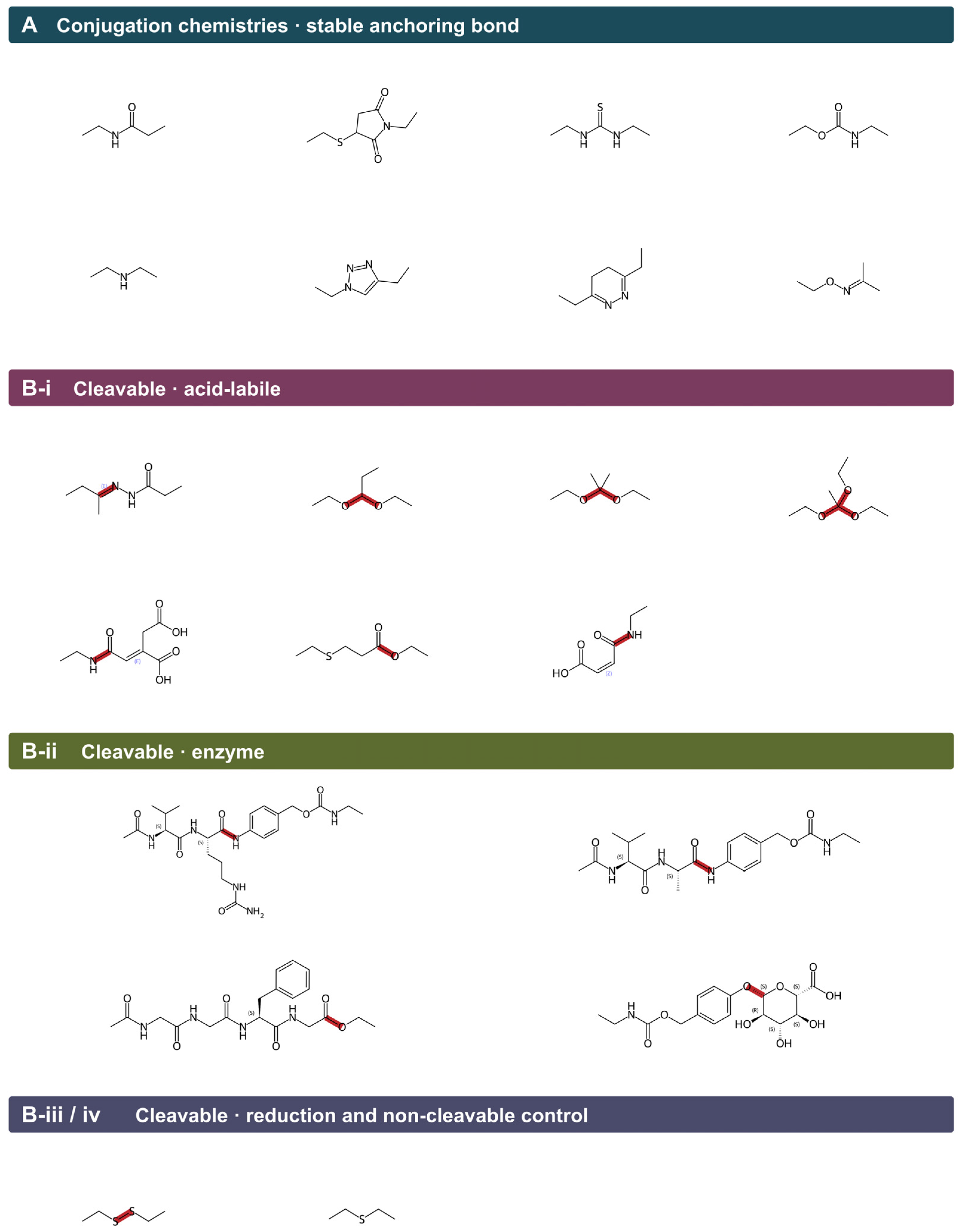
Beyond cleavability, the chemistry by which the cRGD peptide is initially tethered to the nanoparticle dictates conjugation efficiency, regiochemical fidelity, peptide orientation, and surface valency, thereby exerting a decisive influence on the functional integrity of the resulting conjugate. Contemporary RGD–nanoparticle systems leverage several bioconjugation strategies (Table 5 and Table 6), encompassing stable covalent bonds, bioorthogonal click reactions, affinity- or genetically encoded tethering, and non-covalent self-assembly (Figure 10A).
(I)
Stable covalent coupling. One of the most widely adopted strategies is carbodiimide-mediated amide bond formation (EDC/NHS), which facilitates the immobilization of carboxyl- or amine-functionalized peptides directly onto pre-activated nanoparticle surfaces. In this regard, pre-activated carboxylated mesoporous silica nanoparticles demonstrated dual functionalities of cRGD and folic acid for paclitaxel delivery [112] or an RGDK-R/AS1411 aptamer combination to target doxorubicin [113]. A further notable example is the anchoring of lipid dialkyl tails to a peptide N-terminus via a glutamic acid linker and a short C2 spacer [84], thereby establishing a hybrid covalent–self-assembly strategy that exploits bilayer integration to achieve stable and multivalent surface presentation. When cysteine residues are present within the peptide sequence (e.g., c(RGDfC)) or introduced terminally, they confer versatility for thiol-specific reactions, as with maleimide-functionalized PEG–lipid termini or disulfide coupling. Furthermore, a thiolated peptide and amine-bearing PEG termini can also be bridged via heterobifunctional crosslinkers such as succinimidyl 4-(N-maleimidomethyl)cyclohexane-1-carboxylate (SMCC) [138], as implemented in graphene oxide-based photothermal platforms for glioblastoma. Covalent coupling is the prevalent strategy for anchoring RGD peptides to liposomes (e.g., DSPE-PEG2000 in A20FMDV2 liposomes [135]) and for the multivalent display of peptides on PEG–lysine dendrimer scaffolds, with the latter yielding 5- to 10-fold greater cellular uptake than monovalent counterparts [85].
(II)
Bioorthogonal click chemistry. In accordance with strain-promoted azide–alkyne cycloaddition (SPAAC), azide-functionalized cRGD-PEG-N3 can be coupled to alkyne-decorated carriers in the absence of copper, eliminating its toxicity. This was demonstrated by covalent organic frameworks containing doxorubicin that achieved a 95% tumor size reduction with minimal off-target toxicity in triple-negative breast cancer [137]. In light of SPAAC’s orthogonality and cytocompatibility, it is becoming an increasingly popular method for in situ or intracellular conjugation.
(III)
Affinity-based and protein-engineered tethering. Through biotin–streptavidin recognition, biotinylated H2009.1 peptides were tetramerized on streptavidin-coated quantum dots, enabling αvβ6-specific receptor imaging in NSCLC models [39]. In instances where the carrier is biological, integrating the RGD motif into the HI loop of the adenovirus fiber knob can negate chemical conjugation, providing a stoichiometrically tailored, capsid-encoded peptide display with 305-fold transduction selectivity for carcinoma cells [136].
(IV)
Non-covalent and self-assembly strategies. There are several methods for displaying RGD peptides independently of chemical bioconjugation. For example, iRGD associates hydrophobically and electrostatically with human serum albumin nanoparticles [139], while the RGDS-FNIII9/10 fusion protein is passively adsorbed onto the surface of poly(lactic acid) nanoparticles [141]. Moreover, PEGylation can also drive the self-assembly of PR_b conjugates into multivalent spherical nanoparticles (PEGibodies) [140].
The choice of the conjugation strategy should therefore be justified based on the peptide orientation, regiochemical control, scalability, and biocompatibility, since they impact peptide density and avidity of engagement with integrin. Furthermore, the conjugation chemistries summarized in Table 5 are leveraged to establish stable peptide–nanoparticle anchorage, thereby providing the critical structural foundation upon which targeted delivery is built. Concomitantly, payload release at the target site is mediated by a complementary class of stimuli-responsive cleavable bonds, which can be integrated either as an internal element of the targeting linker or independently as part of the payload tethering chemistry, as elaborated in the following section.

6.3.3. Stimuli-Responsive Cleavable Chemistries

Modern innovations in linker design employ bioresponsive release mechanisms that coordinate cargo delivery with microenvironmental triggers pertaining to the target tissue or subcellular compartment. Ideally, a cleavable linker possesses three features: stability in plasma and the extracellular space throughout systemic circulation, rapid and selective cleavage at the intended target, and complete release of the active cargo. Developing responsive cRGD-nanoparticle systems requires an understanding of the nature of cleavable bonds (Figure 10(B-i–B-iii)).
Disulfide (Redox-Responsive) Linkers
There is a distinct redox asymmetry between the extracellular and intracellular compartments of the cell, specifically within cancer cells, providing an ideal niche for the responsiveness of disulfide bonds. Cytosolic glutathione (GSH) concentrations exceed those of the extracellular space by nearly three orders of magnitude, with further augmentations observed in the hypoxic, reductase-rich microenvironment of solid tumors [150,151]. Linkers that connect cRGD peptides via a disulfide bond create conjugates that, upon cellular entry through integrin interaction, engage in thiol–disulfide exchange with cytosolic GSH [114,125].
Linear or cyclic RGD variants bearing a free cysteine offer greater conjugation versatility, permitting direct, site-specific disulfide conjugation with no further coupling reagents [114]. Moreover, in some variants lacking cysteine (e.g., c(RGDfK)), disulfide functionality remains applicable via introduction of heterobifunctional crosslinkers such as succinimidyl 3-(2-pyridyldithio)propionate (SPDP) to lysine ε-amine, albeit at the cost of additional synthetic complexity [152].
This chemistry has been successfully deployed in cRGD-functionalized mesoporous silica nanoparticles [153,154] and cRGDfC–doxorubicin conjugates [155], with disulfide-gated systems exhibiting minimal premature leakage during systemic circulation while concomitantly enabling rapid GSH-triggered payload release upon tumor cell internalization. Some concerns remain regarding the susceptibility of disulfide bonds to premature breakdown by competing albumin thiols and reductases before reaching the target site, limiting the applicability of this approach [156]. Furthermore, the rate of reductive cleavage and, consequently, the tunability of release kinetics can be modulated through steric protection of the disulfide bond, whereby the incorporation of one or two methyl substituents adjacent to the susceptible disulfides confers enhanced plasma stability while judiciously preserving intracellular cleavability [156].
Acid-Labile (pH-Responsive) Linkers
Acid-labile hydrazones are among the most commonly deployed pH-responsive linkers. A pH-labile linker takes advantage of the acidification within endosomes following internalization of nanoparticles, wherein pH falls from approximately 6.5 in early endosomes to around 5.0 in late endosomes and ~4.5 in mature lysosomes [157]. Among the common pH-sensitive linkers, hydrazones were employed in the first-generation antibody–drug conjugate (Gemtuzumab ozogamicin), yet limited with variability in plasma stability [158]. Moreover, other linkers such as acetals and orthoesters can confer tunable acid sensitivity through adjacent electron-donating or electron-withdrawing substituents and further sharpen discrimination between neutral plasma and the acidic endolysosomal compartment accessed following cRGD-mediated integrin uptake [159,160]. Zhang et al. [161] tethered DOX to a dextran backbone through a hydrazone bond, and the cRGD–PEG arm was linked by CuAAC chemistry, enabling a pH-dependent release profile with only ~35% of either payload leaking over 72 h (pH 7.4), and major cargo release of ~92% (pH 5.0).
Enzyme-Responsive Linkers
Solid tumors exhibit aberrant proteolytic activities, entailing overexpressed enzymes like lysosomal cathepsin B and L, cytosolic legumain, stromal matrix metalloproteinases and plasmin [162,163]. Enzyme-responsive linkers can leverage the unique nuances in enzyme levels to promote regioselective delivery, avoiding leakage into the bloodstream. For RGD conjugates, integrin-mediated endocytosis routes the payload to lysosomes, where cysteine cathepsins are predominantly concentrated, thereby rendering cathepsin-cleavable chemistry the natural linker of choice. The dominant motif comprises a Val-Cit (or Val-Ala) dipeptide coupled to a para-aminobenzyloxycarbonyl (PABC) self-immolative spacer, and the payload, which is released unmodified following 1,6-elimination of the spacer. Notably, Paulus and Sewald [164] applied this design to an αvβ3-selective RGD mimetic–MMAE conjugate (Glu-Val-Ala-PABC linker, with the glutamate cap concomitantly suppressing murine carboxylesterase cleavage), achieving low-µM inhibition of αvβ3-positive WM115 cells, no detectable effect on αvβ3-negative M21-L cells, and a targeting index of approximately 2.8-fold over free MMAE. Further evidence was provided by Liang et al. [165], whose direct linker-variable study compared three cRGDfC–doxorubicin conjugates with identical ligand and payload; the Val-Cit-PABC variant yielded the highest cytotoxicity in αvβ3-overexpressing B16 cells (IC50 8.16 ± 1.05 µM versus 10.20 ± 1.97 and 13.40 ± 1.44 µM for the thioether and disulfide variants, respectively), as well as ~1.7- to 2.0-fold greater tumor suppression than the disulfide in syngeneic C57BL/6 mice. Together, these findings establish that linker chemistry, rather than the homing device alone, governs cRGD-conjugate tumor selectivity.
Self-Immolative Spacers and Traceless Release
The chemistry of releasing active cargo itself requires sophisticated design, particularly in cases where loading bulky cargos like camptothecin or monomethyl auristatin E can impose steric hindrance on enzyme engagement [166]. Notably, the PABC self-immolative spacer is essential in this design, as bulky payloads such as MMAE, doxorubicin, and camptothecin sterically hinder cathepsin engagement when directly attached to the dipeptide. Concomitantly, the pharmacological activity of these cargoes is strictly dependent on a free amine or hydroxyl group, which would otherwise remain masked by residual linker fragments, yielding compromised DNA intercalation, topoisomerase inhibition, or tubulin binding [167,168]. Consequently, a self-immolative spacer like PABC is inserted between the enzyme-cleavable dipeptide and the cytotoxic payload. This configuration facilitates the traceless release of the payload upon cathepsin-mediated hydrolysis. Mechanistically, Cathepsin B hydrolyzes the amide bond between the dipeptide and the PABC anilide; the resulting aniline subsequently undergoes spontaneous 1,6-elimination, concomitantly expelling CO2 and a para-quinone methide, thereby liberating the unmodified payload in its pharmacologically active form [169].

6.3.4. Design Trade-Offs Impacting Translation

Developing competent linker architectures for linear or cRGD–nanoparticle systems requires reconciling competing design imperatives, particularly plasma stability versus intracellular cleavage kinetics, hydrophilicity versus synthetic tractability, and cleavage selectivity versus breadth of tumor applicability. It is also essential to ensure that design efficacy has translational relevance to human biology, rather than just being confined to preclinical murine models. For instance, the Val-Cit dipeptide linker undergoes premature cleavage by murine carboxylesterase 1C, causing off-target effects and complicating the translatability of promising RGD–drug and RGD–nanoparticle candidates [170]. Another factor to consider is the regiochemistry of cRGD conjugate design since coupling through the terminal ε-amine of lysine as with c(RGDfK) or the cysteine thiol in c(RGDfC) preserves the integrin-binding RGD motif of the cyclic peptide. Nevertheless, in cases of iRGD, lysine linkage would impact the subsequent binding to neuropilin, which recognizes and internalizes the CendR motif (RGDK/R) [139]. It is therefore important to design the linker in concert with site-specific bioconjugation that does not compromise peptide targeting efficiency. Linker design is therefore shifting from being a passive connector to an active molecular switch converging on multi-responsive, hydrophilic, site-specific, and traceless architectures that dictate the pharmacological fate of peptide conjugates.

7. Future Perspective and Conclusions

Despite extensive research into integrin–RGD binding and promising clinical applications for RGD-based peptides, significant research gaps remain that preclude their broader clinical application. The current understanding of the conformational dynamics of integrin–ligand interactions is incomplete, since most models fail to consider the flexibility of peptides and integrins. There are also concerns related to the stability of linear RGD peptides in vivo, since they are rapidly degraded, and even the cyclic variants need to be further optimized for bioavailability. Research has focused almost exclusively on canonical RGD motifs, while alternative integrin-binding sequences like LDV or KQAGDV and other non-canonical scaffolds remain relatively underexplored. The recent discovery of a tryptathionine-bridged bicyclic peptide featuring a non-canonical norArg-Gly-Asp sequence, which exhibits subnanomolar affinity for αvβ3, demonstrates that selectivity can be engineered from within the RGD motif itself, rather than solely relying on its surrounding residues [171]. Extending this concept to αvβ6 by integrating a bicyclic constraint with non-natural analogues of the RGD residues represents an entirely unexplored avenue.
As research into integrin–RGD interactions advances, interest turns toward designing next-generation peptide ligands that combine high affinity, subtype selectivity, and in vivo stability. There are a number of strategies available; however, cyclic peptides appear to be one of the most compelling, as their constrained conformations minimize binding entropic penalties, improve resistance to proteolysis, and confer higher integrin-binding affinity than their linear counterparts [172,173,174,175,176]. In the wake of the discovery of cyclic penta-, nona- and decapeptides described earlier [48,50], researchers now have access to structural and chemical space that can be systematically explored with advanced computational tools.
An opportunity arises from applying molecular dynamics–based free energy approaches, validated on existing peptides, to the screening of new candidates. A number of methods, including Linear Interaction Energy (LIE), as well as alchemical free energy perturbation or thermodynamic integration, are available to provide detailed quantitative measurements of binding energies [177,178,179,180]. This approach can be implemented systematically to establish predictive models that link peptide sequence, conformational rigidity, and binding affinity to specific integrin subtypes. This minimizes the traditional experimental trial-and-error burden by prioritizing promising candidates for synthesis and further in vitro screening. As well as predicting affinity, free energy calculations are also useful for assessing binding kinetics, conformational transitions, and allosteric regulation of integrins, leading to the development of peptides capable of binding selectively and modulating integrin activation states to desired outcomes [181].
In this context, integrin activation states are defined by the receptor’s distinct conformations between transitions. These include a bent-closed state characterized by low ligand affinity, an intermediate extended-closed state, and an extended-open state with high affinity. The transition to the high-affinity state is facilitated by the swing-out of the hybrid domain and the rearrangement of the βA domain, which reorganizes the MIDAS coordination sphere [13,14]. Ligands engage actively since a peptide that completes the MIDAS coordination within the open headpiece serves to stabilize the high-affinity state, thereby acting as a conformational agonist. In contrast, a peptide that occupies the pocket without facilitating the opening of the headpiece functions as a pure antagonist, also referred to as a “closing” antagonist [5,10]. Designing ligands to influence the equilibrium of these states towards a specific outcome is a deliberate process. The free energy methods previously discussed are particularly effective for this purpose, as they allow for the estimation of the relative stability of each conformer and the differential binding free energy of a candidate peptide with each conformer. This has significant implications for peptide-functionalized nanoparticles, as their avidity and internalization are directly related to the proportion of receptors in the high-affinity state.
However, a focus on cyclic peptides should not mean that linear peptides are overlooked [182], as they remain simple to synthesize and functionalize for conjugation to biomaterials and drug delivery vehicles. Nevertheless, linear peptides are highly flexible and susceptible to proteolysis, requiring innovative stabilization strategies derived from cyclic peptide design to maintain synthetic simplicity while borrowing principles from stapling, backbone cyclization, or unnatural amino acid incorporation [76,183,184]. Merging computational modeling into synthetic chemistry could enable the development of stable, scalable, and cost-effective semi-cyclic or hybrid scaffolds. Additionally, machine learning and AI-driven generative models can further enrich our understanding of integrin-ligand binding by training on simulations of these interactions. The use of such models would allow de novo peptide design that goes beyond the well-established RGD motif, providing an opportunity to identify noncanonical pharmacophores or cryptic binding sequences that might contribute to enhanced specificity (e.g., αvβ3 vs. α5β1) or multifunctional activity.
Studies on nanoparticle uptake often overlook physiologically relevant factors, including protein corona formation, shear stress induced by blood flow, extracellular matrix barriers, tissue cellular heterogeneity, and interactions with immune cells. These factors can significantly influence the targeting specificity, cellular internalization, and biological performance of peptide-functionalized nanoparticles, thereby potentially reducing the predictive accuracy of conventional in vitro models. This leads to a gap between in vitro and in vivo results, limiting translational potential. Focusing on decorating nanoparticles with integrin-specific RGD-based peptide ligands may have potential for targeted cancer therapy and diagnostics. The modular peptide library is expected to fine-tune these delivery systems to enhance both the efficacy and precision of treatments by regulating critical factors such as cell adhesion and internalization. Despite still being underdeveloped, tumor-targeted peptides may offer more effective and tailored treatment options for cancer targeting [24,56,185,186]. Besides their role as therapeutic agents, peptide-functionalized nanoparticles are powerful diagnostic tools that can improve the sensitivity and specificity of imaging modalities for early detection of cancer [39,42,43,187].

Author Contributions

M.S.A.: Conceptualization, Methodology, Software, Investigation, Visualization, Writing—original draft, Writing—review and editing. N.M.: Conceptualization, Supervision, Resources, Writing—review and editing. M.Z.: Conceptualization, Supervision, Resources, Funding acquisition, Writing—review and editing. A.K.M.: Conceptualization, Methodology, Supervision, Resources, Validation, Visualization, Writing—review and editing. All authors have read and agreed to the published version of the manuscript.

Funding

The authors acknowledge Tour de Cure for PhD funding support under Grant No. [RSP-282-2026]. The first author acknowledges the support of a PhD scholarship from Griffith University and the School of Pharmacy and Medical Sciences.

Data Availability Statement

The data presented were included in the Figures and Tables of the current study.

Acknowledgments

During the preparation of this manuscript Claude (Opus 5) was used for figure presentation enhancement. The authors have reviewed and edited the output and take full responsibility for the content of this publication.

Conflicts of Interest

The authors declare no conflict of interest.

References

  1. Pan, L.; Zhao, Y.; Yuan, Z.; Qin, G. Research Advances on Structure and Biological Functions of Integrins. SpringerPlus 2016, 5, 1094. [Google Scholar] [CrossRef] [Scilit] [PubMed]
  2. Srichai, M.B.; Zent, R. Integrin Structure and Function. In Cell-Extracellular Matrix Interactions in Cancer; Springer: New York, NY, USA, 2009; pp. 19–41. [Google Scholar]
  3. Van der Flier, A.; Sonnenberg, A. Function and Interactions of Integrins. Cell Tissue Res. 2001, 305, 285–298. [Google Scholar] [CrossRef] [Scilit] [PubMed]
  4. Koivisto, L.; Heino, J.; Häkkinen, L.; Larjava, H. Integrins in Wound Healing. Adv. Wound Care 2014, 3, 762–783. [Google Scholar] [CrossRef] [Scilit] [PubMed]
  5. Pang, X.; He, X.; Qiu, Z.; Zhang, H.; Xie, R.; Liu, Z.; Gu, Y.; Zhao, N.; Xiang, Q.; Cui, Y. Targeting Integrin Pathways: Mechanisms and Advances in Therapy. Signal Transduct. Target. Ther. 2023, 8, 1. [Google Scholar] [CrossRef] [Scilit] [PubMed]
  6. Niu, G.; Chen, X. Why Integrin as a Primary Target for Imaging and Therapy. Theranostics 2011, 1, 30. [Google Scholar] [CrossRef] [Scilit] [PubMed]
  7. Kapp, T.G.; Rechenmacher, F.; Neubauer, S.; Maltsev, O.V.; Cavalcanti-Adam, E.A.; Zarka, R.; Reuning, U.; Notni, J.; Wester, H.-J.; Mas-Moruno, C. A Comprehensive Evaluation of the Activity and Selectivity Profile of Ligands for RGD-Binding Integrins. Sci. Rep. 2017, 7, 39805. [Google Scholar] [CrossRef] [Scilit] [PubMed]
  8. Nieberler, M.; Reuning, U.; Reichart, F.; Notni, J.; Wester, H.-J.; Schwaiger, M.; Weinmüller, M.; Räder, A.; Steiger, K.; Kessler, H. Exploring the Role of RGD-Recognizing Integrins in Cancer. Cancers 2017, 9, 116. [Google Scholar] [CrossRef] [Scilit] [PubMed]
  9. Zheng, Y.; Leftheris, K. Insights into Protein–Ligand Interactions in Integrin Complexes: Advances in Structure Determinations. J. Med. Chem. 2020, 63, 5675–5696. [Google Scholar] [CrossRef] [Scilit] [PubMed]
  10. Ludwig, B.S.; Kessler, H.; Kossatz, S.; Reuning, U. RGD-Binding Integrins Revisited: How Recently Discovered Functions and Novel Synthetic Ligands (Re-) Shape an Ever-Evolving Field. Cancers 2021, 13, 1711. [Google Scholar] [CrossRef] [Scilit] [PubMed]
  11. Tozer, E.C.; Liddington, R.C.; Sutcliffe, M.J.; Smeeton, A.H.; Loftus, J.C. Ligand Binding to Integrin αIIbβ3 Is Dependent on a MIDAS-like Domain in the B3 Subunit. J. Biol. Chem. 1996, 271, 21978–21984. [Google Scholar] [CrossRef] [Scilit] [PubMed]
  12. Craig, D.; Gao, M.; Schulten, K.; Vogel, V. Structural Insights into How the MIDAS Ion Stabilizes Integrin Binding to an RGD Peptide Under Force. Structure 2004, 12, 2049–2058. [Google Scholar] [CrossRef] [Scilit] [PubMed]
  13. Askari, J.A.; Buckley, P.A.; Mould, A.P.; Humphries, M.J. Linking Integrin Conformation to Function. J. Cell Sci. 2009, 122, 165–170. [Google Scholar] [CrossRef] [Scilit] [PubMed]
  14. Luo, B.-H.; Springer, T.A. Integrin Structures and Conformational Signaling. Curr. Opin. Cell Biol. 2006, 18, 579–586. [Google Scholar] [CrossRef] [Scilit] [PubMed]
  15. Hamidi, H.; Ivaska, J. Every Step of the Way: Integrins in Cancer Progression and Metastasis. Nat. Rev. Cancer 2018, 18, 533–548. [Google Scholar] [CrossRef] [Scilit] [PubMed]
  16. Aoudjit, F.; Vuori, K. Integrin Signaling in Cancer Cell Survival and Chemoresistance. Chemother. Res. Pract. 2012, 2012, 283181. [Google Scholar] [CrossRef] [Scilit] [PubMed]
  17. Niu, J.; Li, Z. The Roles of Integrin Avβ6 in Cancer. Cancer Lett. 2017, 403, 128–137. [Google Scholar] [CrossRef] [Scilit] [PubMed]
  18. Beer, A.J.; Schwaiger, M. Imaging of Integrin Avβ3 Expression. Cancer Metastasis Rev. 2008, 27, 631–644. [Google Scholar] [CrossRef] [Scilit] [PubMed]
  19. Demircioglu, F.; Hodivala-Dilke, K. Avβ3 Integrin and Tumour Blood Vessels—Learning from the Past to Shape the Future. Curr. Opin. Cell Biol. 2016, 42, 121–127. [Google Scholar] [CrossRef] [Scilit] [PubMed]
  20. Lian, Y.; Zeng, S.; Wen, S.; Zhao, X.; Fang, C.; Zeng, N. Review and Application of Integrin Alpha v Beta 6 in the Diagnosis and Treatment of Cholangiocarcinoma and Pancreatic Ductal Adenocarcinoma. Technol. Cancer Res. Treat. 2023, 22, 15330338231189399. [Google Scholar] [CrossRef] [Scilit] [PubMed]
  21. Hausner, S.H.; Bold, R.J.; Cheuy, L.Y.; Chew, H.K.; Daly, M.E.; Davis, R.A.; Foster, C.C.; Kim, E.J.; Sutcliffe, J.L. Preclinical Development and First-in-Human Imaging of the Integrin Avβ6 with [18F] Avβ6-Binding Peptide in Metastatic Carcinoma. Clin. Cancer Res. 2019, 25, 1206–1215. [Google Scholar] [PubMed]
  22. Quigley, N.G.; Steiger, K.; Hoberück, S.; Czech, N.; Zierke, M.A.; Kossatz, S.; Pretze, M.; Richter, F.; Weichert, W.; Pox, C. PET/CT Imaging of Head-and-Neck and Pancreatic Cancer in Humans by Targeting the “Cancer Integrin” Avβ6 with Ga-68-Trivehexin. Eur. J. Nucl. Med. Mol. Imaging 2022, 49, 1136–1147. [Google Scholar] [CrossRef] [Scilit] [PubMed]
  23. Rehm, J.; Winzer, R.; Pretze, M.; Müller, J.; Notni, J.; Hempel, S.; Distler, M.; Folprecht, G.; Kotzerke, J. Avβ6-Integrin Targeted PET/CT Imaging in Pancreatic Cancer Patients Using 68Ga-Trivehexin. Front. Nucl. Med. 2024, 4, 1487602. [Google Scholar] [CrossRef] [Scilit] [PubMed]
  24. Akashi, Y.; Oda, T.; Ohara, Y.; Miyamoto, R.; Kurokawa, T.; Hashimoto, S.; Enomoto, T.; Yamada, K.; Satake, M.; Ohkohchi, N. Anticancer Effects of Gemcitabine Are Enhanced by Co-Administered iRGD Peptide in Murine Pancreatic Cancer Models That Overexpressed Neuropilin-1. Br. J. Cancer 2014, 110, 1481–1487. [Google Scholar] [CrossRef] [Scilit] [PubMed]
  25. Kang, S.; Lee, S.; Park, S. iRGD Peptide as a Tumor-Penetrating Enhancer for Tumor-Targeted Drug Delivery. Polymers 2020, 12, 1906. [Google Scholar] [CrossRef] [Scilit] [PubMed]
  26. Saifi, M.A.; Sathish, G.; Bazaz, M.R.; Godugu, C. Exploration of Tumor Penetrating Peptide iRGD as a Potential Strategy to Enhance Tumor Penetration of Cancer Nanotherapeutics. Biochim. Biophys. Acta (BBA)-Rev. Cancer 2023, 1878, 188895. [Google Scholar] [CrossRef] [Scilit] [PubMed]
  27. Yu, K.-F.; Zhang, W.-Q.; Luo, L.-M.; Song, P.; Li, D.; Du, R.; Ren, W.; Huang, D.; Lu, W.-L.; Zhang, X. The Antitumor Activity of a Doxorubicin Loaded, iRGD-Modified Sterically-Stabilized Liposome on B16-F10 Melanoma Cells: In Vitro and In Vivo Evaluation. Int. J. Nanomed. 2013, 8, 2473–2485. [Google Scholar] [CrossRef] [Scilit] [PubMed]
  28. Sievers, F.; Higgins, D.G. Clustal Omega for Making Accurate Alignments of Many Protein Sequences. Protein Sci. 2018, 27, 135–145. [Google Scholar] [CrossRef] [Scilit] [PubMed]
  29. Meng, E.C.; Pettersen, E.F.; Couch, G.S.; Huang, C.C.; Ferrin, T.E. Tools for Integrated Sequence-Structure Analysis with UCSF Chimera. BMC Bioinform. 2006, 7, 339. [Google Scholar] [CrossRef] [Scilit] [PubMed]
  30. Meng, E.C.; Goddard, T.D.; Pettersen, E.F.; Couch, G.S.; Pearson, Z.J.; Morris, J.H.; Ferrin, T.E. UCSF ChimeraX: Tools for Structure Building and Analysis. Protein Sci. 2023, 32, e4792. [Google Scholar] [CrossRef] [Scilit] [PubMed]
  31. Pettersen, E.F.; Goddard, T.D.; Huang, C.C.; Meng, E.C.; Couch, G.S.; Croll, T.I.; Morris, J.H.; Ferrin, T.E. UCSF ChimeraX: Structure Visualization for Researchers, Educators, and Developers. Protein Sci. 2021, 30, 70–82. [Google Scholar] [CrossRef] [Scilit] [PubMed]
  32. Javid, H.; Oryani, M.A.; Rezagholinejad, N.; Esparham, A.; Tajaldini, M.; Karimi-Shahri, M. RGD Peptide in Cancer Targeting: Benefits, Challenges, Solutions, and Possible Integrin–RGD Interactions. Cancer Med. 2024, 13, e6800. [Google Scholar] [CrossRef] [Scilit] [PubMed]
  33. Bates, R.C. Colorectal Cancer Progression: Integrin Alphavbeta6 and the Epithelial-Mesenchymal Transition (EMT). Cell Cycle 2005, 4, 1350–1352. [Google Scholar] [CrossRef] [Scilit] [PubMed]
  34. Sun, Q.; Shang, Y.; Sun, F.; Dong, X.; Niu, J.; Li, F. Interleukin-6 Promotes Epithelial-Mesenchymal Transition and Cell Invasion through Integrin B6 Upregulation in Colorectal Cancer. Oxidative Med. Cell. Longev. 2020, 2020, 8032187. [Google Scholar] [CrossRef] [Scilit] [PubMed]
  35. Bates, R.C.; Bellovin, D.I.; Brown, C.; Maynard, E.; Wu, B.; Kawakatsu, H.; Sheppard, D.; Oettgen, P.; Mercurio, A.M. Transcriptional Activation of Integrin B6 during the Epithelial-Mesenchymal Transition Defines a Novel Prognostic Indicator of Aggressive Colon Carcinoma. J. Clin. Investig. 2005, 115, 339–347. [Google Scholar] [CrossRef] [Scilit]
  36. Mamuya, F.A.; Duncan, M.K. aV Integrins and TGF-β-induced EMT: A Circle of Regulation. J. Cell. Mol. Med. 2012, 16, 445–455. [Google Scholar] [CrossRef] [Scilit] [PubMed]
  37. Moore, K.M.; Thomas, G.J.; Duffy, S.W.; Warwick, J.; Gabe, R.; Chou, P.; Ellis, I.O.; Green, A.R.; Haider, S.; Brouilette, K. Therapeutic Targeting of Integrin Avβ6 in Breast Cancer. J. Natl. Cancer Inst. 2014, 106, dju169. [Google Scholar] [CrossRef] [Scilit] [PubMed]
  38. Hazelbag, S.; Kenter, G.G.; Gorter, A.; Dreef, E.J.; Koopman, L.A.; Violette, S.M.; Weinreb, P.H.; Fleuren, G.J. Overexpression of the Avβ6 Integrin in Cervical Squamous Cell Carcinoma Is a Prognostic Factor for Decreased Survival. J. Pathol. A J. Pathol. Soc. Gt. Br. Irel. 2007, 212, 316–324. [Google Scholar] [CrossRef] [Scilit] [PubMed]
  39. Elayadi, A.N.; Samli, K.N.; Prudkin, L.; Liu, Y.-H.; Bian, A.; Xie, X.-J.; Wistuba, I.I.; Roth, J.A.; McGuire, M.J.; Brown, K.C. A Peptide Selected by Biopanning Identifies the Integrin Avβ6 as a Prognostic Biomarker for Nonsmall Cell Lung Cancer. Cancer Res. 2007, 67, 5889–5895. [Google Scholar] [CrossRef] [Scilit] [PubMed]
  40. Li, Z.; Lin, P.; Gao, C.; Peng, C.; Liu, S.; Gao, H.; Wang, B.; Wang, J.; Niu, J.; Niu, W. Integrin B6 Acts as an Unfavorable Prognostic Indicator and Promotes Cellular Malignant Behaviors via ERK-ETS1 Pathway in Pancreatic Ductal Adenocarcinoma (PDAC). Tumor Biol. 2016, 37, 5117–5131. [Google Scholar] [CrossRef] [Scilit] [PubMed]
  41. Notni, J.; Reich, D.; Maltsev, O.V.; Kapp, T.G.; Steiger, K.; Hoffmann, F.; Esposito, I.; Weichert, W.; Kessler, H.; Wester, H.-J. In Vivo PET Imaging of the Cancer Integrin Avβ6 Using 68Ga-Labeled Cyclic RGD Nonapeptides. J. Nucl. Med. 2017, 58, 671–677. [Google Scholar] [CrossRef] [Scilit] [PubMed]
  42. Nieberler, M.; Reuning, U.; Kessler, H.; Reichart, F.; Weirich, G.; Wolff, K.-D. Fluorescence Imaging of Invasive Head and Neck Carcinoma Cells with Integrin Avβ6-Targeting RGD-Peptides: An Approach to a Fluorescence-Assisted Intraoperative Cytological Assessment of Bony Resection Margins. Br. J. Oral Maxillofac. Surg. 2018, 56, 972–978. [Google Scholar] [CrossRef] [Scilit] [PubMed]
  43. Roesch, S.; Lindner, T.; Sauter, M.; Loktev, A.; Flechsig, P.; Müller, M.; Mier, W.; Warta, R.; Dyckhoff, G.; Herold-Mende, C. Comparison of the RGD Motif–Containing Avβ6 Integrin–Binding Peptides SFLAP3 and SFITGv6 for Diagnostic Application in HNSCC. J. Nucl. Med. 2018, 59, 1679–1685. [Google Scholar] [CrossRef] [Scilit] [PubMed]
  44. Okabe, M.; Yamamoto, S.; Shiokawa, M.; Hisamatsu, T.; Yamazaki, H.; Nakanishi, R.; Hamada, K.; Kitamoto, H.; Kuwada, T.; Uza, N. Anti-Integrin Avβ6 Antibody as a Biomarker for Diagnosing Ulcerative Colitis: A Nationwide Multicenter Validation Study. J. Gastroenterol. 2025, 60, 86–95. [Google Scholar] [CrossRef] [Scilit] [PubMed]
  45. Marafini, I.; Laudisi, F.; Salvatori, S.; Lavigna, D.; Venuto, C.; Giannarelli, D.; Monteleone, G. Diagnostic Value of Anti-Integrin Avβ6 Antibodies in Ulcerative Colitis. Dig. Liver Dis. 2024, 56, 55–60. [Google Scholar] [CrossRef] [Scilit] [PubMed]
  46. Urquiza, M.; Guevara, V.; Diaz-Sana, E.; Mora, F. The Role of Avβ6 Integrin Binding Molecules in the Diagnosis and Treatment of Cancer. Curr. Org. Chem. 2020, 24, 2393–2411. [Google Scholar] [CrossRef] [Scilit]
  47. DiCara, D.; Rapisarda, C.; Sutcliffe, J.L.; Violette, S.M.; Weinreb, P.H.; Hart, I.R.; Howard, M.J.; Marshall, J.F. Structure-Function Analysis of Arg-Gly-Asp Helix Motifs in Avβ6 Integrin Ligands. J. Biol. Chem. 2007, 282, 9657–9665. [Google Scholar] [CrossRef] [Scilit] [PubMed]
  48. Maltsev, O.V.; Marelli, U.K.; Kapp, T.G.; Di Leva, F.S.; Di Maro, S.; Nieberler, M.; Reuning, U.; Schwaiger, M.; Novellino, E.; Marinelli, L. Stable Peptides Instead of Stapled Peptides: Highly Potent Avβ6-selective Integrin Ligands. Angew. Chem. Int. Ed. 2016, 55, 1535–1539. [Google Scholar] [CrossRef] [Scilit] [PubMed]
  49. D’Amore, V.M.; Donati, G.; Lenci, E.; Ludwig, B.S.; Kossatz, S.; Baiula, M.; Trabocchi, A.; Kessler, H.; Di Leva, F.S.; Marinelli, L. Molecular View on the i RGD Peptide Binding Mechanism: Implications for Integrin Activity and Selectivity Profiles. J. Chem. Inf. Model. 2023, 63, 6302–6315. [Google Scholar] [CrossRef] [Scilit] [PubMed]
  50. Di Leva, F.S.; Tomassi, S.; Di Maro, S.; Reichart, F.; Notni, J.; Dangi, A.; Marelli, U.K.; Brancaccio, D.; Merlino, F.; Wester, H. From a Helix to a Small Cycle: Metadynamics-Inspired Avβ6 Integrin Selective Ligands. Angew. Chem. Int. Ed. 2018, 57, 14645–14649. [Google Scholar] [CrossRef] [Scilit] [PubMed]
  51. Civera, M.; Arosio, D.; Bonato, F.; Manzoni, L.; Pignataro, L.; Zanella, S.; Gennari, C.; Piarulli, U.; Belvisi, L. Investigating the Interaction of Cyclic RGD Peptidomimetics with αVβ6 Integrin by Biochemical and Molecular Docking Studies. Cancers 2017, 9, 128. [Google Scholar] [CrossRef] [Scilit] [PubMed]
  52. Tesauro, D.; Accardo, A.; Diaferia, C.; Milano, V.; Guillon, J.; Ronga, L.; Rossi, F. Peptide-Based Drug-Delivery Systems in Biotechnological Applications: Recent Advances and Perspectives. Molecules 2019, 24, 351. [Google Scholar] [CrossRef] [Scilit] [PubMed]
  53. Bobrin, V.; Zhang, J.; Wu, G. Nanocarrier Strategies for Deep Tumour Penetration. RSC Appl. Polym. 2025, 3, 1403–1427. [Google Scholar] [CrossRef] [Scilit]
  54. Sugahara, K.N.; Braun, G.B.; de Mendoza, T.H.; Kotamraju, V.R.; French, R.P.; Lowy, A.M.; Teesalu, T.; Ruoslahti, E. Tumor-Penetrating iRGD Peptide Inhibits Metastasis. Mol. Cancer Ther. 2015, 14, 120–128. [Google Scholar] [CrossRef] [Scilit] [PubMed]
  55. Zuo, H. iRGD: A Promising Peptide for Cancer Imaging and a Potential Therapeutic Agent for Various Cancers. J. Oncol. 2019, 2019, 9367845. [Google Scholar] [CrossRef] [Scilit] [PubMed]
  56. Lo, J.H.; Hao, L.; Muzumdar, M.D.; Raghavan, S.; Kwon, E.J.; Pulver, E.M.; Hsu, F.; Aguirre, A.J.; Wolpin, B.M.; Fuchs, C.S. iRGD-Guided Tumor-Penetrating Nanocomplexes for Therapeutic siRNA Delivery to Pancreatic Cancer. Mol. Cancer Ther. 2018, 17, 2377–2388. [Google Scholar] [CrossRef] [Scilit] [PubMed]
  57. de Mendoza, T.H.; Mose, E.S.; Botta, G.P.; Braun, G.B.; Kotamraju, V.R.; French, R.P.; Suzuki, K.; Miyamura, N.; Teesalu, T.; Ruoslahti, E. Tumor-Penetrating Therapy for B5 Integrin-Rich Pancreas Cancer. Nat. Commun. 2021, 12, 1541. [Google Scholar] [CrossRef] [Scilit] [PubMed]
  58. Yang, X.; Zuo, Q.; He, Q.; Huang, H.; Tian, H.; Zhang, Z.; Yan, J.; Wang, R.; Hu, K. Conformationally Tuned Cyclic RGD Peptides for Integrin-Subtype-Selective PET/CT Imaging. J. Med. Chem. 2026, 69, 17054–17067. [Google Scholar] [CrossRef] [Scilit] [PubMed]
  59. Quigley, N.G.; Richter, F.; Kossatz, S.; Notni, J. Complexity of Avβ6-Integrin Targeting RGD Peptide Trimers: Emergence of Non-Specific Binding by Synergistic Interaction. RSC Med. Chem. 2023, 14, 2564–2573. [Google Scholar] [CrossRef] [Scilit] [PubMed]
  60. Ma, X.; Zhang, Z.; Barba-Bon, A.; Han, D.; Qi, Z.; Ge, B.; He, H.; Huang, F.; Nau, W.M.; Wang, X. A Small-Molecule Carrier for the Intracellular Delivery of a Membrane-Impermeable Protein with Retained Bioactivity. Proc. Natl. Acad. Sci. USA 2024, 121, e2407515121. [Google Scholar] [CrossRef] [Scilit] [PubMed]
  61. Cheng, Z.; Li, Y.; Zhao, D.; Zhao, W.; Wu, M.; Zhang, W.; Cui, Y.; Zhang, P.; Zhang, Z. Nanocarriers for Intracellular Co-Delivery of Proteins and Small-Molecule Drugs for Cancer Therapy. Front. Bioeng. Biotechnol. 2022, 10, 994655. [Google Scholar] [CrossRef] [Scilit] [PubMed]
  62. Attia, M.S.; Kijanka, G.; Nguyen, N.-T.; Zhang, J.; An, H. Advances and Prospects of RNA Delivery Nanoplatforms for Cancer Therapy. Acta Pharm. Sin. B 2025, 15, 52–96. [Google Scholar] [CrossRef] [Scilit] [PubMed]
  63. Dena, A.S.A.; El-Sherbiny, I.M. Biological Macromolecules for Nucleic Acid Delivery. In Biological Macromolecules; Elsevier: New York, NY, USA, 2022; pp. 479–490. [Google Scholar]
  64. Haupenthal, J.; Baehr, C.; Kiermayer, S.; Zeuzem, S.; Piiper, A. Inhibition of RNAse A Family Enzymes Prevents Degradation and Loss of Silencing Activity of siRNAs in Serum. Biochem. Pharmacol. 2006, 71, 702–710. [Google Scholar] [CrossRef] [Scilit] [PubMed]
  65. Zhang, H.; Vandesompele, J.; Braeckmans, K.; De Smedt, S.C.; Remaut, K. Nucleic Acid Degradation as Barrier to Gene Delivery: A Guide to Understand and Overcome Nuclease Activity. Chem. Soc. Rev. 2024, 53, 317–360. [Google Scholar] [CrossRef] [Scilit] [PubMed]
  66. Zaki, N.M.; Tirelli, N. Gateways for the Intracellular Access of Nanocarriers: A Review of Receptor-Mediated Endocytosis Mechanisms and of Strategies in Receptor Targeting. Expert Opin. Drug Deliv. 2010, 7, 895–913. [Google Scholar] [CrossRef] [Scilit] [PubMed]
  67. de Almeida, M.S.; Susnik, E.; Drasler, B.; Taladriz-Blanco, P.; Petri-Fink, A.; Rothen-Rutishauser, B. Understanding Nanoparticle Endocytosis to Improve Targeting Strategies in Nanomedicine. Chem. Soc. Rev. 2021, 50, 5397–5434. [Google Scholar] [CrossRef] [Scilit] [PubMed]
  68. Dowdy, S.F. Endosomal Escape of RNA Therapeutics: How Do We Solve This Rate-Limiting Problem? RNA 2023, 29, 396–401. [Google Scholar] [CrossRef] [Scilit] [PubMed]
  69. Dwivedi, S.D.; Deshpande, P.; Choudhary, R.; Shree, J. Challenges in Nucleic Acid Delivery. In Nanotechnology for Nucleic Acid Delivery; Apple Academic Press: Palm Bay, FL, USA, 2026; pp. 63–97. [Google Scholar]
  70. Gyanani, V.; Goswami, R. Key Design Features of Lipid Nanoparticles and Electrostatic Charge-Based Lipid Nanoparticle Targeting. Pharmaceutics 2023, 15, 1184. [Google Scholar] [CrossRef] [Scilit] [PubMed]
  71. Mamidi, N.; De Silva, F.F.; Mahmoudsalehi, A.O. Advanced Disease Therapeutics Using Engineered Living Drug Delivery Systems. Nanoscale 2025, 17, 7673–7696. [Google Scholar] [CrossRef] [Scilit] [PubMed]
  72. Vagena, I.-A.; Malapani, C.; Gatou, M.-A.; Lagopati, N.; Pavlatou, E.A. Enhancement of EPR Effect for Passive Tumor Targeting: Current Status and Future Perspectives. Appl. Sci. 2025, 15, 3189. [Google Scholar] [CrossRef] [Scilit]
  73. Deng, X.; Zhang, Z.; Ren, T.; Chen, L. Regulation of Oxidative Stress and Inflammation Caused by Drug Accumulation in the TME Based on EPR-Passive Strategy and Active Targeting. Cancer Nanotechnol. 2025, 16, 40. [Google Scholar] [CrossRef] [Scilit]
  74. Sharma, R.; Borah, S.J.; Bhawna; Kumar, S.; Gupta, A.; Singh, P.; Goel, V.K.; Kumar, R.; Kumar, V. Functionalized Peptide-Based Nanoparticles for Targeted Cancer Nanotherapeutics: A State-of-the-Art Review. ACS Omega 2022, 7, 36092–36107. [Google Scholar] [CrossRef] [Scilit] [PubMed]
  75. Dougherty, P.G.; Sahni, A.; Pei, D. Understanding Cell Penetration of Cyclic Peptides. Chem. Rev. 2019, 119, 10241–10287. [Google Scholar] [CrossRef] [Scilit] [PubMed]
  76. Lalani, N.; Tivari, S.; Jain, V.; Jadeja, Y. Review on Therapeutic Potential of Peptides: Advancements in Synthesis Methods, Linear and Cyclic Peptides, and Strategies for Overcoming Challenges. Pept. Sci. 2024, 116, e24343. [Google Scholar] [CrossRef] [Scilit]
  77. Lucie, S.; Elisabeth, G.; Stéphanie, F.; Guy, S.; Amandine, H.; Corinne, A.-R.; Didier, B.; Catherine, S.; Alexeï, G.; Pascal, D. Clustering and Internalization of Integrin Avβ3 with a Tetrameric RGD-Synthetic Peptide. Mol. Ther. 2009, 17, 837–843. [Google Scholar] [CrossRef] [Scilit] [PubMed]
  78. Yu, C.; Rafiq, N.B.M.; Cao, F.; Zhou, Y.; Krishnasamy, A.; Biswas, K.H.; Ravasio, A.; Chen, Z.; Wang, Y.-H.; Kawauchi, K. Integrin-Beta3 Clusters Recruit Clathrin-Mediated Endocytic Machinery in the Absence of Traction Force. Nat. Commun. 2015, 6, 8672. [Google Scholar] [CrossRef] [Scilit] [PubMed]
  79. Kemker, I.; Feiner, R.C.; Müller, K.M.; Sewald, N. Size-Dependent Cellular Uptake of RGD Peptides. ChemBioChem 2020, 21, 496–499. [Google Scholar] [CrossRef] [Scilit] [PubMed]
  80. Stefanick, J.F.; Ashley, J.D.; Kiziltepe, T.; Bilgicer, B. A Systematic Analysis of Peptide Linker Length and Liposomal Polyethylene Glycol Coating on Cellular Uptake of Peptide-Targeted Liposomes. ACS Nano 2013, 7, 2935–2947. [Google Scholar] [CrossRef] [Scilit] [PubMed]
  81. Stefanick, J.F.; Ashley, J.D.; Bilgicer, B. Enhanced Cellular Uptake of Peptide-Targeted Nanoparticles through Increased Peptide Hydrophilicity and Optimized Ethylene Glycol Peptide-Linker Length. ACS Nano 2013, 7, 8115–8127. [Google Scholar] [CrossRef] [Scilit] [PubMed]
  82. Wang, R.; Xiao, R.; Zeng, Z.; Xu, L.; Wang, J. Application of Poly (Ethylene Glycol)–Distearoylphosphatidylethanolamine (PEG-DSPE) Block Copolymers and Their Derivatives as Nanomaterials in Drug Delivery. Int. J. Nanomed. 2012, 7, 4185–4198. [Google Scholar] [CrossRef] [Scilit] [PubMed]
  83. Sun, J.; Jiang, L.; Lin, Y.; Gerhard, E.M.; Jiang, X.; Li, L.; Yang, J.; Gu, Z. Enhanced Anticancer Efficacy of Paclitaxel through Multistage Tumor-Targeting Liposomes Modified with RGD and KLA Peptides. Int. J. Nanomed. 2017, 12, 1517–1537. [Google Scholar] [CrossRef] [Scilit] [PubMed]
  84. Garg, A.; Tisdale, A.W.; Haidari, E.; Kokkoli, E. Targeting Colon Cancer Cells Using PEGylated Liposomes Modified with a Fibronectin-Mimetic Peptide. Int. J. Pharm. 2009, 366, 201–210. [Google Scholar] [CrossRef] [Scilit] [PubMed]
  85. Gray, B.P.; Li, S.; Brown, K.C. From Phage Display to Nanoparticle Delivery: Functionalizing Liposomes with Multivalent Peptides Improves Targeting to a Cancer Biomarker. Bioconjugate Chem. 2013, 24, 85–96. [Google Scholar] [CrossRef] [Scilit] [PubMed]
  86. Dai, W.; Fan, Y.; Zhang, H.; Wang, X.; Zhang, Q.; Wang, X. A Comprehensive Study of iRGD-Modified Liposomes with Improved Chemotherapeutic Efficacy on B16 Melanoma. Drug Deliv. 2015, 22, 10–20. [Google Scholar] [CrossRef] [Scilit] [PubMed]
  87. Huang, H.; Yang, X.; Li, H.; Lu, H.; Oswald, J.; Liu, Y.; Zeng, J.; Jin, C.; Peng, X.; Liu, J. iRGD Decorated Liposomes: A Novel Actively Penetrating Topical Ocular Drug Delivery Strategy. Nano Res. 2020, 13, 3105–3109. [Google Scholar] [CrossRef] [Scilit]
  88. Guan, J.; Guo, H.; Tang, T.; Wang, Y.; Wei, Y.; Seth, P.; Li, Y.; Dehm, S.M.; Ruoslahti, E.; Pang, H. iRGD-liposomes Enhance Tumor Delivery and Therapeutic Efficacy of Antisense Oligonucleotide Drugs against Primary Prostate Cancer and Bone Metastasis. Adv. Funct. Mater. 2021, 31, 2100478. [Google Scholar] [CrossRef] [Scilit] [PubMed]
  89. Zhao, H.; Wang, J.-C.; Sun, Q.-S.; Luo, C.-L.; Zhang, Q. RGD-Based Strategies for Improving Antitumor Activity of Paclitaxel-Loaded Liposomes in Nude Mice Xenografted with Human Ovarian Cancer. J. Drug Target. 2009, 17, 10–18. [Google Scholar] [CrossRef] [Scilit] [PubMed]
  90. Chen, W.-H.; Lei, Q.; Luo, G.-F.; Jia, H.-Z.; Hong, S.; Liu, Y.-X.; Cheng, Y.-J.; Zhang, X.-Z. Rational Design of Multifunctional Gold Nanoparticles via Host–Guest Interaction for Cancer-Targeted Therapy. ACS Appl. Mater. Interfaces 2015, 7, 17171–17180. [Google Scholar] [CrossRef] [Scilit] [PubMed]
  91. Li, Z.; Cai, W.; Cao, Q.; Chen, K.; Wu, Z.; He, L.; Chen, X. 64Cu-Labeled Tetrameric and Octameric RGD Peptides for Small-Animal PET of Tumor Avβ3 Integrin Expression. J. Nucl. Med. 2007, 48, 1162–1171. [Google Scholar] [CrossRef] [Scilit] [PubMed]
  92. Karmaker, S.; Rosales, P.D.; Tirumuruhan, B.; Viravalli, A.; Boehnke, N. More than a Delivery System: The Evolving Role of Lipid-Based Nanoparticles. Nanoscale 2025, 17, 11864–11893. [Google Scholar] [CrossRef] [Scilit] [PubMed]
  93. Ferraresso, F.; Strilchuk, A.W.; Juang, L.J.; Poole, L.G.; Luyendyk, J.P.; Kastrup, C.J. Comparison of DLin-MC3-DMA and ALC-0315 for siRNA Delivery to Hepatocytes and Hepatic Stellate Cells. Mol. Pharm. 2022, 19, 2175–2182. [Google Scholar] [CrossRef] [Scilit] [PubMed]
  94. Schoenmaker, L.; Witzigmann, D.; Kulkarni, J.A.; Verbeke, R.; Kersten, G.; Jiskoot, W.; Crommelin, D.J. mRNA-Lipid Nanoparticle COVID-19 Vaccines: Structure and Stability. Int. J. Pharm. 2021, 601, 120586. [Google Scholar] [CrossRef] [Scilit] [PubMed]
  95. Kamaly, N.; Yameen, B.; Wu, J.; Farokhzad, O.C. Degradable Controlled-Release Polymers and Polymeric Nanoparticles: Mechanisms of Controlling Drug Release. Chem. Rev. 2016, 116, 2602–2663. [Google Scholar] [CrossRef] [Scilit] [PubMed]
  96. Makadia, H.K.; Siegel, S.J. Poly Lactic-Co-Glycolic Acid (PLGA) as Biodegradable Controlled Drug Delivery Carrier. Polymers 2011, 3, 1377–1397. [Google Scholar] [CrossRef] [Scilit] [PubMed]
  97. Nie, H.; Khew, S.T.; Lee, L.Y.; Poh, K.L.; Tong, Y.W.; Wang, C.-H. Lysine-Based Peptide-Functionalized PLGA Foams for Controlled DNA Delivery. J. Control. Release 2009, 138, 64–70. [Google Scholar] [CrossRef] [Scilit] [PubMed]
  98. Yu, Y.; Li, J.; Song, B.; Ma, Z.; Zhang, Y.; Sun, H.; Wei, X.; Bai, Y.; Lu, X.; Zhang, P. Polymeric PD-L1 Blockade Nanoparticles for Cancer Photothermal-Immunotherapy. Biomaterials 2022, 280, 121312. [Google Scholar] [CrossRef] [Scilit] [PubMed]
  99. Krayukhina, M.A.; Samoilova, N.A.; Yamskov, I.A. Polyelectrolyte Complexes of Chitosan: Formation, Properties and Applications. Russ. Chem. Rev. 2008, 77, 799–813. [Google Scholar] [CrossRef] [Scilit]
  100. Ho, M.-H.; Wang, D.-M.; Hsieh, H.-J.; Liu, H.-C.; Hsien, T.-Y.; Lai, J.-Y.; Hou, L.-T. Preparation and Characterization of RGD-Immobilized Chitosan Scaffolds. Biomaterials 2005, 26, 3197–3206. [Google Scholar] [CrossRef] [Scilit] [PubMed]
  101. Cai, L.-L.; Liu, P.; Li, X.; Huang, X.; Ye, Y.-Q.; Chen, F.-Y.; Yuan, H.; Hu, F.-Q.; Du, Y.-Z. RGD Peptide-Mediated Chitosan-Based Polymeric Micelles Targeting Delivery for Integrin-Overexpressing Tumor Cells. Int. J. Nanomed. 2011, 6, 3499–3508. [Google Scholar] [CrossRef] [Scilit] [PubMed][Green Version]
  102. Kunath, K.; Merdan, T.; Hegener, O.; Häberlein, H.; Kissel, T. Integrin Targeting Using RGD-PEI Conjugates for In Vitro Gene Transfer. J. Gene Med. A Cross Discip. J. Res. Sci. Gene Transf. Its Clin. Appl. 2003, 5, 588–599. [Google Scholar] [CrossRef] [Scilit] [PubMed]
  103. Lesniak, W.G.; Kariapper, M.S.; Nair, B.M.; Tan, W.; Hutson, A.; Balogh, L.P.; Khan, M.K. Synthesis and Characterization of PAMAM Dendrimer-Based Multifunctional Nanodevices for Targeting Avβ3 Integrins. Bioconjugate Chem. 2007, 18, 1148–1154. [Google Scholar] [CrossRef] [Scilit] [PubMed]
  104. Boswell, C.A.; Eck, P.K.; Regino, C.A.; Bernardo, M.; Wong, K.J.; Milenic, D.E.; Choyke, P.L.; Brechbiel, M.W. Synthesis, Characterization, and Biological Evaluation of Integrin Avβ3-Targeted PAMAM Dendrimers. Mol. Pharm. 2008, 5, 527–539. [Google Scholar] [CrossRef] [Scilit] [PubMed]
  105. Lyu, Z.; Ding, L.; Tintaru, A.; Peng, L. Self-Assembling Supramolecular Dendrimers for Biomedical Applications: Lessons Learned from Poly (Amidoamine) Dendrimers. Acc. Chem. Res. 2020, 53, 2936–2949. [Google Scholar] [CrossRef] [Scilit] [PubMed]
  106. Salmain, M.; Bertrand, B. Emerging Anticancer Therapeutic Modalities Brought by Gold Complexes: Overview and Perspectives. Eur. J. Inorg. Chem. 2023, 26, e202300340. [Google Scholar] [CrossRef] [Scilit]
  107. Wildt, B.; Wirtz, D.; Searson, P.C. Triggering Cell Detachment from Patterned Electrode Arrays by Programmed Subcellular Release. Nat. Protoc. 2010, 5, 1273–1280. [Google Scholar] [CrossRef] [Scilit] [PubMed][Green Version]
  108. Xie, L.; Zhi, X.; Xiao, N.; Fang, C.-J.; Yan, C.-H. Constraining the Conformation of Peptides with Au Nanorods to Construct Multifunctional Therapeutic Agents with Targeting, Imaging, and Photothermal Abilities. RSC Adv. 2018, 8, 26517–26522. [Google Scholar] [CrossRef] [Scilit] [PubMed]
  109. Mahmoudi, M.; Sant, S.; Wang, B.; Laurent, S.; Sen, T. Superparamagnetic Iron Oxide Nanoparticles (SPIONs): Development, Surface Modification and Applications in Chemotherapy. Adv. Drug Deliv. Rev. 2011, 63, 24–46. [Google Scholar] [CrossRef] [Scilit] [PubMed]
  110. Pan, L.; He, Q.; Liu, J.; Chen, Y.; Ma, M.; Zhang, L.; Shi, J. Nuclear-Targeted Drug Delivery of TAT Peptide-Conjugated Monodisperse Mesoporous Silica Nanoparticles. J. Am. Chem. Soc. 2012, 134, 5722–5725. [Google Scholar] [CrossRef] [Scilit] [PubMed]
  111. Lu, J.; Shen, H.-H.; Wu, Z.; Wang, B.; Zhao, D.; He, L. Self-Assembly of Bi-Functional Peptides on Large-Pore Mesoporous Silica Nanoparticles for miRNA Binding and Delivery. J. Mater. Chem. B 2015, 3, 7653–7657. [Google Scholar] [CrossRef] [Scilit] [PubMed]
  112. Yan, H.; You, Y.; Li, X.; Liu, L.; Guo, F.; Zhang, Q.; Liu, D.; Tong, Y.; Ding, S.; Wang, J. Preparation of RGD Peptide/Folate Acid Double-Targeted Mesoporous Silica Nanoparticles and Its Application in Human Breast Cancer MCF-7 Cells. Front. Pharmacol. 2020, 11, 898. [Google Scholar] [CrossRef] [Scilit] [PubMed]
  113. Hazeri, Y.; Samie, A.; Ramezani, M.; Alibolandi, M.; Yaghoobi, E.; Dehghani, S.; Zolfaghari, R.; Khatami, F.; Zavvar, T.; Nameghi, M.A. Dual-Targeted Delivery of Doxorubicin by Mesoporous Silica Nanoparticle Coated with AS1411 Aptamer and RGDK-R Peptide to Breast Cancer In Vitro and In Vivo. J. Drug Deliv. Sci. Technol. 2022, 71, 103285. [Google Scholar] [CrossRef] [Scilit]
  114. Huang, Z.; Li, W.; MacKay, J.A.; Szoka, F.C. Thiocholesterol-Based Lipids for Ordered Assembly of Bioresponsive Gene Carriers. Mol. Ther. 2005, 11, 409–417. [Google Scholar] [CrossRef] [Scilit] [PubMed]
  115. Wright, T.H.; Brooks, A.E.; Didsbury, A.J.; Williams, G.M.; Harris, P.W.; Dunbar, P.; Brimble, M.A. Direct Peptide Lipidation through Thiol-Ene Coupling Enables Rapid Synthesis and Evaluation of Self-Adjuvanting Vaccine Candidates. Angew. Chem. Int. Ed. 2013, 125, 10810. [Google Scholar] [CrossRef] [Scilit]
  116. Wang, J.; Chow, D.; Heiati, H.; Shen, W.-C. Reversible Lipidization for the Oral Delivery of Salmon Calcitonin. J. Control. Release 2003, 88, 369–380. [Google Scholar] [CrossRef] [Scilit] [PubMed]
  117. Miyasaki, K.; Han, S.; Carton, O.; Kandell, R.M.; Gunn, J.; Kwon, E.J. Formulation Methods for Peptide-Modified Lipid Nanoparticles. J. Control. Release 2025, 385, 114030. [Google Scholar] [CrossRef] [Scilit] [PubMed]
  118. Darwish, M.; Gao, X.; Shatz, W.; Li, H.; Lin, M.; Franke, Y.; Tam, C.; Mortara, K.; Zilberleyb, I.; Hannoush, R.N. Nanolipoprotein Particles for Co-Delivery of Cystine-Knot Peptides and Fab–Based Therapeutics. Nanoscale Adv. 2021, 3, 3929–3941. [Google Scholar] [CrossRef] [Scilit] [PubMed]
  119. Chen, C.-W.; Lu, D.-W.; Yeh, M.-K.; Shiau, C.-Y.; Chiang, C.-H. Novel RGD-Lipid Conjugate-Modified Liposomes for Enhancing siRNA Delivery in Human Retinal Pigment Epithelial Cells. Int. J. Nanomed. 2011, 6, 2567–2580. [Google Scholar] [CrossRef] [Scilit] [PubMed]
  120. Futaki, S.; Ohashi, W.; Suzuki, T.; Niwa, M.; Tanaka, S.; Ueda, K.; Harashima, H.; Sugiura, Y. Stearylated Arginine-Rich Peptides: A New Class of Transfection Systems. Bioconjugate Chem. 2001, 12, 1005–1011. [Google Scholar] [CrossRef] [Scilit] [PubMed]
  121. Rocco, D.; Ross, J.; Murray, P.E.; Caccetta, R. Acyl Lipidation of a Peptide: Effects on Activity and Epidermal Permeability In Vitro. Drug Des. Dev. Ther. 2016, 10, 2203–2209. [Google Scholar] [CrossRef] [Scilit] [PubMed]
  122. Arshad, S.; Asim, M.H.; Nazir, I.; Shahzadi, I.; Mousli, M.; Bernkop-Schnürch, A. S-Protected Thiolated Nanostructured Lipid Carriers Exhibiting Improved Mucoadhesive Properties. Int. J. Pharm. 2020, 587, 119690. [Google Scholar] [CrossRef] [Scilit] [PubMed]
  123. Handké, N.; Ficheux, D.; Rollet, M.; Delair, T.; Mabrouk, K.; Bertin, D.; Gigmes, D.; Verrier, B.; Trimaille, T. Lysine-Tagged Peptide Coupling onto Polylactide Nanoparticles Coated with Activated Ester-Based Amphiphilic Copolymer: A Route to Highly Peptide-Functionalized Biodegradable Carriers. Colloids Surf. B Biointerfaces 2013, 103, 298–303. [Google Scholar] [CrossRef] [Scilit] [PubMed]
  124. Martínez-Jothar, L.; Doulkeridou, S.; Schiffelers, R.M.; Torano, J.S.; Oliveira, S.; van Nostrum, C.F.; Hennink, W.E. Insights into Maleimide-Thiol Conjugation Chemistry: Conditions for Efficient Surface Functionalization of Nanoparticles for Receptor Targeting. J. Control. Release 2018, 282, 101–109. [Google Scholar] [CrossRef] [Scilit] [PubMed]
  125. Van Der Vlies, A.J.; O’Neil, C.P.; Hasegawa, U.; Hammond, N.; Hubbell, J.A. Synthesis of Pyridyl Disulfide-Functionalized Nanoparticles for Conjugating Thiol-Containing Small Molecules, Peptides, and Proteins. Bioconjugate Chem. 2010, 21, 653–662. [Google Scholar] [CrossRef] [Scilit] [PubMed]
  126. Paramonov, V.M.; Gerstenberg, M.; Sahlgren, C.; Lindén, M.; Rivero-Müller, A. In Vitro Targetability Validation of Peptide-Functionalized Mesoporous Silica Nanoparticles in the Presence of Serum Proteins. Front. Chem. 2020, 8, 603616. [Google Scholar] [CrossRef] [Scilit] [PubMed]
  127. Wu, Z.; Li, L.; Liu, S.; Yakushijin, F.; Yakushijin, K.; Horne, D.; Conti, P.S.; Li, Z.; Kandeel, F.; Shively, J.E. Facile Preparation of a Thiol-Reactive 18F-Labeling Agent and Synthesis of 18F-DEG-VS-NT for PET Imaging of a Neurotensin Receptor–Positive Tumor. J. Nucl. Med. 2014, 55, 1178–1184. [Google Scholar] [CrossRef] [Scilit] [PubMed]
  128. Alley, S.C.; Benjamin, D.R.; Jeffrey, S.C.; Okeley, N.M.; Meyer, D.L.; Sanderson, R.J.; Senter, P.D. Contribution of Linker Stability to the Activities of Anticancer Immunoconjugates. Bioconjugate Chem. 2008, 19, 759–765. [Google Scholar] [CrossRef] [Scilit] [PubMed]
  129. Su, Z.; Xiao, D.; Xie, F.; Liu, L.; Wang, Y.; Fan, S.; Zhou, X.; Li, S. Antibody–Drug Conjugates: Recent Advances in Linker Chemistry. Acta Pharm. Sin. B 2021, 11, 3889–3907. [Google Scholar] [CrossRef] [Scilit] [PubMed]
  130. Nair, D.P.; Podgórski, M.; Chatani, S.; Gong, T.; Xi, W.; Fenoli, C.R.; Bowman, C.N. The Thiol-Michael Addition Click Reaction: A Powerful and Widely Used Tool in Materials Chemistry. Chem. Mater. 2014, 26, 724–744. [Google Scholar] [CrossRef] [Scilit]
  131. Zaleski, M.H.; Chase, L.S.; Hood, E.D.; Wang, Z.; Nong, J.; Espy, C.L.; Zamora, M.E.; Wu, J.; Morrell, L.J.; Muzykantov, V.R. Conjugation Chemistry Markedly Impacts Toxicity and Biodistribution of Targeted Nanoparticles, Mediated by Complement Activation. Adv. Mater. 2025, 37, 2409945. [Google Scholar] [CrossRef] [Scilit] [PubMed]
  132. Chu, C.H. Development of Cyclic Peptide-Conjugated Photosensitizers for Targeted Photodynamic Therapy. Doctoral Dissertation, The Chinese University of Hong Kong, Hong Kong, China, 2021. [Google Scholar]
  133. Upadhyay, R.; Rastogi, S.; Mishra, A.K.; Yadav, S.; Yadav, A.K.; Maurya, S.K. Progress in Strain Promoted Azide-Alkyne Cycloaddition (SPAAC) Reaction and Their Applications. Asian J. Org. Chem. 2025, 14, e00505. [Google Scholar] [CrossRef] [Scilit]
  134. Oliveira, B.L.; Guo, Z.; Bernardes, G. Inverse Electron Demand Diels–Alder Reactions in Chemical Biology. Chem. Soc. Rev. 2017, 46, 4895–4950. [Google Scholar] [CrossRef] [Scilit] [PubMed]
  135. Hodgins, N.O.; Wang, J.T.-W.; Klippstein, R.; Costa, P.M.; Sosabowski, J.K.; Marshall, J.F.; Maher, J.; Al-Jamal, K.T. Investigating In Vitro and In Vivo Avβ6 Integrin Receptor-Targeting Liposomal Alendronate for Combinatory Γδ T Cell Immunotherapy. J. Control. Release 2017, 256, 141–152. [Google Scholar] [CrossRef] [Scilit] [PubMed]
  136. Uusi-Kerttula, H.; Davies, J.A.; Thompson, J.M.; Wongthida, P.; Evgin, L.; Shim, K.G.; Bradshaw, A.; Baker, A.T.; Rizkallah, P.J.; Jones, R. Ad5NULL-A20: A Tropism-Modified, Avβ6 Integrin-Selective Oncolytic Adenovirus for Epithelial Ovarian Cancer Therapies. Clin. Cancer Res. 2018, 24, 4215–4224. [Google Scholar] [CrossRef] [Scilit] [PubMed]
  137. Benyettou, F.; Khair, M.; Prakasam, T.; Varghese, S.; Matouk, Z.; Alkaabi, M.; Pena-Sánchez, P.; Boitet, M.; AbdulHalim, R.; Sharma, S.K. cRGD-Peptide Modified Covalent Organic Frameworks for Precision Chemotherapy in Triple-Negative Breast Cancer. ACS Appl. Mater. Interfaces 2024, 16, 56676–56695. [Google Scholar] [CrossRef] [Scilit] [PubMed]
  138. Akhavan, O.; Ghaderi, E.; Emamy, H. Nontoxic Concentrations of PEGylated Graphene Nanoribbons for Selective Cancer Cell Imaging and Photothermal Therapy. J. Mater. Chem. 2012, 22, 20626–20633. [Google Scholar] [CrossRef] [Scilit]
  139. Sugahara, K.N.; Teesalu, T.; Karmali, P.P.; Kotamraju, V.R.; Agemy, L.; Girard, O.M.; Hanahan, D.; Mattrey, R.F.; Ruoslahti, E. Tissue-Penetrating Delivery of Compounds and Nanoparticles into Tumors. Cancer Cell 2009, 16, 510–520. [Google Scholar] [CrossRef] [Scilit] [PubMed]
  140. Zhang, S.; Ma, X.; Wu, J.; Shen, J.; Shi, Y.; Wang, X.; Xie, L.; Sun, X.; Wu, Y.; Tian, H. Enhanced Radiotheranostic Targeting of Integrin A5β1 with PEGylation-Enabled Peptide Multidisplay Platform (PEGibody): A Strategy for Prolonged Tumor Retention with Fast Blood Clearance. Acta Pharm. Sin. B 2025, 15, 692–706. [Google Scholar] [CrossRef] [Scilit] [PubMed]
  141. Dalzon, B.; Lebas, C.; Jimenez, G.; Gutjahr, A.; Terrat, C.; Exposito, J.-Y.; Verrier, B.; Lethias, C. Poly (Lactic Acid) Nanoparticles Targeting A5β1 Integrin as Vaccine Delivery Vehicle, a Prospective Study. PLoS ONE 2016, 11, e0167663. [Google Scholar] [CrossRef] [Scilit] [PubMed]
  142. Schottelius, M.; Laufer, B.; Kessler, H.; Wester, H.-J. Ligands for Mapping Avβ3-Integrin Expression In Vivo. Acc. Chem. Res. 2009, 42, 969–980. [Google Scholar] [CrossRef] [Scilit] [PubMed]
  143. Auzzas, L.; Zanardi, F.; Battistini, L.; Burreddu, P.; Carta, P.; Rassu, G.; Curti, C.; Casiraghi, G. Targeting Avβ3 Integrin: Design and Applications of Mono-and Multifunctional RGD-Based Peptides and Semipeptides. Curr. Med. Chem. 2010, 17, 1255–1299. [Google Scholar] [CrossRef] [Scilit] [PubMed]
  144. Zhao, R.Y.; Wilhelm, S.D.; Audette, C.; Jones, G.; Leece, B.A.; Lazar, A.C.; Goldmacher, V.S.; Singh, R.; Kovtun, Y.; Widdison, W.C.; et al. Synthesis and Evaluation of Hydrophilic Linkers for Antibody–Maytansinoid Conjugates. J. Med. Chem. 2011, 54, 3606–3623. [Google Scholar] [CrossRef] [Scilit] [PubMed]
  145. Lyon, R.P.; Bovee, T.D.; Doronina, S.O.; Burke, P.J.; Hunter, J.H.; Neff-LaFord, H.D.; Jonas, M.; Anderson, M.E.; Setter, J.R.; Senter, P.D. Reducing Hydrophobicity of Homogeneous Antibody-Drug Conjugates Improves Pharmacokinetics and Therapeutic Index. Nat. Biotechnol. 2015, 33, 733–735. [Google Scholar] [CrossRef] [Scilit] [PubMed]
  146. Conilh, L.; Fournet, G.; Fourmaux, E.; Murcia, A.; Matera, E.-L.; Joseph, B.; Dumontet, C.; Viricel, W. Exatecan Antibody Drug Conjugates Based on a Hydrophilic Polysarcosine Drug-Linker Platform. Pharmaceuticals 2021, 14, 247. [Google Scholar] [CrossRef] [Scilit] [PubMed]
  147. Kubas, H.; Schäfer, M.; Bauder-Wüst, U.; Eder, M.; Oltmanns, D.; Haberkorn, U.; Mier, W.; Eisenhut, M. Multivalent Cyclic RGD Ligands: Influence of Linker Lengths on Receptor Binding. Nucl. Med. Biol. 2010, 37, 885–891. [Google Scholar] [CrossRef] [Scilit] [PubMed]
  148. Boturyn, D.; Coll, J.-L.; Garanger, E.; Favrot, M.-C.; Dumy, P. Template Assembled Cyclopeptides as Multimeric System for Integrin Targeting and Endocytosis. J. Am. Chem. Soc. 2004, 126, 5730–5739. [Google Scholar] [CrossRef] [Scilit] [PubMed]
  149. Liu, S. Radiolabeled Multimeric Cyclic RGD Peptides as Integrin Avβ3 Targeted Radiotracers for Tumor Imaging. Mol. Pharm. 2006, 3, 472–487. [Google Scholar] [CrossRef] [Scilit] [PubMed]
  150. Gamcsik, M.P.; Kasibhatla, M.S.; Teeter, S.D.; Colvin, O.M. Glutathione Levels in Human Tumors. Biomarkers 2012, 17, 671–691. [Google Scholar] [CrossRef] [Scilit] [PubMed]
  151. Saito, G.; Swanson, J.A.; Lee, K.-D. Drug Delivery Strategy Utilizing Conjugation via Reversible Disulfide Linkages: Role and Site of Cellular Reducing Activities. Adv. Drug Deliv. Rev. 2003, 55, 199–215. [Google Scholar] [CrossRef] [Scilit] [PubMed]
  152. Vachutinsky, Y.; Oba, M.; Miyata, K.; Hiki, S.; Kano, M.R.; Nishiyama, N.; Koyama, H.; Miyazono, K.; Kataoka, K. Antiangiogenic Gene Therapy of Experimental Pancreatic Tumor by sFlt-1 Plasmid DNA Carried by RGD-Modified Crosslinked Polyplex Micelles. J. Control. Release 2011, 149, 51–57. [Google Scholar] [CrossRef] [Scilit] [PubMed]
  153. Li, Z.-Y.; Hu, J.-J.; Xu, Q.; Chen, S.; Jia, H.-Z.; Sun, Y.-X.; Zhuo, R.-X.; Zhang, X.-Z. A Redox-Responsive Drug Delivery System Based on RGD Containing Peptide-Capped Mesoporous Silica Nanoparticles. J. Mater. Chem. B 2015, 3, 39–44. [Google Scholar] [CrossRef] [Scilit] [PubMed]
  154. Yu, L.; Yao, L.; Yang, K.; Fei, W.; Chen, Q.; Qin, L.; Liu, S.; Cao, M.; Liu, Q.; Qin, B. cRGD-Modified Core–Shell Mesoporous Silica@ BSA Nanoparticles for Drug Delivery. Polym. Bull. 2022, 79, 10555–10571. [Google Scholar] [CrossRef] [Scilit]
  155. Yu, J.; Mao, X.; Yang, X.; Zhao, G.; Li, S. New Transferrin Receptor-Targeted Peptide–Doxorubicin Conjugates: Synthesis and In Vitro Antitumor Activity. Molecules 2024, 29, 1758. [Google Scholar] [CrossRef] [Scilit] [PubMed]
  156. Kellogg, B.A.; Garrett, L.; Kovtun, Y.; Lai, K.C.; Leece, B.; Miller, M.; Payne, G.; Steeves, R.; Whiteman, K.R.; Widdison, W.; et al. Disulfide-Linked Antibody−Maytansinoid Conjugates: Optimization of In Vivo Activity by Varying the Steric Hindrance at Carbon Atoms Adjacent to the Disulfide Linkage. Bioconjugate Chem. 2011, 22, 717–727. [Google Scholar] [CrossRef] [Scilit] [PubMed]
  157. Bargh, J.D.; Isidro-Llobet, A.; Parker, J.S.; Spring, D.R. Cleavable Linkers in Antibody–Drug Conjugates. Chem. Soc. Rev. 2019, 48, 4361–4374. [Google Scholar] [CrossRef] [Scilit] [PubMed]
  158. Hamann, P.R.; Hinman, L.M.; Hollander, I.; Beyer, C.F.; Lindh, D.; Holcomb, R.; Hallett, W.; Tsou, H.-R.; Upeslacis, J.; Shochat, D.; et al. Gemtuzumab Ozogamicin, A Potent and Selective Anti-CD33 Antibody−Calicheamicin Conjugate for Treatment of Acute Myeloid Leukemia. Bioconjugate Chem. 2002, 13, 47–58. [Google Scholar] [CrossRef] [Scilit] [PubMed]
  159. Cordes, E.; Bull, H. Mechanism and Catalysis for Hydrolysis of Acetals, Ketals, and Ortho Esters. Chem. Rev. 1974, 74, 581–603. [Google Scholar] [CrossRef] [Scilit]
  160. Löw, H.; Mena-Osteritz, E.; von Delius, M. Self-Assembled Orthoester Cryptands: Orthoester Scope, Post-Functionalization, Kinetic Locking and Tunable Degradation Kinetics. Chem. Sci. 2018, 9, 4785–4793. [Google Scholar] [CrossRef] [Scilit] [PubMed]
  161. Zhang, L.; Huang, J.; Buratto, D.; Han, P.; Yang, Z.; Zhou, R. A pH-responsive Nanoparticle Delivery System Containing Dihydralazine and Doxorubicin-based Prodrug for Enhancing Antitumor Efficacy. Aggregate 2024, 5, e434. [Google Scholar] [CrossRef] [Scilit]
  162. Vartak, D.G.; Gemeinhart, R.A. Matrix Metalloproteases: Underutilized Targets for Drug Delivery. J. Drug Target. 2007, 15, 1–20. [Google Scholar] [CrossRef] [Scilit] [PubMed]
  163. Mohamed, M.M.; Sloane, B.F. Multifunctional Enzymes in Cancer. Nat. Rev. Cancer 2006, 6, 764–775. [Google Scholar] [CrossRef] [Scilit] [PubMed]
  164. Paulus, J.; Sewald, N. Synthesis and Evaluation of a Non-Peptide Small-Molecule Drug Conjugate Targeting Integrin αVβ3. Front. Chem. 2022, 10, 869639. [Google Scholar] [CrossRef] [Scilit] [PubMed]
  165. Liang, Y.; Li, S.; Wang, X.; Zhang, Y.; Sun, Y.; Wang, Y.; Wang, X.; He, B.; Dai, W.; Zhang, H. A Comparative Study of the Antitumor Efficacy of Peptide-Doxorubicin Conjugates with Different Linkers. J. Control. Release 2018, 275, 129–141. [Google Scholar] [CrossRef] [Scilit] [PubMed]
  166. Davis, R.A.; Ganguly, T.; Harris, R.; Hausner, S.H.; Kovacs, L.; Sutcliffe, J.L. Synthesis and Evaluation of a Monomethyl Auristatin E─Integrin Avβ6 Binding Peptide–Drug Conjugate for Tumor Targeted Drug Delivery. J. Med. Chem. 2023, 66, 9842–9852. [Google Scholar] [CrossRef] [Scilit] [PubMed]
  167. Dubowchik, G.M.; Firestone, R.A.; Padilla, L.; Willner, D.; Hofstead, S.J.; Mosure, K.; Knipe, J.O.; Lasch, S.J.; Trail, P.A. Cathepsin B-Labile Dipeptide Linkers for Lysosomal Release of Doxorubicin from Internalizing Immunoconjugates: Model Studies of Enzymatic Drug Release and Antigen-Specific In Vitro Anticancer Activity. Bioconjugate Chem. 2002, 13, 855–869. [Google Scholar] [CrossRef] [Scilit] [PubMed]
  168. Doronina, S.O.; Toki, B.E.; Torgov, M.Y.; Mendelsohn, B.A.; Cerveny, C.G.; Chace, D.F.; DeBlanc, R.L.; Gearing, R.P.; Bovee, T.D.; Siegall, C.B.; et al. Development of Potent Monoclonal Antibody Auristatin Conjugates for Cancer Therapy. Nat. Biotechnol. 2003, 21, 778–784. [Google Scholar] [CrossRef] [Scilit] [PubMed]
  169. Alouane, A.; Labruère, R.; Le Saux, T.; Schmidt, F.; Jullien, L. Self-Immolative Spacers: Kinetic Aspects, Structure–Property Relationships, and Applications. Angew. Chem. Int. Ed. 2015, 54, 7492–7509. [Google Scholar] [CrossRef] [Scilit] [PubMed]
  170. Dorywalska, M.; Dushin, R.; Moine, L.; Farias, S.E.; Zhou, D.; Navaratnam, T.; Lui, V.; Hasa-Moreno, A.; Casas, M.G.; Tran, T.-T.; et al. Molecular Basis of Valine-Citrulline-PABC Linker Instability in Site-Specific ADCs and Its Mitigation by Linker Design. Mol. Cancer Ther. 2016, 15, 958–970. [Google Scholar] [CrossRef] [Scilit] [PubMed]
  171. Yang, H.; Pan, H.; Ran, T.; Dong, W.; Pan, W.; Tan, J.; Sun, J.; Süssmuth, R.D.; Su, W.; Yao, G. Identification of an Avβ3-Targeting Bicyclic Peptide with Atypical nor Arg-Gly-Asp Sequence. Commun. Chem. 2026, 9, 83. [Google Scholar] [CrossRef] [Scilit] [PubMed]
  172. Bernhagen, D.; Jungbluth, V.; Gisbert Quilis, N.; Dostalek, J.; White, P.B.; Jalink, K.; Timmerman, P. High-Affinity A5β1-Integrin-Selective Bicyclic RGD Peptides Identified via Screening of Designed Random Libraries. ACS Comb. Sci. 2019, 21, 598–607. [Google Scholar] [CrossRef] [Scilit] [PubMed]
  173. Wang, Y.; Xiao, W.; Zhang, Y.; Meza, L.; Tseng, H.; Takada, Y.; Ames, J.B.; Lam, K.S. Optimization of RGD-Containing Cyclic Peptides against Avβ3 Integrin. Mol. Cancer Ther. 2016, 15, 232–240. [Google Scholar] [CrossRef] [Scilit] [PubMed]
  174. Koivunen, E.; Wang, B.; Ruoslahti, E. Phage Libraries Displaying Cyclic Peptides with Different Ring Sizes: Ligand Specificities of the RGD-Directed Integrins. Bio/Technology 1995, 13, 265–270. [Google Scholar] [CrossRef] [Scilit] [PubMed]
  175. Liu, S. Radiolabeled Cyclic RGD Peptides as Integrin Avβ3-Targeted Radiotracers: Maximizing Binding Affinity via Bivalency. Bioconjugate Chem. 2009, 20, 2199–2213. [Google Scholar] [CrossRef] [Scilit] [PubMed]
  176. Reichart, F.; Maltsev, O.V.; Kapp, T.G.; Räder, A.F.; Weinmüller, M.; Marelli, U.K.; Notni, J.; Wurzer, A.; Beck, R.; Wester, H.-J. Selective Targeting of Integrin Avβ8 by a Highly Active Cyclic Peptide. J. Med. Chem. 2019, 62, 2024–2037. [Google Scholar] [CrossRef] [Scilit] [PubMed]
  177. Zhang, S.; Fan, X.; Malde, A.K.; Gilbert, R.G. Development of a Model for Granule-Bound Starch Synthase I Activity Using Free-Energy Calculations. Int. J. Biol. Macromol. 2023, 253, 127589. [Google Scholar] [CrossRef] [Scilit] [PubMed]
  178. Lee, T.-S.; Allen, B.K.; Giese, T.J.; Guo, Z.; Li, P.; Lin, C.; McGee, T.D., Jr.; Pearlman, D.A.; Radak, B.K.; Tao, Y. Alchemical Binding Free Energy Calculations in AMBER20: Advances and Best Practices for Drug Discovery. J. Chem. Inf. Model. 2020, 60, 5595–5623. [Google Scholar] [CrossRef] [Scilit] [PubMed]
  179. Valdés-Tresanco, M.S.; Valdés-Tresanco, M.E.; Rubio-Carrasquilla, M.; Valiente, P.A.; Moreno, E. Tailored Parameterization of the LIE Method for Calculating the Binding Free Energy of Vps34–Inhibitor Complexes. ACS Omega 2021, 6, 29525–29536. [Google Scholar] [CrossRef] [Scilit] [PubMed]
  180. van Dijk, M.; Ter Laak, A.M.; Wichard, J.D.; Capoferri, L.; Vermeulen, N.P.; Geerke, D.P. Comprehensive and Automated Linear Interaction Energy Based Binding-Affinity Prediction for Multifarious Cytochrome P450 Aromatase Inhibitors. J. Chem. Inf. Model. 2017, 57, 2294–2308. [Google Scholar] [CrossRef] [Scilit] [PubMed]
  181. Tvaroška, I.; Kozmon, S.; Kóňa, J. Molecular Modeling Insights into the Structure and Behavior of Integrins: A Review. Cells 2023, 12, 324. [Google Scholar] [CrossRef] [Scilit] [PubMed]
  182. Hausner, S.H.; Bauer, N.; Davis, R.A.; Ganguly, T.; Tang, S.Y.; Sutcliffe, J.L. The Effects of an Albumin Binding Moiety on the Targeting and Pharmacokinetics of an Integrin Avβ6-Selective Peptide Labeled with Aluminum [18F] Fluoride. Mol. Imaging Biol. 2020, 22, 1543–1552. [Google Scholar] [CrossRef] [Scilit] [PubMed]
  183. Fang, P.; Pang, W.-K.; Xuan, S.; Chan, W.-L.; Leung, K.C.-F. Recent Advances in Peptide Macrocyclization Strategies. Chem. Soc. Rev. 2024, 53, 11725–11771. [Google Scholar] [CrossRef] [Scilit] [PubMed]
  184. Chow, H.Y.; Zhang, Y.; Matheson, E.; Li, X. Ligation Technologies for the Synthesis of Cyclic Peptides. Chem. Rev. 2019, 119, 9971–10001. [Google Scholar] [CrossRef] [Scilit] [PubMed]
  185. Zhang, Q.; Zhang, Y.; Li, K.; Wang, H.; Li, H.; Zheng, J. A Novel Strategy to Improve the Therapeutic Efficacy of Gemcitabine for Non-Small Cell Lung Cancer by the Tumor-Penetrating Peptide iRGD. PLoS ONE 2015, 10, e0129865. [Google Scholar] [CrossRef] [Scilit] [PubMed]
  186. Li, X.; Zhong, H.; Zheng, S.; Mu, J.; Yu, N.; Guo, S. Tumor-Penetrating iRGD Facilitates Penetration of Poly (Floxuridine-Ketal)-Based Nanomedicine for Enhanced Pancreatic Cancer Therapy. J. Control. Release 2024, 369, 444–457. [Google Scholar] [CrossRef] [Scilit] [PubMed]
  187. Flechsig, P.; Lindner, T.; Loktev, A.; Roesch, S.; Mier, W.; Sauter, M.; Meister, M.; Herold-Mende, C.; Haberkorn, U.; Altmann, A. PET/CT Imaging of NSCLC with a Avβ6 Integrin-Targeting Peptide. Mol. Imaging Biol. 2019, 21, 973–983. [Google Scholar] [CrossRef] [Scilit] [PubMed]
Figure 1. Pairwise sequence identity (%) among integrins α and β subunits. Percent sequence identity from the UniProt Align/Clustal Omega [28] Percent Identity Matrix (default parameters); identical residues relative to sequence length. Sequences taken from the indicated PDB constructs; α and β-chains aligned separately.
Macromol 06 00076 g001
Figure 2. Pairwise structural similarity among integrin structures evaluated using Cα RMSD (Å) values. The assessment was conducted through structural alignment facilitated by MatchMaker [29,30], employing the default parameters of UCSF ChimeraX [31].
Macromol 06 00076 g002
Figure 3. Superposition of four integrin α–β heterodimer heads. Headpieces of αIIbβ3 (3ZDY, green), α5β1 (3VI4, red), αvβ6 (4UM9, purple) and αvβ8 (6OM2, light blue), superposed on the β-chain βA domain. Both domains overlay closely across all four subfamilies, confirming a structurally conserved heterodimeric ligand-binding pocket.
Macromol 06 00076 g003
Figure 4. Multiple sequence alignment of the ligand-binding domains of integrin β and α subunits. (a) Alignment of the βA (βI) domain of the human integrin β-subunits β1, β3, β6 and β8 (β residues 118–262, β3 numbering). (b) Alignment of the β-propeller domain of the human integrin α-subunits αIIb, α5 and αV (α residues 159–249, αV numbering). Residues are colored by physicochemical property using the Clustal color scheme (blue, hydrophobic; red, positively charged; magenta, negatively charged; green, polar; cyan, aromatic; pink, cysteine; orange, glycine; yellow, proline). The annotation tracks beneath each alignment show, from top to bottom, sequence conservation, alignment quality, the consensus sequence with its sequence logo, and column occupancy. Alignments were generated and visualized in Jalview v2.11.5.2.
Macromol 06 00076 g004
Figure 5. Cyclic peptide binding interactions with αvβ3 integrin 1L5G (a). Linear peptide interaction with integrins α5β1 (3VI4) (b), αIIbβ3 (2VDQ) (c), αvβ6 (4UM9) (d), and αvβ8 (6OM2) (e). Structures were rearranged in Inkscape, while panel (a) was redrawn with RDKit to fix the original structure.
Macromol 06 00076 g005
Figure 6. 3D representation of different integrins’ interaction with RGD peptides. Integrin α5β1 binding to RGD fragment (a), integrin αvβ3 binding to cyclic RGD pentapeptide (b), and integrin αvβ8 binding to linear RGD peptide (c). In all panels the α-chain is yellow and the β-chain blue, with surfaces and peptides colored by heteroatom (O red, N blue; C in the chain color). RGD peptides are shown as sticks with green carbons, red oxygens, blue nitrogens and white hydrogens.
Macromol 06 00076 g006
Figure 7. 3D image of αvβ6 interacting with peptide (HGRGDLGRLKK). The gray surface of the SDL represents hydrophobic regions, while the white surface indicates areas with extra hydrophobicity compared to other integrins. RGD peptides are shown as sticks with green carbons, red oxygens, blue nitrogens and white hydrogens.
Macromol 06 00076 g007
Figure 8. Chemical structure of the parent cyclic pentapeptides C1 and its derivatives. The structures were generated from SMILES using RDKit version 2026.03.1. The highlighted residues represent the RGD motif.
Macromol 06 00076 g008
Figure 9. Chemical structure of the parent cyclic decapeptide C2 (a) and cyclic nonapeptide C3 (b). The structures were generated from SMILES using RDKit version 2026.03.1. The highlighted residues represent the RGD motif.
Macromol 06 00076 g009
Figure 10. Linker chemistries for peptide–drug and peptide–nanoparticle conjugates. (A) Conjugation chemistries form the stable bond anchoring the peptide to its carrier or payload. (B-iB-iv) Cleavable linkers release the payload on a microenvironmental stimulus. (B-i) Acid-labile linkers are hydrolyzed at endosomal/lysosomal or tumor pH; (B-ii) enzyme-cleavable linkers are processed by lysosomal cathepsins (Val-Cit-PABC, Val-Ala-PABC, GGFG) or β-glucuronidase (β-glucuronide), with the PABC-spacer bearing dipeptides; (B-iii) the reduction-cleavable disulfide responds to cytosolic glutathione; (B-iv) the non-cleavable thioether is a control. Ethyl groups denote attachment points. The red bonds indicate the scissile site, while Panel (A) motifs and the thioether are non-cleavable. The structures were generated from SMILES using RDKit version 2026.03.1.
Macromol 06 00076 g010
Table 1. Structural information for the PDB files of the studied integrins.
PDBIntegrin SubtypeResolution (Å)Interacting Peptide
4UM9αvβ62.50ACE HGRGDLGRLKK NH2
3VI4α5β12.90RGDNP
4WK42.50ACRGDGWC
8XERαvβ33.00c(-RGDfV-)
8XF63.10
8XFG2.80c(-RGDfK-)
8XFO3.00
1L5G3.20c(-RGDf(NMe)V-)
2VDRαIIbβ32.40LGGAKQRGDV
2VDQ2.59HHLGGAKQRGDV
3ZDY2.45GRGDSP
3ZE02.95
3ZE13.00
3ZE22.35
3ZDZ2.75
6OM2αvβ82.77GRRGDLATIHG
Table 2. Pairwise βA-domain and pocket structural similarity across four integrin subfamilies.
Pair% Sequence Identity (βA)Cα-RMSD (Å)Pocket Cα-RMSD (Å)
β3 vs. β662.70.830.49
β1 vs. β659.90.840.43
β3 vs. β159.01.070.50
β8 vs. β349.30.920.57
β8 vs. β153.01.090.45
β8 vs. β653.60.980.53
Cα-RMSD, root-mean-square deviation between equivalent α-carbon atoms (Å); <2 Å indicates the same fold. βA domains (chain B) were superposed with MatchMaker in UCSF ChimeraX [29,30,31]; Cα-RMSD is over all matched pairs, not the pruned subset. Pocket Cα-RMSD covers nine MIDAS-lining residues in that frame, without refitting; the MIDAS magnesium was identified by its coordination to the DxSxS serines and the ligand Asp. PDBs: 3ZDY (αIIbβ3), 3VI4 (α5β1), 4UM9 (αvβ6), 6OM2 (αvβ8).
Table 3. The main integrin residues contacting the arginine (R) and aspartate (D) of the RGD motif.
PDB IDIntegrin SubtypePeptide SequenceArg (R)Asp (D)
1L5Gαvβ3c(-RGDf(NMe)V-)Asp α218
2.76
Asp α150
3.32
Tyr α178
2.93
Ser β121
3.27
Ser β123
3.09
Asn β215
2.82
Arg β216
3.46
Mn β4001
2.65
4UM9αvβ6HGRGDLGRLKKAsp α218
2.82
Phe α177
3.36
Tyr α178
3.46
Asn β218
2.67
Ala β126
3.50
Ser β125
3.08
Ile β219
2.74
Mg β502
2.17
6OM2αvβ8GRRGDLATIHGAsp α218
2.42
Phe α177
3.20
Tyr α178
2.88
Ser β116
3.02
Ser β114
3.09
Asn β207
2.91
Ile β208
3.05
Mg β2001
2.06
3ZDYαIIbβ3GRGDSPTyr α189
3.89
Phe α231
3.18
Tyr α190
4.04
Ala β218
3.20
Asn β215
3.00
Tyr β122
3.09
Arg β214
3.44
Glu β220
3.40
Mg β2001
2.07
2VDQHHLGGAKQRGDVAsp α224
3.05
Tyr α189
2.74
Ser α225
3.02
Tyr α190
3.91
Asn β215
2.72
Ser β121
3.12
Tyr β122
2.78
Ser β123
2.83
Mg β2001
2.12
2VDRLGGAKQRGDVAsp α224
3.03
Tyr α189
2.92
Ser α225
2.65
Tyr α190
3.91
Asn β215
2.86
Ser β121
3.17
Tyr β122
2.85
Ser β123
2.84
Mg β2001
2.13
3VI4α5β1RGDNPAsp α227
3.28
Gln α221
2.91
Ser β132
2.81
Ser β134
2.43
Tyr β133
3.21
Glu β229
2.49
Mg β502
1.91
Salt bridge: carboxylate oxygen to guanidinium nitrogen, ≤4.0 Å Nonpolar contact: closest approach through carbon
Hydrogen bond: N/O to N/O, ≤3.5 Å, at least one oxygen Metal coordination: divalent cation to the RGD aspartate
Long polar contact: as above but 3.5–4.2 Å
Each value represents the shortest heavy-atom separation between that residue and the RGD arginine or aspartate, except for salt bridges (carboxylate to guanidinium) and the metal (cation to carboxylate).
Table 4. Binding affinity of linear and cyclic RGD peptides of varying length toward integrin αvβ6.
No.Peptide LengthSequenceIC50 (nM)Ref.
120YTASARGDLAHLTTTHARHL
[A20FMDV1]
6.80 ± 3.49 *[47]
2NAVPNLRGDLQVLAQKVART
[A20FMDV2]
0.50 ± 0.15 *
3GFTTGRRGDLATIHGMNRPF
[A20LAP]
1.40 ± 0.93 *
410c(-ARGDLAALpP-)8.30[48]
5c(-LARGDLAApP-)13.40
6c(-RGDLAALApP-)14.50
7c(-ARGDLAAApP-)15.40
8c(-ALARGDLApP-)69.90
9c(-LARTDLAApP-)94.50
10c(-AALARGDLpP-)140.00
11c(-ARTDLAALpP-)264.00
12c(-ARGDAAALpP-)320.00
13c(-ALARTDLApP-)495.00
14c(-AALARTDLpP-)720.00
15c(-RTDLAALApP-)1554.00
169c(-FRGDLA-Wp(NMe)K-)0.12
10c(-FRGDLA-Fp(NMe)K-)0.26
11c(-FRGDLA-Fp(NMe)K(Ac)-)0.30
12c(-FRGDLA-Yp(NMe)K-)0.39
13c(-YRGDLA-Fp(NMe)K-)0.40
14c(-WRGDLA-Fp(NMe)K-)0.52
15c(-ARGDLA-FpP-)0.67
16c(-ARGDLA-Fp(NMe)K-)0.70
17c(-FRGDLA-LpP-)1.25
18c(-ARGDLA-KpP-)1.51
19c(-ARGDLA-LpP-)3.00
20c(-ARGDLA-Lp(NMe)K-)4.30
21c(-ARGDLA-Lp(Sar)-)8.10
22c(-ARGDLA-L(NMe)kP-)11.40
23c(-ARGDLD-LpP-)14.70
24c(-ARGDLA-L(Sar)P-)24.20
25c(-ARGDFA-LpP-)63.40
26c(-ARGDLd-LpP-)148.00
27c(-CRGDKGPDC-) [iRGD] 191.00[49]
288c(-ARGDL-LpP-)292.00[48]
297c(-ARGDL-pP-)417.00
305c(-RGD-Cha-E)-CONH223.00 ± 2.00[50]
31c(-RGD-cha-E)-CONH2120.00 ± 19.00
32c(-RGD-Hcha-E-)-CONH233.00 ± 3.00
33c(-RGD-hcha-E-)-CONH2113.00 ± 26.00
34c(-RGD-Chg-E-)-CONH21.30 ± 0.20
35c(-RGD-chg-E-)-CONH2608.00 ± 29.00
36c(-RGD-Phg-E-)-CONH224.00 ± 6.00
37c(-RGD-phg-E-)-CONH236.00 ± 3.00
38c(-RGD-1Nal-E-)-CONH215.00 ± 2.00
39c(-RGD-1nal-E-)-CONH263.00 ± 19.00
40c(-RGD-2Nal-E-)-CONH228.00 ± 6.00
41c(-RGD-2nal-E-)-CONH2577.00 ± 93.00
42c(-RGD-Igl-E-)-CONH257.00 ± 7.00
42c(-RGD-igl-E-)-CONH2441.00 ± 122.00
* EC50 from direct binding to soluble αvβ6 [47]; all other values are competitive solid-phase binding IC50 [48,50].
Table 5. Relevant strategies for RGD peptides conjugation.
SystemConjugation ChemistryChemistry DetailsPeptideLipid/NP SystemRef.
LipidDisulfide bond formation (thiocholesterol-based)Covalent disulfide bridges between thiocholesterol and GSH/Cys allowing surface thiol modification, controlling charge and stability, and enabling selective ligand presentation on particles.
TAT peptide was crosslinked to PEG–DPPE via NHS chemistry, purified, and incorporated into NLPs by micelle transfer.
Cystiene-GSH for surface charge
TAT (GRKKRRQRRRGYG) for targeting
Thiocholesterol-modified, disulfide-tuned surface (GSH/Cys) on cationic DOPE/TAT-PEG lipid nanoparticles[114]
LipidThiol-ene radical coupling (UV-initiated)Radical-mediated addition of Cys-SH to terminal alkene C=C; photoinitiator-free click chemistry; thiyl radical attacks vinyl ether forming thioether; no protecting groups needed*CSKKKKNLVPMVATVDirect lipidation with vinyl palmitate forming self-adjuvating lipopeptides[115]
LipidREAL (Reversible Aqueous Lipidization)—N-palmitoyl cysteinyl 2-pyridyl disulfideN-acylation at Cys1/7 α-amino with palmitic acid; 2-pyridyl disulfide linker enables reversible attachment and cleavable disulfide allows de-lipidation in vivoSalmon calcitonin (polypeptide of 32 amino acid)N-palmitoylated reversible conjugate with Cys1/7 [116]
LipidPost-formulation maleimide–thiol coupling to LNPsDSPE-PEG-Mal inserts into lipid bilayer via hydrophobic DSPE anchor; terminal maleimide reacts with peptide Cys*CGKc(CRGDC), *CRGD, *CGKRK, VCAM-1, RVGDLin-MC3-DMA/DSPC/cholesterol/DSPE-PEG-Mal LNPs[117]
LipidNanolipoprotein particle (NLP) incorporation via dual modificationPeptides were modified with palmitic acid to enable NLP incorporation. Thiol-containing cysteines on Fabs reacted with maleimide-functionalized CKP-NLPs for site-specific conjugation.EETI-II cystine-knot peptide with (N-terminal Lys)DOPC/DSPE-PEG-Mal LNPs[118]
LipidDSPE-PEG-NHS ester to Lys side chain (α-NH2 acetylated)
DSPE-PEG-malemide–thiol chemistry
DSPE-PEG2K-maleimide reacted post-formulation with C-terminal cysteine thiols, as previously discussed. NHS ester selectively acylated Lys ε-amino groups while N-terminal acetylation blocked α-NH2, enabling directional amide conjugation.DGPPR*KPGGGS*C
SPALHFLGGGS*C
SNLAAFPGGGS*C
MPVAVYRGGGS*C
Cholesterol/DLin-MC3-DMA/DSPC/DSPE-PEG-NHS or malemide LNPs[119]
LipidationCholestryl, stearic and lauryl acid N-terminal conjugation forming C18 and C12 amideAmide bond between stearic acid carboxyl and peptide α-NH2; N-terminal selectivity over lysine ε-amino under controlled pHHIV-1 Tat-(48–60), HIV-1 Rev-(34–50), FHV coat-(35–49), and octaarginine (R8)N-terminal fatty acid lipopeptides [120]
LipidAcylation of N- and C-terminalAmide bonds at either α-NH2 (N-term) or α-COOH (C-term) via carbodiimide/NHS activationL-AAPV tetrapeptide (elastase inhibitor)C7 acylated lipopeptide[121]
LipidAmide coupling of palmitic acid to N-terminalAmide bond forms as NHS ester reacts with α-amino of oxidized cysteine dimerL-cystine (oxidized Cys dimer)S-protected thiolated nanostructured lipid carriers[122]
Polymeric conjugateN-acryloxysuccinimide (NAS) ester to Lys-tag and N-terminusActivated ester NAS on PLA-b-P(NAS-co-NVP) copolymer surface reacts with primary amines. Triple-Lys tag provides multiple conjugation sites and forms stable amide bonds.KKK-VQGEESNDK (IL-1β peptide with N-terminal triple-Lys tag)PLA-b-P(NAS-co-NVP) surfactant-coated PLA NPs[123]
Maleimide–thiol coupling (pH 7.0, HEPES buffer, 30 min RT)Thioether bond formation via Michael addition of lysine modified with terminal thiol to maleimide C=C; 2:1 maleimide:thiol molar ratiocRGDfK (Ac-S*CH2CO)PLGA-PEG-maleimide nanoparticles[124]
Pyridyl disulfide–thiol exchangeReversible disulfide formation via thiol–disulfide exchange; DSPE-PEG-pyridyl disulfide reacts with Cys-SH releasing pyridine-2-thione (λmax 343 nm); cleavable by GSH/DTTAc-SDKDSLK*CG-OHCross-linked Pluronic copolymers functionalized with pyridyl disulfide groups.[125]
InorganicAmide linking to carboxylated silica surfaceMesoporous silica was carboxylated using carboxyethylsilanetriol, enabling N-terminal peptide conjugation via EDC/NHSfCfwKTC–threoninol
fCFAATC–threoninol
MSNs covalently capped with peptides[126]
Small moleculeVinylsulfone Michael additionThiol on cysteine reacts with vinylsulfone to form stable thioether bondc(RGDyC)Direct peptide conjugation to 18F-DEG-VS for PET labeling[127]
EETI-II (Ecballium elaterium trypsin inhibitor-II): GCPRRCGKRRCQGICVRCIYRCRKN; VCAM: VHPKQHRGGSKGC; RVG: CK-YTIWMPENPRPGTPCDIFTNSRGKRASNG. The asterisk is placed immediately before the residue involved in the coupling reaction.
Table 6. RGD peptide-conjugated therapeutic nanoparticles targeting integrin subtypes in cancer.
Nanoparticle PlatformRGD PeptideIntegrin/
Other Target
Therapeutic PayloadCancer ModelKey OutcomeConjugation StrategyRef.
A20FMDV2-targeted liposomes (DSPC/Chol)A20FMDV2 (20-mer, FMDV-derived)
NAVPNLRGDLQVLAQKVART
αvβ6Alendronate for γδ T cell immunotherapyMelanoma (A375Pβ6) + pancreatic cancer (PANC0403) in vivoLiposomes showed 7 times higher retention in αvβ6+ compared to αvβ6− tumors and enhanced γδ T cell-mediated killing.Maleimide–thiol coupling to DSPE-PEG2000 lipid terminus[135]
A20FMDV2-redirected oncolytic adenovirus (Ad5)Oncolytic adenovirus (replicating virus expressing transgene, e.g., luciferase; potential for immunotherapies)αvβ6+ carcinoma models including ovarian (SKOV3 xenograft and ascites-derived cells) and breast (BT-20)Ad5NULL-A20 transduced αvβ6+/CAR BT-20 cells 305-fold and primary patient-derived EOC cells 69-fold more efficiently than Ad5, retaining up to a 902-fold advantage in the presence of neutralizing ascites. Systemic delivery in non-tumor-bearing mice reduced hepatic genome copy-number 107-fold and splenic copy-number > 700-fold versus Ad5. Oncolytic Ad5NULL-A20 significantly improved survival in a peritoneal SKOV3 ovarian xenograft model.Genetic insertion of A20 peptide into fiber knob (HI loop) of adenovirus capsid (protein engineering, not chemical conjugation)[136]
H2009.1-tetrameric PEGylated liposomesH2009.1 (20-mer, RGDLATLRQLAQEDGVVGVR)Doxorubicin (DOX)NSCLC (H2009)Tetravalent display of peptides showed 5–10 times superior uptake in αvβ6+ H2009 cells with ~6-fold increase in cytotoxicity compared to monovalent targeted nanoparticles.Thiol–maleimide conjugation using a multi-arm PEG–lysine dendrimer linker to form a tetrameric peptide[85]
H2009.1-peptide quantum dots (Qdot) nanoconjugatesFluorescent Qdots (SAQDot605) for imagingReceptor-specific endocytic uptake of QDots confirmed by anti-αvβ6 antibody blocking (>90% inhibition).Biotin–streptavidin conjugation (biotinylated tetrameric peptide bound to streptavidin-coated Qdot)[39]
Mesoporous silica nanoparticles (MSNs)cRGD + Folic acidαvβ3 + FRαPaclitaxel (PTX)Breast cancer (MCF-7)1.6× higher inhibitory efficacy vs. free PTX; IC50 reduced from 35.25 to 22.21 ng/mL.EDC/NHS amide coupling via PEG linker[112]
RGDK-R octaarginine + AS1411 aptamerαvβ3 + nucleolinDOXBreast cancer (4T1, MCF-7)Increased CHO cell viability and reduced toxicity compared with DOX while improving tumor inhibition with higher tumor accumulation and lower heart uptake.EDC/NHS to carboxylated MSN surface[113]
Covalent organic frameworkscRGD-PEG-N3αvβ3DOXTNBC (MDA-MB-231)Markedly improved tumor targeting and drug accumulation, reducing tumor size by 95% and increasing DOX fluorescence up to 16.5-fold compared to controls, while minimizing off-target toxicity.Copper-free SPAAC click chemistry[137]
Reduced graphene oxide nanoribbons (rGNOR)RGD-based peptide on PEG-amphiphileNIR photothermal therapyGlioblastoma (U87MG)PEGylated rGONR showed 14- and 2.4-fold higher NIR absorption than GONRs and rGO-PEG sheets, achieving ≥97% cell destruction at ≥1.0 μg mL−1 under NIR irradiation while causing <11% cell destruction and 7% DNA fragmentation at the same concentration in the dark.SMCC links amine-PEG to thiol–peptide via maleimide coupling[138]
Human serum albumin NPsiRGD (CRGDKRGPDC)
CRGDRGPDC
CRGDKGPEC
αv integrins → NRP-1 (CendR cascade)PTXProstate (22Rv1), Breast (BT474)The CendR pathway, facilitated by iRGD binding to neuropilin-1, enables deep tumor penetration, resulting in significant inhibition of tumor growth in both models.Higher tumor accumulation was observed—8 times in 22Rv1 and 11 times in BT474—when iRGD non-covalently coated albumin nanoparticles (Abraxane).[139]
PR_b-functionalized DPPC liposomesPR_b (fibronectin mimetic)
KSSPHSRN(SG)5RGDSP
α5β15-Fluorouracil (5-FU)Colon carcinoma (CT26.WT, mouse)Cytotoxicity comparable to free 5-FU, nonetheless with targeted delivery, but superior to control GRGDSP liposomes confirming α5β1 specificity.Peptide N-terminus amide-coupled to C16 dialkyl tails via Glu linker and C2 spacer.[84]
PR_b PEGibody nanoparticles64Cu/177Lu PET radionuclide imaging/therapyα5β1+ B16F10 melanoma in C57BL/6J mouse and BALB/c nude mouse miceEnhanced tumor retention vs. non-PEGylated PR_b (1.26 vs. 0.51/0.35%ID/g at 1 h; 3.40%ID/g at 24 h) with rapid blood clearance while keeping low organ and hematological toxicity.PEGylation-enabled self-assembly into multivalent spherical NPs[140]
PLA nanoparticlesRGDS-FNIII9/10 recombinant proteinHIV-1 p24 antigen (model vaccine)α5β1+ cells fibrosarcoma (HT-1080)RGDS-adsorbed nanoparticles entered cells ~8× more than blank. The binding avidity of IgG antibodies increased, but their levels remained unaffected.RGDS-FNIII9/10 recombinant protein adsorption onto PLA nanoparticle surface[141]
Abbreviations: 5-FU, 5-fluorouracil; %ID/g, percentage of injected dose per gram; CendR, C-end rule pathway; DOX, doxorubicin; DPPC, dipalmitoylphosphatidylcholine; DSPE, distearoylphosphatidylethanolamine; EDC, 1-ethyl-3-(3-dimethylaminopropyl)carbodiimide; FMDV, foot-and-mouth disease virus; FN, fibronectin; FRα, folate receptor alpha; MSN, mesoporous silica nanoparticle; NHS, N-hydroxysuccinimide; NIR, near-infrared; NRP-1, neuropilin-1; NSCLC, non-small cell lung cancer; PEG, polyethylene glycol; PET, positron emission tomography; PLA, poly(lactic acid); PTX, paclitaxel; QDs, quantum dots; SPAAC, strain-promoted azide–alkyne cycloaddition; TNBC, triple-negative breast cancer.
Disclaimer/Publisher’s Note: The statements, opinions and data contained in all publications are solely those of the individual author(s) and contributor(s) and not of MDPI and/or the editor(s). MDPI and/or the editor(s) disclaim responsibility for any injury to people or property resulting from any ideas, methods, instructions or products referred to in the content.