Review

RNA Polymerase II Activity and Nuclear Actin: Possible Roles of Nuclear Tropomyosin, Troponin and Ca2+ in Transcription in Striated Muscle Myocyte Nuclei

by
Amelia J. Koopman
,
Alexandra J. Martin
,
Lauren G. Moore
,
Michelle Rodriguez
and
Prescott Bryant Chase
*
Department of Biological Science, Florida State University, Tallahassee, FL 32306, USA
*
Author to whom correspondence should be addressed.
These authors contributed equally to this work.
Macromol 2025, 5(4), 56; https://doi.org/10.3390/macromol5040056
Submission received: 21 June 2025 / Revised: 4 August 2025 / Accepted: 11 November 2025 / Published: 13 November 2025

Abstract

Ribonucleic acid (RNA) polymerases are macromolecular machines that catalyze the synthesis of RNA macromolecules, the sequences of which are coded for by the sequences of regions of deoxyribonucleic acid (DNA) macromolecules in the nucleus of eukaryotic cells, or nuclei in the case of many mature striated muscle cells, or myocytes, which are in many cases polynucleated. Herein, we review the evidence that transcription, the activity of RNA polymerases that is an essential step in gene expression, and processes related to maturation of eukaryotic RNA can be influenced by the macromolecule actin and its macromolecular complex of filamentous actin and its association with actin-binding proteins in the nucleus. We furthermore hypothesize that the macromolecular complexes of troponin (Tn) and tropomyosin (Tm), which bind actin filaments in the cytoplasm of striated muscle myocytes to form thin filaments and which are also found in the nuclei of striated muscle myocytes and some cancerous cells, could modulate that influence of nuclear actin on transcription when present in a nucleus. Interestingly, troponin and tropomyosin could confer Ca2+ dependence to transcriptional modulation by nuclear actin, a mechanism that would complement Ca2+-dependent modulation of post-translational modifications that influence gene expression.

Graphical Abstract

1. Eukaryotic Transcription and Its Regulation

Transcription is the biological process by which genetic information encoded as DNA is converted into RNA that either serves a functional role in the cell or, as messenger RNA. Transcription provides information necessary for ribosomal protein synthesis via translation. Translated proteins play many essential structural and catalytic roles in cellular functions, including key components of many molecular machines in the cell [1,2,3,4]. Since the discovery of RNA polymerase in 1969, the identification of regulatory mechanisms, including transcription factors, along with the determination of increasingly complex structures, have provided important insights into biologically relevant factors that act to affect the process of transcription [3,5]. Advancements in molecular biology have revealed complexities and nuances of transcriptional regulation, examples of this being the roles of enhancers, promoters, and various chromatin modifications that participate in modulation and control of gene expression. One specific innovation, Chromatin Expansion Microscopy (ChromExM), provides insight into these transcriptional processes described above, and further aids in our understanding of transcription in vivo [6,7]. As technology continues to advance, so does our understanding of this complex process [8,9,10].
Transcriptional regulation is an essential process within cells that serves many functions, such as determining the role and identity of cells within an organism, coordinating cellular activity, and modulating intra- and extra-cellular communication [11]. This process requires both DNA regulatory elements and transcription regulators to work together in order to facilitate gene expression [12]. The regulation of RNA polymerase II (Pol II) function is a prime way that transcription is thereby regulated. Co-regulators modulate Pol II access to promoter regions of DNA, which can directly interact with Pol II and influence its expression or modify the nucleosome or chromatin architecture of the transcribed gene itself, altering transcription [13]. Nucleosomes provide a significant barrier to transcription by Pol II, though when completely or even partially lost, major impacts can occur on the transcribed genes and impact cell survival [14]. This makes the process of transcribing chromatin and both displacing nucleosomes whilst retaining them a pivotal process in proper transcription. Cellular signals, such as physiological, environmental, and developmental signals, play crucial roles in directing the behavior of transcription regulators and other units within the transcription cycle. In addition to regulators, DNA elements within the promoter region and coding sequence also play a large role in transcriptional regulation [12].
In striated muscle tissue, it is essential that cardiac and skeletal myocytes express muscle-specific genes via transcription. These genes encode proteins—many of which exist in muscle type-specific isoforms—such as muscle creatine kinase, all three troponin subunits (troponin C, I and T) and alpha skeletal and cardiac actin, which are required in proper amounts for contraction, ionic homeostasis, electrical conduction and metabolic properties of muscle cells [15,16]. Studies have identified a single transcription factor, myoblast determination protein (MyoD), that is uniquely required to initiate transcription and expression of genes in skeletal muscles. Myogenesis is the formation and development of muscle tissues. Transcription is a crucial part of myogenesis that differentiates the muscle cells via transcription factors. Muscle-specific transcription factors (TFs) activate and inhibit RNA polymerase transcription by binding to DNA and controlling gene expression. The MyoD family controls muscle cell differentiation and development in skeletal muscle [15,17]. Myogenic regulatory factors (MRFs) interact with other TFs to regulate gene expression and muscle development [18]. All these TFs and other regulators interact with one another within striated muscles to regulate transcription. No single transcription factor has been identified in cardiac and smooth muscles. Rather, these muscle types require an array of transcription factors, among them being myocardin [19,20]. Thus, transcription plays a central role in maintaining and building proper cytoarchitecture of striated muscle cells, or the structures responsible for contraction, anchoring, and signaling [21]. Abnormalities in cytoarchitecture transcriptional regulation have been shown to lead to various cardiomyopathies, highlighting the importance of this process [12].
Actin is a protein that plays important cytoskeletal roles in eukaryotic cells, especially for motility. In many cells, actin participates in cell motility and cytokinesis by dynamically polymerizing and depolymerizing. In striated muscle cells, actin forms the stable core of thin filaments while myosin forms thick filaments. Thin and thick filaments are organized into sarcomeres, which are the fundamental contractile unit of striated muscles. Muscle contraction occurs when sarcomeres shorten, which involves thin filaments sliding past thick filaments toward the center of each half of the sarcomere. Filament sliding is driven by actomyosin crossbridge cycling, with one MgATP hydrolyzed per crossbridge cycle. In contrast to actin in the cytoplasm, actin located in the nucleus has been proposed to possess many unique roles, including chromatin remodeling, RNA organization, and transcription regulation. This review will focus primarily on the functions of Pol II—with brief mention of RNA polymerases I and III (Pol I and Pol III, respectively)—and their modulation by nuclear actin such as its role in the formation of the Pol II pre-initiation complex (PIC) and elongation of the transcript [22]. Nuclear actin helps to stimulate transcription when it is added to purified Pol II and helps to recruit Pol II to PICs. Nuclear actin likely plays a role in these aspects of transcription and its regulation in vivo. The presence of muscle-specific actin-binding proteins in myocyte nuclei led us to review and analyze the roles of nuclear actin—possibly in coordination with muscle-specific proteins—as well as interactions with RNA polymerase I, II, and III during transcription in skeletal and muscular cells.

2. RNA Polymerase, the Molecular Machine Responsible for Transcription

Three genetically distinct RNA polymerases (Pol I, Pol II, and Pol III) are common to all eukaryotic cells, while land plants have two additional RNA polymerases (Pol IV and Pol V) [23,24]. Pol I is a large enzyme made up of 14 subunits [25] and can be found in an active or inactive state. Pol I mainly functions to transcribe ribosomal RNA genes in the nucleolus to synthesize ribosomal RNAs (rRNAs) that are the main structural and catalytic components of ribosomes. Distinct rRNAs are the primary components of the large and small ribosomal subunits. This makes Pol I activity essential for cell growth, maintenance and proliferation. Nuclear actin, in coordination with nuclear myosin 1, may be essential for Pol I function, as disruption of actin polymerization inhibits ribosomal maturation [26].
RNA Pol III is similar in structure to Pol I, but with subtle differences. For example, Pol III in eukaryotes is a complex of 17 subunits instead of 14. Functionally, Pol III is primarily known for its specialized transcription of transfer RNA (tRNA), as well as precursors of the 5S rRNA and U6 nuclear RNA [27]. Like Pol I, Pol III is important for the ultimate translation of proteins, and therefore cellular function. Ultimately, Pol III shares a similar structure and function to Pol I, but has important nuances in regard to its role within overall cellular function.
While RNA Pol II is involved in transcribing protein coding genes, it is also responsible for the transcription of many non-coding RNAs such as small nuclear RNAs (snRNAs) and microRNAs (miRNAs). These RNAs, along with other non-coding RNAs, play important roles in RNA processing and gene regulation among myriad other functions [28,29,30]. snRNAs are a component of the spliceosome complex that facilitates the splicing of pre-messenger RNA (pre-mRNA) while miRNAs regulate post-transcriptional gene expression by targeting messenger RNA (mRNA) for degradation or translation repression [31]. RNA Poll III also plays a role in the synthesis of snRNA by being involved in splicing, particularly of U6 snRNA [27,32]. These functions show that RNA polymerases not only play an important role in protein coding but also in regulating gene expression.
Overall transcription involves all three RNA polymerases—Pol I, Pol II, and Pol III. Each polymerase is distinguished by its own binding sites in the human genome, the specific set of genes that it transcribes, and the mechanisms that control it [33]. However, much overlap occurs between the different units in order to seamlessly transcribe required genes in a cell, such as proximal binding sites and similar transcription factors. Another overlap occurs in their structures, with each comprising a multi-subunit assembly of a conserved core, a central cleft where DNA is harbored, a clamp that stabilizes downstream DNA and controls opening of the DNA-binding cleft, and a stalk that extends from the base of the structure to assist in the clamp’s movement (Figure 1A) [34]. Other conserved elements that facilitate proper transcription include fork loops, the rudder, wall, trigger loop, and bridge helix (Figure 1B), each with its own contribution to the process ranging from RNA strand stabilization, directing RNA to the exit channel, separation of RNA from DNA, detecting base pair mismatching, and separating of DNA at the active site, respectively. Presence of a B-core, which binds promoter DNA over the Pol II active site, a B-ribbon domain, which binds Pol II, and the B-linker, which functions to open the promoter, are also important components within these structures, as they aid in binding DNA and the TATA-binding protein within the PIC [9,35,36,37,38,39]. Variation on a structural level is associated with the complexity of the core and types of subunits found there, known as peripheral subunits. Pol I and Pol III are constructed with more subunits, 14 and 17, respectively, than Pol II, which contains only 12 [34]. Differences in subunit composition may account for the differences between transcription initiation, termination, and cleavage of RNA among the three polymerases.
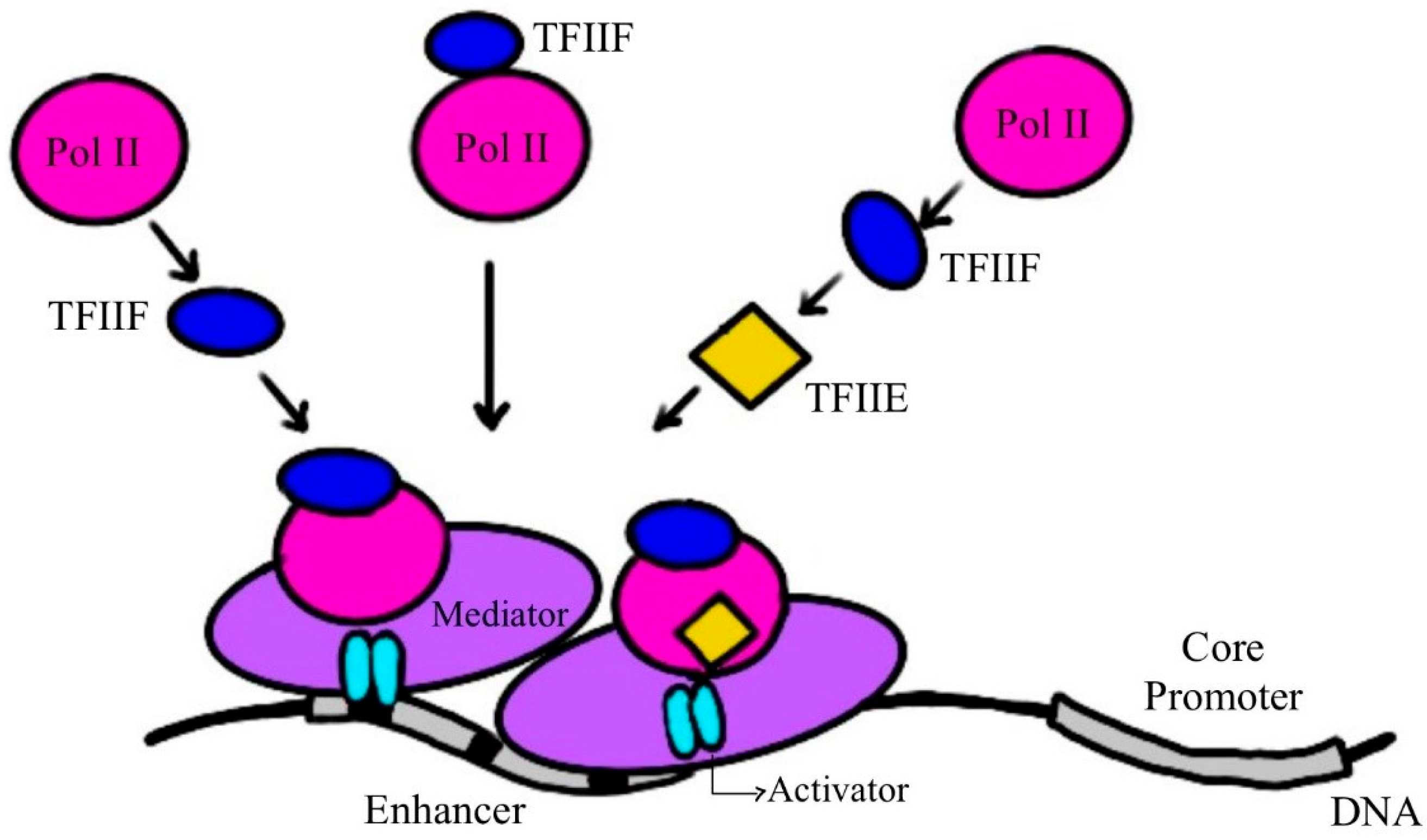
Pol II, which is responsible for transcription of the majority of protein-coding genes and many non-coding RNAs in eukaryotes, must assemble with general transcription factors into the PIC to properly initiate transcription [41]. The PIC is an assembly of Pol II with seven other parts, six transcription factors (TFIIA, TFIIB, TFIID, TFIIE, TFIIF, and TFIIH) and a mediator that increases transcriptional output upon binding to TFs (Figure 2) [42]. Each transcription factor possesses a unique function within the PIC, including stabilization, recruitment, and regulation. In the past, it was challenging to identify the DNA target sequences that led to the formation of the PIC, as the nucleus is highly dynamic and contains thousands of promoters. However, in vivo studies utilizing single-molecule tracking methods within yeast cells showed that a mediator-Pol II interaction was responsible for conducting the transient assembly of the PIC in conjunction with TFIID, TATA box binding protein and Pol II, the former being globally required for transcription initiation [8,43]. Likewise, the sub-diffusion of factors TFIIB and TFIIE and their recruitment to chromatin was found to be coordinated by the mediator rather than TFIID, which instead coordinated the sub-diffusion of TFIIA to chromatin. The mediator plays a pivotal role in transcription by serving as a bridge between transcription factors and Pol II, assisting in PIC assembly, and stimulating phosphorylation at the C-terminal domain of Pol II, promoting elongation [44]. This emphasizes the specific roles of the mediator and TFIID in nuclear exploration and chromatin binding.
Various models have been proposed to explain the nature of PIC assembly. A sequential model of assembly was proposed, positing that assembly is a step-by-step process, or even a two-track model, beginning with TFIID recognizing a specific promoter element, the TATA box-binding protein, to recruit other general TFs to assemble sequentially the core PIC, intermediate PIC, and holo PIC to ultimately form the Pol II complex [45,46]. The dynamic model of PIC assembly, however, depicts the process as a flexible, transient assembly where elements such as Pol II may bind and be recruited independently to chromatin by activators, and without specific order. This introduced the idea that PIC assembly may occur through various pathways rather than one strict mode, and that the outcome of each path may be influenced by the flexible nature in which each component binds [45]. The recruitment of actin has been found to be important for the assembly of the PIC due to its recruitment of RNA Pol II [22].
The intense level of regulation of transcription by Pol II is important for cellular function, as this control has a direct impact on how much protein and which proteins are later translated and expressed. Despite its complexity and strict regulation, just like Pol I and Pol III, Pol II also requires assistance in initiating transcription. Transcription begins when Pol II and its associated transcription factors (TFIIB, -D, -E, -F, and -H) bind to a promoter element, which is either a TATA box or a TATA-less promoter [47]. These two main types of promoters differ in both structure and mechanistic functions, as well as the genes that they encode [48]. Although it has been studied at length and is widely well-understood, the TATA-box sequence is only found associated with a percentage of eukaryotic genes, accounting for only ~3% of the human genome [49]. This review primarily focuses on transcription initiated by the TATA-box sequence, though it is pertinent to note the mechanistic differences between the two. Both TATA-containing and TATA-less promoters depend on TFIID and TATA-binding protein (TBP) for transcription to occur, although the site where TBP binds at TATA-less promoters is hypothesized to be at a TATA-like sequence [48]. Promoters lacking TATA box sequences also greatly depend on other elements, such as the initiator sequence and downstream elements like TBP-associated factors to facilitate the binding of TFIID and coactivate transcription [49,50]. Following a TATA-box based model of transcription, once Pol II is bound to the DNA, the DNA is unwound and separated by DNA Helicase. It is at this point that Pol II begins transcription in the 5′ → 3′ direction of the template strand, in the process denoted as elongation. Once the entire DNA sequence has been fully transcribed, transcription ends when Pol II reaches the end of the coding sequence, the phosphorylation state of its disordered C-terminal domain (CTD) changes, forming a binding site that termination factors are able to bind to and stop transcription [51]. After transcription is halted, Pol II dissociates from the transcription complex and is reused when the process starts again. The CTD plays a role in confining Pol II within regions of the nucleus containing genes that are available for transcription by restricting its diffusion [10], thus decreasing the time between completion of transcription of one gene and formation of PIC for transcription of another.
The eukaryotic transcript is pre-mRNA that must undergo covalent modifications in the nucleus. Many eukaryotic pre-mRNAs undergo splicing, where exon sequences are joined together after the removal of intron sequences. Some transcripts may be alternatively spliced, in which some but not all exon sequences are included in the mature transcript [52], as is found for a number of striated muscle proteins in both vertebrates and invertebrates [53,54,55,56,57,58,59,60]. The mRNA transcript is further modified covalently with a guanosine triphosphate (GTP) cap (a modified guanine nucleotide) and a poly-A tail before ultimately leaving the nucleus as mature mRNA. Interestingly, nuclear actin may be associated with proteins that are involved in mRNA splicing [61], although it may have more general roles in transcriptional regulation, even though it does not show up in existing structures of complexes involving RNA polymerases (Figure 1 and Figure 2). Due to its wide-ranging roles in cells, it is for this reason that the relationship between nuclear actin, RNA Pol II, and transcription more generally should be investigated.

3. Requirement of Nuclear Actin for RNA Pol II Function

Actin is well known for its cytoplasmic and cytoskeletal roles, including its major role in muscle contraction [62,63,64,65,66,67]. Early research found actin not only in the cytosol of muscle cells but also in the nucleus [68,69], although there was concern that the abundance of cytoplasmic actin could confound attempts to detect actin in the nucleus. Though there is preliminary information about nuclear actin’s localization to the nucleus and work with Pol II, much is still unknown about its structure. Nuclear actin is currently known to exist as monomers, small polymers, and rods that assemble dynamically [70,71,72,73,74,75]. Nuclear actin contrasts in more ways than one with cytoplasmic actin; while evidence does not directly show that nuclear actin polymerizes into filaments (F-actin), it does instead show formation of short oligomeric filaments or predominantly monomeric G-actin [76]. One outcome of this structural difference is that it prevents interference with chromatin structure and disruption of the transcription process [77,78]. For instance, polymerization of nuclear actin into rods, forming under stress conditions, can act as an inhibitor to Pol II activity, suggesting its role as a negative inhibitor of transcription [77,78,79]. Nuclear actin can be covalently modified by SUMOylation, a type of post-translational modification involving small ubiquitin-like modifier (SUMO) polypeptides that are targeted to specific lysine residues [79]. Of the four SUMO genetic isoforms in the human genome, two transcripts (SUMO2 and SUMO3) can modify nuclear actin [79]. SUMOylation of nuclear actin distinguishes it from cytoplasmic actin and allows the formation of other nuclear actin structures, such as one with an antiparallel lower dimer. These different configurations of nuclear actin, in comparison with cytoplasmic actin, can accommodate the specialized functions that actin performs in the nucleus.
Polymerization of nuclear actin is regulated by actin-binding proteins that localize actin to the nucleus [80]. Actin-binding proteins such as cofilin, CapG and megakaryocytic acute leukemia (MAL) protein—a G-actin–binding coactivator of the transcription factor SRF (serum response factor)—contain nuclear localization sequences that enable transport of actin into the nucleus [81,82]. Other actin-binding proteins, such as diaphanous-related formins and the actin-related proteins 2/3 (Arp2/3) complex, can localize to the nucleus and regulate nuclear actin dynamics [61,83]. Nuclear actin itself does not contain a nuclear localization sequence [84]. In addition to actin-binding proteins, SUMOylation is necessary for the nuclear localization of actin and is also crucial for the retention of actin in the nucleus [79]. SUMOylation and other actin-binding proteins recruit actin to the nucleus and may also play a role in regulating the structure of nuclear actin.
In addition to post-translational regulation, SUMOylation and actin-binding protein expression is subject to transcriptional regulation as these pathways are also involved in gene expression. Actin-binding proteins may be controlled by upstream Rho GTPases which therefore aid in controlling nuclear actin dynamics [85]. Though the regulation of the SUMOylation system remains a topic of debate, it has been found that the system is upregulated by Ca2+ signaling during keratinocyte differentiation. Specifically, Ca2+ induces transcriptional activation of several components of the SUMOylation system such as SUMO2, SUMO3, and the ubiquitin conjugating enzyme Ubc9 (E2I) [86]. Conditions such as hypoxia induce the expression of SUMO1 RWD-containing SUMOylation enhancer. Ubc9 expression has been found to be linked to different types of carcinomas [87].
Nuclear actin and actin-related proteins are hypothesized to play critical roles in chromatin remodeling. These roles include facilitation of the movement of chromatin and nucleosomes, movement that is vital for initiation of transcription as well as other processes such as DNA recombination and repair [77] and 3D organization of chromatin that is essential for coordination of transcriptional regulation across the genome [88]. One example of this process in action is within the regulation of MAL protein via the Rho-actin signaling pathway [89]. Rho GTPase activation induces actin polymerization, accumulation of F-actin and reduction in G-actin, to which MAL is bound [82]. Once MAL and G-actin interactions are interrupted, MAL can be transported from the cytoplasm and localized to the nucleus where it can bind to the transcription factor serum response factor (SRF), coactivating it. SRF is a mediator protein that helps to relay a rapid transcriptional response from extracellular signals [90]. SRF controls genes associated with many vital functions including cell-cycle progression, neuronal synaptic transmission, and muscle cell differentiation, highlighting its role in gene expression and transcriptional regulation. However, without the presence of the switch/sucrose non-fermentable (SWI/SNF) chromatin-remodeling complex bound to promoter regions of DNA, SRF only binds loosely to chromatin [91]. SWI/SNF binding triggers ATP-dependent chromatin remodeling, rearranging nucleosomes that allow SRF to bind more tightly and carry out transcriptional regulation.
An example of nuclear actin’s more direct role in chromatin remodeling involves the BRG/BRM-associated factor (BAF) chromatin remodeling complex that contains β-actin. The actin component is tightly associated with actin-related protein 4 (ARP4) and is required for the full function of ATPases such as BRG1 within the BAF and SWI/SNF complexes, which provide energy for chromatin remodeling by hydrolyzing MgATP [92]. During development, the BAF complex is critical for gene activation by transforming inaccessible embryonic genes in heterochromatin to accessible states in euchromatin that can be transcriptionally activated [93]. Further, in the absence of β-actin, the ATPase BRG1 dissociates, leading to chromatin reorganization and alteration of its accessibility for transcription and in turn drastically altering expression of genes involved in cellular identity [94]. This demonstrates the influential role of nuclear actin in chromatin remodeling and its necessity for maintaining and regulating gene expression [83].
Nuclear actin is also involved in RNA clustering. RNA clustering helps to organize RNA in specific locations within the nucleus, which helps with the efficacy of gene regulation and cellular responses when they receive an external signal [95]. Regulation of RNA clustering is also thought to promote PIC formation, which is another important role of nuclear actin. Actin is crucial in the formation of PICs and is found associated with Pol II within its C-terminal domain, where it interacts with certain ribonucleoproteins that together recruit the histone acetyltransferase to induce a permissive chromatin environment for transcription initiation [92,96]. Actin is also especially necessary during the elongation phase of transcription, for it helps in the recruitment of positive transcription elongation factor b (P-TEFb), an elongation factor that binds to Pol II and phosphorylates Pol II at residue Ser2, which allows it to continue transcription [97]. Actin’s role in PIC formation has been well substantiated through studies that show when it is absent from the nucleus, the formation of PICs does not occur [22]. This disruption in the formation of PICs occurs because nuclear actin is necessary for TBP to bind to the TATA box sequence, a necessary step for transcription to begin. Chromatin immunoprecipitation assays have also indicated that nuclear actin is recruited to genes ready for transcription [98]. Nuclear actin then recruits Pol II to the PIC, demonstrating the importance of its role in gene expression.
Nuclear actin has been found to play multiple, potentially critical, functional interactions with Pol II. Nuclear actin is structurally associated with Pol II, although most likely indirectly via protein-protein subunit interactions, and has roles in Pol II’s transcription initiation with the PIC and transcription regulation, as discussed above. However, this topic—like the history of nuclear actin in general—has not been without debate and controversy. Studies involving nuclear actin have sometimes been questioned, in part due to nuclear actin not having a unique structure and function, but also because actin that is thought to be localized within the nucleus could instead be due to contamination by cytoplasmic actin, which is highly abundant in the cell [99]. Approaches to studying the role of nuclear actin in transcription and transcriptional regulation prove to be challenging as many in vivo studies of Pol II do not include actin or may be difficult to replicate. Unreliable techniques to capture nuclear actin have made it difficult to visualize its structure and ultimate function in living cells [99].
Compelling evidence strongly supports the role of nuclear actin in transcription. Emerging in vivo studies have introduced new techniques in this area. Findings from Drosophila oogenesis indicate that nuclear actin is prevalent in vivo, with pools of nuclear actin identified by multiple reagents [100]. This reveals that there is much more to be studied regarding the function of actin in the nucleus. In vivo studies involving yeast examine the mechanisms of Pol II in transcription and discuss the involvement of nuclear actin. One study highlights the importance of nuclear actin in the transcriptional regulation of the Pol II PIC [45]. Another notes nuclear actin’s role in chromatin remodeling and Pol II promoter recruitment [101]. Actin-binding proteins, including Wiskott-Aldrich syndrome proteins and Cofilin/ADF have been linked to their involvement in regulating levels of nuclear actin to achieve maximum transcription [92]. This supports the idea that nuclear actin levels can influence transcriptional activity and is required in some amount for proper transcription. Although the structure and function of nuclear actin are not fully understood, in vivo studies have demonstrated its existence and potential roles with Pol II. These studies advocate for the significance of nuclear actin, and as more is uncovered about it, additional questions arise.
Alongside its contribution to nuclear dynamics and the regulation of transcription, nuclear actin also influences the spatial organization of chromatin, which can impact Pol II function. Pol II activity is not only regulated by TFs, but by the three-dimensional location of genes within the nucleus. Each chromosome occupies a defined region within the nucleus known as a chromosome territory. Nuclear actin and other regulatory proteins have been found to participate in chromosome architecture organization and remodeling. For instance, inhibition of F-actin polymerization resulted in increased heterochromatin at the mitotic exit [102], suggesting a role for nuclear actin in nuclear reorganization. Chromosomal territories regulate chromosome accessibility and the extent to which Pol II can bind to chromosomes and initiate transcription. Nuclear actin regulation of chromosome structure and territories suggests that actin can serve as an allosteric regulator in macromolecular assemblies such as chromatin remodeling [22].
Antibodies are able to recognize different types of actin, including distinguishing between cytoplasmic and nuclear β-actin, which indicates that nuclear actin is structurally distinct, even though it is not an isoform coded for by a separate gene in the actin family. Antibodies that uniquely recognize nuclear actin have been shown to have inhibitory effects, therefore affecting transcription and other processes within the cell. Nascent RNA molecules are shown to have associations in the nuclear matrix with actin, which is needed for the synthesis of new transcripts [22]. These antibodies inhibit the functions of nuclear actin and, therefore, inhibit the association of the actin with RNA molecules, leading to a reduction in these RNA molecules. This reduction disrupts a process that is necessary for gene transcription. These antibodies also prevent a short, 15-nucleotide transcript from being produced. This 15-nucleotide transcript is needed for the transition from transcription initiation to elongation. The antibodies inhibit the formation of the PIC due to the fact that actin is needed for this process, as discussed above. This inhibition further suggests that actin acts as a bridge between Pol II and other parts of the PIC [22].
In addition to nuclear actin, however, other possible nucleoskeletal components of transcription have been found [103,104,105]. Nuclear myosin, an isoform of myosin 1, is involved in transcription elongation and regulation of Pol I and Pol II. In Pol I, nuclear myosin 1 (NM1) may play an important role in the stabilization of actin filaments, as both proteins are associated with the entire rDNA repeat [106]. This suggests a potential structural role in facilitating Pol I movement. The roles of Pol I in cell function and growth are suspected to be aided by interactions with nuclear actin and myosin [107,108], suggesting a better understanding of the mechanisms underlying ribosome synthesis and cell proliferation.
Confocal and electron microscopy revealed that NM1 colocalized with Pol II, appearing to be a part of a complex affecting transcription [109]. However, a prevalent nuclear myosin associated with Pol II is nuclear myosin VI (NMVI). NMVI colocalizes with newly transcribed mRNA and Pol II, both enhancing and modulating Pol II-dependent transcription [110]. In vivo studies have shown that NMVI contributes to RNA clustering and localization, thus demonstrating its role as a regulator of gene expression [111]. This suggests that there may be an underlying nuclear actin-myosin component of the Pol II transcription mechanism and regulation, although there may be other contributing factors. Evidence for the nuclear localization of Tm and troponin subunits has been found via immunofluorescence microscopy using antibodies against the three cardiac troponin subunits (troponin C, troponin I and troponin T) and Tm [112,113,114]. Further exploration into nuclear troponin and Tm may reveal their possible roles within the nucleus and transcription regulation.

4. Troponin and Tropomyosin as Potential Modulators of Nuclear Actin and Transcription

In addition to nuclear actin and myosin, thin filament proteins troponin and Tm can be found in the nuclei of striated muscle myocytes [112,115] (Figure 3) and some cancer cells [116]. Troponin and Tm are best known for the role they play in the Ca2+-dependent regulation of striated muscle contraction [66,117]. During muscle contraction, troponin and Tm control the interaction between actin and myosin filaments in a Ca2+-dependent manner [66,67,118,119,120,121], possibly suggesting that they could play a similar role with nuclear actin and myosin. Structurally, Tm is a longer, fibrous protein that lies along the actin filament, blocking myosin binding sites on actin when the skeletal muscle is relaxed [57,66,67,118,119]. Troponin is a protein complex composed of three polypeptide subunits that are coded for by distinct genes and are referred to as troponin C (TnC), troponin I (TnI), and troponin T (TnT). TnC, TnI and TnT have roles in Ca2+ binding, inhibition of the actin-myosin interaction, and anchoring the troponin complex to Tm, respectively [117,122,123,124].
Though their well-established role in Ca2+-dependent regulation of muscle contraction occurs in the cytoplasmic myofilaments of the sarcomere, substantial evidence demonstrates the presence of these proteins within the nucleus of striated muscle cells. Specifically, the cardiac forms of TnI and TnT were detected in association with nuclei of adult human cardiac myocytes [127,128], with TnI being clearly localized within nuclei [128]. In addition, proteomic analysis of isolated cardiac nuclei from rodents also identified cardiac TnC, TnI, and various tropomyosin isoforms like cardiac α-Tm [129]. Finally, immunofluorescence studies have identified all of the components of thin filaments—native cardiac troponin subunits cardiac TnC, TnI and TnT, as well as cardiac α-Tm—within the nuclei of cultured neonatal rat ventricular cardiomyocytes [112].
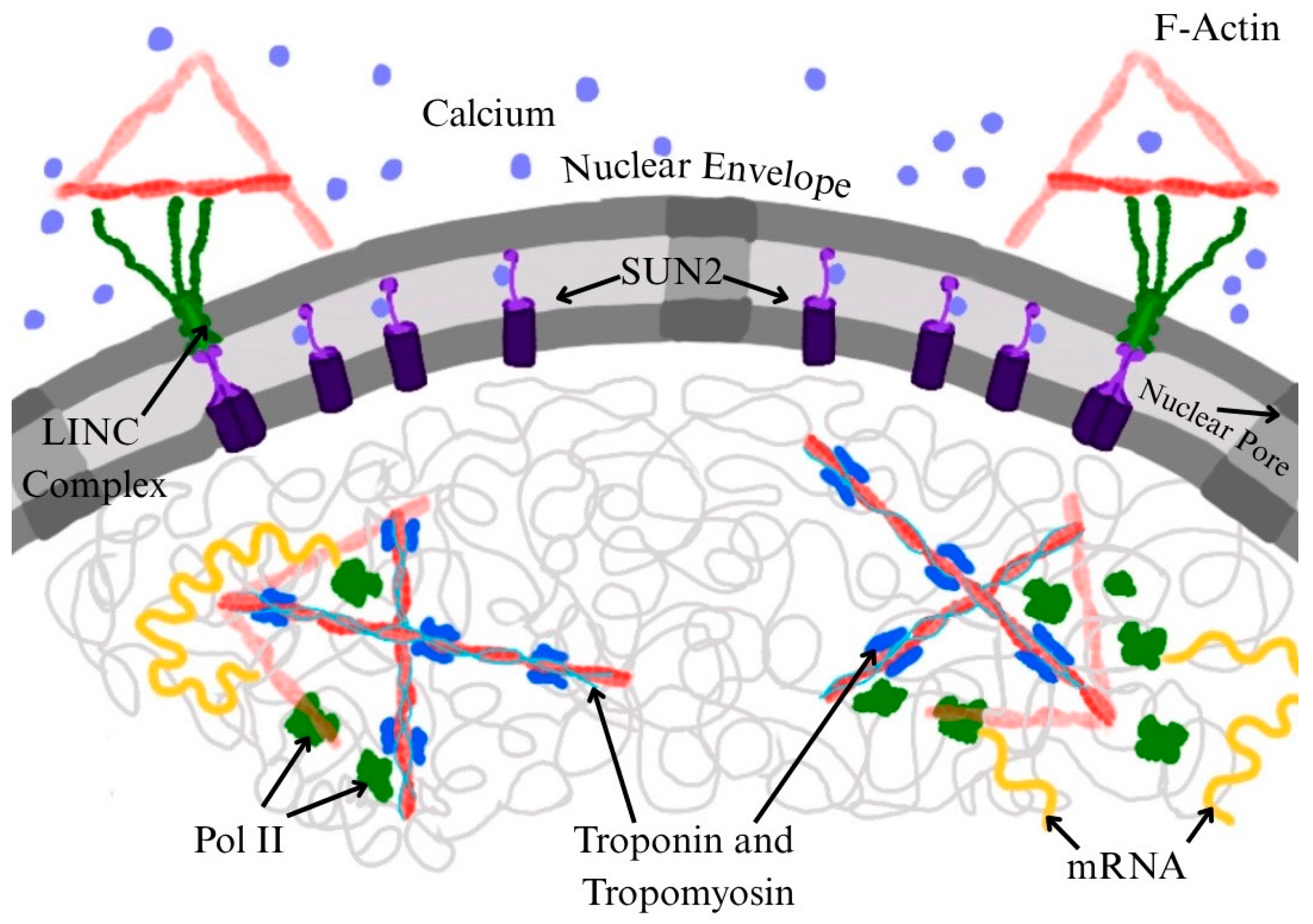
While the presence of all three cardiac troponin subunits has been demonstrated in cardiomyocyte nuclei, it is not definitively known if they assemble onto F-actin-Tm to form thin filaments within the nucleus, or even if they form the ternary troponin complex within the nucleus, as occurs in the sarcomere. However, if actin can polymerize into filaments within the nucleus, it stands to reason that short thin filaments could be formed in striated muscle cell nuclei by associating with Tm and troponin complexes (Figure 3). Furthermore, TnI and TnT have lower solubility on their own in cellular conditions compared to the troponin complex. Actin in the nucleus appears to be highly dynamic, existing in monomeric form or as short oligomers [130]. In studies where drugs were administered to cells that specifically blocked polymerization of actin, transcriptional activity was inhibited. On the other hand, drugs favoring polymerization resulted in regular transcriptional activity. More importantly, it was also found that actin mutants could only restore transcriptional activity when F-actin was stabilized, as opposed to mutants that do not stabilize F-actin [71]. This suggests that nuclear actin involved in transcription must be in the polymerized, filamentous state, which would not only enable thin filament formation in cells where Tm and troponin are present, but also allow actin filaments to serve as tracks for nuclear myosin transport [108,109,110,111].
Interestingly, TnI and TnT can influence DNA and chromatin function, presumably outside of the ternary troponin complex. Tm-TnI in association with actin has an important role as a cytoskeletal complex that helps maintain chromosomal integrity and cell polarity during cell division and development in Drosophila [131]. Mutations of the Tm-TnI complex lead to chromosome fragment loss and mis-localized polarity markers, resulting in defects in mitosis and chromosome segregation [131]. Such effects could be tied to the development of pathological phenotypes, such as cancers and cardiomyopathies, as chromosomal instability and nondisjunction within cells that are common among many cancers [103,116]. Studies using fluorescent skeletal TnT constructs have implied that the troponin complex may be the relevant entity for nuclear localization [132], consistent with earlier bioinformatics analyses [127]. However, assembly of TnT into the troponin complex may not be required for nuclear localization in overexpression studies, due to a potential non-stoichiometric ratio of subunits [132]. TnT3, a form of TnT found in fast skeletal muscle cells, not only localizes in the nucleus but also associates with RNA-polymerases, suggesting a role in transcriptional regulation [132]. Studies that support this also find that TnT3 associates with DNA consensus sequences and modulates transcription, perhaps by acting as a transcription factor [133]. Changes in nuclear TnT3 and its fragments are also highly affected by aging [115]. Thus, troponin subunits may have functions in the nucleus whether or not they assemble into the troponin complex and then assemble into thin filaments with F-actin-Tm.
Along with nuclear troponin and Tm, nuclear Ca2+ has several hypothesized functions in the regulation of neurons and both cardiac and skeletal muscle cells. Given their role as Ca2+-sensors in the striated muscle sarcomere [117], a likely role for troponin and Tm in the nucleus could be to confer Ca2+-regulation to processes involving nuclear actin [72]. Focusing on cardiomyocytes, nuclear Ca2+ functions as a Ca2+-dependent signaling pathway and second messenger. This pathway targets transcription factors with direct or indirect dependence on Ca2+ such as calcineurin-nuclear factor of activated T-cells (CaN-NFAT) and Ca2+/calmodulin-dependent protein kinase II (CaMKII) histone deacetylase (HDAC) [134]. These transcriptional effects and nuclear Ca2+ signals may influence gene expression. Ca2+ signaling itself can be either up- and down-regulated based on changing cellular needs by various pathways, depending on cell type, such as within the feedback loops of NFAT and cyclic-AMP response element-binding (CREB) protein [135]. The regulation of nuclear Ca2+ within cardiomyocytes and its impact on transcription can determine cardiac remodeling, which may lead to diseases such as hypertrophy and heart failure [134,136]. In skeletal muscle cells, inositol trisphosphate (IP3) receptors on the nuclear envelope may organize the nuclear Ca2+ signal in electrically stimulated skeletal muscles [137,138]. This signal is distinct from cytoplasmic Ca2+ involved in muscle contraction. The nuclear Ca2+ signal was found to activate extracellular signal-related kinases 1 and 2 (ERK1/2) mitogen-activated protein kinase (MAPK) signaling that phosphorylates CREB [138]. These phosphorylation-related signaling events lead to changes in transcription and gene expression. Modulation of transcription by nuclear Ca2+ can affect gene expression not only in cardiomyocytes and skeletal muscle cells but also in relation to Pol II and nuclear actin.
Rapid polymerization of nuclear actin is mediated by nuclear Ca2+ signaling, which leads to its functions described above, such as chromatin remodeling and transcriptional regulation. Nuclear actin polymerization triggered by nuclear Ca2+ emanates from the inner nuclear membrane (INM) and is facilitated by INM proteins SUN2 and INF2 in the NIH3T3 fibroblast cell line that likely does not express troponin subunits [139]. Nuclear Ca2+ co-localizes these proteins in the INM, triggering the nuclear localization of actin; this localization of nuclear actin increases with rising levels of nuclear Ca2+. The interaction with SUN2 may reveal more about the structure of nuclear actin. The INF2/SUN2-mediated nuclear actin network promotes the formation of Pol II foci [139]. SUN2 and INF2 interactions increased with elevated nuclear Ca2+, promoting Pol II clustering by the polymerized nuclear actin network. This demonstrates how nuclear Ca2+ and actin have potential roles in the regulation of transcription and its efficiency.
Another mechanism by which actin can modulate transcription is through the yes-associated protein (YAP) and TAZ pathway, a pathway that has been shown to be important for cell fate and differentiation [140]. This mechanism is a primary example of a mechnotransduction pathway in which extracellular constraints lead to intracellular mechanical responses, otherwise known as mechanosensing, further resulting in a cascade of signaling pathways [141]. External mechanical stresses illicit strong responses from cytoskeletal features, namely tubulin, intermediate filaments (desmin in striated muscle), nuclear envelope proteins, and actin; stresses result in extracellular matrix stiffness and cytoskeletal tension, both of which influence the amount of monomeric and filamentous actin within the nucleus [142]. F-actin levels indirectly modulate YAP activity by activating or inhibiting upstream pathways, collectively known as the Hippo signaling pathway. Increased F-actin assembly within the nucleus promotes the YAP pathway by simultaneously inhibiting Hippo signaling and increasing nuclear YAP/TAZ levels and promoting nuclear localization of these proteins [140]. Once in the nucleus, YAP/TAZ associate with DNA-binding proteins and transcription factors to co-activate transcription, specifically of genes involved in cell cycle control relating to cell proliferation and cell death. Misregulation of this pathway has been linked to cellular disorders, especially cancer, leading to tumor growth [141]. Overexpression of active YAP and elevated YAP levels have been reported in skeletal muscle tumors and cancers in both in vitro and in vivo studies, as high YAP levels inhibit other pathways with tumor suppression functions. In this way, YAP plays a significant role in transmitting mechanical cues from the cytoskeleton into a transcriptional cell response.
As detailed, actin plays a large role in many cellular processes, including those within the nucleus that regulate fundamental genetic mechanisms that include transcription, RNA processing, DNA recombination, and nuclear export. While cytoplasmic actin is commonly associated with Tm and the troponin complex in striated muscle myocytes and some cancer cells, nuclear actin has also been detected in cardiac myonuclei that also contain the cardiac isoforms of these proteins [112]. It is unclear whether these proteins assemble into thin filaments under any conditions, and whether they might be present in any other cell types, including undifferentiated cells. A specific form of myosin that has been identified to exist solely in the nucleus is nuclear myosin (NM1), which has a unique structure from cytoplasmic myosin, as it contains a specific nuclear localization sequence within its N-terminus [130]. Given the close relation between actin, myosin, and the Ca2+ regulatory proteins troponin and Tm, it seems probable that these components would be found assembled within the nucleus to carry out various functions in concert. One example of actin and nuclear myosin I synergy is the promotion of transcription by RNA Pol I [130].
Troponin is well known for its Ca2+ binding properties as it contains a Ca2+-binding subunit, TnC. Cytoplasmic Ca2+ is well-known for its role as a second messenger in excitation-contraction coupling, controlling muscle contraction in response to physiological needs. In cardiac muscle specifically, cytoplasmic Ca2+ concentration is heavily regulated. Each heartbeat is governed by an electrical action potential that results in a cytoplasmic Ca2+ transient—a rise in the amount of free cytoplasmic Ca2+ followed by its subsequent removal [134,136]. Ca2+ within the nucleus is also heavily regulated; nuclear Ca2+ participates in regulating gene expression of cardiomyocytes. Cardiac muscle regulates force on a beat-to-beat basis (Starling’s Law) with mechanisms intrinsic to the myofilaments [143,144]. But cardiac muscle must also respond to physiological changes over longer timescales, responses that often involve changes in gene expression that are largely controlled by nuclear Ca2+-dependent signaling pathways. Nuclear Ca2+ directly or indirectly activates specific transcription factors such as NFAT in the CaN-NFAT pathway and CREB via the cyclic-AMP signaling pathway; both TFs and their pathways are required for the reprogramming of cardiomyocytes [134]. Increases in local nuclear Ca2+ levels modulate such transcription factors, in turn activating signaling pathways to regulate or control gene expression within the cell. More research is needed in regard to the excitation-transcription coupling process of Ca2+ to better understand its implication and mechanism within cardiac myocytes and striated muscle cells more generally. Though one underlying principle is clear: there is a clear and vital role of cellular Ca2+ in the regulation of cardiac cells, including transcription of striated muscle cells overall. Troponin’s presence, accompanied by actin and Tm, within nuclei and its role as a Ca2+-binding protein suggest that, in conjunction with each other, these components could serve to regulate transcription in profound ways.
An important note about nuclear function is that the location of nuclei within cells greatly influences their function. Cardiomyocytes can be mono- or multi-nucleated, with nuclei located centrally, surrounded by myofibrils [145,146,147]. Because of the position of the nuclei, the nuclei experience both the Ca2+ transient and mechanical compression every time contraction occurs in the heart. The location of nuclei differs in multi-nucleated skeletal muscle fibers when compared to cardiomyocytes due to the fact that the multi-nucleated nuclei are located in the peripheral region of the cell, between the sarcolemma and the myofibrils, rather than centrally as found in cardiomyocytes. Because skeletal muscle fiber nuclei are not located within the myofibrils, the mechanical impact on the nuclei is not as direct when contraction occurs. Interestingly, recent studies have found associations between nuclei that are centrally located in skeletal muscle fibers with some skeletal myopathies [148,149].
Mechanosensing in myocytes describes the physical, mechanical forces experienced by muscle cell nuclei due to contraction or externally applied forces; it is a concept that can be applied more generally to forces—internal or external—experienced by any cell type. Forces that are not directly experienced by the nuclei (e.g., compression of cardiomyocyte nuclei during systole) are transmitted through the cytoskeletal network, which is composed of actin, microtubules, and intermediate filaments [150]. Mechanical strain still occurs because of the cytoskeleton network deforming around the nuclei, which is transmitted to them by the same cytoskeleton network [150]. These forces can ultimately cause deformation in the nuclear envelope and can change the organization of chromatin and gene expression [151,152].

5. Conclusions

With a growing understanding of nuclear actin’s role in eukaryotic transcription, studies have also revealed its complex role in gene regulation, especially within striated muscle cells, where transcriptional control possesses both structural and signaling components. Evidence supports that nuclear actin serves not solely in a structural role but also as a dynamic regulator involved in chromatin remodeling, RNA clustering, and assembly of the RNA polymerase II PIC. Its cooperation with nuclear myosin, along with recruitment and regulation via mechanisms such as SUMOylation and Ca2+ signaling, further highlights the sophisticated manner in which transcriptional activity is directed in the nucleus.
Emerging evidence suggests that cytoskeletal proteins like troponin and Tm may also localize to the nucleus and interact with nuclear actin in a nuclear Ca2-dependent manner. Their potential roles in modulating nuclear actin complexes within the nucleus could further influence RNA Pol II function beyond the role of nuclear actin alone. Together, these findings and hypotheses highlight what is still unknown about transcriptional regulation in striated muscle biology.
Many studies have reported that troponin and Tm are overexpressed in various cancers, and they may play a role in gene regulation. Troponin T3 (TnT3) contains nuclear localization signals and a leucine zipper DNA-binding domain [132]. This can allow for nuclear import as well as potential transcriptional regulatory activity. TnT3 has also been shown to be involved in the nuclear import of a calcium channel subunit that modulates gene expression in muscle cells [153]. Troponin subunits have been detected in certain cancers [116] and tropomyosin is frequently used as a biomarker in order to detect certain cancers [154], even though their actual involvement in cancer progression is not entirely known. These findings suggest that troponin and Tm have additional roles beyond their typical functions in striated muscles and may also act as transcriptional regulators in other contexts such as cancer. Studying nuclear thin filament components is not only relevant to myocytes, but also potentially in cancer cells that aberrantly express these striated muscle proteins. The hypothesis that Tm and troponin could act as transcriptional modulators opens new possibilities that deserve investigation.

Author Contributions

Conceptualization, A.J.K., A.J.M., L.G.M., M.R. and P.B.C.; writing—original draft preparation, A.J.K., A.J.M., L.G.M. and M.R.; writing—review and editing, A.J.K., A.J.M., L.G.M., M.R. and P.B.C.; visualization, A.J.K., A.J.M., L.G.M. and M.R.; supervision, P.B.C.; project administration, P.B.C.; funding acquisition, A.J.K. and P.B.C. All authors have read and agreed to the published version of the manuscript.

Funding

This research was funded by U.S. National Institutes of Health NHLBI grant number 5R01HL160966 to PBC; and by Florida State University’s Women in Mathematics, Science and Engineering (WIMSE) Research Experience Program (REP) to AJK.

Institutional Review Board Statement

Not applicable.

Data Availability Statement

No new data were created or analyzed in this study.

Acknowledgments

We thank our colleagues, especially Jose R. Pinto, who, over the years, have inspired consideration of the roles of myofilament proteins in the cell nucleus.

Conflicts of Interest

The authors declare no conflicts of interest. The funders had no role in the design of the study; in the collection, analyses, or interpretation of data; in the writing of the manuscript; or in the decision to publish the results.

Abbreviations

The following abbreviations are used in this manuscript:
Arp2/3Actin-related proteins 2/3
ARP4Actin-related protein 4
BAF complexBRG/BRM-associated factor complex
CaMKIICa2+-calmodulin-dependent protein kinase II
CaNCalcineurin
ChromExMChromatin expansion microscopy
CREBCyclic-AMP response element-binding
CTDDisordered carboxy-terminal domain of RNA Pol II
DNADeoxyribonucleic acid
ERK1/2Extracellular signal-related kinases 1 and 2
F-actinFilamentous (polymerized) actin
G-actinGlobular (monomeric) actin
GTPGuanosine triphosphate
HDACHistone deacetylase
INMInner nuclear membrane
IP3Inositol trisphosphate
LINCLinker of nucleoskeleton and cytoskeleton
MALMegakaryocytic acute leukemia protein
MAPKMitogen-activated protein kinase
miRNAsmicroRNAs
MRFsMyogenic regulatory factors
mRNAMessenger ribonucleic acid
MyoDMyoblast determination protein
NFATNuclear factor of activated T-cells
NM1Nuclear myosin 1
NMVINuclear myosin VI
PICPre-initiation complex for transcription
Pol IRNA polymerase I
Pol IIRNA polymerase II
Pol IIIRNA polymerase III
Pol IVRNA polymerase IV
Pol VRNA polymerase V
pre-mRNAPre-messenger RNA
P-TEFbPositive transcription elongation factor b
rDNAGene for a ribosomal RNA
RNARibonucleic acid
rRNARibosomal ribonucleic acid
snRNAssmall nuclear RNAs
SRFSerum response factor
SUMOSmall ubiquitin-like modifier protein
SWI/SNFswitch/sucrose non-fermentable chromatin-remodeling complex
TBPTATA-binding protein
TFsTranscription factors
TnTroponin
TnCTroponin C subunit of troponin
TnITroponin I subunit of troponin
TnTTroponin T subunit of troponin
TmTropomyosin
tRNATransfer ribonucleic acid
Ubc9ubiquitin conjugating enzyme Ubc9 (E2I)
YAPYes-associated protein

References

  1. Piccolino, M. Biological machines: From mills to molecules. Nat. Rev. Mol. Cell Biol. 2000, 1, 149–153. [Google Scholar] [CrossRef] [PubMed]
  2. van den Heuvel, M.G.L.; Dekker, C. Motor proteins at work for nanotechnology. Science 2007, 317, 333–336. [Google Scholar] [CrossRef]
  3. Cramer, P. A Tale of Chromatin and Transcription in 100 Structures. Cell 2014, 159, 985–994. [Google Scholar] [CrossRef]
  4. Trivedi, D.V.; Nag, S.; Spudich, A.; Ruppel, K.M.; Spudich, J.A. The Myosin Family of Mechanoenzymes: From Mechanisms to Therapeutic Approaches. Annu. Rev. Biochem. 2020, 89, 667–693. [Google Scholar] [CrossRef]
  5. Roeder, R.G. 50+ years of eukaryotic transcription: An expanding universe of factors and mechanisms. Nat. Struct. Mol. Biol. 2019, 26, 783–791. [Google Scholar] [CrossRef] [PubMed]
  6. Pownall, M.E.; Miao, L.; Vejnar, C.E.; M’Saad, O.; Sherrard, A.; Frederick, M.A.; Benitez, M.D.J.; Boswell, C.W.; Zaret, K.S.; Bewersdorf, J.; et al. Chromatin expansion microscopy reveals nanoscale organization of transcription and chromatin. Science 2023, 381, 92–100. [Google Scholar] [CrossRef] [PubMed]
  7. Stasevich, T.J.; Kimura, H. An expanded view of transcription. Science 2023, 381, 26–27. [Google Scholar] [CrossRef]
  8. Nguyen, V.Q.; Ranjan, A.; Liu, S.; Tang, X.; Ling, Y.H.; Wisniewski, J.; Mizuguchi, G.; Li, K.Y.; Jou, V.; Zheng, Q.; et al. Spatiotemporal coordination of transcription preinitiation complex assembly in live cells. Mol. Cell 2021, 81, 3560–3575.e6. [Google Scholar] [CrossRef]
  9. Baek, I.; Friedman, L.J.; Gelles, J.; Buratowski, S. Single-molecule studies reveal branched pathways for activator-dependent assembly of RNA polymerase II pre-initiation complexes. Mol. Cell 2021, 81, 3576–3588.e6. [Google Scholar] [CrossRef]
  10. Ling, Y.H.; Ye, Z.; Liang, C.; Yu, C.; Park, G.; Corden, J.L.; Wu, C. Disordered C-terminal domain drives spatiotemporal confinement of RNAPII to enhance search for chromatin targets. Nat. Cell Biol. 2024, 26, 581–592. [Google Scholar] [CrossRef]
  11. Casamassimi, A.; Ciccodicola, A. Transcriptional Regulation: Molecules, Involved Mechanisms, and Misregulation. Int. J. Mol. Sci. 2019, 20, 1281. [Google Scholar] [CrossRef] [PubMed]
  12. Nie, Y.; Song, C.; Tang, H. Transcriptional regulation in cardiovascular diseases. Front. Cardiovasc. Med. 2024, 11, 1360765. [Google Scholar] [CrossRef]
  13. Fuda, N.J.; Ardehali, M.B.; Lis, J.T. Defining mechanisms that regulate RNA polymerase II transcription in vivo. Nature 2009, 461, 186–192. [Google Scholar] [CrossRef]
  14. Filipovski, M.; Soffers, J.H.M.; Vos, S.M.; Farnung, L. Structural basis of nucleosome retention during transcription elongation. Science 2022, 376, 1313–1316. [Google Scholar] [CrossRef]
  15. Olson, E.N. Regulation of muscle transcription by the MyoD family. The heart of the matter. Circ. Res. 1993, 72, 1–6. [Google Scholar] [CrossRef] [PubMed]
  16. Olson, E.N. Signal transduction pathways that regulate skeletal muscle gene expression. Mol. Endocrinol. 1993, 7, 1369–1378. [Google Scholar] [CrossRef]
  17. Li, L.; Olson, E.N. Regulation of muscle cell growth and differentiation by the MyoD family of helix-loop-helix proteins. Adv. Cancer Res. 1992, 58, 95–119. [Google Scholar] [CrossRef]
  18. Braun, T.; Gautel, M. Transcriptional mechanisms regulating skeletal muscle differentiation, growth and homeostasis. Nat. Rev. Mol. Cell Biol. 2011, 12, 349–361. [Google Scholar] [CrossRef]
  19. Wang, Z.; Wang, D.-Z.; Hockemeyer, D.; McAnally, J.; Nordheim, A.; Olson, E.N. Myocardin and ternary complex factors compete for SRF to control smooth muscle gene expression. Nature 2004, 428, 185–189. [Google Scholar] [CrossRef]
  20. Wang, Z.; Wang, D.Z.; Pipes, G.C.; Olson, E.N. Myocardin is a master regulator of smooth muscle gene expression. Proc. Natl. Acad. Sci. USA 2003, 100, 7129–7134. [Google Scholar] [CrossRef] [PubMed]
  21. Estrella, N.L.; Naya, F.J. Transcriptional networks regulating the costamere, sarcomere, and other cytoskeletal structures in striated muscle. Cell. Mol. Life Sci. 2014, 71, 1641–1656. [Google Scholar] [CrossRef] [PubMed]
  22. Zheng, B.; Han, M.; Bernier, M.; Wen, J.-k. Nuclear actin and actin-binding proteins in the regulation of transcription and gene expression. FEBS J. 2009, 276, 2669–2685. [Google Scholar] [CrossRef]
  23. Roeder, R.G.; Rutter, W.J. Multiple forms of DNA-dependent RNA polymerase in eukaryotic organisms. Nature 1969, 224, 234–237. [Google Scholar] [CrossRef] [PubMed]
  24. Huang, K.; Wu, X.-X.; Fang, C.-L.; Xu, Z.-G.; Zhang, H.-W.; Gao, J.; Zhou, C.-M.; You, L.-L.; Gu, Z.-X.; Mu, W.-H.; et al. Pol IV and RDR2: A two-RNA-polymerase machine that produces double-stranded RNA. Science 2021, 374, 1579–1586. [Google Scholar] [CrossRef] [PubMed]
  25. Engel, C.; Sainsbury, S.; Cheung, A.C.; Kostrewa, D.; Cramer, P. RNA polymerase I structure and transcription regulation. Nature 2013, 502, 650–655. [Google Scholar] [CrossRef] [PubMed]
  26. Obrdlik, A.; Louvet, E.; Kukalev, A.; Naschekin, D.; Kiseleva, E.; Fahrenkrog, B.; Percipalle, P. Nuclear myosin 1 is in complex with mature rRNA transcripts and associates with the nuclear pore basket. FASEB J. 2010, 24, 146–157. [Google Scholar] [CrossRef]
  27. Turowski, T.W.; Tollervey, D. Transcription by RNA polymerase III: Insights into mechanism and regulation. Biochem. Soc. Trans. 2016, 44, 1367–1375. [Google Scholar] [CrossRef]
  28. Cech, T.R.; Steitz, J.A. The noncoding RNA revolution-trashing old rules to forge new ones. Cell 2014, 157, 77–94. [Google Scholar] [CrossRef]
  29. Rinn, J.; Guttman, M. RNA Function. RNA and dynamic nuclear organization. Science 2014, 345, 1240–1241. [Google Scholar] [CrossRef]
  30. Kopp, F.; Mendell, J.T. Functional Classification and Experimental Dissection of Long Noncoding RNAs. Cell 2018, 172, 393–407. [Google Scholar] [CrossRef]
  31. Lee, Y.; Kim, M.; Han, J.; Yeom, K.-H.; Lee, S.; Baek, S.H.; Kim, V.N. MicroRNA genes are transcribed by RNA polymerase II. EMBO J. 2004, 23, 4051–4060. [Google Scholar] [CrossRef]
  32. Rissland, O.S.; Norbury, C.J. The Cid1 poly(U) polymerase. Biochim. Biophys. Acta 2008, 1779, 286–294. [Google Scholar] [CrossRef] [PubMed]
  33. Schier, A.C.; Taatjes, D.J. Structure and mechanism of the RNA polymerase II transcription machinery. Genes Dev. 2020, 34, 465–488. [Google Scholar] [CrossRef] [PubMed]
  34. Khatter, H.; Vorländer, M.K.; Müller, C.W. RNA polymerase I and III: Similar yet unique. Curr. Opin. Struct. Biol. 2017, 47, 88–94. [Google Scholar] [CrossRef]
  35. Kostrewa, D.; Zeller, M.E.; Armache, K.-J.; Seizl, M.; Leike, K.; Thomm, M.; Cramer, P. RNA polymerase II-TFIIB structure and mechanism of transcription initiation. Nature 2009, 462, 323–330. [Google Scholar] [CrossRef]
  36. He, Y.; Yan, C.; Fang, J.; Inouye, C.; Tjian, R.; Ivanov, I.; Nogales, E. Near-atomic resolution visualization of human transcription promoter opening. Nature 2016, 533, 359–365. [Google Scholar] [CrossRef]
  37. Vos, S.M.; Farnung, L.; Urlaub, H.; Cramer, P. Structure of paused transcription complex Pol II-DSIF-NELF. Nature 2018, 560, 601–606. [Google Scholar] [CrossRef] [PubMed]
  38. Vos, S.M.; Farnung, L.; Boehning, M.; Wigge, C.; Linden, A.; Urlaub, H.; Cramer, P. Structure of activated transcription complex Pol II-DSIF-PAF-SPT6. Nature 2018, 560, 607–612. [Google Scholar] [CrossRef]
  39. Vos, S.M.; Farnung, L.; Linden, A.; Urlaub, H.; Cramer, P. Structure of complete Pol II-DSIF-PAF-SPT6 transcription complex reveals RTF1 allosteric activation. Nat. Struct. Mol. Biol. 2020, 27, 668–677. [Google Scholar] [CrossRef]
  40. Kettenberger, H.; Armache, K.-J.; Cramer, P. Complete RNA polymerase II elongation complex structure and its interactions with NTP and TFIIS. Mol. Cell 2004, 16, 955–965. [Google Scholar] [CrossRef]
  41. Aibara, S.; Schilbach, S.; Cramer, P. Structures of mammalian RNA polymerase II pre-initiation complexes. Nature 2021, 594, 124–128. [Google Scholar] [CrossRef]
  42. Schier, A.C.; Taatjes, D.J. Everything at once: Cryo-EM yields remarkable insights into human RNA polymerase II transcription. Nat. Struct. Mol. Biol. 2021, 28, 540–543. [Google Scholar] [CrossRef]
  43. Chen, X.; Wang, X.; Liu, W.; Ren, Y.; Qu, X.; Li, J.; Yin, X.; Xu, Y. Structures of +1 nucleosome-bound PIC-Mediator complex. Science 2022, 378, 62–68. [Google Scholar] [CrossRef]
  44. Chen, X.; Yin, X.; Li, J.; Wu, Z.; Qi, Y.; Wang, X.; Liu, W.; Xu, Y. Structures of the human Mediator and Mediator-bound preinitiation complex. Science 2021, 372, eabg0635. [Google Scholar] [CrossRef]
  45. Hou, T.Y.; Kraus, W.L. Come one, come all? Re-evaluating RNA polymerase II pre-initiation complex assembly using single-molecule microscopy. Mol. Cell 2021, 81, 3443–3445. [Google Scholar] [CrossRef]
  46. Chen, X.; Qi, Y.; Wu, Z.; Wang, X.; Li, J.; Zhao, D.; Hou, H.; Li, Y.; Yu, Z.; Liu, W.; et al. Structural insights into preinitiation complex assembly on core promoters. Science 2021, 372, eaba8490. [Google Scholar] [CrossRef]
  47. Liu, X.; Bushnell, D.A.; Kornberg, R.D. RNA polymerase II transcription: Structure and mechanism. Biochim. Biophys. Acta 2013, 1829, 2–8. [Google Scholar] [CrossRef]
  48. Donczew, R.; Hahn, S. Mechanistic Differences in Transcription Initiation at TATA-Less and TATA-Containing Promoters. Mol. Cell Biol. 2018, 38, e00448-17. [Google Scholar] [CrossRef]
  49. Yella, V.R.; Bansal, M. DNA structural features of eukaryotic TATA-containing and TATA-less promoters. FEBS Open Bio. 2017, 7, 324–334. [Google Scholar] [CrossRef]
  50. Pugh, B.F.; Tjian, R. Transcription from a TATA-less promoter requires a multisubunit TFIID complex. Genes Dev. 1991, 5, 1935–1945. [Google Scholar] [CrossRef]
  51. Harlen, K.M.; Churchman, L.S. The code and beyond: Transcription regulation by the RNA polymerase II carboxy-terminal domain. Nat. Rev. Mol. Cell Biol. 2017, 18, 263–273. [Google Scholar] [CrossRef]
  52. Barbosa-Morais, N.L.; Irimia, M.; Pan, Q.; Xiong, H.Y.; Gueroussov, S.; Lee, L.J.; Slobodeniuc, V.; Kutter, C.; Watt, S.; Çolak, R.; et al. The evolutionary landscape of alternative splicing in vertebrate species. Science 2012, 338, 1587–1593. [Google Scholar] [CrossRef]
  53. Ruiz-Opazo, N.; Nadal-Ginard, B. α-tropomyosin gene organization: Alternative splicing of duplicated isotype-specific exons accounts for the production of smooth and striated muscle isoforms. J. Biol. Chem. 1987, 262, 4755–4765. [Google Scholar] [CrossRef]
  54. Schiaffino, S.; Reggiani, C. Molecular diversity of myofibrillar proteins: Gene regulation and functional significance. Physiol. Rev. 1996, 76, 371–423. [Google Scholar] [CrossRef]
  55. Bernstein, S.I.; Milligan, R.A. Fine tuning a molecular motor: The location of alternative domains in the Drosophila myosin head. J. Mol. Biol. 1997, 271, 1–6. [Google Scholar] [CrossRef]
  56. Perry, S.V. Troponin T: Genetics, properties and function. J. Muscle Res. Cell Motil. 1998, 19, 575–602. [Google Scholar] [CrossRef]
  57. Perry, S.V. Vertebrate tropomyosin: Distribution, properties and function. J. Muscle Res. Cell Motil. 2001, 22, 5–49. [Google Scholar] [CrossRef]
  58. Swank, D.M.; Kronert, W.A.; Bernstein, S.I.; Maughan, D.W. Alternative N-terminal regions of Drosophila myosin heavy chain tune muscle kinetics for optimal power output. Biophys. J. 2004, 87, 1805–1814. [Google Scholar] [CrossRef]
  59. Labeit, S.; Lahmers, S.; Burkart, C.; Fong, C.; McNabb, M.; Witt, S.; Witt, C.; Labeit, D.; Granzier, H. Expression of distinct classes of titin isoforms in striated and smooth muscles by alternative splicing, and their conserved interaction with filamins. J. Mol. Biol. 2006, 362, 664–681. [Google Scholar] [CrossRef]
  60. Sheng, J.J.; Jin, J.-P. Gene regulation, alternative splicing, and posttranslational modification of troponin subunits in cardiac development and adaptation: A focused review. Front. Physiol. 2014, 5, 165. [Google Scholar] [CrossRef]
  61. Viita, T.; Kyheröinen, S.; Prajapati, B.; Virtanen, J.; Frilander, M.J.; Varjosalo, M.; Vartiainen, M.K. Nuclear actin interactome analysis links actin to KAT14 histone acetyl transferase and mRNA splicing. J. Cell Sci. 2019, 132, jcs226852. [Google Scholar] [CrossRef]
  62. Huxley, H.E. The mechanism of muscular contraction. Science 1969, 164, 1356–1366. [Google Scholar] [CrossRef]
  63. Cooke, R. The mechanism of muscle contraction. CRC Crit. Rev. Biochem. 1986, 21, 53–118. [Google Scholar] [CrossRef]
  64. Holmes, K.C.; Kabsch, W. Muscle proteins: Actin. Curr. Opin. Struct. Biol. 1991, 1, 270–280. [Google Scholar] [CrossRef]
  65. Pollard, T.D.; Cooper, J.A. Actin, a central player in cell shape and movement. Science 2009, 326, 1208–1212. [Google Scholar] [CrossRef]
  66. Risi, C.M.; Pepper, I.; Belknap, B.; Landim-Vieira, M.; White, H.D.; Dryden, K.; Pinto, J.R.; Chase, P.B.; Galkin, V.E. The Structure of the Native Cardiac Thin Filament at Systolic Ca2+ Levels. Proc. Natl. Acad. Sci. USA 2021, 118, e2024288118. [Google Scholar] [CrossRef]
  67. Risi, C.M.; Belknap, B.; Atherton, J.; Coscarella, I.L.; White, H.D.; Chase, P.B.; Pinto, J.R.; Galkin, V.E. Troponin structural dynamics in the native cardiac thin filament revealed by cryo electron microscopy. J. Mol. Biol. 2024, 436, 168498. [Google Scholar] [CrossRef]
  68. Ohnishi, T.; Kawamura, H.; Tanaka, Y. Actin and Myosin-like Proteins in the Calf Thymus Cell Nucleus. J. Biochem. 1964, 56, 6–15. [Google Scholar] [CrossRef]
  69. Ohnishi, T.; Kawamura, H.; Yamamoto, T. Extraction of a Protein Resembling Actin from the Cell Nucleus of the Calf Thymus. J. Biochem. 1963, 54, 298–300. [Google Scholar] [CrossRef]
  70. Jockusch, B.M.; Schoenenberger, C.-A.; Stetefeld, J.; Aebi, U. Tracking down the different forms of nuclear actin. Trends Cell Biol. 2006, 16, 391–396. [Google Scholar] [CrossRef]
  71. Pederson, T. As functional nuclear actin comes into view, is it globular, filamentous, or both? J. Cell Biol. 2008, 180, 1061–1064. [Google Scholar] [CrossRef]
  72. Baarlink, C.; Wang, H.; Grosse, R. Nuclear actin network assembly by formins regulates the SRF coactivator MAL. Science 2013, 340, 864–867. [Google Scholar] [CrossRef]
  73. Feric, M.; Brangwynne, C.P. A nuclear F-actin scaffold stabilizes ribonucleoprotein droplets against gravity in large cells. Nat. Cell Biol. 2013, 15, 1253–1259. [Google Scholar] [CrossRef]
  74. Kelpsch, D.J.; Tootle, T.L. Nuclear Actin: From Discovery to Function. Anat. Rec. 2018, 301, 1999–2013. [Google Scholar] [CrossRef]
  75. Ulferts, S.; Lopes, M.; Miyamoto, K.; Grosse, R. Nuclear actin dynamics and functions at a glance. J. Cell Sci. 2024, 137, jcs261630. [Google Scholar] [CrossRef]
  76. Hofmann, W.A.; de Lanerolle, P. Nuclear actin: To polymerize or not to polymerize. J. Cell Biol. 2006, 172, 495–496. [Google Scholar] [CrossRef]
  77. Kloc, M.; Chanana, P.; Vaughn, N.; Uosef, A.; Kubiak, J.Z.; Ghobrial, R.M. New Insights into Cellular Functions of Nuclear Actin. Biology 2021, 10, 304. [Google Scholar] [CrossRef]
  78. Serebryannyy, L.A.; Parilla, M.; Annibale, P.; Cruz, C.M.; Laster, K.; Gratton, E.; Kudryashov, D.; Kosak, S.T.; Gottardi, C.J.; de Lanerolle, P. Persistent nuclear actin filaments inhibit transcription by RNA polymerase II. J. Cell Sci. 2016, 129, 3412–3425. [Google Scholar] [CrossRef]
  79. Hofmann, W.A.; Arduini, A.; Nicol, S.M.; Camacho, C.J.; Lessard, J.L.; Fuller-Pace, F.V.; de Lanerolle, P. SUMOylation of nuclear actin. J. Cell Biol. 2009, 186, 193–200. [Google Scholar] [CrossRef]
  80. Pollard, T.D. Actin and Actin-Binding Proteins. Cold Spring Harb. Perspect. Biol. 2016, 8, a018226. [Google Scholar] [CrossRef]
  81. Falahzadeh, K.; Banaei-Esfahani, A.; Shahhoseini, M. The potential roles of actin in the nucleus. Cell J. 2015, 17, 7–14. [Google Scholar] [CrossRef]
  82. Vartiainen, M.K.; Guettler, S.; Larijani, B.; Treisman, R. Nuclear actin regulates dynamic subcellular localization and activity of the SRF cofactor MAL. Science 2007, 316, 1749–1752. [Google Scholar] [CrossRef]
  83. Sen, B.; Xie, Z.; Thomas, M.D.; Pattenden, S.G.; Howard, S.; McGrath, C.; Styner, M.; Uzer, G.; Furey, T.S.; Rubin, J. Nuclear actin structure regulates chromatin accessibility. Nat. Commun. 2024, 15, 4095. [Google Scholar] [CrossRef]
  84. Vandekerckhove, J.; Weber, K. At least six different actins are expressed in a higher mammal: An analysis based on the amino acid sequence of the amino-terminal tryptic peptide. J. Mol. Biol. 1978, 126, 783–802. [Google Scholar] [CrossRef]
  85. Rajakylä, E.K.; Vartiainen, M.K. Rho, nuclear actin, and actin-binding proteins in the regulation of transcription and gene expression. Small GTPases 2014, 5, e27539. [Google Scholar] [CrossRef]
  86. Liu, B.; Shuai, K. Regulation of the sumoylation system in gene expression. Curr. Opin. Cell Biol. 2008, 20, 288–293. [Google Scholar] [CrossRef]
  87. Baek, S.H. A novel link between SUMO modification and cancer metastasis. Cell Cycle 2006, 5, 1492–1495. [Google Scholar] [CrossRef]
  88. Batut, P.J.; Bing, X.Y.; Sisco, Z.; Raimundo, J.; Levo, M.; Levine, M.S. Genome organization controls transcriptional dynamics during development. Science 2022, 375, 566–570. [Google Scholar] [CrossRef]
  89. Miralles, F.; Posern, G.; Zaromytidou, A.-I.; Treisman, R. Actin dynamics control SRF activity by regulation of its coactivator MAL. Cell 2003, 113, 329–342. [Google Scholar] [CrossRef]
  90. Arsenian, S.; Weinhold, B.; Oelgeschläger, M.; Rüther, U.; Nordheim, A. Serum response factor is essential for mesoderm formation during mouse embryogenesis. EMBO J. 1998, 17, 6289–6299. [Google Scholar] [CrossRef]
  91. Zhou, J.; Zhang, M.; Fang, H.; El-Mounayri, O.; Rodenberg, J.M.; Imbalzano, A.N.; Herring, B.P. The SWI/SNF chromatin remodeling complex regulates myocardin-induced smooth muscle-specific gene expression. Arterioscler. Thromb. Vasc. Biol. 2009, 29, 921–928. [Google Scholar] [CrossRef]
  92. Miyamoto, K.; Gurdon, J.B. Transcriptional regulation and nuclear reprogramming: Roles of nuclear actin and actin-binding proteins. Cell. Mol. Life Sci. 2013, 70, 3289–3302. [Google Scholar] [CrossRef]
  93. Alfert, A.; Moreno, N.; Kerl, K. The BAF complex in development and disease. Epigenetics Chromatin 2019, 12, 19. [Google Scholar] [CrossRef]
  94. Petrusová, J.; Manning, J.; Filipp, D. Envisioning a role for nuclear actin in prophase I spermatocytes. Front. Cell Dev. Biol. 2023, 11, 1295452. [Google Scholar] [CrossRef]
  95. Wei, M.; Fan, X.; Ding, M.; Li, R.; Shao, S.; Hou, Y.; Meng, S.; Tang, F.; Li, C.; Sun, Y. Nuclear actin regulates inducible transcription by enhancing RNA polymerase II clustering. Sci. Adv. 2020, 6, eaay6515. [Google Scholar] [CrossRef]
  96. Miyamoto, K.; Gurdon, J.B. Nuclear actin and transcriptional activation. Commun. Integr. Biol. 2011, 4, 582–583. [Google Scholar] [CrossRef]
  97. Core, L.; Adelman, K. Promoter-proximal pausing of RNA polymerase II: A nexus of gene regulation. Genes Dev. 2019, 33, 960–982. [Google Scholar] [CrossRef]
  98. Kyheröinen, S.; Prajapati, B.; Sokolova, M.; Schmitz, M.; Viita, T.; Geyer, M.; Vartiainen, M.K. Actin associates with actively elongating genes and binds directly to the Cdk9 subunit of P-TEFb. J. Biol. Chem. 2024, 300, 105698. [Google Scholar] [CrossRef]
  99. Belin, B.J.; Cimini, B.A.; Blackburn, E.H.; Mullins, R.D. Visualization of actin filaments and monomers in somatic cell nuclei. Mol. Biol. Cell 2013, 24, 982–994. [Google Scholar] [CrossRef]
  100. Wineland, D.M.; Kelpsch, D.J.; Tootle, T.L. Multiple Pools of Nuclear Actin. Anat. Rec. 2018, 301, 2014–2036. [Google Scholar] [CrossRef]
  101. Kapoor, P.; Shen, X. Mechanisms of nuclear actin in chromatin-remodeling complexes. Trends Cell Biol. 2014, 24, 238–246. [Google Scholar] [CrossRef]
  102. Huang, Y.; Zhang, S.; Park, J.-I. Nuclear Actin Dynamics in Gene Expression, DNA Repair, and Cancer. In Nuclear, Chromosomal, and Genomic Architecture in Biology and Medicine. Results and Problems in Cell Differentiation; Springer: Cham, Switzerland, 2022; Volume 70, pp. 625–663. [Google Scholar] [CrossRef]
  103. Saidova, A.A.; Vorobjev, I.A. What Actin and Myosin Do in the Nucleus: New Functions of the Well-Known Proteins. Mol. Biol. 2024, 58, 349–362. [Google Scholar] [CrossRef]
  104. de Lanerolle, P.; Serebryannyy, L. Nuclear actin and myosins: Life without filaments. Nat. Cell Biol. 2011, 13, 1282–1288. [Google Scholar] [CrossRef]
  105. de Lanerolle, P. Nuclear actin and myosins at a glance. J. Cell Sci. 2012, 125 Pt 21, 4945–4949. [Google Scholar] [CrossRef]
  106. Philimonenko, V.V.; Zhao, J.; Iben, S.; Dingova, H.; Kysela, K.; Kahle, M.; Zentgraf, H.; Hofmann, W.A.; de Lanerolle, P.; Hozak, P.; et al. Nuclear actin and myosin I are required for RNA polymerase I transcription. Nat. Cell Biol. 2004, 6, 1165–1172. [Google Scholar] [CrossRef] [PubMed]
  107. Khanna, N.; Hu, Y.; Belmont, A.S. HSP70 transgene directed motion to nuclear speckles facilitates heat shock activation. Curr. Biol. 2014, 24, 1138–1144. [Google Scholar] [CrossRef] [PubMed]
  108. van Steensel, B.; Belmont, A.S. Lamina-Associated Domains: Links with Chromosome Architecture, Heterochromatin, and Gene Repression. Cell 2017, 169, 780–791. [Google Scholar] [CrossRef]
  109. Pestic-Dragovich, L.; Stojiljkovic, L.; Philimonenko, A.A.; Nowak, G.; Ke, Y.; Settlage, R.E.; Shabanowitz, J.; Hunt, D.F.; Hozak, P.; de Lanerolle, P. A myosin I isoform in the nucleus. Science 2000, 290, 337–341. [Google Scholar] [CrossRef] [PubMed]
  110. Vreugde, S.; Ferrai, C.; Miluzio, A.; Hauben, E.; Marchisio, P.C.; Crippa, M.P.; Bussi, M.; Biffo, S. Nuclear myosin VI enhances RNA polymerase II-dependent transcription. Mol. Cell 2006, 23, 749–755. [Google Scholar] [CrossRef]
  111. Hari-Gupta, Y.; Fili, N.; Dos Santos, Á.; Cook, A.W.; Gough, R.E.; Reed, H.C.W.; Wang, L.; Aaron, J.; Venit, T.; Wait, E.; et al. Myosin VI regulates the spatial organisation of mammalian transcription initiation. Nat. Commun. 2022, 13, 1346. [Google Scholar] [CrossRef]
  112. Asumda, F.Z.; Chase, P.B. Nuclear cardiac troponin and tropomyosin are expressed early in cardiac differentiation of rat mesenchymal stem cells. Differentiation 2012, 83, 106–115. [Google Scholar] [CrossRef]
  113. Chase, P.B.; Szczypinski, M.P.; Soto, E.P. Nuclear tropomyosin and troponin in striated muscle: New roles in a new locale? J. Muscle Res. Cell Motil. 2013, 34, 275–284. [Google Scholar] [CrossRef]
  114. Arifulin, E.A.; Sheval, E.V. Non-Canonical Localization of Cardiac Troponins: Expanding Functions or Causing Pathologies? Int. J. Mol. Sci. 2024, 25, 3117. [Google Scholar] [CrossRef] [PubMed]
  115. Zhang, T.; Birbrair, A.; Wang, Z.M.; Taylor, J.; Messi, M.L.; Delbono, O. Troponin T nuclear localization and its role in aging skeletal muscle. Age 2013, 35, 353–370. [Google Scholar] [CrossRef]
  116. Johnston, J.R.; Chase, P.B.; Pinto, J.R. Troponin through the looking-glass: Emerging roles beyond regulation of striated muscle contraction. Oncotarget 2018, 9, 1461–1482. [Google Scholar] [CrossRef]
  117. Gordon, A.M.; Homsher, E.; Regnier, M. Regulation of contraction in striated muscle. Physiol. Rev. 2000, 80, 853–924. [Google Scholar] [CrossRef]
  118. Risi, C.M.; Belknap, B.; White, H.D.; Dryden, K.; Pinto, J.R.; Chase, P.B.; Galkin, V.E. High-resolution cryo-EM structure of the junction region of the native cardiac thin filament in relaxed state. PNAS Nexus 2023, 2, pgac298. [Google Scholar] [CrossRef]
  119. Risi, C.M.; Landim-Vieira, M.; Belknap, B.; Chase, P.B.; Pinto, J.R.; Galkin, V.E. The role of the troponin T interactions with actin in regulation of cardiac thin filament revealed by the troponin T pathogenic variant Ile79Asn. J. Mol. Cell. Cardiol. 2025, 204, 55–67. [Google Scholar] [CrossRef] [PubMed]
  120. Yamada, Y.; Namba, K.; Fujii, T. Cardiac muscle thin filament structures reveal calcium regulatory mechanism. Nat. Commun. 2020, 11, 153. [Google Scholar] [CrossRef] [PubMed]
  121. Oda, T.; Yanagisawa, H.; Wakabayashi, T. Cryo-EM structures of cardiac thin filaments reveal the 3D architecture of troponin. J. Struct. Biol. 2020, 209, 107450. [Google Scholar] [CrossRef]
  122. Ebashi, S.; Kodama, A. Interaction of troponin with F-actin in the presence of tropomyosin. J. Biochem. 1966, 59, 425–426. [Google Scholar] [CrossRef] [PubMed]
  123. Potter, J.D. The content of troponin, tropomyosin, actin, and myosin in rabbit skeletal muscle myofibrils. Arch. Biochem. Biophys. 1974, 162, 436–441. [Google Scholar] [CrossRef] [PubMed]
  124. Potter, J.D.; Gergely, J. The calcium and magnesium binding sites on troponin and their role in the regulation of myofibrillar adenosine triphosphatase. J. Biol. Chem. 1975, 250, 4628–4633. [Google Scholar] [CrossRef] [PubMed]
  125. Isambert, H.; Venier, P.; Maggs, A.C.; Fattoum, A.; Kassab, R.; Pantaloni, D.; Carlier, M.-F. Flexibility of actin filaments derived from thermal fluctuations. Effect of bound nucleotide, phalloidin, and muscle regulatory proteins. J. Biol. Chem. 1995, 270, 11437–11444. [Google Scholar] [CrossRef]
  126. Loong, C.K.P.; Zhou, H.-X.; Chase, P.B. Persistence length of human cardiac α-tropomyosin measured by single molecule direct probe microscopy. PLoS ONE 2012, 7, e39676. [Google Scholar] [CrossRef]
  127. Bergmann, O.; Bhardwaj, R.D.; Bernard, S.; Zdunek, S.; Barnabé-Heider, F.; Walsh, S.; Zupicich, J.; Alkass, K.; Buchholz, B.A.; Druid, H.; et al. Evidence for cardiomyocyte renewal in humans. Science 2009, 324, 98–102. [Google Scholar] [CrossRef]
  128. Bergmann, O.; Zdunek, S.; Alkass, K.; Druid, H.; Bernard, S.; Frisén, J. Identification of cardiomyocyte nuclei and assessment of ploidy for the analysis of cell turnover. Exp. Cell Res. 2011, 317, 188–194. [Google Scholar] [CrossRef]
  129. Franklin, S.; Zhang, M.J.; Chen, H.; Paulsson, A.K.; Mitchell-Jordan, S.A.; Li, Y.; Ping, P.; Vondriska, T.M. Specialized compartments of cardiac nuclei exhibit distinct proteomic anatomy. Mol. Cell. Proteom. 2011, 10, M110.000703. [Google Scholar] [CrossRef]
  130. Ye, J.; Zhao, J.; Hoffmann-Rohrer, U.; Grummt, I. Nuclear myosin I acts in concert with polymeric actin to drive RNA polymerase I transcription. Genes Dev. 2008, 22, 322–330. [Google Scholar] [CrossRef]
  131. Sahota, V.K.; Grau, B.F.; Mansilla, A.; Ferrús, A. Troponin I and Tropomyosin regulate chromosomal stability and cell polarity. J. Cell Sci. 2009, 122, 2623–2631. [Google Scholar] [CrossRef]
  132. Zhang, T.; Birbrair, A.; Delbono, O. Nonmyofilament-associated troponin T3 nuclear and nucleolar localization sequence and leucine zipper domain mediate muscle cell apoptosis. Cytoskeleton 2013, 70, 134–147. [Google Scholar] [CrossRef]
  133. Nunez Lopez, Y.O.; Messi, M.L.; Pratley, R.E.; Zhang, T.; Delbono, O. Troponin T3 associates with DNA consensus sequence that overlaps with p53 binding motifs. Exp. Gerontol. 2018, 108, 35–40. [Google Scholar] [CrossRef] [PubMed]
  134. Ljubojevic, S.; Bers, D.M. Nuclear calcium in cardiac myocytes. J. Cardiovasc. Pharmacol. 2015, 65, 211–217. [Google Scholar] [CrossRef]
  135. Berridge, M.J.; Bootman, M.D.; Roderick, H.L. Calcium signalling: Dynamics, homeostasis and remodelling. Nat. Rev. Mol. Cell Biol. 2003, 4, 517–529. [Google Scholar] [CrossRef]
  136. Ljubojevic, S.; Radulovic, S.; Leitinger, G.; Sedej, S.; Sacherer, M.; Holzer, M.; Winkler, C.; Pritz, E.; Mittler, T.; Schmidt, A.; et al. Early remodeling of perinuclear Ca2+ stores and nucleoplasmic Ca2+ signaling during the development of hypertrophy and heart failure. Circulation 2014, 130, 244–255. [Google Scholar] [CrossRef] [PubMed]
  137. Powell, J.A.; Carrasco, M.A.; Adams, D.S.; Drouet, B.; Rios, J.; Müller, M.; Estrada, M.; Jaimovich, E. IP3 receptor function and localization in myotubes: An unexplored Ca2+ signaling pathway in skeletal muscle. J. Cell Sci. 2001, 114 Pt 20, 3673–3683. [Google Scholar] [CrossRef] [PubMed]
  138. Powell, J.A.; Molgó, J.; Adams, D.S.; Colasante, C.; Williams, A.; Bohlen, M.; Jaimovich, E. IP3 receptors and associated Ca2+ signals localize to satellite cells and to components of the neuromuscular junction in skeletal muscle. J. Neurosci. 2003, 23, 8185–8192. [Google Scholar] [CrossRef]
  139. Ulferts, S.; Grosse, R. SUN2 mediates calcium-triggered nuclear actin polymerization to cluster active RNA polymerase II. EMBO Rep. 2024, 25, 4728–4748. [Google Scholar] [CrossRef]
  140. Dasgupta, I.; McCollum, D. Control of cellular responses to mechanical cues through YAP/TAZ regulation. J. Biol. Chem. 2019, 294, 17693–17706. [Google Scholar] [CrossRef]
  141. Fischer, M.; Rikeit, P.; Knaus, P.; Coirault, C. YAP-Mediated Mechanotransduction in Skeletal Muscle. Front. Physiol. 2016, 7, 41. [Google Scholar] [CrossRef]
  142. Dupont, S.; Morsut, L.; Aragona, M.; Enzo, E.; Giulitti, S.; Cordenonsi, M.; Zanconato, F.; Le Digabel, J.; Forcato, M.; Bicciato, S.; et al. Role of YAP/TAZ in mechanotransduction. Nature 2011, 474, 179–183. [Google Scholar] [CrossRef]
  143. Kobirumaki-Shimozawa, F.; Inoue, T.; Shintani, S.A.; Oyama, K.; Terui, T.; Minamisawa, S.; Ishiwata, S.; Fukuda, N. Cardiac thin filament regulation and the Frank-Starling mechanism. J. Physiol. Sci. 2014, 64, 221–232. [Google Scholar] [CrossRef]
  144. Smith, L.; Tainter, C.; Regnier, M.; Martyn, D.A. Cooperative cross-bridge activation of thin filaments contributes to the Frank-Starling mechanism in cardiac muscle. Biophys. J. 2009, 96, 3692–3702. [Google Scholar] [CrossRef] [PubMed]
  145. Bensley, J.G.; De Matteo, R.; Harding, R.; Black, M.J. Three-dimensional direct measurement of cardiomyocyte volume, nuclearity, and ploidy in thick histological sections. Sci. Rep. 2016, 6, 23756. [Google Scholar] [CrossRef] [PubMed]
  146. Landim-Vieira, M.; Schipper, J.M.; Pinto, J.R.; Chase, P.B. Cardiomyocyte nuclearity and ploidy: When is double trouble? J. Muscle Res. Cell Motil. 2020, 41, 329–340. [Google Scholar] [CrossRef]
  147. Laflamme, M.A.; Murry, C.E. Heart regeneration. Nature 2011, 473, 326–335. [Google Scholar] [CrossRef]
  148. Jungbluth, H.; Gautel, M. Pathogenic mechanisms in centronuclear myopathies. Front. Aging Neurosci. 2014, 6, 339. [Google Scholar] [CrossRef]
  149. Mazzotti, A.L.; Coletti, D. The Need for a Consensus on the Locution “Central Nuclei” in Striated Muscle Myopathies. Front. Physiol. 2016, 7, 577. [Google Scholar] [CrossRef] [PubMed]
  150. Ross, J.A.; Stroud, M.J. THE NUCLEUS: Mechanosensing in cardiac disease. Int. J. Biochem. Cell Biol. 2021, 137, 106035. [Google Scholar] [CrossRef]
  151. Coscarella, I.L.; Landim-Vieira, M.; Rastegarpouyani, H.; Chase, P.B.; Irianto, J.; Pinto, J.R. Nucleus mechanosensing in cardiomyocytes. Int. J. Mol. Sci. 2023, 24, 13341. [Google Scholar] [CrossRef]
  152. Landim-Vieira, M.; Nieto Morales, P.F.; ElSafty, S.; Kahmini, A.R.; Ranek, M.J.; Solís, C. The role of mechanosignaling in the control of myocardial mass. Am. J. Physiol. Heart Circ. Physiol. 2025, 328, H622–H638. [Google Scholar] [CrossRef] [PubMed]
  153. Zhang, T.; Pereyra, A.S.; Wang, Z.-M.; Birbrair, A.; Reisz, J.A.; Files, D.C.; Purcell, L.; Feng, X.; Messi, M.L.; Feng, H.; et al. Calpain inhibition rescues troponin T3 fragmentation, increases Cav1.1, and enhances skeletal muscle force in aging sedentary mice. Aging Cell 2016, 15, 488–498. [Google Scholar] [CrossRef] [PubMed]
  154. Meng, Y.; Huang, K.; Shi, M.; Huo, Y.; Han, L.; Liu, B.; Li, Y. Research Advances in the Role of the Tropomyosin Family in Cancer. Int. J. Mol. Sci. 2023, 24, 13295. [Google Scholar] [CrossRef]
Figure 1. (A) Internal structure of RNA Pol II transcribing DNA; (B) Structure of RNA pol II. PDB: 1Y1V [40]. Note that these structures do not include nuclear actin.
Macromol 05 00056 g001
Figure 2. Recruitment of RNA pol II by the pre-initiation complex (PIC) for transcription of DNA.
Macromol 05 00056 g002
Figure 3. Hypothesized macromolecular organization of actin in association with troponin and tropomyosin within the nucleus. As discussed in the text, actin (pink, cytoplasmic actin; red, nuclear actin) is localized to the nucleus via actin-binding proteins, some with a nuclear localization signal, such as cofilin. Other forms of recruitment involve the mechanotransduction system, with SUN2 (purple) associated with the linker of nucleoskeleton and cytoskeleton (LINC) complex (green in outer nuclear membrane and cytoplasm). Transport of macromolecules such as mature RNAs out of the nucleus and proteins into the nucleus occurs through nuclear pores in the nuclear envelope. Inside the nucleus, actin (red) could interact with nuclear troponin (blue) and tropomyosin (cyan) to form thin filament-like macromolecular complexes that modulate Pol II (green within nucleus) transcriptional activity, the synthesis of mRNA (yellow) based on the sequence of DNA (gray). Note that actin, tropomyosin, and thin filaments are not as rigid as depicted [125,126] and thus could exhibit some flexure over the length scale of a nucleus.
Macromol 05 00056 g003
Disclaimer/Publisher’s Note: The statements, opinions and data contained in all publications are solely those of the individual author(s) and contributor(s) and not of MDPI and/or the editor(s). MDPI and/or the editor(s) disclaim responsibility for any injury to people or property resulting from any ideas, methods, instructions or products referred to in the content.