Article

Supramolecular Structure and Complexation of Gum Arabic in Aqueous Solutions: What Determines Its Protective Functions in Nature and Technologies?

by
Olga S. Zueva
1,*,
Mariya A. Klimovitskaya
2,3,
Polina V. Skvortsova
2,
Tahar Khair
1,
Daria A. Kazantseva
2,4,
Yuliya Abakumova
1 and
Naira R. Gromova
1,5
1
Institute of Electric Power Engineering and Electronics, Kazan State Power Engineering University, Krasnoselskaya St. 51, 420066 Kazan, Russia
2
Kazan Institute of Biochemistry and Biophysics, FRC Kazan Scientific Center of RAS, Lobachevsky St. 2/31, 420111 Kazan, Russia
3
A. Butlerov Chemical Institute, Kazan Federal University, Kremlevskaya St. 18, 420008 Kazan, Russia
4
Department of Electronic Technologies in Mechanical Engineering, Bauman Moscow State Technical University, 2 Baumanskaya Str. 7, 105005 Moscow, Russia
5
Faculty of Physics, St. Petersburg State University, Universitetskaya nab. 7/9, 199034 St. Petersburg, Russia
*
Author to whom correspondence should be addressed.
Macromol 2025, 5(4), 49; https://doi.org/10.3390/macromol5040049
Submission received: 20 June 2025 / Revised: 29 September 2025 / Accepted: 11 October 2025 / Published: 16 October 2025

Abstract

In this work, the associative behavior of Gum Arabic in aqueous solutions was investigated through dynamic light scattering, nuclear magnetic resonance, and transmission and scanning electron microscopy. It was shown that in small associates, the spherical polysaccharide units have predominant sizes of 2–8 and 9–20 nm. The average hydrodynamic diameter of diffusing structural units, calculated on the basis of NMR experiment, turned out to be close to 20 nm, which corresponds with electron microscopy data. Based on geometric considerations and the composition and supramolecular structure of Gum Arabic, we calculated the parameters of branched chains of Gum Arabic. A possible “crown” model of polysaccharide chain association into spherical blocks is presented. The developed model allowed us to describe the effects observed during the time-extended association of Gum Arabic particles (molecules) in aqueous solutions, leading first to blocks’ swelling, then the appearance of local gelation, and only then to the creation of dense protective layers on the surfaces. It was established that the tendency of amphiphilic Gum Arabic molecules to form complexes both among themselves and with various surfaces and the possibility of forming viscous gel-like layers on the interfaces underly its use in many natural, food, technical, and technological applications, including emulsification.

1. Introduction

Gum Arabic (GA), or acacia gum, is an exudate, a translucent resin formed by the drying of juice, secreted by stems and branches of various species of acacia. The gum is secreted when plant tissue is damaged, forming protective layers, i.e., it acts as a natural plaster to protect plant wounds from damage, mold, infections, and insects. In commercial quantities, the resin is collected from wild trees, mainly Acacia Senegal and Acacia Seyal, growing in Africa and western Asia [1,2,3]. In its composition, GA is a mixture of biopolymers formed mainly by anionic heteropolysaccharides of complex composition and structure, containing calcium, magnesium, and potassium ions, and also a small number of polypeptide chains linked into protein–polysaccharide complexes [4,5,6,7], which usually strengthen the structure of the original components [8,9,10,11]. In terms of its functions, GA belongs to protective polysaccharides, the main purpose of which is to modify the properties of the environment when external conditions are changing [12]. This feature of GA suggests that it is particularly useful in various technical and technological applications.
GA dissolves well in water, forming a neutral or slightly acidic solution widely used in various technological processes (primarily in food preparations) [13,14]. The gum is widely used in food, pharmaceutical, and cosmetic industries as an emulsifier, thickener, and stabilizer [15,16,17].
Gum Arabic has healthful properties and antioxidant activity and can be used in medicine [6,18,19,20], e.g., promoting the healing of skin damage [21,22]. As a binder, GA is used to prepare various compositions, paints, and adhesive compositions and to modify concrete [22,23,24,25,26]. It is considered a defoamer and also has good film-forming and texturing properties. These properties allow GA to be used for microencapsulation of medicines and for creating edible protective coatings [27,28,29].
GA makes it possible to obtain stable emulsions with oils even in the presence of electrolytes, and its ability to form highly concentrated solutions with low viscosity makes it possible to use it in the oil recovery processes and as a component of detergent surfactant capable of removing oil contaminants [6,30].
An extremely relevant area of development in the technical sphere is the creation of safe innovative technologies for slowing down the corrosion processes based on the use of “green chemistry” products. It turns out that GA can be successfully used as a natural corrosion inhibitor, the effect of which is due to its ability to form a thin protective film on metal surfaces [31,32,33,34,35,36,37,38].
GA has been successfully applied as a reducing and stabilizing agent in the synthesis of gold nanoparticles for medical applications [39], as well as in the production of silver nanoparticles used in safe innovative technologies for slowing down corrosion processes of metal surfaces [40,41].
Thus, the areas of Gum Arabic application, the beneficial properties of which have been used for more than five thousand years, continue to expand, as evidenced by the increasing number of publications on this topic [22]. We assume that the use of GA in various technological and technical applications is determined by its ability to exhibit protective functions, which, in turn, depend on the composition and structure of not only GA molecules but also their associates arising in aqueous solutions. The purpose of this work is to study the associative behavior of Gum Arabic in aqueous solutions using various methods, including dynamic light scattering, nuclear magnetic resonance, and transmission and scanning electron microscopy.

2. Materials and Methods

2.1. Material

We investigated solutions of dried natural Gum Arabic obtained from the stems and branches of natural strains of Acacia Senegal, used as a protective response to stress conditions or damage to plant tissues. The studied commercial samples of exudate were translucent, amber-colored pieces of dried gum (Figure 1, left). Before use, the GA pieces were washed with water to remove dust and dirt adhering to their surface, dried, and then milled. The resulting GA powder was diluted to the required concentration with Milli-Q water purified with the “Arium mini” ultrapure water system (Sartorius, Gottingen, Germany). No additional purification of GA solutions was carried out.
Structural studies carried out earlier by using various experimental methods have shown that the natural gum is a mixture of calcium, magnesium, and potassium salts of polysaccharide (arabic acid), i.e., it is a complex anionic polysaccharide consisting of hyperbranched polysaccharide chains. During acid hydrolysis, GA is broken down mainly into arabinose, galactose, rhamnose, and glucuronic acid. Its structural units also contain a small portion of protein, which binds polysaccharide chains into protein–polysaccharide complexes [42,43,44].
Despite the fact that the obtained molecular structures do not have the uniformity that bacterial and fungal polysaccharides have, which include clearly defined repeating units of polysaccharide assemblies, the structural variations of each series of samples are small. There is a certain commonality in the composition and structure of the substance under study. The approximate (suggested) scheme of the GA chain structure, proposed in [45], is shown in Figure 1 (right).
According to Kersten et al. [46], on average, GA samples contain 10.5% rhamnose, 27.3% arabinose, 42.4 galactose, 9.3% glucuronic acid, and 0.7% 4-O-methylglucuronic acid in the dry mass (which gives 90.2% total carbohydrates in dry mass). In addition, available literature data [43,46] indicate that the samples include approximately 2.5% protein and 4.0% minerals in dry matter. The molecular weight of the three main GA fractions varies significantly, ranging from approximately 250,000 to over 1,000,000 Daltons [45,47,48]. However, the bulk of the gum (~90%) corresponds to the fraction that has a molecular weight close to MW = 2.5 × 10⁵ Da. The polydispersion index of GA typically ranges from 1.5 to 2.5, which reflects its heterogeneous nature and broad distribution of molecular weights [22,46].

2.2. Dynamic Light Scattering

The study of hydrodynamic radii of emerging structures and their size distribution was carried out using the dynamic light scattering technique using the multi-angle particle size analyzer DLS Photocor Complex (Photocor, Moscow, Russia) equipped with a helium–neon laser (λ = 632.8 nm). Measurements were performed at the 20° scattering angle. For better mixing, the prepared solutions were subjected to ultrasound treatment (Bandelin SONOREX TK52 ultrasonic bath (Bandelin, Berlin, Germany, 100 W, 35 kHz) for 30 min, after which measurements were carried out.

2.3. Electron Microscopy

The TEM and SEM experiments were carried out in the Interdisciplinary Center “Analytical Microscopy” (Kazan Federal University, Kazan). The morphology of freeze-dried samples was analyzed by means of scanning electron microscopy (SEM) with the help of the field emission scanning electron microscope “Merlin” (“Carl Zeiss”, Oberkochen, Germany) at accelerating voltage of 5 kV. The fractured xerogel sections were covered with gold/palladium (80/20) for SEM observations. The initial polysaccharide solution was additionally diluted 10 and 100 times.
To obtain images of structures arising in dilute solutions, transmission electron microscopy (TEM) was performed using the Hitachi HT7700 Exalens electron microscope (Hitachi, Tokyo, Japan). Before measurement, a polysaccharide solution with a concentration of 0.1 mg/mL was placed on a copper grid covered with a formvar film. Then, the sample on the grid was dried for 24 h at room temperature.

2.4. NMR Experiments

NMR spectra were obtained using the Bruker Avance III 600 MHz spectrometer (Bruker, Billerica, MA, USA) equipped with the triple resonance TBI probe, z-gradient, and the BCU05 temperature control unit. The spectra were recorded at a temperature of 25 °C. Furthermore, 10% D2O was added to NMR samples for magnetic field stabilization. The self-diffusion coefficients (SDC) of GA were measured using the standard pulse sequence “stimulated echo” with bipolar gradient pulses stebpgp1s19 (Δ = 50 ms, δ = 10 ms). The SDC was calculated using integration regions of 3.85–3.89 ppm, 3.67–3.71 ppm, 3.48–3.52 ppm, and 1.16–1.18 ppm in NMR spectra. Topspin software was used for data processing and analysis. All NMR experiments were performed at a temperature of 25 °C.

3. Results

3.1. Dynamic Light Scattering

It is known that GA has a heteropolymolecular structure, i.e., it is heterogeneous both in composition and molecular weight, alongside the binding and branching of monomer units [5,47,48,49]. First of all, this fact is associated with the natural origin of GA and depends on its growing conditions, the age of the tree, climatic, soil, and stress conditions, etc.
The complex GA anionic polysaccharide consists of hyperbranched polysaccharide chains partially bound into protein–polysaccharide complexes. GA dissolves well in water, forming solutions or gels, and its good solubility, emulsifying, and thickening properties are widely used in food production. Despite the fact that low-concentration solutions of high-molecular compounds should not form complex spatial structures, we decided to check this by studying possible structuring. Dynamic light scattering studies of structures formed upon GA dissolution in water to concentrations of 2, 5, and 10 mg/mL are presented in Figure 2. The obtained results showed that structuration is observed at all studied concentrations, corresponding to three types of supramolecular structures with hydrodynamic radii Rh: (a) several nanometers; (b) several hundred (~200–300) nanometers; and (c) about 3 μm and more. Table 1 presents the parameters of particle size distribution, averaged by intensity, namely the hydrodynamic radius corresponding to the maximum amplitude of a given peak and the standard deviation.
The results obtained were surprising, as in the transparent low-concentration solutions we expected either not to see the supramolecular structure formation at all or to see structures formed by individual molecules or associates of single molecules. However, the contribution of small structures of 9–11 nm was observed only for GA aqueous solutions with a concentration of 2 mg/mL (Figure 2A). In all other cases, the main contribution was made by particles with a size of several hundred or even more nanometers. It is known that the associative behavior of GA strongly depends on the sampling prehistory. Therefore, the observed discrepancies may be associated with time differences between measurements, as the samples were prepared simultaneously and the measurements were performed sequentially (15 min after each other) as the concentration increased. It should also be noted that the starting material itself has a heteropolymolecular structure. GA’s associative behavior leads to even greater heterogeneity of the resulting supramolecular structures. The device software made it possible to calculate the polydispersity index of the entire system as a whole. However, the presence of several levels of hierarchical organization of GA, which is confirmed by the existence of several peaks in the size distribution, led to a large value of the polydispersity index (~4–5).

3.2. Transmission Electron Microscopy

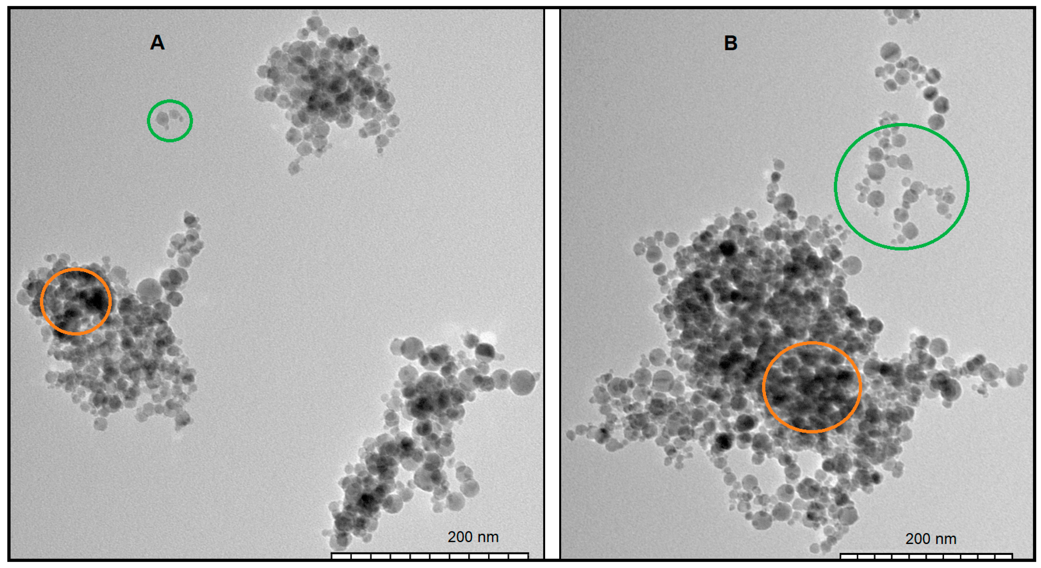
The DLS experimentally revealed features of structure formation and aggregation of GA particles that can be explained on the basis of data obtained through transmission electron microscopy. This method has already been used in the study of GA solutions, but the authors used higher concentrations [50]. During the TEM study of GA solutions, we first identified two areas containing the most characteristic small structural formations, shown in Figure 3. All formations consist of the set of spherical blocks, which we identify as polysaccharide units of GA colloidal structures, including a small part with protein content, as was shown earlier [5]. This fact allows us to conclude that the polysaccharide parts of GA molecules have an almost spherical shape.
Visually, the blocks of smallest conglomerates (highlighted as green circles in Figure 3) correspond to two fractions with predominant size of 2–8 and 9–20 nm. It is also noteworthy that many spherical blocks of larger sizes are in contact with one or two also spherical blocks of smaller sizes. The nature of the conjunctions suggests the presence of a third, thread-like fraction, as in some cases the connection of spheres is not due to direct contact between the blocks but at some distance from each other.
The second feature of Figure 3 was the complete absence of individual spherical blocks, i.e., single polysaccharide spherical blocks were practically absent. Only the conglomerates and larger associates are present, which is very surprising for a low concentration of 0.1 mg/mL of substance consisting almost entirely of polysaccharides. Based on existing ideas about the composition and structure of GA molecules [42,43,51,52,53,54], it can be assumed that colloidal structures representing small conglomerates organized by a few polysaccharide blocks linked by threads (most likely of protein origin) are true molecules, all parts of which are linked by covalent interactions. Moreover, the conglomerates that we compare with GA molecules are prone to the formation of associative supramolecular structures of several hundred nanometers or more in size, which we observe in our studies using the dynamic light scattering method. Because individual spherical blocks are parts of molecules or truncated molecules, they are most susceptible to association. Therefore, they are detected by the nanosizer only in the case of a small GA concentration (Figure 2A). In more concentrated solutions, the probability of their association with other structures increases, and therefore they are not observed.
The third feature is the different clearness of the boundaries of the spherical polysaccharide parts of GA molecules in various places. Where the spherical blocks are located least densely, i.e., in small colloidal structures or along the loose edges of large formations, the polysaccharide parts of molecules have a spherical shape with clear edges. However, as the local density of spherical blocks increases, their edges become blurred, and the blocks themselves begin to swell and merge into shapeless structures (in Figure 4 these areas are highlighted with orange circles), indicating the onset of local gelation. Note that gelation is characterized by the emergence of a three-dimensional network structure between polymer chains. In this case, gelation occurs in a limited volume, i.e., it is local in nature. Considering that GA is an anionic polysaccharide, this fact may indicate a large role of electrostatic interactions within the associates. In [50], it was noted that the existence of acidic sugars in the GA’s composition leads to the ability of gums to bind mono- or divalent cations. The presence of ions associated with biopolymer chains results in changes in solubility, viscosity, and gel formation ability resulting from pH alteration in the aqueous dispersion medium.
The fourth feature relates to the largest associates (Figure 4) existing in solutions simultaneously with those described above. They consist of swollen, adherent balls of increased size, forming even more shapeless structures (Figure 4A), which ultimately results in limited internal gelation and leveling of individual parts of the surface (highlighted by red circles). Such a variety of GA molecular and supramolecular forms in solutions significantly complicates its analysis but allows it to perform the main protective (enveloping) function.
Note that due to the peculiarities of the structures formed on the basis of aggregation of spherical polysaccharide blocks, rather than linear extended chains, the occurrence of regions of internal local gelation and the increase in their number with a growth in GA concentration in solution practically do not change the macroscopic rheological properties of aqueous GA solutions. One of the main reasons why GA is so valued is precisely the fact that gum dissolving in water does not lead to a significant increase in solution viscosity or its gelation [50,55]. This fact is explained by the absence of a network of bonds in the bulk of solution up to high GA concentrations. The emerging bonds are local in nature, leading to the formation of gel-like microclots floating in water (in a low-viscosity GA solution). A rheological feature of such systems is the layer-by-layer shear flow, which is largely determined by the viscosity of the low-molecular layer [56].
It should be noted that the diversity of forms of GA, which exist in solution, is most likely determined by the sample’s prehistory. Similar effects are characteristic of non-ionic surfactants, including, in particular, the block copolymer surfactants [57]. Dissolution or mixing of GA leads to the detachment of molecular subsystems, i.e., the appearance of small conglomerates of spherical particles bound with protein structures through covalent interactions. Over time, their further self-association leads to the appearance of increasingly swelling and sticking formations and reversible local gelation. In this case, some of the established local gel-like structures remain in solution along with newly formed ones. In our case, this fact is understood due to the tenfold dilution and mixing of solution immediately before performing TEM experiments.

3.3. NMR Experiments

To estimate the size of structures, we also conducted NMR experiments to determine the self-diffusion coefficients (SDCs) of particles in aqueous solutions with GA concentrations of 2, 5, and 10 mg/mL. Unlike the dynamic light scattering method, which mainly records the contribution from aggregates existing in the solution, NMR detects the weighted-average self-diffusion of individual molecules that exist in all possible structural states (molecules and different associates) with fast exchange between states [58]. The SDC concentration dependence of polysaccharides when describing the diffusion decay with one and two exponents is shown in Figure 5.
The obtained diffusion decays are close to monoexponential but also may be better described by two components. The values of the fast and slow components do not differ much. When described by two exponents, the values of SDCs differ significantly for different signals. At the same time, the values of the slow component are close to the values when described by one exponent (Figure 5B).
The Stokes–Einstein formula was used to estimate the particle size R,
D S D = k T 6 π η R
The value of KSD was chosen to be D S D = 2.3 10−11 m2/s, corresponding to the extrapolation of obtained SDC values at zero concentration; k = 1.38 × 10⁻²³ J/K is the Boltzmann constant, T = 298 K. The value of solvent viscosity was taken to be η = 8.9 × 10⁻⁴ Pa·s, equal to water’s value at corresponding temperatures. According to the calculations, the average hydrodynamic radius of diffusing particles in the solution is R = 10.6 nm, i.e., their diameter is close to 20 nm. This result agrees quite well with the available images, especially considering the presence of swollen particles shown in Figure 4, evidence of long lifetimes of individual molecules in comparison with the lifetimes of different aggregates. The presence of a fast component also makes it possible to assume (as a large assumption) the presence of smaller units in the solution with sizes 3–8 times smaller than the main ones, which is quite consistent with the fraction of smaller GA particles. A more detailed report on finding self-diffusion coefficients is given in the “How the SDC was measured” section of the Supporting Information.
We also analyzed the NMR proton spectra of aqueous solutions of GA with concentrations of 2, 5, and 10 mg/mL (Figure 6). As a result, we obtained three identical spectra that did not contain any signs of line width changes or chemical shifts of individual lines. Thus, it can be concluded that in the studied concentration range, the NMR spectra do not depend on GA concentration in solution. This fact indicates the absence of strong covalent interactions within the emerging associates.
Figure 6 shows the proton NMR spectrum of 5 mg/mL GA aqueous solution. Monosaccharides H-2 through H-6 protons resonate at 3.5–4.5 ppm, whereas the anomeric H-1 protons can be found at 4.5–5.8 ppm [59]. A signal at 1.2 ppm corresponds to the methyl group in rhamnose. Signals are numbered according to protons in the arabinose moiety (green), galactopyranose moiety (blue), and rhamnose moiety (magenta). Assignments are made based on literature data [60].

4. Discussion

The above-described behavioral features of Gum Arabic in aqueous solutions can be partly explained within the framework of existing concepts of the structure of its molecules, taking into account GA’s main functions. It has long been known that acacia gum contains a portion of protein material covalently bound to the polysaccharide [2,45]. According to classical concepts [45,47,48,51,52,53], GA consists of three fractions, namely, arabino-galactan (AG), glycoprotein (GP), and arabino-galactan protein conjugates (AGPs), differing in the polysaccharide/protein ratio. They also differ in size and hydrophobicity. These components, linked by protein chains prone to aggregation, form a three-dimensional multiscale porous structure [54]. In general, the GA structural unit is described by the “wattle blossom” model, first proposed by Fincher et al. [61], with porous structures of polysaccharides associated with polypeptide chains. This picture corresponds quite well to our image shown in Figure 3. Note that the obtained picture also does not contradict the “twisted hairy rope” model by Qi et al. [53], in which the Gum Arabic molecule consists of a polypeptide backbone of some 400 amino acids with numerous small polysaccharide substituents attached (~30 sugar residues), linked through hydroxyproline [14]. Such a molecular structure allows it to be in solution not only in rodlike but also in pseudospherical conformations.
The obtained results for the molecular sizes allow us to supplement them with some calculations obtained from geometric considerations. According to literature data [51], the main part of gum, constituting ~90% of its total amount, corresponds to the fraction with a molecular weight close to MW = 2.5 × 10⁵ Da. The radius of gyration Rg (from GPC MALLS) and the hydrodynamic radius Rh (from dynamic light scattering) for this fraction have values of ~10 and ~15 nm, respectively, consistent with our data. The Rg/Rh ratio indicates a compact spherical structure. The second component, representing ~10% of the total amount, contains ~10% protein. It has a molecular weight several times greater and, according to many authors, it is a composite. The third component, corresponding to only ~1% of the total amount, mainly contains protein content. Detailed studies of the major molecular fractions and polypeptide backbones, taking into account the number of charges coming from polysaccharidic moieties (i.e., glucuronic acids) and polypeptidic backbone, were carried out in [52].
Thus, for the analysis of the obtained data, it can be considered in the first approximation (excluding polydispersity) that the main structural units in GA solutions are identical spherical blocks of polysaccharide chains with a mass of MW = 2.5 × 10⁵ Da. Taking into account polydispersity, it is generally considered that the molecular weight of gum is in the range MW = 2.5 × 10⁵ ÷ 1.45 × 10⁶ Da [51]. They are connected by protein chains into a colloidal structure (actually a GA molecule), which can take both rodlike and pseudospherical forms with all sorts of intermediates.
For comparison with our data and taking into account the spherical shape of GA polysaccharide units, we can estimate the diameter D of the sphere occupied by blocks with indicated molecular masses. For the estimate, we use the formula for calculating the volume of sphere V through its diameter, as well as the relationship between volume, mass MW, and density ρ, which allows us to write a formula for calculating the diameter in the case of dense packing of chains and the absence of water inside of blocks,
D = 6 m π ρ 1 / 3
Polysaccharide units float in water, so their average density can be taken as the density of water. The mass MW must be converted to the SI system, where 1 Da = 1.6605 × 10⁻²⁷ kg, so m = 4.15 × 10⁻²² kg. Substituting the available data, we find the diameter of polysaccharide unit D = 9.26 nm.
Note that the calculations did not take into account the presence of water inside of the sphere. The presence of water content can be taken into account as follows. If water occupies 50% of the volume in the spheres under consideration, the mass of the polysaccharide unit would increase by two times. This means an increase in diameter by 21/3 = 1.26 times, which gives an average diameter value of D ~ 12 nm, which, taking into account polydispersity, reaches 21 nm. This result is quite consistent with block diameters shown in Figure 3. Accordingly, it can be concluded that the water content in the composition of spherical polysaccharide units occupies at least 50%.
At the next stage, we tried to estimate the length of expanded GA polysaccharide chains by an order of magnitude. For linear polysaccharide chains, such a problem is solved simply. Based on knowledge of the chemical formula, which for cellulose, for example, has the form (C6H10O5)n, we can calculate the molecular weight of one monosaccharide unit mW = 162 Da and then find the number of units, equal to n = MW/mW. Then, taking into account that on average one monosaccharide unit has a linear size of l = 0.515 nm [62], the total chain length L can be calculated using the formula
L = M W m W l .
In the case of branched chains, the situation becomes more complicated. For calculations, it is necessary to know the average unit size of polysaccharide and to take into account the presence and length of side chains. To estimate the average cell size of polysaccharides, we considered another substance, namely, the disaccharide sucrose, consisting of two units and having the formula C12H22O11. Sucrose crystallizes in the monoclinic space group with the room temperature lattice parameters a = 1.08631 nm, b = 0.87044 nm, and c = 0.77624 nm [63]. Accordingly, the size of one unit is 0.54 nm. Because this value is almost the same as the cellulose unit length, and the formula of many other polysaccharides coincides with the formula of cellulose, we decided to take the value l = 0.515 nm for the following calculations.
To account for the side chains, we decided to use Figure 1, which shows a putative molecular structure for an A. Senegal gum. Here, we notice that for every 7 cells in the backbone, there are 30 more side cells. This corresponds to an increase in the mass of the chain cell by 37/7 times. Therefore, taking into account Figure 1, the approximate number of cells n in the backbone of the polysaccharide chain and its length L can be found as
n = 7 37 M W m W = 292 ,
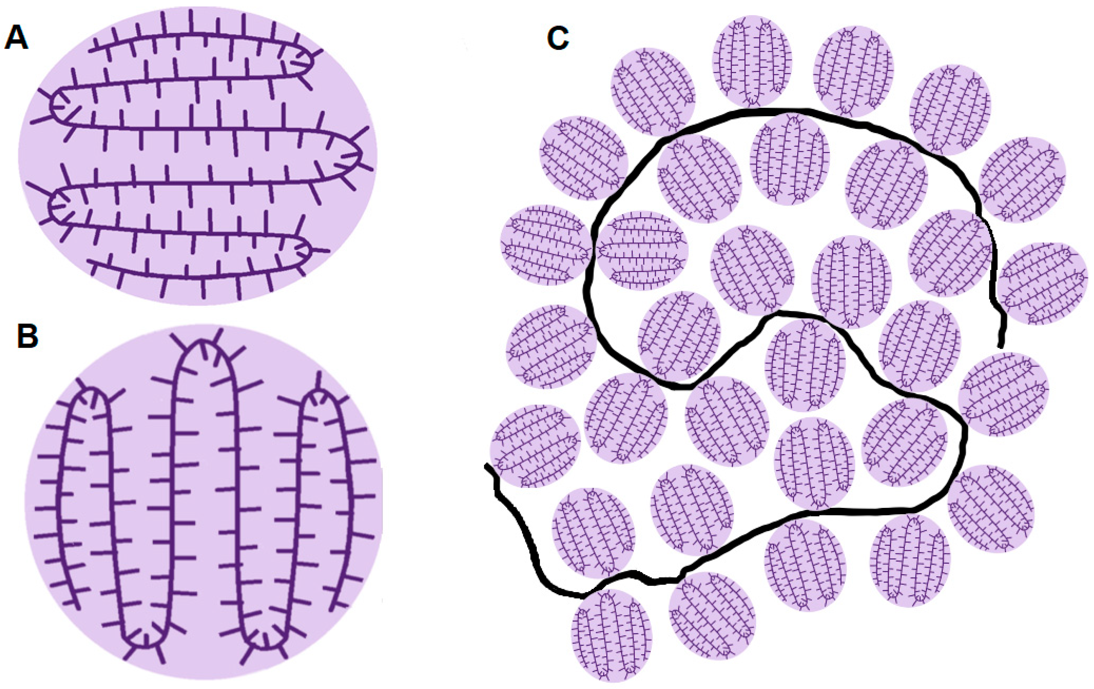
In the next stage, knowing the basic geometric parameters of the polysaccharide chain, it was necessary to develop a model that would describe the effects observed during the association of GA particles in aqueous solutions, leading first to swelling of the blocks and then the appearance of local gelation. In fact, it was necessary to invent a spherical block structure that would allow for rapid swelling (increase in the space between the polysaccharide chains), which is the basis for local gelation. Note that neither a spiral nor a coil allow for a rapid increase in size (swelling). However, a structure resembling a sinusoid wrapped around a cylinder or sphere (something similar to a “crown”) allows for rapid stretching.
Because the average diameter of a polysaccharide block calculated by us (and consistent with Figure 5) is D ~ 12 nm (without taking into account polydispersity), the polysaccharide chain forming it should bend only 12 times. Thus, schematically, the polysaccharide block may look as shown in Figure 7A, only, in the three-dimensional case, the six sections shown in the foreground will correspond to six more in the background, as if forming a “crown”. The width of the chain will be determined by the number of cells in the side chains. In accordance with Figure 1, where side chains of six cells are shown, the maximum width of the chain will correspond to 3 nm. However, when connected into a block (when forming a crown), the side chains will interpenetrate each other or be directed to the center of the spherical structure. In a similar manner, but with increased scale, larger blocks can be built.
Figure 7C shows a possible pseudospherical conformation of the colloidal structure from polysaccharide blocks covalently linked to a polypeptide chain in aqueous solution. Due to polydispersity, the sizes of spherical blocks in Figure 7C may vary.
Indirect confirmation of the “crown” model of the spherical structure of the polysaccharide block developed by us is provided by the results of Sanchez et al. [64], in which a combination of methods, including atomic force microscopy, showed that the morphology of the polysaccharide block can be described as a porous disk-like structure with a central intricate ‘‘network’’ with a diameter of 20 nm and a thickness below 2 nm. Note that the polysaccharide block organized according to the “crown” model, as a result of drying, will turn into a disk of similar dimensions, very similar to that described in [64], and, therefore, the results of that work do not contradict the assumptions put forward by us.
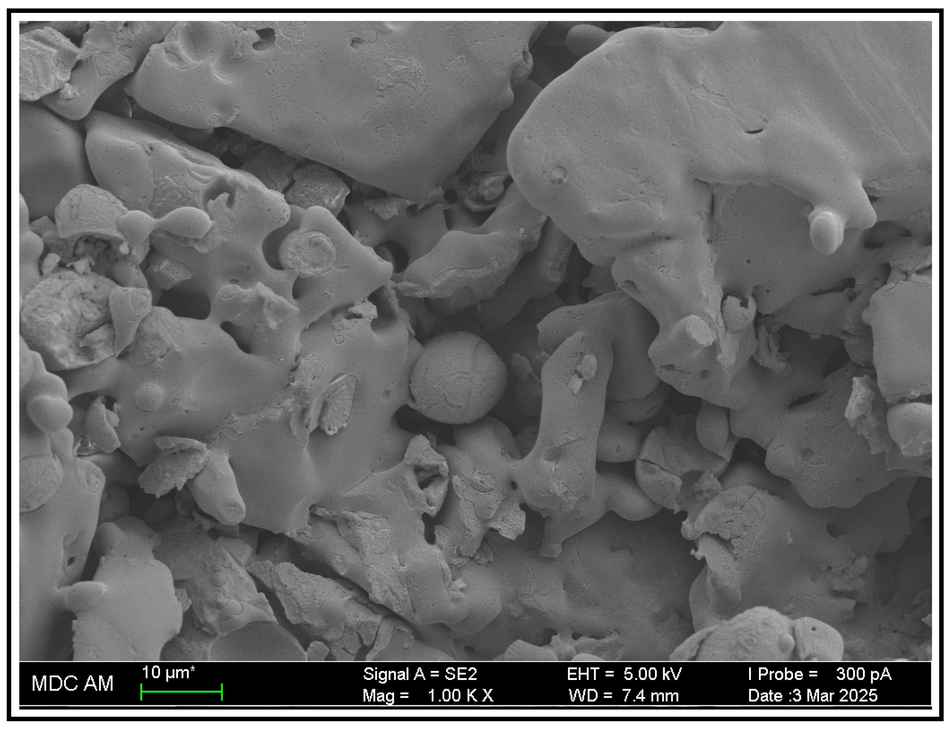
The initial shape and size of a block (Figure 7A) are probably determined during gum formation in response to stress conditions. The compact shape of the spatially separated blocks linked by the polypeptide chain is maintained in solution mainly by ionic interactions. There is no reason for ions to leave their blocks as long as the molecule (“wattle blossom”) is single. However, the integration of several molecules into an associate leads to a convergence between the blocks and even their contact, which allows the ions to leave the block volume and distribute themselves within an associate space. This process causes the side chains to turn to the area outside of the block. Accordingly, the forces holding the “crown” in a compact state become weaker, which leads to stretching of the crown and an increase (swelling) of the block (Figure 7B) up to partial fusion with neighboring blocks of the associate. In fact, local gelation begins. GA starts to perform its main protective and enveloping function. Based on the SEM image (Figure 8), it is clear that GA can create dense protective layers on the surface.
Partial confirmation of the “crown” model can be provided by the remarkable emulsifying properties of colloidal structures (molecules) of GA, which can be explained on the basis of local gelation. The high emulsifying capacity of gum is due to the presence of protein material covalently binding hydrophilic carbohydrate blocks [65]. Therefore, during emulsification, proteins containing non-polar groups are adsorbed at the boundaries of oil microdroplets, and polysaccharide blocks bound by them are turned in one direction (the water side), which leads to their convergence and the emergence of conditions for local gelation. Due to local gelation (swelling of the blocks), layers are formed consisting of swollen spherical polysaccharide blocks, which form a layer with increased viscosity between the oil microdroplets and the dispersion medium. Thus, HA forms thick steric stabilizing layers around the oil microdroplets, performing both an emulsifying and a stabilizing role [46,65,66,67]. This situation is somewhat analogous to the change in water viscosity near ionic surfactant micelles [68,69].
The effect of GA as a corrosion inhibitor can be explained in a similar way; protein chains are adsorbed on metal surfaces, and the polysaccharide blocks facing the water swell and merge, forming protective layers.
Thus, we have a process that is extended over time, allowing nature to realize its functions. At the initial stage, GA associates are permeable to water and the water-soluble medicinal and protective preparations. Over time, on the contrary, a dense gel-like cushion is created, protecting the wound’s surface from unfavorable external conditions.
In [54], it was noted that self-association of GA’s initial molecular structures into large-scale formations leads to a strong multi-scale structure that extends throughout the solution, which is mainly influenced by concentration and ionic repulsion. To the above, we add the need to take into account the time characteristics of the sample, which was also noted in [50].

5. Conclusions

We studied the associative behavior of Gum Arabic in aqueous solutions using dynamic light scattering, nuclear magnetic resonance, and transmission and scanning electron microscopy. Microscopic methods reveal the smallest formations corresponding to conglomerates of polysaccharide blocks covalently linked by protein chains, which are essentially true GA molecules, as well as their associates. It is shown that in small associates, spherical polysaccharide units can be related to two fractions with predominant sizes of 2–8 and 9–20 nm. It is established that in places with the highest concentration of polysaccharide blocks, their boundaries begin to blur and merge, indicating the occurrence of local gelation.
To estimate the size of structures contributing to diffusion motion, the NMR experiments were conducted to determine self-diffusion coefficients of colloidal structures in aqueous solutions with concentrations of 2, 5, and 10 mg/mL of GA. Extrapolation of the SDC values to zero concentration allowed us to calculate the average hydrodynamic diameter of diffusing structures, which turned out to be close to 20 nm, which corresponds quite well to the available images. The presence of a fast component in self-diffusion data also makes it possible to assume the presence of smaller units in the solution with sizes 3–8 times smaller than the main ones, which is quite consistent with the fraction of smaller GA particles.
Based on geometric considerations and ideas about GA’s composition and structure and considering that the main structural units in GA solutions are spherical protein–polysaccharide blocks of biopolymer chains with a mass of MW = 2.5 × 10⁵ Da, we calculated the geometric parameters of branched GA polysaccharide chains. A possible “crown” model of chain association into a polysaccharide block is proposed, which allow us to describe the effects observed during the association of gum particles in aqueous solutions, leading first to the swelling of blocks, then to the appearance of local gelation, and only then to the creation of dense protective layers on the damaged surfaces. The association process, extended over time, allows to implement its functions in nature. At the initial stage, GA associates are permeable to water and water-soluble medicinal and protective preparations. Over time, on the contrary, a dense gel-like cushion is created, protecting the wound’s surface from unfavorable external conditions. Thus, a correct consideration of the processes occurring in GA solutions requires us to take into account time characteristics, i.e., taking into account the sample’s prehistory.
The above leads to the conclusion that the basis of the GA properties necessary for natural, food, technical, and technological applications are as follows:
  • Structural features of the amphiphilic GA molecule, which is a colloidal structure of hydrophilic anionic polysaccharide blocks linked together by a polypeptide chain that includes hydrophobic fragments (“wattle blossom” model);
  • Mobility of parts of the GA molecule and their tendency to form associates and complexes both among themselves and with surfaces;
  • Structural features of the polysaccharide blocks (“crown” model), allowing for rapid swelling and local gelation emergence with an increase in their local concentration while maintaining low solution viscosity up to fairly high GA concentrations;
  • Interfacial activity of amphiphilic GA molecules, creating the possibility of forming viscous layers on interfacial surfaces.

Supplementary Materials

The following supporting information can be downloaded at: https://www.mdpi.com/article/10.3390/macromol5040049/s1, Figure S1: GA diffusion decay approximated by one (black) and two (red) exponentials.

Author Contributions

Conceptualization, O.S.Z.; investigation, P.V.S., M.A.K., D.A.K., and N.R.G.; formal analysis, O.S.Z., T.K., Y.A., and N.R.G.; writing—original draft preparation, O.S.Z. and N.R.G.; writing—review and editing, P.V.S., M.A.K., D.A.K., and O.S.Z.; supervision and project administration, T.K. and O.S.Z. All authors have read and agreed to the published version of the manuscript.

Funding

This research received no external funding.

Institutional Review Board Statement

Not applicable.

Data Availability Statement

The original contributions presented in this study are included in the article/Supplementary Material. Further inquiries can be directed to the corresponding author(s).

Acknowledgments

O.S.Z., N.R.G., and Y.A. give thanks for the support of the KSPEU Strategic Academic Leadership Program (“PRIORITY-2030”). P.V.S. and M.A.K. thank the state assignments to the Kazan Scientific Center of the Russian Academy of Sciences. NMR experiments were performed with equipment of the Collective spectro-analytical center of Kazan Research Center, Russian Academy of Sciences, Kazan. TEM and SEM studies were fulfilled in the Interdisciplinary Center “Analytical Microscopy” (Kazan Federal University, Kazan).

Conflicts of Interest

The authors declare no conflicts of interest.

References

  1. Whistler, R.L. Exudate gums. In Industrial Gums: Polysaccharides and Their Derivatives, 3rd ed.; Whistler, R.L., BeMiller, J.N., Eds.; Academic Press: San Diego, CA, USA, 1993. [Google Scholar]
  2. Williams, P.A.; Phillips, G.O. 11—Gum Arabic. In Handbook of Hydrocolloids, 2nd ed.; Phillips, G.O., Williams, P.A., Eds.; Woodhead Publishing: Saskatoon, UK, 2009. [Google Scholar]
  3. Musa, H.H.; Ahmed, A.A.; Musa, T.H. Chemistry, Biological, and Pharmacological Properties of Gum Arabic. In Bioactive Molecules in Food; Mérillon, J.-M., Ramawat, K.G., Eds.; Springer International Publishing: Berlin/Heidelberg, Germany, 2019. [Google Scholar]
  4. Alftrén, J.; Peñarrieta, J.M.; Bergenståhl, B.; Nilsson, L. Comparison of molecular and emulsifying properties of gum arabic and mesquite gum using asymmetrical flow field-flow fractionation. Food Hydrocoll. 2012, 26, 54–62. [Google Scholar] [CrossRef]
  5. Islam, A.M.; Phillips, G.O.; Sljivo, A.; Snowden, M.J.; Williams, P.A. A review of recent developments on the regulatory, structural and functional aspects of gum Arabic. Food Hydrocoll. 1997, 11, 493–505. [Google Scholar] [CrossRef]
  6. Ali, B.H.; Ziada, A.; Blunden, G. Biological effects of gum arabic: A review of some recent research. Food Chem. Toxicol. 2009, 47, 1–8. [Google Scholar] [CrossRef]
  7. Verbeken, D.; Dierckx, K.; Dewettinck, S. Exudate gums: Occurrence, production, and applications. Appl. Microbiol. Biotechnol. 2003, 63, 10–21. [Google Scholar] [CrossRef]
  8. Makarova, A.O.; Derkach, S.R.; Kadyirov, A.I.; Ziganshina, S.A.; Kazantseva, M.A.; Zueva, O.S.; Gubaidullin, A.T.; Zuev, Y.F. Supramolecular Structure and Mechanical Performance of κ-Carrageenan–Gelatin Gel. Polymers 2022, 14, 4347. [Google Scholar] [CrossRef]
  9. Gubaidullin, A.T.; Galeeva, A.I.; Galyametdinov, Y.G.; Ageev, G.G.; Piryazev, A.A.; Ivanov, D.A.; Ermakova, E.A.; Nikiforova, A.A.; Derkach, S.R.; Zueva, O.S.; et al. Modulation of Structural and Physical-Chemical Properties of Fish Gelatin Hydrogel by Natural Polysaccharides. Int. J. Mol. Sci. 2025, 26, 2901. [Google Scholar] [CrossRef] [PubMed]
  10. Zueva, O.S.; Gubaidullin, A.T.; Makarova, A.O.; Bogdanova, L.R.; Zakharova, L.Y.; Zuev, Y.F. Structural features of composite protein-polysaccharide hydrogel in the presence of a carbon nanomaterial. Russ. Chem. Bull. 2020, 69, 581–589. [Google Scholar] [CrossRef]
  11. Gubaidullin, A.T.; Makarova, A.O.; Derkach, S.R.; Voron’ko, N.G.; Kadyirov, A.I.; Ziganshina, S.A.; Salnikov, V.V.; Zueva, O.S.; Zuev, Y.F. Modulation of Molecular Structure and Mechanical Properties of κ-Carrageenan-Gelatin Hydrogel with Multi-Walled Carbon Nanotubes. Polymers 2022, 14, 2346. [Google Scholar] [CrossRef] [PubMed]
  12. Barak, S.; Mudgil, D.; Taneja, S. Exudate gums: Chemistry, properties and food applications—A review. J. Sci. Food Agric. 2020, 100, 2828–2835. [Google Scholar] [CrossRef]
  13. Padil, V.V.T.; Senan, C.; Cerník, M. “Green” polymeric electrospun fibers based on tree-gum hydrocolloids: Fabrication, characterization and applications. Mater. Biomed. Eng. 2019, 5, 127–172. [Google Scholar] [CrossRef]
  14. Osman, M.E.; Williams, P.A.; Menzies, A.R.; Phillips, G.O. Characterization of commercial samples of gum arabic. J. Agric. Food Chem. 1993, 41, 71–77. [Google Scholar] [CrossRef]
  15. Sanchez, C.; Nigen, M.; Mejia Tamayo, V.; Doco, T.; Williams, P.; Amine, C.; Renard, D. Acacia gum: History of the future. Food Hydrocoll. 2018, 78, 140–160. [Google Scholar] [CrossRef]
  16. Sharkawy, A.; Rodrigues, A.E. Plant gums in Pickering emulsions: A review of sources, properties, applications, and future perspectives. Carbohydr. Polym. 2024, 332, 121900. [Google Scholar] [CrossRef]
  17. Zipkin, A.M.; Wagner, M.; McGrath, K.; Brooks, A.S.; Lucas, P.W. An experimental study of hafting adhesives and the implications for compound tool technology. PLoS ONE 2014, 9, e112560. [Google Scholar] [CrossRef]
  18. Al-Jubori, Y.; Ahmed, N.T.B.; Albusaidi, R.; Madden, J.; Das, S.; Sirasanagandla, S.R. The Efficacy of Gum Arabic in Managing Diseases: A Systematic Review of Evidence-Based Clinical Trials. Biomolecules 2023, 13, 138. [Google Scholar] [CrossRef]
  19. Alobaidi, S. Therapeutic Potential of Gum Arabic (Acacia senegal) in Chronic Kidney Disease Management: A Narrative Review. J. Clin. Med. 2024, 13, 5778. [Google Scholar] [CrossRef] [PubMed]
  20. Ahmed, A.A. 16—Health Benefits of Gum Arabic and Medical Use. In Gum Arabic Structure, Properties, Application and Economics; Mariod, A.A., Ed.; Academic Press: Cambridge, MA, USA, 2018. [Google Scholar] [CrossRef]
  21. Chai, L.; Chen, D.; Ye, L.; Peng, P.; Wang, H.; Al Saleh, N.; Al-Kenani, N.S.; Guo, J.; Li, Q.; Guo, L. Pharmacological targets and therapeutic mechanisms of Arabic gum in treating diabetic wounds: Insights from network pharmacology and experimental validation. Front. Pharmacol. 2025, 16, 1528880. [Google Scholar] [CrossRef] [PubMed]
  22. Mohamed, S.A.; Elsherbini, A.M.; Alrefaey, H.R.; Adelrahman, K.; Moustafa, A.; Egodawaththa, N.M.; Crawford, K.E.; Nesnas, N.; Sabra, S.A. Gum Arabic: A Commodity with Versatile Formulations and Applications. Nanomaterials 2025, 15, 290. [Google Scholar] [CrossRef] [PubMed]
  23. Lelon, J.K.; Jumba, I.O.; Keter, J.K.; Chemuku, W.; Oduor, F.D.O. Assessment of physical properties of gum arabic from Acacia senegal varieties in Baringo District, Kenya. Afr. J. Plant Sci. 2010, 4, 95–98. Available online: https://academicjournals.org/journal/AJPS/article-full-text-pdf/417951A11473 (accessed on 1 June 2025).
  24. Mbugua, R.; Salim, R.; Ndambuki, J. Effect of Gum Arabic Karroo as a water-reducing admixture in cement mortar. Case Stud. Constr. Mater. 2016, 5, 100–111. [Google Scholar] [CrossRef]
  25. Elinwa, A.U.; Abdulbasir, G.; Abdulkadir, G. Gum Arabic as an admixture for cement concrete production. Constr. Build. Mater. 2018, 176, 201–212. [Google Scholar] [CrossRef]
  26. Brzyski, P. The Influence of Gum Arabic Admixture on the Mechanical Properties of Lime-Metakaolin Paste Used as Binder in Hemp Concrete. Materials 2021, 14, 6775. [Google Scholar] [CrossRef]
  27. Nehra, A.; Biswas, D.; Siracusa, V.; Roy, S. Natural Gum-Based Functional Bioactive Films and Coatings: A Review. Int. J. Mol. Sci. 2023, 24, 485. [Google Scholar] [CrossRef]
  28. Morodi, V.; Kaseke, T.; Fawole, O.A. Impact of Gum Arabic Coating Pretreatment on Quality Attributes of Oven-Dried Red Raspberry (Rubus idaeus L.). Fruit. Process. 2022, 10, 1629. [Google Scholar] [CrossRef]
  29. Shoukat, H.; Buksh, K.; Noreen, S.; Pervaiz, F.; Maqbool, I. Hydrogels as potential drug-delivery systems: Network design and applications. Ther. Deliv. 2021, 12, 375–396. [Google Scholar] [CrossRef] [PubMed]
  30. Olabode, O.; Akinsanya, O.; Daramola, O.; Sowunmi, A.; Osakwe, C.; Benjamin, S.; Samuel, I. Effect of Salt Concentration on Oil Recovery During Polymer Flooding: Simulation Studies on Xanthan Gum and Gum Arabic. Polymers 2023, 15, 4013. [Google Scholar] [CrossRef] [PubMed]
  31. Timothy, U.J.; Umoren, P.S.; Solomon, M.M.; Igwe, I.O.; Umoren, S.A. An appraisal of the utilization of natural gums as corrosion inhibitors: Prospects, challenges, and future perspectives. Int. J. Biol. Macromol. 2023, 253, 126904. [Google Scholar] [CrossRef]
  32. Umoren, S.A.; Obot, I.B.; Ebenso, E.E.; Okafor, P.C.; Ogbobe, O.; Oguzie, E.E. Gum arabic as a potential corrosion inhibitor for aluminium in alkaline medium and its adsorption characteristics. Anti Corros. Methods Mater. 2006, 53, 277–282. [Google Scholar] [CrossRef]
  33. Umoren, S.A. Inhibition of aluminium and mild steel corrosion in acidic medium using Gum Arabic. Cellulose 2008, 15, 751–761. [Google Scholar] [CrossRef]
  34. Azzaoui, K.; Mejdoubi, E.; Jodeh, S.; Lamhamdi, A.; Rodriguez-Castellón, E.; Algarra, M.; Zarrouk, A.; Errich, A.; Salghi, R.; Lgaz, H. Eco friendly green inhibitor Gum Arabic (GA) for the corrosion control of mild steel in hydrochloric acid medium. Corros. Sci. 2017, 129, 70–81. [Google Scholar] [CrossRef]
  35. Shen, C.; Alvarez, V.; Koenig, J.D.; Luo, J.-L. Gum Arabic as corrosion inhibitor in the oil industry: Experimental and theoretical studies. Corros. Eng. Sci. Technol. 2019, 54, 444–454. [Google Scholar] [CrossRef]
  36. Hamza, B.; Youssouf, R.; Abdelouahad, C. Gum Arabic as an eco-friendly inhibitor for API 5L X42 pipeline steel in HCl medium. Corros. Sci. 2014, 82, 426–431. [Google Scholar] [CrossRef]
  37. Abu-Dalo, M.A.; Othman, A.A.; Al-Rawashdeh, N.A.F. Exudate gum from Acacia trees as green corrosion inhibitor for mild steel in acidic media. Int. J. Electrochem. Sci. 2012, 7, 9303–9324. [Google Scholar] [CrossRef]
  38. Khair, T.; Abakumova, Y.; Zueva, O. Inhibitive behaviour of Gum Arabic on corrosion process of pipeline steel in acidic environment. E3S Web Conf. 2024, 592, 04011. [Google Scholar] [CrossRef]
  39. Djajadisastra, J.; Sutriyo, P.P.; Pujiyanto, A. Antioxidant activity of gold nanoparticles using gum arabic as a stabilizing agent. Int. J. Pharm. Pharm. Sci. 2014, 6, 462–465. [Google Scholar]
  40. Solomon, M.M.; Gerengi, H.; Umoren, S.A.; Essien, N.B.; Essien, U.B.; Kaya, E. Gum Arabic-silver nanoparticles composite as a green anticorrosive formulation for steel corrosion in strong acid media. Carbohydr. Polym. 2018, 181, 43–55. [Google Scholar] [CrossRef]
  41. Timothy, U.J.; Mamudu, U.; Solomon, M.M.; Umoren, P.S.; Igwe, I.O.; Anyanwu, P.I.; Aharanwa, B.C.; Lim, R.C.; Uchechukwu, T.O.; Umoren, S.A. In-situ biosynthesized plant exudate gums-silver nanocomposites as corrosion inhibitors for mild steel in hydrochloric acid medium. Int. J. Biol. Macromol. 2024, 269, 132065. [Google Scholar] [CrossRef] [PubMed]
  42. Gashua, I.B.; Williams, P.A.; Yadav, M.P.; Baldwin, T.C. Characterisation and molecular association of Nigerian and Sudanese Acacia gum exudates. Food Hydrocoll. 2015, 51, 405–413. [Google Scholar] [CrossRef]
  43. Randall, R.C.; Phillips, G.O.; Williams, P.A. The role of the proteinaceous component on the emulsifying properties of gum Arabic. Food Hydrocoll. 1988, 2, 131–140. [Google Scholar] [CrossRef]
  44. Nie, S.-P.; Wang, C.; Cui, S.W.; Wang, Q.; Xie, M.-Y.; Phillips, G.O. A further amendment to the classical core structure of gum Arabic (Acacia senegal). Food Hydrocoll. 2013, 31, 42–48. [Google Scholar] [CrossRef]
  45. Williams, P.A.; Phillips, G.O.; Stephen, A.M.; Churms, S.C. Gums and Mucilages. In Food Polysaccharides and Their Applications; Stephen, A.M., Ed.; Marcel Dekker Inc.: New York, NY, USA, 1995. [Google Scholar]
  46. Kersten, F.; Martin, D.; van der Schaaf, U.S.; Wefers, D. Gum Arabic—Same but different: Comparative analysis of structural characteristics and emulsifying properties of 20 Acacia senegal samples of various qualities. Food Hydrocoll. 2025, 165, 111231. [Google Scholar] [CrossRef]
  47. Vandevelde, M.-C.; Fenyo, J.-C. Macromolecular distribution of Acacia senegal gum (gum arabic) by size-exclusion chromatography. Carbohydr. Polym. 1985, 5, 251–273. [Google Scholar] [CrossRef]
  48. Defaye, J.; Wong, E. Structural studies of gum arabic, the exudate polysaccharide from Acacia senegal. Carbohydr. Res. 1986, 150, 221–231. [Google Scholar] [CrossRef]
  49. Mahendran, T.; Williams, P.A.; Phillips, G.O.; Al-Assaf, S.; Baldwin, T.C. New insights into the structural characteristics of the arabinogalactan-protein (AGP) fraction of gum arabic. J. Agric. Food Chem. 2008, 56, 9269–9276. [Google Scholar] [CrossRef]
  50. Gashua, I.B.; Williams, P.A.; Baldwin, T.C. Molecular characteristics, association and interfacial properties of gum Arabic harvested from both Acacia senegal and Acacia seyal. Food Hydrocoll. 2016, 61, 514–522. [Google Scholar] [CrossRef]
  51. Randall, R.C.; Phillips, G.O.; Williams, P.A. Fractionation and characterization of gum from Acacia senegal. Food Hydrocoll. 1989, 3, 65–75. [Google Scholar] [CrossRef]
  52. Renard, D.; Lavenant-Gourgeon, L.; Ralet, M.C.; Sanchez, C. Acacia senegal Gum:  Continuum of Molecular Species Differing by Their Protein to Sugar Ratio, Molecular Weight, and Charges. Biomacromolecules 2006, 7, 2637–2649. [Google Scholar] [CrossRef]
  53. Qi, W.; Fong, C.; Lamport, D.T.A. Gum arabic glycoprotein is a twisted hairy rope. A new model based on O-galactosyl hydroxyproline as the polysaccharide attachment site. Plant Physiol. 1991, 96, 848. [Google Scholar] [CrossRef]
  54. Atgié, M.; Garrigues, J.C.; Chennevière, A.; Masbernat, O.; Roger, K. Gum Arabic in solution: Composition and multi-scale structures. Food Hydrocoll. 2019, 91, 319–330. [Google Scholar] [CrossRef]
  55. Li, X.; Fang, Y.; Al-Assaf, S.; Phillips, G.O.; Nishinari, K.; Zhang, H. Rheological study of gum Arabic solutions: Interpretation based on molecular self-association. Food Hydrocoll. 2009, 23, 2394–2402. [Google Scholar] [CrossRef]
  56. Kulichikhin, V.G.; Malkin, A.Y. The Role of Structure in Polymer Rheology: Review. Polymers 2022, 14, 1262. [Google Scholar] [CrossRef]
  57. Zueva, O.S.; Makarova, A.O.; Zvereva, E.R.; Kurbanov, R.K.; Salnikov, V.V.; Turanov, A.N.; Zuev, Y.F. Industrial block copolymer surfactants: Diversity of associative forms and interaction with carbon nanomaterial. J. Mol. Liq. 2022, 359, 119267. [Google Scholar] [CrossRef]
  58. Zuev, Y.F.; Kurbanov, R.K.; Idiyatullin, B.Z.; Us’yarov, O.G. Sodium dodecyl sulfate self-diffusion in premicellar and low-concentrated micellar solutions in the presence of a back-ground electrolyte. Colloid. J. 2007, 69, 444–449. [Google Scholar] [CrossRef]
  59. Cheng, H.N.; Neiss, T.G. Solution NMR Spectroscopy of Food Polysaccharides. Polym. Rev. 2012, 52, 81–114. [Google Scholar] [CrossRef]
  60. Radziej, S.; Scherb-Forster, J.; Schlicht, C.; Eisenreich, W. Fast Identification of Food Thickeners by Nontargeted NMR Spectroscopy. J. Agric. Food Chem. 2021, 69, 3761–3775. [Google Scholar] [CrossRef]
  61. Fincher, G.B.; Stone, B.A.; Clarke, A.E. Arabinogalactan-proteins: Structure, biosynthesis and function. Annu. Rev. Plant Physiol. 1983, 34, 47–70. [Google Scholar] [CrossRef]
  62. Habibi, Y.; Lucia, L.A.; Rojas, O.J. Cellulose nanocrystals: Chemistry, selfassembly, and applications. Chem. Rev. 2010, 110, 3479–3500. [Google Scholar] [CrossRef]
  63. Beevers, C.A.; McDonald, T.R.R.; Robertson, J.H.; Stern, F. The crystal structure of sucrose. Acta Crystallogr. 1952, 5, 689–690. [Google Scholar] [CrossRef]
  64. Sanchez, C.; Lapp, A.; Schmitt, C.; Gaillard, C.; Kolodziejczyk, C.E.; Renard, D. The Acacia gum arabinogalactan fraction is a thin oblate ellipsoid: A new model based on SANS and ab initio calculation. Biophys. J. 2008, 94, 629–639. [Google Scholar] [CrossRef] [PubMed]
  65. Dickinson, E. Hydrocolloids at interfaces and the influence on the properties of dispersed systems. Food Hydrocoll. 2003, 17, 25–39. [Google Scholar] [CrossRef]
  66. Bai, L.; Huan, S.; Li, Z.; McClements, D.J. Comparison of emulsifying properties of food-grade polysaccharides in oil-in-water emulsions: Gum Arabic, beet pectin, and corn fiber gum. Food Hydrocoll. 2017, 66, 144–153. [Google Scholar] [CrossRef]
  67. Ozturk, B.; McClements, D.J. Progress in natural emulsifiers for utilization in food emulsions. Curr. Opin. Food Sci. 2016, 7, 1–6. [Google Scholar] [CrossRef]
  68. Zueva, O.S.; Rukhlov, V.S.; Zuev, Y.F. Morphology of ionic micelles as studied by numerical solution of Poisson equation. ACS Omega 2022, 7, 6174–6183. [Google Scholar] [CrossRef] [PubMed]
  69. Zueva, O.S.; Kazantseva, M.A.; Zuev, Y.F. Nanosized Being of Ionic Surfactant Micelles: An Advanced View on Micellization Process. Colloids Interfaces 2025, 9, 15. [Google Scholar] [CrossRef]
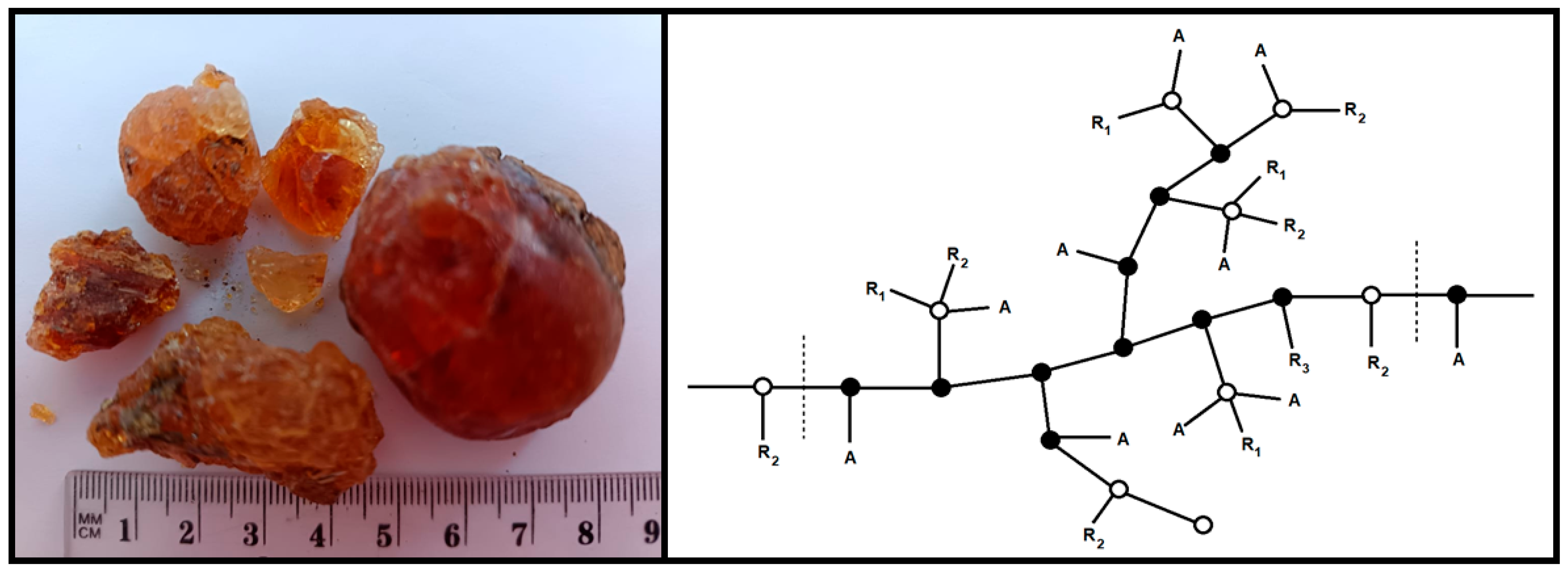
Figure 1. (Left): commercial GA samples. (Right): putative molecular structure for A. senegal gum: A = arabinosyl; ( ) = 3-linked galactose (galactose attached); ( ) = 6-linked galactose (galactose or glucuronic acid attached), or end group; R1 = rhamnose-glucuronic acid (rhamnose may be absent or replaced by Me); R2 = galactose-3arabinose; R3 = arabinose-3arabinose-3arabinose.
Macromol 05 00049 g001
Figure 2. Dynamic light scattering results in GA aqueous solution with concentrations of 2 mg/mL (A), 5 mg/mL (B), and 10 mg/mL (C).
Macromol 05 00049 g002
Figure 3. TEM images of conglomerates and associates of small structures formed in various parts (A,B) of aqueous GA solution.
Macromol 05 00049 g003
Figure 4. TEM images of shapeless structures formed in various parts (A,B) of GA aqueous solution.
Macromol 05 00049 g004
Figure 5. The SDC concentration dependence of polysaccharides when describing the diffusion decay with one (A) and two (B) exponents.
Macromol 05 00049 g005
Figure 6. Proton NMR spectrum of 5 mg/mL GA aqueous solution.
Macromol 05 00049 g006
Figure 7. Schematic representations of a single polysaccharide block (“crown” model) of a high molecular fraction of A. Senegal gum in its initial state (A) and with increasing water content (B); possible pseudospherical conformation of the colloidal structure of branched polysaccharide chains covalently linked to a polypeptide chain in aqueous solution (without taking into account polydispersity) (C).
Macromol 05 00049 g007
Figure 8. SEM image of lyophilized GA solution.
Macromol 05 00049 g008
Table 1. Dynamic light scattering results for GA aqueous solution with different concentrations.
Concentration of GAMaxima of Particle Size Distribution by Intensity Rhmax, nm
Peak 1Peak 2Peak 3
2 mg/mL9 ± 0.83246 ± 46434,000 ± 16,000
5 mg/mL240 ± 3244593 ± 4160
10 mg/mL209 ± 3442894 ± 2004
Disclaimer/Publisher’s Note: The statements, opinions and data contained in all publications are solely those of the individual author(s) and contributor(s) and not of MDPI and/or the editor(s). MDPI and/or the editor(s) disclaim responsibility for any injury to people or property resulting from any ideas, methods, instructions or products referred to in the content.