Article

Interplay Between Protein Phosphatase 2A (PP2A) and SE Translocation (SET) as Macromolecular Target of Anticancer Compounds: A Combined Computational and Experimental Study

by
Giovanni Ribaudo
1,†,
Mario Angelo Pagano
2,*,†,
Margrate Anyanwu
1,
Matteo Giannangeli
1,
Marika Vezzoli
1,
Andrea Visentin
3,
Federica Frezzato
3,
Livio Trentin
3,
Anna Maria Brunati
4 and
Alessandra Gianoncelli
1,*
1
Department of Molecular and Translational Medicine, University of Brescia, Viale Europa 11, 25123 Brescia, Italy
2
Department of Neuroscience and Rehabilitation, University of Ferrara, Via Luigi Borsari 46, 44121 Ferrara, Italy
3
Hematology Unit, Department of Medicine, University of Padova, Via Giustiniani 2, 35128 Padova, Italy
4
Department of Molecular Medicine, University of Padova, Via Gabelli 63, 35121 Padova, Italy
*
Authors to whom correspondence should be addressed.
These authors contributed equally to this work.
Macromol 2025, 5(3), 43; https://doi.org/10.3390/macromol5030043
Submission received: 30 May 2025 / Revised: 10 July 2025 / Accepted: 5 September 2025 / Published: 12 September 2025

Abstract

Cancer represents a leading cause of mortality globally, with its complex biological nature posing significant challenges for treatment. Central to cancer progression are molecular pathways that govern cellular function, among which protein phosphatase 2A (PP2A) plays a vital role. As a serine/threonine phosphatase, PP2A maintains cellular homeostasis by dephosphorylating a broad range of protein substrates and has emerged as a key tumor suppressor. However, PP2A activity can be physiologically inhibited by endogenous regulators such as the SE Translocation (SET) protein. Overexpression of SET has been associated with the loss of PP2A function, promoting hallmark features of cancer. Interestingly, targeting the PP2A/SET interaction has shown therapeutic potential. Indeed, inhibiting SET to reactivate PP2A may restore cellular regulation, induce apoptosis in tumor cells, and attenuate cancer progression. Research efforts have explored compounds such as the endogenous D-erythro-C18-ceramide and the drug fingolimod (FTY720), both known for their ability to reactivate PP2A. In this work, PP2A/SET complex models were generated through a computational approach and, using molecular docking, the interaction of potential SET inhibitors from a library of 26 alkoxy phenyl 1-propan-one derivatives (APPDs) was characterized. Additionally, absorption, distribution, metabolism, and excretion (ADME) predictions were performed to assess pharmacokinetic properties and therapeutic potential. Eventually, the predicted binding affinities were then correlated with biological data to assess the reliability of the models. These findings provide valuable insights into molecule–receptor interactions and lay the groundwork for developing inhibitors with encouraging therapeutic implications.

Graphical Abstract

1. Introduction

Protein phosphatase 2A (PP2A) is a major family of ubiquitously expressed and evolutionarily conserved serine/threonine phosphatases [1,2]. PP2A constitutes up to 1% of total cellular protein in certain tissues [3,4], and, together with protein phosphatase 1 (PP1), accounts for over 90% of serine/threonine phosphatase activity in most tissues and cell types [5]. PP2A plays a pivotal regulatory role in diverse cellular processes, including metabolism, cell cycle progression, DNA replication, transcription, translation, proliferation, metabolism, DNA repair, and apoptosis [6,7,8,9,10,11,12].
Structurally, the active PP2A holoenzyme is a heterotrimer composed of a 65 kDa structural subunit A (PP2A-A) and a 36 kDa catalytic subunit (PP2A-C), which form the core enzyme [6,13,14], as well as a highly variable regulatory B subunit (PP2A-B) [12,15,16].
These three subunits can assemble into several different configurations [4,17]. While PP2A-A and PP2A-C each exist as two isoforms (α and β with 87% and 97% sequence identity, respectively) [6], the B subunit shows exceptional diversity through four structurally distinct families, namely B/B55/PR55 (α, β, γ, δ isoforms), B’/B56/PR61 (α, β, γ1, γ2, γ3, δ, ε isoforms), B’’/PR72/PR130 (α, β PR70/PR48, G5PR), and B’’’ (striatin, SG2NA). The combinatorial assembly of these latter with the core enzyme generates over 70 holoenzyme variants that confer diverse tissue and substrate specificity, subcellular localization, tissue-directed enrichment, and functional regulation [5,6,7,15,16,18,19,20].
PP2A activity is related to a variety of factors including the expression level of the individual subunits, post-translational modifications (particularly phosphorylation and methylation), and the interactions with endogenous regulatory factors [7]. The dysregulation of these mechanisms impacts PP2A activity, triggering pathological phosphorylation-mediated cues. More specifically, mutation or deranged expression of PP2A subunits [21,22], dysregulation in the post-translational modifications involved in the heterotrimer assembly [19,23], or interaction with multiple oncoproteins [1,24,25] impair its well-established tumor suppressor role, with inhibition of phosphatase activity and consequently, overactivation of oncogenic pathways [3,11,26,27,28,29,30,31].
PP2A inhibition has also been identified as a contributing factor in neurodegenerative disorders, such as Alzheimer’s disease, where it is reported to drive tau protein hyperphosphorylation [32,33,34,35], and in autoimmune disorders including multiple sclerosis [35] and systemic lupus erythematosus [36].
Various molecules have been explored and found to act as PP2A activators, both natural compounds, with mechanisms of action remaining poorly characterized (forskolin, carnosic acid, and vitamin E analogs) and drugs already approved for the treatment of human diseases [37]. Among these latter, phenothiazine antipsychotics, such as perphenazine, were shown to induce dephosphorylation of PP2A substrates and induce apoptosis in cancer cells [38], which has led to the have development of compounds devoid of neurotropic effects such as small-molecule activators of PP2A (SMAPs), including DBK-1154, DT-061, and ATUX-8385, and improved heterocyclic activators of PP2A [23,25,39,40,41,42]. Other targets of particular interest, rightfully defined as oncoproteins due to their high expression in a wide range of tumors, are the endogenous inhibitors of PP2A, particularly cancerous inhibitor of protein phosphatase 2A (CIP2A) and SE translocation (SET). These proteins bind to heterotrimeric complexes with specific B subunits and the catalytic C subunit, respectively, thereby inhibiting their function. To date, several compounds have proven effective in antagonizing CIP2A, such as celastrol—a compound used in traditional Chinese medicine—which induces proteasomal degradation of CIP2A, and bortezomib, an FDA-approved proteasome inhibitor, which appears to act at the transcriptional level [37]. However, it is SET, which is the focus of this report, that has attracted considerable scientific interest in recent years as a potential therapeutic target in diseases characterized by PP2A inhibition.
SET is a dimeric protein consisting of two 277-amino acid (aa) monomers. Four isoforms of SET exist, differing at the N-terminus, with the most extensively studied being template-activating factor I (TAF-I) α and TAF-Iβ [23,39]. The N-terminal regions play a critical role in determining the subcellular localization of SET, which is primarily nuclear, albeit also detectable in the cytoplasm, endoplasmic reticulum, and cell membrane [25,40]. SET is organized into distinct functional domains: an α-helix in the N-terminus that is responsible for the dimerization (residues 31–78), the earmuff domain (residues 79–225) which is pivotal for histone and DNA binding, while the C-terminus mediates histone acetyltransferase inhibition. Moreover, the N-terminus region of TAF-Iβ (residues 25–119) is reported to be responsible for PP2A inhibition, both in the monomeric and dimeric conformations [41,42]. SET overexpression has been linked to many human cancers, including non-small cell lung cancer (NSCLC), acute myeloid leukemia (AML), hepatocellular carcinoma (HCC), and head and neck squamous cell carcinoma (HNSCC) among others [25,43,44,45,46].
The PP2A/SET complex has been extensively investigated, with multiple classes of compounds designed to disrupt this complex and restore PP2A activity. Among the most promising SET-targeted agents are the synthetic peptide OP449 [11], non-immunosuppressive FTY720 derivatives such as OSU-2S [47], and certain alkoxy phenyl-1-propanone derivatives (APPDs) [48], which have shown considerable efficacy, especially against various types of leukemia.
To date, the use of computer-aided drug design has demonstrated its ability to significantly accelerate the discovery and refinement of new pharmaceutical compounds. Structure-based computational techniques, particularly protein–protein molecular docking, now enable efficient generation of three-dimensional (3D) interaction models while minimizing experimental limitations. The initial phase of this study focused on computational modeling of the PP2A-ABC/SET (PP2A/SET) complex.
To identify novel PP2A/SET-targeting compounds, we expanded our library of APPDs designed to disrupt this aberrant interaction. This effort built upon our initial identification of the PP2A/SET complex in primary B cells from chronic lymphocytic leukemia (CLL) patients. The expanded compound series was subsequently evaluated across a panel of tumor cell lines to assess therapeutic potential [48,49,50]. The development of APPDs was based on the design of derivatives of FTY720 (also known as fingolimod, and marketed by Novartis as Gilenya™), an agent approved as an oral drug for the treatment of the relapsing–remitting form of multiple sclerosis [51,52]. Although FTY720 itself exhibits anti-tumor activity in pre-clinical models [53,54], its clinical application has been discouraged by immunosuppressive effects, as evidenced by contraindications for patients with active malignancies or immunodeficiency, including chemotherapy-induced immunosuppression, and bradycardia, the recognized dose-limiting toxicity in patients taking FTY720 dose [55,56,57]. To abolish or at least minimize immunosuppression, several molecules analogous to FTY720 have been developed, as described in the scientific literature [56,57,58,59]. In the present study we focused on compounds structurally related to FTY720 and previously reported as 1-phenylpropanone derivatives exhibiting cytotoxic activity on anaplastic thyroid cancer cells [60] and dyclonine, an agent capable of enhancing the cytotoxic effect of proteasome inhibitor GM132 in multiple myeloma cells, though being very weak when used alone [61].
Here, we report the data concerning the ability of such compounds to induce apoptosis in primary B cells from patients with CLL and multiple cancer cell lines including HepG2 and HUH7.5 (hepatocellular carcinoma), SK-N-BE (neuroblastoma), and MDA-MB-231 (triple negative breast cancer), as described in the patent “1-phenylpropanone compounds and use thereof” (filed as 102016000098338 at the Ministry of Economic Development, Italy, and later as PCT/IB2017/056010 under the PCT) [62]. Further, the compounds present in this small in-house library and designed for disrupting the aberrant PP2A/SET complex were screened through computational docking analysis to better elucidate the mechanism of action and decipher the structural basis of the SET/PP2A interaction and consequent phosphatase inhibition.
Moreover, two select compounds, CC11 and GR390, were further investigated for their effects on survival signaling pathways. Eventually, GR390, the most promising compound of this library, was also examined by means of ligand-based computational tools.

2. Materials and Methods

2.1. Computational Studies

Template-free protein–protein docking was performed with the web-based tool HDOCK (http://hdock.phys.hust.edu.cn) which relies on a hybrid algorithm of template-based modeling and ab initio free docking [63]. The structure of the proteins SET and PP2A were retrieved from the RCSB Protein Data Bank (PDB, www.rcsb.org, accessed on 17 April 2025) (PDB ID: 2E50, 2IAE, respectively, SET and PP2A) [13,64]. The SET structure was chosen in accordance with previous drug discovery studies [65]. Even if some higher resolution structures have been deposited in the PDB, the PP2A file was selected as it represents a well-established and referenced model for structural and computational studies [66,67]. To build the complexes between PP2A and SET, the former was used as the receptor. The best-scoring pose resulting from protein–protein docking runs was selected for subsequent studies. The structures were prepared with the UCSF Chimera 1.17.3 (http://www.cgl.ucsf.edu/chimera, accessed on 10 July 2025) [68] tool, Dock Prep, using default settings, i.e., force field (AMBER ff14SB), deleting water molecules, repairing truncated sidechains, adding hydrogens, assigning partial charges, and writing files in mol2 format. UCSF Chimera 1.17.3 was also used for molecular visualization. The ligands were designed with ChemSketch (version 2024, Advanced Chemistry Development, Inc. (ACD/Labs), Toronto, ON, Canada, www.acdlabs.com), while the geometry was optimized with Avogadro 1.2.0—an open-source molecular builder and visualization tool (http://avogadro.cc/) [69]. AutoDock Vina [70] was employed for molecular docking analyses, which were based on a protocol consisting of rigid receptor/flexible ligand docking. A blind docking approach was applied in the analyses, meaning that the grid box encompassed the whole protein. The number of binding modes was set to 10 and the exhaustiveness of search to 8. The calculated binding energy was expressed in -kcal/mol. Absorption, distribution, metabolism, and excretion (ADME) properties were evaluated using the web-based tool SwissADME (http://www.swissadme.ch) [71]. To assess the relationship between the computed binding energy from docking and the experimental EC50 of ligands, a multistep statistical pipeline was implemented. Raw experimental EC50 values and docking-derived binding energy scores were standardized (z-score transformation) to account for unit disparity and to enable direct comparison across datasets. For exploratory data visualization and pattern recognition, unsupervised hierarchical clustering was applied separately for each tumor cell line. Standardized variables were clustered using Manhattan distance and complete linkage, and heatmaps were generated to represent compound-to-model similarity patterns. Heatmaps displaying both row-wise and column-wise dendrograms were generated to illustrate clustering structures and to highlight similarity profiles among compounds and predictive models. The color gradient represents the magnitude of standardized values: blue denotes lower values; red indicates higher values; while yellow reflects values close to the mean, providing a visual baseline against which deviations can be interpreted. These analyses were performed with R Core Team (2024). R: A Language and Environment for Statistical Computing. R Foundation for Statistical Computing, Vienna, Austria, version 4.4.1 (https://www.R-project.org).

2.2. Biological Studies

If not otherwise specified, all reagents were from Sigma-Aldrich (St Louis, MO, USA).

2.2.1. Patient Cohort and Ethical Approval

Peripheral blood samples were obtained from 38 untreated CLL patients who met standard diagnostic criteria (Appendix S1). All participants provided written informed consent in accordance with the Declaration of Helsinki, and the study was approved by the local ethics committee (Regione Veneto on Chronic Lymphocytic Leukemia, number 3259/AO/14, 18 September 2014).
Malignant B cells were isolated from each specimen for subsequent experiments. To ensure biological diversity, CLL cells from 4 to 8 patients representing different disease subsets (Appendix S1) were used in each assay.

2.2.2. Isolation and Purification of CLL Cells

Peripheral blood mononuclear cells (PBMCs) were isolated from CLL patient samples by density gradient centrifugation using Ficoll-Paque™ (GE Healthcare, Uppsala, Sweden). When necessary, further B-cell enrichment was performed using either the RosetteSep™ human B-cell isolation kit (STEMCELL Technologies, Vancouver, BC, Canada), or a sheep red blood cell (SRBC)-based rosetting method (SRBCs from Neomed, Milan, Italy). For SRBC-based T-cell depletion, 25 × 106 PBMCs were incubated on ice with 1 mL of neuraminidase-treated SRBCs. After centrifugation, the cell suspension was layered onto a Ficoll-Paque gradient. Following separation, T cells and SRBCs pelleted, while the B-cell-enriched “non-T fraction” (containing CD19+/CD5+ CLL cells) was collected from the interface. This method consistently yielded >95% purity, as verified by flow cytometry.

2.2.3. Tumor Cell Lines

The SK-N-BE human neuroblastoma cell line was a kind gift of Prof. M. Salvi, University of Padova [72]. The HepG2 and HUH7.5 hepatocarcinoma cell lines were a kind gift of Dr. Amedeo Carraro, University of Verona, and Gualtiero Alvisi, University of Padova, respectively [73,74,75]. The MDA-MB- 231 triple negative breast cancer cell line was a kind gift of Prof. G.L. Beretta, IRCCS, Milan [76].

2.2.4. Apoptosis Assay and Flow Cytometry Analysis

CLL cells (2 × 10⁶ cells/mL) were incubated in RPMI-1640 medium, supplemented with 10% heated-inactivated fetal bovine serum, 2 mM L-glutamine, 100 U/mL penicillin, and 100 μg/mL streptomycin, at 37 °C in a humidified atmosphere containing 5% CO2 in the presence of increasing concentrations of each of the 26 compounds, for 24 h. As for the tumor adherent cell lines, HepG2 (2 × 10⁵ cells per well), HUH7.5, and MDA-MB-231 (1.5 × 10⁵ cells per well), these were seeded on a 6-well plate in Dulbecco’s modified eagle medium (DMEM), supplemented with 10% heat-inactivated fetal bovine serum (FBS) (ICN Flow), 2 mM L-glutamine, 100 U/mL penicillin, and 100 μg/mL streptomycin, and incubated at 37 °C in a humidified 5% CO2 atmosphere. After 24 h, the medium was removed and replaced by DMEM containing each APPD, prepared as described above. After 30 min of incubation, FBS was added, and the cells were incubated for 24 h, then detached with trypsin.
All types of cells (2.5 × 10⁵ CLL cells and 1.0 × 10⁵ for the other cell types) under all experimental conditions were then harvested by centrifugation, washed in PBS, and stained with Annexin V/propidium iodide (PI). Then, 1.0 × 10⁴ cells per sample underwent flow cytometry (FACScalibur Cell Analyzer; BD Biosciences–Immunocytometry Systems), (San Jose, CA, USA), the apoptotic rate being expressed as the sum of Annexin V-positive (early apoptosis) and Annexin V-positive/PI-positive (late apoptosis) cells based upon the quadrant analysis with the use of BD FACS Diva software (version 7.0), and reported as mean percentage ± SD. This enabled us to estimate the half maximal effective concentration (EC50) of each compound, i.e., the concentration at which 50% of each cell type died by apoptosis.

2.2.5. Western Blot Analysis

Whole cell lysates obtained by disrupting cells in lysis buffer (62 mM Tris/HCl buffer, pH 6.8, containing 5% glycerol, 0.5% SDS, and 0.5% β-mercaptoethanol) and immunoprecipitates were run in 10% or 15% SDS-PAGE and transferred to nitrocellulose membranes (Bio-Rad, Hercules, CA, USA). After 1 hour of treatment with 3% bovine serum albumin at room temperature, nitrocellulose membranes were incubated with the appropriate antibodies (anti-PP2Ac/sc-80665 and, anti-I2PP2A/SET/sc-25564 from Santa Cruz Biotechnology, Santa Cruz, CA, USA) overnight. Immunodetection was performed by using the ECL Western blotting substrate (Thermo Fisher Scientific, Strasbourg, France) and visualized with a Kodak Image Station 4000 MM Pro Digital System (Eastman Kodak, Rochester, NY, USA).

2.2.6. PP2A Activity Assay

PP2A activity from samples undergoing the various treatments utilized throughout the study was measured by using the Malachite Green-based Phosphatase Assay Kit 1 (using the peptide substrate K-R-pT-I-R-R) following the manufacturer’s instructions (Merck-Millipore, Billerica, MA, USA). The data concerning PP2A activity is reported as arbitrary units.

2.2.7. Immunoprecipitation Assays

Total cell lysates were immunoprecipitated for 2 h at 4 °C with the appropriate antibody, and immune complexes were recovered by incubation for 1 h with Protein G PLUS-Agarose (Santa Cruz Biotechnology, Santa Cruz, CA, USA). The immunoprecipitated complexes were then washed three times with 50 mM Tris-HCl (pH 7.5), 0.05% NP-40, and a protease inhibitor cocktail (Roche Applied Science, Mannheim, Germany), followed by Western blot analysis.

2.2.8. Statistical Analysis

Histograms depict mean EC50 values ± standard deviation (SD). Statistical significance for in vitro data was defined as p < 0.05. All analyses were performed using GraphPad Prism 5 (GraphPad Software, San Diego, CA, USA).

3. Results and Discussion

3.1. Computational Studies

To generate a representative 3D model of the PP2A and SET interaction, protein–protein docking experiments were performed with the web-based tool HDOCK. By combining different PP2A subunits with SET (Figure 1A), five different models were obtained: PP2A holoenzyme, PP2A in complex with SET monomer, PP2A in complex with SET dimer, SET monomer, and SET dimer (Figure 1B). These models could represent plausible pharmacological intervention scenarios for the compound under investigation and serve as the foundation for subsequent mechanistic studies.
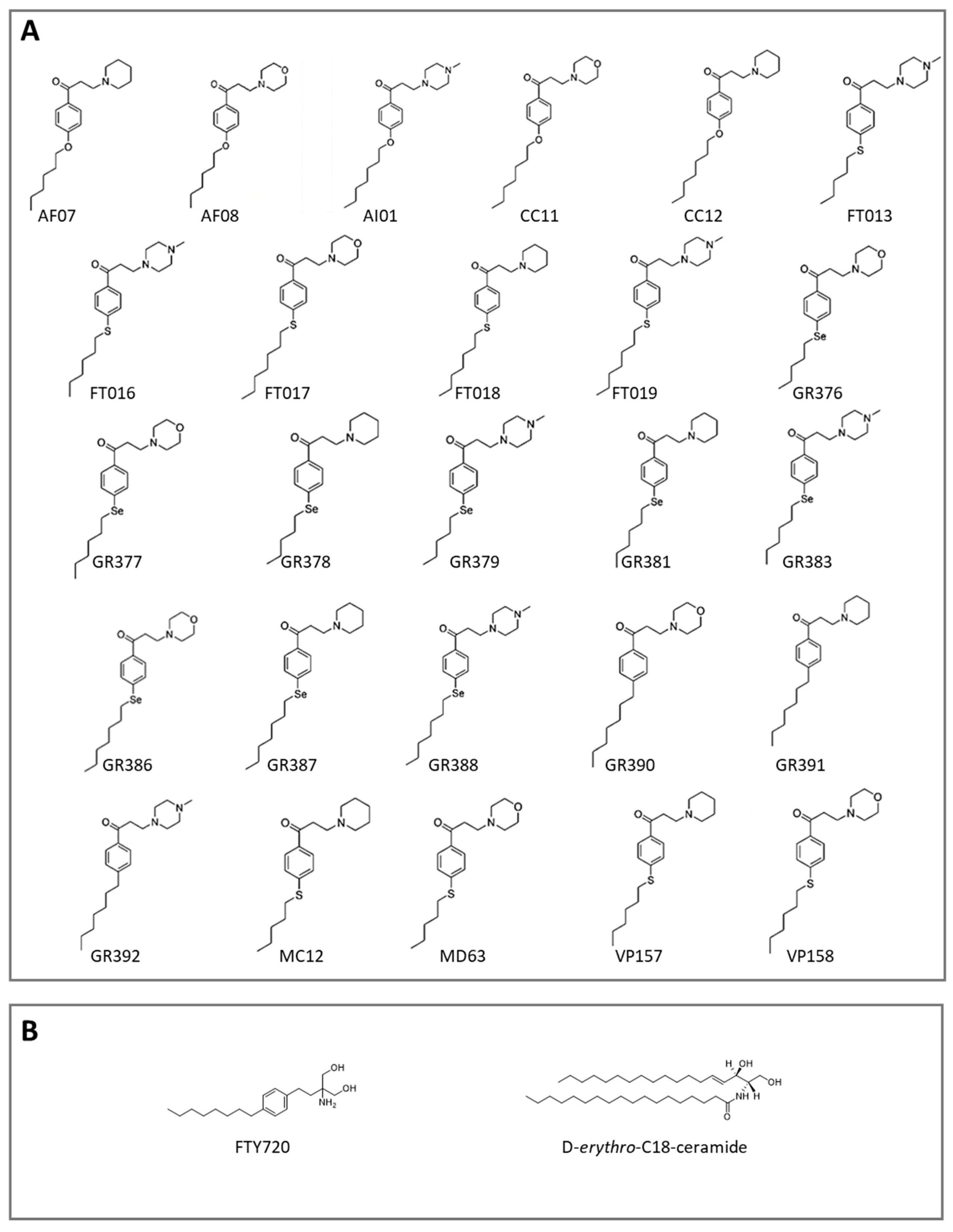
Leveraging the structural framework of ceramide (a physiological PP2A activator) and mechanistic insights from FTY720, we developed a library of 26 newly synthesized compounds (Figure 2A) for structure-based drug discovery [77]. These compounds retained key FTY720-like structural features, including (i) a polar head group and (ii) an aromatic ring to maintain molecular rigidity. Conversely, the phosphorylatable hydroxyl moiety, which mediates immunosuppression and other severe adverse effects such as bradycardia, as previously described, in addition to hindering binding to SET, when phosphorylated, was excluded [56,78]. FTY720 and D-erythro-C18-ceramide (D-e-C18, the natural form of ceramide) were included as positive controls in the computational study (Figure 2B).
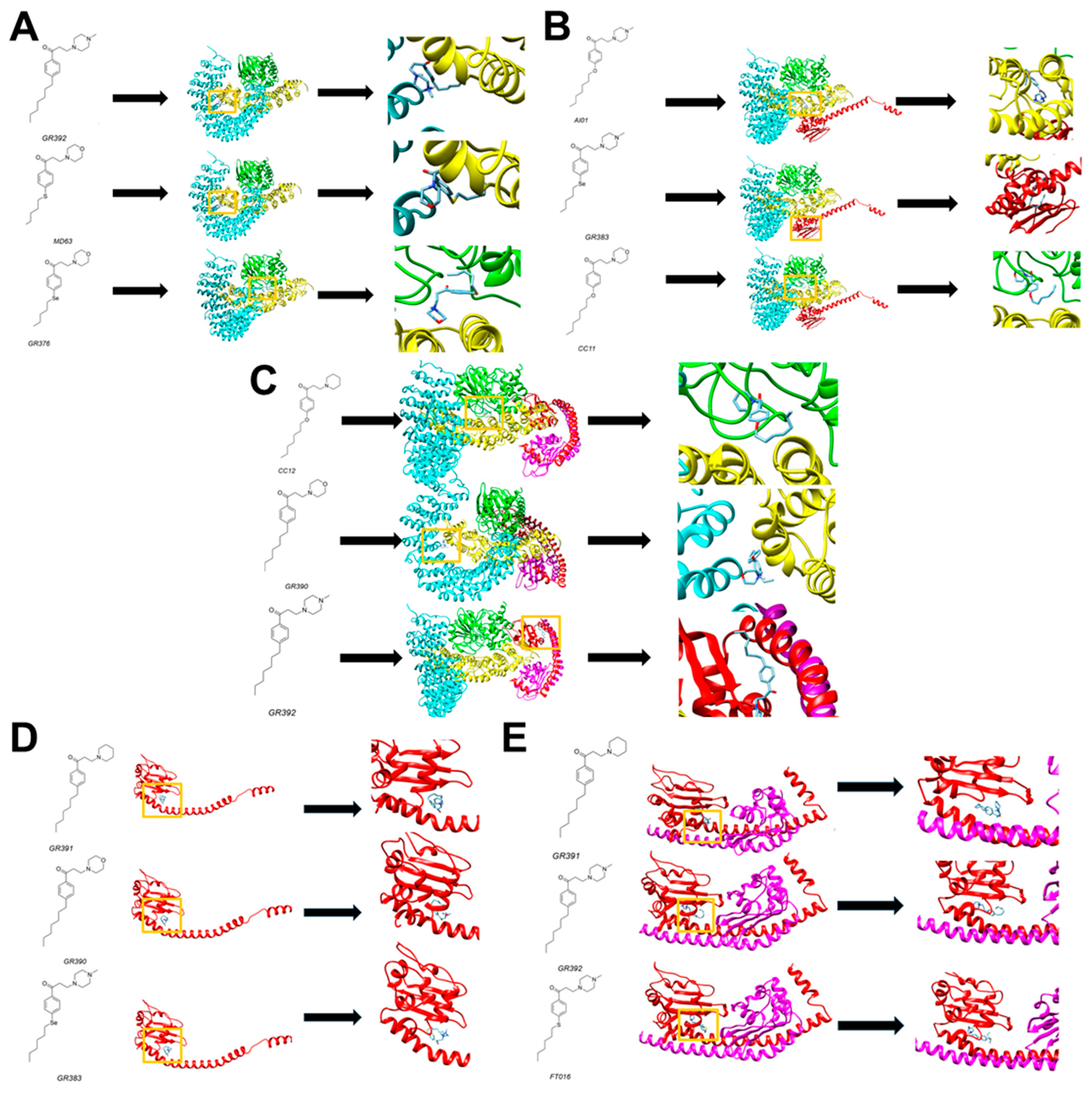
These compounds were analyzed in terms of binding mode and energy scores through blind molecular docking towards the five built receptors. In blind docking, the conformational search is performed within a box that encompasses the whole macromolecular target. While less accurate than a site-specific approach, blind docking is useful in cases when the interaction site is not known, if several different macromolecular targets are considered, and as the preliminary virtual screening approach. Figure 3 illustrates the best poses of the top three ligands for each receptor considered, while Appendix S2 contains all docking scores.
As observed in the different panels of Figure 3, the molecules, in the context of the smaller receptors, tend to cluster in the same position. This is the case of SET monomer and SET dimer models, in which the best scoring molecules target the same site of the proteins in the blind docking experiment (panels D–E). In the case of more complex receptor systems, some differences can be observed. The selenium-containing compounds GR376 and GR383 show different binding sites with respect to other analogs, towards PP2A (A) and PP2A in complex with SET monomer (B). In the case of PP2A in complex with the SET dimer (C), three different sites were observed for the best scoring compounds. In this case, the binding region appears to be influenced by the heteroatom present in the alkyl chain and by the nature of the cyclic amine.

3.2. Biological Studies and ADME Prediction

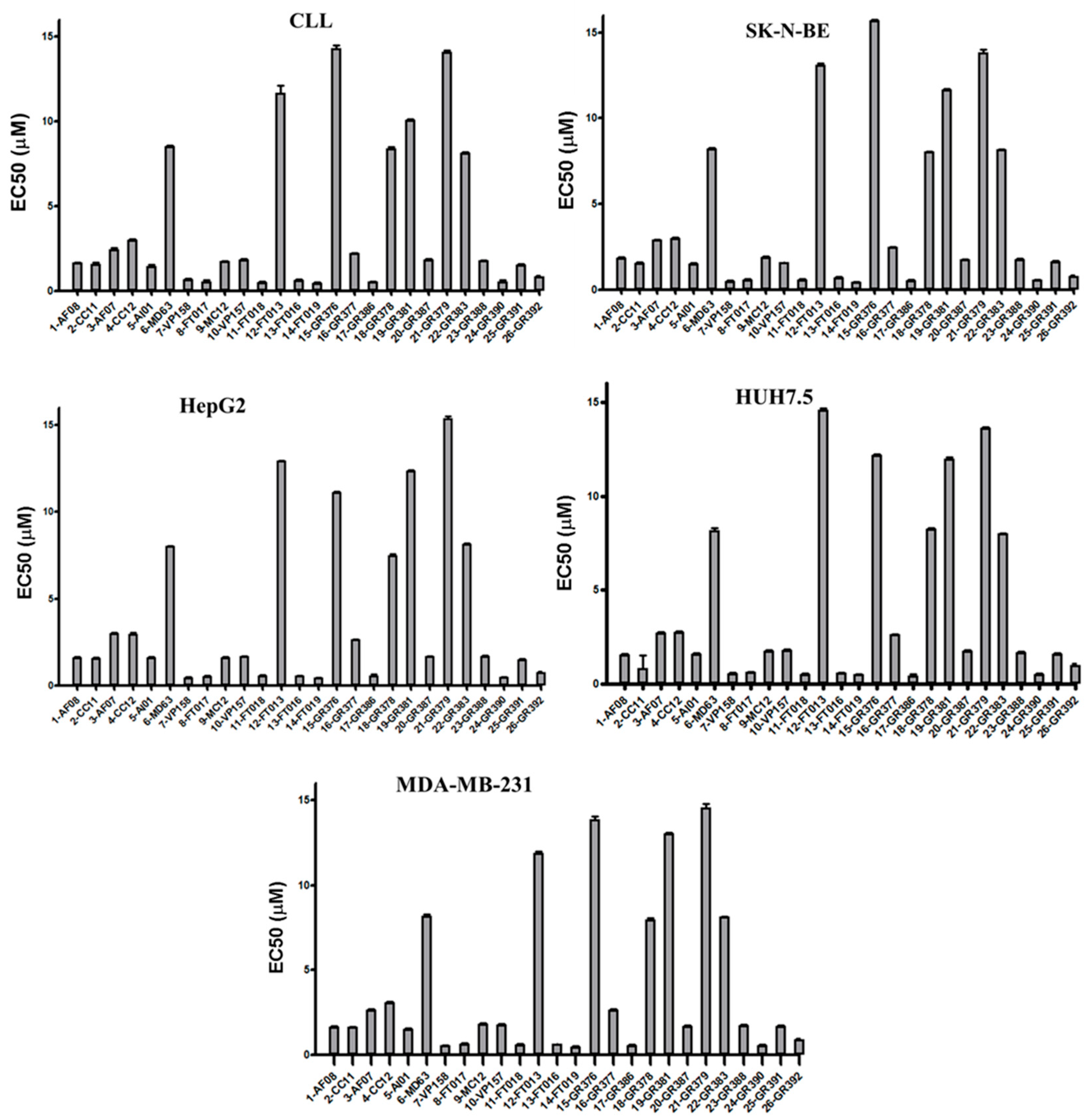
Figure 4 shows the EC50 values obtained from experiments performed in duplicate on CLL cells from four patients, representative of the eight patients whose freshly isolated malignant cells underwent screening with each compound. The data concerning the adherent cell lines, i.e., SK-N-BE, HepG2, Huh7.5, MDA-MB-231, were obtained from experiments performed in quadruplicate and are shown in the same figure. The complete dataset for EC50 calculations is provided in Appendix S3 and the mean values ± SD are detailed in Appendix S4.
Based on comprehensive data analysis and compound stability assessment, GR390 (Compound 24, Appendixes S3 and S4) emerged as one of the most promising candidates for further investigation, comparable to CC11 (Compound 2, Appendixes S3 and S4), which had already shown to elicit CLL apoptosis via SET/PP2A disruption and subsequent PP2A phosphatase reactivation in a previous study [48].
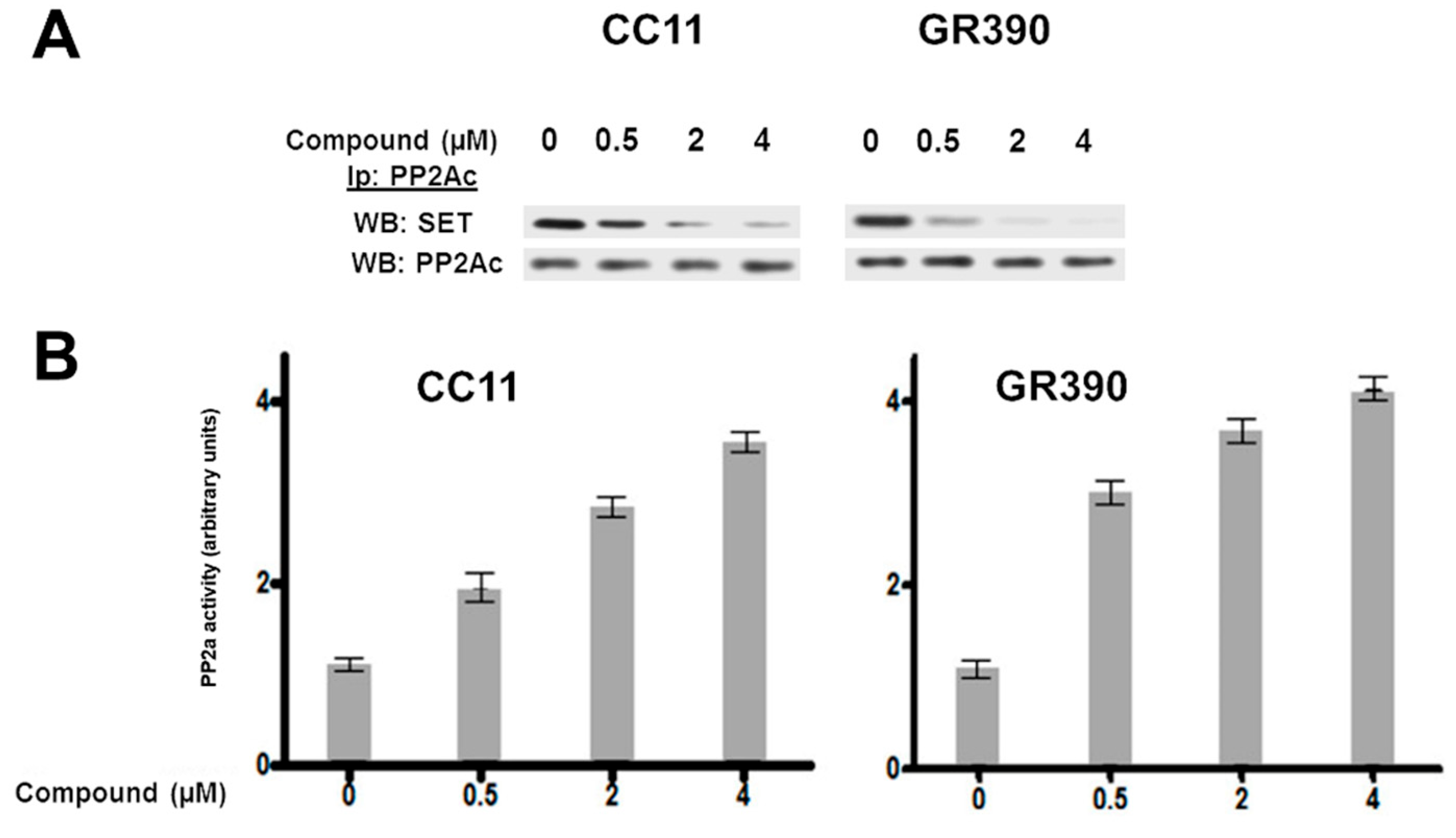
Therefore, we set out to assess whether GR390 was also capable of restoring the activity of the protein phosphatase 2A (PP2A) by removing its endogenous inhibitor SET in CLL cells. To do so, CLL cells were incubated in the presence of increasing concentrations of GR390 or CC11 for 16 h and PP2A was immunoprecipitated with an antibody directed against its catalytic subunit. Western blot analysis was thereafter performed with anti-SET and anti-PP2A antibodies. As shown in Figure 5, GR390 was more effective at displacing the PP2A inhibitor SET than CC11 in this type of cancer (top panels), and subsequently at activating PP2A activity (bottom panel), which is consistent with the more effective apoptotic effect shown when compared to CC11. Interestingly, molecular docking ranked CC11 and GR390 as the highest-affinity ligands for the macromolecular target (Figure 3 and Appendix S2).
In drug development, understanding the factors governing a compound’s concentration and distribution within biological systems is critical. These dynamics are characterized by four fundamental pharmacokinetic parameters: absorption, distribution, metabolism, and excretion (ADME), which collectively determine a drug’s bioavailability and therapeutic efficacy [57].
The in silico ADME evaluation of the lead compound GR390 revealed favorable pharmacokinetic properties. As shown in Appendix S6, the radar plot analysis suggests well predicted bioavailability. Indeed, according to this graph, the molecule has structural features that allow it to fall within the ideal chemical space in terms of lipophilicity, molecular size, polarity, solubility, and saturation. While most parameters fall within ideal ranges, the slightly elevated flexibility index likely attributed to rotational freedom in the aliphatic chain, which is formed by many single bonds. Following Lipinski’s “rule of 5”, the molecule can bear at most one violation to be considered a potential drug to be administered orally. The parameters taken into account by this empirical rule are the following: octanol-water partition coefficient LogP ≤ 5, molecular weight ≤ 500 g/mol, hydrogen bond donors ≤ 5, and hydrogen bond acceptors ≤ 10 [79]. The analysis of the compound GR390 does not report any violation of such parameters; in fact, the molecule presents a LogP of about 4, a molecular weight of 317.47 g/mol, a number of hydrogen bond donors that equal to 0, and a number of hydrogen bond acceptors that equal to 3. Based on the predicted parameters, the compound can undergo passive absorption through the gastrointestinal tract and the blood–brain barrier; it has good lipophilic properties and is moderately soluble in water. GR390 is not a substrate of the main cytochrome P450 isoenzymes (CYP; CYP1A2, CYP2C19, CYP2C9, CYP3A4), which are responsible for the metabolism of many drugs. Thus, this candidate can be defined as a drug-like compound.

3.3. Computational and Biological Data Integration

To assess the relationship between EC50 values and binding energy scores derived from docking studies on five macromolecular models, integrated heatmaps were generated through hierarchical cluster analysis using the Manhattan distance metric, after standardizing the data to normalize the different units of measurement. The experimental EC50 data and computational energy scores reported different units, µM and kcal/mol, respectively, thus standardization was necessary to compare them directly. The standardization process involves calculating the mean for each variable (μ), calculating the standard deviation (SD) for each variable (σ), then each value of variable x is transformed according to the following formula, where z is the standardized value (known also as z-score):
z = x µ σ
Once the data were standardized, the visualization technique using heatmaps in combination with hierarchical cluster analysis was applied to identify possible relationships between EC50 and energy score. Color gradients indicate the relative intensity of binding energy or EC50 response, with blue representing lower values (stronger predicted binding or higher in vitro potency), red representing higher values (weaker binding or lower activity), and yellow corresponds to values near the mean, serving as a visual baseline for interpreting deviations.
Five heatmaps were obtained, one for each cell line (CLL, SK-N-BE, HepG2, HUH7.5, MDA-MB-231). In this case study, it is preferable for z-score values to be below the mean (colored in blue) for two reasons: in docking studies, a lower energy score (indicating a SD value below the mean) generally represents a higher predicted binding affinity between the drug and the biological target. This suggests enhanced target engagement potential, a critical determinant of pharmacological efficacy. As for EC50, a lower value (represented as z-scores and colored of blue) indicates that a lower concentration of the drug is needed to induce 50 percent cell apoptosis. The heatmaps obtained showed recurring patterns of relationship between the EC50 data and the energy scores of the compounds. The five heatmaps are reported in Appendix S7. In particular, the PP2A bound to SET monomer model shows the strongest relationship with the cell lines on which the compounds were evaluated (the model reported in Figure 3B). Thus, this macromolecular arrangement could represent a crucial target for the PP2A activators. Among the compounds, GR390 and CC11, which were highlighted as the most potent compounds in vitro, were among the best candidates (respectively, −7.0 kcal/mol and −7.2 kcal/mol) on that computational model. In addition, drug candidates GR390 and CC11 are part of one of three macro-clusters into which the heatmaps can be divided, where the most promising compounds are found.

4. Conclusions

The aim of this study was to combine computational and in vitro techniques to evaluate a library of 26 APPDs as SET inhibitors, an endogenous PP2A regulator, and to pave the way for a better understanding of the molecular mechanisms in which the two macromolecules are involved to exploit their apoptotic effects. Indeed, inhibiting SET might restore PP2A activity, hindering cancer progression.
In particular, five models resembling the possible scenarios of PP2A-SET interaction were generated through protein–protein docking; subsequently, these models were used as receptors for molecular docking analyses with the 26 APPDs. Then, biological studies highlighted that, out of the 26 compounds, a few of them proved to be significantly effective at inducing apoptosis in the low micromolar and sub-micromolar range in the types of cells used in this study, namely CC11 and GR390, and all were devoid of immunosuppressive activity. Such information may also be relevant to build preliminary structure–activity relationship studies. Indeed, the results demonstrate that the presence of the substituted aromatic ring is tolerated, such as that of the cyclic amine that can be of different nature. Eventually, it must be noted that the most promising compounds bear a 7-carbon atoms alkyl chain.
Moreover, statistical tools revealed consistent associations between computational predictions and experimental outcomes, pointing to one macromolecular assembly as the most plausible target for the compounds. Eventually, GR390 and CC11 were identified among the more promising compounds. Further, ADME parameters of GR390 have been predicted, highlighting a good drug-likeness.
On the other hand, further investigations are warranted to assess their safety and potential to be developed as anticancer drugs.

5. Patents

Invention: “1-Phenylpropanone compounds and use thereof”. Priority Data: 102016000098338, 30 September 2016. International Application Number: PCT/IB2017/056010, 29 September 2017). Internation Publication Number: WO 2018/060947 A1. Inventors: Giuseppe Zagotto, Giovanni Ribaudo, Anna Maria Brunati, Mario Angelo Primo Pagano, Elena Tibaldi, Livio Trentin. Applicant: University of Padova.

Supplementary Materials

The following supporting information can be downloaded at: https://www.mdpi.com/article/10.3390/macromol5030043/s1, Appendix S1. Biological and clinical characteristics of the CLL patients; Appendix S2. Docking scores; Appendix S3. List of APPDs and apoptotic effect thereof; Appendix S4. EC50 values as processed from the data reported in the patent for each compound; Appendix S5. Effect of CC11 and GR390 on the activity of PP2A on CLL cells; Appendix S6. SwissADME prediction; Appendix S7. Heatmaps.

Author Contributions

Conceptualization, G.R.; methodology, G.R. and M.A.P.; software, M.A., M.G. and M.V.; investigation, M.A.P., M.A., A.V., F.F. and M.G.; data curation, G.R., M.A.P., M.V., L.T., A.M.B. and A.G.; writing—original draft preparation, G.R. and M.A.P.; writing—review and editing, A.M.B., L.T. and A.G.; supervision, G.R., M.A.P., A.M.B. and A.G. All authors have read and agreed to the published version of the manuscript.

Funding

The research was supported by University of Brescia and by funds from Associazione Italiana per la Ricerca sul Cancro (AIRC) to L.T. (IG-25024), Associazione italiana contro le leucemie-linfomi e mieloma (AIL), ONLUS ‘Ricerca per Credere nella Vita’ (RCV) odv, and Progetti di Rilevanza Nazionale PRIN PNRR (P2022PSMX4) and Supporting Talent in ReSearch@University of Padua–Starting Grant Be_CL3VER to A.V.

Institutional Review Board Statement

The study was conducted in accordance with the Declaration of Helsinki, and approved by the local ethics committee (Regione Veneto on Chronic Lymphocytic Leukemia, number 3259/AO/14; 18 September 2014).

Informed Consent Statement

Leukemic B cells were obtained from 38 untreated CLL patients, who were enrolled by the Hematology Division of Azienda Ospedale—Università di Padova and met standard diagnostic criteria (Appendix S1). All participants provided informed written consent in accordance with the Declaration of Helsinki.

Data Availability Statement

Data available within the article and Supplementary Materials.

Acknowledgments

G.R. and A.G. are grateful to Valentina Premoli for her experimental support. The authors would also like to thank Giuseppe Zagotto.

Conflicts of Interest

The authors declare no conflicts of interest.

References

  1. Sangodkar, J.; Farrington, C.C.; McClinch, K.; Galsky, M.D.; Kastrinsky, D.B.; Narla, G. All Roads Lead to PP 2A: Exploiting the Therapeutic Potential of This Phosphatase. FEBS J. 2016, 283, 1004–1024. [Google Scholar] [CrossRef] [PubMed]
  2. Orgad, S.; Brewis, N.D.; Alphey, L.; Axton, J.M.; Dudai, Y.; Cohen, P.T.W. The Structure of Protein Phosphatase 2A Is as Highly Conserved as That of Protein Phosphatase I. FEBS Lett. 1990, 275, 44–48. [Google Scholar] [CrossRef]
  3. Westermarck, J.; Hahn, W.C. Multiple Pathways Regulated by the Tumor Suppressor PP2A in Transformation. Trends Mol. Med. 2008, 14, 152–160. [Google Scholar] [CrossRef]
  4. Shi, Y. Serine/Threonine Phosphatases: Mechanism through Structure. Cell 2009, 139, 468–484. [Google Scholar] [CrossRef]
  5. Eichhorn, P.J.A.; Creyghton, M.P.; Bernards, R. Protein Phosphatase 2A Regulatory Subunits and Cancer. Biochim. Biophys. Acta BBA-Rev. Cancer 2009, 1795, 1–15. [Google Scholar] [CrossRef]
  6. Janssens, V.; Goris, J. Protein Phosphatase 2A: A Highly Regulated Family of Serine/Threonine Phosphatases Implicated in Cell Growth and Signalling. Biochem. J. 2001, 353, 417–439. [Google Scholar] [CrossRef]
  7. Seshacharyulu, P.; Pandey, P.; Datta, K.; Batra, S.K. Phosphatase: PP2A Structural Importance, Regulation and Its Aberrant Expression in Cancer. Cancer Lett. 2013, 335, 9–18. [Google Scholar] [CrossRef]
  8. Wlodarchak, N.; Xing, Y. PP2A as a Master Regulator of the Cell Cycle. Crit. Rev. Biochem. Mol. Biol. 2016, 51, 162–184. [Google Scholar] [CrossRef] [PubMed]
  9. Jong, C.J.; Merrill, R.A.; Wilkerson, E.M.; Herring, L.E.; Graves, L.M.; Strack, S. Reduction of Protein Phosphatase 2A (PP2A) Complexity Reveals Cellular Functions and Dephosphorylation Motifs of the PP2A/B′δ Holoenzyme. J. Biol. Chem. 2020, 295, 5654–5668. [Google Scholar] [CrossRef]
  10. Ciccone, M.; Calin, G.A.; Perrotti, D. From the Biology of PP2A to the PADs for Therapy of Hematologic Malignancies. Front. Oncol. 2015, 5, 21. [Google Scholar] [CrossRef] [PubMed]
  11. Perrotti, D.; Neviani, P. Protein Phosphatase 2A: A Target for Anticancer Therapy. Lancet Oncol. 2013, 14, e229–e238. [Google Scholar] [CrossRef]
  12. Haesen, D.; Sents, W.; Lemaire, K.; Hoorne, Y.; Janssens, V. The Basic Biology of PP2A in Hematologic Cells and Malignancies. Front. Oncol. 2014, 4, 347. [Google Scholar] [CrossRef]
  13. Cho, U.S.; Xu, W. Crystal Structure of a Protein Phosphatase 2A Heterotrimeric Holoenzyme. Nature 2007, 445, 53–57. [Google Scholar] [CrossRef]
  14. Xing, Y.; Xu, Y.; Chen, Y.; Jeffrey, P.D.; Chao, Y.; Lin, Z.; Li, Z.; Strack, S.; Stock, J.B.; Shi, Y. Structure of Protein Phosphatase 2A Core Enzyme Bound to Tumor-Inducing Toxins. Cell 2006, 127, 341–353. [Google Scholar] [CrossRef] [PubMed]
  15. Brewer, A.; Sathe, G.; Pflug, B.E.; Clarke, R.G.; Macartney, T.J.; Sapkota, G.P. Mapping the Substrate Landscape of Protein Phosphatase 2A Catalytic Subunit PPP2CA. iScience 2024, 27, 109302. [Google Scholar] [CrossRef]
  16. Ramaswamy, K.; Spitzer, B.; Kentsis, A. Therapeutic Re-Activation of Protein Phosphatase 2A in Acute Myeloid Leukemia. Front. Oncol. 2015, 5, 16. [Google Scholar] [CrossRef]
  17. Fowle, H.; Zhao, Z.; Graña, X. PP2A Holoenzymes, Substrate Specificity Driving Cellular Functions and Deregulation in Cancer. In Advances in Cancer Research; Elsevier: Amsterdam, The Netherlands, 2019; Volume 144, pp. 55–93. ISBN 978-0-12-817155-4. [Google Scholar]
  18. Janssens, V.; Jordens, J.; Stevens, I.; Van Hoof, C.; Martens, E.; De Smedt, H.; Engelborghs, Y.; Waelkens, E.; Goris, J. Identification and Functional Analysis of Two Ca2+-Binding EF-Hand Motifs in the B“/PR72 Subunit of Protein Phosphatase 2A. J. Biol. Chem. 2003, 278, 10697–10706. [Google Scholar] [CrossRef] [PubMed]
  19. Janssens, V.; Longin, S.; Goris, J. PP2A Holoenzyme Assembly: In Cauda Venenum (the Sting Is in the Tail). Trends Biochem. Sci. 2008, 33, 113–121. [Google Scholar] [CrossRef] [PubMed]
  20. Chen, C.; Shi, Z.; Zhang, W.; Chen, M.; He, F.; Zhang, Z.; Wang, Y.; Feng, M.; Wang, W.; Zhao, Y.; et al. Striatins Contain a Noncanonical Coiled Coil That Binds Protein Phosphatase 2A A Subunit to Form a 2:2 Heterotetrameric Core of Striatin-Interacting Phosphatase and Kinase (STRIPAK) Complex. J. Biol. Chem. 2014, 289, 9651–9661. [Google Scholar] [CrossRef]
  21. Janssens, V.; Goris, J.; Van Hoof, C. PP2A: The Expected Tumor Suppressor. Curr. Opin. Genet. Dev. 2005, 15, 34–41. [Google Scholar] [CrossRef]
  22. Ruediger, R.; Pham, H.T.; Walter, G. Disruption of Protein Phosphatase 2A Subunit Interaction in Human Cancers with Mutations in the Aα Subunit Gene. Oncogene 2001, 20, 10–15. [Google Scholar] [CrossRef] [PubMed]
  23. Leonard, D.; Huang, W.; Izadmehr, S.; O’Connor, C.M.; Wiredja, D.D.; Wang, Z.; Zaware, N.; Chen, Y.; Schlatzer, D.M.; Kiselar, J.; et al. Selective PP2A Enhancement through Biased Heterotrimer Stabilization. Cell 2020, 181, 688–701.e16. [Google Scholar] [CrossRef] [PubMed]
  24. Wang, J.; Okkeri, J.; Pavic, K.; Wang, Z.; Kauko, O.; Halonen, T.; Sarek, G.; Ojala, P.M.; Rao, Z.; Xu, W.; et al. Oncoprotein CIP 2A Is Stabilized via Interaction with Tumor Suppressor PP 2A/B56. EMBO Rep. 2017, 18, 437–450. [Google Scholar] [CrossRef]
  25. Dacol, E.C.; Wang, S.; Chen, Y.; Lepique, A.P. The Interaction of SET and Protein Phosphatase 2A as Target for Cancer Therapy. Biochim. Biophys. Acta BBA-Rev. Cancer 2021, 1876, 188578. [Google Scholar] [CrossRef]
  26. Arnold, H.K.; Sears, R.C. A Tumor Suppressor Role for PP2A-B56α Through Negative Regulation of c-Myc and Other Key Oncoproteins. Cancer Metastasis Rev. 2008, 27, 147–158. [Google Scholar] [CrossRef]
  27. Ruvolo, P.P.; Qui, Y.H.; Coombes, K.R.; Zhang, N.; Ruvolo, V.R.; Borthakur, G.; Konopleva, M.; Andreeff, M.; Kornblau, S.M. Low Expression of PP2A Regulatory Subunit B55α Is Associated with T308 Phosphorylation of AKT and Shorter Complete Remission Duration in Acute Myeloid Leukemia Patients. Leukemia 2011, 25, 1711–1717. [Google Scholar] [CrossRef]
  28. Silverstein, A.M.; Barrow, C.A.; Davis, A.J.; Mumby, M.C. Actions of PP2A on the MAP Kinase Pathway and Apoptosis Are Mediated by Distinct Regulatory Subunits. Proc. Natl. Acad. Sci. USA 2002, 99, 4221–4226. [Google Scholar] [CrossRef] [PubMed]
  29. Kauko, O.; Laajala, T.D.; Jumppanen, M.; Hintsanen, P.; Suni, V.; Haapaniemi, P.; Corthals, G.; Aittokallio, T.; Westermarck, J.; Imanishi, S.Y. Label-Free Quantitative Phosphoproteomics with Novel Pairwise Abundance Normalization Reveals Synergistic RAS and CIP2A Signaling. Sci. Rep. 2015, 5, 13099. [Google Scholar] [CrossRef]
  30. Ruvolo, P.P. The Broken “Off” Switch in Cancer Signaling: PP2A as a Regulator of Tumorigenesis, Drug Resistance, and Immune Surveillance. BBA Clin. 2016, 6, 87–99. [Google Scholar] [CrossRef]
  31. Kurimchak, A.; Graña, X. PP2A Holoenzymes Negatively and Positively Regulate Cell Cycle Progression by Dephosphorylating Pocket Proteins and Multiple CDK Substrates. Gene 2012, 499, 1–7. [Google Scholar] [CrossRef]
  32. Qian, W.; Shi, J.; Yin, X.; Iqbal, K.; Grundke-Iqbal, I.; Gong, C.-X.; Liu, F. PP2A Regulates Tau Phosphorylation Directly and Also Indirectly via Activating GSK-3β. J. Alzheimers Dis. 2010, 19, 1221–1229. [Google Scholar] [CrossRef]
  33. Shentu, Y.-P.; Huo, Y.; Feng, X.-L.; Gilbert, J.; Zhang, Q.; Liuyang, Z.-Y.; Wang, X.-L.; Wang, G.; Zhou, H.; Wang, X.-C.; et al. CIP2A Causes Tau/APP Phosphorylation, Synaptopathy, and Memory Deficits in Alzheimer’s Disease. Cell Rep. 2018, 24, 713–723. [Google Scholar] [CrossRef]
  34. Zhang, Q.; Xia, Y.; Wang, Y.; Shentu, Y.; Zeng, K.; Mahaman, Y.A.R.; Huang, F.; Wu, M.; Ke, D.; Wang, Q.; et al. CK2 Phosphorylating I2PP2A/SET Mediates Tau Pathology and Cognitive Impairment. Front. Mol. Neurosci. 2018, 11, 146. [Google Scholar] [CrossRef] [PubMed]
  35. Clark, A.R.; Ohlmeyer, M. Protein Phosphatase 2A as a Therapeutic Target in Inflammation and Neurodegeneration. Pharmacol. Ther. 2019, 201, 181–201. [Google Scholar] [CrossRef] [PubMed]
  36. Sharabi, A.; Kasper, I.R.; Tsokos, G.C. The Serine/Threonine Protein Phosphatase 2A Controls Autoimmunity. Clin. Immunol. 2018, 186, 38–42. [Google Scholar] [CrossRef]
  37. O’Connor, C.M.; Perl, A.; Leonard, D.; Sangodkar, J.; Narla, G. Therapeutic Targeting of PP2A. Int. J. Biochem. Cell Biol. 2018, 96, 182–193. [Google Scholar] [CrossRef] [PubMed]
  38. Gutierrez, A.; Pan, L.; Groen, R.W.J.; Baleydier, F.; Kentsis, A.; Marineau, J.; Grebliunaite, R.; Kozakewich, E.; Reed, C.; Pflumio, F.; et al. Phenothiazines Induce PP2A-Mediated Apoptosis in T Cell Acute Lymphoblastic Leukemia. J. Clin. Investig. 2014, 124, 644–655. [Google Scholar] [CrossRef]
  39. Kastrinsky, D.B.; Sangodkar, J.; Zaware, N.; Izadmehr, S.; Dhawan, N.S.; Narla, G.; Ohlmeyer, M. Reengineered Tricyclic Anti-Cancer Agents. Bioorg. Med. Chem. 2015, 23, 6528–6534. [Google Scholar] [CrossRef]
  40. Westermarck, J.; Neel, B.G. Piecing Together a Broken Tumor Suppressor Phosphatase for Cancer Therapy. Cell 2020, 181, 514–517. [Google Scholar] [CrossRef]
  41. Yang-Schulz, A.; Zacharopoulou, M.; Yilmaz, S.Z.; Banerjee, A.; Saha, S.; Nietlispach, D.; Ohlmeyer, M.; Gur, M.; Itzhaki, L.S.; Bahar, I.; et al. Direct Observation of Small Molecule Activator Binding to Single PR65 Protein. Npj Biosens. 2025, 2, 2. [Google Scholar] [CrossRef]
  42. Bownes, L.V.; Julson, J.R.; Quinn, C.H.; Hutchins, S.C.; Erwin, M.H.; Markert, H.R.; Stewart, J.E.; Mroczek-Musulman, E.; Aye, J.; Yoon, K.J.; et al. The Effects of Protein Phosphatase 2A Activation with Novel Tricyclic Sulfonamides on Hepatoblastoma. J. Pediatr. Surg. 2023, 58, 1145–1154. [Google Scholar] [CrossRef]
  43. Von Lindern, M.; Van Baal, S.; Wiegant, J.; Raap, A.; Hagemeijer, A.; Grosveld, G. Can, a Putative Oncogene Associated with Myeloid Leukemogenesis, May Be Activated by Fusion of Its 3‣ Half to Different Genes: Characterization of the Set. Gene. Mol. Cell. Biol. 1992, 12, 3346–3355. [Google Scholar] [CrossRef]
  44. Nagata, K.; Saito, S.; Okuwaki, M.; Kawase, H.; Furuya, A.; Kusano, A.; Hanai, N.; Okuda, A.; Kikuchi, A. Cellular Localization and Expression of Template-Activating Factor I in Different Cell Types. Exp. Cell Res. 1998, 240, 274–281. [Google Scholar] [CrossRef]
  45. Adachi, Y.; Pavlakis, G.N.; Copeland, T.D. Identification and Characterization of SET, a Nuclear Phosphoprotein Encoded by the Translocation Break Point in Acute Undifferentiated Leukemia. J. Biol. Chem. 1994, 269, 2258–2262. [Google Scholar] [CrossRef]
  46. Yu, G.; Yan, T.; Feng, Y.; Liu, X.; Xia, Y.; Luo, H.; Wang, J.-Z.; Wang, X. Ser9 Phosphorylation Causes Cytoplasmic Detention of I2PP2A/SET in Alzheimer Disease. Neurobiol. Aging 2013, 34, 1748–1758. [Google Scholar] [CrossRef]
  47. Saito, S.; Miyaji-Yamaguchi, M.; Shimoyama, T.; Nagata, K. Functional Domains of Template-Activating Factor-I as a Protein Phosphatase 2A Inhibitor. Biochem. Biophys. Res. Commun. 1999, 259, 471–475. [Google Scholar] [CrossRef] [PubMed]
  48. Muto, S.; Senda, M.; Akai, Y.; Sato, L.; Suzuki, T.; Nagai, R.; Senda, T.; Horikoshi, M. Relationship between the Structure of SET/TAF-Iβ/INHAT and Its Histone Chaperone Activity. Proc. Natl. Acad. Sci. USA 2007, 104, 4285–4290. [Google Scholar] [CrossRef] [PubMed]
  49. Liu, H.; Gu, Y.; Wang, H.; Yin, J.; Zheng, G.; Zhang, Z.; Lu, M.; Wang, C.; He, Z. Overexpression of PP2A Inhibitor SET Oncoprotein Is Associated with Tumor Progression and Poor Prognosis in Human Non-Small Cell Lung Cancer. Oncotarget 2015, 6, 14913–14925. [Google Scholar] [CrossRef] [PubMed]
  50. Cristobal, I.; Garcia-Orti, L.; Cirauqui, C.; Cortes-Lavaud, X.; Garcia-Sanchez, M.A.; Calasanz, M.J.; Odero, M.D. Overexpression of SET Is a Recurrent Event Associated with Poor Outcome and Contributes to Protein Phosphatase 2A Inhibition in Acute Myeloid Leukemia. Haematologica 2012, 97, 543–550. [Google Scholar] [CrossRef]
  51. Kappos, L.; Antel, J.; Comi, G.; Montalban, X.; O’Connor, P.; Polman, C.H.; Haas, T.; Korn, A.A.; Karlsson, G.; Radue, E.W. Oral Fingolimod (FTY720) for Relapsing Multiple Sclerosis. N. Engl. J. Med. 2006, 355, 1124–1140. [Google Scholar] [CrossRef]
  52. Horga, A.; Montalban, X. FTY720 (Fingolimod) for Relapsing Multiple Sclerosis. Expert. Rev. Neurother. 2008, 8, 699–714. [Google Scholar] [CrossRef]
  53. Neviani, P.; Santhanam, R.; Oaks, J.J.; Eiring, A.M.; Notari, M.; Blaser, B.W.; Liu, S.; Trotta, R.; Muthusamy, N.; Gambacorti-Passerini, C.; et al. FTY720, a New Alternative for Treating Blast Crisis Chronic Myelogenous Leukemia and Philadelphia Chromosome–Positive Acute Lymphocytic Leukemia. J. Clin. Investig. 2007, 117, 2408–2421. [Google Scholar] [CrossRef]
  54. Roberts, K.G.; Smith, A.M.; McDougall, F.; Carpenter, H.; Horan, M.; Neviani, P.; Powell, J.A.; Thomas, D.; Guthridge, M.A.; Perrotti, D.; et al. Essential Requirement for PP2A Inhibition by the Oncogenic Receptor C-KIT Suggests PP2A Reactivation as a Strategy to Treat c-KIT+ Cancers. Cancer Res. 2010, 70, 5438–5447. [Google Scholar] [CrossRef]
  55. Zhang, L.; Wang, H.-D.; Ji, X.-J.; Cong, Z.-X.; Zhu, J.-H.; Zhou, Y. FTY720 for Cancer Therapy (Review). Oncol. Rep. 2013, 30, 2571–2578. [Google Scholar] [CrossRef]
  56. White, C.; Alshaker, H.; Cooper, C.; Winkler, M.; Pchejetski, D. The Emerging Role of FTY720 (Fingolimod) in Cancer Treatment. Oncotarget 2016, 7, 23106–23127. [Google Scholar] [CrossRef] [PubMed]
  57. Chen, B.; Roy, S.G.; McMonigle, R.J.; Keebaugh, A.; McCracken, A.N.; Selwan, E.; Fransson, R.; Fallegger, D.; Huwiler, A.; Kleinman, M.T.; et al. Azacyclic FTY720 Analogues That Limit Nutrient Transporter Expression but Lack S1P Receptor Activity and Negative Chronotropic Effects Offer a Novel and Effective Strategy to Kill Cancer Cells in Vivo. ACS Chem. Biol. 2016, 11, 409–414. [Google Scholar] [CrossRef] [PubMed]
  58. Pagano, M.A.; Tibaldi, E.; Molino, P.; Frezzato, F.; Trimarco, V.; Facco, M.; Zagotto, G.; Ribaudo, G.; Leanza, L.; Peruzzo, R.; et al. Mitochondrial Apoptosis Is Induced by Alkoxy Phenyl-1-Propanone Derivatives Through PP2A-Mediated Dephosphorylation of Bad and Foxo3A in CLL. Leukemia 2019, 33, 1148–1160. [Google Scholar] [CrossRef] [PubMed]
  59. Zonta, F.; Pagano, M.A.; Trentin, L.; Tibaldi, E.; Frezzato, F.; Trimarco, V.; Facco, M.; Zagotto, G.; Pavan, V.; Ribaudo, G.; et al. Lyn Sustains Oncogenic Signaling in Chronic Lymphocytic Leukemia by Strengthening SET-Mediated Inhibition of PP2A. Blood 2015, 125, 3747–3755. [Google Scholar] [CrossRef]
  60. Arnold, L.A.; Kosinski, A.; Estébanez-Perpiñá, E.; Guy, R.K. Inhibitors of the Interaction of a Thyroid Hormone Receptor and Coactivators: Preliminary Structure−Activity Relationships. J. Med. Chem. 2007, 50, 5269–5280. [Google Scholar] [CrossRef]
  61. Ju, D.; Wang, X.; Xie, Y. Dyclonine and Alverine Citrate Enhance the Cytotoxic Effects of Proteasome Inhibitor MG132 on Breast Cancer Cells. Int. J. Mol. Med. 2009, 23, 205–209. [Google Scholar]
  62. Hung, M.-H.; Chen, Y.-L.; Chu, P.-Y.; Shih, C.-T.; Yu, H.-C.; Tai, W.-T.; Shiau, C.-W.; Chen, K.-F. Upregulation of the Oncoprotein SET Determines Poor Clinical Outcomes in Hepatocellular Carcinoma and Shows Therapeutic Potential. Oncogene 2016, 35, 4891–4902. [Google Scholar] [CrossRef]
  63. Leopoldino, A.M.; Squarize, C.H.; Garcia, C.B.; Almeida, L.O.; Pestana, C.R.; Sobral, L.M.; Uyemura, S.A.; Tajara, E.H.; Silvio Gutkind, J.; Curti, C. SET Protein Accumulates in HNSCC and Contributes to Cell Survival: Antioxidant Defense, Akt Phosphorylation and AVOs Acidification. Oral Oncol. 2012, 48, 1106–1113. [Google Scholar] [CrossRef]
  64. Goswami, S.; Chiang, C.-L.; Zapolnik, K.; Nunes, J.; Ventura, A.; Mo, X.; Xie, Z.; Lee, L.J.; Baskar, S.; Rader, C.; et al. ROR1 Targeted Immunoliposomal Delivery of OSU-2S Shows Selective Cytotoxicity in t(1;19)(Q23;P13) Translocated B-Cell Acute Lymphoblastic Leukemia. Leuk. Res. 2022, 118, 106872. [Google Scholar] [CrossRef] [PubMed]
  65. Kim, S.B.; Oh, Y.S.; Kim, K.J.; Cho, S.W.; Park, S.K.; Baek, D.J.; Park, E.-Y. Synthesis of PP2A-Activating PF-543 Derivatives and Investigation of Their Inhibitory Effects on Pancreatic Cancer Cells. Molecules 2022, 27, 3346. [Google Scholar] [CrossRef] [PubMed]
  66. Cho, U.S.; Morrone, S.; Sablina, A.A.; Arroyo, J.D.; Hahn, W.C.; Xu, W. Structural Basis of PP2A Inhibition by Small t Antigen. PLoS Biol. 2007, 5, e202. [Google Scholar] [CrossRef] [PubMed]
  67. Scorsato, V.; Lima, T.B.; Righetto, G.L.; Zanchin, N.I.T.; Brandão-Neto, J.; Sandy, J.; Pereira, H.D.; Ferrari, Á.J.R.; Gozzo, F.C.; Smetana, J.H.C.; et al. Crystal Structure of the Human Tip41 Orthologue, TIPRL, Reveals a Novel Fold and a Binding Site for the PP2Ac C-Terminus. Sci. Rep. 2016, 6, 30813. [Google Scholar] [CrossRef]
  68. Pettersen, E.F.; Goddard, T.D.; Huang, C.C.; Couch, G.S.; Greenblatt, D.M.; Meng, E.C.; Ferrin, T.E. UCSF Chimera—A Visualization System for Exploratory Research and Analysis. J. Comput. Chem. 2004, 25, 1605–1612. [Google Scholar] [CrossRef]
  69. Hanwell, M.D.; Curtis, D.E.; Lonie, D.C.; Vandermeersch, T.; Zurek, E.; Hutchison, G.R. Avogadro: An Advanced Semantic Chemical Editor, Visualization, and Analysis Platform. J. Cheminform. 2012, 4, 17. [Google Scholar] [CrossRef]
  70. Trott, O.; Olson, A.J. AutoDock Vina: Improving the Speed and Accuracy of Docking with a New Scoring Function, Efficient Optimization, and Multithreading. J. Comput. Chem. 2010, 31, 455–461. [Google Scholar] [CrossRef]
  71. Daina, A.; Michielin, O.; Zoete, V. SwissADME: A Free Web Tool to Evaluate Pharmacokinetics, Drug-Likeness and Medicinal Chemistry Friendliness of Small Molecules. Sci. Rep. 2017, 7, 42717. [Google Scholar] [CrossRef]
  72. Zonta, F.; Borgo, C.; Quezada Meza, C.P.; Masgras, I.; Rasola, A.; Salvi, M.; Pinna, L.A.; Ruzzene, M. Contribution of the CK2 Catalytic Isoforms α and α’ to the Glycolytic Phenotype of Tumor Cells. Cells 2021, 10, 181. [Google Scholar] [CrossRef] [PubMed]
  73. Masola, V.; Carraro, A.; Zaza, G.; Bellin, G.; Montin, U.; Violi, P.; Lupo, A.; Tedeschi, U. Epithelial to Mesenchymal Transition in the Liver Field: The Double Face of Everolimus in Vitro. BMC Gastroenterol. 2015, 15, 118. [Google Scholar] [CrossRef]
  74. Kawamoto, M.; Yamaji, T.; Saito, K.; Shirasago, Y.; Satomura, K.; Endo, T.; Fukasawa, M.; Hanada, K.; Osada, N. Identification of Characteristic Genomic Markers in Human Hepatoma HuH-7 and Huh7.5.1-8 Cell Lines. Front. Genet. 2020, 11, 546106. [Google Scholar] [CrossRef]
  75. Eberle, C.-A.; Zayas, M.; Stukalov, A.; Pichlmair, A.; Alvisi, G.; Müller, A.C.; Bennett, K.L.; Bartenschlager, R.; Superti-Furga, G. The Lysine Methyltransferase SMYD3 Interacts with Hepatitis C Virus NS5A and Is a Negative Regulator of Viral Particle Production. Virology 2014, 462–463, 34–41. [Google Scholar] [CrossRef]
  76. Bianchi, F.; Sasso, M.; Turdo, F.; Beretta, G.L.; Casalini, P.; Ghirelli, C.; Sfondrini, L.; Ménard, S.; Tagliabue, E.; Campiglio, M. Fhit Nuclear Import Following EGF Stimulation Sustains Proliferation of Breast Cancer Cells. J. Cell. Physiol. 2015, 230, 2661–2670. [Google Scholar] [CrossRef]
  77. Zagotto, G.; Ribaudo, G.; Brunati, A.M.; Pagano, M.A.P.; Tibaldi, E.; Trentin, L. 1-Phenylpropanone Compounds and Use Thereof. WO2018060947A1, 4 May 2018. [Google Scholar]
  78. Saddoughi, S.A.; Gencer, S.; Peterson, Y.K.; Ward, K.E.; Mukhopadhyay, A.; Oaks, J.; Bielawski, J.; Szulc, Z.M.; Thomas, R.J.; Selvam, S.P.; et al. Sphingosine Analogue Drug FTY720 Targets I2PP2A/SET and Mediates Lung Tumour Suppression via Activation of PP2A-RIPK1-dependent Necroptosis. EMBO Mol. Med. 2013, 5, 105–121. [Google Scholar] [CrossRef] [PubMed]
  79. Lipinski, C.A. Drug-like Properties and the Causes of Poor Solubility and Poor Permeability. J. Pharmacol. Toxicol. Methods 2000, 44, 235–249. [Google Scholar] [CrossRef] [PubMed]
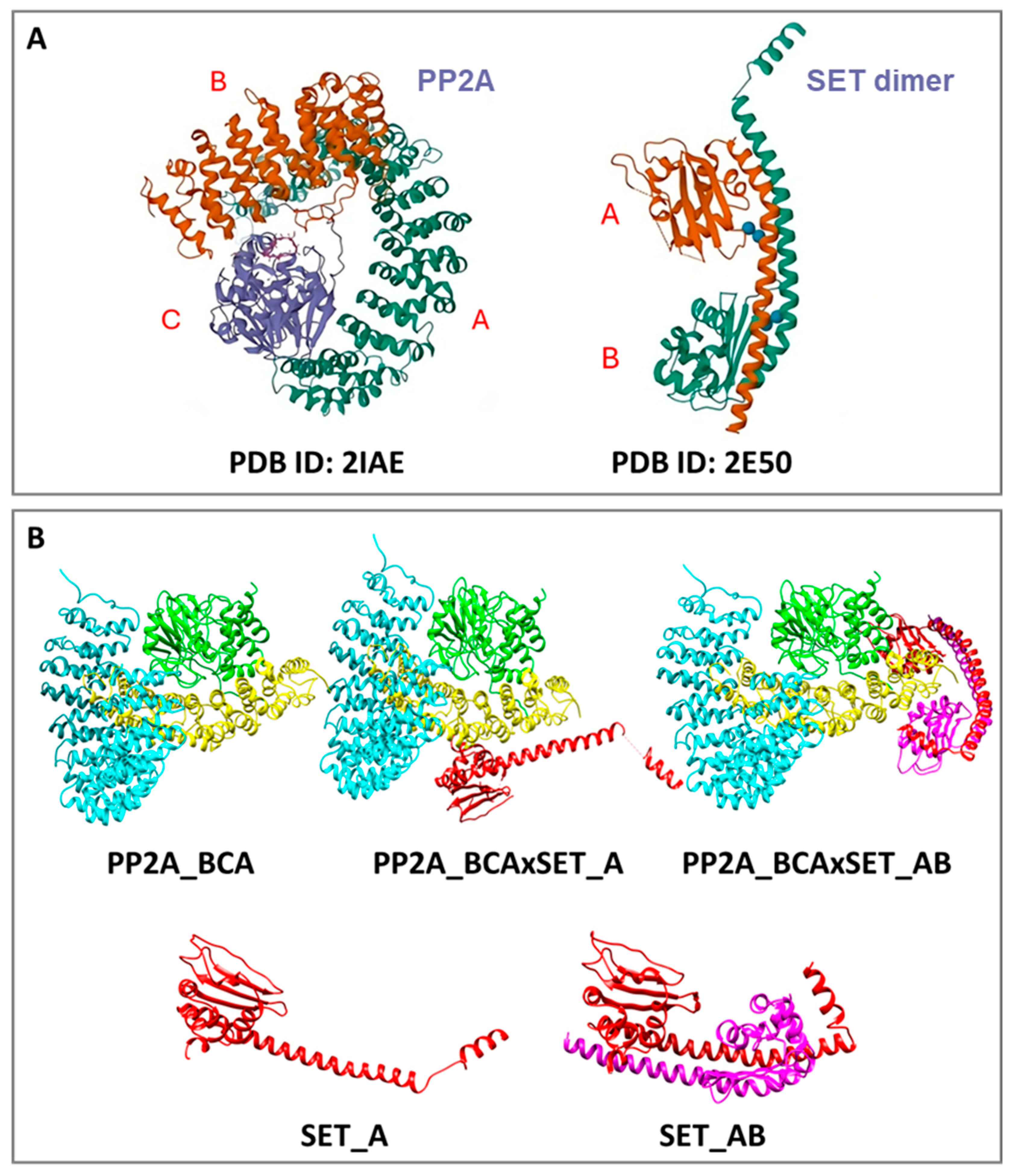
Figure 1. (A) Models retrieved from the RCSB PDB repository; letters A, B and C indicate different chains; (B) 5 models generated through protein–protein docking utilizing HDOCK, including PP2A formed by B, C and A subunit, PP2A (three subunits) in complex with SET monomer, PP2A (three subunits) in complex with the SET dimer, SET monomer, and SET dimer. Panel (A) of the figure was prepared by adapting structures from the Protein Data Bank; Panel (B) was prepared using UCSF Chimera [68].
Macromol 05 00043 g001
Figure 2. Chemical structures of (A) the 26-compound library and (B) the parent compounds FTY720 and D-e-C18.
Macromol 05 00043 g002
Figure 3. Representative docking poses of the three best performing ligands for each receptor in terms of energy score: (A) PP2A, (B) PP2A in complex with SET monomer, (C) PP2A in complex with the SET dimer, (D) SET monomer, (E) SET dimer. The figure was prepared using UCSF Chimera [68].
Macromol 05 00043 g003
Figure 4. EC50 values for CLL, SK-N-BE, HepG2, HUH7.5, and MDA-MB-231 cells are presented as histograms based on the experimental measurements documented in Appendixes S3 and S4.
Macromol 05 00043 g004
Figure 5. (A) Western blot analysis of PP2A immunoprecipitates from total cell lysates of CLL cells; previously incubated increasing concentrations of CC11, and GR390 for 16 h, and eventually analyzed with anti-SET and anti-PP2A antibodies. The results shown in (A) are representative of experiments conducted on CLL cells from 8 patients randomly selected from the all 38 patients included in the study. (B) In vitro PP2A activity of the total cell lysates of CLL in (A) and performed by using a specific phosphopeptide as a substrate. The raw data regarding PP2A activity are reported in Appendix S5.
Macromol 05 00043 g005
Disclaimer/Publisher’s Note: The statements, opinions and data contained in all publications are solely those of the individual author(s) and contributor(s) and not of MDPI and/or the editor(s). MDPI and/or the editor(s) disclaim responsibility for any injury to people or property resulting from any ideas, methods, instructions or products referred to in the content.