Article

Development of a Drug Delivery System with Bacterial Cellulose and Gelatin: Physicochemical and Microbiological Evaluation

Department of Pharmacy, University of Sorocaba (UNISO), Sorocaba 18023-000, Brazil
*
Author to whom correspondence should be addressed.
Macromol 2025, 5(3), 39; https://doi.org/10.3390/macromol5030039
Submission received: 29 April 2025 / Revised: 2 July 2025 / Accepted: 22 August 2025 / Published: 27 August 2025

Abstract

The growing threat of antimicrobial resistance drives the need for innovative and multifunctional therapeutic systems. In this study, a controlled-release system based on a bioactive film composed of gelatin, bacterial cellulose (BC), sericin, citric acid, PEG 400, and nisin was developed for topical applications in infected wound treatment. BC membranes were produced using Komagataeibacter xylinus and enzymatically treated to optimize dispersion within the polymer matrix. The resulting system exhibited a semi-rigid, homogeneous morphology with appropriate visual characteristics for dermatological use. Microbiological assays demonstrated significant antimicrobial activity against Gram-positive (Staphylococcus aureus) and resistant Gram-negative strains (Escherichia coli and Enterobacter cloacae), attributed to the synergistic action of nisin and citric acid, which enhanced bacterial outer membrane permeability. The antioxidant capacity was confirmed through DPPH radical scavenging assays, indicating a progressive release of bioactive compounds over time. Scanning electron microscopy (SEM) analyses revealed good integration of biopolymers within the matrix. These results suggest that the strategic combination of natural biopolymers and antimicrobial agents produced a functional system with improved mechanical properties, a broadened antimicrobial spectrum, and promising potential as a bioactive wound dressing for the treatment of infected skin lesions.

Graphical Abstract

1. Introduction

Bacterial resistance to antimicrobials is an urgent global health issue, with significant implications for morbidity, mortality, and economic costs. The World Health Organization (WHO) has highlighted that antimicrobial resistance (AMR) is responsible for substantial mortality, with an estimated 1.27 million deaths directly attributed to AMR in 2019. Inappropriate and excessive use of antimicrobials in humans, animals, and the environment are the main drivers of this crisis [1].
Before the COVID-19 pandemic, AMR was already a major concern, with high resistance rates reported for common pathogens such as Escherichia coli (E. coli), Staphylococcus aureus (S. aureus), and Klebsiella pneumoniae (K. pneumoniae) [2,3]. The pandemic aggravated the AMR problem due to increased antibiotic use in COVID-19 patients, despite the low prevalence of bacterial coinfections. Although only 8% of hospitalized COVID-19 patients had bacterial coinfections, approximately 75% received antibiotics. This widespread and excessive use contributed to the acceleration of AMR, emphasizing the need for rigorous antibiotic stewardship programs [4]. Studies have shown that the pandemic led to an increase in multidrug-resistant bacteria, particularly in hospital settings [5,6].
Antimicrobial resistance in bacteria such as E. coli, Enterobacter spp., S. aureus, Pseudomonas aeruginosa (P. aeruginosa), and Serratia spp. pose a significant challenge in clinical practice, especially in hospital environments [7,8,9]. Bacteria of the Enterobacterales order, such as E. coli and Enterobacter spp., are part of the human intestinal microbiota but are also associated with nosocomial infections. The emergence of carbapenem-resistant Enterobacterales represents a serious public health problem due to rapid spread, high mortality rates, and limited therapeutic options. Resistance is often linked to the production of extended-spectrum β-lactamases and carbapenemases such as KPC and NDM [9,10].
The economic and public health implications of AMR are substantial, with projected GDP losses and increased healthcare costs by 2050. Additionally, AMR complicates medical procedures, making infections harder to treat and increasing the risk of complications [11,12].
Combating bacterial resistance is crucial to preserve the efficacy of antimicrobial treatments, thereby preventing a post-antibiotic era and protecting public health. This requires a multifaceted approach, including enhanced surveillance, antimicrobial stewardship, and the development of new antimicrobial agents [13,14].
The growing threat of AMR demands the development of innovative antimicrobial agents. Traditional antibiotics are becoming less effective, leading to the search for alternative therapies, including antimicrobial peptides and natural compounds [15,16,17].
The dermal and transdermal application of liquid and semi-solid drugs in skin wound healing presents challenges such as washing away and removal of formulations, interfering with the desired therapeutic effect. As a result, the use of biodegradable polymers in wound dressings has attracted attention for accelerating healing and reducing infections, as well as improving adhesion, enhancing cosmetic properties, and offering better resistance to mechanical influences and water [18,19,20,21].
Our research group has experience in developing biomaterials using bacterial cellulose (BC), a biopolymer with exceptional properties, including high purity, biocompatibility, and mechanical strength. These characteristics make BC an ideal candidate for drug delivery systems, particularly in topical applications [22,23]. The combination of BC with other polymers can generate advanced biomaterials, which can be used in controlled-release systems for drugs and bioactive compounds, such as films, membranes, and hydrogels, which are applied in dressings, sutures, temporary implants, and tissue engineering [24,25].
For topical formulations, biocompatibility and biodegradability are crucial to ensure safety and minimize the environmental impact. Materials such as BC, gelatin, and sericin meet these criteria, making them suitable for biomedical applications [26]. Moreover, the integration of these biopolymers into topical formulations is promising for enhancing antimicrobial efficacy and promoting wound healing. These materials provide a favorable environment for sustained drug release and tissue regeneration [24].
Due to its amphiphilic nature, sericin serves as an effective drug delivery carrier for charged molecules, exhibiting long in vivo half-life and high absorption–desorption capacity [27]. Similarly, PEG 400, a highly water-soluble, low-molecular-weight hydrophilic solvent, enhances topical formulations through solubilization and permeation while also functioning as a plasticizer [28,29]. Furthermore, integrating these biopolymers into topical formulations is promising for enhancing antimicrobial efficacy and promoting wound healing. These materials provide a conducive environment for sustained drug release and tissue regeneration [27].
Antimicrobial peptides are being studied as alternatives to traditional antibiotics due to their unique mode of action, which disrupts bacterial membranes [30]. Similarly, gelatin microparticles have been explored as carriers for antimicrobial agents. When loaded with peptides such as nisin, these particles exhibit synergistic effects [31].
Motivated by the search for combinations of multiple antimicrobial agents and biopolymers capable of promoting synergistic effects and broadening the efficacy of formulations, this work is aimed at developing a multifunctional bioactive material. For this purpose, a matrix composed of gelatin, bacterial cellulose, sericin, citric acid, PEG 400, and nisin was used, targeting effective antimicrobial action against resistant bacteria. These combinations were strategically designed to act on multiple bacterial pathways, contributing to reducing the likelihood of microbial resistance development.

2. Materials and Methods

2.1. Production of the Release System

BC membranes were produced as described by Jozala et al. [32] using the Komagataeibacter xylinus strain (ATCC 53582), which was cultured in Hestrin–Schramm medium, which is composed of glucose (20 g/L), bacteriological peptone (5 g/L), yeast extract (5 g/L), anhydrous sodium phosphate (2.7 g/L), and monohydrated citric acid (1.5 g/L). The cultures were incubated statically in 250 mL Erlenmeyer flasks containing 50 mL of the medium at 30 °C for 4 days, resulting in the formation of membranes approximately 10 cm in diameter. After this period, the membranes were carefully removed and subjected to alkaline treatment with a 1 M NaOH solution at 60 °C for 1 h and 30 min to remove cellular residues. They were then washed with distilled water until they reached a neutral pH (6.5 to 7.0) and stored in sterilized flasks containing phosphate-buffered saline (PBS, pH 7.0), followed by autoclaving at 121 °C for 15 min for sterilization.
The enzymatic treatment was adapted from the study by Soeiro et al. [33], who reported that 4 h of incubation at 50 °C using cellulase from Trichoderma reesei effectively reduces BC aggregation, improving dispersion and uniformity in film matrices. Initially, 5 g of BC was homogenized in 15 mL of distilled water and kept under gentle agitation at 50 °C (500 rpm). The enzymatic treatment was carried out without pH adjustment, using the natural pH of the aqueous suspension, which ranged between 5.0 and 7.0, making it compatible with cellulase activity. Then, 500 µL of cellulase (Sigma Aldrich- St. Louis, MA, USA, from Trichoderma reesei, aqueous solution, ≥700 units/g, approximately 420 U) was added, and the mixture was kept under constant agitation for 4 h to allow for fragmentation of BC aggregates. After this time, the temperature was raised to 100 °C and maintained for 1 h and 30 min to ensure complete enzyme inactivation.
The release system formulation was prepared utilizing components from Table 1 by dissolving 15 g of powdered gelatin in 100 mL of distilled water, which was heated to 80 °C under constant stirring until complete solubilization. After cooling the solution to approximately 40 °C, the following components were incorporated into 10 mL of the gelatin solution: 2 g of treated BC, 0.45 g of citric acid (crosslinking agent), 0.2 g of PEG 400 (plasticizer), 1.5 g of sericin (functional biopolymer), and 0.1 g of nisin (antimicrobial agent). The mixture was homogenized until a uniform solution was obtained. The formulation was then poured into a sterile Petri dish and left to rest at room temperature (±25 °C) for 48 h for drying and film formation. After this period, the film was carefully removed from the dish and stored under controlled conditions until analysis. Additionally, a control formulation without nisin and sericin (BE) was produced to compare the antimicrobial and antioxidant activity of the system containing the antimicrobial peptide; all reagents are from Synth-SP/Brazil. To prepare the release system, the components outlined in Table 1 were used:

2.2. Determination of the Minimum Inhibitory Concentration

The minimum inhibitory concentration was determined against the strains E. coli (ATCC 25922), P. aeruginosa (ATCC 9721), and S. aureus (ATCC 10390). The assay followed the methodology described by Ataíde et al. [34] and Mazzola et al. [35], who used sterile 96-well plates. Each well initially received 100 µL of tryptic soy broth, except for well 1, which received 200 µL of solutions of the antimicrobial agents sericin and nisin, which were tested both individually and in combination. From well 1, serial dilutions of 100 µL were performed up to well 11. Subsequently, 10 µL of the microbial inoculum (106 CFU) was added to wells 1 through 12, excluding well 11, which served as a sterility control. The plates were incubated at 37 °C for 24 h. After incubation, 5 µL of the content from each well was plated on Petri dishes containing tryptic soy agar (TSA) to verify the presence or absence of bacterial growth by observing the colonies formed.

2.3. Diffusion Assay

The agar diffusion assay was conducted to evaluate the antimicrobial activity of the developed film as a controlled-release system for bioactive compounds. The resistant strains used in this study, including Escherichia coli, Serratia marcescens, Enterobacter cloacae, and Pseudomonas aeruginosa, were isolated from hospital findings and kindly provided by a partner laboratory. The ATCC-numbered strains were also evaluated. The methodology followed the protocols described by Santos et al. [36].
The films were carefully placed at the center of Petri dishes containing TSA medium, which were previously inoculated with bacterial suspensions standardized to a concentration of 106 colony-forming units per milliliter (CFU/mL). The dishes were then refrigerated (4 °C) overnight to allow passive diffusion of the active compounds from the film into the culture medium. After this, the dishes were incubated at 37 °C for 24 h.
Antimicrobial efficacy was assessed by observing and measuring the inhibition halos formed around the films. The inhibition zones were measured using a digital caliper in millimeters (mm), providing precise quantification of the antimicrobial effects against the tested bacterial strains.

2.4. Antioxidant Activity

The antioxidant activity of the films was evaluated based on the reaction between the stable radical 1,1-diphenyl-2-picrylhydrazyl (DPPH) and the active compounds in the formulation, as described by Brand-Williams et al. [37]. For the assay, the films were placed in 24-well plates, and 1 mL of the DPPH solution was added to each well. The mixture was left to rest for 1 h to allow interactions between the free radicals and the film’s antioxidant agents.
At regular 15 min intervals, aliquots of the reaction solution were collected and subjected to spectrophotometric reading at a wavelength of 515 nm. The reduction in absorbance over time served as an indicator of the system’s antioxidant capacity, reflecting the film’s efficiency in neutralizing DPPH radicals.

2.5. Scanning Electron Microscopy

Scanning electron microscopy (SEM) is a morphological characterization technique that uses a focused electron beam to scan the sample surface, generating secondary and backscattered signals that provide detailed information about the material’s topography, structure, and, in some cases, chemical composition, as described by Goldstein et al. [38]. For this analysis, the samples were previously coated with a thin layer of gold, a procedure necessary to enhance surface electrical conductivity and, consequently, improve image quality, enabling clearer visualization of both the surface and internal structures of the material, as described by Egerton [39]. After this the film was set with carbon tape and metallized for 2 min (DH-29010SCTR Smart Coater - JEOL, Tokyo, Japan, Model IT200). SEM images were observed on the scanning electron microscopy equipment (JEOL, Tokyo, Japan, Model IT200) and obtained using an accelerating voltage of 20 kV.

3. Results

3.1. Analysis of the Release System

After producing the film using the previously described methodology, it was possible to observe the formation of a material with specific physical characteristics: the film appeared as a semi-rigid and translucent structure, allowing partial light transmission, which gave it a smooth visual appearance. Additionally, the presence of bacterial cellulose (BC) flakes distributed across its surface could be identified with the naked eye, indicating proper incorporation of the biopolymer into the system’s matrix. Another relevant point was the absence of odor, an important characteristic for formulations intended for topical use, reinforcing its potential for dermatological applications. These initial results align with expectations and are seen in Figure 1.

3.2. Results from Antimicrobial Assays: Minimum Inhibitory Concentration and Agar Diffusion

The obtained results demonstrated that nisin has significant antimicrobial activity, establishing itself as the active compound in the developed system. During the assays, nisin was able to inhibit microbial growth, exhibiting a minimum inhibitory concentration of 0.00125 mg/mL for S. aureus and 0.0125 mg/mL for E. coli and P. aeruginosa. In contrast, sericin did not show antimicrobial activity against the evaluated bacterial strains.
In the agar diffusion test, the drug-release film demonstrated high efficacy against Gram-negative bacteria, indicating an expanded spectrum of action and a prolonged antimicrobial effect. Compared to previous assays in Santos et al.’s study [23], it was observed that nisin, when evaluated alone, exhibited significant antimicrobial activity against Gram-positive bacteria but showed no efficacy against Gram-negative bacteria due to its well-known permeability limitation against the outer membrane of these microorganisms. However, the blank film (BE) demonstrated antimicrobial activity against the Gram-negative bacteria E. coli and E. cloacae, even in the absence of nisin. This result suggests that the citric acid present in the formulation may have contributed to the enhanced antimicrobial activity [40]. No activity was observed against the remaining tested strains, which further emphasizes the selectivity of the formulation. As shown in Table 2, where E5 refers to the film containing nisin and sericin (experimental formulation). BE refers to the film without nisin and sericin (blank film).
The antimicrobial activity of the BE and E5 films was compared using Student’s t-test, revealing a significant effect of both treatment and time (p < 0.05). E5 demonstrated significantly larger inhibition zones than BE across all tested strains. The E5 film exhibited considerable inhibition zones against all tested strains, with notable results for E. coli (ATCC 25922; 21.68 mm ± 0.59), P. aeruginosa (ATCC 9721; 20.35 mm ± 0.33), and S. aureus (ATCC 10390; 18.96 mm ± 0.15). These results indicate broad-spectrum antimicrobial activity, indicating effectiveness against both Gram-positive and Gram-negative bacteria, including resistant strains such as E. cloacae, P. aeruginosa, and E. coli. BE (lacking nisin and sericin) showed activity against only two strains: E. coli (ATCC 25922), with an inhibition zone of 8.95 mm, and resistant E. coli, with an inhibition zone of 5.18 mm. Although present, these values are significantly lower than those observed for the E5 film, suggesting that BE may exert some effect in synergy with citric acid, but it does not demonstrate relevant antimicrobial activity.

3.3. Matrix Antioxidant Capacity

After recording the analysis time points, the triplicate data were processed and graphically represented. The film exhibited antioxidant activity, with a significantly higher radical scavenging effect observed for the E5 formulation compared to the BE control. This suggests that nisin and/or sericin are primarily responsible for the antioxidant effect. Since citric acid was also present in the BE control and did not result in a comparable antioxidant response, its isolated contribution in this system appears limited, as observed in Figure 2.
Antioxidant activity (Figure 2) increased over time in the E5 formulation, whereas the BE control showed minimal response. Student’s t-test revealed a significant effect of both treatment and time (p < 0.05), with E5 displaying significantly higher antioxidant activity than BE from 10 min onward.

3.4. Microscopic Observations

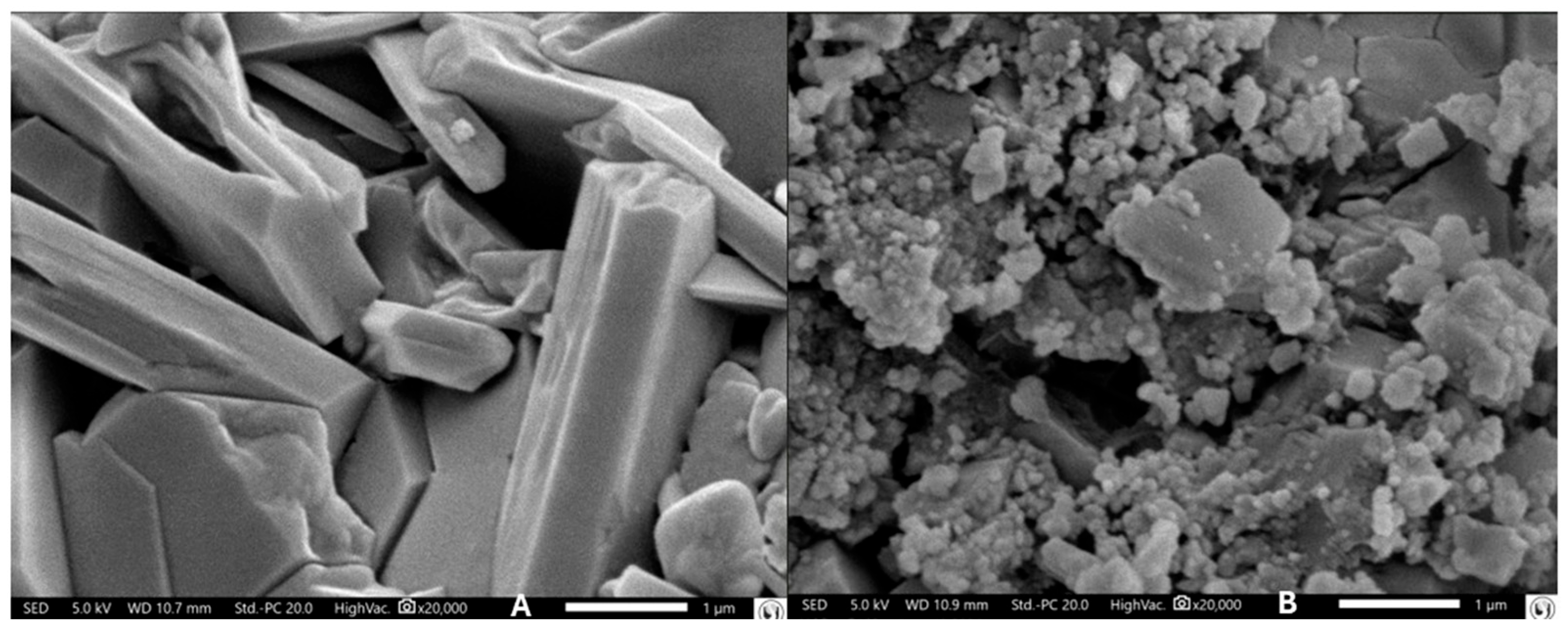
The SEM images revealed that gelatin forms a general coating on the film surface; however, the presence of surface irregularities and clusters, especially near the edges, indicates incomplete dispersion of nisin and sericin. Figure 3 shows the film surface morphology. In Figure 3B the aggregates suggest that the components were successfully incorporated into the matrix. As observed in Figure 3A, nisin and sericin may accumulate on the surface. This could result in greater dispersion of the peptide, potentially enhancing its antimicrobial activity.

4. Discussion

The results obtained in this study clearly demonstrate the efficacy of a multifunctional bioactive system developed through the combination of gelatin, bacterial cellulose, sericin, citric acid, PEG 400, and nisin. The initial proposal to create a material with enhanced mechanical properties coupled with antimicrobial and antioxidant activities was confirmed by the conducted assays, validating the working hypotheses that predicted a functional matrix with potential for biomedical applications, particularly as topical wound dressings.
Specialized studies emphasize that films produced exclusively with gelatin present significant limitations, including high stiffness and fragility, which compromise their performance in applications requiring flexibility and mechanical resistance [41,42]. In the current study, the incorporation of enzymatically processed bacterial cellulose proved to be an effective strategy to overcome these deficiencies. Morphological and surface analyses indicated a more cohesive and resistant matrix, albeit with some structural heterogeneity likely associated with the dispersion of cellulose flakes. These findings align with previous studies demonstrating that bacterial cellulose, due to its nanofibrillar structure, can enhance both the mechanical properties and water retention capacity of polymeric films [43].
As a protein-based biopolymer, gelatin interacts with cellulose nanofibers by filling interstitial spaces and modifying surface topography [44]. Scanning electron microscopy reveals that gelatin addition results in a more compact matrix with reduced surface porosity, as gelatin molecules intertwine with the bacterial cellulose fibrous network.
This uniformity in gelatin distribution is crucial to ensure that the film maintains adequate mechanical properties and that the release of bioactive compounds, when applied in drug delivery systems, is controlled and homogeneous. The viscoelastic behavior of the films, which is highly dependent on this integrated microstructure, is also enhanced by the combination of the two biopolymers, providing a balance between the stiffness of BC and the flexibility of gelatin [44,45].
Another key component in the formulation was sericin, a natural silk-derived protein that demonstrated relevant functional properties. Its incorporation enhanced the film’s elasticity and cohesion while reinforcing the polymeric structure and helping prevent desiccation—an essential characteristic for prolonged topical applications [46]. Sericin promotes the formation of more resistant and hydrated polymeric networks [47]. The synergistic interaction between sericin, PEG 400, and citric acid yielded a final material with excellent physicochemical stability and functionality. PEG 400 acted as a humectant and plasticizing agent, promoting greater malleability and resistance to drying, while citric acid, in addition to acidifying the medium, contributed to the formulation’s stability [48].
The association between nisin with citric acid and PEG 400 significantly enhances its antimicrobial activity, especially against Gram-negative bacteria, which have natural resistance barriers. Citric acid acts as a chelating agent, removing ions such as calcium and magnesium that stabilize the outer membrane of Gram-negative bacteria [40]. This destabilization facilitates the entry of nisin and increases its antimicrobial activity. Although PEG 400 has no direct antimicrobial action, it can act as a vehicle, helping nisin penetrate bacterial barriers. In combination with citric acid, PEG 400 improves system stability and facilitates nisin’s action at the target site [49,50].
While nisin is widely recognized for its activity against Gram-positive bacteria, its action against Gram-negative bacteria is typically limited due to the presence of an outer membrane. The article [51] reports that nisin treated with EDTA can sensitize Gram-negative bacteria, making them susceptible to the action of nisin. A similar effect may have occurred with the combination of nisin and citric acid, as observed in our formulation.
The microorganisms used in these experiments exhibit resistance mechanisms that are well documented in the literature. Enterobacter cloacae expresses endogenous AmpC and ESBL production, often combined with low permeability due to alterations in OmpC/OmpF porins and activation of efflux pumps [52]. Serratia marcescens, in turn, combines β-lactamases (chromosomal cephalosporinases and ESBLs), lipopolysaccharide modifications that hinder drug penetration, and efflux pumps (ABC and RND) [53]. Pseudomonas aeruginosa employs multiple strategies, including chromosomal AmpC, ESBLs (PER-1 and VEB-1), carbapenemases (IMP, NDM, and OXA), to target modifications (e.g., DNA gyrase via Qnr) and efflux systems [54]. Escherichia coli, especially ESBL- or MBL-producing strains, shows plasmid-mediated resistance—often via qnr genes as well as via the use of efflux pumps and porin mutations [55].
In our tests, the E5 film demonstrated significant efficacy, effectively overcoming this multi-resistance barrier. A substantial reduction in bacterial load was observed for all tested strains, suggesting sustained and concentrated release of antimicrobial agents. This resistance-breaking effect can be explained by a triple mechanism: (i) reaching concentrations that saturate β-lactamases; (ii) effective penetration into the biofilm matrix, inhibiting adaptive mechanisms; and (iii) maintaining continuous antimicrobial pressure, thereby minimizing escape through efflux or mutation.
Thus, our positive results support the hypothesis that controlled-release antimicrobial systems can effectively neutralize multiple resistance mechanisms, offering a promising outlook for clinical applications, particularly in infections caused by multidrug-resistant strains.
These data corroborate previous investigations that point to bacterial cellulose as an excellent support for the controlled release of antimicrobials, as demonstrated by Santos et al. [36] and Shao et al. [56]. The presence of enlarged inhibition halos against S. aureus and the significant action against Gram-negative bacteria suggest that the film architecture favored the stability and activity of nisin, making it more efficient even against typically resistant pathogens.
The antioxidant capacity, evaluated through DPPH assays, reflects the system’s ability to neutralize reactive oxygen species, which is desirable in wound dressings as it may reduce oxidative damage in lesions and promote the healing process [57]. Although both sericin and citric acid have demonstrated antioxidant activity in the literature [58,59], this effect was not observed in our study for the BE control, which contains these components. In contrast, the E5 film, which includes nisin in addition to sericin and citric acid, exhibited a measurable antioxidant effect.
Overall, the obtained results reinforce the hypothesis that the rational association of natural biopolymers with bioactive compounds can generate versatile and effective systems for both pharmaceutical and biomedical applications.
Recent studies also demonstrate that biopolymers such as chitosan, collagen, alginate, and hyaluronic acid, when combined with natural bioactive compounds (e.g., curcumin, quercetin, Aloe vera, and Centella asiatica), result in formulations with antimicrobial, antioxidant, and anti-inflammatory properties, which collectively promote wound healing [60,61,62,63]. In a recent study, Bessalah et al. [45] evaluated the potential of an innovative biopolymer composed of gelatin, chitosan, and Moringa leaf extract (G–CH–M) for biomedical applications. The material demonstrated hemocompatible, anti-inflammatory, antioxidant, and antibacterial properties, acting effectively against both Gram-positive and Gram-negative bacteria.
Our results support the hypothesis indicated in the literature [64,65,66] that the rational combination of natural biopolymers and bioactive compounds can lead to the development of versatile and effective systems for pharmaceutical and biomedical applications. Notably, the integration of both antimicrobial and antioxidant properties into a single bioactive matrix is particularly advantageous for topical formulations targeting infected skin lesions, where the presence of multidrug-resistant microorganisms and oxidative stress significantly impairs the tissue regeneration process. These findings highlight the therapeutic potential of such associations in promoting more effective treatments for complex skin injuries [64].

5. Conclusions

The development of a multifunctional bioactive film based on gelatin, bacterial cellulose, sericin, citric acid, PEG 400, and nisin has proven to be an innovative and effective approach for pharmaceutical and biomedical applications, particularly as a topical dressing for treating skin lesions. The results demonstrate not only superior mechanical properties but also remarkable antimicrobial activity, including Gram-negative and resistant strains, along with significant antioxidant capacity.
This study represents an initial step in the development of a bioactive system for topical application in infected wounds. The results obtained indicate promising performance in terms of antimicrobial and antioxidant activity; however, it is important to recognize that the complete validation of the formulation still requires a more in-depth characterization of its mechanical properties, as well as additional evaluations of biocompatibility and biological safety. Therefore, future studies will be directed toward conducting these complementary assessments to ensure the efficacy and safety of the proposed material.
The synergistic interaction among the natural components endowed the material with stability, flexibility, and functionality, particularly through controlled release of bioactive compounds, which are the key advantage for localized therapies. Nisin showed efficacy in inhibiting multidrug-resistant microorganisms, while the other constituents contributed to the system’s structural integrity and biofunctional performance.
These advances represent a significant step forward in integrating biotechnology with materials engineering, creating a versatile therapeutic platform adaptable to the demands of acute and chronic wound treatment. Thus, the developed film not only offers a promising alternative to conventional systems but also stands out as a leading candidate in the field of smart dressings and regenerative medicine, with real potential to impact patients’ quality of life.

Author Contributions

Conceptualization: A.F.J., P.L.M.A., S.O., and L.S.; methodology, G.P.M., V.S., N.L.A.I., and A.C.C.; software, L.S., D.G., and P.L.M.A.; formal analysis, G.P.M.; writing—original draft preparation, G.P.M. and A.C.C.; writing—review and editing, A.F.J., P.L.M.A., L.S., and D.G.; supervision, A.F.J. All authors contributed equally to the manuscript. All authors have read and agreed to the published version of the manuscript.

Funding

This research was funded by the University of Sorocaba (Uniso) and the Coordination for the Improvement of Higher Education Personnel (CAPES).

Institutional Review Board Statement

Not applicable.

Informed Consent Statement

Not applicable.

Data Availability Statement

Data are contained within the article.

Conflicts of Interest

The authors declare no conflicts of interest.

References

  1. World Health Organization. Antimicrobial Resistance. 2023. Available online: https://www.who.int/news-room/fact-sheets/detail/antimicrobial-resistance (accessed on 28 April 2025).
  2. Akova, M. Epidemiology of antimicrobial resistance in bloodstream infections. Virulence 2016, 7, 252–266. [Google Scholar] [CrossRef] [PubMed]
  3. Abdelaziz Abdelmoneim, S.; Mohamed Ghazy, R.; Anwar Sultan, E.; Hassaan, M.A.; Anwar Mahgoub, M. Antimicrobial resistance burden pre and post-COVID-19 pandemic with mapping the multidrug resistance in Egypt: A comparative cross-sectional study. Sci. Rep. 2024, 14, 7176. [Google Scholar] [CrossRef]
  4. World Health Organization. WHO Reports Widespread Overuse of Antibiotics in Patients Hospitalized with COVID-19. 2024. Available online: https://www.who.int/news/item/26-04-2024-who-reports-widespread-overuse-of-antibiotics-in-patients--hospitalized-with-covid-19 (accessed on 28 April 2025).
  5. Langford, B.J.; So, M.; Simeonova, M.; Leung, V.; Lo, J.; Kan, T.; Raybardhan, S.; Sapin, M.E.; Mponponsuo, K.; Farrell, A.; et al. Antimicrobial resistance in patients with COVID-19: A systematic review and meta-analysis. Lancet Microbe 2023, 4, e179–e191. [Google Scholar] [CrossRef]
  6. Petrakis, V.; Panopoulou, M.; Rafailidis, P.; Lemonakis, N.; Lazaridis, G.; Terzi, I.; Papazoglou, D.; Panagopoulos, P. The Impact of the COVID-19 Pandemic on Antimicrobial Resistance and Management of Bloodstream Infections. Pathogens 2023, 12, 780. [Google Scholar] [CrossRef]
  7. Botelho, J.; Grosso, F.; Peixe, L. Antibiotic resistance in Pseudomonas aeruginosa—Mechanisms, epidemiology and evolution. Drug Resist. Updates 2019, 44, 100640. [Google Scholar] [CrossRef]
  8. MacKinnon, M.C.; McEwen, S.A.; Pearl, D.L.; Lyytikäinen, O.; Jacobsson, G.; Collignon, P.; Gregson, D.B.; Valiquette, L.; Laupland, K.B. Increasing incidence and antimicrobial resistance in Escherichia coli bloodstream infections: A multinational population-based cohort study. Antimicrob. Resist. Infect. Control 2021, 10, 131. [Google Scholar] [CrossRef]
  9. van Duin, D.; Paterson, D.L. Multidrug-Resistant Bacteria in the Community. Infect. Dis. Clin. N. Am. 2020, 34, 709–722. [Google Scholar] [CrossRef]
  10. Leite Mdos, S.; Gusmão Ado, C.; Gontijo Bde, A.V.; Garcia, P.G. Antimicrobial resistance profile of Escherichia coli isolated from urine specimens of an Intensive Care Unit patients. Rev. Bras. Anál. Clín. 2020, 52, 243–247. [Google Scholar] [CrossRef]
  11. Salam, M.A.; Al-Amin, M.Y.; Salam, M.T.; Pawar, J.S.; Akhter, N.; Rabaan, A.A.; Alqumber, M.A.A. Antimicrobial Resistance: A Growing Serious Threat for Global Public Health. Healthcare 2023, 11, 1946. [Google Scholar] [CrossRef] [PubMed]
  12. Tizhe, D.T.; Ogra, I.O.; Apollos, S.D.; Enesi, K.O.; Yohanna, A.; Aminu, R.; Kwaga, J.K.P. Antimicrobial resistance as a global public health threat: The way forward. EUREKA Life Sci. 2024, 52–57. [Google Scholar] [CrossRef]
  13. Mancuso, G.; Midiri, A.; Gerace, E.; Biondo, C. Bacterial Antibiotic Resistance: The Most Critical Pathogens. Pathogens 2021, 10, 1310. [Google Scholar] [CrossRef]
  14. Halawa, E.M.; Fadel, M.; Al-Rabia, M.W.; Behairy, A.; Nouh, N.A.; Abdo, M.; Olga, R.; Fericean, L.; Atwa, A.M.; El-Nablaway, M.; et al. Antibiotic action and resistance: Updated review of mechanisms, spread, influencing factors, and alternative approaches for combating resistance. Front. Pharmacol. 2023, 14, 1305294. [Google Scholar] [CrossRef]
  15. Ferraboschi, P.; Ciceri, S.; Grisenti, P. Applications of Lysozyme, an Innate Immune Defense Factor, as an Alternative Antibiotic. Antibiotics 2021, 10, 1534. [Google Scholar] [CrossRef]
  16. Singha, B.; Singh, V.; Soni, V. Alternative therapeutics to control antimicrobial resistance: A general perspective. Front. Drug Discov. 2024, 4, 1385460. [Google Scholar] [CrossRef]
  17. Prima, M.J.; Hassan, M.; Sharma, J. Novel Approaches for Combating Antibiotic Resistance in Pathogenic Bacteria. Microb. Bioact. 2023, 6, 1–8. [Google Scholar] [CrossRef]
  18. Pünnel, L.C.; Lunter, D.J. Film-Forming Systems for Dermal Drug Delivery. Pharmaceutics 2021, 13, 932. [Google Scholar] [CrossRef] [PubMed]
  19. Ventola, C.L. The Antibiotic Resistance Crisis: Part 1: Causes and Threats. P T 2015, 40, 277–283. [Google Scholar] [PubMed]
  20. Nussbaum, S.R.; Carter, M.J.; Fife, C.E.; DaVanzo, J.; Haught, R.; Nusgart, M.; Cartwright, D. An economic evaluation of the impact, cost, and Medicare policy implications of chronic nonhealing wounds. Value Health 2018, 21, 27–32. [Google Scholar] [CrossRef]
  21. Williamson, D.A.; Carter, G.P.; Howden, B.P. Current and emerging topical antibacterials and antiseptics: Agents, action, and resistance patterns. Clin. Microbiol. Rev. 2017, 30, 827–860. [Google Scholar] [CrossRef] [PubMed]
  22. Ataíde, J.A.; de Carvalho, N.M.; Rebelo, M.A.; Chaud, M.V.; Grotto, D.; Gerenutti, M.; Rai, M.; Mazzola, P.G.; Jozala, A.F. Bacterial nanocellulose loaded with bromelain: Assessment of antimicrobial, antioxidant, and physical-chemical properties. Sci. Rep. 2017, 7, 2–10. [Google Scholar] [CrossRef] [PubMed]
  23. dos Santos, C.A.; dos Santos, G.R.; Soeiro, V.S.; dos Santos, J.R.; de Araujo Rebelo, M.; Chaud, M.V.; Gerenutti, M.; Grotto, D.; Pandit, R.; Rai, M.; et al. Bacterial nanocellulose membranes combined with nisin: A strategy to prevent microbial growth. Cellulose 2018, 25, 6681–6689. [Google Scholar] [CrossRef]
  24. Kupnik, K.; Primožič, M.; Kokol, V.; Leitgeb, M. Nanocellulose in Drug Delivery and Antimicrobially Active Materials. Polymers 2020, 12, 2825. [Google Scholar] [CrossRef]
  25. Liang, S. Advances in drug delivery applications of modified bacterial cellulose-based materials. Front. Bioeng. Biotechnol. 2023, 11, 1252706. [Google Scholar] [CrossRef]
  26. Pandey, A.; Singh, M.K.; Singh, A. Bacterial cellulose: A smart biomaterial for biomedical applications. J. Mater. Res. 2024, 39, 2–18. [Google Scholar] [CrossRef]
  27. Lamboni, L.; Gauthier, M.; Yang, G.; Wang, Q. Silk sericin: A versatile material for tissue engineering and drug delivery. Biotechnol. Adv. 2015, 33, 1855–1867. [Google Scholar] [CrossRef]
  28. D’souza, A.A.; Shegokar, R. Polyethylene glycol (PEG): A versatile polymer for pharmaceutical applications. Expert Opin. Drug Deliv. 2016, 13, 1257–1275. [Google Scholar] [CrossRef]
  29. Strickley, R.G. Solubilizing excipients in oral and injectable formulations. Pharm. Res. 2004, 21, 201–230. [Google Scholar] [CrossRef]
  30. Mohamed, M.F.; Abdelkhalek, A.; Seleem, M.N. Evaluation of short synthetic antimicrobial peptides for treatment of drug-resistant and intracellular Staphylococcus aureus. Sci. Rep. 2016, 6, 29707. [Google Scholar] [CrossRef] [PubMed]
  31. Hren, M.; Trček, J.; Šakanović, A.; Obradović, H.; Kreft, M.E.; Hribernik, S.; Gorgieva, S. Nisin-loaded gelatin microparticles for the enhanced bioactivity of bacterial nanocellulose. Int. J. Biol. Macromol. 2025, 305, 141203. [Google Scholar] [CrossRef] [PubMed]
  32. Jozala, A.F.; Pértile, R.A.N.; dos Santos, C.A.; de Carvalho Santos-Ebinuma, V.; Seckler, M.M.; Gama, F.M.; Pessoa, A., Jr. Bacterial cellulose production by Gluconacetobacter xylinus by employing alternative culture media. Appl. Microbiol. Biotechnol. 2014, 99, 1181–1190. [Google Scholar] [CrossRef] [PubMed]
  33. Soeiro, V.S.; Silva-Carvalho, R.; Martins, D.; Parpot, P.; Grotto, D.; Chaud, M.V.; da Gama, F.M.P.; Jozala, A.F. Alginate-amphotericin B nanocomplexes covered by nanocrystals from bacterial cellulose: Physico-chemical characterization and in vitro toxicity. Sci. Rep. 2021, 11, 23944. [Google Scholar] [CrossRef]
  34. Andrews, J.M. Determination of minimum inhibitory concentrations. J. Antimicrob. Chemother. 2001, 48 (Suppl. 1), 5–16, Erratum in J. Antimicrob. Chemother. 2002, 49, 1049. [Google Scholar] [CrossRef] [PubMed]
  35. Mazzola, P.G.; Jozala, A.F.; Novaes LCde, L.; Moriel, P.; Penna, T.C.V. Minimal inhibitory concentration (MIC) determination of disinfectant and/or sterilizing agents. Braz. J. Pharm. Sci. 2009, 45, 241–248. [Google Scholar] [CrossRef]
  36. dos Santos, G.R.; Soeiro, V.S.; Talarico, C.F.; Ataide, J.A.; Lopes, A.M.; Mazzola, P.G.; Oliveira, T.J.; Oliveira Junior, J.M.; Grotto, D.; Jozala, A.F. Bacterial Cellulose Membranes as Carriers for Nisin: Incorporation, Antimicrobial Activity, Cytotoxicity and Morphology. Polymers 2022, 14, 3497. [Google Scholar] [CrossRef] [PubMed]
  37. Brand-Williams, W.; Cuvelier, M.E.; Berset, C. Use of a Free Radical Method to Evaluate Antioxidant Activity. LWT Food Sci. Technol. 1995, 28, 25–30. [Google Scholar] [CrossRef]
  38. Goldstein, J.I.; Newbury, D.E.; Michael, J.R.; Ritchie, N.W.M.; Scott, J.H.J.; Joy, D.C. Scanning Electron Microscopy and X-Ray Microanalysis, 4th ed.; Springer: New York, NY, USA, 2018. [Google Scholar]
  39. Egerton, R.F. Physical Principles of Electron Microscopy: An Introduction to TEM, SEM, and AEM, 2nd ed.; Springer: Cham, Switzerland, 2016. [Google Scholar]
  40. Burel, C.; Kala, A.; Purevdorj-Gage, L. Impact of pH on citric acid antimicrobial activity against Gram-negative bacteria. Lett. Appl. Microbiol. 2020, 72, 332–340. [Google Scholar] [CrossRef]
  41. Luo, Q.; Hossen, M.A.; Zeng, Y.; Dai, J.; Li, S.; Qin, W.; Liu, Y. Gelatin-based composite films and their application in food packaging: A review. J. Food Eng. 2022, 313, 110762. [Google Scholar] [CrossRef]
  42. Tamo, A.K. Nanocellulose-based hydrogels as versatile materials with interesting functional properties for tissue engineering applications. J. Mater. Chem. B 2024, 12, 7692–7759. [Google Scholar] [CrossRef]
  43. Soeiro, V.S.; Tundisi, L.L.; Amaral, V.A.; Batain, F.; Mazzola, P.G.; Tambourgi, E.B.; de Oliveira Júnior, J.M.; Chaud, M.V.; Grotto, D.; Aranha, N.; et al. Bacterial nanocellulose and fibroin: Natural products to produce a structure membranes. Matéria 2021, 26, e13086. [Google Scholar] [CrossRef]
  44. Roy, S.; Biswas, D.; Rhim, J.W. Gelatin/Cellulose Nanofiber-Based Functional Nanocomposite Film Incorporated with Zinc Oxide Nanoparticles. J. Compos. Sci. 2022, 6, 223. [Google Scholar] [CrossRef]
  45. Bessalah, S.; Faraz, A.; Dbara, M.; Khorcheni, T.; Hammadi, M.; Ajose, D.J.; Saeed, S.I. Antibacterial, anti-biofilm, and anti-inflammatory properties of gelatin-chitosan-moringa-biopolymer-based wound dressings toward Staphylococcus aureus and Escherichia coli. Pharmaceuticals 2024, 17, 545. [Google Scholar] [CrossRef]
  46. Aad, R.; Dragojlov, I.; Vesentini, S. Sericin Protein: Structure, Properties, and Applications. J. Funct. Biomater. 2024, 15, 322. [Google Scholar] [CrossRef]
  47. Liu, J.; Shi, L.; Deng, Y.; Zou, M.; Cai, B.; Song, Y.; Wang, Z.; Wang, L. Silk sericin-based materials for biomedical applications. Biomaterials 2022, 287, 121638. [Google Scholar] [CrossRef]
  48. Domján, A.; Bajdik, J.; Pintye-Hódi, K. Understanding of the Plasticizing Effects of Glycerol and PEG 400 on Chitosan Films Using Solid-State NMR Spectroscopy. Macromolecules 2009, 42, 4667–4673. [Google Scholar] [CrossRef]
  49. Zhang, Y.; Lane, M.E.; Moore, D.J. An Investigation of the Influence of PEG 400 and PEG-6-Caprylic/Capric Glycerides on Dermal Delivery of Niacinamide. Polymers 2020, 12, 2907. [Google Scholar] [CrossRef]
  50. Knop, K.; Hoogenboom, R.; Fischer, D.; Schubert, U.S. Poly(ethylene glycol) in drug delivery: Pros and cons as well as potential alternatives. Angew. Chem. Int. Ed. Engl. 2010, 49, 6288–6308. [Google Scholar] [CrossRef] [PubMed]
  51. Vessoni, C.; Jozala, A.F.; Gentille, T.R.; Pessoa, A.; Cholewa, O. Detection of Nisin Expression by Lactococcus lactis Using Two Susceptible Bacteria to Associate the Effects of Nisin with EDTA. Appl. Biochem. Biotechnol. 2006, 129, 334–346. [Google Scholar] [CrossRef]
  52. Yang, F.C.; Yan, J.J.; Hung, K.H.; Wu, J.J. Characterization of Ertapenem-Resistant Enterobacter cloacae in a Taiwanese University Hospital. J. Clin. Microbiol. 2012, 50, 223–226. [Google Scholar] [CrossRef] [PubMed]
  53. Tavares-Carreon, F.; De Anda-Mora, K.; Rojas-Barrera, I.C.; Andrade, A. Serratia marcescens antibiotic resistance mechanisms of an opportunistic pathogen: A literature review. PeerJ 2023, 11, e14399. [Google Scholar] [CrossRef] [PubMed]
  54. Moradali, M.F.; Ghods, S.; Rehm, B.H. Pseudomonas aeruginosa lifestyle: A paradigm for adaptation, survival, and persistence. Front. Cell Infect. Microbiol. 2017, 7, 39. [Google Scholar] [CrossRef]
  55. Rozwadowski, M.; Gawel, D. Molecular factors and mechanisms driving multidrug resistance in uropathogenic Escherichia coli—An update. Genes 2022, 13, 1397. [Google Scholar] [CrossRef]
  56. Shao, W.; Liu, H.; Wang, S.; Wu, J.; Huang, M.; Min, H.; Liu, X. Controlled release and antibacterial activity of tetracycline hydrochloride-loaded bacterial cellulose composite membranes. Carbohydr. Polym. 2016, 145, 114–120. [Google Scholar] [CrossRef]
  57. Araujo, L.; Nunes, K.; Mello, J.; Nakamura, C.; Gomes, R.; Bergamasco, R. Evaluation of the Antioxidant, Photoprotective and Wound Healing Capacity of Guazuma ulmifolia lam. Extracts in L-929 Cells Exposed to UV-A and UV-B Irradiation. J. Braz. Chem. Soc. 2024, 35, e-20230149. [Google Scholar] [CrossRef]
  58. Takechi, T.; Wada, R.; Fukuda, T.; Harada, K.; Takamura, H. Antioxidant activities of two sericin proteins extracted from cocoon of silkworm (Bombyx mori) measured by DPPH, chemiluminescence, ORAC and ESR methods. Biomed. Rep. 2014, 2, 364–369. [Google Scholar] [CrossRef]
  59. Singh, S.K.; Kaldate, R.; Bisht, A. Chapter 4.5—Citric acid, antioxidant effects in health. In Antioxidants Effects in Health; Nabavi, S.M., Sanches Silva, A., Eds.; Elsevier eBooks: Amsterdam, The Netherlands, 2022; pp. 309–322. [Google Scholar] [CrossRef]
  60. Luo, L.; Wu, Y.; Li, C.; Zou, Y.; Huang, L.; Liang, Y.; Ren, J.; Liu, Y.; Lin, Q. Elaboration and characterization of curcumin-loaded soy soluble polysaccharide (SSPS)-based nanocarriers mediated by antimicrobial peptide nisin. Food Chem. 2021, 336, 127669. [Google Scholar] [CrossRef] [PubMed]
  61. Fernandes, A.S.; Rodrigues, P.; Pintado, M.; Tavaria, F.K. A systematic review of natural products for skin applications: Targeting inflammation, wound healing, and photo-aging. Phytomedicine 2023, 115, 154824. [Google Scholar] [CrossRef]
  62. Jangra, N.; Singla, A.; Puri, V.; Dheer, D.; Chopra, H.; Malik, T.; Sharma, A. Herbal bioactive-loaded biopolymeric formulations for wound healing applications. RSC Adv. 2025, 15, 12402–12442. [Google Scholar] [CrossRef]
  63. Bai, Q.; Hu, F.; Gou, S.; Gao, Q.; Wang, S.; Zhang, W.; Zhang, Y.; Lu, T. Curcumin-loaded chitosan-based hydrogels accelerating S. aureus-infected wound healing. Int. J. Biol. Macromol. 2024, 259 Pt 1, 129111. [Google Scholar] [CrossRef]
  64. Falbo, F.; Spizzirri, U.G.; Restuccia, D.; Aiello, F. Natural compounds and biopolymers-based hydrogels join forces to promote wound healing. Pharmaceutics 2023, 15, 271. [Google Scholar] [CrossRef]
  65. Zhou, Z.; Zhang, D.; Ning, X.; Jin, L.; Lin, Y.; Liang, C.; Wen, X.; Huang, T.; Zhou, J.; Zhang, Y. An antibacterial, antioxidant and hemostatic hydrogel accelerates infectious wound healing. J. Nanobiotechnol. 2025, 23, 49. [Google Scholar] [CrossRef] [PubMed]
  66. Haririan, Y.; Asefnejad, A. Biopolymer hydrogels and synergistic blends for tailored wound healing. Int. J. Biol. Macromol. 2024, 279 Pt 4, 135519. [Google Scholar] [CrossRef] [PubMed]
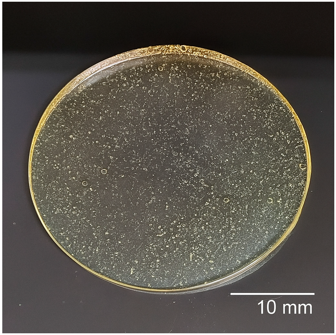
Figure 1. The E5 film formulated with gelatin, bacterial cellulose (BC), citric acid, PEG 400, sericin, and nisin.
Macromol 05 00039 g001
Figure 2. Antioxidant activity of the film over time (minutes).
Macromol 05 00039 g002
Figure 3. Scanning electron microscopy of the films at 20,000× magnification: (A) BE and (B) the E5 film.
Macromol 05 00039 g003
Table 1. Quantitative composition per mL for the preparation of the E5 film.
ComponentAmount (per 10 mL Solution)Function
Gelatin1.5 gMatrix/base
BC *2 gStructural agent
PEG 4000.2 gPlasticizer
Citric acid0.45 gCrosslinker
Sericin **1.5 gFunctional agent
Nisin **0.1 gAntimicrobial
* BC—bacterial cellulose; ** indicates the control formulation produced without the corresponding component.
Table 2. Average inhibition zone results (in mm) using the film as an antimicrobial agent, tested in triplicate against the following microorganisms.
MicroorganismsE5 Halo (mm)SDBE Halo (mm)SD
S. aureus (ATCC 10390)18.96±0.1500
E. coli (ATCC 25922)21.68±0.598.95±0.06
P. aeruginosa (ATCC 9721)20.35±0.3300
E. cloacae R18.34±0.1300
S. marcescens R13.34±0.1600
P. aeruginosa R18.54±0.2400
E. coli R17.40±0.115.18±0.09
R Resistant microorganisms.
Disclaimer/Publisher’s Note: The statements, opinions and data contained in all publications are solely those of the individual author(s) and contributor(s) and not of MDPI and/or the editor(s). MDPI and/or the editor(s) disclaim responsibility for any injury to people or property resulting from any ideas, methods, instructions or products referred to in the content.