Article

Whole Transcriptome Analysis of the Mouse Placenta Following Radiation-Induced Growth Restriction

by
Shayenthiran Sreetharan
1,2,3,
Sujeenthar Tharmalingam
1,4,5,
Cameron Hourtovenko
1,
Felix Tubin
1,
Christopher D. McTiernan
1,
Christopher Thome
1,4,5,
Neelam Khaper
2,6,
Douglas R. Boreham
1,4,
Simon J. Lees
2,6 and
T.C. Tai
1,4,5,*
1
Medical Sciences Division, NOSM University, 935 Ramsey Lake Rd., Sudbury, ON P3E 2C6, Canada
2
Medical Sciences Division, NOSM University, 955 Oliver Rd., Thunder Bay, ON P7B 5E1, Canada
3
Department of Physics and Astronomy, McMaster University, 1280 Main St W, Hamilton, ON L8S 4L8, Canada
4
School of Natural Sciences, Laurentian University, Sudbury, ON P3E 2C6, Canada
5
Health Sciences North Research Institute, Sudbury, ON P3E 2H2, Canada
6
Department of Biology, Lakehead University, Thunder Bay, ON P7B 5E1, Canada
*
Author to whom correspondence should be addressed.
Radiation 2025, 5(4), 35; https://doi.org/10.3390/radiation5040035
Submission received: 12 September 2025 / Revised: 6 November 2025 / Accepted: 14 November 2025 / Published: 24 November 2025

Simple Summary

The placenta is a tissue that provides numerous functions to the developing mammalian offspring, and dysregulation within the placenta following stressor exposure can cause adverse outcomes. In this study, pregnant mice were irradiated with high-dose x-ray irradiation, causing intrauterine growth restriction, and transcriptomic analysis of the placenta was completed. A number of gene targets and dysregulated pathways were identified, offering insight into the impact that radiation can have on the developing placenta and offspring.

Abstract

High doses of ionizing radiation during prenatal development can cause growth restriction, or a decrease in growth of the developing offspring. This outcome of intrauterine growth restriction (IUGR) can predispose the offspring to lifelong health outcomes, which is referred to as developmental programming. The role of the placenta in radiation-induced IUGR was investigated using a mouse model. Pregnant BALB/cAnNCrl mice were externally irradiated with 1.82 Gy x-ray irradiation on gestational day 14.5. Fetoplacental units were collected on gestational day 18.5, and growth restriction was observed in irradiated offspring. Whole placenta samples from growth restricted and sham-irradiated groups were analyzed via RNA-sequencing analysis. Differential gene expression (DEG) analysis revealed a total of 166 DEGs in the irradiated samples. Validation of these DEG findings were completed using RT-qPCR analysis. Gene ontology (GO) analysis of the DEGs supported the involvement of autoimmune response and dysregulation in retinol (vitamin A) metabolism in the placenta. Upstream prediction analysis identified a number of potential regulators responsible for the DEG profiles including Nppb, Myod1 and genes of the classic complement system (Complement C1q chains C1qa, C1qb, C1qc). Overall, these findings present an overview of the dysregulation in the mouse placenta following an acute, high-dose radiation exposure.

1. Introduction

Exposure of the developing fetus to a physiological stressor can result in adverse effects, such as a decreased or stunted growth, also referred to as growth restriction. Growth restriction can manifest in utero prior to birth, which is also known as intrauterine growth restriction (IUGR) or fetal growth restriction (FGR). IUGR can be broadly defined as a failure of the developing conceptus to reach an adequate growth potential. Clinically, IUGR in humans is characterized by fetal weight below the 10th percentile accounting for gestational age [1]. IUGR is an obstetric outcome of concern, as there are reported associations between IUGR and increased neonatal death or other adverse effects during development [2]. There is also a growing body of evidence that correlates growth restriction (low birth weight or IUGR) with an increased susceptibility for disease later in life, such as hypertension [3,4]. Exposures to these stressors during in utero development can therefore program offspring to have lifelong effects on health and disease after birth. Example models of developmental programming include dietary interventions [5], exposure to glucocorticoid compounds [6], hypoxia exposure [7] or exposure to harmful substances like tobacco [8].
The developing fetus is known to be sensitive to ionizing radiation exposure, with numerous adverse effects observed following higher dose radiation exposures. In humans, antenatal radiation exposure increased the risk of various adverse birth outcomes including low birth weight [9]. This includes the in utero exposure of Japanese atomic bomb survivors from mothers that were pregnant at the time of the bombings. A recent analysis of mortality in this cohort from 1950 to 2012 concluded that the significant correlation between radiation dose and mortality was likely mediated by small head size, low birth weight and parental loss [10]. This includes an increase in cancerous and non-cancerous causes of death. This suggests that growth restriction effects following antenatal radiation exposure may lead to potentially life-long alterations in the offspring.
One of the largest regulators of fetal growth during gestation is the placenta. The development of the human and mouse placenta has been reviewed previously [11,12]. The placenta serves a diverse set of functions to the developing offspring including respiration (transport of gases), nutrition, excretion, endocrine and immunity functions. These processes and functions are critical for the normal development and growth of the developing offspring. Placental dysfunction has been a known to cause IUGR [13]. Altered function or dysregulation within the placenta could therefore result in a wide range of outcomes to the developing offspring. Changes within the placenta following exposure to different stressors have been studied in humans and animal models. The placenta is also a complex network of cells that interface the mother and fetus, and alterations to cells within the different layers of the placenta could also have adverse effects on the offspring.
The goal of this study was to investigate the effects of high-dose whole-body x-ray irradiation on the mouse placenta. The present study irradiated pregnant Balb/c mice with a single irradiation of 1.82 Gy. This is a very large single exposure, with a corresponding equivalent dose of 1.82 Sv in humans. Outside of a very limited number of accidental human exposures, this is not an environmentally relevant exposure. Previous work from our group did not observe significant growth restriction when animals were irradiated with approximately 1.0 Gy gamma radiation, which is why the present study employed a high-dose exposure to observe radiation-induced growth restriction. To understand the complex molecular and cellular dysregulation, whole-placental RNA-sequencing analysis was performed to identify the global transcriptional response to radiation exposure and elucidate potential dysregulated pathways.

2. Methods

2.1. Animals and Irradiation Treatment

Male and female 7–8 week old wild-type BALB/cAnNCrl mice (Charles River Laboratory; Montreal, QC, Canada) were used in this study. Animals were housed at the Lakehead University Animal Care Facility, and all described procedures were approved by the Lakehead University Animal Care Committee (AUP# 1467646). All authors complied with the ARRIVE (Animal Research: Reporting In Vivo Experiments) guidelines during this study. Animals were maintained on a 12:12 light-dark cycle with 40–70% room humidity. Food was available to animals ad libitum, with free access to water. The only exception was the removal of food and water from the animal cage during irradiations. Female breeder mice were fed the ProLab RMH 2000 breeding diet (content of protein = 18%, fat = 9% and fiber = 4%). Male mice were fed a standard lab chow diet for the duration of the study.
Animals were irradiated on gestational day 14.5 with 1.82 Gy x-rays, delivered at dose-rate of 0.838 Gy per minute. These irradiation conditions were verified using thermoluminescent dosimeters (Mirion Technologies GDS Inc.; Oak Ridge, TN, USA). Irradiations were performed using the XRAD320 cabinet x-ray irradiation system (Precision X-ray Inc.; Madison, WI, USA), with a kVp of 320 kV. Individually housed pregnant animals were irradiated in their home cage (with the food and water removed) to minimize stress during irradiation. Sham-irradiated control animals were handled and manipulated to simulate the irradiation exposure conditions without exposure to radiation. Briefly, sham-irradiated control animals were transported to the irradiator via lab cart as the irradiated animals were also transported. Sham-irradiated controls were then placed inside the cabinet irradiator and held for the same duration as the irradiation time of the treated animals, with the x-ray beam turned off. Sham-irradiated animals were then returned to the breeding room following irradiation.
Pregnant animals were euthanized on gestational day 18.5 via isofluorane anesthetic administration prior to cervical dislocation, and fetoplacental units were collected. Placenta and conceptus samples were weighed to verify growth restriction in the irradiation group. The fetal tail was collected and flash frozen for sex determination genotyping. The placenta was also flash frozen on dry ice and stored at −80 °C until further processing.

2.2. Genotyping

Sex determination of the offspring was performed using endpoint PCR. Frozen tail samples were digested overnight in an SDS-based tail buffer with proteinase K (20 mg/mL; ThermoFisher Scientific, Mississauga, ON, Canada), following the StarrLab protocol [14]. Following digestion, DNA was extracted from samples using the phenol/chloroform methods outlined by The Jackson Laboratory [15]. Sex determination was assessed by using a pair of primers (Forward: 5′-CTGAAGCTTTTGGCTTTGAG-3′ and Reverse: 5′-CCACTGCCAAATTCTTTGG-3′), which allowed for simultaneous amplification of homologous genes Jarid1c and Jarid1d from both the X and Y chromosomes, respectively [16]. Here, amplification of the 331 base pair Jarid1d gene indicates a female genotype, whereas amplification of the 302 base pair Jarid1c gene indicates a male genotype. PCR was performed in 25 μL reactions with the following: 12.5 μL of Bi2M-2xPCR Mix (containing 0.02 U/μL Taq DNA polymerase, reaction buffer, 3 mM MgCl2, 0.4 mM of each dNTP; Bio-Mine Scientific, Sudbury, ON, Canada), 1.25 μL of each forward and reverse primers (500 nM final concentration), and 10 μL of diluted DNA template (prepared at a concentration of 10 ng/μL with a total of 100 ng template per reaction). PCR was performed using the S1000 Thermal Cycler (Bio-Rad Laboratories; Mississauga, ON, Canada), under the following conditions as per manufacturer guidelines: 2 min at 95 °C for initial denaturation (1 cycle), 2 min for the denaturation, annealing and extension steps (30 s at 95 °C for denaturation, 30 s at 57 °C for annealing and 1 min at 72 °C for extension with a total of 35 cycles), 5 min at 72 °C for final extension (1 cycle) and held at 4 °C until the plate was removed. Following PCR of the tail lysate, the amplified product was resolved on a 2% agarose gel stained with ethidium bromide. Adult mouse tissue samples from male and female mice were also amplified and run on the same gel to serve as positive controls for sex determination.

2.3. RNA Extraction and cDNA Synthesis

RNA was extracted from whole placenta samples using the QIAGEN AllPrep DNA/RNA/miRNA Universal Kit (QIAGEN; Toronto, ON, Canada) following manufacturer’s instructions. The whole placenta sample was divided into two pieces due to size, 1.2 mL of lysis buffer (Buffer RLT Plus, QIAGEN) and a stainless-steel bead added to each tube and then homogenized using a Tissuelyser (QIAGEN) for 2 cycles at 30 Hz for 2 min per cycle. The lysate from the two halves of the placenta were combined in a single tube and this whole tissue lysate was used for extraction as per manufacturer instructions. RNA was analyzed using a Nanodrop (ThermoFisher Scientific, Mississauga, ON, Canada) to verify yield and purity prior to cDNA synthesis. A total of 8 placenta samples for each sex was extracted for both sham-irradiated control and the 1.82 Gy treatment group.
DNase treatment and cDNA synthesis were performed using the Bi2M-SSRT2 reverse transcription kit (Bio-Mine Scientific, Sudbury, ON, Canada) according to the manufacturer’s instructions. Briefly, DNase treatment was completed in 10 μL reactions with the following: 2 μg RNA, 1 μL 10× gDNA buffer, 1 μL gDNA remover (DNase), with the balance of the total volume being made up using DNAse/Rnase-free water. Reaction was incubated at 37 °C for 2 min. Following DNase treatment, 1 μL random hexamers and 4 μL DNAse/Rnase-free water were added to the reaction and incubated at 65 °C for 5 min. The final step included the addition of 4 μL of 5× reaction buffer and 1 μL reverse transcriptase enzyme bringing the final reaction volume to 20 μL. Reactions were run for 5 min at 25 °C, followed by 15 min at 55 °C and a final enzyme inactivation step of 30 s at 85 °C.

2.4. Transcriptomic Profiling and Secondary Analyses

RNA-seq analysis was performed on whole-placental samples collected from 8 male and 8 female samples from each treatment group. Only one male and one female fetoplacental unit was analyzed from a given dam, in order to minimize any parental bias in the analysis. Total RNA was subjected to RNA-sequencing library preparation using the NEBNext® Ultra™ II Directional RNA Library Kit (NEB E7765S) (New England BioLabs; Whitby, ON, Canada). The samples were quantified following library preparation using the NEBNext® Library Quant Kit (NEB E7630S). The samples were then pooled at equimolar concentrations and sequenced at The Centre for Applied Genomics (Toronto, ON, Canada) on a NovaSeq 6000 Illumina platform at approximately 50 million reads per sample (paired-end 150 base pair reads). The raw sequencing data was then further processed in-house using the following bioinformatics toolkits. The DRAGEN FASTQ Toolkit (DRAGEN Inc.) was used for quality check and reading trims. The RNA-Seq Alignment application (BaseSpace) was used to align reads to the mouse reference genome (mm10/GRCm38) and perform transcript count analysis. RNA-Seq Differential Expression application (Illumina Inc., San Diego, CA, USA) was used to complete differential expression analysis based on the DESeq2 platform using the Mus musculus (UCSC mm10) RefSeq gene annotation. Differential gene selection criteria were determined based on <−1.5 or >1.5 gene fold change, <0.05 for false discovery rate (FDR) corrected p-values, and a minimum average read count of 30 transcripts per million. iPathwayGuide (Advaita Bioinformatics, Ann Arbor, MI, USA) was used to perform secondary sequencing analysis including gene ontology and upstream regulator analysis as reported previously [17,18,19].

2.5. Reverse Transcription-Quantitative Polymerase Chain Reaction (RT-qPCR)

Forward and reverse primers for genes of interest were designed using the NCBI Primer Blast Tool [20]. Criteria for primer design were target sequence length of 75–150 base pairs, 50–60% GC content and predicted melting temperatures between 57 and 63 °C. Primers were also designed to span exon–exon junctions where possible, in order to limit the possibility of unintended detection of genomic DNA in the samples. Primers were custom-ordered from IDT Technologies (Integrated DNA Technologies; Coraville, IA, USA) and were validated by plotting critical threshold (Cq) values against a 6-point cDNA serial dilution series on a logarithmic scale. The reaction efficiency for each primer pair was calculated using the formula [Efficiency = 10(−1/slope) − 1]. Primers with a tested reaction efficiency between 90 and 110% and an R2 value of ≥0.99 were deemed to be validated and were used in RT-qPCR analysis.
Methods for RT-qPCR reactions have been previously reported [18]. Briefly, reactions were performed using the Quantstudio 5 qPCR instrument (ThermoFisher Scientific, Mississauga, ON, Canada) in 15 μL reactions performed in duplicate using the Bi2M-2xqPCR (Bio-mine Scientific; Sudbury, ON, Canada) mix according to the manufacturer’s recommendations. All genes analyzed were normalized to 3 independent control housekeeping genes (Polr2a, Actb and Ubc). These housekeeping genes have been previously reported in the literature to be suitable reference genes for mouse placental tissue and were also validated for this study [21]. Relative mRNA transcript expression for each target gene analyzed was presented following the ΔΔCq method, with mRNA fold increase being 2 ΔΔCq = 2(ΔCt gene of interest − ΔCq housekeeping genes) [22].

2.6. Statistical Analysis

Conceptus and placental weight data are presented in the form of a box-and-whisker plot. Weight data was analyzed using a 2-way Analysis of Variance (ANOVA) with test factors of treatment group and offspring sex used. ANOVA was performed using GraphPad Prism 8.4.3 (GraphPad Software; Boston, MA, USA).
Statistical testing methods for RNA-seq and qPCR data have been previously described [17,18,19]. Statistical methods for secondary analyses of transcriptomics data were performed using iPathwayGuide, with the statistical methods employed described in Draghici et al. [23]. When reporting p-values for significant DEGs, GO terms, enriched pathways or predicted upstream regulators, the false discovery rate-adjusted p-value (FDR p-adj) is being reported, unless otherwise stated.

3. Results

3.1. Conceptus and Placental Weight

Following collection of fetoplacental units, the conceptus and whole-placenta were weighed (presented in Figure 1). A significant effect of irradiation treatment (2-way ANOVA; F1,33 = 105.4, p < 0.0001) was detected in the conceptus weight analysis (Figure 1a), with an approximately 25% decrease in the weight of both male and female offspring in the irradiated group. Neither the sex of the offspring nor the interaction (treatment × sex) were significant for conceptus weight. No effects of either factor, or their interaction, were significant for placenta weight (Figure 1b).

3.2. Whole Transcriptome Analysis and Differentially Expressed Genes

The RNA-seq analysis identified 31 upregulated differentially expressed genes (DEGs) and 135 downregulated DEGs, for a total of 166 DEGs that met significance criteria in irradiated samples compared to sham-irradiated controls. A summary of the top 10 upregulated and downregulated DEGs are provided in Figure 2a,b, respectively, with DEGs ranked by fold-change. Top upregulated DEGs included Cpne5 (Copine V; fold change = +2.75), Slc9b2 (Solute carrier family 9, subfamily B NHA2, cation proton antiporter 2; fold change = +2.50) and Kcnk18 (Potassium channel, subfamily K, member 18; fold change = +2.22) and top downregulated DEGs included Sycn (Syncollin; fold change = −6.75), Aldh1a2 (Aldehyde dehydrogenase family 1, subfamily A1; fold change = −3.23) and Cd163 (CD163 antigen; fold change = −2.92. A volcano plot visualizing all 166 DEGs that were significant in this analysis is provided in Figure 2c. A full list of the 166 DEGs identified in this analysis are available in Supplementary Table S1.
When considering sex differences in the present analysis, 13 genes were found to be significantly different between the sexes (Figure 2d). In total, 6 of the 13 genes were found to be differentially expressed in both the sham and 1.82 Gy conditions (Uty, Eif2s3y, Xist, Taf1, Ddx3y and Kdm5d). There was greater expression of the Xist and Taf1 genes in the female samples relative to sham, with the other 4 genes having greater expression in the male samples. Further, 6 genes were identified to be different between the sexes specifically in the 1.82 Gy group (Dnmt3l, Amds1, Kcnh2, Slc6a14, Trpm2 and Arl14epl), with all of these genes having greater expression in the female samples, with the exception of Trpm2. A single gene (Slc22a18) was unique to the sham condition, with greater expression in the male samples. Due to the minimal evidence for sexual dimorphism for the observed fetal growth restriction following high-dose irradiation treatment, the RNA-seq results for the remainder of the data analysis are presented for the pooled sex analysis of male and female samples for each treatment condition.

3.3. RT-qPCR Validation

These RNA-seq findings were validated using RT-qPCR. A comparison of RNA-seq and RT-qPCR fold change values are presented in Table 1. There was consensus between the two techniques for all of the tested genes. This was true for genes that did not change with irradiation as well as DEGs that were both upregulated and downregulated following irradiation.

3.4. Gene Ontology Analysis

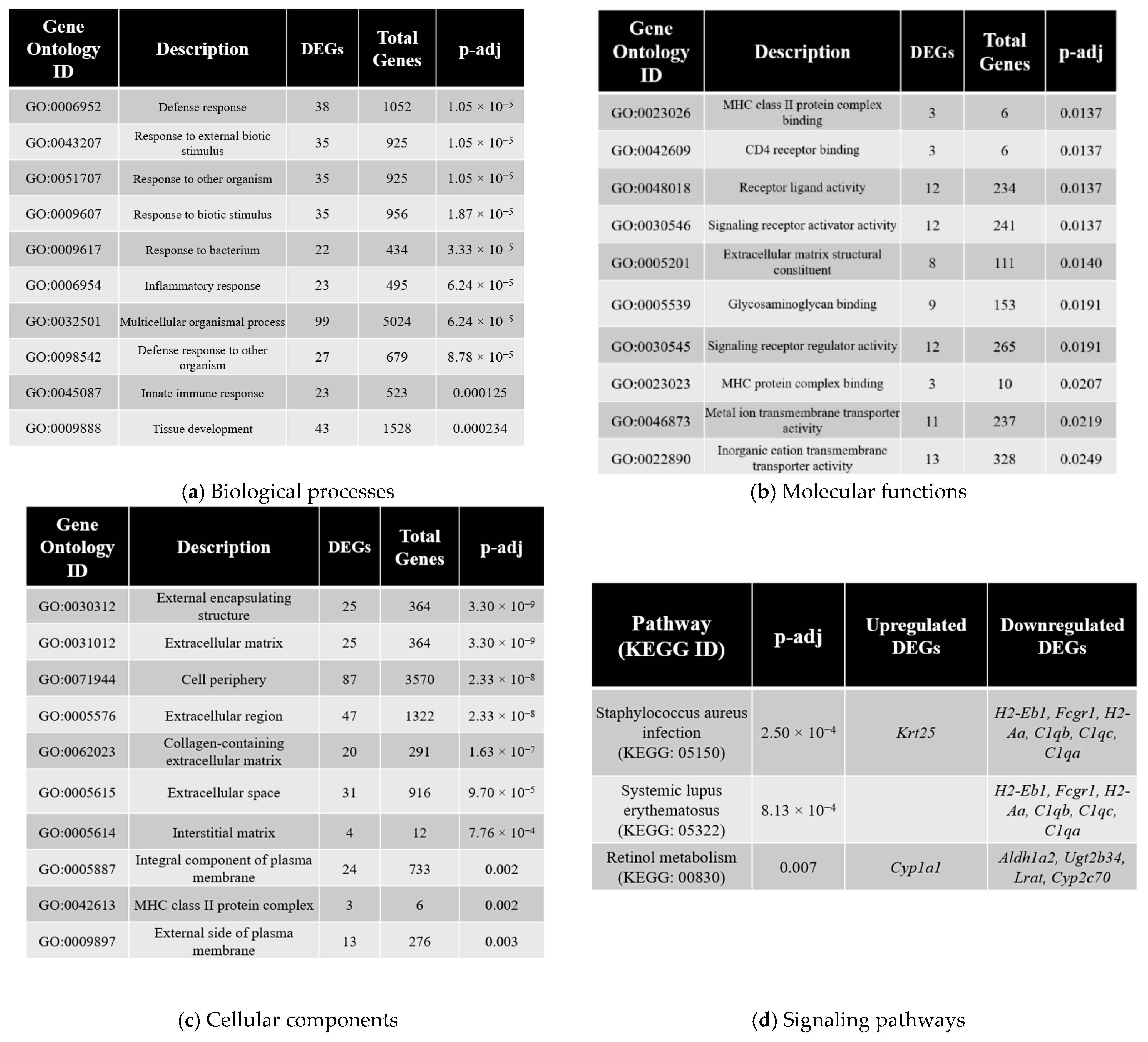
Gene ontology (GO) enrichment analysis was performed on the identified DEGs, in an attempt to better understand the specific biological processes that may be impacted. The iPathwayGuide analysis software (2020 release version) was used to hierarchically organize the DEGs within annotated GO units identified by the GO database (Gene Ontology Consortium database 2021-Nov4) using iPathwayGuide’s proprietary “impact” and “perturbation” analyses. Top 10 enriched GO terms were categorized into categories of biological processes (Figure 3a), molecular functions (Figure 3b) or cellular components (Figure 3c) and are presented in order of increasing adjusted p-value (p-adj). The total number of DEGs identified under each GO term category are listed, in addition to the total number of genes annotated within the GO database for that category.
The top identified biological processes included a number of pathways related to a stimulus response by the cell, including defense response, response to external biotic stimulus, response to other organism, response to biotic stimulus and response to bacterium (Figure 3a). Other noteworthy identified biological processes include inflammatory response and innate immune response, which suggest the involvement of the autoimmune and inflammatory networks. This is of note, based on the pathway enrichment results that also suggest the involvement of autoimmune and inflammatory response-related pathways. The top identified molecular functions include MHC class II protein complex binding and CD4 receptor binding, which are related to activation of inflammatory cytokines. The remaining top identified molecular functions comprise various ligand binding and receptor activity processes (Figure 3b). The top identified cellular components appear to suggest the involvement of the cell periphery, based on the identified targets of external encapsulating membrane, extracellular matrix, cell periphery and extracellular region (Figure 3c).

3.5. Pathway Enrichment Analysis

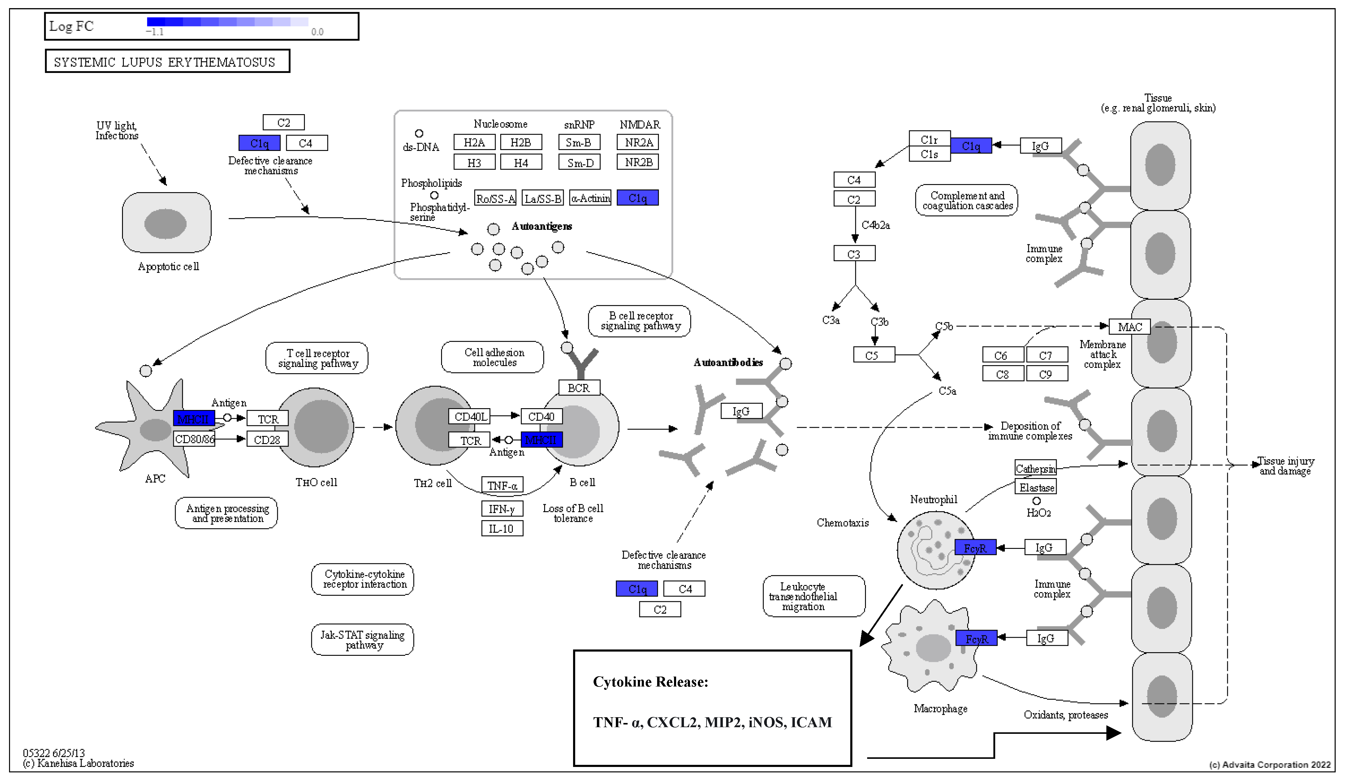
The DEGs were also analyzed using iPathwayGuide’s pathway enrichment analysis, which aims to identify dysregulated molecular signaling families and pathways in the irradiated placenta samples compared to sham-irradiated controls. Global network enrichment analysis identified three significantly affected pathways (FDR p-adj < 0.05; Figure 3d). These pathways included Staphylococcus aureus infection (p = 2.50 × 10⁻⁴), systemic lupus erythematosus (p = 8.13 × 10⁻⁴) and retinol metabolism (p = 0.007). The Staphylococcus aureus infection and systemic lupus erythematosus pathways both involve an autoimmune response by the cell, with a number of DEGs related to immune and complement system identified to be dysregulated in these pathways. The specific DEGs that were either identified to be significantly upregulated or downregulated for the systemic lupus erythematosus and retinol metabolism pathways are identified in Figure 4 and Figure 5, respectively. The majority of DEGs were downregulated in these three pathways, with Krt25 and Cyp1a1 being the only identified upregulated DEGs in the analysis.

3.6. Predicted Upstream Regulator Analysis

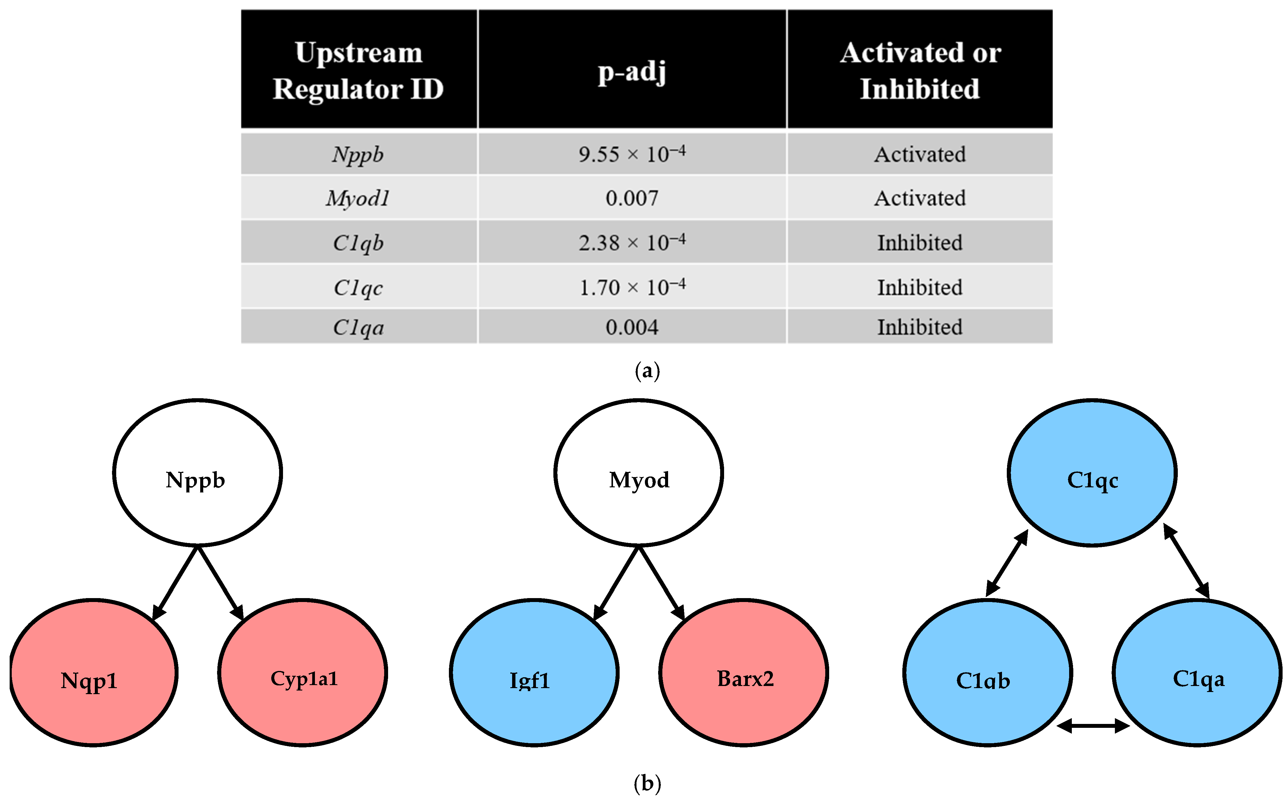
Secondary RNA-seq analysis using iPathwayGuide allowed for the identification of potential “master upstream regulators”, which may act as upstream transcription regulators based on the identified DEGs. The analysis further categorizes predicted upstream regulators as being activated (present) or inhibited (absent). For example, an identified upstream regulator predicted to be activated would be upregulated in treated (irradiated) samples compared to sham-irradiated controls. Conversely, upstream regulators predicted to be inhibited would be downregulated following irradiation. The iPathwayGuide software analysis predicted myogenic differentiation 1 (Myod1) and natriuretic peptide B (Nppb) to be activated, and the components of the complement system (C1qa, C1qb, C1qc) were predicted to be inhibited (Figure 6).

4. Discussion

The objective of the present study was to identify the transcriptional perturbations in placental samples that experienced radiation-induced growth restriction. The goal of this analysis was to better under any dysregulation occurring at the placenta level, which may be responsible for mediating the growth restriction effects observed in the present study. High-dose irradiation effects on the placenta are not well known, and this study has identified a number of novel findings that shed further light on the placental dysregulation underlying the observed intrauterine growth restriction.
The effects of ionizing radiation exposure on the placenta are relatively understudied. One practical reason why this may be true is due to difficulties in collection of placentae as a result of placentophagy (consumption of the placenta and afterbirth) that occurs in various animal species, including rodents [24]. A previous study by Kanter et al. (2014) [25] also investigated the effects of high-dose irradiation in the placenta in a mouse model. The authors irradiated C57Bl/6HNsd mice with 4 Gy gamma radiation on gestational day 13.5 and quantified fetoplacental measurements on day 17.5. Similarly to the present study, the authors were able to demonstrate growth restriction, based on a significantly decreased embryo weight. The authors additionally reported a significant decrease in placental weight, which was not observed in the present study. The authors also assessed placental gene expression; however, there were largely unchanged differences in the placental genes analyzed. With the significant placental growth restriction that was reported by the study authors, this was a surprising result. It is important to note that the authors utilized a microarray approach for gene expression profiling, while we used a next-generation RNA-seq approach. The difference in results could be explained by the fact that the microarray approach profiles predefined transcripts through hybridization, while the RNA-seq method is more encompassing and allows for full sequencing of the whole transcriptome [26]. There are also novel mechanistic insights that are capable with RNA-seq analysis, such as the pathway enrichment or predicted upstream regulator analysis, which is not capable with microarray technology. Nonetheless, one of the placental genes examined by Kanter et al. (2014) [25] that was not significantly affected with irradiation was Igf2; a finding supported by the present study, which showed no significant change in Igf2 gene expression following RNA-seq analysis. Interestingly, Igf1 was one of the significantly downregulated DEGs in the present study, which would provide support for the possible involvement of Igf1, rather than Igf2 specifically, in the observed growth restriction. The present study identified a total of 166 DEGs that met the statistical significance criteria, which may represent targets genes and pathways being affected by placental irradiation that could underlie the observed growth restriction. An important conclusion from the Kanter et al. (2014) [25] study that should be kept in mind when studying fetal growth restriction is the use of whole-body irradiation to the pregnant dam. A consequence of this is that direct irradiation of the fetus may be driving the observed growth effects, independent of the placenta. This is a limitation of the present study, in that both the placenta and fetus were exposed to the x-ray exposures, however the reported RNA-seq results would suggest at least a partial involvement of the placenta in the development of fetal programming.
When considering the totality of the GO terms that were found to be statistically significant in the transcriptomic analysis, there is evidence for the involvement of the autoimmune system and associated inflammation. This is based on identified GO terms that are directly related to the autoimmune system. This included biological processes of inflammatory response (GO:0006954) and innate immune response (GO:0045087). In addition, MHC class II protein complex binding (GO:0023026) and CD4 receptor binding (GO:0042609) were among the top identified molecular function GO terms that were significant in the transcriptomic analysis. Further evidence supporting the possibility of the involvement of an autoimmune response following irradiation was the findings of the pathway enrichment analysis. Among the three signaling pathways that were identified to be statistically significant (p-adj < 0.05), two were related to the autoimmune system, which were Staphylococcus aureus infection (KEGG: 05150) and systemic lupus erythematosus (KEGG:05322). Although the biological features of these two signaling pathways are rather different (with one being the response to bacterial infection and the other being an autoimmune disease), both involve the immune system and an induced inflammatory response. The immune and inflammatory responses to infection by Staphylococcus aureus have been previously reported [27,28]. There is also evidence for adverse pregnancy outcomes in pregnant patients with systemic lupus erythematosus, which includes fetal wastage [29], preterm birth [30] and, importantly, intrauterine growth restriction [31]. This could suggest that high-dose irradiation during pregnancy may trigger similar placental signaling pathways as systemic lupus erythematosus or infection with Staphylococcus aureus, leading to placental dysregulation and ultimately IUGR in the developing offspring. A summary of autoimmune pathways of systemic lupus erythematosus with the specific DEGs identified in the present analysis are provided in Figure 4. The identified DEGs that were significantly downregulated are visualized with blue shading, suggesting an altered autoimmune response following irradiation that may have mechanistic overlap with systemic lupus erythematosus at a cellular level.
Retinol metabolism was another pathway that was identified to be potentially involved in the irradiation response. Vitamin A (or retinol) is an essential nutrient that plays a role in a number of critical biological processes ranging from development and growth to reproduction [32]. Retinoids (which includes retinol and its metabolites) are made available to the developing offspring exclusively through the maternal circulation during gestation [33]. A number of studies have been performed to determine the different rates at which retinoids can cross the placenta [34]. The function of retinol (and its associated metabolites) occur through the actions of retinoic acid, which is a lipid-soluble hormone that interacts with numerous gene targets via receptor-mediated pathways [35]. Another pathophysiology that has been reported to involve dysregulation in retinol metabolism is placental insufficiency [36]. A summary of the DEGs identified in the present study in relation to the metabolic pathways for retinol products is provided in Figure 5. One of the identified DEGs, the retinaldehyde reductase DHRS3, was an overlapping DEG with a previous study that investigated the effects of dexamethasone administration in pregnant Wistar Kyoto rats and subsequent RNA-seq analysis in adrenal glands [18]. Although the prenatal stressor (glucocorticoid administration), target tissue of interest (adrenal gland) and study model (Wistar Kyoto rat) differed from the present study, this parallel finding provides potential evidence for the dysregulation of the retinoid metabolism in multiple tissues. This is of relevance as carotenoid and retinoid compounds have been reported to function as peroxyl radical-scavenging antioxidants [37], which could suggest that a large dose of radiation may be too great of an oxidative stress insult that is overwhelming this system. Retinoids have also been implicated in prenatal lung development [38], which would make pulmonary endpoints in irradiated offspring an interesting avenue for future work. There is interesting mechanistic overlap with other factors that are known to be involved in programming responses, such as maternal nutrient availability or enhanced oxidative stress during development. Although we did not quantify food and water consumption of the pregnant dams, there was a significant decrease in maternal weight gain in the 1.82 Gy group in the days following irradiation compared to sham-irradiated controls. It is also conceivable to assume excess free radical production at the time of irradiation and an altered oxidative stress homeostasis. Further testing would be required in future work, and the role of maternal nutrient availability and oxidative stress and its downstream impacts on the retinoid network would be interesting avenues for future research.
There was evidence for the dysregulation of cytochrome P450 enzyme genes (CYPs) in three separate components of the retinol metabolic pathway. This was for the conversion of all-trans-Retinoate to all-trans-4-Hydroxy-retinoic acid and subsequently all-trans-4-Oxoretinoic acid or conversion to all-trans-18-Hydroxy-retinoic acid. Interestingly, CYP2 was downregulated whereas CYP1A1 was upregulated in the observed DEGs. This could potentially represent a compensatory action by the placental cell in response to the CYP2 downregulation. CYPs are a superfamily of enzymes that are involved with the metabolism of xenobiotic compounds and other physiological metabolites. This is therefore not surprising that CYPs are found in the liver, although it is not specific to this organ, with notably a number of CYPs found to be expressed within the human and mammalian placenta [39]. The highlighted CYPs, CYP1A1 and CYP2 could represent specific targets of interest within the framework of developmental programming. For example, there is evidence that in utero exposure to tobacco increases CYP1A1 expression (a significantly upregulated DEG that was also identified in our analysis) and causes altered methylation status of different xenobiotic response elements with suggestions that CYP1A1 methylation could serve as a molecular biomarker for tobacco exposure in utero [40,41]. CYP2E1 was also reported to be significantly decreased in a dexamethasone-induced model of programming in rats, although this was in a different tissue of interest (the adrenal gland), suggesting that dysregulation of CYPs could be occurring in various tissues, rather than being restricted to the placenta. Another identified gene that was significantly downregulated in the present analysis was lecithin:retinol acyltransferase (Lrat), which is an essential enzyme for mediating retinol storage through esterification of hydrolyzed retinyl esters [42]. Taken together, the secondary analysis of transcriptomic data suggests that antenatal radiation exposure alters the metabolism of different retinoid compounds (through the involvement of CYPs), and represents a potential future avenue of research when considering IUGR or responses to radiation exposure in utero.
The upstream regulator analysis revealed a number of potential candidate “master regulators” that may control or influence the expression levels of the DEGs identified in the present analysis. Two genes of interest, Myod1 and Nppb, were predicted to be activated (upregulated with irradiation; Figure 6a). The downstream target DEGs of these respective genes are also identified in the same Figure 6b. The downregulated DEG in the analysis, Igf1, is an important growth hormone that is also a downstream target of Myod1. The growth hormone/IGF-1 axis has been previously implicated during IUGR, and, furthermore, there is evidence for an epigenetic dysregulation occurring in the axis [43,44,45]. A theoretical epigenetic modification of Myod1 (such as change in DNA methylation or interaction with a microRNA) could explain the observed decrease in Igf1 gene expression with no observed change in the upstream regulator. Cyp1a1 was previously discussed in the retinol metabolism dysregulation section, and therefore its upstream regulator Nppb could also be a target of the retinol metabolism network that is being affected with the radiation treatment. Three genes involved in the classic complement system were predicted to be inhibited with irradiation treatment, which were C1qa, C1qb and C1qc. Interestingly, activation (rather than inhibition) of the complement system (and specifically C1q) has been reported in the literature as a pathophysiological mechanism underlying preeclampsia and associated placental dysfunction during pregnancy, which has also been validated using animal models [46,47]. C1q plays an important role in trophoblast migration and spinal artery remodeling, and therefore downregulation by radiation could impact the normal development of the placenta. Further detailed analysis of the structure of the placenta following radiation exposure would be required to confirm this hypothesis. A study by Agostinis et al. (2010) [48] compared placental markers in wildtype and Cq1 knockout mice, which would serve as an interesting comparison with the observed downregulation of C1q components in the present RNA-seq findings. The authors reported that C1q−/− mice exhibited increased fetal resorption rates, decreased fetal weight and reduced litter size compared to wildtype animals. The authors also reported altered placental labyrinth development and decidual vessel remodeling. The complement system functions together with the immune system (reviewed by Sarma & Ward, 2011 [49]), which could provide further support for an immune dysregulation underlying the growth restriction observed in the present study following high-dose irradiation. It is important to note that despite a statistically significant outcome in the identified upstream regulator targets, further testing and validation is required. The upstream regulator analysis identifies indirect regulatory relationships or shared downstream targets as opposed to direct transcriptional activation within placental tissue. Further validation is required to ensure biological accuracy of these findings, as it is possible for false-positive results due to maternal or fetal contamination in placental samples.
One aspect of the transcriptomic analysis was the detection of significant DEGs between sexes within each treatment condition. There is interest in performing the transcriptomic analysis independently for both sexes, as there is evidence in the literature that the growth restriction and other programming responses of stressors in utero can result in sexually dimorphic responses, including responses detected at the placental level [50,51,52]. A summary Venn diagram of the results are provided in Figure 2c. Of the 13 genes that were significant between the sexes, 6 were different irrespective of the experimental treatment. Unsurprisingly, all six of these genes were located on either X or Y chromosome, and further, the Ddx3y and Xist genes have been previously proposed to be used as placental gene markers for fetal sex determination [53,54]. The remaining seven genes that were differentially expressed between the sexes had variable functions, ranging from genes encoding channel and carrier proteins (Trpm2, Kcnh2, Slc6a14) to DNA methyltransferase enzymes (Dnmt3l). When reviewing the available literature, there is a lack of data that would suggest a sex-specific role of these genes in the growth restriction response. The combination of the lack of sex differences in fetoplacental growth restriction (Figure 1) and the small number of sexually dimorphic gene expression in the placenta identified in the RNA-seq analysis does not provide strong evidence for any sex-specific effects within the placenta following irradiation in this model. This does not preclude the possibility that sexually dimorphic responses could emerge within the placenta at a later life stage.
To our knowledge this study is the first to perform transcriptomic analysis of placenta tissue following high-dose irradiation-induced growth restriction in an animal model. Other models of IUGR have also reported placental transcriptomic or gene expression results, which make them interesting comparisons to the observed results in the present study. Steinhauser et al. (2021) [55] studied the placental transcriptomic response in sheep following maternal nutrient restriction (50% of recommended intake). Similarly to this study, the authors identified MHC class II protein binding as one of their significant molecular function GO terms and evidence for dysregulation of genes related to natural killer cell-mediated cytotoxicity underlying the observed fetal weight in the cohort. It should be noted that nutritional intervention models for placental studies can also occur with overnutrition designs, such as the studies by Gallou-Kabani et al. (2010) [56] and Gabory et al. (2012) [57]. In these studies, the authors reported sex-specific dysregulation of placental genes and epigenetic components in offspring from mice that were maintained on a high-fat diet for the first 15 days of gestation. Another example of an overnutrition model was the study by de Barros Mucci et al. (2020) [58], in which the authors studied the effects of diet-induced maternal obesity on the placental transcriptome in a mouse model. RNA-seq analysis in this study identified dysregulation in targets and pathways related to labyrinth zone development and vascular development in this model. When comparing the identified placental DEGs in these nutritional intervention studies, there was minimal to no overlap in the specific genes that were dysregulated in the present analysis compared to these studies. This would suggest that the molecular changes within the placenta following an altered nutritional homeostasis (whether it is via nutrient restriction or overnutrition) is different from the dysregulation that occurs following high-dose radiation exposure. One challenge with interpreting results across different nutritional intervention studies is the differences in specific micro- and macro-nutrients between study diets. This is an important consideration to keep in mind when comparing and contrasting the findings between different nutritional intervention studies.
Outside of nutritional interventions, Chu et al. (2019) [59] performed placental transcriptomics analysis in a hypoxia-exposed mouse model. This study found further evidence for increased cardiometabolic pathologies by evaluating parameters such as blood pressure and plasma leptin levels once offspring were 4 months of change. An example of the observed placental outcomes was dysregulation of the Hsd11b2 gene, which encodes the enzyme that is critical for glucocorticoid availability to the developing offspring. This study was valuable because the authors were able to connect placental transcriptomic changes following hypoxia exposure to altered in vivo physiological responses in animals. Chemical stressors have also been tested in these placental stressor studies, such as the report by Mao et al. (2020) [60] that tested the effects of endocrine-disrupting chemicals, bisphenol A (BPA) and an inert analog bisphenol S (BPS). Mice were fed 200 ug/kg BPA or BPS and, overall, there was evidence for a similar pattern of placental effects from both compounds. No major areas of overlap were identified when considering the present study, which could suggest that the placental effects of compounds such as BPA may be distinct from the observed changes following high-dose radiation exposure. Studies have also performed profiling for placental changes in placental tissue from human IUGR samples. For example, Majewska et al. (2019) [61] performed RNA-Seq analysis on human IUGR-affected placental samples and secondary analysis suggested involvement of immune and inflammatory disorders related to the IUGR and associated preeclampsia.
Considering these findings in hypoxia and chemical stressors on placental gene regulation and placental responses, there is clearly an interesting mechanistic overlap between radiation exposure and these other stressor models. Despite some similarities in underlying cellular and tissue-level processes, there are generally transcriptional and protein-level changes in different targets following high-dose radiation exposure. Under hypoxic conditions, the placenta has been reported to enhance angiogenesis and maintenance of oxygen and nutrient exchange, largely mediated through the HIF signaling pathway [62]. Chemical exposure has been reported to impact oxidative stress and xenobiotic detoxification pathways, such as Nrf2 and Cyp family genes [63,64]. In contrast, there is a large body of evidence, across various model systems, that has demonstrated that high-dose radiation exposure generally dysregulates genes and pathways associated with processes that include DNA damage, DNA repair, cell-cycle regulation and apoptosis [65]. There are many factors such as the type of radiation that the system is exposed to, absorbed dose, dose-rate or dose fractionation that can strongly impact the exact changes observed, but there are generally conserved responses observed at the cellular level, and further functional testing is needed to elucidate the downstream impacts of these changes on placental structure and function.

5. Conclusions

The present study has identified a number of novel potential targets for future investigation in regard to placental function. This is important for the identification of potential mechanisms underlying placental dysregulation following an exposure to high doses of radiation. It is apparent that the pattern of gene dysregulation of the placenta has similarities and differences to other placental stressor models, which could suggest interesting overlaps in the underlying mechanism of IUGR as a consequence of a stressor such as high-dose radiation exposure. A summary of the overlapping mechanisms based on placental gene expression or transcriptomic evidence in other stressor models is provided in Figure 7. The value in the novel data that was produced from this study is that it provides an indication of placental dysregulation triggered by high-dose radiation exposure, which could be used as a predictor or biomarker of radiation-induced IUGR. Maternal interventions targeting the different dysregulated pathways within the placenta (retinol metabolism, autoimmune effects comparable to systemic lupus erythematosus, altered complement system) represent an exciting avenue of future research that can be explored using the results of the present analysis. More research is needed in this area to further elucidate the pathophysiology of radiation-induced IUGR, and this research provides crucial information of the role of the developing placenta in this response.

Supplementary Materials

The following supporting information can be downloaded at: https://www.mdpi.com/article/10.3390/radiation5040035/s1, A full list of the 166 DEGs identified in this analysis are available in Supplementary Table S1.

Author Contributions

S.S. and S.T. contributed to the overall design, experimentation and writing of the manuscript. C.H. performed the RT-qPCR experiments. C.D.M., C.H. and F.T. performed the genotyping experiments. C.T., N.K., D.R.B., S.J.L. and T.C.T. contributed to project conceptualization, project management, funding acquisition, training, and writing of the manuscript. All authors have read and agreed to the published version of the manuscript.

Funding

Funding was provided by a Natural Sciences and Engineering Research Council of Canada-Collaborative Research and Development (NSERC-CRD-CRDPJ/494077-16) grant and the Nuclear Innovation Institute. SS was funded by a NSERC postgraduate graduate scholarship (PGS-D) and the Michael John Page PhD, Graduate Research Award.

Institutional Review Board Statement

The animal study protocol was approved by the Institutional Animal Ethics Committee of Lakehead University for studies involving animals. All animal work was completed in accordance with the Animal Use Protocol.

Data Availability Statement

The raw data of the findings of this report are available from the corresponding author, upon a reasonable request.

Acknowledgments

The authors thank staff from Lakehead University’s Animal Care Facility for providing support with animal care. The authors also acknowledge and thank the Kyoto Encyclopedia of Genes and Genomes (KEGG) for approving inclusion of the KEGG pathway diagrams in this publication.

Conflicts of Interest

D.R.B. was previously employed by the company Bruce Power. The other authors declare no conflict of interest.

References

  1. Scifres, C.M.; Nelson, D.M. Intrauterine growth restriction, human placental development and trophoblast cell death: IGR, placental development and trophoblast cell death. J. Physiol. 2009, 587, 3453–3458. [Google Scholar] [CrossRef]
  2. Bernstein, I.M.; Horbar, J.D.; Badger, G.J.; Ohlsson, A.; Golan, A. Morbidity and mortality among very-low-birth-weight neonates with intrauterine growth restriction. Am. J. Obstet. Gynecol. 2000, 182, 198–206. [Google Scholar] [CrossRef]
  3. Barker, D.J. The fetal origins of adult hypertension. J. Hypertension. Suppl. Off. J. Int. Soc. Hypertens. 1992, 10, S39–S44. [Google Scholar]
  4. Barker, D.J.; Bagby, S.P.; Hanson, M.A. Mechanisms of Disease: In utero programming in the pathogenesis of hypertension. Nat. Clin. Pract. Nephrol. 2006, 2, 700–707. [Google Scholar] [CrossRef]
  5. Lee, H.-S. Impact of Maternal Diet on the Epigenome during In Utero Life and the Developmental Programming of Diseases in Childhood and Adulthood. Nutrients 2015, 7, 9492–9507. [Google Scholar] [CrossRef]
  6. Seckl, J.R. Glucocorticoid programming of the fetus; adult phenotypes and molecular mechanisms. Mol. Cell. Endocrinol. 2001, 185, 61–71. [Google Scholar] [CrossRef]
  7. Fajersztajn, L.; Veras, M.M. Hypoxia: From Placental Development to Fetal Programming: Influence of Hypoxia on Development. Birth Defects Res. 2017, 109, 1377–1385. [Google Scholar] [CrossRef] [PubMed]
  8. Maritz, G.S.; Harding, R. Life-long Programming Implications of Exposure to Tobacco Smoking and Nicotine Before and Soon After Birth: Evidence for Altered Lung Development. Int. J. Environ. Res. Public Health 2011, 8, 875–898. [Google Scholar] [CrossRef] [PubMed]
  9. Frangione, B.; Hinton, P.; Villeneuve, P.J. Low-dose ionizing radiation and adverse birth outcomes: A systematic review and meta-analysis. Int. Arch. Occup. Environ. Health 2023, 96, 77–92. [Google Scholar] [CrossRef] [PubMed]
  10. Sugiyama, H.; Misumi, M.; Sakata, R.; Brenner, A.V.; Utada, M.; Ozasa, K. Mortality among individuals exposed to atomic bomb radiation in utero: 1950–2012. Eur. J. Epidemiol. 2021, 36, 415–428. [Google Scholar] [CrossRef] [PubMed]
  11. Turco, M.Y.; Moffett, A. Development of the human placenta. Development 2019, 146, dev163428. [Google Scholar] [CrossRef]
  12. Watson, E.D.; Cross, J.C. Development of Structures and Transport Functions in the Mouse Placenta. Physiology 2005, 20, 180–193. [Google Scholar] [CrossRef]
  13. Chaddha, V.; Viero, S.; Huppertz, B.; Kingdom, J. Developmental biology of the placenta and the origins of placental insufficiency. Semin. Fetal Neonatal Med. 2004, 9, 357–369. [Google Scholar] [CrossRef]
  14. StarrLab. DNA Extraction from Tail Clips. Available online: https://sites.google.com/a/umn.edu/starrlab/protocols/dna/dna-extraction-from-tail-clips/ (accessed on 24 October 2022).
  15. The Jackson Laboratory. DNA Isolation Protocols. Available online: https://www.jax.org/jax-mice-and-services/customer-support/technical-support/genotyping-resources/dna-isolation-protocols/ (accessed on 24 October 2022).
  16. Clapcote, S.J.; Roder, J.C. Simplex PCR assay for sex determination in mice. BioTechniques 2005, 38, 702–706. [Google Scholar] [CrossRef]
  17. Pirkkanen, J.; Tharmalingam, S.; Morais, I.H.; Lam-Sidun, D.; Thome, C.; Zarnke, A.M.; Benjamin, L.V.; Losch, A.C.; Borgmann, A.J.; Sinex, H.C.; et al. Transcriptomic profiling of gamma ray induced mutants from the CGL1 human hybrid cell system reveals novel insights into the mechanisms of radiation-induced carcinogenesis. Free Radic. Biol. Med. 2019, 145, 300–311. [Google Scholar] [CrossRef]
  18. Tharmalingam, S.; Khurana, S.; Murray, A.; Lamothe, J.; Tai, T.C. Whole transcriptome analysis of adrenal glands from prenatal glucocorticoid programmed hypertensive rodents. Sci. Rep. 2020, 10, 18755. [Google Scholar] [CrossRef]
  19. Al-khayyat, W.; Pirkkanen, J.; Dougherty, J.; Laframboise, T.; Dickinson, N.; Khaper, N.; Lees, S.J.; Mendonca, M.S.; Boreham, D.R.; Tai, T.C.; et al. Overexpression of FRA1 (FOSL1) Leads to Global Transcriptional Perturbations, Reduced Cellular Adhesion and Altered Cell Cycle Progression. Cells 2023, 12, 2344. [Google Scholar] [CrossRef] [PubMed]
  20. Ye, J.; Coulouris, G.; Zaretskaya, I.; Cutcutache, I.; Rozen, S.; Madden, T.L. Primer-BLAST: A tool to design target-specific primers for polymerase chain reaction. BMC Bioinform. 2012, 13, 134. [Google Scholar] [CrossRef] [PubMed]
  21. Solano, M.E.; Thiele, K.; Kowal, M.K.; Arck, P.C. Identification of suitable reference genes in the mouse placenta. Placenta 2016, 39, 7–15. [Google Scholar] [CrossRef] [PubMed]
  22. Livak, K.J.; Schmittgen, T.D. Analysis of Relative Gene Expression Data Using Real-Time Quantitative PCR and the 2−ΔΔCT Method. Methods 2001, 25, 402–408. [Google Scholar] [CrossRef]
  23. Draghici, S.; Khatri, P.; Tarca, A.L.; Amin, K.; Done, A.; Voichita, C.; Georgescu, C.; Romero, R. A systems biology approach for pathway level analysis. Genome Res. 2007, 17, 1537–1545. [Google Scholar] [CrossRef]
  24. Kristal, M.B.; DiPirro, J.M.; Thompson, A.C. Placentophagia in Humans and Nonhuman Mammals: Causes and Consequences. Ecol. Food Nutr. 2012, 51, 177–197. [Google Scholar] [CrossRef] [PubMed]
  25. Kanter, D.J.; O’Brien, M.B.; Shi, X.-H.; Chu, T.; Mishima, T.; Beriwal, S.; Epperly, M.W.; Wipf, P.; Greenberger, J.S.; Sadovsky, Y. The impact of ionizing radiation on placental trophoblasts. Placenta 2014, 35, 85–91. [Google Scholar] [CrossRef]
  26. Rao, M.S.; Van Vleet, T.R.; Ciurlionis, R.; Buck, W.R.; Mittelstadt, S.W.; Blomme, E.A.G.; Liguori, M.J. Comparison of RNA-Seq and Microarray Gene Expression Platforms for the Toxicogenomic Evaluation of Liver From Short-Term Rat Toxicity Studies. Front. Genet. 2019, 9, 636. [Google Scholar] [CrossRef]
  27. Seyyed Mousavi, M.N.; Mehramuz, B.; Sadeghi, J.; Alizadeh, N.; Oskouee, M.A.; Kafil, H.S. The pathogenesis of Staphylococcus aureus in autoimmune diseases. Microb. Pathog. 2017, 111, 503–507. [Google Scholar] [CrossRef]
  28. Liu, Q.; Mazhar, M.; Miller, L.S. Immune and Inflammatory Reponses to Staphylococcus aureus Skin Infections. Curr. Dermatol. Rep. 2018, 7, 338–349. [Google Scholar] [CrossRef] [PubMed]
  29. Watson, R.M.; Lane, A.T.; Barnett, N.K.; Bias, W.B.; Arnett, F.C.; Provost, T.T. Neonatal lupus erythematosus. A clinical, serological and immunogenetic study with review of the literature. Medicine 1984, 63, 362–378. [Google Scholar] [CrossRef]
  30. Wei, S.; Lai, K.; Yang, Z.; Zeng, K. Systemic lupus erythematosus and risk of preterm birth: A systematic review and meta-analysis of observational studies. Lupus 2017, 26, 563–571. [Google Scholar] [CrossRef]
  31. Guillotin, V.; Bouhet, A.; Barnetche, T.; Richez, C.; Truchetet, M.-E.; Seneschal, J.; Duffau, P.; Lazaro, E. Hydroxychloroquine for the prevention of fetal growth restriction and prematurity in lupus pregnancy: A systematic review and meta-analysis. Jt. Bone Spine 2018, 85, 663–668. [Google Scholar] [CrossRef]
  32. Blomhoff, R.; Blomhoff, H.K. Overview of retinoid metabolism and function. J. Neurobiol. 2006, 66, 606–630. [Google Scholar] [CrossRef] [PubMed]
  33. Kim, Y.-K.; Wassef, L.; Hamberger, L.; Piantedosi, R.; Palczewski, K.; Blaner, W.S.; Quadro, L. Retinyl Ester Formation by Lecithin:Retinol Acyltransferase Is a Key Regulator of Retinoid Homeostasis in Mouse Embryogenesis. J. Biol. Chem. 2008, 283, 5611–5621. [Google Scholar] [CrossRef]
  34. Marceau, G.; Gallot, D.; Lemery, D.; Sapin, V. Metabolism of Retinol During Mammalian Placental and Embryonic Development. In Vitamins & Hormones; Elsevier: Amsterdam, The Netherlands, 2007; pp. 97–115. [Google Scholar]
  35. Balmer, J.E.; Blomhoff, R. Gene expression regulation by retinoic acid. J. Lipid Res. 2002, 43, 1773–1808. [Google Scholar] [CrossRef]
  36. Huang, L.-T.; Chou, H.-C.; Lin, C.-M.; Chen, C.-M. Uteroplacental Insufficiency Alters the Retinoid Pathway and Lung Development in Newborn Rats. Pediatr. Neonatol. 2016, 57, 508–514. [Google Scholar] [CrossRef] [PubMed]
  37. Edge, R.; Truscott, T. Singlet Oxygen and Free Radical Reactions of Retinoids and Carotenoids—A Review. Antioxidants 2018, 7, 5. [Google Scholar] [CrossRef]
  38. Fernandes-Silva, H.; Araújo-Silva, H.; Correia-Pinto, J.; Moura, R.S. Retinoic Acid: A Key Regulator of Lung Development. Biomolecules 2020, 10, 152. [Google Scholar] [CrossRef]
  39. Pasanen, M. The expression and regulation of drug metabolism in human placenta. Adv. Drug Deliv. Rev. 1999, 38, 81–97. [Google Scholar] [CrossRef]
  40. Suter, M.; Abramovici, A.; Showalter, L.; Hu, M.; Shope, C.D.; Varner, M.; Aagaard-Tillery, K. In utero tobacco exposure epigenetically modifies placental CYP1A1 expression. Metabolism 2010, 59, 1481–1490. [Google Scholar] [CrossRef] [PubMed]
  41. Janssen, B.G.; Gyselaers, W.; Byun, H.-M.; Roels, H.A.; Cuypers, A.; Baccarelli, A.A.; Nawrot, T.S. Placental mitochondrial DNA and CYP1A1 gene methylation as molecular signatures for tobacco smoke exposure in pregnant women and the relevance for birth weight. J. Transl. Med. 2017, 15, 5. [Google Scholar] [CrossRef] [PubMed]
  42. Spiegler, E.; Kim, Y.-K.; Wassef, L.; Shete, V.; Quadro, L. Maternal–fetal transfer and metabolism of vitamin A and its precursor β-carotene in the developing tissues. Biochim. Et Biophys. Acta (BBA)-Mol. Cell Biol. Lipids 2012, 1821, 88–98. [Google Scholar] [CrossRef]
  43. Laviola, L.; Perrini, S.; Belsanti, G.; Natalicchio, A.; Montrone, C.; Leonardini, A.; Vimercati, A.; Scioscia, M.; Selvaggi, L.; Giorgino, R.; et al. Intrauterine Growth Restriction in Humans Is Associated with Abnormalities in Placental Insulin-Like Growth Factor Signaling. Endocrinology 2005, 146, 1498–1505. [Google Scholar] [CrossRef]
  44. Setia, S.; Sridhar, M.G. Changes in GH/IGF-1 Axis in Intrauterine Growth Retardation: Consequences of Fetal Programming? Horm. Metab. Res. 2009, 41, 791–798. [Google Scholar] [CrossRef] [PubMed]
  45. Álvarez-Nava, F.; Lanes, R. GH/IGF-1 Signaling and Current Knowledge of Epigenetics; a Review and Considerations on Possible Therapeutic Options. Int. J. Mol. Sci. 2017, 18, 1624. [Google Scholar] [CrossRef]
  46. Regal, J.F.; Burwick, R.M.; Fleming, S.D. The Complement System and Preeclampsia. Curr. Hypertens. Rep. 2017, 19, 87. [Google Scholar] [CrossRef] [PubMed]
  47. Pierik, E.; Prins, J.R.; van Goor, H.; Dekker, G.A.; Daha, M.R.; Seelen, M.A.J.; Scherjon, S.A. Dysregulation of Complement Activation and Placental Dysfunction: A Potential Target to Treat Preeclampsia? Front. Immunol. 2020, 10, 3098. [Google Scholar] [CrossRef]
  48. Agostinis, C.; Bulla, R.; Tripodo, C.; Gismondi, A.; Stabile, H.; Bossi, F.; Guarnotta, C.; Garlanda, C.; De Seta, F.; Spessotto, P.; et al. An Alternative Role of C1q in Cell Migration and Tissue Remodeling: Contribution to Trophoblast Invasion and Placental Development. J. Immunol. 2010, 185, 4420–4429. [Google Scholar] [CrossRef] [PubMed]
  49. Sarma, J.V.; Ward, P.A. The complement system. Cell Tissue Res. 2011, 343, 227–235. [Google Scholar] [CrossRef]
  50. Pennington, K.A.; Harper, J.L.; Sigafoos, A.N.; Beffa, L.M.; Carleton, S.M.; Phillips, C.L.; Schulz, L.C. Effect of Food Restriction and Leptin Supplementation on Fetal Programming in Mice. Endocrinology 2012, 153, 4556–4567. [Google Scholar] [CrossRef]
  51. Tarrade, A.; Panchenko, P.; Junien, C.; Gabory, A. Placental contribution to nutritional programming of health and diseases: Epigenetics and sexual dimorphism. J. Exp. Biol. 2015, 218, 50–58. [Google Scholar] [CrossRef]
  52. Kalisch-Smith, J.I.; Simmons, D.G.; Dickinson, H.; Moritz, K.M. Review: Sexual dimorphism in the formation, function and adaptation of the placenta. Placenta 2017, 54, 10–16. [Google Scholar] [CrossRef]
  53. Hoch, D.; Novakovic, B.; Cvitic, S.; Saffery, R.; Desoye, G.; Majali-Martinez, A. Sex matters: XIST and DDX3Y gene expression as a tool to determine fetal sex in human first trimester placenta. Placenta 2020, 97, 68–70. [Google Scholar] [CrossRef]
  54. Ontsouka, E.; Lüthi, M.; Zaugg, J.; Schroeder, M.; Albrecht, C. Establishment and validation of an approach allowing unequivocal fetal sex determination based on placental sex-specific genes. Placenta 2021, 112, 132–134. [Google Scholar] [CrossRef]
  55. Steinhauser, C.B.; Lambo, C.A.; Askelson, K.; Burns, G.W.; Behura, S.K.; Spencer, T.E.; Bazer, F.W.; Satterfield, M.C. Placental Transcriptome Adaptations to Maternal Nutrient Restriction in Sheep. Int. J. Mol. Sci. 2021, 22, 7654. [Google Scholar] [CrossRef]
  56. Gallou-Kabani, C.; Gabory, A.; Tost, J.; Karimi, M.; Mayeur, S.; Lesage, J.; Boudadi, E.; Gross, M.-S.; Taurelle, J.; Vigé, A.; et al. Sex- and Diet-Specific Changes of Imprinted Gene Expression and DNA Methylation in Mouse Placenta under a High-Fat Diet. PLoS ONE 2010, 5, e14398. [Google Scholar] [CrossRef]
  57. Gabory, A.; Ferry, L.; Fajardy, I.; Jouneau, L.; Gothié, J.-D.; Vigé, A.; Fleur, C.; Mayeur, S.; Gallou-Kabani, C.; Gross, M.-S.; et al. Maternal Diets Trigger Sex-Specific Divergent Trajectories of Gene Expression and Epigenetic Systems in Mouse Placenta. PLoS ONE 2012, 7, e47986. [Google Scholar] [CrossRef]
  58. de Barros Mucci, D.; Kusinski, L.C.; Wilsmore, P.; Loche, E.; Pantaleão, L.C.; Ashmore, T.J.; Blackmore, H.L.; Fernandez-Twinn, D.S.; Carmo, M.D.G.T.D.; Ozanne, S.E. Impact of maternal obesity on placental transcriptome and morphology associated with fetal growth restriction in mice. Int. J. Obes. 2020, 44, 1087–1096. [Google Scholar] [CrossRef] [PubMed]
  59. Chu, A.; Casero, D.; Thamotharan, S.; Wadehra, M.; Cosi, A.; Devaskar, S.U. The Placental Transcriptome in Late Gestational Hypoxia Resulting in Murine Intrauterine Growth Restriction Parallels Increased Risk of Adult Cardiometabolic Disease. Sci. Rep. 2019, 9, 1243. [Google Scholar] [CrossRef] [PubMed]
  60. Mao, J.; Jain, A.; Denslow, N.D.; Nouri, M.-Z.; Chen, S.; Wang, T.; Zhu, N.; Koh, J.; Sarma, S.J.; Sumner, B.W.; et al. Bisphenol A and bisphenol S disruptions of the mouse placenta and potential effects on the placenta–brain axis. Proc. Natl. Acad. Sci. USA 2020, 117, 4642–4652. [Google Scholar] [CrossRef]
  61. Majewska, M.; Lipka, A.; Paukszto, L.; Jastrzebski, J.; Szeszko, K.; Gowkielewicz, M.; Lepiarczyk, E.; Jozwik, M.; Majewski, M. Placenta Transcriptome Profiling in Intrauterine Growth Restriction (IUGR). Int. J. Mol. Sci. 2019, 20, 1510. [Google Scholar] [CrossRef] [PubMed]
  62. Fryer, B.H.; Simon, M.C. Hypoxia, HIF and the Placenta. Cell Cycle 2006, 5, 495–498. [Google Scholar] [CrossRef]
  63. Bischoff, L.J.M.; Kuijper, I.A.; Schimming, J.P.; Wolters, L.; Braak, B.T.; Langenberg, J.P.; Noort, D.; Beltman, J.B.; Van De Water, B. A systematic analysis of Nrf2 pathway activation dynamics during repeated xenobiotic exposure. Arch. Toxicol. 2019, 93, 435–451. [Google Scholar] [CrossRef]
  64. Esteves, F.; Rueff, J.; Kranendonk, M. The Central Role of Cytochrome P450 in Xenobiotic Metabolism—A Brief Review on a Fascinating Enzyme Family. J. Xenobiotics 2021, 11, 94–114. [Google Scholar] [CrossRef]
  65. Hall, E.J.; Giaccia, A.J. Radiobiology for the Radiologist; Wolters Kluwer: Philadelphia, PA, USA, 2019; p. 597. [Google Scholar]
  66. Gundogan, F.; Gilligan, J.; Qi, W.; Chen, E.; Naram, R.; De La Monte, S.M. Dose effect of gestational ethanol exposure on placentation and fetal growth. Placenta 2015, 36, 523–530. [Google Scholar] [CrossRef]
  67. Simmers, M.D.; Hudson, K.M.; Baptissart, M.; Cowley, M. Epigenetic control of the imprinted growth regulator Cdkn1c in cadmium-induced placental dysfunction. Epigenetics 2022, 18, 2088173. [Google Scholar] [CrossRef]
  68. Gonzalez, P.N.; Gasperowicz, M.; Barbeito-Andrés, J.; Klenin, N.; Cross, J.C.; Hallgrímsson, B. Chronic Protein Restriction in Mice Impacts Placental Function and Maternal Body Weight before Fetal Growth. PLoS ONE 2016, 11, e0152227. [Google Scholar] [CrossRef]
  69. Kelly, A.C.; Powell, T.L.; Jansson, T. Placental function in maternal obesity. Clin. Sci. 2020, 134, 961–984. [Google Scholar] [CrossRef]
  70. Li, B.; Chang, S.; Liu, C.; Zhang, M.; Zhang, L.; Liang, L.; Li, R.; Wang, X.; Qin, C.; Zhang, T.; et al. Low Maternal Dietary Folate Alters Retrotranspose by Methylation Regulation in Intrauterine Growth Retardation (IUGR) Fetuses in a Mouse Model. Med. Sci. Monit. 2019, 25, 3354–3365. [Google Scholar] [CrossRef]
  71. Cao, C.; Prado, M.A.; Sun, L.; Rockowitz, S.; Sliz, P.; Paulo, J.A.; Finley, D.; Fleming, M.D. Maternal Iron Deficiency Modulates Placental Transcriptome and Proteome in Mid-Gestation of Mouse Pregnancy. J. Nutr. 2021, 151, 1073–1083. [Google Scholar] [CrossRef]
  72. Arias, A.; Schander, J.A.; Bariani, M.V.; Correa, F.; Domínguez Rubio, A.P.; Cella, M.; Cymeryng, C.B.; Wolfson, M.L.; Franchi, A.M.; Aisemberg, J. Dexamethasone-induced intrauterine growth restriction modulates expression of placental vascular growth factors and fetal and placental growth. Mol. Hum. Reprod. 2021, 27, gaab006. [Google Scholar] [CrossRef] [PubMed]
  73. Czamara, D.; Dieckmann, L.; Röh, S.; Kraemer, S.; Rancourt, R.C.; Sammallahti, S.; Kajantie, E.; Laivuori, H.; Eriksson, J.G.; Räikkönen, K.; et al. Betamethasone administration during pregnancy is associated with placental epigenetic changes with implications for inflammation. Clin. Epigenetics 2021, 13, 165. [Google Scholar] [CrossRef] [PubMed]
  74. James, J.L.; Whitley, G.S.; Cartwright, J.E. Pre-eclampsia: Fitting together the placental, immune and cardiovascular pieces. J. Pathol. 2010, 221, 363–378. [Google Scholar] [CrossRef] [PubMed]
  75. Magid, M.S.; Kaplan, C.; Sammaritano, L.R.; Peterson, M.; Druzin, M.L.; Lockshin, M.D. Placental pathology in systemic lupus erythematosus: A prospective study. Am. J. Obstet. Gynecol. 1998, 179, 226–234. [Google Scholar] [CrossRef] [PubMed]
  76. Bordt, E.A.; Shook, L.L.; Atyeo, C.; Pullen, K.M.; De Guzman, R.M.; Meinsohn, M.-C.; Chauvin, M.; Fischinger, S.; Yockey, L.J.; James, K.; et al. Maternal SARS-CoV-2 infection elicits sexually dimorphic placental immune responses. Sci. Transl. Med. 2021, 13, eabi7428. [Google Scholar] [CrossRef] [PubMed]
  77. Yung, H.W.; Cox, M.; Patot, M.T.; Burton, G.J. Evidence of endoplasmic reticulum stress and protein synthesis inhibition in the placenta of non-native women at high altitude. FASEB J. 2012, 26, 1970–1981. [Google Scholar] [CrossRef] [PubMed]
Figure 1. Fetoplacental weight following irradiation. Fetoplacental weight measurements of conceptus weight (a) and placental weight (b) following 1.82 Gy x-ray irradiation on gestational day 14.5. The effect of irradiation treatment was significant for conceptus weight (*; 2-way ANOVA; p < 0.05). Sample sizes were n = 9 for all treatment groups, with the exception of 1.82 Gy, female cohort (n = 10). All studied conceptus and placental samples were from unique dams, to minimize any maternally driven effects.
Radiation 05 00035 g001
Figure 2. Differentially expressed gene (DEG) analysis. DEGs identified following RNA-seq transcriptomic analysis of irradiated placenta samples. A list of the top 10 identified upregulated (a) and downregulated (b) DEGs are presented, ranked in order of the magnitude of fold change. p-adjusted values are reported for each DEG. (c) Volcano plot of the 166 DEGs presented in terms of their measured change (x-axis; log fold-change) and the significance of the change (y-axis). Dotted lines represent threshold cut-off values of +1.5 or −1.5 fold-change (corresponding to a log fold-change of +0.6 and −0.6, respectively). Grey circles indicate genes that did not meet the threshold for statistical significance. Top dysregulated DEGs in panels (a,b) are annotated. Volcano plot figure was made using the VolcaNoseR app (version 1), hosted by Hyugens Science. (d) Venn diagram of sex-specific DEGs.
Radiation 05 00035 g002
Figure 3. Gene ontology and pathway enrichment results. Top 10 enriched GO terms were categorized as biological processes (a), molecular functions (b), cellular components (c) and signaling pathways (d), and are ranked by false discovery rate (FDR) adjusted p-value (FDR p-adjusted value < 0.05).
Radiation 05 00035 g003
Figure 4. Autoimmune and complement pathways in Systemic Lupus Erythematosus (KEGG: 05322). The KEGG (Kyoto Encyclopedia of Genes and Genomes) pathway diagram is superimposed with the degree of perturbation (which accounts for both the gene’s measured fold change (FC) and for the accumulated perturbation that is propagated down from upstream genes). The greatest magnitude of negative perturbation is shown in dark blue. The legend identifies the values on the color gradient of log FC. Pathway diagram was produced in iPathwayGuide (Advaita Bioinformatics).
Radiation 05 00035 g004
Figure 5. Retinol metabolism pathway (KEGG: 00830). The KEGG (Kyoto Encyclopedia of Genes and Genomes) pathway diagram is superimposed with the degree of perturbation (which accounts for both the gene’s measured fold change (FC) and for the accumulated perturbation that is propagated down from upstream genes). The greatest magnitude of negative perturbation is shown in dark blue, and the greatest magnitude of positive perturbation is shown in dark red. The legend identifies the values on the color gradient of log FC. Pathway diagram was produced in iPathwayGuide (Advaita Bioinformatics).
Radiation 05 00035 g005
Figure 6. Predicted upstream regulators. (a) Five genes: Nppb (Natriuretic Peptide B), Myod1 (Myogenic Differentiation 1), Complement C1q chains C1qa, C1qb and C1qc were identified as potential upstream regulators that were activated (increased expression) or inhibited (decreased expression) with the 1.82 Gy irradiation treatment, with significant p-adjusted values (<0.05). (b) Interaction map of the predicted upstream regulators with their downstream upregulated (red), downregulated (blue) or unchanged (white) DEG targets.
Radiation 05 00035 g006
Figure 7. Placental IUGR (intrauterine growth restriction) stressor models. Summary diagram of different stressor models of IUGR that have been reported to involve placental dysregulation or effects. Figure created with BioRender.com. Superscript references in Figure 7: 1. Suter et al. (2016) [40]; 2. Gundogen et al. (2015) [66]; 3. Simmers et al. (2022) [67]; 4. Mao et al. (2020) [60]; 5. Gonzalez et al. (2016) [68]; 6. Aravidou et al. (2015) [25]; 7. de Barros Mucci et al. (2020) [58]; 8. Kelly et al. (2020) [69]; 9. Li et al. (2019) [70]; 10. Cao et al. (2021) [71]; 11. Arias et al. (2021) [72]; 12. Czamara et al. (2021) [73]; 13. James et al. (2010) [74]; 14. Magid et al. (1998) [75]; 15. Bordt et al. (2021) [76]; 16. Yung et al. (2012) [77]; 17. Chu et al. (2019) [59]; 18. Kanter et al. (2014) [25].
Radiation 05 00035 g007
Table 1. Quantitative reverse transcription polymerase chain reaction (RT-qPCR) validation of RNA-seq fold change values. A comparison of fold change (relative to sham-irradiated controls) for the RNA-seq and RT-qPCR methods is presented for each gene. p-adjusted values are reported for each respective fold change.
GeneSexFold Change
(RNA-Seq)
p-adjFold Change
(RT-qPCR)
p-adj
Rasl11bFemale−1.000.994−1.020.972
Male+1.000.999−1.010.999
Hsd11b2Female−1.040.947−1.110.273
Male−1.020.972−1.030.965
Sec61bFemale+1.040.827+1.080.701
Male−1.030.878+1.080.643
Mtmr7Female+1.020.951−1.000.999
Male−1.020.932−1.010.998
Slc5a5Female+1.891.08 × 10−4+1.250.111
Male+1.923.26 × 10−4+1.481.30 × 10−3
Wsb1Female−1.140.210−1.204.40 × 10−3
Male−1.160.152−1.070.575
Dio2Female−2.210.0286−1.490.0469
Male−2.160.117−1.808.80 × 10−3
Disclaimer/Publisher’s Note: The statements, opinions and data contained in all publications are solely those of the individual author(s) and contributor(s) and not of MDPI and/or the editor(s). MDPI and/or the editor(s) disclaim responsibility for any injury to people or property resulting from any ideas, methods, instructions or products referred to in the content.