
Chernobyl’s Aftermath: Multiple Manifestations of Basalioma in a Patient after Radioactive Contamination in 1986


 , ,
, , 
Abstract
Simple Summary
Abstract
1. Introduction
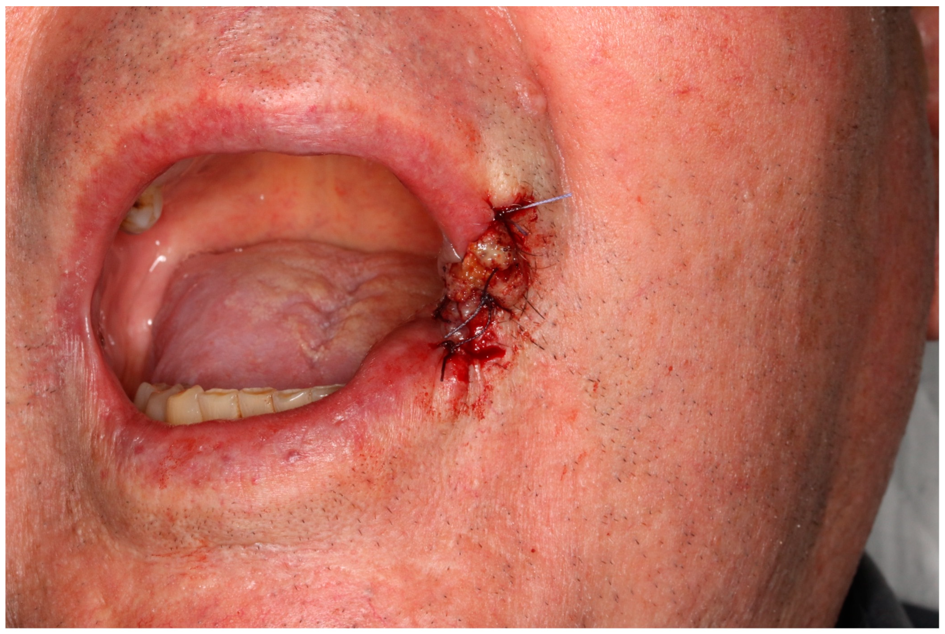
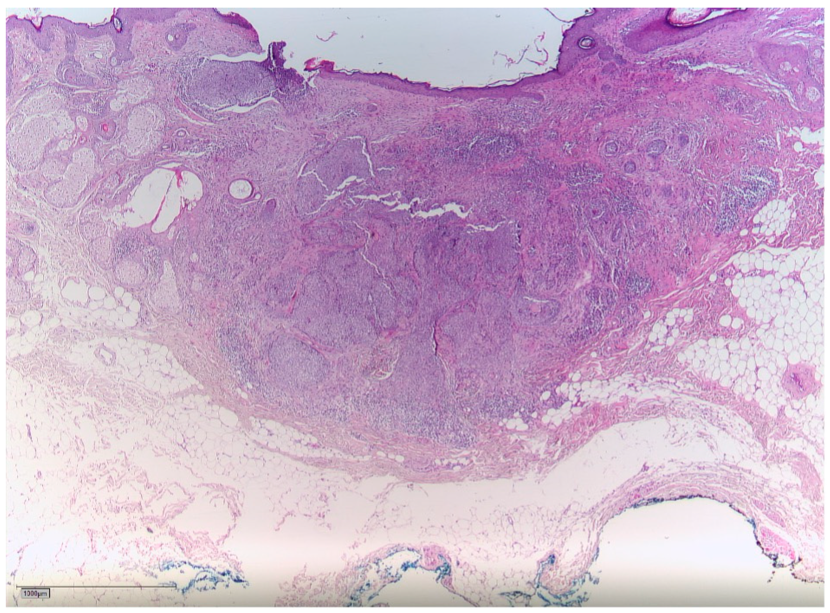
2. Case Report
3. Discussion
4. Conclusions
Author Contributions
Funding
Institutional Review Board Statement
Informed Consent Statement
Data Availability Statement
Conflicts of Interest
References
- International Atomic Energy Agency. The Chernobyl Accident: Updating of INSAG-1. A Report by the International Nuclear Safety Advisory Group; Safety Series; International Atomic Energy Agency: Vienna, Austria, 1992; Volume INSAG-7, 136p. [Google Scholar]
- McCall, C. Chernobyl disaster 30 years on: Lessons not learned. Lancet 2016, 387, 1707–1708. [Google Scholar] [CrossRef]
- Sugiyama, H.; Misumi, M.; Kishikawa, M.; Iseki, M.; Yonehara, S.; Hayashi, T.; Soda, M.; Tokuoka, S.; Shimizu, Y.; Sakata, R.; et al. Skin Cancer Incidence among Atomic Bomb Survivors from 1958 to 1996. Radiat. Res. 2014, 181, 531–539. [Google Scholar] [CrossRef]
- Karagas, M.R.; McDonald, J.A.; Greendberg, E.R.; Stukel, T.A.; Weiss, J.E.; Baron, J.A.; Stevens, M.M. For the Skin Cancer Prevention Study Group Risk of Basal Cell and Squamous Cell Skin Cancers After Ionizing Radiation Therapy. JNCI J. Natl. Cancer Inst. 1996, 88, 1848–1853. [Google Scholar] [CrossRef]
- Eagan, J.T., Jr.; Jones, C.T. Cutaneous Cancers in an Interventional Cardiologist: A Cautionary Tale: Cutaneous cancers in an interventional cardiologist. J. Intervent. Cardiol. 2011, 24, 49–55. [Google Scholar] [CrossRef]
- Shore, R.E.; Albert, R.E.; Pasternack, B.S. Follow-up Study of Patients Treated by X-ray Epilation for Tinea Capitis: Resurvey of Post-Treatment Illness and Mortality Experience. Arch. Environ. Health Int. J. 1976, 31, 21–28. [Google Scholar] [CrossRef]
- Mizuno, T.; Tokuoka, S.; Kishikawa, M.; Nakashima, E.; Mabuchi, K.; Iwamoto, K.S. Molecular basis of basal cell carcinogenesis in the atomic-bomb survivor population: p53 and PTCH gene alterations. Carcinogenesis 2006, 27, 2286–2294. [Google Scholar] [CrossRef][Green Version]
- Baheti, A.D.; Tirumani, S.H.; Giardino, A.; Rosenthal, M.H.; Tirumani, H.; Krajewski, K.; Ramaiya, N.H. Basal Cell Carcinoma: A Comprehensive Review for the Radiologist. Am. J. Roentgenol. 2015, 204, W132–W140. [Google Scholar] [CrossRef]
- Lang, B.; Balermpas, P.; Bauer, A. S2k-Leitlinie Basalzellkarzinom der Haut (Aktualisierung 2017/2018). J. Dtsch. Dermatol. Ges. 2019, 17, 94–104. [Google Scholar]
- Fosko, S.W. Positron Emission Tomography for Basal Cell Carcinoma of the Head and Neck. Arch. Dermatol. 2003, 139, 1141. [Google Scholar] [CrossRef]
- Weiss, G.J.; Korn, R.L. Metastatic basal cell carcinoma in the era of hedgehog signaling pathway inhibitors: Metastatic Basal Cell Carcinoma. Cancer 2012, 118, 5310–5319. [Google Scholar] [CrossRef] [PubMed]
- Leisenring, W.; Friedman, D.L.; Flowers, M.E.D.; Schwartz, J.L.; Deeg, H.J. Nonmelanoma Skin and Mucosal Cancers After Hematopoietic Cell Transplantation. J. Clin. Oncol. 2006, 24, 1119–1126. [Google Scholar] [CrossRef] [PubMed]
- Prysyazhnyuk, A.; Gristchenko, V.; Fedorenko, Z.; Gulak, L.; Fuzik, M.; Slipenyuk, K.; Tirmarche, M. Twenty years after the Chernobyl accident: Solid cancer incidence in various groups of the Ukrainian population. Radiat. Environ. Biophys. 2007, 46, 43–51. [Google Scholar] [CrossRef] [PubMed]
- Bouville, A.; Likhtarev, I.A.; Kovgan, L.N.; Minenko, V.F.; Shinkarev, S.M.; Drozdovitch, V.V. Radiation dosimetry for highly contaminated belarusian, russian and ukrainian populations, and for less contaminated populations in europe. Health Phys. 2007, 93, 487–501. [Google Scholar] [CrossRef] [PubMed][Green Version]
- Romanenko, A.; Morimura, K.; Wanibuchi, H.; Salim, E.I.; Kinoshita, A.; Kaneko, M.; Vozianov, A.; Fukushima, S. Increased oxidative stress with gene alteration in urinary bladder urothelium after the Chernobyl accident. Int. J. Cancer 2000, 86, 790–798. [Google Scholar] [CrossRef]
- El Ghissassi, F.; Baan, R.; Straif, K.; Grosse, Y.; Secretan, B.; Bouvard, V.; Benbrahim-Tallaa, L.; Guha, N.; Freeman, C.; Galichet, L.; et al. A review of human carcinogens—Part D: Radiation. Lancet Oncol. 2009, 10, 751–752. [Google Scholar] [CrossRef]
- Williams, D. Radiation carcinogenesis: Lessons from Chernobyl. Oncogene 2008, 27, S9–S18. [Google Scholar] [CrossRef]
- Cardis, E.; Krewski, D.; Boniol, M.; Drozdovitch, V.; Darby, S.C.; Gilbert, E.S.; Akiba, S.; Benichou, J.; Ferlay, J.; Gandini, S.; et al. Estimates of the cancer burden in Europe from radioactive fallout from the Chernobyl accident. Int. J. Cancer 2006, 119, 1224–1235. [Google Scholar] [CrossRef]
- Gottlöber, P.; Steinert, M.; Weiss, M.; Bebeshko, V.; Belyi, D.; Nadejina, N.; Stefani, F.H.; Wagemaker, G.; Fliedner, T.M.; Peter, R.U. The Outcome of Local Radiation Injuries: 14 Years of Follow-up after the Chernobyl Accident. Radiat. Res. 2001, 155, 409–416. [Google Scholar] [CrossRef]
- Athar, M.; Li, C.; Kim, A.L.; Spiegelman, V.S.; Bickers, D.R. Sonic Hedgehog Signaling in Basal Cell Nevus Syndrome. Cancer Res. 2014, 74, 4967–4975. [Google Scholar] [CrossRef]
- Epstein, E.H. Basal cell carcinomas: Attack of the hedgehog. Nat. Rev. Cancer 2008, 8, 743–754. [Google Scholar] [CrossRef]
- Kasper, M.; Jaks, V.; Hohl, D.; Toftgård, R. Basal cell carcinoma—Molecular biology and potential new therapies. J. Clin. Investig. 2012, 122, 455–463. [Google Scholar] [CrossRef] [PubMed]
- Pazzaglia, S.; Mancuso, M.; Tanori, M.; Atkinson, M.J.; Merola, P.; Rebessi, S.; Di Majo, V.; Covelli, V.; Hahn, H.; Saran, A. Modulation of Patched-Associated Susceptibility to Radiation Induced Tumorigenesis by Genetic Background. Cancer Res. 2004, 64, 3798–3806. [Google Scholar] [CrossRef] [PubMed]
- Beinke, C.; Braselmann, H.; Meineke, V. Establishment of an x-ray standard calibration curve by conventional dicentric analysis as prerequisite for accurate radiation dose assessment. Health Phys. 2010, 98, 261–268. [Google Scholar] [CrossRef] [PubMed]
- Abend, M.; Azizova, T.; Müller, K.; Dörr, H.; Senf, S.; Kreppel, H.; Rusinova, G.; Glazkova, I.; Vyazovskaya, N.; Schmidl, D.; et al. Gene Expression Analysis in Mayak Workers With Prolonged Occupational Radiation Exposure. Health Phys. 2014, 106, 664–676. [Google Scholar] [CrossRef]
- Tebel, K.; Boldt, V.; Steininger, A.; Port, M.; Ebert, G.; Ullmann, R. GenomeCAT: A versatile tool for the analysis and integrative visualization of DNA copy number variants. BMC Bioinform. 2017, 18, 19. [Google Scholar] [CrossRef]
- Chlebicka, I.; Stefaniak, A.; Matusiak, Ł.; Szepietowski, J. Basal cell carcinoma: What new can be learned aboutthe most common human cancer? A cross-sectionalprospective study of 180 cases in a single centre. Adv. Dermatol. Allergol. 2021, 38, 1086–1091. [Google Scholar] [CrossRef]


Disclaimer/Publisher’s Note: The statements, opinions and data contained in all publications are solely those of the individual author(s) and contributor(s) and not of MDPI and/or the editor(s). MDPI and/or the editor(s) disclaim responsibility for any injury to people or property resulting from any ideas, methods, instructions or products referred to in the content. |
© 2023 by the authors. Licensee MDPI, Basel, Switzerland. This article is an open access article distributed under the terms and conditions of the Creative Commons Attribution (CC BY) license (https://creativecommons.org/licenses/by/4.0/).
Share and Cite
Ebeling, M.; Steinestel, K.; Grunert, M.; Schramm, A.; Wilde, F.; Pietzka, S.; Sakkas, A. Chernobyl’s Aftermath: Multiple Manifestations of Basalioma in a Patient after Radioactive Contamination in 1986. Radiation 2023, 3, 203-210. https://doi.org/10.3390/radiation3040016
Ebeling M, Steinestel K, Grunert M, Schramm A, Wilde F, Pietzka S, Sakkas A. Chernobyl’s Aftermath: Multiple Manifestations of Basalioma in a Patient after Radioactive Contamination in 1986. Radiation. 2023; 3(4):203-210. https://doi.org/10.3390/radiation3040016
Chicago/Turabian StyleEbeling, Marcel, Konrad Steinestel, Michael Grunert, Alexander Schramm, Frank Wilde, Sebastian Pietzka, and Andreas Sakkas. 2023. "Chernobyl’s Aftermath: Multiple Manifestations of Basalioma in a Patient after Radioactive Contamination in 1986" Radiation 3, no. 4: 203-210. https://doi.org/10.3390/radiation3040016
APA StyleEbeling, M., Steinestel, K., Grunert, M., Schramm, A., Wilde, F., Pietzka, S., & Sakkas, A. (2023). Chernobyl’s Aftermath: Multiple Manifestations of Basalioma in a Patient after Radioactive Contamination in 1986. Radiation, 3(4), 203-210. https://doi.org/10.3390/radiation3040016