Article

Chlorography or Chlorotyping from the Decomposition of Chlorophyll and Natural Pigments in Leaves and Flowers as a Natural Alternative for Photographic Development

by
Andrea D. Larrea Solórzano
*,
Iván P. Álvarez Lizano
,
Pablo R. Morales Fiallos
,
Carolina E. Maldonado Cherrez
and
Carlos S. Suárez Naranjo
Facultad de Diseño y Arquitectura, Universidad Técnica de Ambato, Ambato 180207, Ecuador
*
Author to whom correspondence should be addressed.
J. Zool. Bot. Gard. 2025, 6(3), 41; https://doi.org/10.3390/jzbg6030041
Submission received: 16 June 2025 / Revised: 29 July 2025 / Accepted: 5 August 2025 / Published: 7 August 2025

Abstract

This study explores the use of chlorography as a natural photographic developing technique that utilizes the decomposition of chlorophyll and other plant pigments through the action of sunlight. The developed images corresponded to previous research on changes in the iconography of the indigenous Salasaka people. In this context, this experimental project on natural photography is oriented toward the conservation of the ancestral knowledge of this community and the understanding of the native flora of Ecuador. We investigated the application of the contact image transfer technique with positive transparencies on leaves and flowers of 30 different species that grow in the Ecuadorian highlands, including leaves of vascular plants, as well as rose petals. The results showed that the clarity and contrast of chlorography depended on the plant species and exposure time. It was observed that fruit-bearing species produced more visible images than the leaves of other plants and rose petals, with species from the Passifloraceae family proving particularly effective. We interpreted these findings within the framework of plant photophysical mechanisms, proposing an inverse relationship between development efficiency and species’ non-photochemical quenching (NPQ) capacity. Furthermore, we interpreted the findings in relation to the photobleaching of pigments and compared chlorography with other natural photographic processes such as anthotypes. Key factors influencing the process were identified, such as the type of leaf, the intensity and duration of light, and the hydration of the plant material. It is concluded that chlorography is a viable, non-toxic, and environmentally friendly photographic alternative with potential applications in art, education, and research, although it presents challenges in terms of image permanence and reproducibility.

1. Introduction

Chlorography, also known as chlorotyping or phytography, represents a completely natural photographic development process that is carried out directly on the leaves and flowers of various plants. This technique is distinguished by its sustainable and ecological nature, as it exclusively utilizes the internal compounds present in plant material without requiring the addition of external chemicals [1]. This approach aligns with the growing global awareness and increased priority placed on sustainability across multiple sectors, including the field of photography. Indeed, there is a growing interest in environmentally friendly practices within art, science, and education, ranging from individual artists and educational programs to community-level initiatives focused on ecological balance.
Photography, in essence, is both an art and a science, founded upon the ability to capture images through the action of light on sensitive materials. Throughout its history, this discipline has evolved from complex chemical processes that required darkrooms and often toxic substances to the contemporary digital era. However, in parallel with the main avenues of development, there has always been an interest in exploring alternative and unconventional methods for creating photographic images. These alternative processes encompass a wide range of rediscovered historical techniques and novel approaches that deviate from standard gelatin silver photography or digital printing.
A particularly intriguing category is that of natural photography, which employs organic materials, specifically of plant origin, as the photosensitive medium. This approach leverages the inherent properties of plant pigments, which are the natural compounds responsible for color in plants that also perform vital functions in light absorption for photosynthesis and photoprotection. The fascination lies in the fact that these same pigments can undergo light-induced chemical changes, thereby laying the foundation for natural photographic image formation.
Two prominent examples of natural photography that utilize the principle of the photobleaching of plant pigments are chlorophyll printing (also known as chlorotyping) and anthotypes. Chlorophyll printing is a process in which images are formed directly on live or freshly picked plant leaves, using sunlight to bleach the chlorophyll in the exposed areas [1]. On the other hand, anthotypes employ photosensitive emulsions extracted from various plants, which are applied to paper and exposed to sunlight.
To contextualize chlorography, it is useful to recall the history of photography, which in its early stages explored various non-traditional methods [2]. In the 19th century, several alternative processes emerged [3], such as anthotypes, cyanotypes, and other techniques that were not silver-based [4]. Chlorography can be situated within this historical context as a continuation of the exploration of natural photosensitive materials for image creation. Specifically, chlorography is a technique within alternative photography that utilizes the photosensitivity of plant pigments directly within the vegetal material itself. Early explorations and the contemporary resurgence of this technique by artists such as Binh Danh [5] and others demonstrate its continued relevance and artistic potential in the modern era.
The fundamental scientific principle behind chlorography is the decomposition or photobleaching of the chlorophyll [6] and other natural pigments [7] present in leaves and flowers when exposed to light [8]. Light energy induces chemical changes in the pigment molecules, which leads to a discoloration or alteration of color in the exposed areas. This process is based on the inherent light sensitivity of plant pigments, a natural phenomenon related to photosynthesis and photoprotection [9]. Chlorophyll, the predominant green pigment in plants, primarily absorbs light in the red and blue regions of the visible spectrum. When exposed to intense light, particularly ultraviolet (UV) light, the chlorophyll molecule can decompose, losing its green color [10]. Other pigments present in plants, such as carotenoids (yellows and oranges) and anthocyanins (reds, purples, and blues), can also be sensitive to light and undergo alterations in their chemical structure and color.
A significant advantage of chlorography is its avoidance of the harsh and potentially toxic chemicals employed in traditional photographic development [9]. This stands in contrast to the environmental concerns associated with chemical photography. This aspect aligns with the broader movement toward sustainable, non-toxic artistic practices and a heightened environmental consciousness across various fields.
Due to its interdisciplinary nature, chlorography holds potential for diverse applications in the fields of art, science, and education, fostering creativity, learning, and scientific inquiry. In art, it enables the creation of unique, ephemeral photographic works that are integrated with nature. In education, it can serve as an engaging and practical method for teaching photography, botany, and environmental sciences. In scientific research, it offers an avenue for exploring plant physiology, pigment properties, and the interactions of light with biological materials.
Therefore, the primary objective of this article is to delve into the scientific principles, historical context, and methodological considerations of chlorography as a promising natural alternative for photographic development using leaves and flowers.
Although the flora of the Ecuadorian Andes has been the subject of extensive botanical and ethnobotanical investigations documenting its diversity [11] and traditional uses [12], there is a notable absence of systematic research on the photographic and photochemical characteristics of these native species. The existing literature on chlorography tends to focus on a limited set of commonly cultivated or temperate climate plants, leaving a vast reservoir of tropical and high Andean flora unexplored in this field. Therefore, this study addresses a critical gap by pioneering the systematic evaluation of native Andean flora for chlorographic development.
We used chlorography to reveal traditional Salasaka patterns on native plant leaves, establishing a physical connection between natural materials and cultural symbols, thus providing an innovative medium for the preservation of ancestral knowledge. Salasaka iconography, traditionally expressed in textiles, is not merely decorative but constitutes a complex visual language that codifies their worldview, relationship with the environment, and historical memory. By transposing these sacred and communal symbols onto native plant leaves—elements that in Andean culture are themselves powerful representations of Pachamama, or Mother Earth—this project generates a new, living medium for cultural transmission.
This process can be understood as a form of biocultural translation, where the symbolic language of a culture is materialized in the biological substrate of its territory. In this way, chlorography transcends simple documentation to become an active tool for revitalization, creating artifacts that simultaneously embody intangible heritage and regional biodiversity, offering a novel avenue for the conservation and dialogue of knowledge in a contemporary context.
This approach lays the foundation for what might be called ethnophotobotany, a subdiscipline devoted to studying the photographic properties of local flora in the context of their cultural significance and use.

1.1. Theoretical Framework to Support Photographic Practice

Alternative photographic development encompasses a variety of techniques that deviate from the traditional methods of chemical photography. These techniques not only offer new avenues for artistic expression but also foster a deeper connection with materials and the environment.
Among the most prominent techniques are the cyanotype [13], anthotype [14], chlorotype, and the use of earth pigments, each with its own specific characteristics and processes.
Cyanotype is an alternative photographic technique characterized by the use of iron salts to create images in a distinctive cyan-blue color [9]. This technique was developed in the 19th century by John Herschel and is particularly noted for its association with Anna Atkins, who is regarded as the first female photographer and a pioneer in the use of cyanotype to document flora. Her most famous work, Photographs of British Algae: Cyanotype Impressions, published in 1843 [13], is recognized as the first book illustrated with photographs, containing 398 plates of British algae [15].
Cyanotyping involves the preparation of an iron salt emulsion that is applied to surfaces such as paper or fabric. After drying, an object or photographic negative is placed upon it and exposed to sunlight, triggering a chemical reaction that fixes the image. This process is distinguished by its simplicity and accessibility, enabling photographic experimentation without the need for a specialized laboratory [16].
In addition to its unique aesthetic, the cyanotype method has recognized educational and therapeutic potential. It is utilized in artistic workshops accessible to all ages and abilities due to its intuitive and non-technical process [13]. This technique has also been integrated into educational environments to teach the principles of chemistry and photography, facilitating a hands-on and visual learning experience.
On the other hand, the anthotype process employs natural pigments extracted from plants to create images, leveraging the photosensitivity of these pigments once applied to a substrate and exposed to sunlight. This method not only provides an artistic medium but also promotes awareness regarding the use of natural materials and sustainability in photography.
Chlorotyping utilizes chemical compounds present in leaves and flowers [1], such as chlorophyll and tannic acids, to print images naturally, and is distinguished by its ecological approach of using organic materials and dispensing with additional chemicals [7]. This process can be divided into several stages, beginning with the selection of suitable leaves. This requires evaluating the foliar photosensitivity potential in different plant species, which is crucial for optimizing the quality of the generated images [8]. The choice of leaf or flower petal type is fundamental, as different species can exhibit significant variations in their capacity to retain and degrade chlorophyll under solar exposure. This directly impacts the final outcome of the image, a characteristic that is associated with the type of plant pigments utilized and their internal photochemistry.
The diversity of colors in the plant kingdom is attributed to a variety of pigments [17], organic compounds that absorb and reflect light [18] at different wavelengths. These pigments not only lend color to flowers and leaves but are also key actors in fundamental biological processes, particularly photosynthesis and photoprotection.
The most relevant plant pigments for natural photographic processes include chlorophylls, carotenoids, anthocyanins, flavonoids, betalains, and tannins, among others.
Chlorophylls are the primary green pigments essential for photosynthesis. Their molecular structure is based on a porphyrin ring with a central magnesium atom and a phytol tail [19]. They strongly absorb light in the blue and red regions of the spectrum while reflecting green light, which explains the characteristic color of leaves.
Chlorophyll is the key agent in chlorophyll printing or chlorography. This process utilizes the leaf itself as the photographic substrate rather than an extract, operating on the principle of photobleaching. Prolonged exposure to intense sunlight, through a high-contrast negative placed directly onto a living leaf (often while still attached to the plant) [1], causes the decomposition of chlorophyll molecules in the exposed areas. This results in a whitish or yellowish image against the green background of the leaf.
Two primary types of chlorophyll exist in higher plants, chlorophyll a and chlorophyll b, both of which are photosensitive. It is their degradation that enables image formation. In contrast to the anthotype process, which requires the preparation of an emulsion, chlorophyll printing is a more direct method that reveals the intricate relationship between light and plant life.
Carotenoids contribute colors ranging from yellow to orange and red (such as β-carotene, lutein, and xanthophylls) [20]. They possess a polyene chain structure. Their primary function is to act as accessory pigments in photosynthesis, absorbing blue-green to green light, and as crucial photoprotective agents, dissipating excess light energy as heat. During the growing season, their color is often masked by the abundance of chlorophyll. This type of pigment is present in many plants, including carrots, tomatoes, and marigold flowers.
Although they fulfill a photoprotective function, these same chemicals are susceptible to long-term photodegradation, which makes them viable for the anthotype process. Generally, carotenoids are more photostable than anthocyanins, meaning that the exposure times required to create an anthotype with them can be considerably longer.
Furthermore, they offer a palette of warm tones that complements the colors provided by anthocyanins. Rich sources of carotenoids for natural photography include turmeric, bell pepper, and marigolds.
Anthocyanins are responsible for red, purple, and blue colors [21] and belong to the flavonoid subclass. They dissolve in cell sap, and their color is pH-sensitive. Their functions include attracting pollinators, providing UV protection, and defense, and they are often synthesized in leaves during autumn. Anthocyanins are perhaps the most studied and utilized pigments in the anthotype technique, owing to their notable photosensitivity.
Their relevance to this type of photographic process is due to several key factors, including their degree of photosensitivity and their degradation by ultraviolet (UV) and visible light. It is precisely this discoloration that enables image formation. The area of paper coated with an anthocyanin emulsion that is exposed to light will fade, while the area covered by a negative will retain its color.
Furthermore, the color of anthocyanins is often pH-dependent. This offers an additional variable for artists, who can alter the hue of their emulsions by adjusting the acidity, for example, with lemon juice or sodium bicarbonate. This type of natural compound is found in high concentrations in common plants such as berries (e.g., blueberries, blackberries), red cabbage, red roses, hibiscus, and poppies, making them readily accessible for experimentation.
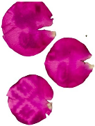
The presence of anthocyanins and carotenoids in rose petals, owing to their degree of concentration, makes these petals suitable for photographic development processes similar to chlorography.
Flavonoids represent a broad class of pigments that includes anthocyanins and flavonols. Their functions include UV protection, signaling, and defense. The primary function of their conspicuous colors is the attraction of pollinators (e.g., insects, birds) and seed dispersers. The color patterns, often species-specific, act as visual cues that guide animals to flowers for pollination or to fruits for their consumption and subsequent seed dispersal.
Anthocyanins are the most prominent pigments for natural photographic processes like anthotyping, due to their notable photosensitivity to visible and UV light that results in color change or bleaching. Meanwhile, other flavonoids also interact with light. The high UV absorbance of flavones and flavonols means these can act as UV filters if included in an emulsion, potentially altering exposure times or protecting other, more labile pigments from rapid UV degradation. However, their own transformation into visible images is less common or drastic than that of anthocyanins.
Flavonoids are potent antioxidants. They help to neutralize reactive oxygen species (ROS) generated during metabolic or environmental stress (such as high light irradiance), thereby protecting cells from oxidative damage. This function is intrinsically linked to their chemical structure, which allows them to absorb light and stabilize free radicals.
Betalains are a class of red and yellow pigments found in a more restricted group of plants, notably in beetroot (red betacyanins) and in prickly pear cactus flowers (yellow betaxanthins) [22]. They are mutually exclusive with anthocyanins; a plant will produce one or the other, but not both. Research into the applications of betalains, including in fields such as dye-sensitized solar cells (DSSCs), analogous to the photographic process, highlights their photosensitivity.
Betalains are known for their vibrant and intense colors. Beetroot juice is one of the most popular and effective emulsions for beginners in anthotyping, due to its strong tinctorial strength and relative sensitivity to light. Like other pigments, betalains degrade upon exposure to light, enabling the creation of images. Their stability can also be affected by factors such as pH and temperature.
Finally, reference is made to tannins, which are astringent, often brown-hued compounds used in botanical contact printing as mordants. Although they are not typically the primary agents responsible for the vivid and bright colors associated with pigments like anthocyanins or carotenoids, tannins play a crucial role as natural pigments, particularly in generating more subdued, earthy colors and in modifying and fixing other colors. Their contribution to color is often the result of oxidation, polymerization, or complexation reactions with metals.
Tannins are secondary plant metabolites, characterized by their ability to precipitate proteins (which confers their astringency) and form complexes with other macromolecules. They are primarily divided into two major groups [23].
Hydrolysable tannins are esters of a polyol (generally glucose) with phenolic acids such as gallic acid (gallotannins) or ellagic acid (ellagitannins). They can be hydrolyzed by weak acids or enzymes.
Condensed tannins (proanthocyanidins) are polymers of flavan-3-ol units. They are not easily hydrolyzed but can decompose in an acidic medium to yield red-colored anthocyanidins.
Although many tannins in their pure form may be colorless or pale yellow to light brown, they contribute significantly to the coloration of many plant parts, such as bark (oaks, acacias), wood, senescent leaves (brown and reddish autumn colors), and some fruits (particularly unripe ones) [24]. The color associated with tannins is often intensified or developed through oxidation (enzymatic or non-enzymatic) and polymerization.
Upon degradation under acidic conditions, condensed tannins can release red anthocyanidins, contributing to reddish hues. One of the most important properties of tannins in color generation is their ability to form colored complexes with metal ions. The classic example is the reaction of tannins (particularly gallotannins) with iron salts to produce dark blue, black, or gray complexes, which form the basis of iron gall ink, one of the most historically significant inks [25]. Other metals such as aluminum, copper, or chromium can also form complexes with tannins, resulting in different hues that have been exploited in traditional dyeing [26].
The interaction of light with these pigments can lead to “photodegradation” or “photobleaching” [6], an irreversible process in which the pigment molecule is destroyed by the action of light. This occurs when a pigment absorbs a photon, transitions to an excited state, and undergoes chemical reactions—often oxidative—that alter its structure and lead to loss of color. The presence of oxygen (photooxidation) and other environmental factors such as temperature, humidity, and pH can influence the rate and extent of this degradation. Ultraviolet (UV) and blue light are particularly effective at photobleaching many pigments, and higher light intensity or a longer exposure time generally results in greater bleaching.
It is important to recognize that plant pigments evolved for light capture and photoprotection, processes that involve controlled photochemical reactions [27]. In photography, however, their uncontrolled degradation is exploited. This underscores a fundamental difference in how these molecules function in vivo versus in photographic processes. Whereas plants possess mechanisms to manage light energy and minimize damage, the isolation of pigments or the use of an entire leaf outside its protective physiological context allows degradation to occur more readily, thereby forming the photographic image. The very mechanisms that plants utilize to survive high irradiance are those that must be understood or overcome when using them for photography.
The diversity of pigments in plants, each with distinct absorption spectra and sensitivities, directly determines the potential color palette and light requirements of natural photographic processes. The variety of pigments available in different plant sources (chlorophylls, carotenoids, anthocyanins, etc.) dictates their suitability for a given process and the resulting color and sensitivity. This intrinsic diversity contributes to the experimental nature and variability observed in techniques such as anthotype and chorotype or chlorography.
The following Table 1, summarizes the key properties of plant pigments relevant to natural photography.

1.2. Photochemical Principles of Chlorography

The fundamental scientific principle of chlorography is the photodegradation of chlorophyll and other plant pigments. This is not a simple decomposition process but rather a cascade of photochemical reactions initiated by the absorption of photons, primarily in the UV and blue spectral regions [28]. An initial step in chlorophyll degradation in vivo and in vitro is the loss or replacement of the central magnesium ion (Mg2+) of the porphyrin ring, a process known as pheophytinization, which converts chlorophyll (bright green) to pheophytin (olive-green to grayish). This molecular alteration is critical for the color change and contrast observed in the final image.
Furthermore, chlorophyll itself acts as a potent photosensitizer. Upon absorbing light energy, it can transition to an excited triplet state and transfer that energy to molecular oxygen (O2), generating reactive oxygen species (ROS), such as singlet oxygen (1O2) [29]. These ROS are extremely potent oxidizing agents that attack and irreversibly destroy both neighboring chlorophyll molecules and other pigments such as carotenoids, in a self-propagating photooxidative bleaching process essential for image formation. However, the efficiency of this degradation process is directly modulated by the plant’s inherent photoprotective mechanisms. The most important of these is non-photochemical quenching (NPQ). NPQ is a set of physiological processes that allow the plant to safely dissipate excess absorbed light energy as heat, thus avoiding photooxidative damage [30]. The efficiency of NPQ, which varies greatly between species and is often correlated with adaptation to high-irradiance environments such as the Andes, directly competes with the photodegradation pathway [31]. Therefore, the suitability of a leaf for chlorography depends on a delicate balance: it must be sufficiently photosensitive to allow degradation but not so susceptible that the leaf structure collapses prematurely. To a large extent, the observed variability in exposure times and image quality among different Andean plant species can be attributed to interspecific differences in the efficiency of their NPQ mechanisms [32].

2. Materials and Methods

The creation of this series on chlorotypes and anthotypes resulted from the link between an ethnographic study focused on understanding the visual changes that occurred in the graphic art of the Salasaka people from 1960 to 2018 and the exploration of alternative photographic development practices during the isolation period of the COVID-19 pandemic.
In the exploratory research on the iconography of the Salasaka people, during the specified period, processes of aesthetic transculturation that occurred in this indigenous community of the Ecuadorian Andes—renowned for their textile craftsmanship—were identified. As part of this previous investigation, a large number of images associated with the community’s production of tapestries, sashes, and drums were collected and cataloged.
Toward the end of 2020, these two moments converged to give rise to a new artistic research experience, born from the visual interest in utilizing the images collected in the ethnographic study on the Salasaka and using nearby natural resources that were available during the period of isolation.
Thus, the idea arose to revive several alternative photographic development techniques with natural products that could be sourced without violating the health protocols established at that time. This marked the beginning of the exploration into creating chlorotypes and anthotypes with plants found in the province of Tungurahua (a region located in the central highlands of Ecuador).
Initially, the collection of this material was limited to the leaves from the potted plants and gardens of this study’s initial author. The collection range for the plant substrate was subsequently expanded to other locations within the province of Tungurahua.
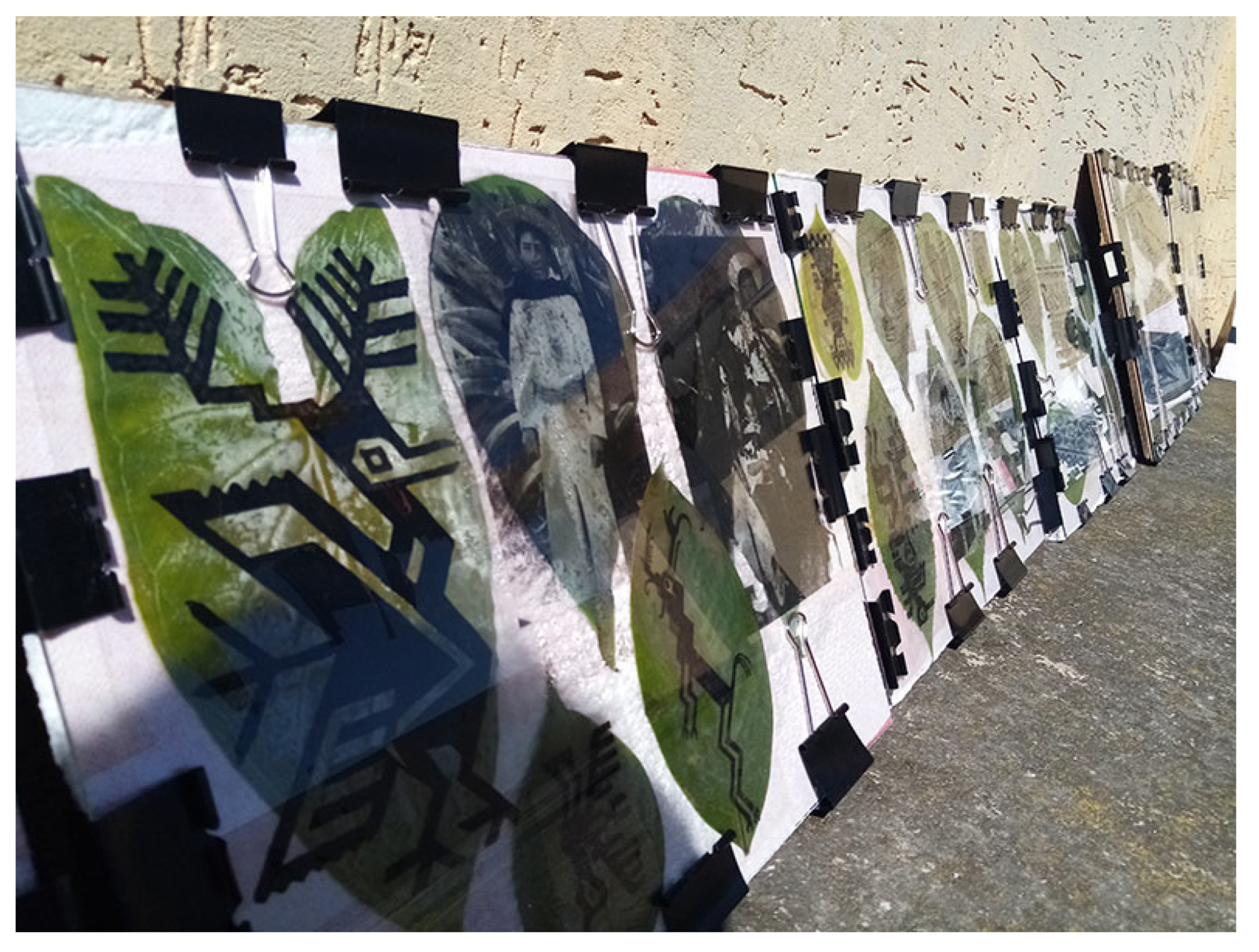
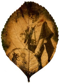
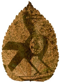
Between 2021 and 2024, approximately 1000 trials were conducted on various types of leaves and, subsequently, flower petals to identify the most suitable species for this natural development process (Figure 1). In 2023, a series of tests on the encapsulation of leaves also commenced, aimed at prolonging their preservation.
This process led to the conception of an exhibition proposal that would come to fruition as the exhibition “Origen vivo” (Living Origin), which opened in April 2024 in Quito and included an installation of chlorotypes.

2.1. Chlorophyll Development Process

Chlorophyll printing is a natural photographic technique that enables the transfer of images directly onto the surface of plant leaves. Its origins can be traced back to early observations of how light affected plant coloration, a phenomenon known long before the formal development of photography. While Sir John Herschel conducted early research into the photographic properties of chlorophyll in the 19th century, the modern application of this technique to create recognizable photographic images is largely attributed to contemporary artists.
Heather Ackroyd and Dan Harvey pioneered the technique of projecting images onto growing grass, using light to influence chlorophyll synthesis and thereby create ephemeral “photographs” in art installations [33]. Subsequently, the Vietnamese artist Binh Danh [5] significantly refined the concept by applying it to individual leaves, often from his own garden. He is recognized for developing methods to preserve these delicate organic images, typically by encapsulating them in resin [1].
The process of chlorotyping or chlorography is based on exploring the artistic potential of using diverse species of vascular and non-vascular plants, flowers, and other natural species such as fungi. These serve as a substrate in place of paper to generate images, which are often linked to themes of memory, nature, and sustainability.
The process for creating a chlorophyll print involves several key steps, which are summarized as follows.

2.1.1. Leaf Selection

Suitable leaves are chosen, which ideally should be large, flat, and healthy, although successful results have been achieved with leaves smaller than 4 cm. The choice of plant species is crucial, as different types of leaves such as those from pumpkin, oak, fig, ivy, or granadilla (passion fruit) yield varying results in terms of color, texture, and light sensitivity. For fibrous leaves, it is recommended that the stem be cut at an angle to ensure the pressing process is uniform across the entire leaf structure.
Although freshly cut leaves are preferred, access to collection sites sometimes required preserving leaves for up to a maximum of three weeks before their use in the chlorotype process. For preservation, leaves were kept dry inside a plastic bag under refrigeration (Figure 2). For best results, the stored leaves were wrapped in absorbent paper towel to prevent accumulation of moisture. The process cannot be performed once the leaf is dry.

2.1.2. Image Preparation

For the development process, a positive transparency of the desired image is required. This can be a laser print on acetate or an illustration on plastic film created with permanent markers that block sunlight. A high-contrast black and white digital image is often used for optimal results, as this helps to clearly delineate the areas that will be bleached from those that will remain dark. In this case, the print is made from a photographic positive, in contrast to the cyanotype process, which utilizes a negative.

2.1.3. Setup for Development

The leaf and the positive transparency are placed together in a contact frame. A rigid surface (such as a board or a sheet of pressboard) and a sheet of glass are recommended to ensure firm and uniform contact between the transparency and the leaf, which is crucial for image sharpness. Materials that could prematurely dry the leaf should be avoided. For prolonged exposures, the leaf can be kept hydrated by allowing its stem to protrude from the frame into a water source.

2.1.4. Exposure

The assembly is placed under direct sunlight. Exposure time varies considerably, ranging from a few hours for young, freshly cut leaves, to longer periods. For example, an image can be developed on a granadilla leaf under such conditions in three to four hours of direct exposure. Most of the leaves tested required between one day and up to three weeks to generate an image, depending on the leaf type, solar intensity, season, and meteorological conditions (Figure 3). It is necessary to monitor the progress periodically, taking care not to displace the transparency. An abrupt change in weather can cause the internal chemical reaction to cease, a phenomenon analogous to the fogging of a photograph in traditional development.

2.1.5. Drying and Preservation

Once the image has formed satisfactorily, the leaf is removed from the setup and dried between sheets of absorbent paper. Given that chlorophyll prints are inherently ephemeral, preservation is a critical step. Methods such as resin encapsulation, chemical baths (e.g., alcohol, copper sulfate), or the application of sealants (e.g., wax, varnish) are used to arrest degradation and protect the image from light and physical deterioration.

2.1.6. Environmental Parameter Monitoring and Measurement

To improve experimental reproducibility and allow correlational analysis between environmental conditions and photographic results, key environmental parameters were monitored during the exposure phase. Qualitative descriptions such as direct sunlight was complemented with quantitative metrics.
Light Intensity: Ultraviolet (UV) irradiance was measured using a calibrated UV radiometer (UV PowerMap® II - EIT 2.0, Leesburg-US), positioned parallel to the contact frame to simulate incidence on the leaf. Measurements were recorded in watts per square meter (W/m2) or milliwatts per square centimeter (mW/cm2), noting the measured spectral range [34]. Data were recorded at hourly intervals during exposure periods to calculate an average irradiance for each test.
Temperature and Humidity: Ambient air temperature (in degrees Celsius, °C) and relative humidity (RH, in %) were recorded using a calibrated digital thermo-hygrometer [35]. The sensor was placed in close proximity to the experimental setups but protected from direct sunlight to ensure the accurate measurement of air conditions.
Given the conditions of the current study, which focused more on an empirical development process to identify the types of species that allow or prevent the development of a natural image, measurements were not taken for all the tracked species; instead, a purposive sample of three species was taken.
These quantitative data, summarized in Table 2, are important for correlating environmental conditions with the development times and image quality obtained for different species and seasons, laying the groundwork for future predictive models of the chlorographic process. A second stage of this project plans to measure all identified species.
Exposure to light remains a key factor for the developed leaf. Even when attempts are made to halt the chemical process of photodecomposition, ambient light continues to deteriorate the image. Under direct light exposure, the developed images lasted for approximately one year before beginning to degrade. Climatic conditions are also influential; in the dry climate of the Ecuadorian highlands, chlorotypes are more durable, and the leaves do not warp. In contrast, leaves exposed to the humid climate of the coastal regions warped and developed fungus, and unencapsulated images deteriorated within a period of approximately three months.
The detailed mechanism behind chlorophyll printing centers on the photobleaching of chlorophyll. The image is formed as sunlight, particularly UV radiation, degrades the chlorophyll molecules in the exposed areas of the leaf. The areas of the leaf shielded from light by the transparency or object retain their chlorophyll, maintaining their original green color and thereby creating the necessary contrast for the image. As chlorophyll degrades in the exposed areas, the underlying yellow and orange carotenoids may become visible, contributing to the tones in these parts of the image.
A fundamental characteristic of chlorophyll printing or development is that the substrate (the leaf or flower petal) is itself the photosensitive material (Figure 4), in contrast to techniques that involve coating an inert material like paper. This means that the biological state of the leaf—its freshness, type, thickness, and pigment concentration—directly impacts the photographic outcome. The image forms as the leaf withers and its chlorophyll and other natural pigments degrade, a process intimately linked to the life and death of the leaf. This deep connection with the specific plant used, including its natural structures such as veins and textures, renders each print unique and unpredictable, a true collaboration with nature.
The inherent impermanence of chlorotypes is another defining aspect. Although preservation methods exist, the fleeting nature of the image, which continues to fade over time if not adequately protected, is not merely a technical limitation but a characteristic that is often exploited artistically (Table 3). The tension between the fragility of the organic support and the attempt to render it permanent through fixation adds layers of meaning to the work. The following table details several preservation methods.

3. Results

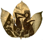
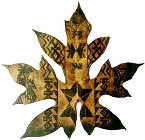
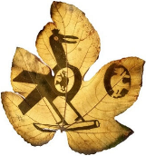
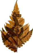
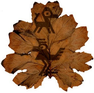
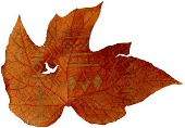
Chlorotyping, also known as chlorography or phytotyping, is an alternative photographic process that utilizes plant leaves as a substrate for image generation, leveraging the photosensitivity of the chlorophyll they contain. This process is conducted via exposure to sunlight, which induces a chemical reaction resulting in the degradation of the leaf’s own chemical compounds. These compounds oxidize in the areas exposed to light, while the areas shielded from light retain their original hue, thus creating the image (Figure 5).
As described in the Materials and Methods section, climatic variables such as solar intensity and humidity play a decisive role in the development process. The required exposure time varies depending on the type of leaf.
From the experiments conducted, it was inferred that granadilla leaves are the most versatile. An image can be developed on very young leaves in approximately two to three hours of solar exposure. In contrast, more fibrous leaves, such as those from fig, monstera, or certain types of orchids, required up to three weeks of daily exposure lasting 10 to 12 h.
Continuous monitoring is necessary during the development process, as in some cases, the liquid content of the leaves evaporates very rapidly. This disintegrates the leaf pulp, causing the leaf to become skeletonized. The following Table 4, presents a graphical sample of the results obtained during the experimental process.
Table 4 presents a sample of 30 species, including fruit-bearing plants, shrubs, and flowers. All are vascular plants that grow in the central region of Ecuador and were tested for the production of chlorotypes.
For each selected species, the approximate direct solar exposure time under natural conditions throughout the year is documented. The development process was conducted in the city of Ambato, located in the province of Tungurahua, at an altitude of 2580 m above sea level.
Additional tests were also conducted in coastal cities, which have higher solar incidence and elevated temperatures, conditions that shorten the exposure times required to develop the image.
The average solar exposure for thin, non-fibrous leaves is typically between 6 and 10 h, whereas for thick leaves, the exposure can require several days. Trials have been conducted to generate prints using UV lamps, but the exposure time triples compared to that required when using sunlight.
Furthermore, trials have been conducted on living leaves still attached to the plant, specifically, with granadilla and sunflower. However, although an image can be obtained, the exposure time is extremely long—around one month—and the resulting image lacks complete sharpness. This is because the leaf continues to grow, while the transparency used for the transfer maintains a constant size.
Each print is a reflection of the specific characteristics of the vegetal material utilized, meaning no two images are identical. This uniqueness becomes an asset for artists, who can explore the diversity of local flora and create works representative of their environment. This experimental approach not only highlights the technique’s versatility but also opens new possibilities for contemporary artistic creation by integrating natural elements into the creative process.

4. Discussion

The results obtained not only validate chlorography as a viable photographic technique but also position it as a practice rich in interdisciplinary implications. By analyzing the findings through the lenses of contemporary art, biodiversity conservation, and ethnobotany, the technique emerges as a powerful medium for exploring and communicating the complex relationships between human culture, science, and the natural world.
Chlorotyping is situated at the confluence of botany, contemporary art, and cultural heritage. It aligns with the bio-art and eco-art movements [36], which use biological materials and processes to critically address humanity’s relationship with the environment [37]. In this case, the leaf is not merely a substrate but the medium itself: its physiology, biochemistry, and life and death cycle are integral to the creative process. This practice forces the viewer to confront plant blindness, a cognitive bias that leads to ignoring plants in the environment. By transforming a common leaf into a photographic object carrying a cultural image, chlorotyping elevates the plant to a dynamic actor [38], sensitive to light and loaded with history.

4.1. Interpretation of Photographic Variability Between Species

The results demonstrate a marked difference in the suitability of different plant species for chlorography, manifested primarily in the required exposure times (Table 4). This variability cannot be attributed solely to the initial chlorophyll concentration. Rather, it is the result of a complex interaction of anatomical, physiological, and, crucially, photoprotective factors.
Anatomically, characteristics such as leaf thickness, waxy cuticle density, and water content influence light penetration and the rate of dehydration, affecting the process. Species with thick, leathery leaves, such as Monstera deliciosa, present a greater physical barrier to light and a greater water reserve, which is likely to contribute to their long exposure times.
However, the explanation for this variability lies in the plant’s photoprotective mechanisms. As previously noted, a plant’s ability to dissipate excess light energy through non-photochemical quenching (NPQ) is in direct competition with the photodegradation process required for chlorography to function [39]. Species that have proven most efficient for chlorography, such as Passiflora ligularis and Cucurbita maxima, are postulated to possess NPQ systems that saturate relatively easily under the high irradiance of the equatorial Andes.
Once their heat dissipation capacity is exceeded, the excess absorbed energy inevitably diverts toward the formation of reactive oxygen species (1O2), initiating the photodegradation cascade that forms the image [40]. Conversely, species that require prolonged exposure times likely possess more robust NPQ mechanisms or a greater capacity for xanthophyll cycling, allowing them to resist photooxidative damage for longer [41]. This perspective transforms chlorography from a mere artistic process to a qualitative visualization of a plant’s photoprotective capacity.

4.2. Contextualizing Chlorography Versus Other Alternative Techniques

Although anthotyping and chlorography are both organic and based on photobleaching, they differ fundamentally. Anthotyping uses an emulsion extracted from plant pigments (commonly anthocyanins from flowers or berries) that is applied to an inert substrate such as paper. Chlorography, on the other hand, uses live or freshly cut leaves as an integrated substrate and emulsion system [1]. This entails key differences.
Regarding the primary photosensitive pigments, chlorotyping focuses almost exclusively on chlorophyll, the predominant green pigment in leaves. The yellow and orange carotenoids present in leaves can become visible as the chlorophyll degrades, contributing to the hues in exposed areas. In contrast, a much wider range of pigments can be extracted from various plant parts, with anthocyanins (red, purple, blue) and carotenoids (yellow, orange) being the most common, but also including betalains, flavonoids, and tannins, allowing a greater diversity of initial colors in the emulsion.
In both cases, the fundamental mechanism of photobleaching is light-induced. Light, especially UV light, excites the pigments, triggering chemical reactions that result in color loss in the exposed areas. Areas protected by a transparency or object retain their original color, creating the contrast of the image. Both are positive processes, where light bleaches the pigment.
The methodologies differ significantly, as chlorophyll development uses the plant leaf as a direct photosensitive substrate, which involves steps such as selecting suitable leaves and considering their biological state and physical structure. Anthotypes, on the other hand, require the preparation of an emulsion from plant extracts, which is then applied to an inert substrate such as paper [42]. This involves steps such as grinding, diluting, and straining the plant material, as well as coating and drying the paper.
Chlorography intrinsically incorporates the texture, veins, and biological structure of the leaf into the final image, creating a unique fusion between image and support. Anthotypes tend to produce images with a more painterly or watercolor-like quality on a uniform surface.
Cyanotypes rely on the photoreduction of iron salts to create a Prussian blue image. The contrast with chlorography is stark. As an inorganic chemical process versus an organic one, it uses a photographic negative to produce a positive image, while chlorography uses a positive to bleach the leaf (a positive–positive process). The result is a distinctive, monochromatic aesthetic, very different from the earthy, natural tones of chlorography.
The present study contributes significantly to the field by providing a systematic analysis of 30 Andean species, surpassing the more general descriptions of previous work which explored a smaller number of ornamental species. By placing the practice in the context of contemporary artists such as Binh Danh, who has popularized the technique in exploring themes of memory and war, and Ackroyd and Harvey, pioneers in the use of live plants as photographic canvas, this work connects scientific research with a vibrant current artistic practice.
Permanence is a key challenge for both processes, as images are inherently ephemeral and continue to fade with exposure to light if not preserved. Various preservation methods have been developed for both, such as resin encapsulation or the use of sealants for chlorophyll prints and dark storage for anthotypes. The duration of exposure is significantly different. While chlorophyll prints, depending on the leaf type, can produce results in a few hours or sometimes take days or weeks, anthotypes often require several weeks of exposure.
Aesthetically, chlorotypes often present a delicate, sometimes ghostly appearance, with the texture and structure of the leaf integrated into the image. Anthotypes can vary widely in color and tone depending on the pigment source, often with a watercolor-like quality.

4.3. Chlorography as a Tool for Ethnobotany and Cultural Conservation

Beyond its technical and artistic interest, this project demonstrates the potential of chlorography as a tool for ethnographic research and practice and for cultural conservation. By using the technique to print iconographic designs of the Salasaka people on the leaves of plants native to their own territory, the process transcends mere representation. It becomes a form of material ethnobotany.
The resulting artifact, a cultural image printed on a local biological support, creates a physical and inseparable link between the symbol and the ecosystem from which the culture emerges, embodying the Andean worldview, where culture and nature are not separate spheres. This approach offers a powerful means for cultural revitalization. Being non-toxic, low-cost, and accessible, the process is ideal for community workshops. It allows community members, especially the youngest, to reconnect with their visual heritage and, simultaneously, learn to identify and value the local flora. The practice of chlorography thus becomes a pedagogical act that teaches cultural history and botany in an integrated and practical way.

4.4. Technical Challenges in the Face of Ephemerality and Conservation

The main technical and conceptual challenge of chlorography is the impermanence of the image. As an organic process, the sheet continues to degrade even after the image has been formed, especially if exposed to light, moisture, or oxygen. However, this ephemerality is not only a technical limitation; it is also one of the technique’s most powerful qualities from an artistic and conceptual perspective [43]. The transient nature of chlorography resonates with themes of memory, fragility, the cycle of life and death, and humanity’s relationship with an ever-changing natural world [44].
However, for these works to be exhibited, collected, and studied, conservation is a practical necessity. The discussion of how to preserve ephemeral art is an active field in contemporary conservation, where the tension between preserving the material object and respecting the artist’s conceptual intent is debated [45]. In the context of this study, practical methods for stabilizing chlorotypes were explored. Two methods stand out for their effectiveness.
Resin encapsulation involves embedding the dried leaf in a block of UV-filtering, transparent epoxy resin [1]. The resin provides robust structural support, protects the leaf from physical damage, and, crucially, isolates it from oxygen and ambient humidity, two of the main agents of degradation [46]. Furthermore, the UV filter mitigates continuous light damage. This method is currently the standard for the long-term preservation of chlorotypes [5].
Copper sulfate chemical fixation uses a copper sulfate (CuSO4) bath in a chemical approach to color stabilization [1]. The underlying mechanism involves a transmetalation reaction. The copper ion (Cu2+) in the solution, being more stable at the center of the porphyrin ring, displaces the original magnesium ion (Mg2+) from the chlorophyll molecule [47]. This forms a copper–chlorophyllin complex, which is significantly more stable and resistant to photodegradation and acidification than the original chlorophyll [46]. The result is a more permanent green color, although often with a slightly bluish hue. This method, although interventional, offers a way to preserve the green color of the leaf without the need for complete encapsulation.
Both methods offer viable solutions to the problem of permanence, allowing these ephemeral works to be integrated into long-term collections and archives [48].

4.5. Limitations and Future Perspectives

It is essential to recognize the limitations of this exploratory study, principally, the lack of control and quantification of environmental variables. Solar irradiance, temperature, and humidity were not systematically measured, which prevents establishing precise quantitative correlations. These limitations open clear avenues for future research.Studies in controlled environments should replicate the experiments using artificial light sources that enable control and quantification of irradiance (W/m2) and spectral composition.
Physiological and biochemical analysis should correlate chlorotyping results with quantitative measurements of leaf parameters, such as spectrophotometric pigment quantification and chlorophyll fluorescence measurements to assess response to light stress.
To deepening ethnobotanical collaboration, projects should be developed in direct collaboration with members of the Salasaka community to select plants based on their cultural significance and create a dialogic research process.

5. Conclusions

This study reexamines chlorography not only as an alternative photographic technique, but also as a robust interdisciplinary research tool. The main conclusions, derived directly from the results and discussion, are as follows.
The viability of chlorography was successfully demonstrated in a diverse set of 30 plant species from the Ecuadorian Andean ecosystem. The systematic investigation identified the genera Passiflora and Cucurbita as particularly effective substrates, characterized by fast development times and high image quality.
A theoretical and scientific framework has been established to explain the variability observed in the results. It is postulated that the efficiency of chlorographic development is inversely proportional to the photoprotective capacity of the plant, specifically to the efficiency of its non-photochemical quenching (NPC) system. This finding links, for the first time, artistic practice with plant physiology at the molecular level.
This research validates chlorography as a powerful interdisciplinary bridge. By merging the ancestral iconography of the Salasaka people with native flora, the technique becomes a means of material ethnobotany, offering a tangible, sustainable, and non-toxic avenue for the conservation and transmission of cultural heritage.
While ephemerality is an inherent and conceptually rich feature of the technique, effective conservation methods have been explored. Resin encapsulation and chemical fixation with copper sulfate offer practical avenues for the long-term preservation of these works, ensuring their place in collections and archives.
In short, this work elevates chlorography from an artistic curiosity to a field of research with profound scientific, cultural, and conservation implications.

Author Contributions

Conceptualization, A.D.L.S. and I.P.Á.L.; methodology, A.D.L.S.; validation, I.P.Á.L., P.R.M.F. and C.E.M.C.; formal analysis, A.D.L.S. and C.S.S.N.; investigation, A.D.L.S.; resources, P.R.M.F.; data curation, C.S.S.N. and C.E.M.C.; writing—original draft preparation, A.D.L.S.; writing—review and editing, I.P.Á.L. and P.R.M.F.; supervision, C.S.S.N. and C.E.M.C.; project administration, A.D.L.S. All authors have read and agreed to the published version of the manuscript.

Funding

This research: entitled “DESIGN AND MEMORY. MEANINGS EXPOSED IN SALASACA’S ICONOGRAPHY”, was funded by the Research and Development Department–DIDE of the Technical University of Ambato, Ecuador, through Resolution No. UTA-CONIN-2023-0082-R.

Data Availability Statement

This article analyzes for the first time the relationship between embroidered and painted iconography and the ritual places of the Salasaka community.

Acknowledgments

We would like to express our gratitude to the Research and Development Department–DIDE of the Technical University of Ambato, which promoted the execution of this research, as well as to the Faculty of Design and Architecture. We also extend our gratitude to the Salasaka people and the Parish Council of the community, which has allowed us to carry out this project.

Conflicts of Interest

The authors declare no conflicts of interest.

References

  1. Larrea Solórzano, A. Sin triturar la pulpa: Prácticas de clorotipia con hojas de Tungurahua. Index Rev. De Arte Contemp. 2023, 9, 134–144. [Google Scholar] [CrossRef]
  2. MET–Department of Photographs. The Daguerreian Era and Early American Photography on Paper, 1839–1860. In Heilbrunn Timeline of Art History; The Metropolitan Museum of Art: New York, NY, USA, 2004. [Google Scholar]
  3. The New York Public Library. An Introduction to Photographic Processes; The New York Public Library: New York, NY, USA, 2013. [Google Scholar]
  4. Michaud, L. Alternative Process Photography: Beyond Digital and Film; Senior Honors Projects; University of Rhode Island: Kingston, RI, USA, 2017; p. 454. [Google Scholar]
  5. Danh, B. Binh Danh: The Enigma of Belonging; Radius Books: Santa Fe, NM, USA, 2023. [Google Scholar]
  6. Lingvay, M.; Akhtar, P.; Sebők-Nagy, K.; Páli, T.; Lambrev, P.H. Photobleaching of Chlorophyll in Light-Harvesting Complex II Increases in Lipid Environment. Front. Plant Sci. 2020, 11, 849. [Google Scholar] [CrossRef]
  7. Blones Borges, J.M.; Mora, A.J.; Giráldez De Luca, M.D. Potencialidad en fotosensibilidad foliar, para tres especies de plantas ornamentales, en la generación de imágenes, bajo la técnica fotográfica alternativa y experimental de la clorotipia. Rev. Crit. Con Cienc. 2024, 2, 39–52. [Google Scholar] [CrossRef]
  8. Torres Canela, O.; Gurieva, N. Fotografía impresa al natural: La clorotipia como procedimiento alternativo de impresión de imágenes en plantas. Zincografía 2024, 8, 15. [Google Scholar] [CrossRef]
  9. Hannavy, J. Encyclopedia of Nineteenth-Century Photography; Routledge: New York, NY, USA, 2008. [Google Scholar]
  10. Murillo, A. Analysis Of Light Exposure Time And Its Effect On Photosynthetic Pigment Chlorophyll A And Β Degradation. Res. J. Life Sci. Bioinform. Pharm. Chem. Sci. 2021, 7, 17–29. [Google Scholar]
  11. Cerón Martínez, C. Plantas medicinales de los Andes ecuatorianos. In Botánica Económica de los Andes Centrales; Universidad Mayor de San Andrés: La Paz, Bolivia, 2006. [Google Scholar]
  12. Zambrano-Intriago, L.; Buenaño-Allauca, M.; Mancera-Rodríguez, N.; Jiménez-Romero, E. Estudio etnobotánico de plantas medicinales utilizadas por los habitantes del área rural de la Parroquia San Carlos, Quevedo, Ecuador. Univ. Y Salud 2015, 17, 97–111. [Google Scholar]
  13. Kmet, A.; Drozdíková, A.; Nagyová, S.; Ikhardt, P. The cyanotype process and its potential in chemistry education. J. Chem. Educ. 2023, 100, 2367–2372. [Google Scholar] [CrossRef]
  14. Fabbri, M. Anthotype Emulsions, Volume 2: The collective research from photographers on World Anthotype Day 2023. In Anthotype Emulsions—The Collective Research from Photographers on World Anthotype Day; Alternative Photography: Micamera, Italy, 2023. [Google Scholar]
  15. Saska, H. Anna atkins: Photographs of british algae. Bull. Detroit Inst. Arts 2010, 84, 8–15. [Google Scholar] [CrossRef]
  16. Moreno-Saez, C.; Gutiérrez-Párraga, T.C. La cianotipia como medio de formación e implementación de actividades artísticas para educadores que trabajan con enfermos de Alzheimer. Arte Individuo Y Soc. 2017, 29, 109–126. [Google Scholar] [CrossRef]
  17. Wang, H.; Fan, Y.; Yang, Y.; Zhang, H.; Li, M.; Sun, P.; Zhang, X.; Xue, Z.; Jin, W. Classification of rose petal colors based on optical spectrum and pigment content analyses. Hortic. Environ. Biotechnol. 2023, 64, 153–166. [Google Scholar] [CrossRef]
  18. Martínez Girón, J.; Martínez, J.A.; García Hurtado, L.; Cuaran, J.; Ocampo, Y. Pigmentos vegetales y compuestos naturales aplicados en productos cárnicos como colorantes y/o antioxidantes: Revisión. Inventum 2016, 11, 51–62. [Google Scholar] [CrossRef]
  19. Schelbert, S.; Aubry, S.; Burla, B.; Agne, B.; Kessler, F.; Krupinska, K.; Hortensteiner, S. Pheophytin Pheophorbide Hydrolase (Pheophytinase) Is Involved in Chlorophyll Breakdown during Leaf Senescence in Arabidopsis. Plant Cell 2009, 21, 767–785. [Google Scholar] [CrossRef] [PubMed]
  20. Frank, H.; Cogdell, R. Carotenoids in Photosynthesis. Photochem. Photobiol. 1996, 63, 257–264. [Google Scholar] [CrossRef]
  21. Castañeda-Ovando, A.; Pacheco-Hernández, M.; Páez-Hernández, M.; Rodríguez, J.; Galán-Vidal, C. Chemical studies of anthocyanins: A review. Food Chem. 2009, 113, 859–871. [Google Scholar] [CrossRef]
  22. Strack, D.; Vogt, T.; Schliemann, W. Recent advances in betalain research. Phytochemistry 2003, 62, 247–269. [Google Scholar] [CrossRef]
  23. Khanbabaee, K.; van Ree, T. Tannins: Classification and Definition. Nat. Prod. Rep. 2001, 18, 641–649. [Google Scholar]
  24. Falcão, L.; Araújo, M. Tannins characterization in historic leathers by complementary analytical techniques ATR-FTIR, UV-Vis and chemical tests. J. Cult. Herit. 2013, 14, 499–508. [Google Scholar] [CrossRef]
  25. Pizzicato, B.; Pacifico, S.; Cayuela, D.; Mijas, G.; Riba-Moliner, M. Advancements in Sustainable Natural Dyes for Textile Applications: A Review. Molecules 2023, 28, 5954. [Google Scholar] [CrossRef] [PubMed]
  26. Danila, A.; Muresan, E.; Chirila, L.; Coroblea, M. Natural Dyes Used in Textiles: A Review. In International Symposium “Technical Textiles—Present and Future”., 2021 ed; Sciendo: Warsaw, Poland, 2021. [Google Scholar]
  27. Barber, J.; Andersson, B. Too much of a good thing: Light can be bad for photosynthesis. Trends Biochem. Sci. 1992, 17, 61–66. [Google Scholar] [CrossRef]
  28. Petrović, S.; Zvezdanović, J.; Marković, D. Chlorophyll degradation in aqueous mediums induced by light and UV-B irradiation: An UHPLC-ESI-MS study. Radiat. Phys. Chem. 2017, 141, 8–16. [Google Scholar] [CrossRef]
  29. Verduin, J.; den Uijl, M.; Peters, R.; van Bommel, M. Photodegradation Products and their Analysis in Food. J. Food Sci. Nutr. 2020, 6, 67. [Google Scholar] [CrossRef]
  30. Murakami, A.; Kim, E.; Minagawa, J.; Takizawa, K. How much heat does non-photochemical quenching produce? Front. Plant Sci. 2024, 15, 1367795. [Google Scholar] [CrossRef] [PubMed]
  31. Ruban, A.V. Nonphotochemical Chlorophyll Fluorescence Quenching: Mechanism and Effectiveness in Protecting Plants from Photodamage. Plant Physiol. 2016, 170, 1903–1916. [Google Scholar] [CrossRef] [PubMed]
  32. Zaks, J.; Amarnath, K.; Kramer, D.; Niyogi, K.; Fleming, G. A kinetic model of rapidly reversible nonphotochemical quenching. Proc. Natl. Acad. Sci. USA 2012, 109, 15757–15762. [Google Scholar] [CrossRef]
  33. Foundation, P. Anthotypes & Other Organic Processes I. Available online: https://www.penumbrafoundation.org/anthotypes-other-organic-processes-i (accessed on 15 February 2025).
  34. Waveform Lighting. Understanding UV-A Irradiance, and Requirements for Various Applications. Available online: https://www.waveformlighting.com/tech/understanding-uv-irradiance-and-requirements-for-various-applications (accessed on 16 March 2025).
  35. Both, A.J.; Benjamin, L.; Franklin, J.; Holroyd, G.; Incoll, L.D.; Lefsrud, M.G.; Pitkin, G. Guidelines for measuring and reporting environmental parameters for experiments in greenhouses. Plant Methods 2015, 11, 43. [Google Scholar] [CrossRef] [PubMed]
  36. Linköping University. The Eco- and Bioart Lab. Available online: https://liu.se/en/research/the-eco-and-bioart-lab (accessed on 15 February 2025).
  37. Zhang, L.; Shen, T. Integrating Sustainability into Contemporary Art and Design: An Interdisciplinary Approach. Sustainability 2024, 16, 6539. [Google Scholar] [CrossRef]
  38. Puljar D’Alessio, S. Dynamic Relations between Plants and Humans at the Intersection of Art, the Humanities, and Botanical Sciences: Four Instances of Contemporary Art in Croatia. Nar. Umjet. Hrvat. Časopis Za Etnol. I Folk. 2024, 61, 2. [Google Scholar] [CrossRef]
  39. Lu, D.; Zhang, Y.; Zhang, A.; Lu, C. Non-Photochemical Quenching: From Light Perception to Photoprotective Gene Expression. Int. J. Mol. Sci. 2022, 23, 687. [Google Scholar] [CrossRef]
  40. Zhao, X.; Chen, T.; Feng, B.; Zhang, C.; Peng, S.; Zhang, X.; Fu, G.; Tao, L. Non-photochemical Quenching Plays a Key Role in Light Acclimation of Rice Plants Differing in Leaf Color. Front. Plant Sci. 2017, 7, 1968. [Google Scholar] [CrossRef] [PubMed]
  41. Lambrev, P.; Miloslavina, Y.; Jahns, P.; Holzwarth, A. On the relationship between non-photochemical quenching and photoprotection of Photosystem II. Biochim. Biophys. Acta (BBA) Bioenerg. 2012, 1817, 760–769. [Google Scholar] [CrossRef]
  42. Zuluaga, A. Antotipias e impresiones de clorofila: Capturando la esencia fotosensible de las plantas a través del revelado solar. Folios Rev. De La Fac. De Comun. Y Filol. 2023, 47–48, 92–99. [Google Scholar]
  43. Arnold, E. Photography, composition, and the ephemeral city. Area 2021, 53, 659–670. [Google Scholar] [CrossRef]
  44. Som, S. Ephemeral Art-Impermanence in Material and Concept in the Work of Indian Contemporary Artists; National Institute of Fashion Technlolgy: New Delhi, India, 2020. [Google Scholar]
  45. Hornbeck, S. Intersecting conservation approaches to ethnographic and contemporary art: Ephemeral art at the National Museum of African Art. Objects Spec. Group Postprints 2013, 20, 207–226. [Google Scholar]
  46. Woods, A. A Method of Preserving the Green Color of Plants for Exhibition Purposes. Bot. Gaz. 1897, 24, 206–209. [Google Scholar] [CrossRef]
  47. Higdon, J.; Kopec, R. Linus Pauling Institute. 2004. Available online: https://lpi.oregonstate.edu/mic/dietary-factors/phytochemicals/chlorophyll-metallo-chlorophyll-derivatives (accessed on 20 July 2025).
  48. Phillips, M. The Effects of Aqueous Immersion Wash Treatments on Platinum Photographs. Top. Photogr. Preserv. 2001, 9, 103–114. [Google Scholar]
Figure 1. Photograph of a Salasaka musician developed on a passion fruit leaf.
Jzbg 06 00041 g001
Figure 2. Process of selecting and cleaning leaves for development.
Jzbg 06 00041 g002
Figure 3. Solar exposure process for chlorotype generation.
Jzbg 06 00041 g003
Figure 4. Differences between negative development of a begonia leaf and positive development of a malanga leaf.
Jzbg 06 00041 g004
Figure 5. Chlorotypes with images of Salasaka revealed in calla, geranium, and passionflower leaves.
Jzbg 06 00041 g005
Table 1. Basic plant pigments for development and their properties.
Pigment ClassCharacteristic ColorPrincipal Absorption RangePrimary Functions in PlantsRelevance in Natural Photography
ChlorophyllsGreenBlue, RedLight absorption for photosynthesis.Primary pigment in chlorophyll printing (chlorotypes).
CarotenoidsYellow, Orange, RedBlue-Green, BlueAccessory pigments in photosynthesis, photoprotection (dissipation of excess energy).Present in leaves (revealed upon chlorophyll degradation), extracted for anthotypes.
AnthocyaninsRed, Purple, BlueGreen, Blue-Green, BluePollinator attraction, UV protection, defense, signaling. Color is pH-sensitive.Extracted for anthotypes (wide range of colors, pH sensitivity).
FlavonoidsYellow, OtherUV, Blue (some)UV protection, signaling, defense.Contribute to the color and properties of some anthotype emulsions.
BetalainsRed, YellowBlue, GreenColoration, defense.Extracted for anthotypes (source of pigments in some plants).
TanninsBrownUVAstringency, defense.Used as mordants in botanical contact printing, can be light-sensitive.
Table 2. Quantitative environmental parameters recorded during exposure tests.
Plant Species (Common Name)Exposure Duration (h)Average UV Irradiance (W/m2)Average Temperature (°C)Average Relative Humidity (%)
Granadilla (Passiflora ligularis)3–1235.521.555
Higo (Ficus carica)2434.820.858
Costilla de Adán (Monstera deliciosa)40–8032.119.562
Granadilla (Passiflora ligularis)3–1235.521.555
Table 3. Preservation methods for chlorographs.
Preservation MethodProcess DescriptionPrimary Purpose
Resin Encapsulation (Epoxy)The dry, imaged leaf is completely submerged in liquid epoxy resin, which then cures to form a solid, transparent block.Physical protection and arresting degradation from light/oxygen.
Chemical Baths (Copper Sulfate)The leaf is submerged in a dilute solution of copper sulfate or other chemical fixatives in an attempt to stabilize the chlorophyll and other pigments.To deactivate chlorophyll, halt enzymatic reactions, or replace ions (e.g., Mg with Cu).
Application of Sealants (Wax, Varnish)A thin layer of beeswax, microcrystalline wax, or a UV-filtering spray varnish is applied to the surface of the dry leaf.To physically protect the surface and block UV light.
Pressing and Storage in DarknessThe leaf is carefully pressed between blotting paper to remove all moisture and is then stored in an acid-free environment in complete darkness.To stabilize the image in a more environmentally friendly manner.
Table 4. Sample of chlorotypes in various types of leaves.
No.Plant DescriptionScientific and Common Name (Spanish)Developed ImageApproximate Exposure Time
1Passiflora ligularis, or sweet granadilla, is part of the Passifloraceae family. The plant is a vine and is native to the Andes of northwestern South America.Passiflora ligularis/GranadillaJzbg 06 00041 i001Depending on the age of the leaf, between 3 and 12 h.
2Curuba or taxo are species of the Passifloraceae family. They are climbing plants or vines, native to the Andean regions of South America.Curuba/TaxoJzbg 06 00041 i0028 h
3A shrub-like plant belonging to the Solanaceae family, native to the Andes.Solanum quitoense/NaranjillaJzbg 06 00041 i00316 h (two days of sun exposure)
4Known as squash or pumpkin, it is an annual herbaceous plant of the Cucurbitaceae family. It is a creeping or climbing plant with long stems.Cucurbita maxima/ZapalloJzbg 06 00041 i0044 h
5Vasconcellea pubescens, also known as mountain papaya, papayuela, or chamburo, is an evergreen shrub or small tree belonging to the Caricaceae family.Vasconcellea pubescens/JigachoJzbg 06 00041 i00516 h (two days of sun exposure)
6An annual herbaceous plant belonging to the legume family (Fabaceae).Phaseolus vulgaris/FréjolJzbg 06 00041 i006Between 6 and 8 h
7Ficus carica, commonly called the fig tree, is a tree or shrub of the Moraceae family, which produces the fruit known as the fig.Ficus carica/HigoJzbg 06 00041 i00724 h (three days of sun exposure)
8Eucalyptus camaldulensis is a tree of the genus Eucalyptus. It is a species planted in many parts of the world.Eucalyptus/Eucalipto rojoJzbg 06 00041 i008Between 8 and 10 h
9The parrot’s beak heliconia belongs to a genus that includes more than 100 species of tropical plants, native to South America, Central America, and Indonesia.Heliconia rostrata/PlatanilloJzbg 06 00041 i009Between 8 and 10 h
10Also known as “Elephant Ear”, it is a perennial herbaceous plant of the Araceae family. Its leaves are large and heart-shaped, and its stems are blackish-purple.Colocasia fontanesii Schott/Araceae—oreja de elefanteJzbg 06 00041 i0106 h
11Calla lilies are perennial rhizomatous herbaceous plants of the Araceae family, known as arum lily, water lily, or calla lily.Zantedeschia aethiopica/CalaJzbg 06 00041 i0118 h
12An ornamental perennial plant belonging to the genus Pelargonium and the Geraniaceae family. It is a perennial, shrubby, or succulent plant with showy flowers.Geranium/GeranioJzbg 06 00041 i0124 h
13A perennial and woody climbing plant belonging to the Araliaceae family. It is known for its ability to climb using adventitious roots that allow it to adhere to various supports.Hedera helix/HiedraJzbg 06 00041 i01316 h (two days of sun exposure)
14A shrub of the mallow family (Malvaceae). It is an evergreen plant that can reach up to 5 m in height.Hibiscus/CucardaJzbg 06 00041 i0148 h
15A tree species belonging to the Euphorbiaceae family, famous for its latex known as dragon’s blood, which has extraordinary healing properties.Croton urucurana Baillon/Sangre de dragónJzbg 06 00041 i015Between 4 and 6 h
16A climbing plant of the Solanaceae family, characterized by its large, yellow, trumpet-shaped flowers.Solandra grandiflora/Copa de oroJzbg 06 00041 i0168 h
17A tropical, climbing plant of the Araceae family, belonging to the species Monstera deliciosa.Monstera/Costilla de AdánJzbg 06 00041 i017Between 40 and 80 h (five to ten days of sun exposure)
18Commonly known as achocha or caigua, it is an annual herbaceous climbing plant of the Cucurbitaceae family.Cyclanthera pedata/AchochaJzbg 06 00041 i018Between 4 and 6 h
19A perennial climbing plant belonging to the Asteraceae family.Delairea odorata/Hiedra amarillaJzbg 06 00041 i01916 h (two days of sun exposure)
20A tree belonging to the genus Micropholis and the Sapotaceae family. It is a plant endemic to Brazil.Micropholis crotonoides/PierreJzbg 06 00041 i020Between 40 and 80 h (five to ten days of sun exposure)
21A shrub-like plant of the Melastomataceae family, also called the glory bush.Pleroma urvilleanum/Sietecueros Nazareno BrasileñoJzbg 06 00041 i021Between 24 and 40 h (three to five days of sun exposure)
22A plant of the Polypodiaceae family. It is an epiphytic fern.Phlebodium decumanum/HelechoJzbg 06 00041 i022Between 6 and 10 h
23A shrub of the genus Nicotiana, the same genus as tobacco, from the Solanaceae family.Nicotiana glauca/Palán palánJzbg 06 00041 i023Between 6 and 10 h
24A plant species belonging to the Acanthaceae family.Acanthus mollis/AcantoJzbg 06 00041 i02416 h (two days of sun exposure)
25A slow-growing perennial palm belonging to the Arecaceae family.Licuala grandis/Palma de abanicoJzbg 06 00041 i025Between 24 and 40 h (three to five days of sun exposure)
26A perennial herbaceous plant of the genus Begonia.Begonia sericoneura/BegoniaJzbg 06 00041 i026Between 8 and 10 h
27An epiphytic herbaceous plant of the Gesneriaceae family. It belongs to the genus Columnea.Columnea medicinalis/Punta de flechaJzbg 06 00041 i027Between 8 and 10 h
28A deciduous tree of the genus Tilia, belonging to the mallow family (Malvaceae).Tilia europaea/TiloJzbg 06 00041 i028Between 6 and 8 h
29A perennial herbaceous plant with wide, lobed leaves.Malva sylvestris/MalvaJzbg 06 00041 i029Between 12 and 16 h (one to two days of sun exposure)
30Flower petal. The genus Rosa is composed of a well-known group of generally thorny and flowering shrubs, primary representatives of the rose family (Rosaceae).Rosa grandiflora/RosaJzbg 06 00041 i0308 h
Disclaimer/Publisher’s Note: The statements, opinions and data contained in all publications are solely those of the individual author(s) and contributor(s) and not of MDPI and/or the editor(s). MDPI and/or the editor(s) disclaim responsibility for any injury to people or property resulting from any ideas, methods, instructions or products referred to in the content.