Article

Cell Viability of Skin Tissue Collected from Postmortem Neotropical Deer: A Novel Perspective for Conservation Biotechnology

by
Luciana Diniz Rola
*,
Iara Maluf Tomazella
,
Eluzai Dinai Pinto Sandoval
,
Jorge Alfonso Morales-Donoso
,
Carolina Heloisa de Souza Borges
and
José Maurício Barbanti Duarte
*
Núcleo de Pesquisa e Conservação de Cervídeos (NUPECCE), Departamento de Zootecnia, Universidade Estadual Paulista, Jaboticabal 4884-900, SP, Brazil
*
Authors to whom correspondence should be addressed.
J. Zool. Bot. Gard. 2025, 6(2), 31; https://doi.org/10.3390/jzbg6020031
Submission received: 11 April 2025 / Revised: 16 May 2025 / Accepted: 3 June 2025 / Published: 6 June 2025

Abstract

Considering the taxonomic uncertainties of Neotropical deer species, as well as the threat status of many of them, new studies and strategies for their maintenance are urgently needed. Obtaining live cells is of great importance for the conservation of wild species in order to allow cytogenetic and molecular studies to be carried out and for the construction of genomic resource banks. In order to increase the genetic diversity stored in these banks, the possibility of collecting skin fragments from dead animals (e.g., run over, hunted, deaths related to disease or natural causes) becomes a valuable source and a last alternative for obtaining material from these individuals. However, the interval between the death of the animal and the collection of tissue can directly interfere with the quality of the sample obtained and it is therefore essential to identify the maximum time during which viable cells are still found. Thus, this study sought to establish a protocol for the collection, storage, cryopreservation, and cultivation of skin obtained postmortem from individuals of the species Subulo gouazoubira (gray brocket deer) and Mazama rufa (red brocket deer). The collection of tissue fragments at different postmortem intervals (0 h, 1 h, 2 h, 3 h, 4 h, 5 h, 6 h, 7 h, 8 h, 9 h, 10 h, and 11 h) was evaluated. The tissues were analyzed for fibroblast cell viability immediately after collection. Their ability to undergo cryopreservation was evaluated based on techniques that can be directly applied to samples obtained in the field and their subsequent thawing and success of cell cultures was performed in the laboratory. Regarding the genetic integrity of the cells, the number of metaphases was observed by the mitotic index. The cell viability presented by the samples always remained above 60%. It was possible to establish cell cultures even with the tissues obtained 11 h after the death of the individuals; however, they required twice as many days to reach bottle confluence compared to the cultures performed with the tissues obtained 0 h after the death of the individuals. The results suggest that the best rates of cell viability, time to reach confluence, and number of metaphases per cell (mitotic index) are found in skin fragments collected up to 5 h after the death of individuals when their carcasses are kept at room temperature.

Graphical Abstract

1. Introduction

Many Neotropical deer species have exhibited a declining population trend, with estimated reductions in their geographic distribution ranging from 40% to 90% [1]. Their conservation status is considerably more critical when compared to that of other mammalian taxa worldwide. While approximately 25% of all mammal species are currently classified as threatened and 15% as data-deficient [2], the scenario is more alarming for Neotropical deer: 53% are listed under some level of threat, and 17.6% are considered data-deficient due to insufficient studies confirming their actual status [3]. As wild populations decline, there is a corresponding reduction in genetic diversity and an increase in consanguineous mating (inbreeding), thereby elevating the risk of extinction [4,5].
Taxonomic ambiguities further complicate the conservation of these species. It is suggested that numerous species remain undescribed, and for many, accurate differentiation through traditional methodologies is particularly challenging. Morphological homoplasy among several taxa highlights the presence of cryptic species complexes [6], where morphology proves to be an unreliable diagnostic tool. Similarly, while molecular genetic studies using mitochondrial or nuclear markers have been informative, they have occasionally lacked the resolution needed to distinguish evolutionary lineages, which were only clarified through cytogenetics [6,7]. Thus, karyotypic analysis has emerged as a highly relevant approach, as it facilitates species-level differentiation, and the establishment of reproductive isolation, which is essential for taxonomic validation [6,8,9].
Following species validation, it becomes possible to initiate foundational studies to address knowledge gaps regarding ecological distribution (extent of occurrence and area of occupancy) and population structure (number of locations and subpopulations). Such information is crucial for the development of targeted conservation strategies, including the designation of protected areas to support species that have previously been overlooked or unknown [10].
Nonetheless, cytogenetic analyses require the collection of viable cells from living or recently deceased individuals, as the preparation of chromosome spreads necessitates metabolically active cells. In addition to their relevance in cytogenetic research, somatic cells represent a valuable biological resource [11,12], especially in the establishment of germplasm banks aimed at preserving the genetic diversity of threatened populations. Reproductive biotechnologies allow for the conservation and potential future use of various cell types to reintroduce alleles that may be lost in wild populations. These technologies include somatic cell nuclear transfer (SCNT) and the differentiation of stem cells into gametes [10]. However, obtaining viable cells from cervids is particularly challenging due to their behavioral and ecological characteristics. Many species are elusive, stress-prone, and highly adapted to forested environments, with some being extremely rare. Therefore, recovering and cryopreserving fibroblasts from deceased individuals can be a valuable strategy, enabling the collection of biological material from animals affected by fatal events such as roadkill, poaching, domestic dog attacks, disease, wildfires, and other causes. While freshly collected samples can be used directly for cell culture, in field conditions, it is rarely possible to access nearby laboratories with the necessary infrastructure [10]. Thus, the cryopreservation of tissue samples becomes a critical procedure, with the time interval between sample collection and processing being a determining factor for cell viability.
Postmortem, cellular components degrade progressively, which can compromise the integrity of genetic material, rendering subsequent genetic analyses unviable [13]. The rate at which postmortem cellular activity ceases is influenced by several variables, including environmental conditions (e.g., ambient temperature and humidity) and intrinsic factors of the animal (e.g., age and health status) [13]. Therefore, the present study aimed to assess the cellular viability of skin tissue collected following the death of individuals from two species of Neotropical deer, whose carcasses were maintained under natural field conditions (i.e., without temperature control and in non-sterile environments). Different time intervals between death and the collection of biological material were evaluated. Fresh and cryopreserved skin samples were subjected to cell culture to determine the viability of the derived fibroblast cell lines.

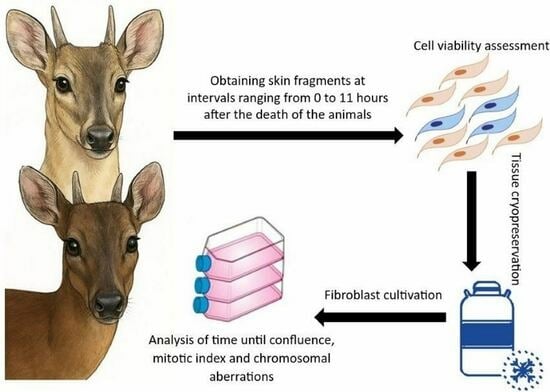
2. Materials and Methods

This work was evaluated and approved by the Ethics and Animal Welfare Committee (CEBEA) of the Faculty of Agricultural and Veterinary Sciences (FCAV) of São Paulo State University (UNESP), Jaboticabal campus, under protocol Nº. 005433/19. The study complies with the ethical principles in animal experimentation adopted by the Brazilian College of Animal Experimentation (COBEA).

2.1. Sample Collection

Two individuals were used in this study—a female of the species Mazama rufa and a male of the species Subulo gouazoubira—both housed in captivity at the Deer Research and Conservation Center (NUPECCE), Department of Animal Science, School of Agricultural and Veterinary Sciences, São Paulo State University (UNESP), Jaboticabal Campus. The animals were chemically restrained using a combination of anesthetic agents—7 mg/kg of ketamine hydrochloride and 1 mg/kg of xylazine hydrochloride—administered intramuscularly via manual injection within a restraining box, followed by euthanasia through intravenous administration of 1 g of sodium thiopental.
The carcasses were maintained in a room located directly across from the laboratory, without temperature control, where ambient temperature ranged between 20 °C and 25 °C. Skin fragments were collected immediately after euthanasia and subsequently at regular intervals of one hour for a total period of 11 h, resulting in twelve collection time points (0 h, 1 h, 2 h, 3 h, 4 h, 5 h, 6 h, 7 h, 8 h, 9 h, 10 h, and 11 h). This interval was chosen because the study aims to maintain the carcass under non-sterile environmental conditions, simulating field settings. The typical tropical climate, characterized by high temperatures and humidity, promotes both tissue degradation and rapid microorganism proliferation, where extended postmortem periods may yield less consistent data on the viability and functionality of fibroblasts.
Samples were collected from the inner thigh region, which had been previously shaved and disinfected using 70% ethanol. Excised skin fragments measured approximately 2 cm2 and were immediately immersed in McCoy’s medium (Sigma-Aldrich, St. Louis, MO, USA) supplemented with a high concentration of antibiotics (500 mg/L gentamicin and 20 mg/L amphotericin B). After collection, samples were immediately transferred to the laboratory, within less than one minute, thus minimizing degradation risks. Upon arrival at the laboratory, each skin sample was divided into two equal parts: one portion was used to assess cellular viability in fresh tissue, while the other was cryopreserved in liquid nitrogen for subsequent thawing and cell culture procedures. The cultures were maintained until cell confluence and chromosome preparations were obtained to assess the number of metaphases per cell, based on analysis of the mitotic index, as described below.

2.2. Cell ViabilityCell Dissociation

Cell Dissociation

Upon arrival at the laboratory, skin fragments stored in McCoy’s medium were handled under laminar flow and washed with phosphate-buffered saline (PBS). For cell viability analysis, tissue fragments were mechanically minced into ~1 mm3 pieces in a dish containing 3 mL of collagenase I (Sigma-Aldrich, Taufkirchen, Germany) (1 mg/mL) diluted in Dulbecco’s Modified Eagle medium (DMEM), high glucose, and pyruvate (Cultilab, Campinas, Brazil). The digestion was performed in a 5% CO2 incubator at 37 °C for 30 min, with manual agitation every 5 min. The content was centrifuged at 200× g for 5 min, and the supernatant was discarded. The resulting cell pellet was resuspended in 4.5 mL of DMEM for viability assessment.

2.3. Cell Viability Counting and Calculation

To determine cell viability, a 50 µL aliquot of the cell suspension was mixed with 50 µL of 0.4% Trypan Blue (Gibco, 1:1 dilution). After mixing, the cell suspension was loaded into a Neubauer counting chamber and examined under a light microscope at 10× magnification. Viable (unstained) and non-viable (blue-stained) cells were counted in the four quadrants [14]. The total number of viable cells was calculated using the following formula:
Total viable cells = (Mean number of viable cells) × 10,000 × 2 × 4.5
where the mean refers to the average of cells in quadrants A, B, C, and D; 10,000 is the conversion factor per mL; 2 accounts for dilution (50 µL Trypan Blue + 50 µL cell suspension); and 4.5 is the total volume of DMEM used for resuspension [14]. Cell viability (%) was calculated based on Louis & Siegel [14] as follows:
Cell viability = (Viable cells/Total cells [viable + non-viable]) × 100.

2.4. Cryopreservation of Skin Fragments

For cryopreservation, each skin fragment was first washed by sequentially transferring it through three separate Petri dishes containing 4 mL of PBS for 2 min each, to ensure thorough cleaning prior to freezing. After washing, each piece was placed in a cryotube containing 1.0 mL of freezing medium (McCoy’s medium supplemented with 20% inactivated equine serum, 6.25% DMSO, 100 mg/mL PVP, 4.25 µL gentamicin, and 8.5 µL amphotericin B). Tubes were stored at 4 °C for 3.5 h, then exposed to liquid nitrogen vapor (2 cm above the liquid surface) for 30 min, and, finally, immersed in liquid nitrogen for long-term storage.

2.5. Thawing and Cell Culture of Cryopreserved Fragments

Thawing of cryopreserved fragments was performed by transferring the cryotube from liquid nitrogen directly into a 37 °C water bath for about 1.5 min until the freezing medium was completely liquefied. The fragment was then manipulated under laminar flow, washed three times with PBS, and mechanically minced into ~1 mm3 pieces in a Petri dish containing 1 mL of DMEM and 1 mL of fetal bovine serum (FBS). Fragments were transferred to culture flasks and supplemented with 1.0 mL of DMEM enriched with 50% FBS, 50 mg/L gentamicin sulfate, and 2 mg/L amphotericin B. Cultures were incubated at 37 °C in a 5% CO2 atmosphere and maintained without disturbance for 48 h [15].
After that, cultures were monitored daily and medium changes occurred every 48 h. Cell proliferation and confluence were subjectively evaluated based on the rate of cell expansion within the culture flask and cell morphology, using an inverted optical microscope, until 100% confluence was reached. All evaluations were performed by two independent, blinded observers who were unaware of the animal source or time point of the samples under examination.

2.6. Cell Harvesting

Once cultures reached full confluence, cells were treated with 60 µL of 0.016% colchicine for 30 min at 37 °C. Afterward, cells were detached using 0.25% trypsin and centrifuged at 200× g for 5 min. The supernatant was discarded, and the pellet resuspended in 5 mL of hypotonic KCl solution (0.067 M) for 20 min at 37 °C. The cell pellet was then fixed with 5 mL of Carnoy’s fixative (methanol/acetic acid, 3:1), centrifuged, and the fixation process repeated three times.

2.7. Mitotic Index Analysis

Fixed cells were dropped onto microscope slides and stained using conventional Giemsa staining. Slides were analyzed under brightfield microscopy using an Olympus BX60 microscope (Olympus Corporation, Tokyo, Japan) (100× objective) equipped with a Zeiss AxioCam MRm camera (Carl Zeiss Microimaging GmbH, Jena, Germany) and AxioVision Release 4.8.2 software. For each culture, 1000 cells were evaluated. The mitotic index (MI) was calculated as the ratio of metaphase cells to the total number of cells analyzed, expressed as a percentage [16,17].

2.8. Chromosomal Alteration Analysis

For each sampling time point, between 20 and 60 metaphases were analyzed using bright-field microscopy with an oil immersion objective lens (magnification: 1000×), in order to assess chromosomal stability based on the presence and number of chromosomal alterations observed per sample. The observed chromosomal alterations were counted and classified into the following categories: ring (A), dicentric chromosome (Dic), chromatid gap (Gct), chromosome gap (Gcr), chromatid break (Qct), chromosome break (Qcr), triradial figure (Ft), quadriradial figure (Fq), and rearrangement (Re) [18,19,20].

2.9. Statistical Analysis

Due to the accession of tissue collected for only one individual per species, cell viability, and mitotic index data were treated descriptively. For each individual, postmortem time series were constructed showing viability and mitotic index at each collection interval. To explore potential differences between species, we performed simple linear regression analyses modeling each parameter (viability and mitotic index) as a function of postmortem time. Interaction terms (Time × Species) were included in the models to assess whether the rate of change over time differed between species. Regarding chromosomal analysis, the number of chromosomal alterations was statistically evaluated using Tukey’s test, performed with the statistical software GraphPad Prism 7.0.

3. Results

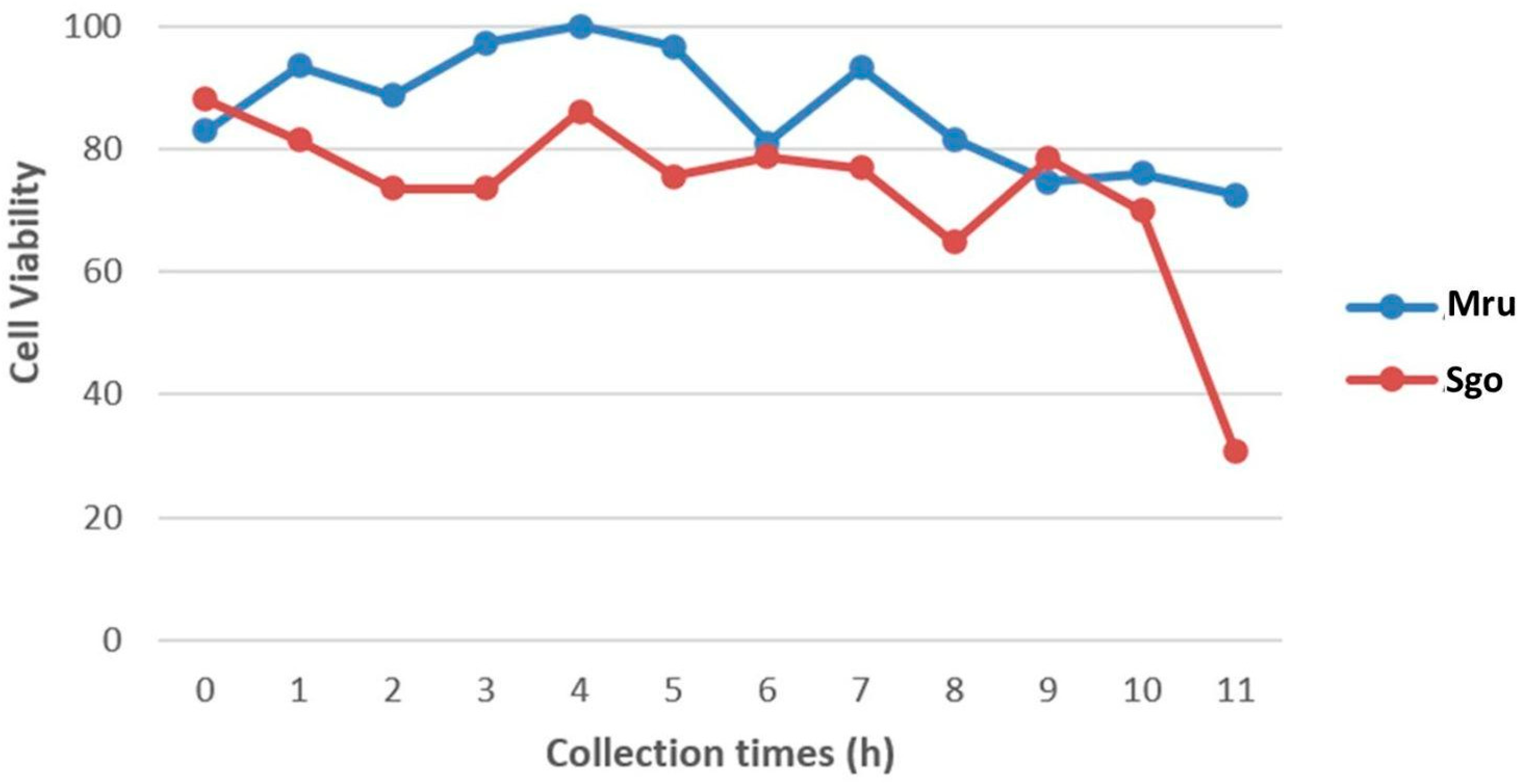
3.1. Cell Viability of Fresh Tissues

As expected, a decrease in the number of viable cells was observed over time after the death of both animals (Figure 1). Tissues collected immediately after euthanasia showed cell viability greater than 80%. This number of viable cells slightly decreased in red brocket tissue collected after 11 h, and the reduction was more pronounced in gray brocket tissue, where cell viability dropped below 40%. Although the number of viable cells at later time points was considerably lower than in the initial h, the data indicate that it is still feasible to obtain viable cells from tissue fragments collected up to 11 h postmortem. An exploratory linear regression analysis was conducted to compare the postmortem decline in cell viability between the two species. The individual Mazama rufa exhibited an initial viability of approximately 95.9%, with a decreasing trend of 1.7% per hour (p = 0.055), whereas the individual Subulo gouazoubira showed a similar but steeper decline. However, the difference in slopes between species was not statistically significant (interaction term: p = 0.41) (Supplementary Materials, Figure S1). These results, although limited by the absence of biological replication, suggest a possible interspecific difference in the temporal preservation of cell viability.

3.2. Cryopreservation Methodology

Cryopreservation was carried out according to the methods used by Oliveira and collaborators [12]. The culture of cryopreserved skin fragments proved to be viable at all collection time points for both individuals evaluated in this study. Following thawing, the skin fragments were able to adhere to the bottom of the culture flasks, indicating that the cryopreservation protocol did not result in substantial damage to the tissue samples. Furthermore, these fragments demonstrated the capacity to resume cell division, as fibroblasts migrating from the tissue were observed to proliferate and spread throughout the culture surface.

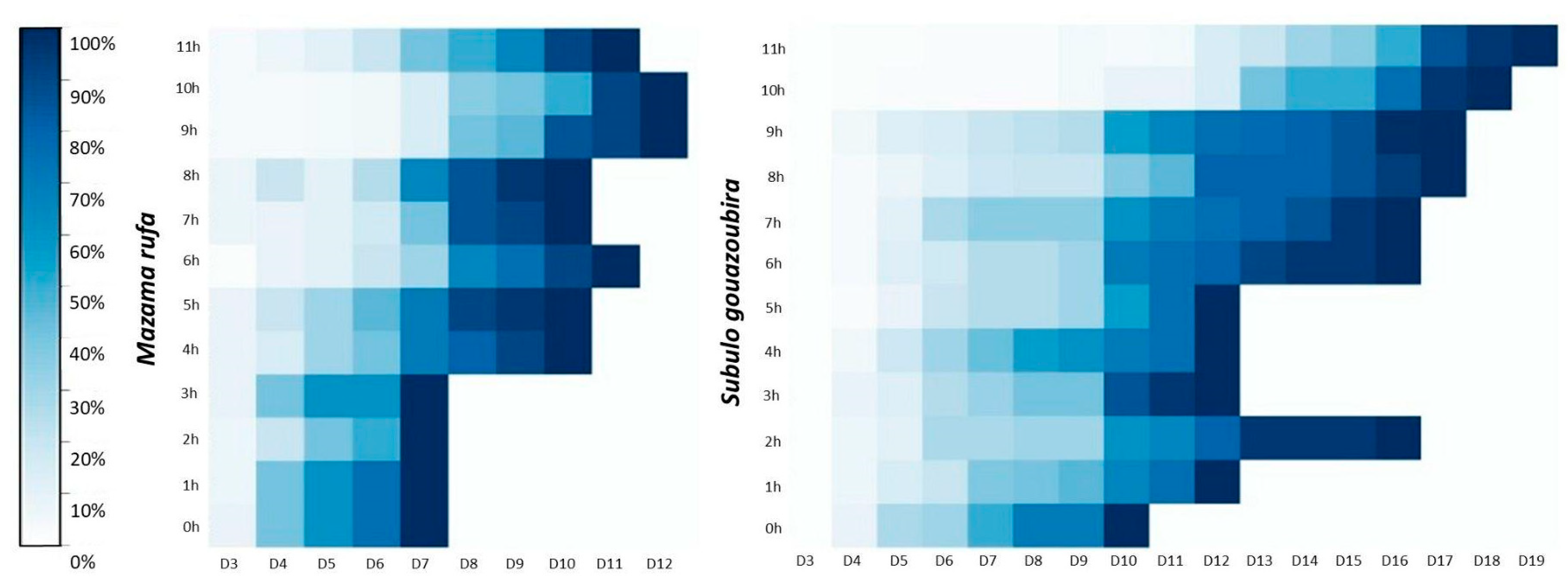
3.3. Confluence Analysis of Cell Cultures Derived from Cryopreserved Skin Fragments

Confluence monitoring was conducted daily for all samples starting on day three (D3) after the initiation of cell culture. In Mazama rufa, tissue fragments adhered to the flask surface by the third day (D3), whereas in Subulo gouazoubira, adherence was first observed on the fourth day (D4) (Figure 2). Cell cultures from Mazama rufa reached full confluence within a maximum of 12 days, which was notably shorter than the time required for Subulo gouazoubira, in which cultures took up to 19 days to reach confluence (Figure 2). In Mazama rufa, cell cultures established from samples collected within the first 3 h postmortem reached full confluence simultaneously on the seventh day (D7) of cultivation. In contrast, for Subulo gouazoubira, samples collected from the 1st to the 5th h postmortem reached confluence by day 12 of culture, with the exception of the sample collected at two h postmortem.

3.4. Mitotic Index Analysis

Mitotic index analysis showed a range from 0.2 h to 0.8 h in the individual M. rufa (Animal 1), with the lowest value after 10 h of time point collection (Figure 3). In the individual S. gouzoubira, a higher number of metaphases was observed, mainly in samples collected up to 5 h postmortem with mitotic index ranging from 0.1 h to 1.9 h (Figure 3). After this 6 h time point, the number of metaphases in S. gouzoubira declined dramatically, approaching the values observed in M. rufa. In S. gouzoubira, a marked decrease in metaphase count was observed in the sample collected 2 h postmortem. Considering that this same sample also showed delayed confluence, as previously reported, it is possible that a specific issue occurred during its processing, negatively affecting sample quality. An exploratory linear regression analysis was also performed to compare the temporal variation in mitotic index between the two species. The model revealed a statistically significant interaction between time and species (p = 0.042), indicating that the rate of decrease in mitotic activity differed between Mazama rufa and Subulo gouazoubira (Supplementary Materials, Figure S2). While M. rufa showed a relatively stable mitotic index over time, S. gouazoubira exhibited a steeper decline, suggesting greater sensitivity to postmortem delay. These findings support a potential species-specific pattern in cell division dynamics after death, although the results should be interpreted with caution due to the limited sample size.

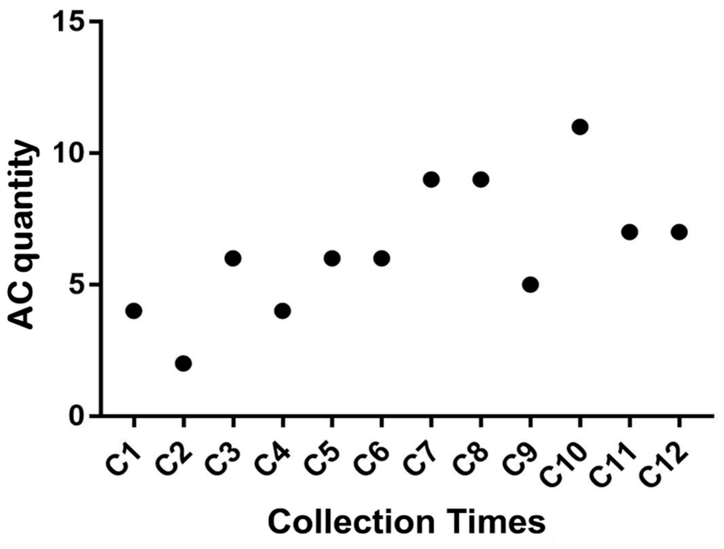
3.5. Chromosomal Alteration Analysis

A total of 780 metaphases were analyzed to assess chromosomal stability based on the presence and frequency of chromosomal alterations observed in each sampling time point. Among the nine types of chromosomal alterations evaluated, only four were detected: Gct, Gcr, Qct, and Qcr. The most frequently observed alteration was Gct, followed by Qct, Qcr, and Gcr, respectively.
As shown in Figure 4, although a gradual increase in the average number of chromosomal alterations was observed in both Mazama rufa and Subulo gouazoubira as the postmortem interval increased—particularly after the sixth collection point (5 h postmortem)—these differences were not statistically significant. This result suggests that chromosomal structures remain stable for up to 11 h after the animal’s death.
Although it is possible to obtain viable tissue suitable for cryopreservation, subsequent thawing, and use in cytogenetic studies up to 11 h postmortem, the data presented herein suggest that the highest rates of cell viability, shortest time to reach confluence, greatest number of metaphases per cell (mitotic index), and highest chromosomal stability are achieved when skin fragments are collected within the first 5 h postmortem.

4. Discussion

The establishment of fibroblast cell lines from endangered species has been proposed as a practical approach to animal preservation, not only safeguarding valuable genetic material but also providing a robust resource for biological research [11]. They could provide an essential resource for further studies regarding reproductive biotechniques, and therefore be very useful as an ex situ conservation strategy. Although the collection of reproductive tissues from Neotropical deer species represents a promising strategy to enhance the genetic diversity stored in biobanks, protocols for cryopreservation and in vitro cultivation of cells derived from these tissues are still in the early stages of development. Therefore, studies suggest that using fibroblasts from Neotropical cervids in assisted reproductive technologies, including somatic cell nuclear transfer (SCNT), holds promise despite ongoing practical challenges. For instance, research on the Subulo gouazoubira species demonstrated successful production of blastocysts through interspecies cloning using bovine cytoplasts [21]. Moreover, it was shown that, following a freeze–thaw cycle, passages 4 and 7 of these cell lines maintained good viability in terms of survival rates, while passage 10 exhibited reduced quality [22].
The collection of tissue samples from animals postmortem represents a valuable opportunity to preserve the genetic material of individuals, particularly those of rare or elusive species [23]. In Brazil, it is estimated that approximately 475 million animals are killed annually on roads, equating to 15 roadkill incidents per second [24]. Specific studies conducted in the state of São Paulo estimate an average of 0.6 medium- to large-sized mammals killed per kilometer per year on toll roads over a ten-year monitoring period [25]. These figures support the potential significance of postmortem sampling in roadkill cases as a means of contributing to the maintenance of genetic diversity in cryobanks. Therefore, implementing policies that encourage highway concessionaires to collect animal carcasses could provide an efficient pathway for acquiring samples from forest-dwelling and elusive species, such as many Neotropical deer.
Since death results in the cessation of blood circulation and subsequent oxygen deprivation, a cascade of tissue degradation begins [26,27]. In this study, the analysis of freshly collected tissue allowed for the evaluation of sample quality, as low viability would significantly compromise the effectiveness of cryopreservation. Trypan Blue staining remains the most commonly used method to differentiate viable from non-viable cells and serves as a proxy for the proliferative capacity of a given sample [28,29,30].
Upon evaluating cellular viability in samples collected at different postmortem intervals, it was observed that most samples from M. rufa exhibited a higher number of viable cells compared to those from S. gouazoubira. This observation may be attributed to individual variation or possibly species-specific traits, as M. rufa typically has noticeably thicker skin than S. gouazoubira.
Fibroblasts began to migrate out from the skin fragments between the third day (M. rufa) and the fourth day (S. gouazoubira) of culture, a timeframe consistent with previous studies on fibroblast outgrowth from Neotropical deer skin tissues [12,13]. Regarding the time to reach full confluence, all cultures achieved 100% confluence within a timeframe comparable to that typically observed for brocket deer fibroblasts derived from in vivo-collected samples that were immediately cryopreserved and later thawed for culture [12].
Previous studies have demonstrated that viable and cultivable cells can be obtained up to 10 days after the death of animals such as sheep, rabbits, and goats. However, these studies involved samples stored under controlled and sterile conditions at room temperature [13,31,32,33]. In contrast, this study demonstrated the presence of viable cells and sterility in cultured fibroblast lines up to 11 h postmortem, under field conditions without temperature control or sterile handling, thus simulating a scenario found in nature.
Mitotic index analysis was used to morphologically assess cell cycle stages, quantifying the number of cells reaching metaphase during mitosis [16]. However, the maximum mitotic potential may not have been fully represented in this experiment, as cultures were harvested only upon reaching 100% confluence—a point at which cellular proliferation generally begins to decline [16,34]. Given that fibroblasts proliferate rapidly and are capable of reaching adequate mitotic indices, it was unnecessary to use mitogens to stimulate cell division in order to obtain a moderate number of metaphases [28].
Cytogenetic analyses provide insight into whether postmortem cellular degradation affects chromosomal structure and stability through the observation of chromosomal alterations [31]. Some of the alterations observed in this study may not necessarily be associated with postmortem degradation but could instead reflect inherent chromosomal fragility reported in some Neotropical deer species [35]. Nevertheless, although chromosomal alterations were detected in some metaphases, their frequency did not differ significantly among samples collected at different time points. This finding indicates that stable, intact chromosomes can still be obtained up to 11 h postmortem.
Both the number of viable cells and the mitotic index declined progressively with increasing postmortem interval, consistent with the gradual death of skin cells, including adult stem cells, which are responsible for rapid cellular proliferation [13]. The aforementioned oxygen deprivation results in progressive tissue decomposition due to lysosomal enzyme activity [13]. Although the data were obtained from a single individual per species, this study suggests that optimal results are achieved when skin samples from M. rufa and S. gouazoubira are collected within 5 h postmortem under uncontrolled environmental conditions of temperature and sterility. In addition, the exploratory linear regression analysis of mitotic index and cell viability distributions suggests consistent interspecific differences that should be confirmed in studies with a larger sample size.

5. Conclusions

This study demonstrated that cells obtained from postmortem animal tissue remain chromosomally stable, making them suitable for cytogenetic analyses. Based on the combined analysis of the data presented in this study, it can be concluded that fibroblast cell lines can be successfully established from skin tissue collected up to 11 h postmortem from animals kept under non-controlled temperature conditions and without aseptic procedures. However, based on higher cell viability rates, shorter time to reach confluence, and greater numbers of metaphases per cell (mitotic index), it is suggested that optimal results can be achieved when skin fragments are collected within 5 h postmortem.

Supplementary Materials

The following supporting information can be downloaded at: https://www.mdpi.com/article/10.3390/jzbg6020031/s1, Figure S1: Simple linear regression analysis of cell viability as a function of postmortem time. Interaction terms (Time × Species). Mru = Mazama rufa individual. Sgo = Subulo gouazoubira individual; Figure S2: Simple linear regression analysis of mitotic index as a function of postmortem time. Interaction terms (Time × Species). Mru = Mazama rufa individual. Sgo = Subulo gouazoubira individual.

Author Contributions

Conceptualization, I.M.T. and J.M.B.D.; methodology, I.M.T.; formal analysis, L.D.R. and I.M.T.; investigation, L.D.R., I.M.T., E.D.P.S., J.A.M.-D. and C.H.d.S.B.; resources, J.M.B.D.; writing—original draft preparation, L.D.R. and I.M.T.; writing—review and editing, L.D.R., I.M.T. and E.D.P.S.; visualization, L.D.R., I.M.T., E.D.P.S., J.A.M.-D., C.H.d.S.B. and J.M.B.D.; supervision, J.M.B.D. All authors have read and agreed to the published version of the manuscript.

Funding

This research was supported by the Fundação de Amparo à Pesquisa do Estado de São Paulo (FAPESP) process n◦ 2017/07014-8.

Institutional Review Board Statement

The animal study protocol was approved by the Ethics and Animal Welfare Committee (CEBEA) of the Faculty of Agricultural and Veterinary Sciences (FCAV) of São Paulo State University (UNESP), Jaboticabal campus, under protocol Nº. 005433/19, approved 16 May 2019.

Data Availability Statement

The original contributions presented in this study are included in the article/Supplementary Material. Further inquiries can be directed to the corresponding author.

Acknowledgments

We would like to thank other members of the Deer Research and Conservation Center for their collaboration, including the laboratory technician João Boer for helping with the equipment and preparing solutions, and the animal caretakers, who kept them healthy and well-being until the time of euthanasia of the individuals. The authors have reviewed and edited the output and take full responsibility for the content of this publication.

Conflicts of Interest

The authors declare no conflicts of interest.

Abbreviations

The following abbreviations are used in this manuscript:
SCNT Somatic cell nuclear transfer
DMEM Dulbecco’s Modified Eagle medium
DMSODimethyl sulfoxide
PVPPolyvinylpyrrolidone
FBSFetal bovine serum
PBSPhosphate-buffered saline
MI Mitotic Index
R Ring
Dic Dicentric chromosome
Gct Chromatid gap
Gcr Chromosome gap
Qct Chromatid break
Qcr Chromosome break
FtTriradial figure
FqQuadriradial figure
ReRearrangement

References

  1. Weber, M.; Gonzalez, S. Latin American deer diversity and conservation: A review of status and distribution. Écoscience 2003, 10, 443–454. [Google Scholar] [CrossRef]
  2. Schipper, J.; Chanson, J.S.; Chiozza, F.; Cox, N.A.; Hoffmann, M.; Katariya, V.; Lamoreux, J.; Rodrigues, A.S.L.; Stuart, S.N.; Temple, H.J.; et al. The status of the world’s land and marine mammals: Diversity, threat, and knowledge. Science 2008, 322, 225–230. [Google Scholar] [CrossRef]
  3. The IUCN Red List of Threatened Species. Available online: https://www.iucnredlist.org/ (accessed on 31 March 2025).
  4. Frankham, R.; Ballou, J.D.; Briscoe, D.A. A Primer of Conservation Genetics; Cambridge University Press: Cambridge, UK, 2004; 220p. [Google Scholar]
  5. Keller, L.F.; Biebach, I.; Ewing, S.R.; Hoeck, P.E.A. Then genetics of Reintroductions: Inbreding and Genetic Drift. In Reintroduction Biology, Integrating Science and Management; Ewen, J.G., Armstrong, D.P., Parker, K.A., Seddon, P.J., Eds.; Wiley-Blackwell: Hoboken, NJ, USA, 2012; 499p. [Google Scholar]
  6. González, S.; Duarte, J.M.B. Speciation, evolutionary history and conservation trends of neotropical deer. Mastozool. Neotrop. 2020, 27, 37–47. [Google Scholar] [CrossRef]
  7. Maran, M.L.H. Filogenia Molecular de Mazama americana (Artiodactyla: Cervidae) como auxílio na Resolução das Incertezas Taxonômicas. Master’s Thesis, University of São Paulo, São Paulo, Brazil, 2016. Available online: https://repositorio.unesp.br/handle/11449/142902 (accessed on 11 April 2025).
  8. Ryder, O.A.; Onuma, M. Viable cell culture banking for biodiversity characterization and conservation. Annu. Rev. Anim. Biosci. 2018, 6, 83–97. [Google Scholar] [CrossRef]
  9. Comizzoli, P.; Holt, W. Breakthroughs and new horizons in reproductive biology of rare and endangered animal species. Biol. Reprod. 2019, 101, 514–525. [Google Scholar] [CrossRef]
  10. Rola, L.D.; Buzanskas, M.E.; Melo, L.M.; Chaves, M.S.; Freitas, V.J.F.; Duarte, J.M.B. Assisted reproductive technology in neotropical deer: A model approach to preserving genetic diversity. Animals 2021, 11, 1961. [Google Scholar] [CrossRef]
  11. Mastromonaco, G.F.; King, W.A. Cloning in companion animal, non-domestic and endangered species: Can the technology become a practical reality? Reprod. Fertil. Dev. 2007, 19, 748–761. [Google Scholar] [CrossRef]
  12. Oliveira, L.C.; Vacari, G.Q.; Duarte, J.M.B. A method to freeze skin samples for cryobank: A test of some cryoprotectants for an endangered deer. Biopreserv. Biobank. 2023, 22, 211–216. [Google Scholar] [CrossRef]
  13. Singh, M.; Ma, X. In vitro culture of fibroblast-like cells from sheep ear skin stored at 25–26 °C for 10 days after animal death. Int. J. Biol. 2014, 6, 96–102. [Google Scholar] [CrossRef]
  14. Louis, K.S.; Siegel, A.C. Cell viability analysis using trypan blue: Manual and automated methods. In Mammalian Cell Viability: Methods and Protocols; Stoddart, M.J., Ed.; Humana Press: Totowa, NJ, USA, 2011; pp. 7–12. [Google Scholar]
  15. Verma, R.S.; Babu, A. Human Chromosomes: Principles and Techniques, 2nd ed.; McGraw-Hill: New York, NY, USA, 1995; p. 419. [Google Scholar]
  16. O’Connor, P.M.; Ferris, D.K.; Pagano, M.; Draetta, G.; Pines, J.; Hunter, T.; Longo, D.L.; Kohn, K.W. G2 delay induced by nitrogen mustard in human cells affects cyclin A/cdk2 and cyclin B1/cdc2-kinase complexes differently. J. Biol. Chem. 1993, 268, 8298–8308. [Google Scholar] [CrossRef]
  17. Galloway, S.M.; Aardema, M.J.; Ishidate, M., Jr.; Ivett, J.L.; Kirkland, D.J.; Morita, T.; Moses, P.; Sofuni, T. Report from working group on in vitro tests for chromosomal aberrations. Mutat. Res. 1994, 312, 241–261. [Google Scholar] [CrossRef]
  18. Evans, H.J.; O’Riordan, M.L. Human peripheral blood lymphocytes for the analysis of chromosome aberrations in mutagen tests. Mutat. Res. 1975, 31, 135–148. [Google Scholar] [CrossRef] [PubMed]
  19. Savage, J.R.K. Classification and relationships of induced chromosomal structural changes. J. Med. Genet. 1975, 12, 103–122. [Google Scholar]
  20. Savage, J.R.K.; Simpson, P.J. FISH “painting” patterns resulting from complex exchanges. Mutat. Res. 1994, 312, 51–60. [Google Scholar] [CrossRef] [PubMed]
  21. Melo, L.M.; Silva, S.B.; Magalhães, L.C.; Cortez, J.V.; Kumar, S.; Duarte, J.M.; Rola, L.D.; Chaves, M.S.; Freitas, V.J.F. The use of somatic cell nuclear transfer to obtain interspecific cloned embryos from brown brocket deer karyoplast and bovine cytoplast: Embryo development and nuclear gene expression. Theriogenol. Wild 2022, 1, 100001. [Google Scholar] [CrossRef]
  22. Magalhaes, L.C.; Bhat, M.H.; Freitas, J.L.; Melo, L.M.; Teixeira, D.I.; Pinto, L.C.; Câmara, L.M.C.; Duarte, J.M.B.; Freitas, V.J.F. The effects of cryopreservation on different passages of fibroblast cell culture in brown brocket deer (Mazama gouazoubira). Biopreserv. Biobank. 2017, 15, 463–468. [Google Scholar] [CrossRef]
  23. Huijsmans, T.E.R.G.; Hassan, H.A.; Smits, K.; Van Soom, A. Postmortem collection of gametes for the conservation of endangered mammals: A review of the current state-of-the-art. Animals 2023, 13, 1360. [Google Scholar] [CrossRef] [PubMed]
  24. Centro Brasileiro de Ecologia de Estradas (CBEE). Atropelômetro. 2019. Available online: https://ecoestradas.com.br/ (accessed on 14 March 2024).
  25. Abra, F.D.; Huijser, M.P.; Magioli, M.; Bovo, A.A.A.; de Barros, K.M.P.M. An estimate of wild mammal roadkill in São Paulo state, Brazil. Heliyon 2021, 7, e06084. [Google Scholar] [CrossRef]
  26. Zdravkovic, M. Ultrastructural changes of renal epithelial cells during post-mortem autolysis—Experimental work. Med. Pregl. 2010, 63, 15–20. [Google Scholar] [CrossRef]
  27. Neta, L.B.Q.; Lira, G.P.O.; Borges, A.A.; de Oliveira Santos, M.V.; Silva, M.B.; de Oliveira, L.R.M.; Silva, A.R.; de Oliveira, M.F.; Pereira, A.F. Influence of storage time and nutrient medium on recovery of fibroblast like cells from refrigerated collared peccary (Pecari Tajacu Linnaeus, 1758) skin. In Vitro Cell Dev. Biol. Anim. 2018, 54, 486–495. [Google Scholar] [CrossRef]
  28. Pegg, D.E. Viability assays for preserved cells, tissues and organs. Cryobiology 1989, 26, 212–231. [Google Scholar] [CrossRef] [PubMed]
  29. Hartford, J.B. Cell culture. In Current Protocols in Cell Biology; Bonifacino, J.S., Hartford, J.B., Lippincott-Schwartz, J., Eds.; John Wiley & Sons: Hoboken, NJ, USA, 1998; pp. 101–102. [Google Scholar]
  30. Stoddart, M.J. (Ed.) Cell viability assays: Introduction. In Mammalian Cell Viability: Methods and Protocols; Humana Press: Totowa, NJ, USA, 2011; pp. 1–6. [Google Scholar]
  31. Silvestre, M.A.; Saeed, A.M.; Cervera, R.P.; Escriba, M.J.; Garcia-Ximenez, F. Rabbit and pig ear skin sample cryobanking: Effects of storage time and temperature of the whole ear extirpated immediately after death. Theriogenology 2003, 59, 1469–1477. [Google Scholar] [CrossRef] [PubMed]
  32. Silvestre, M.A.; Sanchez, J.P.; Gomez, E.A. Vitrification of goat, sheep, and cattle skin samples from whole ear extirpated after death and maintained at different storage times and temperatures. Cryobiology 2004, 49, 221–229. [Google Scholar] [CrossRef] [PubMed]
  33. Singh, M.; Ma, X.; Sharma, A. Effect of postmortem time interval on in vitro culture potential of goat skin tissues stored at room temperature. Vitr. Cell. Dev. Biol.—Anim. 2012, 48, 478–482. [Google Scholar] [CrossRef]
  34. Jackman, J.; O’Connor, P.M. Methods for synchronizing cells at specific stages of the cell cycle. In Current Protocols in Cell Biology; Bonifacino, J.S., Hartford, J.B., Lippincott-Schwartz, J., Yamada, K.M., Eds.; John Wiley & Sons: Hoboken, NJ, USA, 1998; pp. 8.3.1–8.3.20. [Google Scholar]
  35. Vargas-Munar, D.S.F. Relação entre Fragilidade Cromossômica e Trocas entre Cromátides Irmãs com a Variabilidade Cariotípica de Cervídeos Brasileiros. Master’s Dissertation, Paulista State University, São Paulo, Brazil, 2003. [Google Scholar]
Figure 1. Viability of cells from skin fragments expressed as a percentage, in Mru (Mazama rufa) and Sgo (Subulo gouazoubira) collected at different collection time points.
Jzbg 06 00031 g001
Figure 2. Degree of cell confluence (%) in cultures derived from postmortem tissue samples of Mazama rufa and Subulo gouazoubira. Cell cultures were evaluated daily, starting on the third day of culture (D3), until reaching 100% confluence. Tissue collection time points: 0 h, 1 h, 2 h, 3 h, 4 h, 5 h, 6 h, 7 h, 8 h, 9 h, 10 h, and 11 h. Right panel: color bar indicates cell confluence percentage, ranging from 0% (white) to 100% (dark blue).
Jzbg 06 00031 g002
Figure 3. Mitotic index after skin tissue collection at different postmortem times of Mru (Mazama rufa) and Sgo (Subulo gouazoubira).
Jzbg 06 00031 g003
Figure 4. Average number of chromosomal alterations (ACs) observed at different sampling time points.
Jzbg 06 00031 g004
Disclaimer/Publisher’s Note: The statements, opinions and data contained in all publications are solely those of the individual author(s) and contributor(s) and not of MDPI and/or the editor(s). MDPI and/or the editor(s) disclaim responsibility for any injury to people or property resulting from any ideas, methods, instructions or products referred to in the content.

