Review

Immune Responses to Filarial Nematodes: A Mechanistic Evaluation of Evasion and Modulation Strategies

Department of Biochemistry, Institute of Science, Banaras Hindu University, Varanasi 221005, India
*
Author to whom correspondence should be addressed.
Immuno 2025, 5(4), 57; https://doi.org/10.3390/immuno5040057
Submission received: 26 September 2025 / Revised: 1 November 2025 / Accepted: 11 November 2025 / Published: 26 November 2025

Abstract

Filarial parasites are long-lived organisms that cause extreme morbidity due to pathological manifestations, including lymphedema, hydrocele, and elephantiasis. Understanding the hosts’ immune responses to filarial parasites is crucial to developing new and effective anti-filarial treatments. The review thoroughly examines and summarises immunological modulation, evasion strategies, and filarial–host immune interactions to provide an updated knowledge of the immune evasion manoeuvres used by filarial parasites. An extensive literature search was conducted using databases such as PubMed, Google Scholar, ScienceDirect, Web of Science, and Scopus to identify articles published mostly between 2000 and 2025 that focus on the crucial molecular, cellular, and immunomodulatory strategies of filarial parasites. The immune evasion mechanisms include the modulation of effector T cells, induction of apoptosis in immune cells, the release of immunomodulatory proteins, and the induction of regulatory immune cell populations, thereby ensuring the mutual survival of both the parasite and the host. An antigen-specific T helper 2 (Th2) response and an increase in Interleukin-10 (IL-10) producing CD4+ T cells, along with a suppressed T helper 1 (Th1) response, are the key immunological characteristics of filarial pathogenesis. This antigen-specific T-cell hyporesponsiveness seems necessary for keeping the long-term infection going, which often involves large parasite densities. This review summarises filarial parasites’ mechanisms and strategies in regulating host immune responses and will facilitate future studies on the filarial pathogenesis, leading to the development of novel anti-filarial therapeutics.

1. Introduction

Lymphatic Filariasis (LF) is an undermining parasitic disease mainly caused by Wuchereria bancrofti and, to some extent, by Brugia malayi and Brugia timori. More than 90% of infections are bancroftian in nature [1]. LF affects over 120 million people around the globe, with millions more people at risk, mainly in tropical and subtropical regions [2]. The disease targets the lymphatic system, causing inflammation, lymphatic dysfunction, and eventual development of lymphedema, elephantiasis, and hydrocele. The manifestation and severity of disease are caused by the influence of host immune response, which decides whether the individual remains asymptomatic, develops acute inflammation, or progresses to severe chronic pathology [3]. Immune mechanisms during infection not only direct the clinical manifestation but also influence the susceptibility, parasite persistence, and therapeutic response [4].

2. Global Filarial Endemicity

LF is considered endemic in tropical and subtropical areas worldwide, affecting the population of Sub-Saharan Africa, Madagascar, several Western Pacific island nations, territories, and parts of the Caribbean and sporadically in America, India, and Southeast Asia. W. bancrofti is the most widespread causative agent, responsible for nearly 90% cases, found in many tropical and subtropical regions, whereas Brugia spp. are more geographically limited, primarily found in Southeast Asia. Over 657 million people in 39 countries worldwide remain threatened by LF and require preventive chemotherapy just to interrupt the spread of this parasitic infection. According to the World Health Organisation (WHO), the global baseline estimate of people affected by LF was 25 million men with hydrocele and over 15 million people with lymphoedema, while at least 36 million people remain with chronic disease manifestations [5].
India has historically been a significant contributor to the global burden of LF, with estimates showing millions of people at risk. According to the National Vector-borne Disease Control Program (NVBDCP), 345 districts were reported endemic to LF, with around 740 million people at risk. Since 2000, an intensive Mass Drug Administration (MDA) of over 9.7 billion doses has been delivered. Despite these efforts, as of 2023, the number of cases reported in India is around 619,000 lymphedema cases and 126,000 hydrocele cases [6]. In India, the majority of cases are of W. bancrofti, while B. malayi is restricted to its states that are Kerala, Karnataka, and Odisha. Among the endemic states, high prevalence of the disease can be seen in Bihar, Uttar Pradesh, and Odisha, while other states like Andhra Pradesh, Tamil Nadu, Jharkhand, and Karnataka also have notable LF burden [7].

3. Clinical Diagnosis

The clinical diagnosis of filariasis relies on the identification of microfilariae in blood, skin, or ocular tissues. Blood smears are frequently used by clinicians to diagnose W. bancrofti, Loa loa, Mansonella perstans, and M. ozzardi [8]. Blood must be withdrawn at a particular point in time, either during the day or at night, depending on the microfilariae’s periodicity [9]. The improved identification of microfilariae in blood or other bodily fluids can be achieved using concentration techniques like the membrane filtration technique and Knott’s technique, followed by microscopic examination [10]. Skin snips are suggested for identifying infections caused by M. streptocerca and O. volvulus present in skin tissues [11]. Polymerase chain reaction (PCR) and loop-mediated isothermal amplification are two well-known molecular approaches for diagnosing filariasis. Developed countries employ PCR, whereas developing nations utilise loop-mediated isothermal amplification [12]. Immunochromatographic tests (ICTs) and the Filariasis Test Strip (FTS), which is a Rapid Diagnostic Test (RDT), are the best ways to diagnose W. bancrofti that utilises circulating filarial antigens [13,14]. A slit-lamp examination could reveal O. volvulus microfilariae in the ocular area. Antibody tests are typically conducted for W. bancrofti and B. malayi, and they are extremely specific. Wb-SXP1 is a recombinant protein antigen used to diagnose lymphatic filariasis that has been identified in each stage of the life cycle of W. bancrofti [15]. Wb-SXP-1 identifies the specific antifilarial antibodies for W. bancrofti and B. malayi [10]. Imaging technologies such as ultrasonography and lymphoscintigraphy help in diagnosis by demonstrating lymphatic obstruction or live adult worms [16,17]. They additionally assist in diagnosing early complications. Patients with dirofilariasis may have nodules or cavities on a chest X-ray, which are frequently called “coin lesions” [18] Patients with Tropical Pulmonary Eosinophilia (TPE) may have diffuse infiltrates on the same test [8].

4. Filariasis: Causative Organisms

Filariasis encompasses several diseases caused by filarial worms, and the most common type is Lymphatic Filariasis, which includes Bancroftian and Malayan filariasis. Bancroftian filariasis commonly involves inflammation of the spermatic cord, epididymitis, orchitis, and hydrocele [19]. Elephantiasis, particularly of the legs, is a common symptom, whereas Malayan filariasis is caused by B. malayi and B. timori [20]. This type also results in elephantiasis, often affecting the legs below the knee and occasionally the arms beyond the elbow. Lymphadenitis (swollen and painful lymph nodes) is another symptom [21]. LF primarily encompasses the tropical regions of Asia, the Americas, the Pacific, and Africa, as well as countries such as Indonesia, Malaysia, and Thailand [22].
Other major types of filariasis are Onchocerciasis (River blindness), Loiasis (African eye worm), Mansonellosis, and Dracunculiasis [23]. Onchocerciasis is caused by O. volvulus and transmitted by blackflies that breed near fast-flowing rivers [24]. It is endemic to sub-Saharan Africa, with smaller foci in Yemen, Uganda, Cameroon, and other regions of Latin America, and affects around 18 million people. This disease is characterised by severe pruritic dermatitis, depigmentation (“Leopard skin”), itchy skin, and potential blindness due to eye damage caused by migration of microfilariae into the eyes [25]. Loiasis, or “African eye worm,” is caused by Loa loa, transmitted by Chrysops/deerflies, and is confined to the rainforest of Central and West Africa. Around 10 million people are affected by the disease. Loa loa causes itchy, non-painful swellings, and worms may be seen moving across the eyes; higher microfilarial loads may lead to severe conditions such as the development of adverse neurological effects [26]. Mansonellosis, caused by M. streptocerca, M. ozzardi, and M. pertans, is one of the most neglected filarial infections, despite infecting a large population across Central Africa, South America, and the Caribbean. Clinical symptoms of fever, fatigue, joint and abdominal pain, along with pruritus, and subcutaneous swellings are often reported [27].
Dracunculiasis or Guinea worm disease, caused by Dracunculus medinensis, is water-borne rather than vector-borne unlike other filarial diseases. Transmission occurs through contaminated water ingestion containing copepods harbouring infective larvae. The adult female worm migrates through subcutaneous tissues and emerges painfully through the skin, causing blisters, ulcers, or hives, fever, and itching with diarrhoea, nausea, and vomiting. Once widespread across Africa and Asia, Dracunculiasis is now on the verge of eradication due to proper water filtration, safe potable water programs, and surveillance [28]. Several animal models have been widely utilised to study filariae, contributing to our understanding of how the immune system reacts to filarial invasion and the immunomodulation caused by them [29]. A summary of the causative agents, globally impacted regions, available animal models, suggested treatments, causes, symptoms, and vectors involved in transmitting different kinds of filariasis is presented in Table 1.

5. Pathophysiology

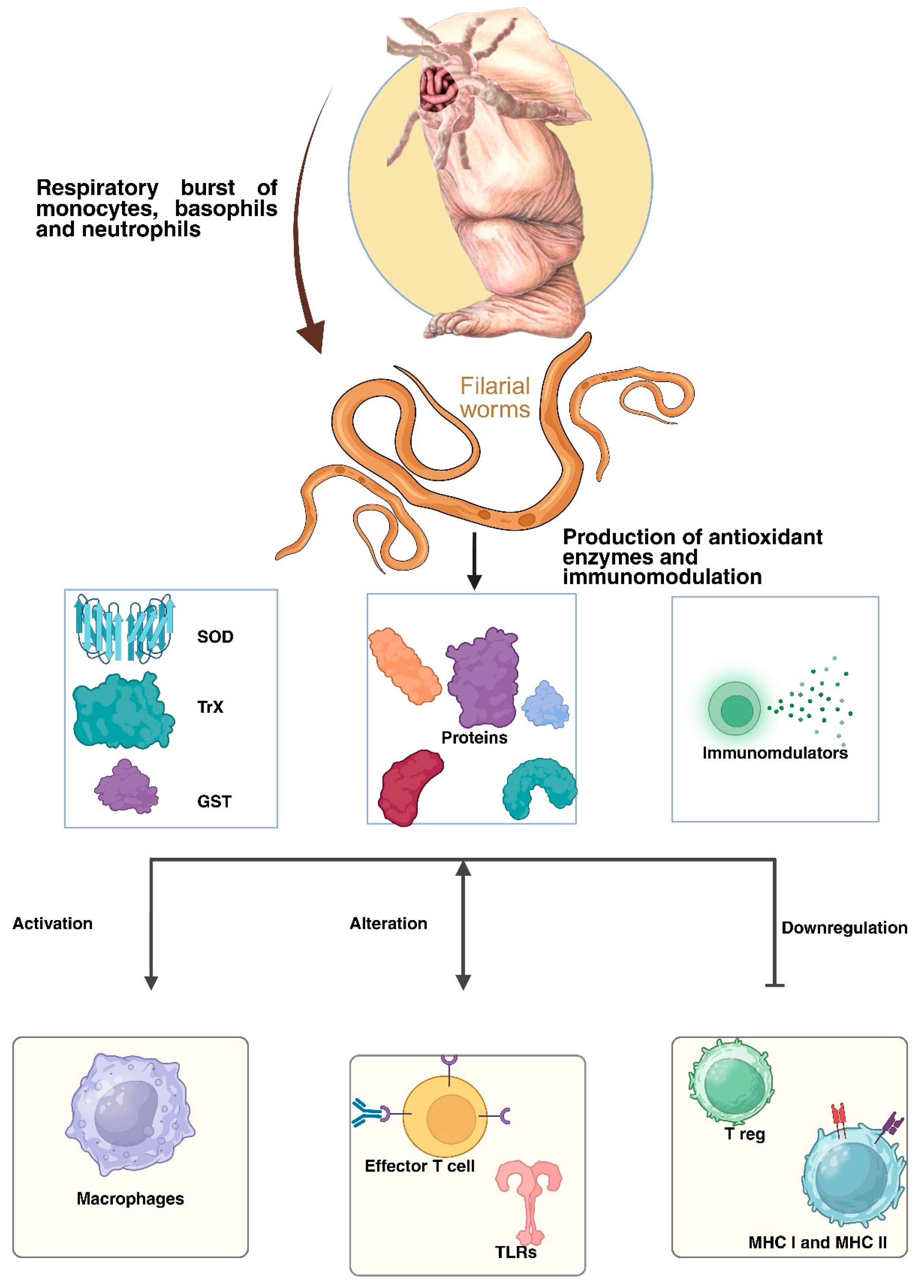
LF pathophysiology comprises a complex interplay between parasite, host’s immune response, and secondary bacterial or fungal infection, leading to lymphatic damage and chronic inflammation [32]. Infection is primarily caused by W. bancrofti, transmitted by mosquitoes. The infective larval stage (L3) enters the human host during a mosquito bite. It migrates to the lymphatic system, where it matures into an adult worm that can survive for years in lymphatics and lymph nodes, releasing microfilariae into the bloodstream, causing persistent antigenic stimulation [4,32]. The human immune system recognises filarial antigen and mounts both innate and adaptive immune responses (Figure 1).
During the asymptomatic phase, the worm manipulates the human immune system to avoid detection. They induce regulatory response by T-reg cells and promote anti-inflammatory cytokines such as IL-10 and Transforming Growth Factor-beta (TGF-β), which suppress immune reactions, allowing the worms to persist while preventing significant tissue damage [4]. The parasite also secretes a few immunomodulatory molecules that inhibit the activation of dendritic cells (DCs) and effector lymphocytes, maintaining a delicate balance between host tolerance and parasite survival [33].
When an adult worm dies or releases the endosymbiont Wolbachia, a potent inflammatory immunological response is triggered, marking the beginning of the acute phase. After infiltrating the lymphatics, neutrophils, eosinophils, and macrophages release pro-inflammatory cytokines such as Tumour Necrosis Factor-α (TNF-α), Interleukin-1β (IL-1β), and Interleukin-6 (IL-6), resulting in lymphangitis, adenolymphangitis, fever, and excruciating swelling [1]. Recurrent bouts of adenolymphangitis might result from secondary bacterial infections that enter through weakened skin and worsen inflammation. Persistent infections cause the lymphatic system to undergo irreversible structural deformations and functional abnormalities. Fibrosis, valvular incompetence, and lymphatic dilatation (lymphangiectasia) are brought on by endothelial damage, persistent inflammation, and recurrent immunological activation. Elephantiasis, lymphedema, and lymphatic blockages are the outcomes of these alterations [34]. Areas of immunological hyperactivation and immune repression coexist in the chronic stage, which is indicative of a dysregulated immune milieu. In male patients, hydrocele develops due to impaired lymphatic drainage in the scrotal region, reflecting localised lymphatic compromise [35]. Furthermore, frequent bacterial superinfections can promote fibrosis and severe deformity due to tissue disorganisation and damage. Understanding these mechanisms is not only critical for developing targeted therapies but also for managing complex and grotesque pathophysiologies.

6. Clinical Stages

LF usually has three stages, but the length and complexity of each stage depend on the number and life stages of parasites present, the host’s immunity, and the number of times the person has been bitten by an infected mosquito [36]. Asymptomatic, acute, and chronic are the three primary clinical stages that an LF-infected patient goes through. The majority of infected individuals are asymptomatic, while some experience acute attacks and a smaller number develop chronic, disfiguring symptoms [5,32]. Asymptomatic cases show no symptoms because they harbour circulating microfilariae and adult worms without any clinical signs. This lack of pathology is due to immune modulation induced by adult parasites. Worms release molecules that have an immunosuppressive nature, allowing the parasites to survive for several years in their hosts without any visible symptoms [4].
After the asymptomatic phase, the infected cases progress to the acute stages. In this phase, some adult worms would have died, resulting in a strong and uncontrolled immune response being mounted against the released antigens, toxins, and other products [37]. Most acute LF cases suffer from fever, pain, swelling, as well as inflammation in the lymph nodes and vessels, while in many cases, bacteria enter through the damaged skin and worsen the conditions [1]. Eventually, after suffering from several years of filarial infections, the lymph vessels become damaged and blocked, which leads to fluid buildup in legs, arms, or genitals known as elephantiasis, which is the typical LF chronic stage [38]. Chronic pathology reflects a combination of immune hyperactivation in some functionalities and suppression in others, compounded by repeated secondary infections that accelerate tissue remodelling and disability [39] (Table 2).
Since the filarial infections are non-deadly, the parasites persist in their hosts for a considerable amount of time and slowly but constantly incur irreversible damage to the host’s immune system [33]. Filarial worms are capable of thriving in their hosts despite being surrounded by immune effector cells, antibodies, and other immunological chemicals [45]. This is achieved through the utilisation of highly calculated immune evasion and immune modulation strategies, including immunosuppression, immunological tolerance, and the modulation of Th-2 responses, to mention only a few [46]. Filarial parasites emit a range of stage- and gender-specific products to accomplish long-term survival as well as to move, localise, and reside through/into various host organs, tissues, and anatomical compartments as well as to avoid the host’s immune responses [47].
The host immune response to filarial parasites is characterised by a Th2 profile, which includes the secretion of cytokines such as Interleukin-4 (IL-4), Interleukin-5 (IL-5), Interleukin-9 (IL-9), Interleukin-10 (IL-10), and Interleukin-13 (IL-13), as well as the production of antibody isotypes Immunoglobulin G1 (IgG1), Immunoglobulin G4 (IgG4), and Immunoglobulin E (IgE). This response is also marked by an increase in eosinophils, basophils, mast cells, and alternatively activated macrophages (AAMs) [48]. Although filarial parasite-induced Th2 responses are a typical host response, they must be initiated by interaction with a wide variety of cell types, including (1) stromal cells; (2) DCs and macrophages; (3) eosinophils; (4) mast cells; (5) basophils; and (6) epithelial [49] and innate helper cells [48]. Chronic infection leads to modulation of these conventional type-2 responses by adaptive and natural regulatory T cells, alternatively activated macrophages, eosinophils, and potentially other unidentified cell populations [40] (Table 3).
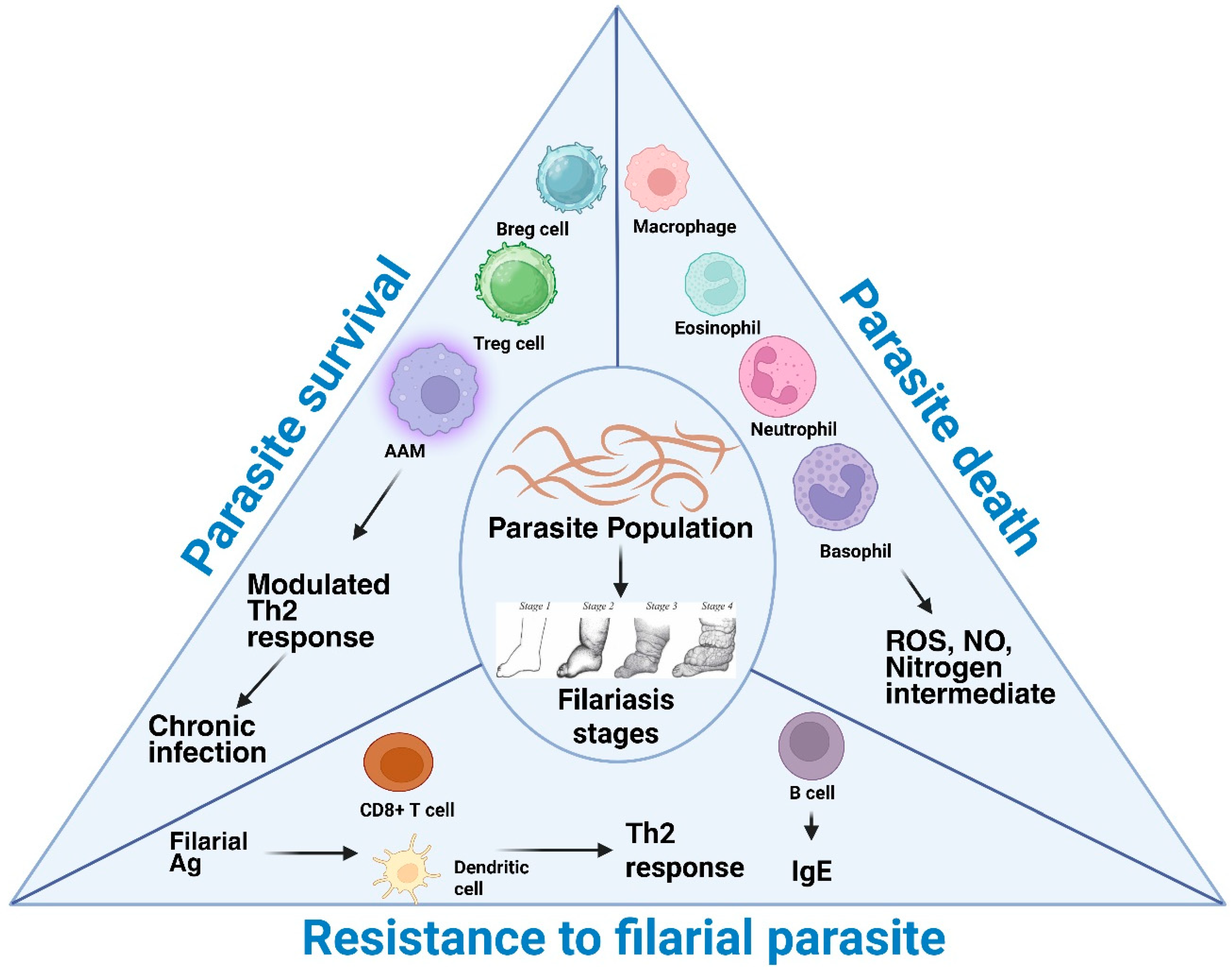
Individuals showing occult manifestations show no classic filariasis symptoms or blood microfilariae but harbour them in tissues, presenting with atypical syndromes [55,56]. These include TPE, filarial arthritis, glomerulopathy, Endomyocardial Fibrosis (EMF), and breast granulomas. TPE, caused by W. bancrofti and B. malayi, presents with cough, fever, breathlessness, and eosinophilia, progressing to fibrosis if untreated [57]. Filarial arthritis affects large joints, mimicking rheumatoid arthritis but remains benign and responsive to DEC [58,59]. Glomerulopathy shows immune complex-mediated nephritis, while EMF, seen in equatorial areas, is linked circumstantially to filariasis. Breast granulomas mimic malignancy, but histology confirms filarial origin [60]. The induction of regulatory T-cells, the modulation of effector T-cells and antigen-presenting cells (APCs), and the death of responder cells are also the main variables that affect the control of immune response in filarial infection [61]. This review highlights the key immune evasion strategies of the filarial parasites, which help them to establish and maintain parasitism (Figure 2).
By comprehending the molecular underpinnings of the host–parasite connection and the immune avoidance strategies used by filarial parasites, new therapeutic strategies to combat filarial diseases need to be developed.

7. Immunomodulation via Parasite-Derived Molecules

The developing filarial parasite passes through several stages within the human host, and during this process, it releases several chemicals and molecules specifically pertaining to the developmental stage and/or gender. The spectrum of these molecules not only represents the parasites’ distinct developmental processes and survival strategies at each stage but also represents their manipulative immune evasion tactics for establishing infection [47].
In general, lipid peroxidation is defined as the process by which oxidants, such as free radicals or nonradical species, attack lipids containing one or more carbon-carbon double bonds, particularly polyunsaturated fatty acids (PUFAs), resulting in hydrogen abstraction from a carbon and oxygen insertion, producing hydroperoxides and lipid peroxyl radicals as previously mentioned [62]. One of the primary impacts of oxidant generation is the destruction of membrane lipids, or more specifically, their polyunsaturated fatty acyl moieties, via the chain reaction of lipid peroxidation. Lipid peroxidation destroys biological membranes and produces chemicals that disrupt other molecules. In response to membrane lipid peroxidation, and according to specific cellular metabolic circumstances and repair capacities, the cells may promote either cell survival or induce cell death. Under physiological or low lipid peroxidation rates (subtoxic conditions), the cells stimulate their maintenance and survival through constitutive antioxidant defence systems or signalling pathways activation that upregulate antioxidant proteins, resulting in an adaptive stress response. By contrast, under medium or high lipid peroxidation rates (toxic conditions), the extent of oxidative damage overwhelms repair capacity, and the cells induce apoptosis or necrosis, programmed cell death; both processes eventually lead to molecular cell damage, which may facilitate the development of various pathological states and accelerated ageing [63,64].
It has been repeatedly shown that MDA, a more reliable and measurable indicator of lipid peroxidation, is higher in LF patients. Individuals infected with W. bancrofti and B. malayi have been found to have elevated serum MDA, which is associated with increased oxidative damage and decreased antioxidant enzyme conditioning (superoxide dismutase, catalase, glutathione peroxidase). In chronic and occult filariasis, but not in mf carriers, a significant degree of oxidative stress has been observed in terms of an increase in pro-oxidative effect (measured by serum MDA levels) and a simultaneous reduction in anti-oxidative defence (measured by serum SOD activity) when compared to endemic healthy controls. These findings suggest that oxidative stress production has significance in cases of active infection, chronic infection, and even occult infections where the parasite is sessile in the host [65].
Oxidative stress is present in both occult cases, where the parasite remains sessile in host tissue and may still cause local disease, and actively infected chronic cases, confirming that the oxidative environment of a filarial infection is prolonged. MDA is a good indicator of the degree of oxidative stress in LF since it primarily reflects the level of oxidative injury and antigenic load. MDA can create MDA-protein conjugates that act as neoantigens and trigger immunological and inflammatory reactions by forming covalent adducts with proteins and nucleic acids [66]. Filarial parasites produce a significant quantity of antioxidant enzymes and non-enzymatic antioxidants, like α-tocopherol, thioredoxin peroxidase, GST, TrxR, glutathione (GSH), ascorbic acid, superoxide dismutases, and translationally controlled tumour protein. These are very important for protecting against free radicals that are made when the host immune cells attack [37,53,54,55]. Acetylcholinesterases secreted by B. malayi may prevent fluid accumulation in the gut and hinder parasite elimination. Calreticulin from B. malayi interacts with human C1q and inhibits subsequent classical complement pathways [67]. The thioredoxin reductase (TrxR) from Setaria cervi exhibited anti-inflammatory effects in macrophages by inhibiting the TLR4/NF-κB pathway, down-regulating the inflammasome pathway, and activating macrophages [68].
Additionally, other prospective immunomodulators have been identified within the filarial genome, such as serpins and cystatins (which modulate antigen processing and presentation to T cells), indoleamine 2, 3-dioxygenase (IDO) genes (a potent immunomodulatory molecule), and the Wnt family of developmental regulators (which modulate immune activation) [69]. Upon infection, B. malayi larvae release a large amount of the protein called Brugia malayi abundant larval transcript (Bm-ALT), which is linked to the upregulation of the GATA-3 transcription factor in macrophages and the subsequent induction of a Th2 immune response. Furthermore, Bm-ALT stimulates suppressor of cytokine signalling 1 (SOCS-1), suppresses IFN-γR-associated JAK kinase in macrophages, and disrupts stimuli necessary for pro-inflammatory Th1 cell differentiation [70]. The BmK1 protein derived from B. malayi specifically inhibits voltage-gated potassium (Kv) 1.3 channels and diminishes IFN-γ production in CCR7-effector memory T cells; it does not affect naïve or central memory T cells [71].
Researchers have demonstrated that PC alone, PC-BSA, and PC associated with ES-62 directly inhibit polyclonal activation of B cells in a PKC-dependent manner, suggesting that PC-containing compounds in B. malayi can mimic immunosuppressive effects [72]. Significantly, PC is absent in the ES-62 homolog, leucyl aminopeptidase (LAP), in B. malayi, but is present in another secretory protein known as N-acetylglucosaminyltransferase [73]. Treg cells and IL-10 are known to stimulate the synthesis of IgG4 in B cells. Immunosuppressive IgG4 cannot activate the complement system or generate antibody-dependent cell-mediated cytotoxicity (ADCC) upon binding to CD16 on neutrophils and eosinophils [74].
Certain filarial compounds affect the immune response without relying on receptors, for instance, B. malayi’s Cystatins (BmCys), in addition to receptor-mediated regulation of immune cells [75], inhibits host cysteine proteases and asparaginyl endopeptidase, hinders antigen presentation on APCs, and diminishes T cell priming [76]. An unusual filarial immunomodulator is the B. malayi polyprotein “ladder,” gp15/400, which manipulates the immune response in a metabolite-dependent fashion. This retinoid-binding protein is believed to facilitate vitamin A absorption by host tissues [77]. Retinoic acid, a metabolite of vitamin A, promotes TGF-β’s inhibition of IL-6-induced Th17 cell proliferation while facilitating the development of Fox head box p3 (FOXP3+) Treg cells [78].
Leucyl aminopeptidase, a homolog of Acanthocheilonema vitae’s ES-62, a phosphoryl-containing glycoprotein, is one of the glycoproteins released by filarial parasites B. malayi and W. bancrofti [79]. In various immune cells, ES-62 has diverse functions. ES-62 attaches with TLR4 in monocytes, followed by subsequent internalisation of ES-62–TLR4 complexes, facilitating sequestration and caveolae/lipid raft-mediated, proteasome-independent destruction of protein kinase C-α (PKC-α), a crucial factor responsible for the interaction of FcεRI receptor present on mast cells for their activation [80].
A tiny hapten-like moiety termed ES-62, which is present in the excretory secretions of filarial parasites, contains the phosphorylcholine (PC) molecule. It has demonstrated immunomodulatory characteristics [81] that are mainly 1. Downregulating the proliferation of CD4+ T cells and conventional B cells, 2. Downregulating the production of IL-4 and IFN-γ, 3. Increasing the production of IL-10 by B1B cells, leading to their enhanced proliferation, 4. Activating antigen-presenting cells to promote Th2 development and suppress Th1 reactions [82].
In dendritic cells, ES-62 binds to TLR-4 and inhibits PKC-γ associated with TLR-4, upregulates and sequesters p62 and LC3 (autophagosome machinery components), causes their autophagolysosomal destruction, and inhibits the LPS-driven release of IL-6, IL-12p70, and TNF-α from these cells [83]. The degradation of PKC-δ induced by ES-62 is expected to impede dendritic cell development and motility, the production of IL-12p40/p70, MHC II-antigen presentation, and Th1 polarisation [84].
Upon microbial or parasitic infections, the damaged epithelial cells produce alarmins such as Thymic stromal lymphopoietin (TSLP), IL-25, and IL-33. Alarmins cause type-2 innate lymphoid cells (ILCs) to become activated, which aids in the development of anti-parasitic immunity and intensifies type-2 inflammation [85]. Due to the inadequacy of cutaneous ILC activation, langerhans cells (LC) and DCs stay inactive and do not trigger an ILC2-dependent inflammatory response; as a result, the L3 in the cutaneous tissue successfully invades the host tissues. Additionally, there is still much to learn about the filarial chemicals that are responsible for this alteration of the cutaneous immune response [86].

8. Molecular Mimicry

Innate immune cells possess pattern recognition receptors (PRRs) that identify certain pathogen-associated molecular patterns (PAMPs) and activate intracellular pathways that elicit pro-inflammatory responses. Toll-like receptors (TLRs), NLRs, RLRs, CLRs, and ALRs are subcategories of PRRs [87]. TLRs and CLRs are the primary targets of glycan-conjugated products (glycoproteins and glycolipids) during immunomodulation. This parasitic survival technique, termed “glycan gimmickry,” not only modifies TLR expression but also effectively manipulates its intracellular signalling [88,89]. Filarial parasites utilise an intriguing strategy called molecular mimicry, where they express molecules on their surface that mimic the host’s glycan antigens. These host-like glycan antigens found on filarial surfaces are successful in thwarting the host’s immunological response [90]. Researchers have found that the antigens of human filarial parasites mimic several host compounds. TGFβ-R signalling is used to trigger the immunosuppressive mechanisms of B. malayi’s adult and microfilariae stages’ expression of TGH-2, a human TGF-β homolog [91,92]. BmHsp12.6, a small heat shock protein, is upregulated during the transition of B. malayi L3 larvae from mosquitoes to their mammalian hosts [93]. The upregulated BmHsp 12.6 may protect the parasite proteins from destruction within the host by carrying out their chaperone activities. Because BmHsp 12.6 binds to IL-10R, it exhibits IL-10-like properties and can modulate the host-immune responses [94]. Microfilariae of W. bancrofti and B. malayi release prostaglandins, predominantly PGE2 [95]. PGE2 serves multiple functions in immune modulation [96], including the activation of FOXP3+ Treg cells, vasodilation, and the suppression of platelet aggregation [97].
It has been demonstrated that the presence of cytokine- and chemokine-like molecules in filarial parasites affect the host immune response. Some of these homologs are functionally similar to TGF-β and macrophage migration inhibitory factor (MIF) [77]. A recent investigation of the filarial parasite genome has shown a significant number of human cytokine and chemokine mimics and/or antagonists within the genome [77]. This encompasses members of the interleukin-16 (IL-16) family, an IL-5 receptor antagonist, an interferon regulatory factor, a homolog of SOCS-7, and two members of the chemokine-like family [69].

9. Modulation of TLR and NF-κB Signalling Cascades Induced by Filarial Parasites

The discovery and characterisation of the TLR family have generated renewed interest in the domain of innate immunity. These receptors play a crucial role in microbial detection, the activation of antimicrobial genes, and the regulation of adaptive immune responses [98]. TLRs play a pivotal role in recognising the ‘molecular fingerprints’ of microbial infections, activating distinct signalling pathways, and regulating dendritic cell maturation and Th cell differentiation [99]. TLRs are expressed by macrophages and DCs, as well as by T and B lymphocytes [100]. TLRs are membrane-spanning, non-catalytic receptors that recognise structurally conserved compounds from infections and control the subsequent immune response [101]. There are currently thirteen TLRs that have been identified, including TLR1–TLR13, of which TLR1–TLR9 are shared by mice and humans. TLR11 and TLR13 are found in the endosomal compartments of mice but are absent from the human genome, while TLR10 is nonfunctional in mice because of a retrovirus insertion [102]. TLR1, TLR2, TLR4, TLR5, and TLR6 are localised on the plasma membrane, while TLR3, TLR7, TLR8, and TLR9 are found within the endosomes of leukocytes [103]. These receptors are present on diverse immune and non-immune cells in various combinations to identify a wide array of PAMPs, thus establishing a connection between the innate and adaptive immune systems [104,105]. Typically, TLRs form homodimers, with the notable exception of TLR2, which tends to preferentially associate as a heterodimer with either TLR1 or TLR6, and in certain instances, with TLR10 [106,107]. Pathogen-encoded TLR ligands can be classified into three distinct categories: lipids and lipopeptides (TLR2/TLR1; TLR2/TLR6; TLR4), nucleic acids (TLR3, TLR7, TLR8, TLR9), and proteins (TLR5 and, in mice, TLR11) [108]. TLR10 has been demonstrated to interact with a range of ligands, although the specific natural ligand(s) remain under investigation. Proposed ligands encompass HIV-1 gp41 and lipopolysaccharides (LPS) [107].
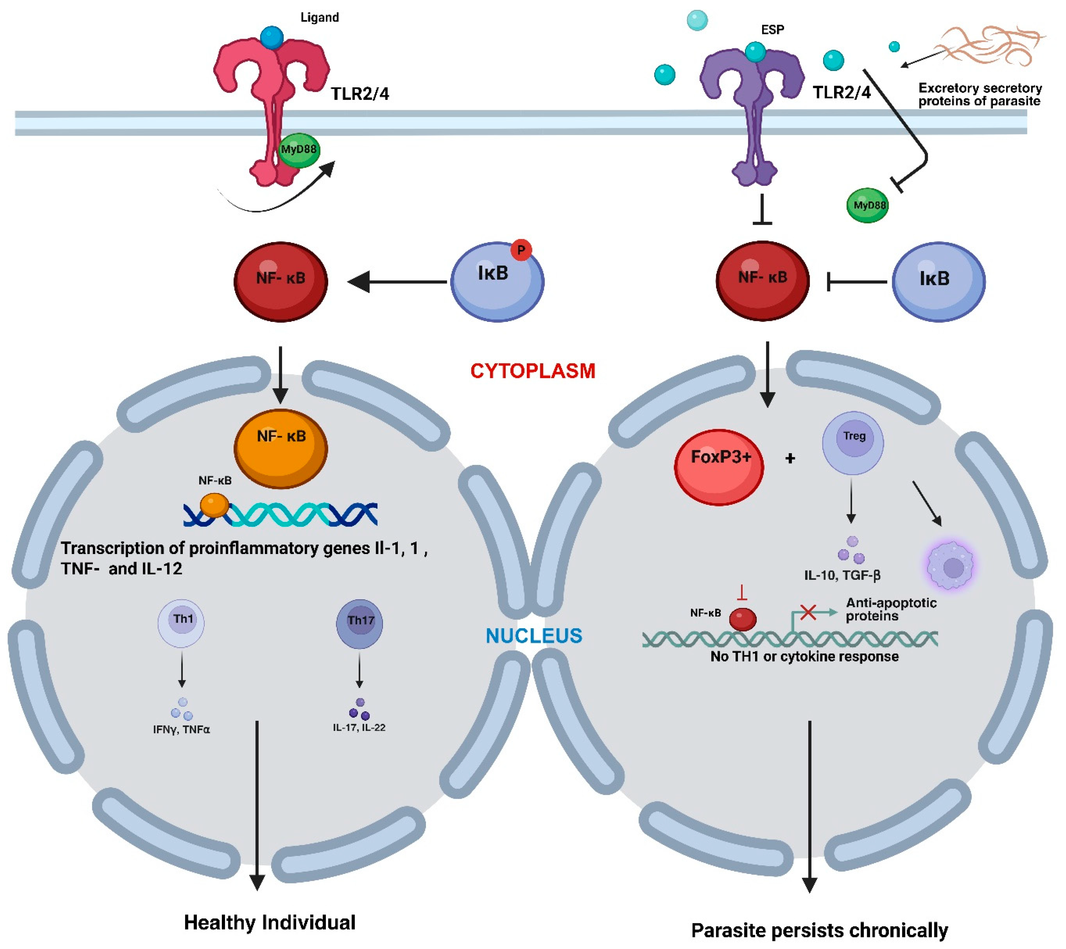
TLRs activate the MyD88 pathway, which subsequently leads to the activation of MAPK and facilitates the translocation of nuclear factor kappa B (NF-kB) into the nucleus. NF-kB facilitates the transcription and production of pro-inflammatory cytokines [109]. NF-κB is essential in mediating responses to a wide range of external stimuli, making it a key component in various physiological and pathological processes, as well as a significant orchestrator of the immune response [110,111,112]. Before activation, latent NF-κB dimers are associated with inhibitors (IkB) that sequester the transcription factor within the cytoplasm. In the majority of cells, NF-κB is linked with IkBα, IkBβ, or IkBε; furthermore, p105 and p100 possess an inhibitor within their sequence [113,114], thereby also acting as inhibitors, while p50 and p52 homo- and heterodimers suppress NFκB-dependent transcription. The translocation of NF-κB to the nucleus and its activation require the dissociation from the inhibitor, which occurs upon the phosphorylation of the inhibitor by an IκB kinase (IKK) [115].
Filarial parasites such as W. bancrofti and B. malayi release excretory/secretory products (ESPs) that interact with TLR2 and TLR4 in a non-stimulatory or suppressive manner, hence biasing immune responses towards tolerance instead of inflammation [116]. These ESPs comprise glycoproteins, lipids, and extracellular vesicles that disrupt standard TLR signalling, specifically the MyD88-NF-κB pathway [117]. Rather than eliciting pro-inflammatory cytokines, ESP-TLR interaction stimulates the production of anti-inflammatory cytokines such as IL-10 and TGF-β, which eventually contribute to immunological suppression in LF infections [118]. The suppression reinforces the FOXP3+ regulatory T cell (Treg) and AAMs, which makes the suppression even stronger. Both of these inhibit NF-κB activation and eventually slow down the antigen presentation [118,119].
Thus, the canonical NF-κB signalling pathway is obstructed, resulting in the host immune system’s inability to initiate an efficient Th1 response, hence enabling the parasite to persist chronically without being eliminated [116,120]. Hence, helminths such as filarial nematodes manipulate the host’s TLR-NFκB signalling to create a hyporesponsive environment that prolongs their survival and transmission into their host’s body [121] (Figure 3).

10. Effector T Cell Modulation

Effector T-cells are induced by immunoregulatory cytokines to suppress immunological responses and promote immune tolerance. This alters the Th2 response and activates AAMs. Effector T-cell responses can be suppressed or regulated Via several mechanisms, including CTLA-4 and PD-1 [122]. Human filarial infections notably exhibit elevated levels of CTLA-4 and PD-1, and the inhibition of CTLA-4 can partially reinstate the immunological response in cells from infected individuals [123]. The T cells from filaria-infected patients have shown characteristic anergy symptoms, including elevated expression of E3 ubiquitin ligases and decreased IL-2 production [124]. Although the majority of immunological research on filarial infections has focused on immune responses triggered by filarial antigens, investigating the immune responses elicited by live parasites reveals intriguing insights. Active parasites significantly inhibit the production of both Th1 and Th2 cytokines in response to L3 and Mf stages, resulting in reduced levels of Th1 (IFN-γ and TNF-α) and Th2 (IL-4 and IL-5) cytokines [124]. This is associated with a diminished induction of T-bet (the principal Th1 transcription factor) and GATA-3 (the principal Th2 transcription factor) mRNA, with a markedly elevated expression of Foxp3, TGF-β, CTLA-4, PD-1, ICOS, and IDO. The increased activation of anergy-inducing factors Cbl-b, c-Cbl, Itch, and NEDD4 facilitates the impairment of effector T-cell functions. Consequently, many regulatory variables such as IL-10, TGF-β, and nTregs (perhaps through PD-1 and CTLA-4) have been associated with the attenuation of immune responses in active filarial infection and may play a crucial role in the development of chronic pathologies.
Filarial parasites cause the downregulation of MHC class I and class II, along with cytokines and other genes associated with antigen presentation in dendritic cells, thereby diminishing their ability to activate CD4+ T cells [125]. Furthermore, live parasites have demonstrated the ability to downregulate MHC class II, IL-18, and many genes associated with antigen presentation in skin-resident Langerhans cells [126]. The interaction between filarial parasites and macrophages has been demonstrated to elicit AAMs. Due to the production of regulatory molecules such as IL-10, TGF-β, and PD-L2, these macrophages may primarily assume a regulatory function in filarial infections [46]. Anti-inflammatory macrophages can decrease T-cell responses Via arginase-1 production and PD-L2 expression, while also suppressing classical macrophage inflammation and recruitment through arginase-1, resistin-like molecule-α (RELMα), triggering receptor expressed on myeloid cells 2 (TREM2), and other chemicals [127].

11. Role of Regulatory T Cells in Filarial Evasion and Pathogenesis

Regulatory T cells are identified by a specific marker, Foxhead box P3, or FOXP3. Other markers of Tregs include CD25, cytotoxic T-lymphocyte-associated protein-4 (CTLA-4), inducible T-cell costimulator (ICOS), and T-cell immunoreceptor with Ig and ITIM domains [128]. Surface molecules, such as CTLA-4, LAG-3, FasL, PD1, and cell-surface-bound TGF-β, help in engaging Tregs with their corresponding cognate ligands on the target cells to deliver cytotoxic factors and suppressive signals, including granzyme B, cyclic adenosine monophosphate (cAMP), and the FasL-Fas apoptotic signal [129].
The regulatory T cell population consists of two subgroups: Foxp3+ population and the Foxp3- population. The Foxp3 population exerts down-modulatory effects through their production of IL-10 in the case of Tr1 and IL-35 from Tr35 T cells [130]. The classical Foxp3+ Treg population can be divided into two primary groups based on their origin. Thymic regulatory T cells (tTregs), also known as natural Tregs with the phenotype CD4+ CD25+ CTLA4+ FOXP3+, develop in the thymus due to negative selection and have self-antigen specificity [131]. They have previously been referred to as Helios+ Tregs. On the other hand, peripherally induced regulatory T cells (pTregs) are generated from peripheral Th cells in a process that depends on TGF-β. Various subtypes of iTreg cells have been identified, such as converted Foxp3+ Tregs, Tr1 cells that mainly secrete IL-10 and small amounts of TGF-β, Th3 cells that mainly secrete TGF-β, Tr35 cells that primarily secrete IL-35 and regulatory Th17 (Treg17) cells that secrete IL-17, have high levels of aryl hydrocarbon receptor (AhR) and IL-10, but produce significantly lower levels of IL-22 [132,133,134,135,136,137,138].
Following an infection with B. malayi, Tregs exhibit higher levels of CD103, a beta integrin associated with tissue residency, indicating that Treg retention at the infection site is important for their function. When human filarial infections occur, peripheral T cell populations express more CTLA-4, and therapy with anti-CTLA-4 Abs increases the cytokine responsiveness of T cells in filarial patients [139]. To sustain their successful parasitism, numerous parasites exploit the regulatory network of the host immune system to diminish the immunological response directed against them [140,141]. The role of regulatory T cells in controlling immune responses, which lead to enhanced pathogen survival and ultimately long-term persistence of the pathogen, has been reviewed previously [142]. Nematodes are effective inducers of Treg responses, and Immunoregulatory T cells (Tregs) play a central role in the regulatory network of the host. A tolerant, asymptomatic carrier state that benefits the parasite and the host by allowing continued transmission with little clinical damage may be established by Tregs in chronic human infections such as filariasis [143]. Treg cells interact with filarial parasites to cause regulated inflammation by secreting immunosuppressive cytokines such as TGF-β and IL-10 [144].
Human filariasis, caused by nematodes such as O. volvulus or W. bancrofti, results in an increase in Tr1 cells, which, during infection, generate most of the IL-10 produced [145]. Tr1 cells are a specific subset of regulatory T cells. These cells release elevated levels of IL-10 but do not express Foxp3. Tr1 cells are crucial for maintaining immunological homeostasis and serve as important regulators in the immune network [146]. Tr1 cells, together with Tregs, play a crucial role in facilitating the switch to IgG4 antibody production during helminthiases, hence promoting an anti-inflammatory phenotype [147]. In conclusion, during helminth infection, parasite-induced modulation of regulatory T cells plays a critical role in the establishment and maintenance of a tolerant state.
During the chronic phase of filarial infection, there is an increase in the levels of regulatory cytokines such as IL-10 and TGF-β, as well as the antibody isotype IgG4. Research has indicated that in individuals with sub-clinical microfilaraemic W. bancrofti infection, neutralising antibodies to IL-10 (and to a lesser extent TGF-β) markedly improved the downregulated antigen-specific proliferative response [148]. According to research, populations infected with W. bancrofti had higher levels of regulatory B and T-cell subsets that make the cytokine IL-10, which helps in the survival of the parasite [149].
W. bancrofti’s sheath antigen stimulates TLR-4 signalling in DCs, which induces the production of Th1 and Treg cells [150]. The presence of various Treg subsets, such as Foxp3-expressing CD4+ cells that express TGF-β (Th3) and Foxp3-CD4+ cells that express IL-10 (Tr1), is indicative of the connection between chronic filarial infection and host immune modulation via Tregs, especially in the microfilaremics [124,151,152,153]. During the early stages of filarial infection, Treg cells and their suppressive cytokines actively regulate Th1, Th2, and Th-17-driven inflammatory responses. Additionally, these Treg cells influence the B-cell response, which supports the persistent filarial infection [154].
Adult B. malayi and their microfilariae secrete the human TGF-β orthologue as a pathogenic factor to counteract the host’s efficient immune responses. Following activation, antigen-specific Tr1 cells infected with O. volvulus exhibit increased levels of CTLA-4, which facilitates the suppression of other T-cell activities [155]. Patients with lymphoedema who have the W. bancrofti infection exhibit significantly higher Th1 and Th17 responses and lower Treg levels compared to those without symptoms. In contrast, hyper-reactive onchocerciasis (river blindness) patients lack FOXP3+ CD25 (high) Tregs [156].

12. Apoptosis Induction in Host Immune Cells

Another route of immune evasion is the capability of the filarial parasites to trigger apoptosis in host cells. In vivo, apoptosis of CD4+ T cells has been seen in experimental murine models of filarial infection. Moreover, Brugia microfilariae have demonstrated the ability to interact with DCs and NK cells, subsequently inducing their death [125,157].
By inducing apoptosis to decrease the number of immune cells, filarial parasites prolong their infection. B. malayi L3 stimulates NK cells to secrete IFN-γ and TNF-α, thereby promoting cell death through the caspase-dependent pathway [157]. B. malayi Mf influences human DCs in two ways: (1) by altering their functionality and (2) by inducing apoptosis, leading to an antigen-specific T cell hypo-responsiveness. Microfilariae engage with human DCs to form cell-parasite aggregates, induce DC apoptosis through TRAIL and TNF-α pathways, and hinder IL-12 production, hence restricting CD4+ T cell activation and proliferation [84,125]. Furthermore, besides inducing death, microfilariae also induce autophagy in DCs by obstructing the phosphorylation of mTOR and its downstream proteins (p70S6K1 and 4E-BP1), enhancing Beclin1 phosphorylation, promoting LC3II induction, and facilitating p62 degradation [158]. Recently, researchers discovered that B. malayi microfilariae secrete extracellular vesicles that dendritic cells rapidly absorb. These extracellular vesicles are abundant in unique miRNAs that specifically target the mTOR signalling pathway [159]. W. bancrofti has been demonstrated to cause FasL-expressing B-1 cells (caused by increased IL-10 levels) to induce the death of CD4+ T cells, leading to a hypo-immune reaction in infected individuals [160]. The filarial parasites employ several other strategies and unidentified processes in addition to these to secure their existence and avoid causing significant harm to their host’s body, thus establishing a productive infection for several years. (Figure 4). Collectively, these immune mechanisms establish the equilibrium between parasite persistence and resistance to filarial infection during various stages of disease progression.

13. Conclusions and Future Prospects

Filarial parasites employ a range of evasion strategies to effectively disable the host’s innate and adaptive immune responses, ensuring their continued parasitism. These parasites use several molecular and cellular strategies to evade host immune responses, including immunomodulation aided by various chemicals and parasite-derived molecules, molecular mimicry, in which the parasites express molecules on their surface that mimic the host’s glycan antigens, alteration of TLR expression and NF-κB signalling cascades, as well as the induction and growth of regulatory immune cell populations, such as regulatory B cells (Bregs), regulatory T cells (Tregs) and AAMs. These mechanisms create a hyporesponsive environment that allows the persistence and survival of parasites in their hosts. The molecular factors and immune evasion mechanisms of parasites present potential targets for the development of specific diagnostics, treatments, and vaccines aimed at alleviating the chronic pathophysiological symptoms associated with the disease. The immunological mechanisms necessary for the induction, expansion, and maintenance of anti-helminth responses are still under exploration.
Most of the understanding of anti-filarial immune responses has been deduced from rodent infections, which do not exhibit typical filarial pathologies [29]. Canines and felines function as natural hosts and efficient models for B. malayi infection, exhibiting symptoms such as lymphedema and lymphangitis but only for transient periods. Non-human primates and ferrets are regarded as excellent models for researching B. malayi because their lymphatic pathologies resemble those of human LF. The infections manifest in a manner that is comparable to that of a human infection, characterised by lymphatic pathology. Hence, non-human primates are considered to be one of the best models that are available for studying the host–parasite relationships and immunity to LF, but handling these animals is expensive and is extremely challenging due to ethical considerations and other concerns [161].
Present-day anti-filarial drug combination treatment (Ivermectin, Diethylcarbamazine citrate and Albendazole (IDA)) has proven largely ineffective against the adult filarial worms [162,163]. With the LF elimination deadline approaching, gaining further insight into the mechanisms of response production by using non–human primates are now becoming more important for uncovering immunomodulatory pathways and designing antifilarial drugs targeting adult parasitic stages too. Consequently, a deeper understanding of the molecular and functional mechanisms behind filarial immune evasion could provide specific and focused targets for the creation of anti-filarial treatments.
Understanding parasite biology and host immune responses has been greatly assisted by a wide range of current animal models, each with specific advantages and disadvantages. These models encompass canines, felines, rodents, and non-human primates. Animal models are crucial for formulating treatment strategies and exploring the pathogenesis of clinical lymphedema. The many aspects of lymphatic dysfunction and lymphedema that are seen in humans are not fully replicated in these animal models. Additionally, the entire lifespan of human filariae has not yet been confirmed in vivo, and immunocompromised animals may have reduced treatment efficacy. Recently, several studies have been conducted showing the anti-filarial and macrofilaricidal effects of nutraceuticals like Emodin, Naringin, Ocimum sanctum, and Centratherum anthelminticum, as well as silver nanoparticles derived from Ocimum and andrographolide. However, due to the unavailability of suitable animal models, these anti-filarial leads are still in an ambivalent and disconcerted state [163,164,165,166]. Similarly, recent developments of anti-filarial multi-epitopic cocktail vaccines are stalled for further validations in animal models that can host similar immune responses as human beings and can exhibit LF pathologies to at least some extent [167]. Therefore, more research on animal models is needed to enhance our testing of new treatment approaches in preliminary experiments and to enable access to all stages of the parasite life cycle, which is now a crucial issue of ethical concern. Future research should concentrate on creating novel, ethically significant, and physiologically relevant representative animal models that could more accurately mimic human infections which would serve as a milestone in achieving the elimination of LF transmission.

Author Contributions

T.S.: Conceptualisation and writing—original draft (lead). S.S. and A.T.: Writing and editing. S.K.: Editing. A.S.: Supervision. All authors have read and agreed to the published version of the manuscript.

Funding

This research received no external funding.

Institutional Review Board Statement

Not applicable.

Informed Consent Statement

Not applicable.

Data Availability Statement

No new data were created or analyzed in this study.

Acknowledgments

Tripti Singh is grateful to the University Grants Commission (UGC), New Delhi, for providing a Senior Research Fellowship (NTA Ref. No. 211610077377). Shivani Sharma is thankful to the University Grants Commission (UGC), New Delhi (NTA Ref. No. 221610005833) for providing a Senior Research Fellowship. Animesh Tripathi is grateful to Banaras Hindu University for providing a UGC Non-NET Fellowship. Sunil Kumar is grateful to the NBCFDC JRF (Ref. No. 231620090211) Junior Research Fellowship, University Grants Commission (UGC), New Delhi. Anchal Singh is grateful to UP-CST (Grant ID: P 26/0156), Lucknow, India. The authors also thank the DBT-BHU Interdisciplinary School of Life Sciences for providing laboratory space.

Conflicts of Interest

The authors declare no competing interest.

Abbreviations

The following abbreviations are used in this manuscript:
AAMsAlternatively Activated Macrophages
ADCCAntibody-dependent cell-mediated toxicity
AhRaryl hydrocarbon receptor
ALRAbsent in Melanoma 2-like receptor
APCsAntigen Presenting Cells
Bm-ALTBrugia malayi abundant larval transcript
BmHspBrugia malayi heat shock protein
BmK1Brugia malayi K1
cAMPCyclic adenosine monophosphate
Cbl-bCasitas B-lineage lymphoma proto-oncogene-b
c-CblCasitas B-lineage lymphoma
CCR7C-C chemokine receptor 7
CD 8Cluster of Differentiation 8
CD16Cluster of Differentiation 16
CD4Cluster of Differentiation 4
CLRC-type lectin receptor
CTLA-4Cytotoxic T- T-lymphocyte-associated protein 4
DCDendritic cells
DECDiethylcarbamazine citrate
EMFEndomyocardial Fibrosis
ES-62Excretory-secretory-62
ESPExcretory-secretory Product
FasLFas ligand
FcεRIFc epsilon receptor I
FOXP3Forkhead Box P3
FTSFilariasis Test Strip
GATA-3GATA binding protein 3
gpGlycoprotein
GSHGlutathione
HIV-1Human Immundodeficiency virus-1
ICOSInducible Co-stimulator
ICTImmunochromatographic test
IDOIndoleamine 2,3-dioxygenase
IFN-γInterferon-gamma
IgEImmunoglobulin E
IgG1Immunoglobulin G1
IgG4Immunoglobulin G4
IKKIκB kinase
ILInterleukin
IL-1βInterleukin-1β
IL-6Interleukin-6
ILCsInnate lymphoid cells
ITIMImmunoreceptor Tyrosine-based Inhibition Motif
JAKJanus kinases
LAG-3Lymphocyte Activation Gene
LAPLeucyl aminopeptidase
LC3Microtubule-associated protein light-chain 3
LPSLipopolysaccharides
MAPKMitogen-activated protein kinase
MDAMass Drug Administration
MFMicrofilariae
MHCMajor histocompatibility complex
MIFMacrophage Migration Inhibitory Factor
mTORmammalian target of rapamycin
MyD88Myeloid differentiation primary response 88
NEDD4Neural precursor cell-expressed developmentally downregulated protein 4
NFκBNuclear factor kappa B
NK cellsNatural Killer cells
NLRNod-like receptor
NVBDCPNational Vector-borne Disease Control Program
PAMPsPathogen-Associated Molecular Patterns
PCPhosphorylcholine
PC-BSAPhosphorylcholine-Bovine serum albumin
PCRPolymerase Chain Reaction
PD-1Programmed cell death protein 1
PD-L2Programmed cell death-ligand 2
PGE2Prostaglandin E2
PKCProtein kinase C
RDTRapid Diagnostic Test
RELMαResistin-like molecule-α
RLRRIG-I-like receptor
ROSReactive Oxygen Species
SAESevere adverse effects
SOCSSuppressor of Cytokine Signalling
SODSuperoxide Dismutase
TGF-βTransforming Growth Factor-beta
Th1T helper 1
Th2T helper 2
TLRToll-like receptor
TNF-αTumour Necrosis Factor-α
TPETropical Pulmonary Eosinophilia
Tr1Type 1 regulatory
TRAILTumour necrosis factor-related apoptosis-inducing ligand
TREM2Triggering receptor expressed on myeloid cells 2
TrXThioredoxin
TSLPThymic stromal lymphopoietin
WHOWorld Health Organisation

References

  1. Taylor, M.J.; Hoerauf, A.; Bockarie, M. Lymphatic filariasis and onchocerciasis. Lancet 2010, 376, 1175–1185. [Google Scholar] [CrossRef]
  2. World Health Organization. Lymphatic Filariasis (Elephantiasis). Available online: https://www.who.int/health-topics/lymphatic-filariasis#tab=tab_1 (accessed on 27 October 2025).
  3. Rajamanickam, A.; Babu, S. Unraveling the dynamics of human filarial infections: Immunological responses, host manifestations, and pathogen biology. Pathogens 2025, 14, 223. [Google Scholar] [CrossRef]
  4. Babu, S.; Nutman, T.B. Immunology of lymphatic filariasis. Parasite Immunol. 2014, 36, 338–346. [Google Scholar] [CrossRef]
  5. World Health Organization. Lymphatic Filariasis Key Facts. Available online: https://www.who.int/news-room/fact-sheets/detail/lymphatic-filariasis (accessed on 27 October 2025).
  6. National Center for Vector Borne Diseases Control. Magnitude of Disease. Available online: https://ncvbdc.mohfw.gov.in/index4.php?lang=1&level=0&linkid=455&lid=3732 (accessed on 27 October 2025).
  7. National Center for Vector Borne Diseases Control. List of 348 LF Endemic Districts. Available online: https://ncvbdc.mohfw.gov.in/WriteReadData/l892s/12145485381746781807.pdf (accessed on 27 October 2025).
  8. Juergens, A.L.; Goldin, J. Filariasis. Available online: https://www.ncbi.nlm.nih.gov/books/NBK556012/#:~:text=Diagnosis%20relies%20on%20identifying%20microfilariae,presence%20of%20living%20adult%20worms (accessed on 27 October 2025).
  9. World Health Organization. Diagnosis and Treatment Lymphatic Filariasis (Elephantiasis). Available online: https://www.who.int/teams/control-of-neglected-tropical-diseases/lymphatic-filariasis/diagnosis-and-treatment#:~:text=Circulating%20microfilariae%20can%20be%20detected,the%20periodicity%20of%20the%20microfilariae (accessed on 27 October 2025).
  10. Mathison, B.A.; Couturier, M.R.; Pritt, B.S. Diagnostic identification and differentiation of microfilariae. J. Clin. Microbiol. 2019, 57, e00706-19. [Google Scholar] [CrossRef]
  11. Shaukat, A.; Aleem, M.; Kanwal, A.; Kalim, A.; Kalim, F.; Memoon, A.; Siddique, M.; Samad, M.; Shaukat, R.; Shaukat, I. Recent advances in diagnosis of filariasis. Zoonosis Unique Sci. Publ. Faisalabad Pak. 2023, 2, 45–57. [Google Scholar]
  12. Mendoza, N.; Li, A.; Gill, A.; Tyring, S. Filariasis: Diagnosis and treatment. Dermatol. Ther. 2009, 22, 475–490. [Google Scholar] [CrossRef] [PubMed]
  13. Chandy, A.; Thakur, A.S.; Singh, M.P.; Manigauha, A. A review of neglected tropical diseases: Filariasis. Asian Pac. J. Trop. Med. 2011, 4, 581–586. [Google Scholar] [CrossRef]
  14. Weil, G.J.; Curtis, K.C.; Fakoli, L.; Fischer, K.; Gankpala, L.; Lammie, P.J.; Majewski, A.C.; Pelletreau, S.; Won, K.Y.; Bolay, F.K. Laboratory and field evaluation of a new rapid test for detecting Wuchereria bancrofti antigen in human blood. Am. J. Trop. Med. Hyg. 2013, 89, 11. [Google Scholar] [CrossRef]
  15. Mageto, S.K.; Waihenya, R.W.; Mwangi, A.W.; Rotich, P.K.; Munyao, M.M.; Teya, T.; Irekwa, R.M.; Yego, J.J.; Njoroge, C.W.; Kanyita, G.N.; et al. Expression and Evaluation of Wb-SXP-1 and Wb-123 Recombinant Antigens as Potential Diagnostic Biomarkers for Lymphatic Filariasis. Am. J. Mol. Biol. 2023, 13, 95–112. [Google Scholar] [CrossRef]
  16. Suresh, S.; Kumaraswami, V.; Suresh, I.; Rajesh, K.; Suguna, G.; Vijayasekaran, V.; Ruckmani, A.; Rajamanickam, M. Ultrasonographic diagnosis of subclinical filariasis. J. Ultrasound Med. 1997, 16, 45–49. [Google Scholar] [CrossRef] [PubMed]
  17. Rai, P.; Mahajan, A.; Shukla, S.; Pokar, N. Imaging and management of lymphedema in the era of precision oncology. Br. J. Radiol. 2025, 98, 619–629. [Google Scholar] [CrossRef] [PubMed]
  18. Kuthi, L.; Zombori, T.; Tiszlavicz, L.; Hegedűs, F.; Almási, S.; Baráth, B.; Almakrami, M.; Ej, M.J.; Barta, N.; Ujfaludi, Z. Emerging human pulmonary dirofilariasis in Hungary: A single center experience. Diagn. Pathol. 2024, 19, 85. [Google Scholar] [CrossRef]
  19. Simonsen, P.; Fischer, P.; Hoerauf, A.; Weil, G. Manson’s Tropical Diseases, 23rd ed.; The Filariases; Elsevier: Amsterdam, The Netherlands, 2014; pp. 737–765. [Google Scholar]
  20. Liat, L.B.; Liliana, K.; Sudomo, M.; Joesoef, A. Status of brugian filariasis research in indonesia and future studies. Bul. Penelit. Kesehat. 1985, 13, 31–55. [Google Scholar]
  21. Ehrens, A.; Hoerauf, A.; Hübner, M.P. Eosinophils in filarial infections: Inducers of protection or pathology? Front. Immunol. 2022, 13, 983812. [Google Scholar] [CrossRef] [PubMed]
  22. Bhuvaneswari, A.; Shriram, A.N.; Raju, K.H.K.; Kumar, A. Mosquitoes, lymphatic filariasis, and public health: A systematic review of Anopheles and Aedes surveillance strategies. Pathogens 2023, 12, 1406. [Google Scholar] [CrossRef]
  23. Nanduri, J.; Kazura, J.W. Clinical and laboratory aspects of filariasis. Clin. Microbiol. Rev. 1989, 2, 39–50. [Google Scholar] [CrossRef]
  24. Murdoch, M. Mapping the burden of onchocercal skin disease. Br. J. Dermatol. 2021, 184, 199–207. [Google Scholar] [CrossRef]
  25. Gyasi, M.E.; Okonkwo, O.N.; Tripathy, K. Onchocerciasis. In StatPearls; [Internet]; StatPearls Publishing: Treasure Island, FL, USA, 2023. [Google Scholar]
  26. Elouardi, C.; Lefort, A.; Deconinck, L.; Peiffer-Smadja, N.; Houzé, S.; Argy, N. Imported loiasis: Diagnostic and therapeutic challenges. Infect. Dis. Now. 2025, 55, 105053. [Google Scholar] [CrossRef]
  27. Downes, B.; Jacobsen, K. A systematic review of the epidemiology of mansonelliasis. Afr. J. Infect. Dis. 2010, 4, 7–14. [Google Scholar] [CrossRef]
  28. Simonetti, O.; Zerbato, V.; Di Bella, S.; Luzzati, R.; Cavalli, F. Dracunculiasis over the centuries: The history of a parasite unfamiliar to the West. Le. Infez. Med. 2023, 31, 257. [Google Scholar]
  29. Risch, F.; Ritter, M.; Hoerauf, A.; Hübner, M.P. Human filariasis—Contributions of the Litomosoides sigmodontis and Acanthocheilonema viteae animal model. Parasitol. Res. 2021, 120, 4125–4143. [Google Scholar] [CrossRef] [PubMed]
  30. Bakowski, M.A.; McNamara, C.W. Advances in antiwolbachial drug discovery for treatment of parasitic filarial worm infections. Trop. Med. Infect. Dis. 2019, 4, 108. [Google Scholar] [CrossRef]
  31. Sangshetti, J.N.; Shinde, D.B.; Kulkarni, A.; Arote, R. Two decades of antifilarial drug discovery: A review. RSC Adv. 2017, 7, 20628–20666. [Google Scholar] [CrossRef]
  32. Nutman, T.B. Insights into the pathogenesis of disease in human lymphatic filariasis. Lymphat. Res. Biol. 2013, 11, 144–148. [Google Scholar] [CrossRef]
  33. Chulanetra, M.; Chaicumpa, W. Revisiting the mechanisms of immune evasion employed by human parasites. Front. Cell. Infect. Microbiol. 2021, 11, 702125. [Google Scholar] [CrossRef]
  34. Chakraborty, S.; Gurusamy, M.; Zawieja, D.C.; Muthuchamy, M. Lymphatic filariasis: Perspectives on lymphatic remodeling and contractile dysfunction in filarial disease pathogenesis. Microcirculation 2013, 20, 349–364. [Google Scholar] [CrossRef] [PubMed]
  35. Medeiros, Z.M.; Vieira, A.V.; Xavier, A.T.; Bezerra, G.S.; Lopes, M.d.F.C.; Bonfim, C.V.; Aguiar-Santos, A.M. Lymphatic filariasis: A systematic review on morbidity and its repercussions in countries in the Americas. Int. J. Environ. Res. Public. Health 2021, 19, 316. [Google Scholar] [CrossRef]
  36. Azhar, S.; Abdullah, M.; Qamar, W.; Iqbal, M.; Asghar, S.; Ajaz, R.; Iqbal, H.; Sohail, M.; Tahir, S.; Assad, M. Basic insights into lymphatic filariasis. Zoonosis Unique Sci. Publ. Faisalabad Pak. 2023, 2, 73–88. [Google Scholar]
  37. Jungmann, P.; Figueredo-Silva, J.; Dreyer, G. Bancroftian lymphadenopathy: A histopathologic study of fifty-eight cases from northeastern Brazil. Am. J. Trop. Med. Hyg. 1991, 45, 325–331. [Google Scholar] [CrossRef]
  38. Kumaraswami, V. The clinical manifestations of lymphatic filariasis. Trop. Med. Sci. Pract. Lymphat. Filariasis 2000, 1, 103–125. [Google Scholar] [CrossRef]
  39. Bennuru, S.; Nutman, T.B. Lymphangiogenesis and lymphatic remodeling induced by filarial parasites: Implications for pathogenesis. PLoS Pathog. 2009, 5, e1000688. [Google Scholar] [CrossRef]
  40. Babu, S.; Nutman, T.B. Immunopathogenesis of lymphatic filarial disease. Semin. Immunopathol. 2012, 34, 847–861. [Google Scholar] [CrossRef]
  41. DeVries, C.R. Basic science of lymphatic filariasis. Indian. J. Urol. 2005, 21, 5–8. [Google Scholar] [CrossRef]
  42. Mues, K.E.; Lammie, P.J.; Klein, M.; Kleinbaum, D.G.; Addiss, D.; Fox, L.M. Changes in antibody levels during and following an episode of acute adenolymphangitis (ADL) among lymphedema patients in Léogâne, Haiti. PLoS ONE 2015, 10, e0141047. [Google Scholar] [CrossRef] [PubMed]
  43. Babu, S.; Nutman, T.B. Vascular Responses in Human Lymphatic Filariasis. In Vascular Responses to Pathogens; Elsevier: Amsterdam, The Netherlands, 2016; pp. 209–220. [Google Scholar]
  44. Dierks, C.; Tober-Lau, P.; Veletzky, L.; Wang, Z.; Zühlke, B.; Ludwig, D.; Niewienda, A.; Freiwald, A.; Bardtke, L.; Kroneberg, P. Plasma Proteomics Reveals Distinct Signatures in Occult and Microfilaremic Loa loa Infections. J. Infect. Dis. 2025, 232, jiaf344. [Google Scholar] [CrossRef] [PubMed]
  45. Maizels, R.M.; Gomez-Escobar, N.; Gregory, W.F.; Murray, J.; Zang, X. Immune evasion genes from filarial nematodes. Int. J. Parasitol. 2001, 31, 889–898. [Google Scholar] [CrossRef]
  46. Allen, J.E.; Loke, P.N. Divergent roles for macrophages in lymphatic filariasis. Parasite Immunol. 2001, 23, 345–352. [Google Scholar] [CrossRef]
  47. Moreno, Y.; Geary, T.G. Stage-and gender-specific proteomic analysis of Brugia malayi excretory-secretory products. PLoS Neglected Trop. Dis. 2008, 2, e326. [Google Scholar] [CrossRef]
  48. Allen, J.E.; Maizels, R.M. Diversity and dialogue in immunity to helminths. Nat. Rev. Immunol. 2011, 11, 375–388. [Google Scholar] [CrossRef]
  49. Jafari, N.; Abediankenari, S. Role of microRNAs in immunoregulatory functions of epithelial cells. BMC Immunol. 2024, 25, 84. [Google Scholar] [CrossRef]
  50. Miller, J.F. The function of the thymus and its impact on modern medicine. Science 2020, 369, eaba2429. [Google Scholar] [CrossRef]
  51. Maizels, R.M.; McSorley, H.J. Regulation of the host immune system by helminth parasites. J. Allergy Clin. Immunol. 2016, 138, 666–675. [Google Scholar] [CrossRef]
  52. Ajendra, J.; Allen, J.E. Neutrophils: Friend or foe in Filariasis? Parasite Immunol. 2022, 44, e12918. [Google Scholar] [CrossRef]
  53. Joardar, N.; Babu, S.P.S. A review on the druggability of a thiol-based enzymatic antioxidant thioredoxin reductase for treating filariasis and other parasitic infections. Int. J. Biol. Macromol. 2020, 142, 125–141. [Google Scholar] [CrossRef]
  54. Kwarteng, A.; Asiedu, E.; Koranteng, K.K.; Asiedu, S.O. Highlighting the relevance of CD8+ T cells in filarial infections. Front. Immunol. 2021, 12, 714052. [Google Scholar] [CrossRef]
  55. Lie Kian Joe, L.K.J. Occult filariasis: Its relationship with tropical pulmonary eosinophilia. Am. J. Trop. Med. Hyg. 1962, 11, 646–651. [Google Scholar] [CrossRef] [PubMed]
  56. Beaver, P.C. Filariasis without microfilaremia. Am. J. Trop. Med. Hyg. 1970, 19, 181–189. [Google Scholar] [CrossRef] [PubMed]
  57. Udwadia, F.E. Tropical eosinophilia—A correlation of clinical, histopathologic and lung function studies. Dis. Chest 1967, 52, 531–538. [Google Scholar] [CrossRef] [PubMed]
  58. Ismail, M.; Nagaratnam, N. Arthritis, possibly due to filariasis. Trans. R Soc. Trop. Med. Hyg. 1973, 67, 405–409. [Google Scholar] [CrossRef]
  59. Chaturvedi, P.; Gawdi, A.; Dey, S. Occult filarial infections. Natl. Med. J. India 1990, 3, 7–9. [Google Scholar]
  60. Chandrasoma, P.; Mendis, K.N. Filarial infection of the breast. Am. J. Trop. Med. Hyg. 1978, 27, 770–773. [Google Scholar] [CrossRef] [PubMed]
  61. Maizels, R.M.; Yazdanbakhsh, M. Immune regulation by helminth parasites: Cellular and molecular mechanisms. Nat. Rev. Immunol. 2003, 3, 733–744. [Google Scholar] [CrossRef]
  62. Yin, H.; Xu, L.; Porter, N.A. Free radical lipid peroxidation: Mechanisms and analysis. Chem. Rev. 2011, 111, 5944–5972. [Google Scholar] [CrossRef] [PubMed]
  63. Reis, A.; Spickett, C.M. Chemistry of phospholipid oxidation. Biochim. Biophys. Acta (BBA)-Biomembr. 2012, 1818, 2374–2387. [Google Scholar] [CrossRef] [PubMed]
  64. Volinsky, R.; Kinnunen, P.K. Oxidized phosphatidylcholines in membrane-level cellular signaling: From biophysics to physiology and molecular pathology. FEBS J. 2013, 280, 2806–2816. [Google Scholar] [CrossRef]
  65. Pal, B.; Kulkarni, S.; Bhandari, Y.; Ganesh, B.B.; Goswami, K.; Reddy, M. Lymphatic filariasis: Possible pathophysiological nexus with oxidative stress. Trans. R. Soc. Trop. Med. Hyg. 2006, 100, 650–655. [Google Scholar] [CrossRef]
  66. Ayala, A.; Muñoz, M.F.; Argüelles, S. Lipid peroxidation: Production, metabolism, and signaling mechanisms of malondialdehyde and 4-hydroxy-2-nonenal. Oxidative Med. Cell. Longev. 2014, 2014, 360438. [Google Scholar] [CrossRef]
  67. Yadav, S.; Gupta, S.; Selvaraj, C.; Doharey, P.K.; Verma, A.; Singh, S.K.; Saxena, J.K. In silico and in vitro studies on the protein-protein interactions between Brugia malayi immunomodulatory protein calreticulin and human C1q. PLoS ONE 2014, 9, e106413. [Google Scholar] [CrossRef]
  68. Joardar, N.; Bhattacharya, R.; Halder, S.; Sen, A.; Biswas, S.R.; Jana, K.; Babu, S.P.S. Filarial thioredoxin reductase exerts anti-inflammatory effects upon lipopolysaccharide induced inflammation in macrophages. Int. J. Biol. Macromol. 2021, 193, 1379–1390. [Google Scholar] [CrossRef]
  69. Desjardins, C.A.; Cerqueira, G.C.; Goldberg, J.M.; Hotopp, J.C.D.; Haas, B.J.; Zucker, J.; Ribeiro, J.M.; Saif, S.; Levin, J.Z.; Fan, L. Genomics of Loa loa, a Wolbachia-free filarial parasite of humans. Nat. Genet. 2013, 45, 495–500. [Google Scholar] [CrossRef]
  70. Gomez-Escobar, N.; Bennett, C.; Prieto-Lafuente, L.; Aebischer, T.; Blackburn, C.C.; Maizels, R.M. Heterologous expression of the filarial nematode alt gene products reveals their potential to inhibit immune function. BMC Biol. 2005, 3, 8. [Google Scholar] [CrossRef]
  71. Chhabra, S.; Chang, S.C.; Nguyen, H.M.; Huq, R.; Tanner, M.R.; Londono, L.M.; Estrada, R.; Dhawan, V.; Chauhan, S.; Upadhyay, S.K. Kv1. 3 channel-blocking immunomodulatory peptides from parasitic worms: Implications for autoimmune diseases. FASEB J. 2014, 28, 3952. [Google Scholar] [CrossRef] [PubMed]
  72. Harnett, W.; Harnett, M. Inhibition of murine B cell proliferation and down-regulation of protein kinase C levels by a phosphorylcholine-containing filarial excretory-secretory product. J. Immunol. 1993, 151, 4829–4837. [Google Scholar] [CrossRef] [PubMed]
  73. Hewitson, J.P.; Harcus, Y.M.; Curwen, R.S.; Dowle, A.A.; Atmadja, A.K.; Ashton, P.D.; Wilson, A.; Maizels, R.M. The secretome of the filarial parasite, Brugia malayi: Proteomic profile of adult excretory–secretory products. Mol. Biochem. Parasitol. 2008, 160, 8–21. [Google Scholar] [CrossRef]
  74. Adjobimey, T.; Hoerauf, A. Induction of immunoglobulin G4 in human filariasis: An indicator of immunoregulation. Ann. Trop. Med. Parasitol. 2010, 104, 455–464. [Google Scholar] [CrossRef]
  75. Khatri, V.; Chauhan, N.; Kalyanasundaram, R. Parasite cystatin: Immunomodulatory molecule with therapeutic activity against immune mediated disorders. Pathogens 2020, 9, 431. [Google Scholar] [CrossRef] [PubMed]
  76. Manoury, B.; Gregory, W.F.; Maizels, R.M.; Watts, C. Bm-CPI-2, a cystatin homolog secreted by the filarial parasite Brugia malayi, inhibits class II MHC-restricted antigen processing. Curr. Biol. 2001, 11, 447–451. [Google Scholar] [CrossRef]
  77. Hewitson, J.P.; Grainger, J.R.; Maizels, R.M. Helminth immunoregulation: The role of parasite secreted proteins in modulating host immunity. Mol. Biochem. Parasitol. 2009, 167, 1–11. [Google Scholar] [CrossRef]
  78. Mucida, D.; Park, Y.; Kim, G.; Turovskaya, O.; Scott, I.; Kronenberg, M.; Cheroutre, H. Reciprocal TH17 and regulatory T cell differentiation mediated by retinoic acid. Science 2007, 317, 256–260. [Google Scholar] [CrossRef]
  79. Bunte, M.J.; Schots, A.; Kammenga, J.E.; Wilbers, R.H. Helminth glycans at the host-parasite interface and their potential for developing novel therapeutics. Front. Mol. Biosci. 2022, 8, 1358. [Google Scholar] [CrossRef]
  80. Melendez, A.J.; Harnett, M.M.; Pushparaj, P.N.; Wong, W.S.F.; Tay, H.K.; McSharry, C.P.; Harnett, W. Inhibition of FcεRI-mediated mast cell responses by ES-62, a product of parasitic filarial nematodes. Nat. Med. 2007, 13, 1375–1381. [Google Scholar] [CrossRef]
  81. Harnett, W.; McInnes, I.B.; Harnett, M.M. ES-62, a filarial nematode-derived immunomodulator with anti-inflammatory potential. Immunol. Lett. 2004, 94, 27–33. [Google Scholar] [CrossRef]
  82. Harnett, W.; Harnett, M.M. Helminth-derived immunomodulators: Can understanding the worm produce the pill? Nat. Rev. Immunol. 2010, 10, 278–284. [Google Scholar] [CrossRef] [PubMed]
  83. Eason, R.J.; Bell, K.S.; Marshall, F.A.; Rodgers, D.T.; Pineda, M.A.; Steiger, C.N.; Al-Riyami, L.; Harnett, W.; Harnett, M.M. The helminth product, ES-62 modulates dendritic cell responses by inducing the selective autophagolysosomal degradation of TLR-transducers, as exemplified by PKCδ. Sci. Rep. 2016, 6, 37276. [Google Scholar] [CrossRef] [PubMed]
  84. Semnani, R.T.; Venugopal, P.G.; Leifer, C.A.; Mostböck, S.; Sabzevari, H.; Nutman, T.B. Inhibition of TLR3 and TLR4 function and expression in human dendritic cells by helminth parasites. Blood J. Am. Soc. Hematol. 2008, 112, 1290–1298. [Google Scholar] [CrossRef]
  85. Herbert, D.B.R.; Douglas, B.; Zullo, K. Group 2 innate lymphoid cells (ILC2): Type 2 immunity and helminth immunity. Int. J. Mol. Sci. 2019, 20, 2276. [Google Scholar] [CrossRef] [PubMed]
  86. Cotton, R.; McDonald-Fleming, R.; Boyd, A.; Spates, K.; Nutman, T.; Tolouei Semnani, R. Brugia malayi infective larvae fail to activate Langerhans cells and dermal dendritic cells in human skin. Parasite Immunol. 2015, 37, 79–91. [Google Scholar] [CrossRef]
  87. Li, D.; Wu, M. Pattern recognition receptors in health and diseases. Signal Transduct. Target. Ther. 2021, 6, 291. [Google Scholar] [CrossRef]
  88. Tawill, S.; Le Goff, L.; Ali, F.; Blaxter, M.; Allen, J.E. Both free-living and parasitic nematodes induce a characteristic Th2 response that is dependent on the presence of intact glycans. Infect. Immun. 2004, 72, 398–407. [Google Scholar] [CrossRef]
  89. Van Die, I.; Cummings, R.D. Glycan gimmickry by parasitic helminths: A strategy for modulating the host immune response? Glycobiology 2010, 20, 2–12. [Google Scholar] [CrossRef]
  90. Ludin, P.; Nilsson, D.; Mäser, P. Genome-wide identification of molecular mimicry candidates in parasites. PLoS ONE 2011, 6, e17546. [Google Scholar] [CrossRef] [PubMed]
  91. Gomez-Escobar, N.; Gregory, W.F.; Maizels, R.M. Identification of tgh-2, a filarial nematode homolog of Caenorhabditis elegans daf-7 and human transforming growth factor β, expressed in microfilarial and adult stages of Brugia malayi. Infect. Immun. 2000, 68, 6402–6410. [Google Scholar] [CrossRef] [PubMed]
  92. Maizels, R.M.; Smits, H.H.; McSorley, H.J. Modulation of host immunity by helminths: The expanding repertoire of parasite effector molecules. Immunity 2018, 49, 801–818. [Google Scholar] [CrossRef] [PubMed]
  93. Shiny, C.; Krushna, N.S.; Babu, S.; Elango, S.; Manokaran, G.; Narayanan, R.B. Recombinant Wolbachia heat shock protein 60 (HSP60) mediated immune responses in patients with lymphatic filariasis. Microbes Infect. 2011, 13, 1221–1231. [Google Scholar] [CrossRef]
  94. Dakshinamoorthy, G.; Samykutty, A.K.; Munirathinam, G.; Shinde, G.B.; Nutman, T.; Reddy, M.V.; Kalyanasundaram, R. Biochemical characterization and evaluation of a Brugia malayi small heat shock protein as a vaccine against lymphatic filariasis. PLoS ONE 2012, 7, e34077. [Google Scholar] [CrossRef]
  95. Liu, L.X.; Weller, P.F. Intravascular filarial parasites inhibit platelet aggregation. Role of parasite-derived prostanoids. J. Clin. Investig. 1992, 89, 1113–1120. [Google Scholar] [CrossRef]
  96. Kalinski, P. Regulation of immune responses by prostaglandin E2. J. Immunol. 2012, 188, 21–28. [Google Scholar] [CrossRef]
  97. Oyesola, O.O.; Tait Wojno, E.D. Prostaglandin regulation of type 2 inflammation: From basic biology to therapeutic interventions. Eur. J. Immunol. 2021, 51, 2399–2416. [Google Scholar] [CrossRef]
  98. Medzhitov, R.; Janeway Jr, C. The Toll receptor family and microbial recognition. Trends Microbiol. 2000, 8, 452–456. [Google Scholar] [CrossRef]
  99. Medzhitov, R. Toll-like receptors and innate immunity. Nat. Rev. Immunol. 2001, 1, 135–145. [Google Scholar] [CrossRef]
  100. Lien, E.; Ingalls, R.R. Toll-like receptors. Crit. Care Med. 2002, 30, S1–S11. [Google Scholar] [CrossRef]
  101. Medzhitov, R.; Preston-Hurlburt, P.; Janeway, C.A., Jr. A human homologue of the Drosophila Toll protein signals activation of adaptive immunity. Nature 1997, 388, 394–397. [Google Scholar] [CrossRef]
  102. Takeda, K.; Akira, S. Toll-like receptors. Curr. Protoc. Immunol. 2015, 109, 14.12.1–14.12.10. [Google Scholar] [CrossRef]
  103. Duan, T.; Du, Y.; Xing, C.; Wang, H.Y.; Wang, R.-F. Toll-like receptor signaling and its role in cell-mediated immunity. Front. Immunol. 2022, 13, 812774. [Google Scholar] [CrossRef] [PubMed]
  104. Akira, S. Mammalian Toll-like receptors. Curr. Opin. Immunol. 2003, 15, 5–11. [Google Scholar] [CrossRef]
  105. Kawai, T.; Akira, S. Pathogen recognition with Toll-like receptors. Curr. Opin. Immunol. 2005, 17, 338–344. [Google Scholar] [CrossRef]
  106. Akira, S.; Takeda, K. Toll-like receptor signalling. Nat. Rev. Immunol. 2004, 4, 499–511. [Google Scholar] [CrossRef]
  107. Riva, F.; Muzio, M. Updates on Toll-Like Receptor 10 Research. Eur. J. Immunol. 2025, 55, e202551840. [Google Scholar] [CrossRef]
  108. Barrat, F.J.; Coffman, R.L. Development of TLR inhibitors for the treatment of autoimmune diseases. Immunol. Rev. 2008, 223, 271–283. [Google Scholar] [CrossRef] [PubMed]
  109. Franco, L.H.; Fleuri, A.K.; Pellison, N.C.; Quirino, G.F.; Horta, C.V.; de Carvalho, R.V.; Oliveira, S.C.; Zamboni, D.S. Autophagy downstream of endosomal Toll-like receptor signaling in macrophages is a key mechanism for resistance to Leishmania major infection. J. Biol. Chem. 2017, 292, 13087–13096. [Google Scholar] [CrossRef] [PubMed]
  110. Zhou, J.; Zhou, Q.; Zhang, T.; Fan, J. C7ORF41 regulates inflammation by inhibiting NF-κB signaling pathway. BioMed Res. Int. 2021, 2021, 7413605. [Google Scholar] [CrossRef]
  111. Su, C.-M.; Wang, L.; Yoo, D. Activation of NF-κB and induction of proinflammatory cytokine expressions mediated by ORF7a protein of SARS-CoV-2. Sci. Rep. 2021, 11, 13464. [Google Scholar] [CrossRef] [PubMed]
  112. Barnabei, L.; Laplantine, E.; Mbongo, W.; Rieux-Laucat, F.; Weil, R. NF-κB: At the borders of autoimmunity and inflammation. Front. Immunol. 2021, 12, 716469. [Google Scholar] [CrossRef]
  113. Inoue, J.-i.; Kerr, L.D.; Kakizuka, A.; Verma, I.M. IκBγ, a 70 kd protein identical to the C-terminal half of p110 NF-κB: A new member of the IκB family. Cell 1992, 68, 1109–1120. [Google Scholar] [CrossRef] [PubMed]
  114. Solan, N.J.; Miyoshi, H.; Carmona, E.M.; Bren, G.D.; Paya, C.V. RelB cellular regulation and transcriptional activity are regulated by p100. J. Biol. Chem. 2002, 277, 1405–1418. [Google Scholar] [CrossRef] [PubMed]
  115. Oeckinghaus, A.; Ghosh, S. The NF-κB family of transcription factors and its regulation. Cold Spring Harb. Perspect. Biol. 2009, 1, a000034. [Google Scholar] [CrossRef]
  116. Hotterbeekx, A.; Perneel, J.; Vieri, M.K.; Colebunders, R.; Kumar-Singh, S. The secretome of filarial nematodes and its role in host-parasite interactions and pathogenicity in onchocerciasis-associated epilepsy. Front. Cell. Infect. Microbiol. 2021, 11, 662766. [Google Scholar] [CrossRef]
  117. Venugopal, P.G.; Nutman, T.B.; Semnani, R.T. Activation and regulation of toll-like receptors (TLRs) by helminth parasites. Immunol. Res. 2009, 43, 252–263. [Google Scholar] [CrossRef]
  118. Bhoj, P.; Togre, N.; Khatri, V.; Goswami, K. Harnessing immune evasion strategy of lymphatic filariae: A therapeutic approach against inflammatory and infective pathology. Vaccines 2022, 10, 1235. [Google Scholar] [CrossRef]
  119. Rajasekaran, S.; Anuradha, R.; Bethunaickan, R. TLR specific immune responses against helminth infections. J. Parasitol. Res. 2017, 2017, 6865789. [Google Scholar] [CrossRef]
  120. Bąska, P.; Norbury, L.J. The role of nuclear factor kappa B (NF-κB) in the immune response against parasites. Pathogens 2022, 11, 310. [Google Scholar] [CrossRef]
  121. McManus, C.M.; Maizels, R.M. Regulatory T cells in parasite infections: Susceptibility, specificity and specialisation. Trends Parasitol. 2023, 39, 547–562. [Google Scholar] [CrossRef]
  122. Taylor, M.D.; van der Werf, N.; Maizels, R.M. T cells in helminth infection: The regulators and the regulated. Trends Immunol. 2012, 33, 181–189. [Google Scholar] [CrossRef] [PubMed]
  123. Steel, C.; Nutman, T.B. CTLA-4 in filarial infections: Implications for a role in diminished T cell reactivity. J. Immunol. 2003, 170, 1930–1938. [Google Scholar] [CrossRef]
  124. Babu, S.; Blauvelt, C.P.; Kumaraswami, V.; Nutman, T.B. Regulatory networks induced by live parasites impair both Th1 and Th2 pathways in patent lymphatic filariasis: Implications for parasite persistence. J. Immunol. 2006, 176, 3248–3256. [Google Scholar] [CrossRef]
  125. Semnani, R.T.; Liu, A.Y.; Sabzevari, H.; Kubofcik, J.; Zhou, J.; Gilden, J.K.; Nutman, T.B. Brugia malayi microfilariae induce cell death in human dendritic cells, inhibit their ability to make IL-12 and IL-10, and reduce their capacity to activate CD4+ T cells. J. Immunol. 2003, 171, 1950–1960. [Google Scholar] [CrossRef]
  126. Semnani, R.T.; Law, M.; Kubofcik, J.; Nutman, T.B. Filaria-induced immune evasion: Suppression by the infective stage of Brugia malayi at the earliest host-parasite interface. J. Immunol. 2004, 172, 6229–6238. [Google Scholar] [CrossRef] [PubMed]
  127. Gallagher, I.; Nair, M.G.; Zang, X.; Brombacher, F.; Mohrs, M.; Allison, J.P.; Allen, J.E. Alternative activation is an innate response to injury that requires CD4+ T cells to be sustained during chronic infection. J. Immunol. 2007, 179, 3926–3936. [Google Scholar]
  128. Georgiev, P.; Charbonnier, L.-M.; Chatila, T.A. Regulatory T cells: The many faces of Foxp3. J. Clin. Immunol. 2019, 39, 623–640. [Google Scholar] [CrossRef]
  129. Grover, P.; Goel, P.N.; Greene, M.I. Regulatory T cells: Regulation of identity and function. Front. Immunol. 2021, 12, 750542. [Google Scholar] [CrossRef]
  130. Collison, L.W.; Workman, C.J.; Kuo, T.T.; Boyd, K.; Wang, Y.; Vignali, K.M.; Cross, R.; Sehy, D.; Blumberg, R.S.; Vignali, D.A. The inhibitory cytokine IL-35 contributes to regulatory T-cell function. Nature 2007, 450, 566–569. [Google Scholar] [CrossRef]
  131. Abbas, A.K.; Benoist, C.; Bluestone, J.A.; Campbell, D.J.; Ghosh, S.; Hori, S.; Jiang, S.; Kuchroo, V.K.; Mathis, D.; Roncarolo, M.G. Regulatory T cells: Recommendations to simplify the nomenclature. Nat. Immunol. 2013, 14, 307–308. [Google Scholar] [CrossRef]
  132. Grazia Roncarolo, M.; Gregori, S.; Battaglia, M.; Bacchetta, R.; Fleischhauer, K.; Levings, M.K. Interleukin-10-secreting type 1 regulatory T cells in rodents and humans. Immunol. Rev. 2006, 212, 28–50. [Google Scholar] [CrossRef]
  133. Mak, T.W.; Saunders, M.E.; Jett, B.D. Primer to the Immune Response; Newnes; Academic Cell: Cambridge, MA, USA, 2013. [Google Scholar]
  134. Xing, Y.; Hogquist, K.A. T-cell tolerance: Central and peripheral. Cold Spring Harb. Perspect. Biol. 2012, 4, a006957. [Google Scholar] [CrossRef] [PubMed]
  135. Singh, B.; Schwartz, J.A.; Sandrock, C.; Bellemore, S.M.; Nikoopour, E. Modulation of autoimmune diseases by interleukin (IL)-17 producing regulatory T helper (Th17) cells. Indian. J. Med. Res. 2013, 138, 591–594. [Google Scholar] [PubMed]
  136. Pellerin, L.; Jenks, J.A.; Bégin, P.; Bacchetta, R.; Nadeau, K.C. Regulatory T cells and their roles in immune dysregulation and allergy. Immunol. Res. 2014, 58, 358–368. [Google Scholar] [CrossRef]
  137. Bellemore, S.; Nikoopour, E.; Schwartz, J.; Krougly, O.; Lee-Chan, E.; Singh, B. Preventative role of interleukin-17 producing regulatory T helper type 17 (Treg17) cells in type 1 diabetes in non-obese diabetic mice. Clin. Exp. Immunol. 2015, 182, 261–269. [Google Scholar] [CrossRef]
  138. Sharma, R.; Kinsey, G.R. Regulatory T cells in acute and chronic kidney diseases. Am. J. Physiol.-Ren. Physiol. 2018, 314, F679–F698. [Google Scholar] [CrossRef] [PubMed]
  139. McSorley, H.J.; Harcus, Y.M.; Murray, J.; Taylor, M.D.; Maizels, R.M. Expansion of Foxp3+ regulatory T cells in mice infected with the filarial parasite Brugia malayi. J. Immunol. 2008, 181, 6456–6466. [Google Scholar] [CrossRef]
  140. Yurchenko, E.; Tritt, M.; Hay, V.; Shevach, E.M.; Belkaid, Y.; Piccirillo, C.A. CCR5-dependent homing of naturally occurring CD4+ regulatory T cells to sites of Leishmania major infection favors pathogen persistence. J. Exp. Med. 2006, 203, 2451–2460. [Google Scholar] [CrossRef]
  141. Belkaid, Y. Regulatory T cells and infection: A dangerous necessity. Nat. Rev. Immunol. 2007, 7, 875–888. [Google Scholar] [CrossRef]
  142. Belkaid, Y.; Blank, R.B.; Suffia, I. Natural regulatory T cells and parasites: A common quest for host homeostasis. Immunol. Rev. 2006, 212, 287–300. [Google Scholar] [CrossRef]
  143. Maizels, R.; Yazdanbakhsh, M. Regulation of the immune response by helminth parasites: Cellular and molecular mechanisms. Nat. Rev. Immunol. 2006, 3, 733. [Google Scholar] [CrossRef] [PubMed]
  144. Ouaissi, A. Regulatory cells and immunosuppressive cytokines: Parasite-derived factors induce immune polarization. BioMed Res. Int. 2007, 2007, 094971. [Google Scholar] [CrossRef] [PubMed]
  145. Metenou, S.; Nutman, T.B. Regulatory T cell subsets in filarial infection and their function. Front. Immunol. 2013, 4, 305. [Google Scholar] [CrossRef]
  146. Song, Y.; Wang, N.; Chen, L.; Fang, L. Tr1 cells as a key regulator for maintaining immune homeostasis in transplantation. Front. Immunol. 2021, 12, 671579. [Google Scholar] [CrossRef]
  147. Satoguina, J.S.; Weyand, E.; Larbi, J.; Hoerauf, A. T regulatory-1 cells induce IgG4 production by B cells: Role of IL-10. J. Immunol. 2005, 174, 4718–4726. [Google Scholar] [CrossRef]
  148. King, C.L.; Mahanty, S.; Kumaraswami, V.; Abrams, J.S.; Regunathan, J.; Jayaraman, K.; Ottesen, E.A.; Nutman, T.B. Cytokine control of parasite-specific anergy in human lymphatic filariasis. Preferential induction of a regulatory T helper type 2 lymphocyte subset. J. Clin. Investig. 1993, 92, 1667–1673. [Google Scholar] [CrossRef]
  149. Ritter, M.; Osei-Mensah, J.; Debrah, L.B.; Kwarteng, A.; Mubarik, Y.; Debrah, A.Y.; Pfarr, K.; Hoerauf, A.; Layland, L.E. Wuchereria bancrofti-infected individuals harbor distinct IL-10-producing regulatory B and T cell subsets which are affected by anti-filarial treatment. PLoS Neglected Trop. Dis. 2019, 13, e0007436. [Google Scholar] [CrossRef] [PubMed]
  150. Mukherjee, S.; Karnam, A.; Das, M.; Babu, S.P.S.; Bayry, J. Wuchereria bancrofti filaria activates human dendritic cells and polarizes T helper 1 and regulatory T cells via toll-like receptor 4. Commun. Biol. 2019, 2, 169. [Google Scholar] [CrossRef]
  151. Babu, S.; Kumaraswami, V.; Nutman, T.B. Transcriptional control of impaired Th1 responses in patent lymphatic filariasis by T-box expressed in T cells and suppressor of cytokine signaling genes. Infect. Immun. 2005, 73, 3394–3401. [Google Scholar] [CrossRef] [PubMed]
  152. Taylor, M.D.; LeGoff, L.; Harris, A.; Malone, E.; Allen, J.E.; Maizels, R.M. Removal of regulatory T cell activity reverses hyporesponsiveness and leads to filarial parasite clearance in vivo. J. Immunol. 2005, 174, 4924–4933. [Google Scholar] [CrossRef]
  153. Taylor, M.D.; Harris, A.; Babayan, S.A.; Bain, O.; Culshaw, A.; Allen, J.E.; Maizels, R.M. CTLA-4 and CD4+ CD25+ regulatory T cells inhibit protective immunity to filarial parasites in vivo. J. Immunol. 2007, 179, 4626–4634. [Google Scholar] [CrossRef]
  154. Mariano, F.S.; Gutierrez, F.R.; Pavanelli, W.R.; Milanezi, C.M.; Cavassani, K.A.; Moreira, A.P.; Ferreira, B.R.; Cunha, F.Q.; Cardoso, C.R.; Silva, J.S. The involvement of CD4+ CD25+ T cells in the acute phase of Trypanosoma cruzi infection. Microbes Infect. 2008, 10, 825–833. [Google Scholar] [CrossRef] [PubMed]
  155. Satoguina, J.; Mempel, M.; Larbi, J.; Badusche, M.; Löliger, C.; Adjei, O.; Gachelin, G.; Fleischer, B.; Hoerauf, A. Antigen-specific T regulatory-1 cells are associated with immunosuppression in a chronic helminth infection (onchocerciasis). Microbes Infect. 2002, 4, 1291–1300. [Google Scholar] [CrossRef] [PubMed]
  156. White, M.P.; McManus, C.M.; Maizels, R.M. Regulatory T-cells in helminth infection: Induction, function and therapeutic potential. Immunology 2020, 160, 248–260. [Google Scholar] [CrossRef]
  157. Babu, S.; Blauvelt, C.P.; Nutman, T.B. Filarial parasites induce NK cell activation, type 1 and type 2 cytokine secretion, and subsequent apoptotic cell death. J. Immunol. 2007, 179, 2445–2456. [Google Scholar] [CrossRef]
  158. Narasimhan, P.B.; Bennuru, S.; Meng, Z.; Cotton, R.N.; Elliott, K.R.; Ganesan, S.; McDonald-Fleming, R.; Veenstra, T.D.; Nutman, T.B.; Tolouei Semnani, R. Microfilariae of Brugia malayi inhibit the mTOR pathway and induce autophagy in human dendritic cells. Infect. Immun. 2016, 84, 2463–2472. [Google Scholar] [CrossRef]
  159. Ricciardi, A.; Bennuru, S.; Tariq, S.; Kaur, S.; Wu, W.; Elkahloun, A.G.; Arakelyan, A.; Shaik, J.; Dorward, D.W.; Nutman, T.B. Extracellular vesicles released from the filarial parasite Brugia malayi downregulate the host mTOR pathway. PLoS Neglected Trop. Dis. 2021, 15, e0008884. [Google Scholar] [CrossRef]
  160. Mishra, R.; Panda, S.K.; Sahoo, P.K.; Bal, M.; Satapathy, A. Increased Fas ligand expression of peripheral B-1 cells correlated with CD4+ T-cell apoptosis in filarial-infected patients. Parasite Immunol. 2017, 39, e12421. [Google Scholar] [CrossRef]
  161. Kalyanasundaram, R.; Khatri, V.; Chauhan, N. Advances in vaccine development for human lymphatic filariasis. Trends Parasitol. 2020, 36, 195–205. [Google Scholar] [CrossRef]
  162. Won, K.Y.; Gass, K.; Biamonte, M.; Dagne, D.A.; Ducker, C.; Hanna, C.; Hoerauf, A.; Lammie, P.J.; Njenga, S.M.; Noordin, R. Diagnostics to support elimination of lymphatic filariasis—Development of two target product profiles. PLoS Neglected Trop. Dis. 2021, 15, e0009968. [Google Scholar] [CrossRef]
  163. Kumar, V.; Mishra, A.; Singh, A. Identification of promising nutraceuticals against filarial immune-modulatory proteins: Insights from in silico and ex vivo studies. RSC Adv. 2022, 12, 22542–22554. [Google Scholar] [CrossRef] [PubMed]
  164. Mishra, A.; Kumar, S.; Singh, A. Biosynthesis and characterization of Ocimum sanctum green silver nanoparticles and unravelling their enhanced anti-filarial activity through a HRAMS proteomics approach. RSC Adv. 2024, 14, 5893–5906. [Google Scholar] [CrossRef] [PubMed]
  165. Mishra, A.; Kumar, V.; Kumar, S.; Singh, H.; Singh, A. HRAMS Proteomics Insights on the Anti-Filarial Effect of Ocimum sanctum: Implications in Phytochemical-Based Drug-Targeting and Designing. Proteomes 2024, 13, 2. [Google Scholar] [CrossRef] [PubMed]
  166. Kumar, S.; Mishra, A.; Singh, S.P.; Singh, A. Anti-filarial efficacy of Centratherum anthelminticum: Unravelling the underlying mechanisms through biochemical, HRAMS proteomics and MD simulation approaches. RSC Adv. 2024, 14, 25198–25220. [Google Scholar] [CrossRef]
  167. Kumar, S.; Mishra, A.; Kumar, V.; Singh, T.; Singh, A.K.; Singh, A. Designing and comparative analysis of anti-oxidant and heat shock proteins based multi-epitopic filarial vaccines. BMC Infect. Dis. 2024, 24, 1436. [Google Scholar] [CrossRef]
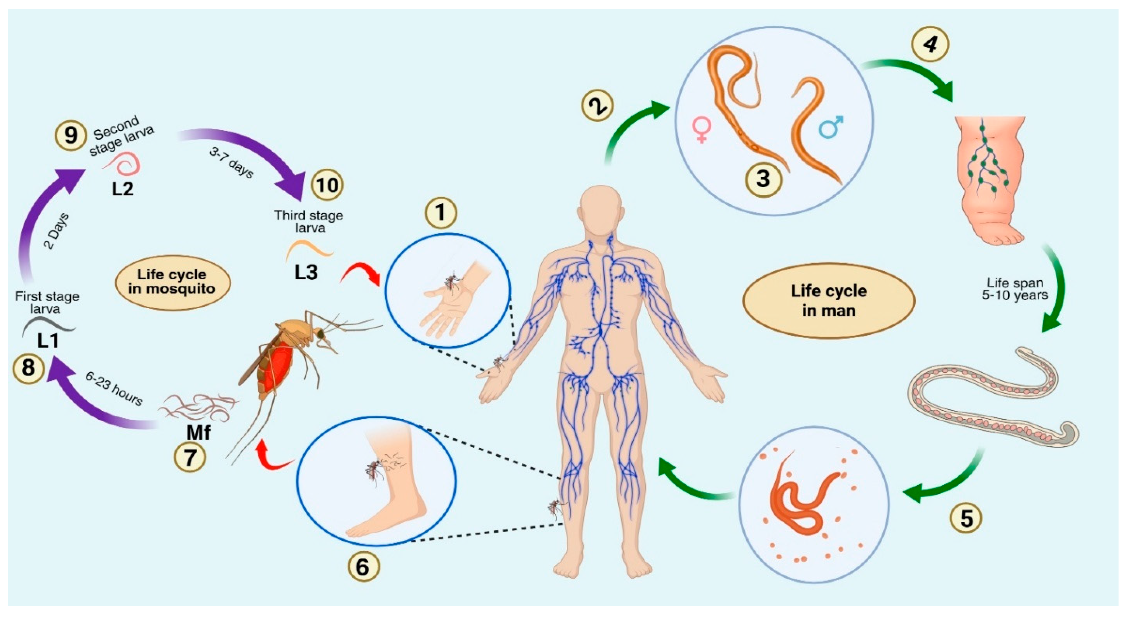
Figure 1. Life cycle of Wuchereria bancrofti 1. Mosquito bites a normal person, entry of infective larva (L3) into the human through the mosquito bite. 2. Migration of larvae into local lymphatics and lymph nodes, preferably into the lymphatics of the spermatic cord. 3. Development into adult worms, both male and female. 4. Inflammation and obstruction of lymphatics, leading to lymphedema. 5. Female adult worm produces microfilariae, which circulate in the lymphatics and the bloodstream. 6. Entry of microfilariae into another mosquito during a blood meal. 7. After sheathing, sheathed microfilariae penetrate the midgut of the mosquito and migrate to the thoracic muscles. 8. Microfilariae develop into the L1 stage. 9. L1 stage larvae moult into L2 stage and further moult into L3 infective stage. 10. Migration of L3 larvae to the head and proboscis of the mosquito, and during the blood meal, enter the human body.
Immuno 05 00057 g001
Figure 2. An overview of immune evasion tactics employed by filarial parasites. The diagram categorises the mechanism by which filarial parasites evade host immune responses to ensure their survival. During respiratory bursts, monocytes, basophils, and neutrophils generate ROS (reactive oxygen species) to eliminate the parasite; as a result, filarial worms release antioxidant enzymes, which include SOD (superoxide dismutase), TrX (thioredoxin), and glutathione S-transferase (GST), some proteins, as well as immunomodulators. This, as a result, facilitates immune evasion by activating macrophages and other immune cells, by altering effector T cells and toll-like receptors (TLRs), and by downregulating antigen presentation through major histocompatibility complex (MHC) I and major histocompatibility complex (MHC) II with the assistance of T regulatory cells. Hence, filarial worms can minimise the oxidative damage and regulate host immunity, thus facilitating chronic infections.
Immuno 05 00057 g002
Figure 3. TLR—NFκB signalling in healthy and chronically infected individuals. The schematic depicts the activation of TLR-NFκB signalling under normal physiological settings and during chronic parasite persistence. During normal signalling, TLR2/4 interacts with lipopolysaccharides of pathogens, and after that recruits MyD88, which eventually results in IκB phosphorylation and NFκB activation and translocation to the nucleus, which facilitates the production of pro-inflammatory genes IL-1, TNF-α, and IL-12, and the subsequent Th1 and Th17 response aids in pathogen clearance. During chronic infection, the excretory secretory products of the parasite engage with TLR2/4, modifying downstream signalling, which induces Foxp3+ and Treg cells and secretion of anti-inflammatory cytokines (IL-10, TGF-β) and anti-apoptotic proteins, inhibiting NFκB-mediated pro-inflammatory gene transcription. Thus, there is a reduced Th1/cytokine response, which enables the parasite’s existence.
Immuno 05 00057 g003
Figure 4. Outcomes of interaction between different immune cells involved in the anti-filarial immune response. The diagram depicts the immunological response affecting parasite population dynamics and progression of filariasis. On the left, parasite survival is facilitated by B and T regulatory cells in addition to AAMs that are responsible for the Th2 response and, due to their modulation, result in chronic infection. At the base, antigen presentation by dendritic cells stimulated CD8+ T cells and again induced a Th2 response, which resulted in the production of IgE by B cells. On the right side of the triangle, macrophages, eosinophils, neutrophils, and basophils result in the parasite’s death by secreting ROS (reactive oxygen species), NO (nitric oxide), and nitrogen intermediates.
Immuno 05 00057 g004
Table 1. Different filarial parasites and their characteristics.
S.No.Filarial ParasiteAssociated
Disease
Vector InvolvedCause of SymptomsRecommended TreatmentAffected RegionsExperimental ModelReferences
1.Oncocerca volvulus,
Oncocerca ochengi
OnchocerciasisBlackflies (Simulium spp.)Microfilariae (MF)-related immune responseIvermectin (generally in co-endemic areas), not advised in case of areas co-endemic with loiasisSub-Saharan Africa, Yemen, Uganda, Cameroon, small foci in South AmericaMice in the case of Oncocerca ochengi[21]
2.Wuchereria bancrofti, Brugia malayi, Brugia timoriLymphatic filariasis
(Elephantiasis)
Mosquito species
(Aedes, Anopheles, Culex, Mansonia, etc.)
Adult worm-specific immune responses
TPE: lung mf trapping
Diethylcarbamazine citrate (DEC), Ivermectin, Albendazole (Different combinations of these drugs are given in areas with co-endemicity)Tropical regions of Asia, America, the Pacific, Africa, and countries like Indonesia, Malaysia, ThailandFerrets, Mice, and Jirds[30]
3.Loa loaLoiasisChrysops fliesDue to the migration of adult worms, Calabar swelling, severe reactions to DEC treatment, and hypereosinophiliaDEC or Albendazole (Due to the possibility of SAEs, treatment is not always advised)West and Central AfricaPrimates (Baboons) and rodents[31]
4.Mansonella perstans, Mansonella ozzardi, Mansonella streptocercaMansonellosisMidges of the genus CulicoidesDue to adult worm migration, ocular symptoms brought on by MF migrationDoxycyclineEastern, Western, and Central Africa, parts of South and Central America, Caribbean islandsNo known
animal model
till date
[31]
Table 2. Summary of clinical stages of filariasis, highlighting disease status and symptoms.
S.No.Clinical Stages in IndividualsHealthy/DiseasedParasitic StagesCirculating Filarial AntigensSymptomsReferences
1.NormalHealthyNoneAbsentNil[40]
2.Endemic NormalsHealth status is questionable as they are living in endemic areas, but have not been tested for filarial infections[4]
3.Microfilaraemic/AsymptomaticDiseasedMicrofilariae in blood, live adult worms in lymphaticsPresentClinically asymptomatic[41]
4.Acute Clinical DiseaseDiseasedAdult worms in lymphaticsPresentEpisodes of lymphangitis, filarial fever, lymph nodes, localised inflammation[42]
5.Chronic PathologyDiseasedUsually, dead adult worms are present in the lymphaticsPresentLymphedema, elephantiasis, and hydrocele[43]
6.OccultDiseasedAdult worms present, but no circulating microfilariaeMay or may not be presentSymptoms include TPE, restrictive pulmonary changes, filarial arthritis, glomerulonephritis, and breast abscesses, among others.[44]
Table 3. Functions of various types of immune cells involved in the anti-filarial immune response.
S.No.Cell TypeLocationFunctions and RolesReferences
1.Regulatory T cellsThymus and PeripheryMaintains tolerance and prevents pathologies. Present in high levels in the asymptomatic stage and low levels in the chronic stage[50,51]
2.Regulatory B cellsBlood circulation and inflammation sitePerform Immune regulatory mechanisms by secreting immunosuppressive cytokines. Induce Treg cells, suppressing CD4+, CD8+ T cells and Natural Killer (NK) cells [33]
3.EosinophilsDerived from bone marrow, later migrates to tissueContributes both to the protection and development of filarial pathology[21]
4.NeutrophilsCirculates in blood later migrate to tissueRelease toxins to eliminate parasites. Involved in protective immune responses and pathological aggravation of disease.[52]
5.Alternatively Activated Macrophages Reside in Blood and TissueBlood-derived AAMs perform immune regulatory roles whereas tissue-resident AAMs responsible for fibrosis seen during chronic infections[4]
6.Dendritic cellsPresent in epithelial tissuesParasite-derived products interact with DCs to initiate profound changes in immune responses leading to suppressed inflammation suitable for prolonged survival of parasites characteristic of chronic infections.[53]
7.CD4+ T cellsThymus and peripheral blood circulationInvolved in parasite clearance along with type-2 cytokines. Presence of Treg balances Th1/Th2 responses.[54]
8.CD8+ T cellsThymus and peripheral blood circulationInvolved in the cytotoxic killing of filarial parasites with the help of type-2 cytokines, persistence of filaria antigens contributes to chronic pathology[54]
Disclaimer/Publisher’s Note: The statements, opinions and data contained in all publications are solely those of the individual author(s) and contributor(s) and not of MDPI and/or the editor(s). MDPI and/or the editor(s) disclaim responsibility for any injury to people or property resulting from any ideas, methods, instructions or products referred to in the content.