Article

PD-1 Expression in Endometriosis

by
José Lourenço Reis
1,*,
Catarina Martins
2,3,
Miguel Ângelo-Dias
2,3,
Natacha Nurdine Rosa
4,
Luís Miguel Borrego
2,3,5 and
Jorge Lima
1,2,3
1
Department of Obstetrics and Gynecology, Hospital da Luz Lisboa, 1500-650 Lisboa, Portugal
2
Comprehensive Health Research Center, NOVA Medical School, Faculdade de Ciências Médicas, NMS|FCM, Universidade NOVA de Lisboa, 1169-056 Lisboa, Portugal
3
Immunology Department, NOVA Medical School, Faculdade de Ciências Médicas, NMS|FCM, Universidade NOVA de Lisboa, 1169-056 Lisboa, Portugal
4
UCD School of Medicine, University College Dublin, D04 V1W8 Dublin, Ireland
5
Department of Imunoallergy, Hospital da Luz Lisboa, 1500-650 Lisboa, Portugal
*
Author to whom correspondence should be addressed.
Immuno 2025, 5(4), 49; https://doi.org/10.3390/immuno5040049
Submission received: 27 August 2025 / Revised: 26 September 2025 / Accepted: 16 October 2025 / Published: 17 October 2025
(This article belongs to the Section Reproductive Immunology)

Abstract

Background: Endometriosis, believed by many to be rooted in immunology, is a chronic disease. Upregulation of programmed cell death protein 1 (PD-1) in immune cells may compromise their defensive function, a mechanism demonstrated in the context of cancer spread. This study aims to explore the potential involvement of PD-1 in the pathophysiology and progression of endometriosis. A total of 62 patients who underwent laparoscopic surgery were analyzed, with 47 diagnosed with endometriosis and 15 serving as controls. We collected peritoneal fluid and peripheral blood samples during surgery and examined them using flow cytometry. Using a panel of monoclonal antibodies, the samples were stained and the expression of PD-1 in immune cells was evaluated. Results: We observed a statistically significant rise in the percentage of the CD56+ CD16+ NK cell subset expressing PD-1 within the peritoneal fluid of endometriosis patients compared to the control group (p = 0.021). Similarly, we found that PD-1 expression on immune cells significantly differed based on factors such as body mass index and smoking habits. Moreover, peritoneal subsets of PD-1+ T and NK cells showed an increase in patients presenting symptomatic endometriosis and those with more widespread disease. Conclusions: Our evaluation of the inhibitory PD-1 receptor has strengthened the potential connection between immune escape mechanisms often seen in cancer cells and those in endometriotic cells. This concept could pave the way for future research in the field of immunomodulation and endometriosis.

1. Background

Endometriosis is a common chronic gynecological condition, often associated with symptoms like painful menstruation, persistent pelvic pain, and infertility. It is characterized by the abnormal presence of endometrial cells, typically located on the ovaries or the pelvic lining [1]. Despite its long history and prevalence among women of reproductive age (one in ten), the condition’s cause remains a mystery [2]. Presently, treatment predominantly involves hormone suppression, which unfortunately proves ineffective for numerous patients [3]. Since the 1990s, the role of the immune system in the development and progression of endometriosis has been conjectured [4,5]. Although the precise mechanism remains unclear, recent emphasis on the study of natural killer (NK) cells and their receptors, spurred by advances in flow cytometry, has made this theory increasingly significant. A comprehensive recent review has further explored this subject [6].
PD-1, a crucial inhibitory checkpoint, was first identified on T, B, and myeloid cells, but has also been recently found on NK cells [7]. Independent studies on diverse cancer types, including ovarian cancer, multiple myeloma, Kaposi sarcoma, and lung and digestive cancers, have revealed that tumor-infiltrating PD-1+ NK cells produce significantly fewer cytokines upon interaction with the tumor [7]. Ahmadzadeh et al. revealed that most tumor-infiltrating T lymphocytes primarily express PD-1, a trait linked to an exhausted phenotype and diminished effector function [8]. Given that tumor cells often use the PD-1 axis to avoid the immune system, it is logical that this pathway has been explored extensively to devise new treatments intended to augment the anti-tumor immune response. The activity of these immune checkpoints can be curtailed by using blocking antibodies that hinder ligand-receptor interactions, both in the lab and the body [9,10,11,12].
This study aims to investigate PD-1 expression in immune cells from both peripheral blood and peritoneal fluid in women with endometriosis. Similar to what has been observed in oncology, altered PD-1 expression in immune cells may represent one of the mechanisms by which these cells evade immune surveillance. Drawing parallels between the immune escape strategies of cancer cells and endometriotic cells could offer important insights into the development of future immunomodulatory therapies for endometriosis.

2. Methods

2.1. Study Design and Subjects

This cross-sectional study, conducted over an 18-month period, included 62 female patients. Among these, 47 had endometriosis (EM) and 15 served as controls without endometriosis. All participants are adult women of reproductive age, selected from the hospital’s outpatient clinic and who have undergone laparoscopic surgery at the Hospital da Luz Lisboa and Hospital Beatriz Ângelo, those in the endometriosis group to treat this disease and those in the control group to treat pathologies other than endometriosis. Their gynecological issues were treated surgically under general anesthesia.
Endometriosis was initially diagnosed following the ESHRE criteria based on clinical and imaging findings and subsequently confirmed through histology [13].
We excluded women who were pregnant within the last year, those suspected of complications from pelvic inflammatory disease, and those with immunologic, endocrine, cancer, or other chronic disorders.
The Ethics Committees at NOVA Medical School, Hospital da Luz Lisboa, and Beatriz Ângelo approved this study. We provided each patient with essential information about the research, including the use of their biological materials for scientific analysis. Every patient granted their explicit written consent before participating in the study.

2.2. Clinical and Demographic Information

We recorded each patient’s age, race, body mass index (BMI), smoking status, hormonal medication use, and the time span between endometriosis diagnosis and surgery. In terms of BMI, normal weight was defined as a BMI between 18.5 and 24.9, overweight as a BMI between 25 and 29.9, and obesity as a BMI of 30 or above.
Clinical data on various symptoms was gathered to assess the severity of the disease. This included dysmenorrhea, dyspareunia, chronic pelvic pain, dyschesia, bowel issues such as constipation, perimenstrual diarrhea, bloating and rectorrhagia, urinary tract symptoms like hematuria and dysuria, and the presence of extra-pelvic endometriosis. Pain was assessed by the Numerical Rating Scale. The American Society for Reproductive Medicine’s established criteria were employed to categorize the severity of the disease [14].

2.3. Sample Collection

During the procedure, peritoneal fluid (PF) and peripheral blood (PB) samples were collected from each patient. To minimize blood contamination, PF was procured at the surgery’s outset using EDTA-coated tubes. After surgery, these samples were stored and transported at room temperature to NOVA Medical School’s immunology laboratory. All PF samples were processed on the day of collection, while blood samples were processed on the day of the collection or kept at room temperature and processed the following morning, as per the laboratory availability.

2.4. Analysis of Immunophenotyping Using Flow Cytometry

A single-platform method with TrucountTM tubes (BD Biosciences) was used to obtain the absolute counts of major leucocyte populations and identify lymphoid compartment in each PB and PF sample. In this procedure, each sample (50 μL for PB samples and 100–200 μL for PF samples) was stained with CD45 for 15 min in a TrucountTM tube, and afterwards cells were lysed with BD FACS lysing solution (BD Biosciences) for another 15 min. All samples were immediately acquired after the lysis period, without any further washing step, according to the manufacturer’s instructions [15].
Another procedure was applied to characterize the major populations within the lymphoid compartment. For that purpose, the PB and PF samples were stained with an additional panel of monoclonal antibodies, including CD45, CD3, CD8, CD16, CD56, CD57, and PD-1. A thorough inventory of all antibodies employed is available in the Supplementary Materials (Table S1).
To concentrate cells in these samples, all PF samples were centrifuged for 5 min at 1800 rpm before the immunophenotyping procedure [16]. After the centrifugation step, the supernatant was discarded, and the pellet was resuspended in 300 μL of BD FACS Flow (BD Biosciences), and incubated with the monoclonal antibodies. The PB samples (100 μL) were directly incubated. In both cases, though, a lyse-wash technique was used, including a 20-min incubation with the monoclonal antibodies, followed by a 15-min lysis using BD FACS lysing solution (BD Biosciences). Following this step, the cells were centrifuged, rinsed with BD FACS Flow Solution (BD Biosciences) and fixed with BD Cell Fix Solution (BD Biosciences).
For all the aforementioned procedures, cell suspensions were acquired in a BD FACS Canto II (BD Biosciences, San Jose, CA, USA), with BD FACS Diva software, version 8.0.2 (BD Biosciences). Data analysis was performed with the InfinicytTM (version 2.0) and FlowJoTM (version 10.6.2)) software (both from BD Biosciences). Figure 1 illustrates the gating strategy, similar to others previously defined [17,18]. This includes the assessment of PD-1 and CD57 in T and NK cell subsets, using a Fluorescence Minus One (FMO) approach to confirm the distinction of positive events for each relevant marker.

2.5. Statistical Analysis

The study participants’ characteristics were summarized using descriptive statistics, which included demographic, clinical, and immunophenotyping data. We represented categorical variables as absolute and relative frequencies. If over 20% of expected values were less than 5 in contingency tables, we used Fisher’s exact test to compare the two groups and used the chi-squared test otherwise. The presentation of continuous variables was done either by indicating the mean (standard deviation) (minimum–maximum) or the median along with the first-to-third quartile (Q1–Q3). In the Supplementary Tables S1–S3, continuous variables are shown as average (standard deviation) [95% confidence interval (95% CI)], median, and quartiles (Q1–Q3). We obtained 95% CIs for normally distributed variables using a Student’s t-distribution approximation, using bootstrap resampling for others. To compare two separate groups, we used the Student’s t-test for normally distributed continuous data and applied the Wilcoxon rank-sum test otherwise. For comparing two dependent groups, we employed a paired-sample t-test if data was distributed normally, and the Wilcoxon signed-rank test when it was not. For more than two groups, we used analysis of variance (ANOVA) with a follow-up Tukey’s HSD paired-comparison test for normal distributions, and the Kruskal–Wallis test with a follow-up Dunn’s paired-comparison test otherwise. We assessed distribution normality with the Shapiro–Wilk test. Because of the study’s exploratory nature, we did not calculate a formal sample size. We considered a p-value less than 0.05 statistically significant. We executed all analyses with SAS software (Version 9.4).

3. Results

3.1. Demographic and Clinical Characteristics of Patients

Table 1 and Table 2 display the demographic and clinical attributes of the control group (n = 15) and women suffering from endometriosis (n = 47). No significant age differences were observed between the groups, with a mean age of 38.5 years in the control group and 36.2 years in the endometriosis group. The majority of women were Caucasian. As anticipated, the only noteworthy disparity between the groups was in relation to infertility history, where it was significantly more common in women with endometriosis (p = 0.003) (Table 1).
The clinical data from women with endometriosis highlights several important points. On average, there was a 2.8 ± 2.52 (1–14) year gap between the diagnosis of endometriosis and surgery. The majority of patients (78.7%) were classified in stages III or IV. A high percentage of these patients reported experiencing dysmenorrhea (93.6%), with the majority (76.6%) describing the pain as severe. Other commonly reported symptoms included dyspareunia (59.6%), chronic pelvic pain (40.4%), dyschesia (34.0%), bowel issues (31.9%), urinary tract problems (10.6%), and rectorrhagia (8.5%) (Table 2). Lastly, 33% of women in the control group and 66% of women with endometriosis were receiving hormone therapy.

3.2. PD-1 in Endometriosis: Expression Patterns in PB and PF

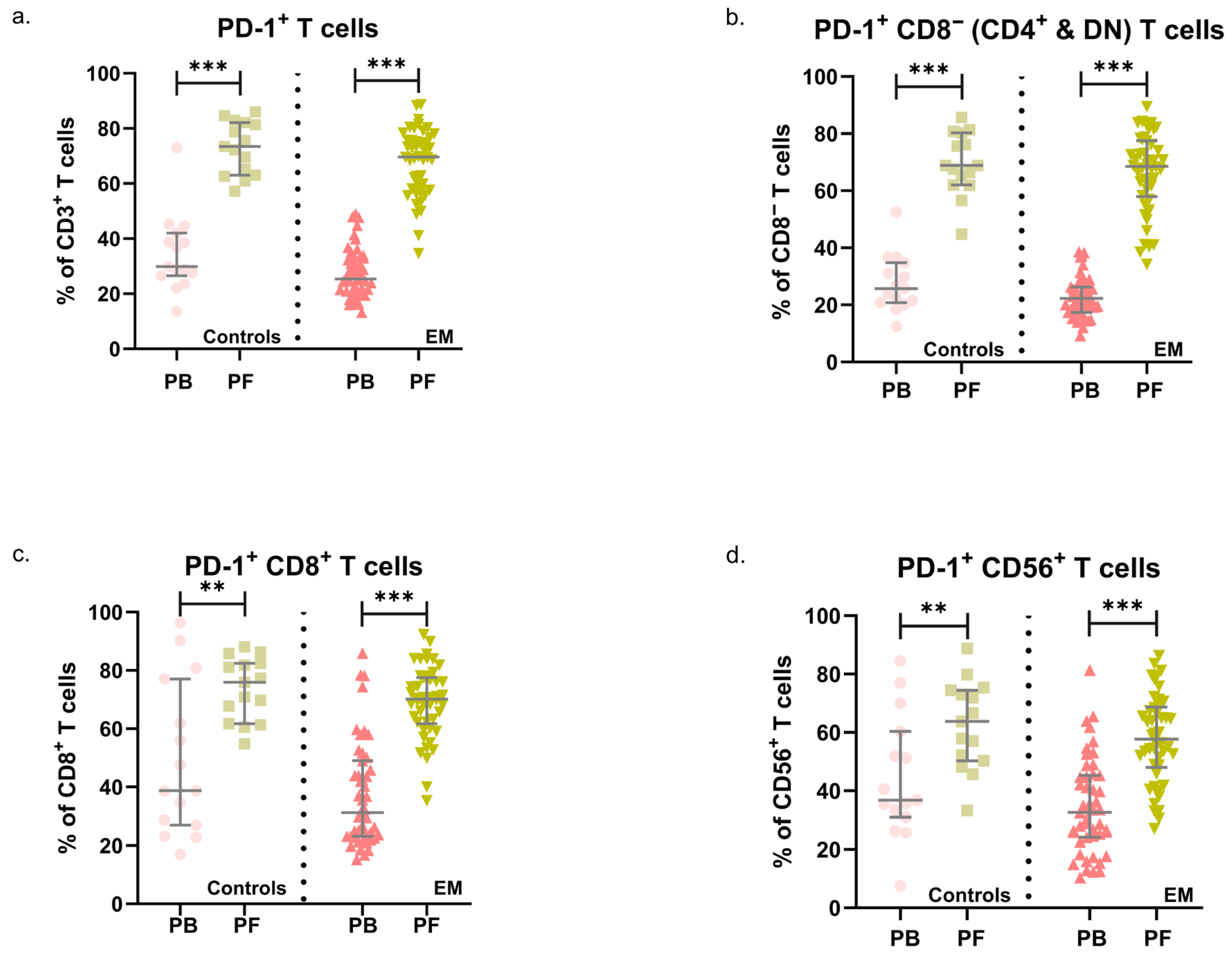
Table S2 in the Supplementary Materials showcases a comprehensive characterization of primary circulating and peritoneal lymphoid subsets in both the control group and women diagnosed with endometriosis. We focused on analyzing the expression of programmed cell death protein 1 (PD-1) within these cells. Generally, we observed higher percentages of T cells expressing PD-1 (p ≤ 0.024) in PF samples relative to PB. This was the case for both endometriosis patients and the control group (Figure 2).
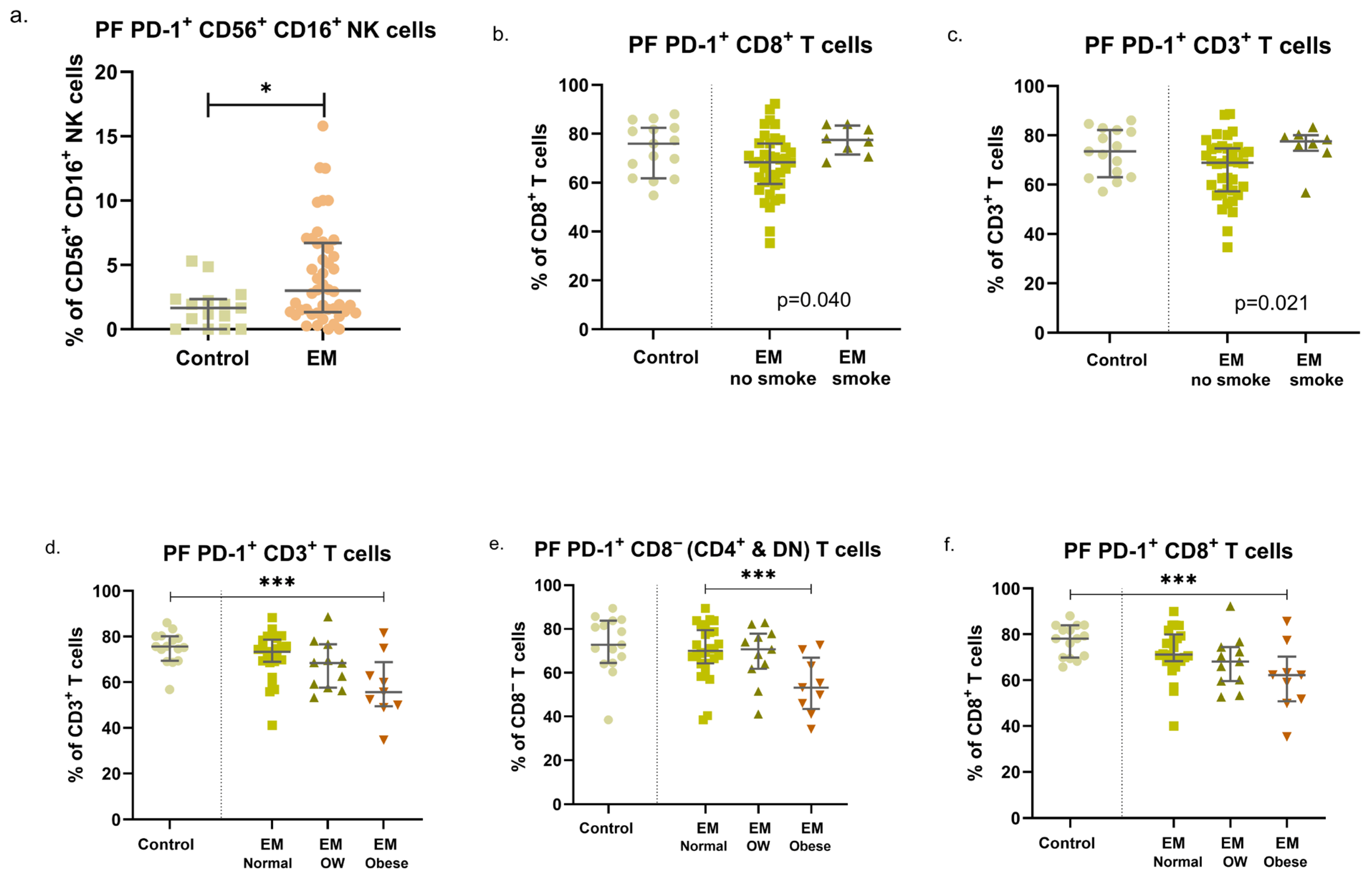
In this study, we identified a higher percentage of a particular type of peritoneal NK cells, namely, PD-1+ CD56+ CD16+ NK cells, in women with endometriosis (p = 0.021) (Figure 3a). No additional differences were found in other NK or T-cell subsets across both groups and compartments.
Patients’ clinical conditions and lifestyles can influence the expression of this marker. Notably, smoking habits showed a significant impact, displaying differences in the groups for peritoneal PD-1+ total T cells (p = 0.021) and PD-1+ CD8+ T cells (p = 0.040). Higher levels of both subsets were found in smoking EM women (Figure 3b,c). All the control participants in the study were non-smokers.
After comparing women with and without endometriosis, the overweight group displayed differences in the expression of PD-1 by peritoneal T cells (p ≤ 0.041) (Figure 3d–f). Though the sample size of this subgroup was small (n = 4), overweight control women demonstrated a potential upward trend in the percentages of PD-1+ cells across all T-cell subgroups evaluated (Total CD3+ T cells, CD8+ T cells, CD8 [CD4+ and Double negative, DN] T cells, and CD56+ T cells; Figure 3d–f). On the other hand, women with endometriosis showed declining values of all PD-1+ subsets, transitioning from normal weight to overweight and obese subgroups. Furthermore, women with endometriosis and obesity exhibited the lowest levels, specifically for PD-1+ CD8 (CD4+ and DN) T cells when compared to normal-weight women with endometriosis (Figure 3e).

3.3. Endometriosis, Previous Pregnancies, and Fertility Status: Modulation of PD-1 Expression in Circulating and Peritoneal Immune Cells

We also observed differing patterns of PD-1 expression in women who have been pregnant and those who have not. In examining circulating T cells, non-pregnant EM women demonstrated lower levels of PD-1+ CD8 (CD4+ and DN) T cells (p = 0.041) compared to others. Moreover, in EM women grappling with fertility issues, we found diminished levels of both PD-1+ CD56+ T cells (p = 0.039) and PD-1+ total T cells (p = 0.005) in peripheral blood samples, especially when compared with EM women and controls who are not struggling with fertility concerns.
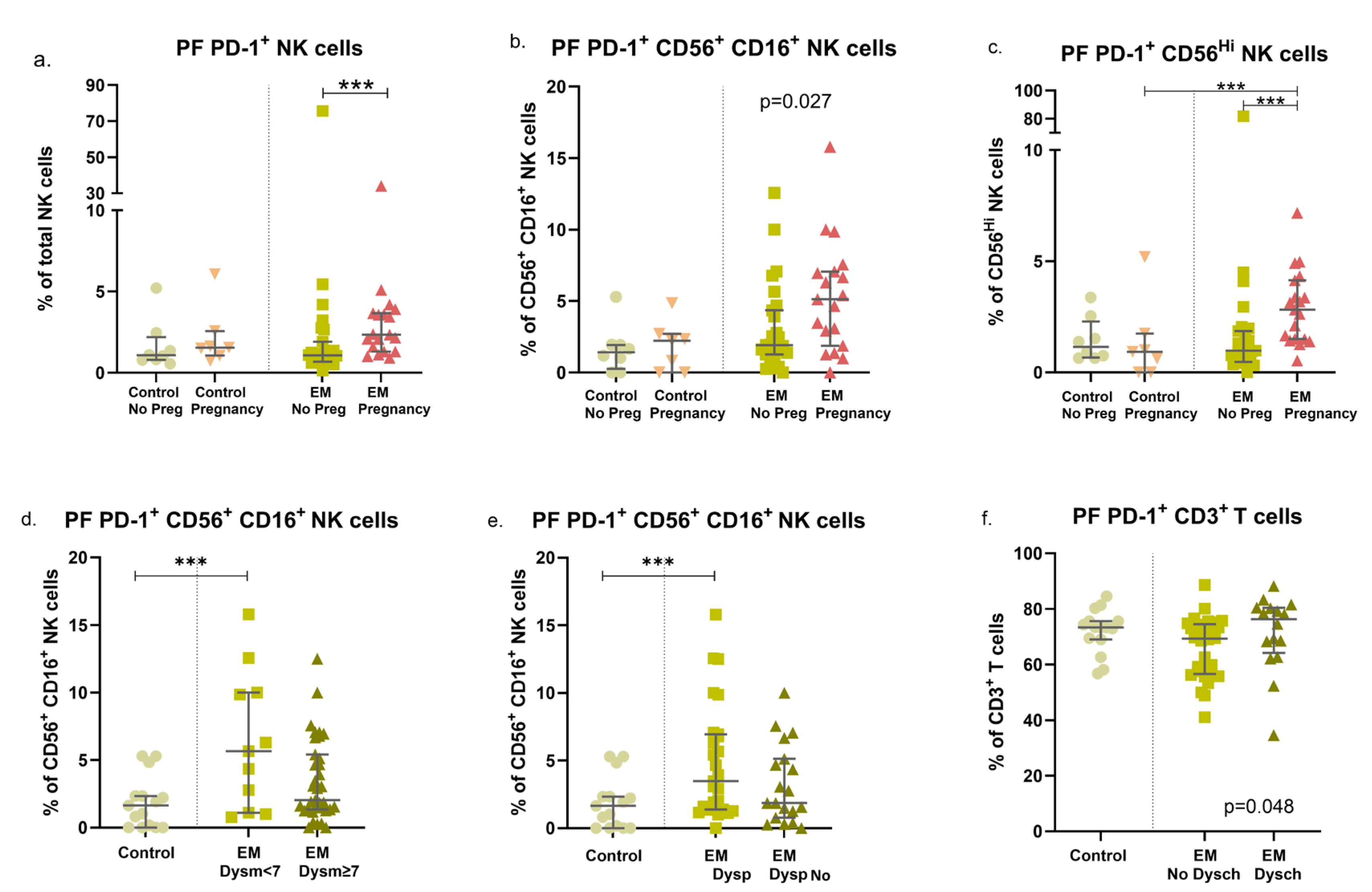
Upon examining peritoneal populations, we noticed a rise in several PD-1+ NK subsets in endometriosis (EM) women who had previously been pregnant (PD-1+ total NK cells: p = 0.017; PD-1+ CD56+ CD16+ NK cells: p = 0.027; PD-1+ CD56Hi NK cells: p = 0.002) (Figure 4a–c). On the other hand, there was a decrease in these subsets in EM women who had not been pregnant when compared to their EM counterparts with past pregnancies. It was also observed that EM women without a history of infertility exhibited a significant increase in PD-1+ CD56+ CD16+ NK cells, not only compared to fertile controls but also compared to infertile EM women (p = 0.031).

3.4. EM Symptoms Are Associated with Different Expression Patterns of PD-1 in NK and T-Cell Subsets

Our results further revealed that in endometriosis, particularly in peritoneal populations, the expression of PD-1 can vary significantly among EM patients with diverse symptoms. Complete data are presented in Supplementary Materials (Table S3).
We observed a trend toward higher percentages of peritoneal PD-1+ CD56+ CD16+ NK cells in women with endometriosis who experienced dysmenorrhea, although this trend was not statistically significant (p = 0.069). Upon further separation of the EM women based on the severity of dysmenorrhea, we found higher levels of this cell subset in women with mild to moderate symptoms (p = 0.028) (Figure 4d).
In patients with endometriosis experiencing dyspareunia, we observed higher percentages of peritoneal PD-1+ total NK cells, as well as PD-1+ CD56+ CD16+ NK cells (p = 0.035 and p = 0.029, respectively) (Figure 4e). In the peripheral blood of women not experiencing this symptom, there were decreased levels of PD-1+ total T cells (p = 0.021) and PD-1+ CD8 (CD4+ and DN) T cells (p = 0.049). While there was a notable increase in PD-1+ total NK cells in EM patients with dyspareunia (p = 0.052), this was not statistically significant, especially in severe cases (T cells: p = 0.038; total NK cells: p = 0.074).
Dyschesia is a common symptom in EM patients. In our group, a trend was found associating higher levels of PF PD-1+ CD8+ T cells (p = 0.052) and PD-1+ total T cells (p = 0.048) (Figure 4f) with this symptom. However, no significant variations were observed when comparing patients with mild, moderate, or severe symptoms.
Similarly, an increase in peritoneal PD-1+ CD56+ CD16+ NK cells was observed in endometriosis patients without rectal bleeding (p = 0.021). This trend was also present in patients without chronic pelvic pain (p = 0.057) and those without bowel endometriosis (p = 0.056). Lastly, endometriosis patients with urinary tract infiltration demonstrated increased percentages of PD-1+ total NK cells (p = 0.042). These patients likewise exhibited higher percentages of circulating PD-1+ CD8+ T cells (p = 0.037), PD-1+ CD56+ T cells (p = 0.036), and PD-1+ total T cells (p = 0.029).

4. Discussion

In our study, we analyzed PD-1 expression in NK and T-cell subsets from the PF and PB of women, both with and without endometriosis. To our knowledge, this study utilizes the largest patient sample to assess PD-1 expression in both compartments.
We observed a statistically significant increase in the percentage of CD56+ CD16+ NK cells expressing PD-1 in the PF of patients with endometriosis compared to the control group. This difference was not detected in PB. Likewise, no significant differences were observed in PD-1 expression on T cells in either the PF or PB.
NK cells, a vital component of the innate immune system, play an essential role in early defense mechanisms against various types of aggression through their natural cytotoxicity activation, which is not antibody-dependent [19,20,21]. The increase in PD-1-expressing NK cells in peritoneal fluid is particularly important to emphasize, as these cells play a crucial role in the initial defense within this microenvironment. This is the same environment where endometriotic cells attempt to implant and spread, especially within the pelvic cavity. Interestingly, this only impacts CD56+ CD16+ NK cells. NK cells expressing CD16 serve as potent mediators of antibody-dependent cellular cytotoxicity, in addition to their inherent cytotoxicity [19,22]. On the other hand, CD56-expressing cells possess strong immunostimulatory effector capabilities, such as producing T helper 1 cytokines and having effective cytotoxic potential [23]. Therefore, our findings show that increased PD-1 expression was detected in a subset of NK cells that are, in theory, functionally more competent to perform local immune surveillance against the early dissemination of endometriotic cells. This upregulation of PD-1 may impair the protective role of NK cells in controlling ectopic endometrial cell spread, a mechanism previously described in oncology, where PD-1-mediated immune evasion facilitates tumor progression [7].
When analyzing variations based on demographic and clinical factors, we discovered intriguing findings. In our study, BMI significantly impacted the expression of the PD-1 marker on T cells in patients with endometriosis, especially within the peritoneal cavity. In women with endometriosis, all PD-1+ immune cell subsets showed a progressive decrease in frequency across increasing BMI categories, from normal weight to overweight and obesity. Notably, women with both endometriosis and obesity exhibited the lowest levels of PD-1+ CD8 T cells when compared to their normal-weight counterparts. Although the influence of BMI on the immune system, including various aspects of the immune response, has long been recognized [24], its specific effect on PD-1 expression remains unexplored in prior research. Obesity is associated with chronic low-grade inflammation [25], which could modulate PD-1 expression in immune cells, potentially leading to the downregulation of PD-1 in T cells within the peritoneal cavity. In the context of endometriosis, the peritoneal immune environment is already altered, with increased inflammation and immune dysregulation. The supplementary influence of obesity-related inflammation might uniquely impact local T-cell populations, explaining the distinct PD-1 expression patterns seen in obese versus normal-weight patients.
In our examination of smoking habits, we discovered a heightened occurrence of PD-1+ total T cells and PD-1+ CD8+ T cells in the peritoneal fluid of endometriosis patients who smoke. This implies a potential association with disease progression in such individuals. It is well-known that smoking can modify immune responses, including NK cell function, by promoting chronic inflammation and adjusting immune checkpoint pathways [26]. The augmented PD-1 expression on T cells in smokers could signify an immunosuppressive state impacted by oxidative stress and inflammation related to tobacco use. However, these findings should be interpreted with caution, as the number of smoking patients with endometriosis was small (n = 8), and there were no smokers in the control group.
When considering obstetric history, we observed an increase in several PD-1+ NK cell subsets within the peritoneal populations of women with endometriosis who had previous pregnancies. Notably, our findings revealed a significant rise in PD-1+ CD56+ CD16+ NK cells in the peritoneal fluid of women without a history of infertility compared to those who were infertile. Although no current studies examine the relationship between PD-1 and fertility, our results suggest a potential link between this marker and a woman’s fecundity, particularly in patients with endometriosis. This hypothesis certainly merits further investigation.
When we categorized the patients based on the typical symptoms of endometriosis, we observed distinct patterns of PD-1 marker expression, with a higher prevalence in those with widespread disease, particularly within the peritoneal cell population. Specifically, we found increased levels of PD-1+ CD56+ CD16+ NK cells and PD-1+ total NK cells in patients suffering from dyspareunia, with the count of PD-1+ total NK cells significantly higher in those experiencing severe dyspareunia compared to those with milder symptoms. Dyschesia, a symptom often associated with bowel involvement, was linked to elevated levels of PD-1+ total T cells in the peritoneal fluid of our cohort. In endometriosis patients with urinary tract involvement, there was a substantial increase in PD-1+ total NK cells in the peritoneal cavity, alongside higher percentages of circulating PD-1+ CD8+ T cells, PD-1+ CD56+ T cells, and PD-1+ total T cells. Overall, these findings suggest an increase in PD-1 expression in the immune cells of patients with more severe or symptomatic endometriosis.
Looking at published studies evaluating the PD-1 marker in women with endometriosis, the available data is scarce. Sobstyl, M. et al. examined the expression of PD-1 and PD-L1 on T and B lymphocytes, but their analysis was limited to the peripheral blood of patients with endometriosis. They found a significantly higher expression of both PD-1 and PD-L1 on T and B lymphocytes in endometriosis patients compared to the control group [27]. Another recent study [28] examined the PD-1 expression on NK cells in PF and PB. Although limited to 11 controls and 11 women with endometriosis, the study found a higher percentage of PD-1+ NK cells in the PF of patients with stage IV endometriosis compared to controls [28]. Wu et al. [29] conducted a study on eutopic and ectopic endometria and blood samples from 15 women with endometriosis and 15 healthy controls. They used immunohistochemistry and western blot analysis to detect PD-1/PD-L1 expression in endometrial tissues. Flow cytometry was used for blood analysis. Their research revealed increased PD-1/PD-L1 expression in both eutopic and ectopic endometria of patients with endometriosis. Additionally, they reported a heightened PD-1 expression in CD41/CD81 T cells in PB [29].
Walankiewicz et al. [30] examined PB samples from patients with endometriosis and a control group using flow cytometry. They found a significantly higher percentage of PD-1 and PD-L1-positive CD4+ T cells, CD8+ T cells, and CD19+ B cells in the patients compared to the control group [30]. Suszczyk et al. [31] used flow cytometry to study expressions of PD-L1/PD-L2 on myeloid (m-) and plasmacytoid (p-) dendritic cells (DCs) in the PB and PF of both endometriosis patients and healthy individuals. They found a higher proportion of mDCs and pDCs expressing PD-L1 or PD-L2 in the PF of endometriosis patients compared to their plasma, indicating the possible influence of the peritoneal cavity environment on immune response modulation and the development or progression of endometriosis. However, this study only included PB samples from the control group, and the exclusion of endometriosis in this group was based solely on clinical inquiries, not direct pelvic observation through laparoscopy [31]. Okşaşoğlu et al. [32] collected PB samples from women with endometriosis during surgery or examination, as well as from healthy individuals. Using an ELISA platform, they found elevated serum levels of PD-1 in women suffering from endometriosis [32]. Nero et al. [33] used immunohistochemistry assays on formalin-fixed paraffin-embedded tissue samples and found a higher PD-1/PD-L1 expression in endometriosis-associated ovarian cancer than in benign endometriosis-related diseases [33].
Our study offers a distinct clinical perspective, differing significantly from previously examined methodologies and parameters. We identified patterns of abnormal PD-1 expression in relation to specific clinical symptoms such as dysmenorrhea, dyspareunia, and dyschezia, investigating the correlation between immune cell subsets and these symptoms. Additionally, we systematically evaluated the influence of demographic factors, including BMI and smoking status, on both systemic and local PD-1 expression in control and endometriosis patients. A key distinction of our study is that the exclusion of endometriosis in the control group was confirmed through laparoscopic surgery with direct pelvic visualization, providing a more reliable method for verifying the absence of the disease, rather than relying solely on clinical evaluation. A strength of our investigation is its detailed examination of specific NK and T-cell subsets and immune receptors, deepening the understanding of the immunological landscape of endometriosis at the cellular level, which may have important implications for its pathophysiology.
This study provides valuable insights into the immunologic profile of PB and PF concerning PD-1 expression, though it acknowledges certain limitations. Despite our sample size being larger than previous studies in this field, it remains insufficient for drawing definitive conclusions. The findings demonstrate complex interactions within each subpopulation, with PD-1 expression observed in NK and T-cell subsets across various demographic and clinical contexts. However, a major limitation of this study is that PD-1 expression was assessed only at the phenotypic level, without evaluation of its functional implications. The presence of PD-1 does not necessarily reflect cellular exhaustion or altered activity, and therefore these results should be interpreted with caution. Future research incorporating functional assays, such as cytotoxicity tests and cytokine profiling, will be essential to clarify the biological significance of PD-1 expression in endometriosis. Finally, while the immune changes observed may contribute to the implantation and growth of endometriotic cells, they could also reflect a response to the disease itself.

5. Conclusions

Our findings suggest that the immune system may be a key factor in the spread of endometriotic cells, uncovering potential abnormalities in primary defense mechanisms driven by NK and T-cell dysfunction within the pelvic microenvironment. This dysfunction may be associated with elevated PD-1 inhibitory receptor expression, which appears more prominent in patients with severe symptoms and disease extending beyond the pelvis. A common pathway for PD-1 modulation may exist between endometriosis and cancer. Further research is required to confirm these findings and investigate immunomodulatory treatment options for endometriosis, similar to the use of checkpoint inhibitors in cancer therapy. This is particularly important given the limitations of current hormone-based therapies, which not only reduce their effectiveness but also cause side effects that hinder long-term use.

Supplementary Materials

The following supporting information can be downloaded at: https://www.mdpi.com/article/10.3390/immuno5040049/s1, Table S1: List of antibodies used in the immunophenotyping panel; Table S2: Summary of major circulating and peritoneal lymphoid subsets in the control group and in women with endometriosis; Table S3: Characterization of circulating and peritoneal lymphoid subsets in the control group and in EM women with different clinical manifestations.

Author Contributions

J.L.R.: Conceptualization, Methodology, Validation, Investigation, Data curation, Formal analysis, Writing—Original Draft. C.M.: Conceptualization, Methodology, Validation, Investigation, Formal analysis, Writing—Original Draft, Writing—Review and Editing. M.Â.-D.: Conceptualization, Methodology, Validation, Investigation, Formal analysis, Writing—Original Draft. N.N.R.: Conceptualization, Methodology, Validation, Investigation, Writing—Review and Editing. L.M.B.: Conceptualization, Methodology, Validation, Investigation, Formal analysis, Writing—Review and Editing. J.L.: Conceptualization, Methodology, Validation, Investigation, Data curation, Formal analysis, Writing—Original Draft, Writing—Review and Editing, Project administration, Funding acquisition, Resources. All authors were involved in the critical revision of the manuscript and take responsibility for the accuracy or integrity of any part of the work. All authors have read and agreed to the published version of the manuscript.

Funding

This research was partially funded by the Hospital da Luz Lisboa under the initiative “Luz Investigação”, Gedeon Richter Portugal, and LifeWell Pharmaceutical & Healthcare.

Institutional Review Board Statement

Informed consent was obtained from all subjects involved in the study. The study was conducted following the Declaration of Helsinki and approved by the Ethics committees of NOVA Medical School | Universidade NOVA de Lisboa (nº122/2020/CEFCM approved on 24 February 2020), Beatriz Ângelo Hospital (3394/2020_MJH/MAB/NO approved on 6 November 2020) and Luz Lisboa Hospital (CES/53/2021/ME approved on 23 November 2021).

Data Availability Statement

No datasets were generated or analysed during the current study.

Acknowledgments

The authors would like to acknowledge Jorge Gonçalves (Scientific ToolBox Consulting, Lisbon, Portugal) for statistical analysis and Sofia Nunes (Scientific ToolBox Consulting, Lisbon, Portugal), for providing medical writing assistance and technical editing.

Conflicts of Interest

The authors declare no conflicts of interest. The funders had no role in the design of the study; in the collection, analyses, or interpretation of data; in the writing of the manuscript; or in the decision to publish the results.

References

  1. Fukui, A.; Mai, C.; Saeki, S.; Yamamoto, M.; Takeyama, R.; Kato, T.; Ukita, Y.; Wakimoto, Y.; Yamaya, A.; Shibahara, H. Pelvic endometriosis and natural killer cell immunity. Am. J. Reprod. Immunol. 2021, 85, e13342. [Google Scholar] [CrossRef]
  2. Shafrir, A.L.; Farland, L.V.; Shah, D.K.; Harris, H.R.; Kvaskoff, M.; Zondervan, K.; Missmer, S.A. Risk for and consequences of endometriosis: A critical epidemiologic review. Best Pract. Res. Clin. Obstet. Gynaecol. 2018, 51, 1–15. [Google Scholar] [CrossRef]
  3. Capezzuoli, T.; Rossi, M.; La Torre, F.; Vannuccini, S.; Petraglia, F. Hormonal drugs for the treatment of endometriosis. Curr. Opin. Pharmacol. 2022, 67, 102311. [Google Scholar] [CrossRef]
  4. Tanaka, E.; Sendo, F.; Kawagoe, S.; Hiroi, M. Decreased natural killer cell activity in women with endometriosis. Gynecol. Obstet. Investig. 1992, 34, 27–30. [Google Scholar] [CrossRef] [PubMed]
  5. Vinatier, D.; Dufour, P.; Oosterlynck, D. Immunological aspects of endometriosis. Hum. Reprod. Update 1996, 2, 371–384. [Google Scholar] [CrossRef] [PubMed]
  6. Reis, J.L.; Rosa, N.N.; Angelo-Dias, M.; Martins, C.; Borrego, L.M.; Lima, J. Natural Killer Cell Receptors and Endometriosis: A Systematic Review. Int. J. Mol. Sci. 2022, 24, 331. [Google Scholar] [CrossRef]
  7. Pesce, S.; Greppi, M.; Tabellini, G.; Rampinelli, F.; Parolini, S.; Olive, D.; Moretta, L.; Moretta, A.; Marcenaro, E. Identification of a subset of human natural killer cells expressing high levels of programmed death 1: A phenotypic and functional characterization. J. Allergy Clin. Immunol. 2017, 139, 335–346.e333. [Google Scholar] [CrossRef] [PubMed]
  8. Ahmadzadeh, M.; Johnson, L.A.; Heemskerk, B.; Wunderlich, J.R.; Dudley, M.E.; White, D.E.; Rosenberg, S.A. Tumor antigen-specific CD8 T cells infiltrating the tumor express high levels of PD-1 and are functionally impaired. Blood 2009, 114, 1537–1544. [Google Scholar] [CrossRef]
  9. Concha-Benavente, F.; Kansy, B.; Moskovitz, J.; Moy, J.; Chandran, U.; Ferris, R.L. PD-L1 Mediates Dysfunction in Activated PD-1+ NK Cells in Head and Neck Cancer Patients. Cancer Immunol. Res. 2018, 6, 1548–1560. [Google Scholar] [CrossRef]
  10. Liu, J.; Chen, Z.; Li, Y.; Zhao, W.; Wu, J.; Zhang, Z. PD-1/PD-L1 Checkpoint Inhibitors in Tumor Immunotherapy. Front. Pharmacol. 2021, 12, 731798. [Google Scholar] [CrossRef]
  11. Tang, Q.; Chen, Y.; Li, X.; Long, S.; Shi, Y.; Yu, Y.; Wu, W.; Han, L.; Wang, S. The role of PD-1/PD-L1 and application of immune-checkpoint inhibitors in human cancers. Front. Immunol. 2022, 13, 964442. [Google Scholar] [CrossRef]
  12. Makuku, R.; Khalili, N.; Razi, S.; Keshavarz-Fathi, M.; Rezaei, N. Current and Future Perspectives of PD-1/PDL-1 Blockade in Cancer Immunotherapy. J. Immunol. Res. 2021, 2021, 6661406. [Google Scholar] [CrossRef]
  13. Becker, C.M.; Bokor, A.; Heikinheimo, O.; Horne, A.; Jansen, F.; Kiesel, L.; King, K.; Kvaskoff, M.; Nap, A.; Petersen, K.; et al. ESHRE guideline: Endometriosis. Hum. Reprod. Open 2022, 2022, hoac009. [Google Scholar] [CrossRef]
  14. Andrews, W.C.; Buttram, V.C., Jr.; Weed, J.C.; Hammond, C.B.; Thomas, H.H.; Behrman, S.J. Revised American Fertility Society classification of endometriosis: 1985. Fertil. Steril. 1985, 43, 351–352. [Google Scholar] [CrossRef] [PubMed]
  15. Hensley, T.R.; Easter, A.B.; Gerdts, S.E.; De Rosa, S.C.; Heit, A.; McElrath, M.J.; Andersen-Nissen, E. Enumeration of major peripheral blood leukocyte populations for multicenter clinical trials using a whole blood phenotyping assay. J. Vis. Exp. 2012, 67, e4302. [Google Scholar] [CrossRef] [PubMed]
  16. Takahashi, K.; Kurashina, K.; Saito, S.; Kanamaru, R.; Ohzawa, H.; Yamaguchi, H.; Miyato, H.; Hosoya, Y.; Lefor, A.K.; Sata, N.; et al. Flow cytometry-based analysis of tumor-leukocyte ratios in peritoneal fluid from patients with advanced gastric cancer. Cytometry B Clin. Cytom. 2021, 100, 666–675. [Google Scholar] [CrossRef] [PubMed]
  17. Bernson, E.; Huhn, O.; Karlsson, V.; Hawkes, D.; Lycke, M.; Cazzetta, V.; Mikulak, J.; Hall, J.; Piskorz, A.M.; Portuesi, R.; et al. Identification of Tissue-Resident Natural Killer and T Lymphocytes with Anti-Tumor Properties in Ascites of Ovarian Cancer Patients. Cancers 2023, 15, 3362. [Google Scholar] [CrossRef]
  18. Ustiuzhanina, M.O.; Vavilova, J.D.; Boyko, A.A.; Streltsova, M.A.; Kust, S.A.; Kanevskiy, L.M.; Sapozhnikov, A.M.; Iskhakov, R.N.; Gubernatorova, E.O.; Drutskaya, M.S.; et al. Coordinated Loss and Acquisition of NK Cell Surface Markers Accompanied by Generalized Cytokine Dysregulation in COVID-19. Int. J. Mol. Sci. 2023, 24, 1996. [Google Scholar] [CrossRef]
  19. Bryceson, Y.T.; March, M.E.; Ljunggren, H.G.; Long, E.O. Activation, coactivation, and costimulation of resting human natural killer cells. Immunol. Rev. 2006, 214, 73–91. [Google Scholar] [CrossRef]
  20. Moretta, L.; Montaldo, E.; Vacca, P.; Del Zotto, G.; Moretta, F.; Merli, P.; Locatelli, F.; Mingari, M.C. Human natural killer cells: Origin, receptors, function, and clinical applications. Int. Arch. Allergy Immunol. 2014, 164, 253–264. [Google Scholar] [CrossRef]
  21. Vivier, E.; Tomasello, E.; Baratin, M.; Walzer, T.; Ugolini, S. Functions of natural killer cells. Nat. Immunol. 2008, 9, 503–510. [Google Scholar] [CrossRef]
  22. Perussia, B. Fc receptors on natural killer cells. Curr. Top. Microbiol. Immunol. 1998, 230, 63–88. [Google Scholar] [CrossRef]
  23. Van Acker, H.H.; Capsomidis, A.; Smits, E.L.; Van Tendeloo, V.F. CD56 in the Immune System: More Than a Marker for Cytotoxicity? Front. Immunol. 2017, 8, 892. [Google Scholar] [CrossRef]
  24. Ilavská, S.; Horváthová, M.; Szabová, M.; Nemessányi, T.; Jahnová, E.; Tulinská, J.; Líšková, A.; Wsolová, L.; Staruchová, M.; Volkovová, K. Association between the human immune response and body mass index. Hum. Immunol. 2012, 73, 480–485. [Google Scholar] [CrossRef]
  25. Kawai, T.; Autieri, M.V.; Scalia, R. Adipose tissue inflammation and metabolic dysfunction in obesity. Am. J. Physiol. Cell Physiol. 2021, 320, C375–C391. [Google Scholar] [CrossRef] [PubMed]
  26. Caliri, A.W.; Tommasi, S.; Besaratinia, A. Relationships among smoking, oxidative stress, inflammation, macromolecular damage, and cancer. Mutat. Res. Rev. Mutat. Res. 2021, 787, 108365. [Google Scholar] [CrossRef] [PubMed]
  27. Sobstyl, M.; Mertowska, P.; Mertowski, S.; Zaborek-Łyczba, M.; Dudziński, D.; Polak, G.; Grywalska, E. The PD-1/PD-L1 Gateway: Peripheral Immune Regulation in the Pathogenesis of Endometriosis. Int. J. Mol. Sci. 2024, 25, 6775. [Google Scholar] [CrossRef] [PubMed]
  28. Hosseinzadeh, R.; Moini, A.; Hosseini, R.; Fatehnejad, M.; Yekaninejad, M.S.; Javidan, M.; Changaei, M.; Feizisani, F.; Rajaei, S. A higher number of exhausted local PD1+, but not TIM3+, NK cells in advanced endometriosis. Heliyon 2024, 10, e23294. [Google Scholar] [CrossRef]
  29. Wu, L.; Lv, C.; Su, Y.; Li, C.; Zhang, H.; Zhao, X.; Li, M. Expression of programmed death-1 (PD-1) and its ligand PD-L1 is upregulated in endometriosis and promoted by 17beta-estradiol. Gynecol. Endocrinol. 2019, 35, 251–256. [Google Scholar] [CrossRef]
  30. Walankiewicz, M.; Grywalska, E.; Polak, G.; Korona-Glowniak, I.; Witt, E.; Surdacka, A.; Kotarski, J.; Rolinski, J. The Increase of Circulating PD-1- and PD-L1-Expressing Lymphocytes in Endometriosis: Correlation with Clinical and Laboratory Parameters. Mediat. Inflamm. 2018, 2018, 7041342. [Google Scholar] [CrossRef]
  31. Suszczyk, D.; Skiba, W.; Zardzewiały, W.; Pawłowska, A.; Włodarczyk, K.; Polak, G.; Tarkowski, R.; Wertel, I. Clinical Value of the PD-1/PD-L1/PD-L2 Pathway in Patients Suffering from Endometriosis. Int. J. Mol. Sci. 2022, 23, 11607. [Google Scholar] [CrossRef] [PubMed]
  32. Okşaşoğlu, B.; Hepokur, C.; Misir, S.; Yildiz, Ç.; Sönmez, G.; Yanik, A. Determination of PD-1 expression in peripheral blood cells in patients with endometriosis. Gynecol. Endocrinol. 2021, 37, 157–161. [Google Scholar] [CrossRef] [PubMed]
  33. Nero, C.; Romito, I.; Spadola, S.; Romito, A.; Turco, L.C.; Cosentino, F.; De Ninno, M.; Catena, U.; De Cicco Nardone, A.; Moroni, R.; et al. Infiltrating T lymphocytes and programmed cell death protein-1/programmed death-ligand 1 expression in endometriosis-associated ovarian cancer. Fertil. Steril. 2022, 117, 160–168. [Google Scholar] [CrossRef] [PubMed]
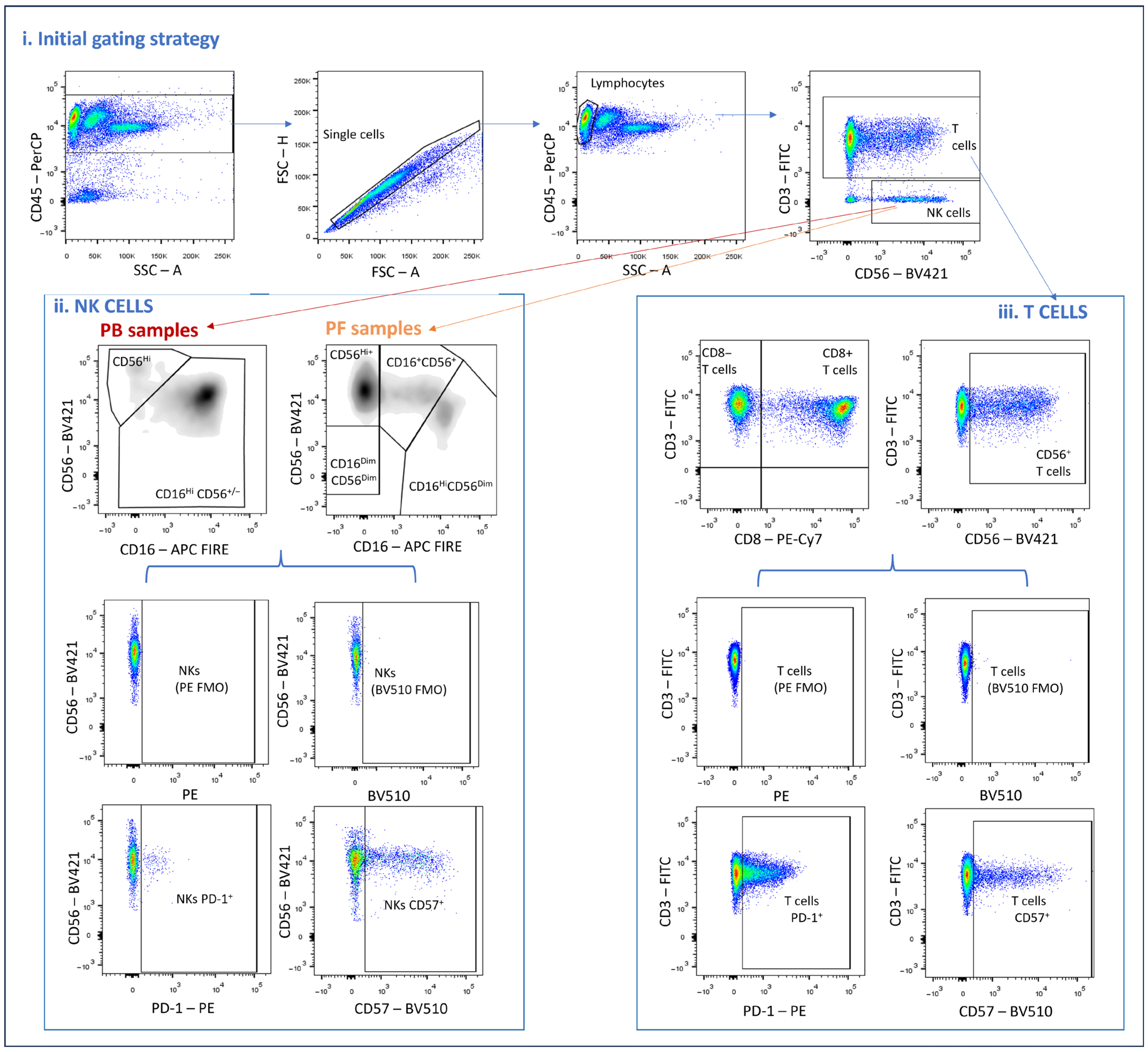
Figure 1. Gating strategy used to characterize PD-1 in lymphocyte subsets of peripheral blood and peritoneal fluid samples. (i) Gating Strategy used for the identification of major lymphocyte subsets in Peritoneal Fluid (PF) and Peripheral blood (PB) samples, i.e., T cells (CD45Hi SSCLo CD3+) and NK cells (CD45Hi SSCLo CD3 CD56+), within the single cell region; (ii) Gating strategy used for the identification of distinct NK cell subsets in PF (CD56Hi; CD16+ CD56+; CD16HiCD56Dim) and PB CD56Hi; CD16HiCD56Dim) samples, along with the subsequent analysis of PD-1 and CD57 in these populations (FMO tubes presented in the upper row); (iii) Gating strategy used for the identification of CD8+ and CD8 (CD4+ and DN) T cells, along with CD56+ T cells. The percentage of cells expressing PD-1 and CD57 was further analyzed in each one of these subsets. (FMO tubes presented in the upper row). DN, Double negative; FMO, Fluorescence Minus One.
Immuno 05 00049 g001
Figure 2. PD-1 expressing T cells in PF and PB samples between EM and controls. (ad) % of PD-1+ cells in (a) total T cells, (b) CD8 (CD4+ and DN) T cells, (c) CD8+ T cells, and (d) CD56+ T cells. Differences were tested with paired-samples t-test (a,c) and Wilcoxon rank-sum test (b,d). Scatter plots are presented with individual values and the lines represent the median with IQR. The control group consists of 15 peripheral blood and peritoneal fluid samples, and the EM group, 47 peripheral blood and peritoneal fluid samples. ** p < 0.01; *** p < 0.001. DN, Double negative; EM, endometriosis; IQR, interquartile range; PB, peripheral blood; PF, peritoneal fluid.
Immuno 05 00049 g002
Figure 3. PD-1 expression in peritoneal cells according to weight subgroups and smoking habits. (a) % of PD-1+ CD56+ CD16+ NK cells in controls and women with endometriosis; (b,c) % of PD-1+ (b) CD8+ T cells and (c) total T cells in controls and EM according to smoking habits; and (df) % of PD-1+ (d) total T cells, (e) CD8 (CD4+ and DN) T cells, and (f) CD8+ T cells in controls and EM according to weight. Differences were tested with the Wilcoxon rank-sum test (a), ANOVA with post-hoc Tukey HSD test for paired-comparison (b,f), and Kruskal–Wallis with post-hoc Dunn’s test for paired-comparison (ce). Test. The control group considers 15 peripheral blood and peritoneal fluid samples, and the EM group, 47 peripheral blood and peritoneal fluid samples. Considering the EM subgroups, the EM smoke subgroup had n = 8; considering the BMI, the normal BMI subgroup had n = 27, the overweight n = 11, and the obese n = 9). Scatter plots are presented with individual values and the lines represent the median with IQR. * p-value < 0.05; *** p-value < 0.001. DN, Double negative; EM, endometriosis; IQR, interquartile range; OW, overweight; PF, peritoneal fluid.
Immuno 05 00049 g003
Figure 4. PD-1 expression in peritoneal cells according to fertility status. (ac) Percentages of circulating PD-1+ CD8 (CD4+ and DN) T cells (a) in controls and EM women according to pregnancy history, and PD-1+ CD56+ T cells and PD-1+ total T cells (b,c) in controls and EM women with and without fertility issues. (d,e) % of PD-1+ total NK cells (d), PD-1+ CD56+ CD16+ NK cells (e), and PD-1+ CD56Hi NK cells (f) in controls and EM women according to pregnancy history. Non-Pregnant controls n = 8, Pregnant controls, n = 7; non-Pregnant EM, n = 28; Pregnant EM, n = 19. Controls without fertility issues, n = 14; EM without fertility issues, n = 24; EM with fertility issues, n = 23. All differences were tested with Kruskal–Wallis with post-hoc Dunn’s test for paired-comparison. Scatter plots are presented with individual values and the lines represent the median with IQR *** p-value < 0.001. DN, Double negative; EM, endometriosis; IQR, interquartile range; PF, peritoneal fluid.
Immuno 05 00049 g004
Table 1. Demographic and clinical characteristics of the control group and women with endometriosis.
CharacteristicsControl
(n = 15)
Endometriosis
(n = 47)
p-Value
Age, mean ± SD (min–max), yr38.5 ± 9.20 (20–55)36.2 ± 6.3 (31–42)0.434 a
Race, n (%) 0.113 b
White15 (100)40 (85.1)
Black0 (0)7 (14.9)
BMI, mean ± SD (min–max), kg/m223.2 ± 3.0 (17–28)25.2 ± 5.6 (20.8–28.0)0.430 a
Fertile, n (%) 0.003 b
Yes14 (93.3)24 (51.1)
No1 (6.7)23 (48.9)
Any Pregnancy, n (%) 0.087 b
Yes7 (46.7)19 (40.4)
No8 (53.3)28 (59.6)
Smoker, n (%) 0.087 b
Yes0 (0)8 (17.0)
No15 (100)39 (83.0)
Hormonal therapy, n (%) 0.090 b
Yes5 (33.3)31 (66.0)
No10 (66.7)16 (34.0)
BMI, body mass index; max, maximum; min, minimum; n, number of patients; SD, standard deviation; yr, years. a Wilcoxon rank-sum test. b Chi-squared test.
Table 2. Endometriosis characteristics of the included patients.
CharacteristicsEndometriosis
(n = 47)
Endometriosis diagnosis, mean ± SD (min–max), yr2.8 ± 2.5 (1–14)
Endometriosis severity, n (%)
Stage I/II10 (21.3)
Stage III/IV37 (78.7)
Dysmenorrhea complaints, n (%)44 (93.6)
Dysmenorrhea severity, n (%)
None/Mild/Moderate (<7/10)11 (23.4)
Severe (≥7/10)36 (76.6)
Dyspareunia complaints, n (%)28 (59.6)
Chronic pelvic pain, n (%)19 (40.4)
Dyschesia complaints, n (%)16 (34.0)
Bowel symptoms, n (%)15 (31.9)
Urinary tract symptoms, n (%)5 (10.6)
Rectorrhagia. n (%)4 (8.5)
Extra-pelvic endometriosis, n (%)2 (4.3)
max, maximum; min, minimum; n, number of patients; SD, standard deviation; yr, years.
Disclaimer/Publisher’s Note: The statements, opinions and data contained in all publications are solely those of the individual author(s) and contributor(s) and not of MDPI and/or the editor(s). MDPI and/or the editor(s) disclaim responsibility for any injury to people or property resulting from any ideas, methods, instructions or products referred to in the content.