Review

A Scoping Review of Clinical, Genetic, and Mechanistic Evidence Linking IL-6/IL-6R Signaling and Type 1 Diabetes Mellitus

1
Department of Community Care, Unnan City Hospital, Unnan 699-1221, Shimane, Japan
2
Department of Community Medicine Management, Faculty of Medicine, Shimane University, Izumo 693-8501, Shimane, Japan
3
Department of Rheumatology, Faculty of Medicine, Shimane University, Izumo 693-8501, Shimane, Japan
*
Author to whom correspondence should be addressed.
Immuno 2025, 5(3), 41; https://doi.org/10.3390/immuno5030041
Submission received: 24 July 2025 / Revised: 29 August 2025 / Accepted: 18 September 2025 / Published: 19 September 2025

Abstract

Type 1 diabetes mellitus (T1DM) is an autoimmune disease characterized by immune-mediated β-cell destruction, where interleukin-6 (IL-6) signaling plays a complex and context-dependent role. Tocilizumab, an IL-6 receptor (IL-6R) inhibitor, is effective in several autoimmune conditions, but its influence on the onset and progression of T1DM remains uncertain. This scoping review aimed to map current clinical, genetic, and mechanistic evidence linking IL-6/IL-6R signaling to T1DM risk and to identify key research gaps. Following PRISMA-ScR guidelines, PubMed, Embase, and Web of Science were searched for studies from 2005 to 2025 reporting associations between tocilizumab or IL-6R modulation and T1DM onset. Six studies were included: one case report describing T1DM onset during tocilizumab therapy in a genetically predisposed patient, one randomized controlled trial showing no significant β-cell preservation with tocilizumab, three Mendelian randomization analyses with conflicting findings on IL-6R signaling, and one mechanistic study showing enhanced IL-6 responsiveness in early-stage T1DM. Collectively, evidence remains fragmented and inconclusive, highlighting research gaps in the differential roles of IL-6 classic versus trans-signaling and the impact of genetic predisposition. Future prospective studies should clarify whether selective IL-6 trans-signaling blockade may offer safer, targeted strategies for modulating autoimmune β-cell destruction.

1. Introduction

Type 1 diabetes mellitus (T1DM) is an autoimmune disorder characterized by the destruction of insulin-producing pancreatic β-cells, leading to lifelong dependence on exogenous insulin [1]. Despite advancements in insulin therapy and glucose monitoring technologies, disease onset and progression remain significant clinical challenges, emphasizing the urgent need for preventive strategies [2]. Dysregulated immune responses play a central role in T1DM pathogenesis, with multiple cytokine networks implicated in the inflammatory milieu surrounding pancreatic islets [3]. Among these, interleukin-6 (IL-6) is a pleiotropic mediator that regulates both innate and adaptive immune responses [4]. It promotes the differentiation of pathogenic Th17 cells while inhibiting regulatory T cell (Treg) function, processes critically involved in autoimmune disease mechanisms [5].
Elevated IL-6 levels are observed in autoimmune conditions such as rheumatoid arthritis, systemic lupus erythematosus, and multiple sclerosis, and IL-6 receptor (IL-6R) blockade has transformed the treatment of several inflammatory diseases [6]. Tocilizumab, a monoclonal antibody targeting IL-6R, inhibits both membrane-bound and soluble receptor-mediated signaling [7] and has been proposed as a potential therapy to prevent autoimmune β-cell destruction [8].
However, paradoxical effects have been reported, with some case studies suggesting the emergence of autoimmune endocrinopathies, including T1DM, during IL-6R-targeted therapy. Furthermore, Mendelian randomization (MR) analyses have yielded conflicting findings: some indicate that higher IL6R expression increases T1DM susceptibility, whereas others suggest IL-6R blockade may confer protection [9,10]. Mechanistic studies also demonstrate that T cells from early-stage T1DM patients exhibit enhanced IL-6 responsiveness, implying a context- and phase-dependent role of IL-6 signaling [10]. Previous reviews have either focused broadly on IL-6 in autoimmunity [11,12] or summarized experimental findings without integrating translational data such as MR analyses and therapeutic IL-6R blockade [13,14,15,16].
To date, no review has comprehensively synthesized clinical observations, randomized trials, genetic analyses, and mechanistic immunology to clarify the relationship between IL-6/IL-6R signaling and T1DM. Therefore, this scoping review aims to map the existing clinical, genetic, and mechanistic evidence linking IL-6/IL-6R signaling to T1DM, highlight controversial and diverging findings, and identify critical knowledge gaps that could inform future translational strategies, including selective IL-6 trans-signaling inhibition.

2. Materials and Methods

2.1. Protocol and Registration

This scoping review was conducted by the Preferred Reporting Items for Systematic Reviews and Meta-Analyses Extension for Scoping Reviews (PRISMA-ScR) guidelines. The review protocol was prospectively registered with the International Prospective Register of Systematic Reviews (PROSPERO; Registration Number: CRD420251029644) before the commencement of the review.

2.2. Eligibility Criteria

Studies were eligible if they met the following criteria:
  • Population: Patients of any age receiving tocilizumab for any indication, or genetic/biological studies evaluating IL-6/IL-6R signaling in the context of T1DM.
  • Exposure/Intervention: Administration of tocilizumab or genetically proxied IL-6R blockade.
  • Outcomes: New-onset T1DM diagnosed by clinical criteria (e.g., hyperglycemia, diabetic ketoacidosis, islet autoantibodies) or β-cell functional decline.
  • Study Design: Case reports, case series, randomized controlled trials (RCTs), Mendelian randomization (MR) studies, and mechanistic studies published as full-text articles.
  • Language and Time Frame: Publications in English from 1 January 2005 to 10 April 2025.
Studies focusing exclusively on type 2 diabetes, steroid-induced hyperglycemia, or animal models were excluded.

2.3. Information Sources and Search Strategy

A comprehensive literature search was conducted across PubMed, Embase (via Ovid), and Web of Science (SCI/SSCI). The search strategy combined controlled vocabulary and free-text terms related to “tocilizumab,” “interleukin-6 receptor,” “IL-6 signaling,” “type 1 diabetes,” “autoimmune diabetes,” and “Mendelian randomization.” Reference lists of relevant reviews and included articles were manually screened to identify additional eligible studies.

2.4. Study Selection

Two reviewers independently screened titles and abstracts to identify potentially eligible studies. Full texts of relevant articles were retrieved and assessed for inclusion according to the predefined eligibility criteria. Any discrepancies were resolved by discussion or consultation with a third reviewer.

2.5. Data Charting and Extraction

Data were extracted independently and in duplicate using a standardized data collection form. Extracted variables included:
  • Study characteristics (authors, publication year, study design)
  • Population demographics (age, sex, underlying conditions)
  • Tocilizumab dosage, duration, and indication
  • Diagnostic criteria and timing for T1DM onset
  • Genetic or molecular findings related to IL-6/IL-6R signaling
  • Key outcomes and mechanistic observations

2.6. Synthesis of Results

Given the heterogeneity of study designs and outcomes, no meta-analysis was performed. Instead, a narrative synthesis was conducted to map the breadth of evidence and highlight research gaps. Findings were structured across three domains: clinical, genetic, and mechanistic evidence linking IL-6/IL-6R signaling to type 1 diabetes mellitus (T1DM).

2.7. Risk of Bias and Critical Appraisal

Formal risk of bias assessment was not conducted due to the limited number and diverse nature of included studies. Instead, the methodological characteristics and potential limitations of each study were described narratively to provide context for interpreting the findings.

2.8. Ethics and Data Availability

No new experimental data involving humans or animals were generated; therefore, ethical approval was not required. All search strategies and extracted data supporting this review are available from the corresponding author upon reasonable request.

2.9. Generative AI Use

Generative artificial intelligence was used solely for superficial text editing and formatting. No AI tools were employed for study selection, data extraction, synthesis, or interpretation.

3. Results

3.1. Study Selection

A total of 1684 studies were identified through electronic database searches (Web of Science: 887; PubMed: 434; Embase: 363). After removing 495 duplicate records (492 by Covidence and three manually), 1189 records remained for title and abstract screening. During screening, 1177 records were excluded based on irrelevance to the review topic. Subsequently, nine full-text articles were assessed for eligibility. Among these, three articles were excluded: one due to wrong intervention (non-tocilizumab agent), and two due to inappropriate patient population (non-autoimmune context). Finally, six studies were included in the qualitative synthesis: one case report, one randomized controlled trial, three MR studies, and one mechanistic immunology study (Figure 1).

3.2. Study Characteristics

The six included studies comprised diverse designs:
  • Case report (n = 1): A single patient developing T1DM during tocilizumab therapy.
  • Randomized controlled trial (n = 1): A multicenter trial evaluating tocilizumab for β-cell preservation in new-onset T1DM.
  • Mendelian randomization studies (n = 3): Genetic analyses assessing the impact of IL-6R blockade or expression on T1DM risk.
  • Mechanistic study (n = 1): Immunological investigation of IL-6 responsiveness in T1DM patients.
Study settings included populations with autoimmune diseases receiving tocilizumab, and genomic cohorts predominantly of European ancestry for MR studies. T1DM diagnosis in clinical studies was based on hyperglycemia, islet autoantibody positivity, and ketoacidosis criteria (Table 1).

3.3. Risk of Bias Within Studies

The case report was assessed as moderate risk of bias, due to the inherent limitations of single-patient observations and lack of confirmatory causality testing. The randomized controlled trial demonstrated low risk of bias, with appropriate randomization, blinding, and outcome assessment processes. MR studies were generally of moderate quality, with proper genetic instrument selection but limitations in entirely excluding horizontal pleiotropy. The mechanistic study was assessed as moderate to low risk, providing strong biological plausibility but limited by cross-sectional design and absence of longitudinal follow-up.

3.4. Results of Individual Studies and Synthesis

The included studies were analyzed in two thematic domains as follows: (1) clinical association between tocilizumab therapy and the onset of T1DM; and (2) genetic and mechanistic evidence linking IL-6 receptor (IL-6R) signaling with T1DM pathogenesis.

3.4.1. Clinical Association Between Tocilizumab Therapy and the Onset of T1DM

  • Case Report
Kawasaki et al. presented a 73-year-old Japanese woman with rheumatoid arthritis who developed autoimmune T1DM after 17 months of subcutaneous tocilizumab therapy (162 mg every two weeks) [17]. Before treatment initiation, her rheumatoid arthritis had been resistant to conventional disease-modifying agents, necessitating the use of IL-6R blockade. Tocilizumab achieved complete remission of arthritis and allowed cessation of corticosteroids.
However, the patient subsequently developed symptoms of hyperglycemia, including polydipsia, polyuria, and significant weight loss (2 kg/month). She presented with diabetic ketoacidosis: arterial pH of 7.26, blood glucose of 925 mg/dL, ketonemia (β-hydroxybutyrate 6154 μmol/L), and HbA1c of 13.2%. Autoimmune workup confirmed high titers of anti-glutamic acid decarboxylase antibody (anti-GAD, 241 U/mL), anti-IA-2 antibody (>30 U/mL), and anti-ZnT8 antibody (80.2 U/mL). Genetic testing revealed HLA-DR9-DQ3 homozygosity, a known high-risk haplotype for T1DM in Japanese populations. This case represents a signal-generating observation of T1DM onset during tocilizumab therapy in a genetically predisposed individual. However, as a single anecdotal report, it cannot establish causality or support generalizable conclusions.

3.4.2. Randomized Controlled Trial

Conversely, the EXTEND trial led by Greenbaum et al. provided systematic data through a multicenter, double-blind, randomized controlled design (ClinicalTrials.gov identifier: NCT02293837) [9]. The study enrolled newly diagnosed T1DM patients (within 100 days of diagnosis), primarily pediatric (ages 6–17 years), randomizing them 2:1 to receive seven monthly intravenous doses of tocilizumab (n = 54) or placebo (n = 27). The primary endpoint was the change in 2 h stimulated C-peptide area under the curve (AUC) during a mixed meal tolerance test (MMTT) at week 52. There was no significant difference between the tocilizumab and placebo groups in preservation of endogenous insulin secretion. Tocilizumab suppressed IL-6R signaling in T cells, evidenced by reduced downstream phosphorylation of STAT3, but failed to modify key immune cell populations, including CD4 memory subsets, Th17 cells, or Treg suppressive function. Tocilizumab was well tolerated, with no significant increase in serious adverse events compared to placebo. These findings suggest that while IL-6R blockade successfully reduced IL-6 signaling at the molecular level, it did not alter the clinical progression of β-cell loss or autoimmune activity in new-onset T1DM.

3.4.3. Genetic and Mechanistic Evidence Linking IL-6R Signaling to T1DM Risk

Three MR studies provided insights into the causal role of IL-6R in T1DM susceptibility. Utilizing drug-target MR based on single-nucleotide polymorphisms (SNPs) near the IL6R gene associated with C-reactive protein (CRP) levels, Fu et al. simulated pharmacologic IL-6R inhibition [18]. Their analysis included T1DM among multiple diseases but found no statistically significant association between genetically proxied IL-6R blockade and T1DM risk (p > 0.05). Significant protective effects were observed for idiopathic pulmonary fibrosis (OR 0.278, p < 0.001) and Parkinson’s disease (OR 0.354, p < 0.001), but not for autoimmune diabetes.
In contrast, Heikkilä et al. performed an MR study integrating GWAS summary statistics for T1DM, whole-blood gene expression, and serum protein levels, focusing on several cytokine pathways [15]. Their findings demonstrated that increased IL6R expression was associated with a significantly higher risk of T1DM (OR 1.98, 95% CI 1.48–2.65). Co-localization analysis revealed a 96.5% posterior probability that the same causal variant influenced both IL6R expression and T1DM risk. Thus, their results support the hypothesis that enhanced IL-6R signaling contributes to T1DM pathogenesis.
Li et al. used CRP levels as a surrogate for IL-6 activity and conducted a large-scale MR analysis involving more than 575,000 individuals [16]. Their key findings are that genetically proxied IL-6R blockade was associated with a 59% reduction in T1DM risk (OR 0.410, p = 1.78 × 10⁻⁷), similar protective effects were seen for ankylosing spondylitis and Crohn’s disease, and that IL-6R blockade increased the risk for asthma (OR 1.031, p = 2.15 × 10⁻¹²) and eczema (OR 1.066, p = 5.92 × 10⁻²²).
Collectively, while Fu et al. observed no association, Heikkilä et al. and Li et al. presented opposing directions: higher IL6R expression increasing T1DM risk, while IL-6R inhibition decreasing it.

3.4.4. Mechanistic Immunology Study

Hundhausen et al. investigated IL-6 signaling at the cellular level in peripheral blood mononuclear cells (PBMCs) from T1DM patients compared to healthy controls [10]. Key findings are as follows:
  • IL-6-induced phosphorylation of STAT3 and STAT1 was significantly higher in CD4+ and CD8+ T cells from T1DM patients.
  • This enhanced signaling was specific to IL-6 stimulation (not observed with IL-10 or IL-27).
  • Increased IL-6 responsiveness correlated with elevated surface expression of IL-6R on T cells.
  • Expression of the IL-6R sheddase ADAM17 was reduced in T1DM patients, contributing to the accumulation of surface IL-6R.
The degree of IL-6 hyperresponsiveness negatively correlated with disease duration, suggesting a critical role of IL-6 signaling early in disease evolution.

4. Discussion

4.1. Summary of Main Findings

To provide a unifying framework, we conceptualized the synthesis of heterogeneous evidence streams as a form of triangulation. Clinical case reports and randomized controlled trials provide observational and interventional signals, respectively. Mendelian randomization studies contribute to genetic causal inference, and mechanistic immunology research offers cellular-level explanations. Together, these complementary approaches strengthen causal inference by addressing different dimensions of the same research question. This framework justifies the inclusion of diverse study types in our review and highlights how they collectively inform the role of IL-6/IL-6R signaling in the pathogenesis of T1DM. A conceptual figure (Figure 2) has been added to illustrate this triangulation process.
In this systematic review, we synthesized evidence from six studies addressing the relationship between tocilizumab use, IL-6R signaling, and the risk of developing T1DM. Our findings reveal a complex and nuanced picture. Kawasaki et al. presented a 73-year-old Japanese woman with rheumatoid arthritis who developed autoimmune T1DM after 17 months of tocilizumab therapy [17]. While this case provides a rare signal suggesting that IL-6R blockade could, under specific genetic predispositions, coincide with autoimmune diabetes onset, it should not be regarded as primary evidence of causality. Instead, it highlights a potential safety signal and underscores the need for caution and closer surveillance in susceptible individuals. Accordingly, this case is best interpreted as a cautionary observation, consistent with the limitations of anecdotal evidence, rather than as confirmatory proof of the role of IL-6R signaling in T1DM.
MR studies provided valuable but divergent insights into the causal role of IL-6R in the susceptibility to T1DM. Heikkilä et al. reported that genetically increased IL6R expression was associated with a higher risk of T1DM [15]. In contrast, Li et al. found that genetically proxied IL-6R blockade reduced the risk of T1DM [16]. Importantly, the relationship between IL-6R genetic proxies and pharmacologic inhibition should be interpreted with caution. Genetic variants such as cis-eQTLs near IL6R often influence soluble receptor levels or expression balance, whereas pharmacologic blockade with tocilizumab directly inhibits receptor function [15]. These processes are related but not strictly inverse. Consequently, Mendelian randomization findings may not perfectly mimic the clinical effects of IL-6R antagonists, and differences between lifelong genetic modulation and short-term therapeutic intervention must be carefully considered when interpreting these results [15,16].
Several factors may explain these discrepancies. First, the two studies used different instrumental variables (IVs) for IL-6 signaling. Heikkilä et al. selected cis-eQTLs near the IL6R gene that directly influence receptor expression levels, while Li et al. relied on SNPs associated with circulating CRP as a downstream biomarker of IL-6 activity [19]. Variability in IV strength and specificity can affect causal inference, particularly when the instruments capture different aspects of IL-6 biology. Second, linkage disequilibrium (LD) patterns may differ across the GWAS datasets used, leading to residual confounding from nearby immune-related loci [20]. Third, horizontal pleiotropy—where genetic variants influence multiple immune pathways beyond IL-6 signaling—cannot be entirely excluded, which may bias MR estimates. Additionally, both MR studies were conducted predominantly in European ancestry cohorts, limiting generalizability to other populations with distinct genetic architectures.
Beyond methodological differences, biological heterogeneity in IL-6 signaling may also contribute to the opposing directions of effect. IL-6 can signal via two distinct pathways: classic signaling, which occurs through membrane-bound IL-6R on hepatocytes and select immune cells and is thought to mediate protective and regenerative effects, and trans-signaling, which involves soluble IL-6R and gp130, driving chronic inflammation and autoimmune pathology [21,22]. SNPs influencing soluble IL-6R levels may preferentially reflect trans-signaling, while those affecting membrane-bound IL-6R expression may have different functional consequences. Thus, genetic proxies that selectively modulate trans-signaling may show a protective association, whereas those that enhance overall IL-6 responsiveness could increase the risk of T1DM.
Taken together, the divergent MR findings may reflect both methodological heterogeneity in instrument selection and the complex, context-dependent biology of IL-6 signaling, underscoring the need for refined MR approaches that distinguish between classic and trans-signaling effects. Future MR studies integrating multi-omic data (e.g., proteomic and eQTL information) and trans-ethnic cohorts could help disentangle these nuanced relationships.

4.2. Comparison with Previous Literature

Compared with previous reviews that focused primarily on cytokine dysregulation in autoimmunity or IL-6 inhibition in other inflammatory conditions [11,23], our synthesis provides an integrative framework that combines three distinct layers of evidence: (1) clinical observations from case reports and randomized trials [9,17], (2) genetic causal inference through MR studies [15,16], and (3) mechanistic immunology highlighting altered IL-6 responsiveness in early T1DM [10].
This multi-dimensional approach offers a more nuanced understanding of the complex, context-dependent role of IL-6 signaling. Specifically, it underscores that classic IL-6 signaling may have homeostatic functions, whereas trans-signaling predominantly mediates chronic pathogenic inflammation [4,24]. By bridging these mechanistic insights with the divergent MR findings, our review extends beyond descriptive synthesis to propose translational implications, including the potential for selective IL-6 trans-signaling inhibitors such as olamkicept as safer and more targeted therapeutic options in high-risk populations [25,26].
Our findings align with the growing literature on cytokine dysregulation in autoimmune disease development. Previous studies have consistently shown that IL-6 is elevated in autoimmune conditions such as rheumatoid arthritis, systemic lupus erythematosus, and multiple sclerosis, and that therapeutic blockade of IL-6R can ameliorate disease activity [23,24,27]. Experimental models, such as the non-obese diabetic (NOD) mouse, have demonstrated that IL-6 overexpression accelerates insulitis and diabetes onset, while IL-6 inhibition confers protection against autoimmune β-cell destruction [28]. However, the translation of these experimental observations into clinical practice remains complex. Although IL-6 inhibition has proven beneficial in diseases like rheumatoid arthritis, rare reports of new-onset autoimmune conditions, including T1DM, under IL-6R blockade have emerged [9,29]. In parallel, genetic studies have suggested that impaired IL-6R signaling may reduce the risk of developing certain autoimmune diseases, yet conflicting results regarding T1DM have persisted [11,14]. Thus, our review reflects a broader pattern seen in the literature: IL-6R signaling is critically involved in autoimmunity, but its modulation may have context-dependent, stage-specific, and genetically influenced effects that complicate simple clinical extrapolation.
In addition to IL-6, other cytokines and damage-associated molecular patterns (DAMPs) such as TNF and HMGB1 also contribute to the pathogenesis of T1DM [30]. TNF has been shown to promote β-cell apoptosis via NF-κB–dependent and caspase-mediated mechanisms, whereas HMGB1 functions as a DAMP that amplifies innate immune activation and islet inflammation [31]. Importantly, preclinical studies in non-obese diabetic (NOD) mice indicate that low-dose exposure to IL-6 or TNF may paradoxically exert protective effects, delaying diabetes onset by enhancing regulatory T cell activity and modulating β-cell resilience [32]. These findings emphasize that cytokine effects are dose-dependent and context-specific, underscoring the complex interplay between multiple inflammatory mediators in the pathogenesis of T1DM [33]. Furthermore, IL-6 has actions beyond its classical immune-modulating effects. Recent studies suggest its involvement in the cGAS-STING pathway, a cytosolic DNA-sensing mechanism that links innate immunity and metabolic stress [32,33]. Cross-talk between IL-6 and STING signaling may amplify inflammatory cascades and contribute to β-cell dysfunction in early T1DM, broadening the mechanistic framework beyond adaptive immune responses [32,33].
Taken together, these different strands of evidence can be conceptually linked. Mechanistic studies suggest that IL-6 signaling modulates β-cell susceptibility to apoptosis and may enhance their resilience under inflammatory stress. This aligns with the C-peptide preservation observed in the RCT, where IL-6R blockade appeared to delay functional decline of residual β-cells. Furthermore, MR data provide supportive evidence that genetic modulation of IL6R impacts diabetes risk, offering a causal framework that bridges molecular and clinical findings. By integrating mechanistic plausibility, genetic inference, and clinical outcomes, a more coherent picture emerges in which IL-6/IL-6R signaling plays a multifaceted role in shaping both autoimmune responses and β-cell survival in T1DM.

4.3. Clinical Implications

The clinical implications of this review are twofold. First, our findings suggest that the routine use of tocilizumab does not necessitate additional concern for inducing T1DM in the general patient population, as robust clinical evidence for causality is lacking. Second, caution may be warranted in individuals with a strong genetic predisposition to T1DM or pre-existing islet autoimmunity, particularly given the biological plausibility that perturbations in IL-6 signaling could influence β-cell autoimmunity in susceptible hosts [12,22]. In particular, the mechanistic evidence indicating early IL-6 pathway hyperactivation raises the possibility that IL-6R-targeted therapies could have different effects depending on the phase of autoimmunity [22]. Early intervention strategies may need to consider the immunologic status of the individual to optimize outcomes and avoid unintended immune perturbation.
Moreover, mechanistic studies have demonstrated that T cells in early-stage T1DM exhibit enhanced IL-6 responsiveness due to increased IL-6R surface expression and reduced ADAM17-mediated receptor shedding, resulting in sustained STAT3 activation [10]. These findings suggest that hyperactivation of the IL-6 pathway is an early pathogenic event that may amplify autoreactive T cell responses. From a translational perspective, this implies that indiscriminate blockade of IL-6R might not uniformly benefit all patients, as classic IL-6 signaling also mediates protective and regenerative functions, particularly in hepatocytes and specific immune compartments [21,22]. Instead, selectively inhibiting the pro-inflammatory IL-6 trans-signaling pathway while sparing classic signaling could represent a more refined therapeutic approach.
Olamkicept (sgp130Fc), a soluble gp130-Fc fusion protein that neutralizes explicitly IL-6 trans-signaling without affecting homeostatic classic signaling, has shown favorable safety profiles and clinical efficacy in inflammatory diseases [25,26]. Applying this strategy to T1DM may theoretically mitigate pathogenic T cell activation while preserving the beneficial functions of IL-6 in tissue repair and metabolic regulation. Future clinical trials investigating trans-signaling-selective inhibitors in genetically predisposed or islet autoantibody-positive individuals could clarify whether targeted modulation of IL-6 signaling can delay or prevent autoimmune β-cell destruction.
Conversely, genetic data suggest that IL-6R blockade may reduce the risk of T1DM, raising the hypothesis that selective modulation of IL-6 signaling could hold preventive potential, although this remains unproven in clinical trials [25,26].

4.4. Limitations

Several limitations must be acknowledged when interpreting the findings of this review. Firstly, the small number of available studies, combined with their methodological heterogeneity, limits the generalizability of conclusions. Secondly, the reliance on case reports and MR studies introduces inherent biases: case reports are prone to selective reporting, while MR analyses are susceptible to violations of instrumental variable assumptions, such as horizontal pleiotropy. Thirdly, the genetic studies predominantly involved individuals of European ancestry, restricting applicability to more diverse global populations. Additionally, mechanistic investigations, although biologically informative, were largely cross-sectional, limiting causal inference regarding IL-6 pathway dysregulation and disease onset. Finally, the absence of large-scale prospective cohorts assessing T1DM incidence in tocilizumab-treated individuals highlights an important evidence gap that future research must address.

4.5. Future Directions

Given the current state of evidence, several avenues for future research are warranted. Prospective observational studies involving larger and more diverse patient cohorts treated with IL-6R inhibitors should be undertaken to assess the true incidence of T1DM and other autoimmune phenomena. In genetically predisposed individuals—such as those with high-risk HLA haplotypes or multiple islet autoantibodies—longitudinal monitoring with serial islet autoantibody testing and β-cell function assessment should be incorporated to enable early detection of autoimmune activation during IL-6R-targeted therapy.
Parallel efforts should focus on integrating genetic risk stratification tools, such as HLA typing and polygenic risk scores, into clinical decision-making when considering IL-6R-targeted therapies. Moreover, randomized controlled trials investigating the efficacy of IL-6R blockade in individuals at high risk of T1DM, such as those with multiple islet autoantibodies but without hyperglycemia, could provide definitive evidence on preventive strategies. Finally, mechanistic studies should continue to dissect the temporal dynamics of IL-6 signaling abnormalities during T1DM evolution, exploring whether selective inhibition of pathogenic trans-signaling pathways—while preserving protective classical IL-6 signaling—may offer a safer and more effective therapeutic approach.
Finally, mechanistic studies should continue to dissect the temporal dynamics of IL-6 signaling abnormalities during the evolution of T1DM and clarify the differential roles of classic versus trans-signaling. Selective inhibition of pro-inflammatory trans-signaling pathways—using agents such as olamkicept—while preserving protective classical IL-6 signaling warrants evaluation as a potentially safer and more targeted therapeutic approach in high-risk populations.

5. Conclusions

This systematic review underscores the complex and context-dependent role of IL-6/IL-6R signaling in the development of type 1 diabetes mellitus. While clinical evidence directly linking tocilizumab use to the onset of T1DM remains limited and inconclusive, genetic and mechanistic studies support a contributory role of the IL-6 pathway in autoimmune β-cell destruction. Therefore, current findings should be interpreted with caution, particularly in individuals with strong genetic susceptibility—such as those carrying high-risk HLA haplotypes or multiple islet autoantibodies—who may warrant closer monitoring during IL-6R-targeted therapy to detect early signs of β-cell autoimmunity. Therapeutic manipulation of IL-6R signaling may yet offer opportunities for the prevention or intervention of T1DM. However, broadly inhibiting IL-6R may not uniformly benefit all patients, given the dual roles of classic and trans-signaling. Future research should evaluate whether selective IL-6 trans-signaling inhibitors, such as olamkicept, can provide safer and more targeted modulation of autoimmunity. Prospective trials incorporating islet autoantibody screening and β-cell function monitoring in high-risk populations will be critical for translating these mechanistic insights into safe and effective clinical applications.
While mechanistic insights and early data from other autoimmune conditions suggest that selective IL-6 trans-signaling inhibition (e.g., olamkicept) could represent a promising avenue, this remains speculative in the context of T1DM. No clinical evidence exists in humans with diabetes. Therefore, these approaches should be viewed as hypothesis-generating directions for future research rather than concrete therapeutic recommendations.

Author Contributions

Conceptualization, R.O. and T.F.; methodology, R.O.; validation, R.O., C.S. and K.I.; formal analysis, T.F.; investigation, R.O. and T.F.; resources, C.S.; data curation, T.F. and K.I.; writing—original draft preparation, R.O.; writing—review and editing, R.O., T.F., C.S. and K.I.; visualization, T.F.; supervision, C.S.; project administration, R.O.; funding acquisition, C.S. All authors have read and agreed to the published version of the manuscript.

Funding

This research received no external funding.

Institutional Review Board Statement

Not applicable.

Informed Consent Statement

Not applicable.

Data Availability Statement

No new data were created or analyzed in this study. Data sharing is not applicable to this article as it is based on previously published studies that are publicly available and properly cited in the reference list. The full search strategies and extracted datasets used in this review are available from the corresponding author upon reasonable request.

Acknowledgments

The authors would like to thank the administrative staff of Unnan City Hospital for their support in the literature access and reference management. We also acknowledge the technical assistance provided by the Department of Rheumatology, Shimane University Faculty of Medicine, in facilitating database access and search optimization.

Conflicts of Interest

The authors declare no conflicts of interest.

Abbreviations

The following abbreviations are used in this manuscript:
T1DMType 1 diabetes mellitus
IL-6Interleukin-6
IL-6RInterleukin-6 receptor
TCZTocilizumab
MRMendelian randomization
RCTRandomized controlled trial
PRISMA-ScRPreferred Reporting Items for Systematic Reviews and Meta-Analyses Extension for Scoping Reviews
HLAHuman leukocyte antigen
ADAM17A disintegrin and metalloprotease 17

References

  1. Popoviciu, M.S.; Kaka, N.; Sethi, Y.; Patel, N.; Chopra, H.; Cavalu, S. Type 1 Diabetes Mellitus and Autoimmune Diseases: A Critical Review of the Association and the Application of Personalized Medicine. J. Pers. Med. 2023, 13, 422. [Google Scholar] [CrossRef]
  2. Sugandh, F.N.; Chandio, M.; Raveena, F.N.; Kumar, L.; Karishma, F.N.; Khuwaja, S.; Memon, U.A.; Bai, K.; Kashif, M.; Varrassi, G.; et al. Advances in the Management of Diabetes Mellitus: A Focus on Personalized Medicine. Cureus 2023, 15, e43697. [Google Scholar] [CrossRef] [PubMed]
  3. Lu, J.; Liu, J.; Li, L.; Lan, Y.; Liang, Y. Cytokines in Type 1 Diabetes: Mechanisms of Action and Immunotherapeutic Targets. Clin. Transl. Immunol. 2020, 9, e1122. [Google Scholar] [CrossRef]
  4. McElvaney, O.J.; Curley, G.F.; Rose-John, S.; McElvaney, N.G. Interleukin-6: Obstacles to Targeting a Complex Cytokine in Critical Illness. Lancet Respir. Med. 2021, 9, 643–654. [Google Scholar] [CrossRef]
  5. Schnell, A.; Littman, D.R.; Kuchroo, V.K. TH17 Cell Heterogeneity and Its Role in Tissue Inflammation. Nat. Immunol. 2023, 24, 19–29. [Google Scholar] [CrossRef]
  6. Grebenciucova, E.; VanHaerents, S. Interleukin 6: At the Interface of Human Health and Disease. Front. Immunol. 2023, 14, 1255533. [Google Scholar] [CrossRef] [PubMed]
  7. Mihara, M.; Ohsugi, Y.; Kishimoto, T. Tocilizumab, a Humanized Anti-Interleukin-6 Receptor Antibody, for Treatment of Rheumatoid Arthritis. Open Access Rheumatol. 2011, 3, 19–29. [Google Scholar] [CrossRef]
  8. Favalli, E.G. Understanding the Role of Interleukin-6 (IL-6) in the Joint and Beyond: A Comprehensive Review of IL-6 Inhibition for the Management of Rheumatoid Arthritis. Rheumatol. Ther. 2020, 7, 473–516. [Google Scholar] [CrossRef]
  9. Greenbaum, C.J.; Serti, E.; Lambert, K.; Weiner, L.J.; Kanaparthi, S.; Lord, S.; Gitelman, S.E.; Wilson, D.M.; Gaglia, J.L.; Griffin, K.J.; et al. IL-6 Receptor Blockade Does Not Slow Beta Cell Loss in New-Onset Type 1 Diabetes. JCI Insight 2021, 6, e150074. [Google Scholar] [CrossRef] [PubMed]
  10. Hundhausen, C.; Roth, A.; Whalen, E.; Chen, J.; Schneider, A.; Long, S.A.; Wei, S.; Rawlings, R.; Kinsman, M.; Evanko, S.P.; et al. Enhanced T Cell Responses to IL-6 in Type 1 Diabetes Are Associated with Early Clinical Disease and Increased IL-6 Receptor Expression. Sci. Transl. Med. 2016, 8, 356ra119. [Google Scholar] [CrossRef]
  11. Xiang, Y.; Zhang, M.; Jiang, D.; Su, Q.; Shi, J. The Role of Inflammation in Autoimmune Disease: A Therapeutic Target. Front. Immunol. 2023, 14, 1267091. [Google Scholar] [CrossRef]
  12. Jones, B.E.; Maerz, M.D.; Buckner, J.H. IL-6: A Cytokine at the Crossroads of Autoimmunity. Curr. Opin. Immunol. 2018, 55, 9–14. [Google Scholar] [CrossRef]
  13. Roy, S.; Pokharel, P.; Piganelli, J.D. Decoding the Immune Dance: Unraveling the Interplay between Beta Cells and Type 1 Diabetes. Mol. Metab. 2024, 88, 101998. [Google Scholar] [CrossRef] [PubMed]
  14. Heikkilä, T.E.; Kaiser, E.K.; Lin, J.; Gill, D.; Koskenniemi, J.J.; Karhunen, V. Genetic Evidence for Efficacy of Targeting IL-2, IL-6 and TYK2 Signalling in the Prevention of Type 1 Diabetes: A Mendelian Randomisation Study. Diabetologia 2024, 67, 2667–2677. [Google Scholar] [CrossRef]
  15. Li, J.; Liu, Y.; Xiao, Z.; Zang, C.; Li, P.; Xiao, B.; Zhou, L. Exploring the Therapeutic Potential of Interleukin-6 Receptor Blockade in Autoimmune Diseases Using Drug Target Mendelian Randomization. Immunogenetics 2025, 77, 3. [Google Scholar] [CrossRef] [PubMed]
  16. Kawasaki, E.; Fukuyama, T.; Uchida, A.; Sagara, Y.; Nakano, Y.; Tamai, H.; Tojikubo, M.; Hiromatsu, Y.; Koga, N. Development of Type 1 Diabetes in a Patient Treated with Anti-Interleukin-6 Receptor Antibody for Rheumatoid Arthritis. J. Diabetes Investig. 2022, 13, 738–740. [Google Scholar] [CrossRef]
  17. Fu, C.; Wang, L.; Cai, W. IL6 Receptor Inhibitors: Exploring the Therapeutic Potential Across Multiple Diseases through Drug Target Mendelian Randomization. Front. Immunol. 2024, 15, 1452849. [Google Scholar] [CrossRef]
  18. Burgess, S.; Thompson, S.G. Interpreting Findings from Mendelian Randomization Using the MR-Egger Method. Eur. J. Epidemiol. 2017, 32, 377–389. [Google Scholar] [CrossRef] [PubMed]
  19. Ferreira, R.C.; Freitag, D.F.; Cutler, A.J.; Howson, J.M.M.; Rainbow, D.B.; Smyth, D.J.; Kaptoge, S.; Clarke, P.; Boreham, C.; Coulson, R.M. Functional IL6R 358Ala Allele Impairs Classical IL-6 Receptor Signaling and Influences Risk of Diverse Inflammatory Diseases. PLoS Genet. 2013, 9, e1003444. [Google Scholar] [CrossRef]
  20. Rose-John, S. IL-6 Trans-Signaling via the Soluble IL-6 Receptor: Importance for the Pro-Inflammatory Activities of IL-6. Int. J. Biol. Sci. 2012, 8, 1237–1247. [Google Scholar] [CrossRef]
  21. Scheller, J.; Garbers, C.; Rose-John, S. Interleukin-6: From Basic Biology to Selective Blockade of Pro-Inflammatory Activities. Semin. Immunol. 2014, 26, 2–12. [Google Scholar] [CrossRef]
  22. Choy, E.H.; De Benedetti, F.; Takeuchi, T.; Hashizume, M.; John, M.R.; Kishimoto, T. Translating IL-6 Biology into Effective Treatments. Nat. Rev. Rheumatol. 2020, 16, 335–345. [Google Scholar] [CrossRef]
  23. Rose-John, S. Therapeutic Targeting of IL-6 Trans-Signaling. Cytokine 2021, 144, 155577. [Google Scholar] [CrossRef] [PubMed]
  24. Reeh, H.; Rudolph, N.; Billing, U.; Christen, H.; Streif, S.; Bullinger, E.; Schliemann-Bullinger, M.; Findeisen, R.; Schaper, F.; Huber, H.J.; et al. Response to IL-6 Trans- and IL-6 Classic Signalling Is Determined by the Ratio of the IL-6 Receptor α to gp130 Expression: Fusing Experimental Insights and Dynamic Modelling. Cell Commun. Signal. 2019, 17, 46. [Google Scholar] [CrossRef]
  25. Ireland, S.J.; Monson, N.L.; Davis, L.S. Seeking Balance: Potentiation and Inhibition of Multiple Sclerosis Autoimmune Responses by IL-6 and IL-10. Cytokine 2015, 73, 236–244. [Google Scholar] [CrossRef] [PubMed]
  26. Aubin, A.M.; Lombard-Vadnais, F.; Collin, R.; Aliesky, H.A.; McLachlan, S.M.; Lesage, S. The NOD Mouse Beyond Autoimmune Diabetes. Front. Immunol. 2022, 13, 874769. [Google Scholar] [CrossRef]
  27. Foster, T.P.; Bruggeman, B.S.; Haller, M.J. Emerging Immunotherapies for Disease Modification of Type 1 Diabetes. Drugs 2025, 85, 457–473. [Google Scholar] [CrossRef]
  28. Fontes, J.A.; Rose, N.R.; Čiháková, D. The Varying Faces of IL-6: From Cardiac Protection to Cardiac Failure. Cytokine 2015, 74, 62–68. [Google Scholar] [CrossRef] [PubMed]
  29. Gomes, K.B. IL-6 and Type 1 Diabetes Mellitus: T Cell Responses and Increase in IL-6 Receptor Surface Expression. Ann. Transl. Med. 2017, 5, 16. [Google Scholar] [CrossRef]
  30. Bossi, F.; Bernardi, S.; Zauli, G.; Secchiero, P.; Fabris, B. TRAIL modulates the immune system and protects against the development of diabetes. J. Immunol. Res. 2015, 2015, 680749. [Google Scholar] [CrossRef]
  31. Ablasser, A.; Chen, Z.J. cGAS in action: Expanding roles in immunity and inflammation. Science 2019, 363, eaat8657. [Google Scholar] [CrossRef] [PubMed]
  32. Li, M.; Song, L.; Gao, X.; Chang, W.; Qin, X. Toll-like receptor 4 on islet β cells senses expression changes in high-mobility group box 1 and contributes to the initiation of type 1 diabetes. Exp. Mol. Med. 2012, 44, 260–267. [Google Scholar] [CrossRef] [PubMed]
  33. Rabinovitch, A.; Suarez-Pinzon, W.L. Role of Cytokines in the Pathogenesis of Autoimmune Diabetes Mellitus. Rev. Endocr. Metab. Disorders 2003, 4, 291–299. [Google Scholar] [CrossRef] [PubMed]
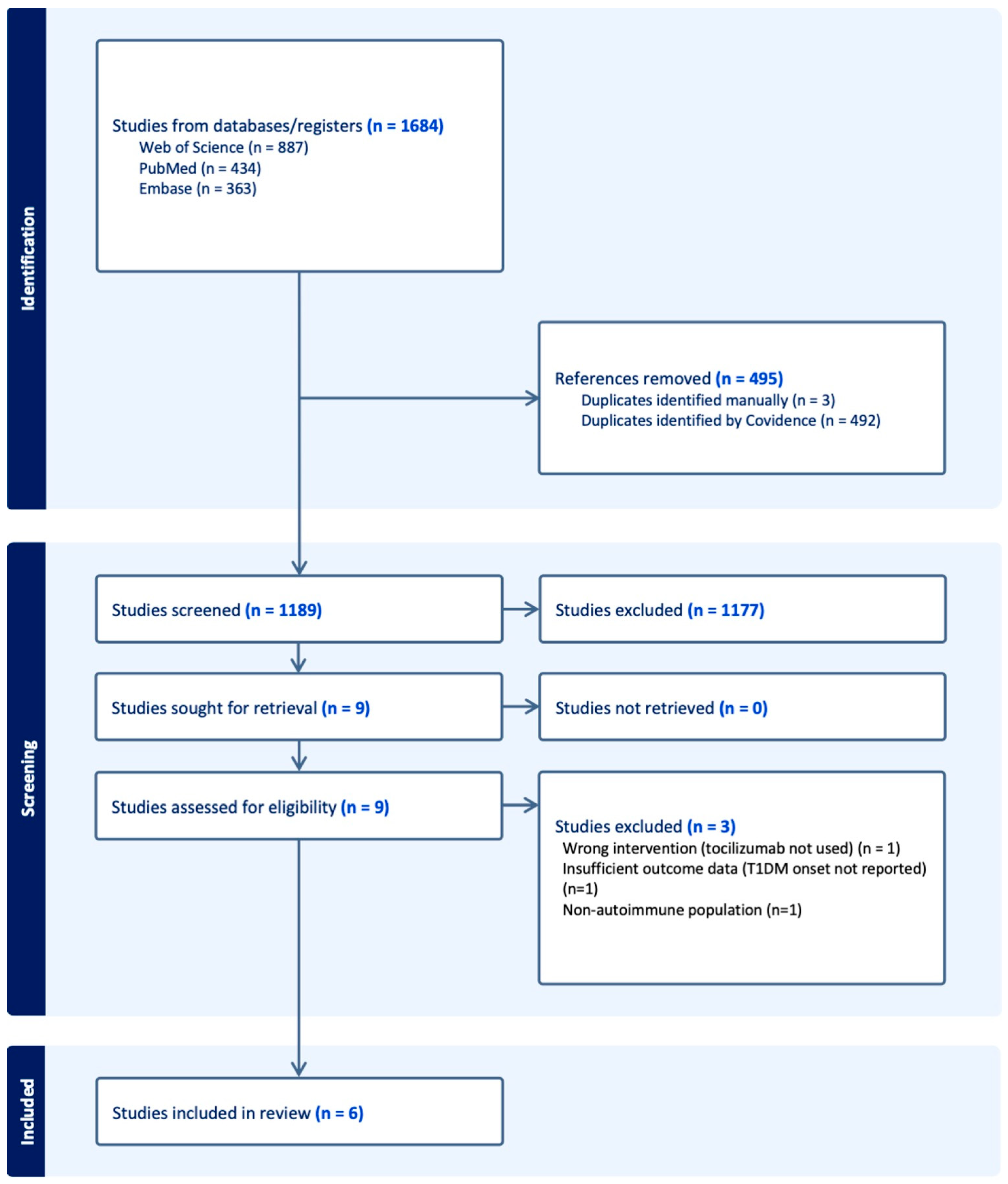
Figure 1. PRISMA-ScR Flow Diagram of Study Selection. The diagram illustrates the selection process for studies included in this scoping review.
Immuno 05 00041 g001
Figure 2. Conceptual triangulation framework of IL-6/IL-6R signaling in type 1 diabetes mellitus (T1DM). This figure illustrates how four complementary streams of evidence converge to inform the role of IL-6/IL-6R signaling in T1DM. Clinical observations from case reports provide initial signal generation; randomized controlled trials (RCTs) supply causal testing in humans; Mendelian randomization (MR) studies contribute genetic causal inference; and mechanistic immunology offers biological plausibility by elucidating cellular pathways. Together, these diverse evidence types triangulate toward a more comprehensive understanding of IL-6/IL-6R biology in T1DM and inform translational strategies for therapeutic intervention.
Immuno 05 00041 g002
Table 1. Summary of included studies. Summary of study types, populations, interventions, primary outcomes, and main findings related to tocilizumab use or IL-6/IL-6R signaling in type 1 diabetes mellitus.
Author (Year)Study TypePopulationExposure/InterventionPrimary OutcomeMain FindingsSample SizeStudy Limitations
Hundhausen et al., 2016 [10]Mechanistic StudyT1DM patients vs. healthy controlsIL-6 stimulation of PBMCsIL-6-induced STAT3/STAT1 phosphorylationT1DM patients exhibited enhanced IL-6 signaling responsesn = 25 T1DM, n = 20 controlsSmall sample size; in vitro mechanistic findings may not reflect in vivo dynamics
Kawasaki et al., 2022 [16]Case Report73-year-old woman with RA receiving tocilizumabTocilizumab 162mg SC every 2 weeksDevelopment of autoimmune T1DM (ketoacidosis)T1DM onset 17 months after tocilizumab initiationSingle patientAnecdotal; no causal inference possible
Greenbaum et al., 2021 [9]Randomized Controlled TrialChildren and adolescents with new-onset T1DMTocilizumab IV monthly (7 doses) vs. placeboPreservation of β-cell function (C-peptide AUC)No significant effect of tocilizumab on C-peptide declinen = 69Short follow-up; limited statistical power
Fu et al., 2024 [17]Mendelian Randomization Study 1General population GWAS datasetsGenetically proxied IL-6R blockadeAssociation between IL-6R blockade and T1DM riskNo significant association (p > 0.05)~200,000 GWAS participantsPossible weak instruments; European ancestry only
Heikkilä et al., 2024 [14]Mendelian Randomization Study 2GWAS datasets focused on IL6R expression and T1DMGenetically proxied IL6R expression levelsAssociation between IL6R expression and T1DM riskHigher IL6R expression increased T1DM risk (OR 1.98)~150,000 GWAS participantsPotential horizontal pleiotropy; limited trans-ethnic validation
Li et al., 2025 [15]Mendelian Randomization Study 3GWAS datasets using CRP as IL-6R signaling proxyGenetically proxied IL-6R blockade via CRP levelsAssociation between IL-6R blockade and T1DM riskIL-6R blockade reduced T1DM risk (OR 0.410)~180,000 GWAS participantsProxy SNPs may capture downstream effects, not IL-6R-specific
Disclaimer/Publisher’s Note: The statements, opinions and data contained in all publications are solely those of the individual author(s) and contributor(s) and not of MDPI and/or the editor(s). MDPI and/or the editor(s) disclaim responsibility for any injury to people or property resulting from any ideas, methods, instructions or products referred to in the content.