Article

Pathogenic Mechanisms of Collagen TypeⅦA1 (COL7A1) and Transporter Protein Transport and Golgi Organization 1 (TANGO1) in Rheumatoid Arthritis: A New Therapeutic Target

1
Department of Integrative & Functional Biology, Council of Scientific & Industrial Research–Institute of Genomics and Integrative Biology (CSIR–IGIB), Delhi University Campus, Mall Road, Delhi 110007, India
2
Academy of Scientific and Innovative Research (AcSIR), Ghaziabad 201002, India
3
All India Institute of Medical Sciences (AIIMS), Ansari Nagar, New Delhi 110029, India
*
Author to whom correspondence should be addressed.
Immuno 2024, 4(4), 461-478; https://doi.org/10.3390/immuno4040029
Submission received: 29 August 2024 / Revised: 25 October 2024 / Accepted: 28 October 2024 / Published: 6 November 2024

Abstract

Rheumatoid arthritis (RA) is an autoimmune disorder causing chronic inflammation primarily due to collagen regulation and transport imbalances. Collagen VII A1(COL7A1), a major component of anchoring fibrils, regulates inflammation via interacting with its transporter protein Transport and Golgi organization 2 homologs (TANGO1). The study revealed a significant increase in COL7A1 levels in both the plasma and PBMCs of RA patients. Additionally, a positive correlation between COL7A1 and ACCPA (anti-cyclic citrullinated peptide antibody) levels was observed among RA patients. TANGO1 mRNA expression was also found to be elevated in PBMCs. The knockdown of COL7A1 in RA synoviocytes using siRNA affected the expression of TANGO1 and inflammatory genes. Western blot analysis showed that COL7A1 si-RNA in TNF-α-induced SW982 cells reduced the expression of COL7A1, TANGO1, and NF-kBp65. The mRNA expression of inflammatory genes TNF-α, NF-kB p65, and IL-6 simultaneously decreased after the knockdown of COL7A1, as measured by qRT-PCR. An in silico analysis found 20 common interacting proteins of COL7A1 and TANGO1, with pathway enrichment analysis linking them to antigen presentation, class I and II MHC, and adaptive immunity pathways in RA. Among the common proteins, The DisGeNET database depicted that COL1A1, MIA3, SERPINH1, and GORASP1 are directly linked to RA. The molecular docking analysis of COL7A1 and TANGO1 revealed strong interaction with a −1013.4 energy-weighted score. Common RA-used drugs such as Adalimumab, Golimumab, and Infliximab were found to inhibit the interaction between COL7A1 and TANGO1, which can further impede the transport of COL7A1 from ER exit sites, indicating COL7A1 and TANGO1 as potential therapeutic targets to diminish RA progression.

1. Introduction

Collagen dysregulation and its differential expressions cause the emergence of various acute and chronic diseases, including Rheumatoid arthritis (RA) [1]. RA is a chronic, autoimmune disorder, majorly affecting the synovial joints, followed by cartilage damage with collagen as the main component of the cartilage matrix and a major target of cartilage regeneration [2]. A key event in RA pathogenesis is the irreversible damage to the cartilage collagen network, and the majorly studied collagens in this regard are Ⅱ, Ⅳ, Ⅵ, Ⅸ, and Ⅹ collagens, where collagen Ⅱ has been considered a prominent protein in RA [3]. Since the cartilage matrix is mainly composed of the collagen network, which is a key factor in the regeneration of cartilage [4], it is essential to have a comprehensive understanding of the various functions and effects of collagen to fully comprehend the mechanism behind RA development that may help in the identification of specific therapeutic targets for effective treatment strategies.
Collagen type VII A1 (COL7A1) is a protein composed of a triple-helical collagen domain, with two non-collagenous domains on either side. It is located in the epithelial basement membrane and helps to form anchoring fibrils. These fibrils may aid in organizing and securing the epithelial basement membrane by interacting with proteins found in the extracellular matrix (ECM) [5]. The interaction between cells and the extracellular matrix (ECM) is vital in the development of Rheumatoid arthritis (RA). The regulation of the composition and structure of the ECM in a targeted manner has been considered an effective approach for treating arthritis [6]. Thereby, we speculate that COL7A1 may play a major role in RA, as the maintenance of fibril assembly is a crucial factor in RA development [7]. Previous studies based on Genome-wide association have assessed the potential association of COL7A1 with the development of accelerated atherosclerosis in RA patients [8], which indicates the potential of this collagen in regulating RA progression. However, the direct impact of COL7A1 in RA pathophysiology has not been explored to date. Based on the above literature report, we believe that identification and validation of its presence in RA plasma and its role in RA might bring a new path in the development of therapeutic targets for new treatment strategies.
COL7A1 is well known for its role in diseases like recessive dystrophic epidermolysis bullosa (RDEB). COL7A1 expression is upregulated in response to stimuli such as the growth factor TGF-β2, contributing to the development of skin fragility [9]. Various mutations and dysregulations of COL7A1 have been linked to skin disorders of differing severities [10]. COL7A1 is critical in wound healing, as it facilitates the attachment of the epidermis to the dermis, promotes the migration of keratinocytes and fibroblasts, regulates protein trafficking and autophagy, and influences cytokine production [11]. Due to its linkage with laminin–integrin interactions and the formation of anchoring fibrils, COL7A1 is considered as a crucial adhesion molecule [12]. The abnormal expression of COL7A1 negatively impacts the cellular environment, leading to proteome alterations and changes in the expression of TGF-β and matrix metalloproteinases (MMPs) [13]. This suggests that COL7A1, being vital for maintaining cellular homeostasis, may also play a significant role in RA development, warranting further exploration.
Collagens are transported to the Golgi vesicles from the endoplasmic reticulum (ER) for their cellular programming. Transport And Golgi Organization 1 (TANGO1), encoded by the MIA3 gene, a transmembrane protein, acts as a cargo receptor for the bulky collagens to export collagens from the ER [14]. TANGO1 acts as an organizer by recruiting ER Golgi Intermediate Compartment (ERGIC) membranes at ER exit sites to form a ring-like structure for the proper secretion of COL7A1 [15]. Studies have shown that the absence of TANGO1 had an adverse effect on COL7A1 secretion (from chondrocytes, fibroblasts, endothelial cells, and mural cells) and caused various abnormalities, including disrupted bone mineralization and bone loss leading to neonatal lethality [16]. It also causes severe collagenopathy related to dentinogenesis imperfecta, short stature, skeletal abnormalities, diabetes mellitus, and mild intellectual disability [17]. An abnormal expression of TANGO1 in oral squamous cell carcinoma promoted angiogenesis and lymph angiogenesis [18], which suggested that TANGO1 and its linked protein COL7A1 probably have an important impact in determining RA progression as angiogenesis is a marked factor of RA pathogenesis.
Here, in this study, we conducted an expression analysis of COL7A1 and its transporter protein TANGO1 and determined their impact on the in vitro cellular environment and inflammation during RA pathogenesis. Further, we viewed the pharmacological impact of targeting COL7A1 and TANGO1 by utilizing commonly prescribed RA drugs through in silico studies suggesting a new therapeutic target for attenuating the RA progression.

2. Materials and Methods

2.1. Clinical Samples

Blood samples (n = 30) were collected from RA patients from the Department of Rheumatology, All India Institute of Medical Sciences (AIIMS), New Delhi, India, who met the revised 2010 American College of Rheumatology (ACR) and European League Against Rheumatism (EULAR) diagnosis criteria. Similarly, blood samples (n = 30) were collected from healthy controls (HCs) with no prior ailment and joint inflammation. The medical history of each patient was collected (Supplementary Table S1). Blood samples were collected into EDTA-coated vacutainer tubes (Becton, Dickinson and Company, Franklin Lakes, NJ, USA) and centrifuged (1300× g, 15 min) at 4 °C. Plasma samples were separated and stored at −80 °C for further study [19].

2.2. Western Blot (WB) and Enzyme-Linked Immunosorbent Assay (ELISA) in Plasma Samples

For WB analysis, 3 pooled plasmas of RA and HC (n = 10 each pool) samples were used, and 20 µg of protein estimated by Bradford assay was run in 6% sodium dodecyl sulfate–polyacrylamide gel electrophoresis (SDS-PAGE). Anti-COL7A1 (1:1000, 4 °C overnight) (sc-33710, Santa Cruz Biotechnology Inc., Paso Robles, CA, USA) was used as the primary antibody, and densitometry values were normalized with total protein as a loading control. Quantitative ELISA was performed with RA plasma (n = 30) and HC (n = 30) samples using a Collagen type Ⅶ ELISA kit (ELK3163, ELK Biotechnology, Denver, CO, USA). The absorbance was observed at 450nm using an ELISA plate reader (Spectra Max Plus 384, Molecular Devices LLC, San Jose, CA, USA) [20].

2.3. Peripheral Blood Mononuclear Cell (PBMC) Isolation, RNA Isolation, and Quantitative Real-Time Polymerase Chain Reaction (qRT-PCR)

PBMCs are the primary immune cells in the human body and offer discriminatory immune responses toward inflammation. Therefore, we checked the protein level and mRNA level of COL7A1 and TANGO1 in PBMCs. PBMCs were isolated by centrifuging the blood of RA patients (n = 6) and HC (n = 6) using Histopaque reagent. The pellet was washed, and total RNA was extracted using Tri-Xtract Reagent (G-biosciences) cDNA was synthesized using the cDNA Synthesis Kit (G-biosciences) according to the manufacturer’s procedure. The level of mRNA expression was determined by qRT-PCR (Roche Light Cycler® 480 Instrument-II real-time PCR system) using human-specific primer sequences and an internal reference Glyceraldehyde 3-phosphate dehydrogenase (GAPDH), a glycolytic enzyme. The reactions were quantitatively evaluated using the 2−ΔΔCT formula, calculated the relative fold gene expression of samples [20]. Primers are shown in Supplementary Table S2 (see details in the Supplementary Materials).

2.4. In Vitro Analysis

2.4.1. Cell Culture and In Vitro Knockdown of COL7A1

SW982 (human synovial fibroblast synoviocytes) cell line was purchased from the National Centre for Cell Science (NCCS). The cells were cultured in a T-25 flask in Dulbecco’s Modified Eagle’s Medium (DMEM) containing antibiotic solution, which was supplemented with 10% FBS and kept at 37 °C humidified incubator with 5% CO2. The cells were grown until 80% confluency followed by transfection with si-COL7A1 (1–50 nM) (sc-43066, Santa Cruz Biotechnology Inc., Paso Robles, CA, USA) using lipofectamine™ RNAiMax (4 µL/mL) transfection reagent (Invitrogen, Carlsbad, CA, USA) in complete media (DMEM with 10% FBS) to knock down the expression of COL7A1 in the cells. The cells were then grown further in the transfected media for 48 h [21].

2.4.2. WB Analysis and qRT-PCR

SW982 cells were grown in DMEM containing 10% FBS in six-well plate and transfected with si-COL7A1 for 48 h. The cells were stimulated with Tumor necrosis factor-alpha (TNF-α) (10 ng/mL) for 24 h in a serum-free medium. The cells were collected in lysis buffer Radioimmunoprecipitation assay buffer (RIPA buffer) containing phosphatase and protease inhibitors. A total of 40 µg sample protein, determined by the Bicinchoninic Acid Protein Assay (BCA) method, was run in 6% SDS-PAGE. The separated proteins were transferred to the NC membrane (G Biosciences) by semi-dry transfer (Bio-Rad Laboratories, Hercules, CA, USA) unit, which was blocked for 3 h with 3% BSA and incubated overnight at 4 °C with primary antibodies of COL7A1 (sc-33710, Santa Cruz Biotechnology Inc., Paso Robles, CA, USA), TANGO1 (sc-393916, Santa Cruz Biotechnology Inc., Paso Robles, CA, USA), Nuclear factor kappa-light-chain-enhancer of activated B cells (NF-κB) p65 (sc-8008, Santa Cruz Biotechnology Inc., Paso Robles, CA, USA) (1:2000). Vinculin (sc-73614, Santa Cruz Biotechnology Inc., Paso Robles, CA, USA) (1:5000) was used as loading control. The membrane was then washed with Tris buffered saline with 1% Tween-20 (TBST), incubated with horseradish peroxidase (HRP)-conjugated anti-mouse secondary antibody (1:10,000, Jackson, West Grove, PA, USA) for 1 h at room temperature (RT) and developed with the ChemiDoc system (Bio-Rad Laboratories (Singapore) Pvt. Ltd.) using a light-sensitive developer. Total RNA was then isolated and subjected to cDNA preparation, and mRNA expression of COL7A1 and TANGO1, Interleukin 6 (IL-6), TNF-α, and NF-κB p65 was evaluated and quantitated using the 2−ΔΔCT formula. Human-specific primer sequences are shown in Supplementary Table S2.

2.5. In Silico Analysis

2.5.1. Screening of COL7A1- and TANGO1-Interacting Proteins and Association with RA

To investigate the relationship between COL7A1 and TANGO1 and their potential involvement in the development of RA, we first retrieved a list of the top 100 proteins that interact with COL7A1 and TANGO1 using the STRING database. We then compared these lists using Venny 2.1.0, an interactive tool for creating Venn diagrams, to identify the proteins that are common to both lists [22]. The common proteins were then matched with RA-associated proteins from the DisGeNET database to screen out proteins directly linked to RA pathogenesis [23].

2.5.2. Protein–Protein Interaction (PPI) Network Construction of Selected Proteins

PPI network of the commonly selected proteins, COL7A1 and TANGO1, was constructed by Cytoscape 3.8.2, a software platform for generating networks, visualization, and analysis, along with identifying the relationship of target proteins with respective compounds and viewing the pathways and diseases involved. The PPI network was created according to the confidence degree of targeted proteins and their related RA-linked pathways [24,25].

2.5.3. Gene Ontology (GO) Enrichment Analysis of Selected Proteins

To elucidate the properties associated with COL7A1 and TANGO1, the associated biological processes, cellular components, and molecular functions of these proteins, along with their common interacting proteins, were interpreted by Cytoscape 3.8.2. The results were represented by GraphPad 9.0.

2.5.4. Pathway Enrichment Analysis of Selected Proteins

To illustrate the involved signaling pathways link with COL7A1 and TANGO1, pathway analysis of the 20 selected proteins was processed for Kyoto Encyclopedia of Genes and Genomes (KEGG) and Reactome pathway enrichment analysis via Cytoscape 3.8.2 and was represented by GraphPad 9.0.

2.5.5. Preparation of Target Proteins for Molecular Docking

Protein structures were extracted from the Research Collaboratory for Structural Bioinformatics Protein Data Bank (RCSB PDB) of the Heat Shock Protein 47 (Hsp47)–collagen complex [PDB ID: 4AU3] and TANGO1 [PDB ID: 7R3M]. The structures were visualized using PyMOL 3.0 software and edited by removing water molecules [26,27].

2.5.6. Preparation of Ligands

The structure of three monoclonal antibodies, Adalimumab [PDB ID: 3WD5], Golimumab [PDB ID: 5YOY], and Infliximab [PDB ID: 5VH3], which are commonly used drugs in RA, were extracted from PDB [28]. These structures were then visualized and prepared for molecular docking analysis using PyMOL 3.0 software, which involved the removal of water and extra molecules to ensure accuracy [27].

2.5.7. Active Binding Site Prediction

The active site prediction of the protein structure was found by using the Computed Atlas of Surface Topography of Proteins (CASTp 3.0) at http://cast.engr.uic.edu (accessed on 2 April 2024). A binding site of 13,436.366 Å3 for volume and 3141.616 Å2 for surface area was predicted for 4AU3 (Hsp47- collagen complex) [29].

2.5.8. Molecular Docking

The interaction of COL7A1 and TANGO1 was conducted by molecular docking using a protein–protein docking software, ClusPro 2.0 server [30,31]. Protein structures were extracted from the protein data bank of the Hsp47–collagen complex or Collagen VII [PDB ID: 4AU3], TANGO1 [PDB ID: 7R3M], and docked. An energy score was obtained that served as a quantitative measure of their interaction strength [30].
Subsequently, to explore potential therapeutic interventions of the RA-prescribed drugs, three monoclonal antibodies, Adalimumab [PDB ID: 3WD5], Golimumab [PDB ID: 5YOY], Infliximab [PDB ID: 5VH3], were docked with TANGO1, yielding three distinct drug–TANGO1 complexes and their respective energy scores. These individual complexes were then docked with the collagen VII complex, and the obtained energy scores were compared with the initial COL7A1 and TANGO1 interaction scores. The binding sites of the protein–protein interactions were obtained and represented by LigPlot+ v.2.2 [32], a software that automatically creates two-dimensional schematic representations of binding sites involved in protein interactions.

2.6. Statistical Analysis

All non-parametric Mann–Whitney tests were performed using GraphPad Prism 9.0. Statistical analysis was performed with the paired Student’s t-test to compare the data between two groups, and one-way analysis of variance (ANOVA) was used to compare data among multiple groups. The obtained p-values were represented by asterisks on the graph (* p ≤ 0.05, ** p ≤ 0.01, *** p ≤ 0.001, **** p ≤ 0.0001).
GraphPad Prism 9.0 is a powerful software that integrates biostatistics, nonlinear regression for curve fitting, and scientific graphing into one comprehensive tool. It enables to efficiently organize, analyze, and graph repeated experiments, select the appropriate statistical tests, and interpret the outcomes. Prism offers a comprehensive solution for seamless data analysis and advanced graphing capabilities, providing all necessary functionalities within a single, integrated program.

3. Results

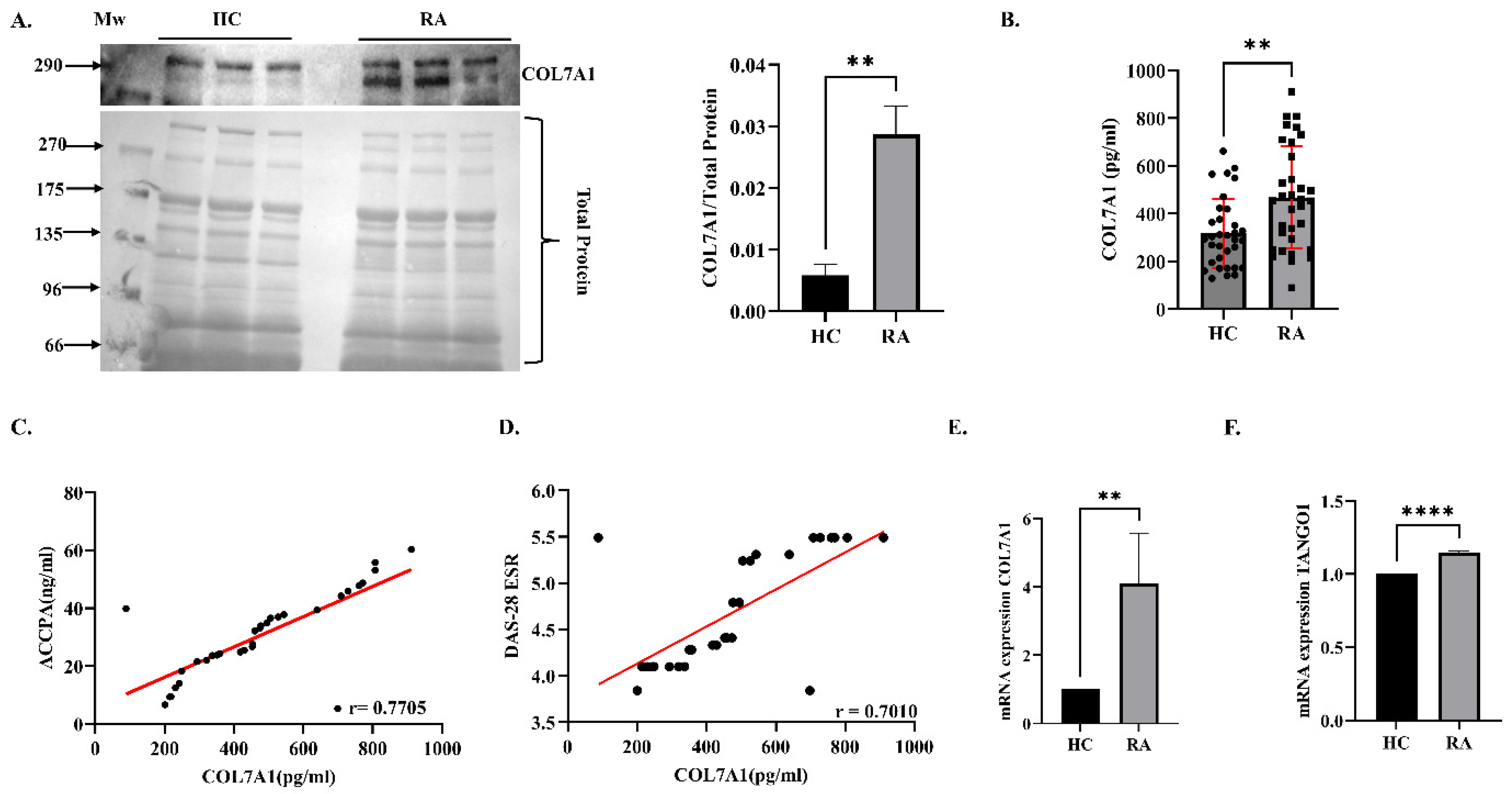
3.1. Expression Analysis of COL7A1 in Plasma and PBMCs

The protein expression analysis of COL7A1 was found to be 3-fold (p = 0.0037) upregulated in RA blood plasma (n = 30) compared to HC (n = 30) by WB (Figure 1A). The concentration level of COL7A1 in RA plasma samples (n = 30) was validated through a quantitative ELISA and a significant upregulation (1.47-fold, p-value = 0.0015) was observed in RA samples compared to HC (Figure 1B). A positive correlation was observed between COL7A1 and ACCPA (r = 0.7705) and also between COL7A1 and DAS28-ESR levels (0.7010) in RA patients (Figure 1C,D), indicating a direct correlation of an increased level of COL7A1 increased level with RA progression. Furthermore, the increased mRNA expression of COL7A1 (p = 0.0056) and TANGO1 (p = 0.0001) was also found in the PBMCs of RA patients compared to HC PBMCs after normalization with GAPDH as a loading control (Figure 1E,F).

3.2. Effect of Knockdown of COL7A1 on Inflammation in SW982 Cells (In Vitro Analysis)

To investigate the effect of COL7A1 on RA inflammation and its transporter protein TANGO1, we used the siRNA of COL7A1 for transient silencing. In order to standardize the dose of si-COL7A1, an in vitro study was conducted in the SW982 cell line and an WB analysis of COL7A1 expression at various concentrations of the siRNA treatment ranging from 1 to 50 nM was conducted. COL7A1 expression was significantly downregulated (~48%) at 50 nM concentration, so this dose was selected for further experiments (Supplementary Figure S1). For COL7A1 (1.2-fold) (p = 0.0304) and a major inflammatory marker of RA synoviocytes, NF-κB p65 expression levels (~2-fold) (p = 0.0001) were significantly decreased upon COL7A1 knockdown in TNF-α-induced SW982 cells. TANGO1 expression was also found to be decreased significantly by ~2.5-fold (p = 0.0144) after silencing COL7A1 (Figure 2A). We also found a significant decrease in the mRNA expression of COL7A1 (p < 0.0012) and its transporter protein TANGO1 (p = 0.0001), and prominent inflammatory cytokines such as IL-6 (p = 0.0077), TNF-α (p = 0.0008), and NF-κB p65 (p = 0.0001) (Figure 2B) after si-COL7A1 treatment, indicating that COL7A1 has an important role in mediating inflammation in RA synoviocytes.

3.3. Targeted Therapeutic Approach of COL7A1-Associated Proteins in RA (In Silico Analysis)

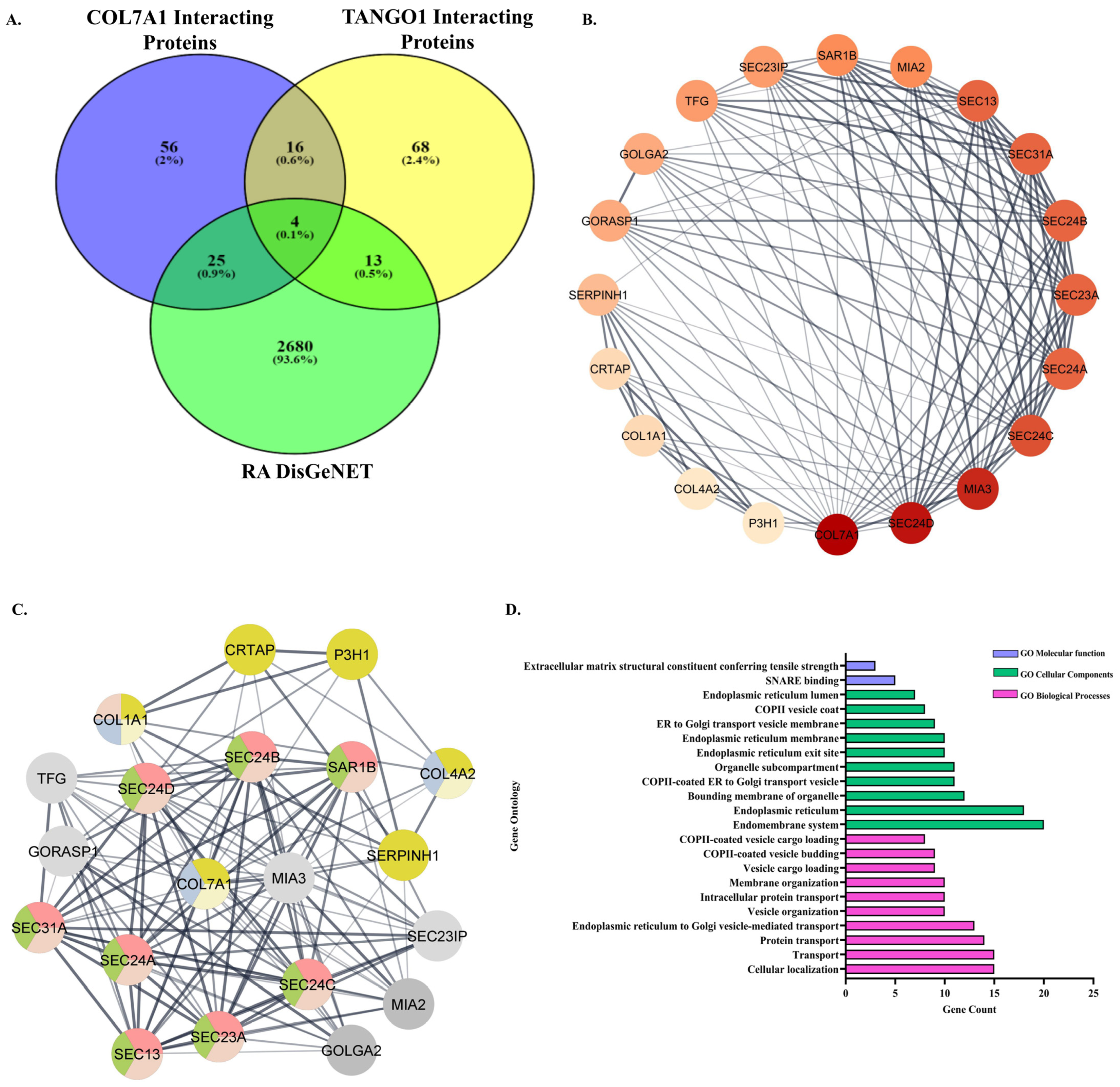
3.3.1. Retrieval of Common Interacting Proteins of COL7A1 and TANGO1 and Association with RA

The association of COL7A1 and TANGO1 in RA pathophysiology was further explored by retrieving the interacting proteins (Top 100) of COL7A1 and TANGO1 consecutively using the STRING 12.0 database. After comparing the respective lists of interacting proteins, 20 proteins were found to be common: SEC24A, SEC23A, SEC24B, GORASP1, SEC23IP, SEC24D, COL7A1, P3H1, SEC24C, COL1A1, GOLGA2, SEC31A, SEC13, MIA3, SERPINH1, TFG, COL4A2, MIA2, SAR1B, and CRTAP. These proteins were then matched with RA-associated genes that were retrieved from the DisGeNET database. It revealed four common proteins (Figure 3A), namely COL1A1, MIA3, SERPINH1, and GORASP1, which were found to have a link with RA with gene–disease association scores of 0.01, 0.01, 0.02, and 0.1, respectively. These interacting proteins of COL7A1 and TANGO1 that are found to be involved in RA pathophysiology indicate a strong association of COL7A1 and TANGO1 in mediating RA progression.

3.3.2. PPI Network of the Common Interacting Proteins of COL7A1 and TANGO1

A PPI network was established to elucidate the relationship between the 20 common proteins retrieved from the above in silico analysis. The color of the node was found to be directly correlated with the degree of contribution of the respective node (each node representing each protein) in the network. The color of nodes like COL7A1, SEC24D, MIA3, SEC24C, SEC24A, and SEC23A, with their degree values of 19, 18, 17, 15, 14, and 14, are comparatively darker, representing the major contributions of these proteins in the network and their interactions (Figure 3B).
Further, a pathway analysis of these 20 proteins revealed the involvement of various RA-linked signaling pathways associated with the proteins, such as antigen presentation; folding, assembly, and peptide loading of class I MHC; MHC Class II antigen presentation; collagen biosynthesis and modifying enzymes; adaptive immune system; collagen degradation; and integrin cell surface interactions-related pathway. The major proteins associated with the pathways were COL7A1, SEC24D, SEC24B, SAR1B, COL4A2, COL1A1, SEC31A, SEC24A, SEC13, SEC23A, and SEC24C (Figure 3C). This suggested that most of the common interacting proteins between COL7A1 and TANGO1 are directly or indirectly linked with RA pathogenesis by modulating majorly the collagen network and inflammatory pathways.

3.3.3. Gene Ontology (GO) Enrichment Analysis

The biological properties of the 20 common proteins, including their biological processes, cellular components, and molecular functions, were elucidated by Cytoscape 3.8.2 to illustrate the association of the proteins with RA and its pathogenesis. A total of 43 GO entries were retrieved. Amongst them, 19 entries were found to be associated with biological processes, which primarily include the regulation of cellular localization, transport, ER to Golgi-mediated transport, vesicle, and membrane organization. Two entries were of molecular functions, which include Soluble N-ethylmaleimide-sensitive factor activating protein receptor (SNARE) binding, extracellular matrix structural constituent conferring tensile strength, and 22 cellular component entries included in the Endomembrane system, endoplasmic reticulum, the bounding membrane of the organelle and the COPII-coated ER to Golgi transport vesicle (Figure 3D depicts the top 10 according to p value < 0.05). This GO enrichment indicated that the proteins are majorly linked with ER and Golgi transport networks and maintain the extracellular matrix.

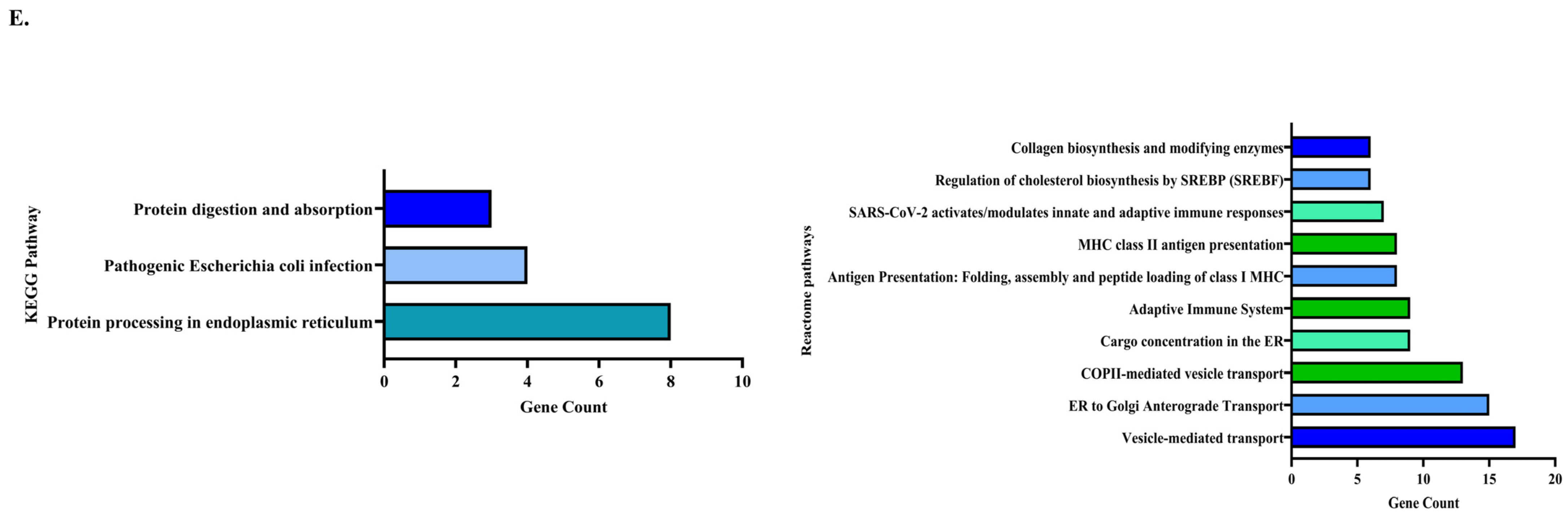
3.3.4. Pathway Enrichment Analysis

To analyze the signaling pathways associated with the 20 common proteins, a target pathway network was created using Cytoscape 3.8.2. The reactome pathway enrichment of the proteins derived 20 related pathways, mainly depicting an association with vesicle-mediated transport, ER to Golgi Anterograde Transport, COPII-mediated vesicle transport, cargo concentration in the ER, and the adaptive immune system (the top 10 according to p-value). We also performed a KEGG pathway analysis of these 20 proteins to enrich more pathways and to deduce their association with RA. The analysis majorly showed the pathways related to protein processing in the endoplasmic reticulum and protein digestion and absorption, which has a major impact during RA, as cells undergo ER stress during the disease condition [27] (Figure 3E). Overall, the in silico enrichment analysis showed that COL7A1, TANGO1, and their interacting proteins play a major role in the development of RA via the modulation of the collagen network, ER to Golgi transport, and inflammatory signal transduction.

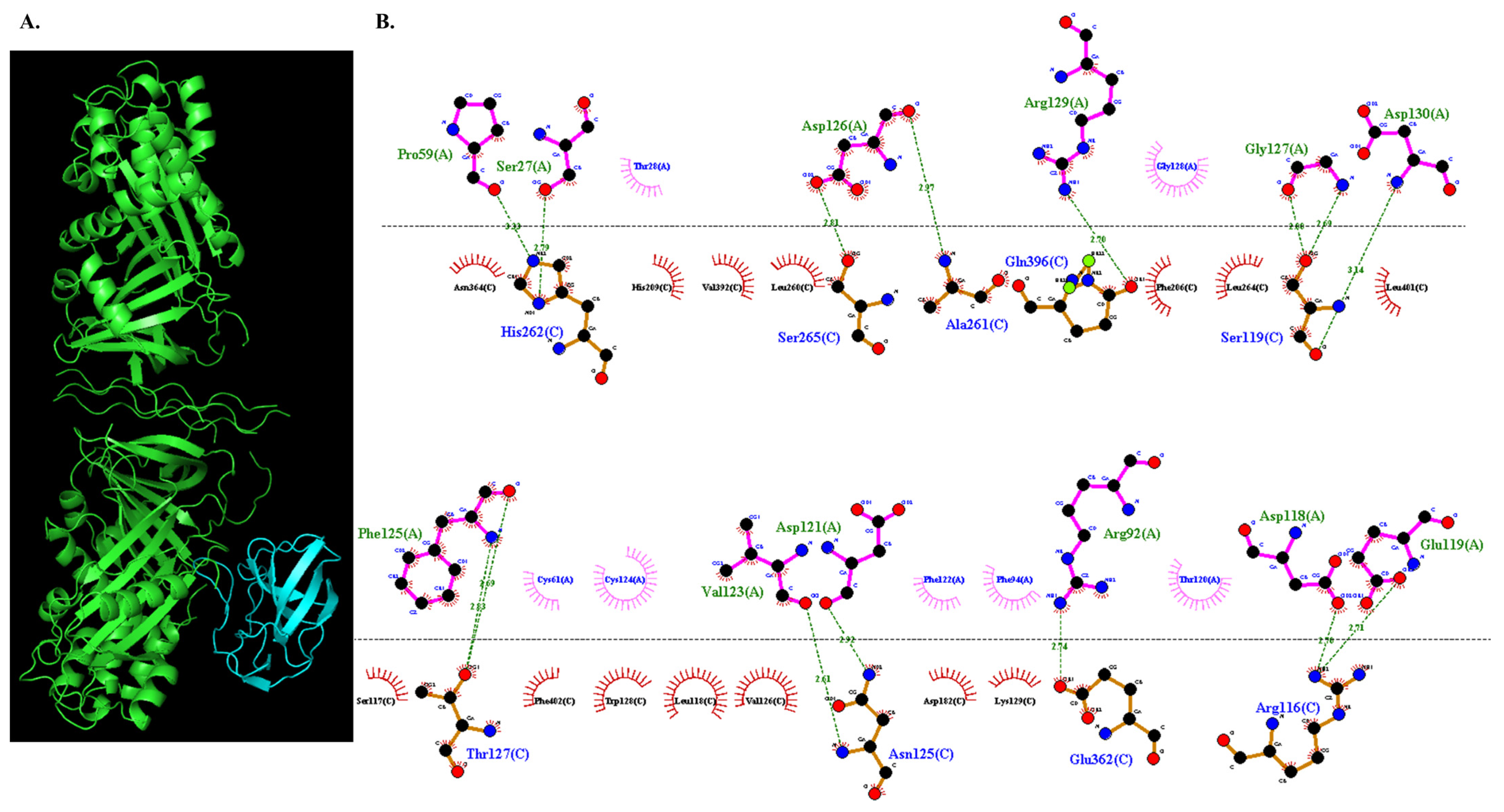
3.3.5. Molecular Docking of COL7A1 and TANGO1

To deduce whether COL7A1 and TANGO1 proteins can be utilized as promising therapeutic targets to minimize RA progression, we initially performed a docking analysis of COL7A1 and TANGO1 to visualize their interaction, which is a major factor in promoting RA occurrence. COL7A1 and TANGO1 proteins interacted with a binding energy with a weighted score of −1013.4 (Figure 4A). The A chain of TANGO1 was found to interact with the C chain of COL7A1. A total of 15 interactions were observed. These 15 interactions between the residues of COL7A1 and TANGO1 involved in the bonding were [TANGO1:COL7A1]—(A.Asp.118:C.Arg.116; A.Glu.119:C.Arg.116; A.Asp.130:C.Ser.119; A.Gly.127:C.Ser.119; A.Gly.127:C.Ser.119; A.Asp.121:C.Asn.125; A.Val.123:C.Asn.125; A.Phe.125:C.Thr.127; A.Phe.125:C.Thr.127; A.Pro.59:C.His.262; A.Ser.27:C.His.262; A.Asp.126:C.Ala.261; A.Asp.126: C.Ser.265; A.Arg.92:C.Glu.362; A.Arg.129:C.Gln.396) (Table 1, Figure 4B).

3.3.6. Docked Complex Preparation of Drugs with TANGO1

To demonstrate that targeting the interaction of COL7A1 and TANGO1 could be a potential therapeutic strategy for achieving remission in RA, we selected three widely used monoclonal antibodies, Adalimumab [PDB ID: 3WD5], Golimumab [PDB ID: 5YOY] and Infliximab [PDB ID: 5VH3], commonly prescribed drugs for RA patients [28], to target the COL7A1-TANGO1 complex. Primarily, the drugs were individually docked with the TANGO1 protein. The docking analysis yielded drug–protein interactions with −794.2, −966.7, and −844.9 energy-weighted scores, respectively (Supplementary Figure S2). These TANGO1–drug complexes were further used for interaction with COL7A1.

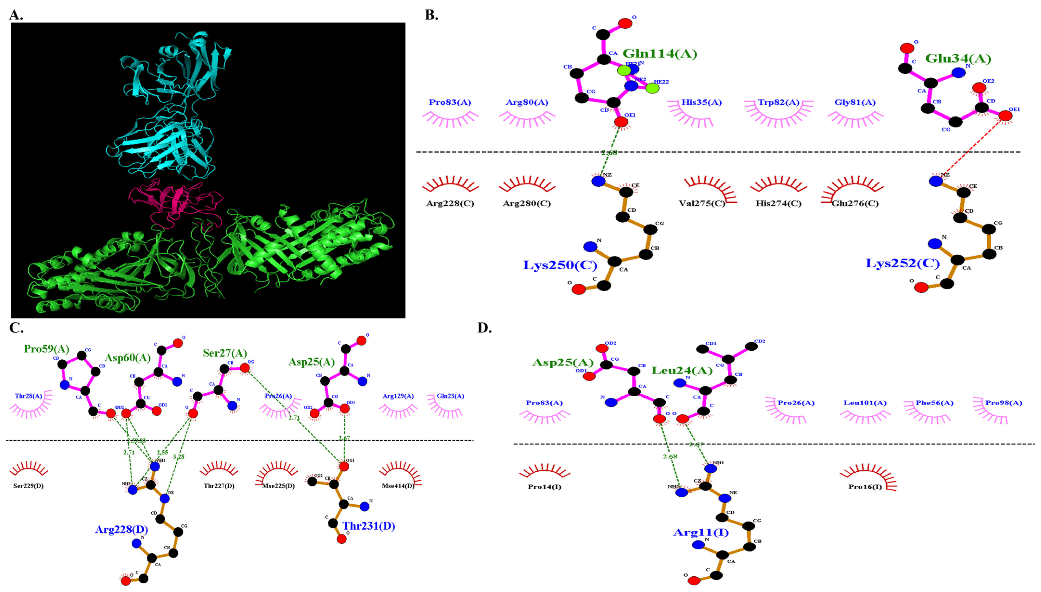
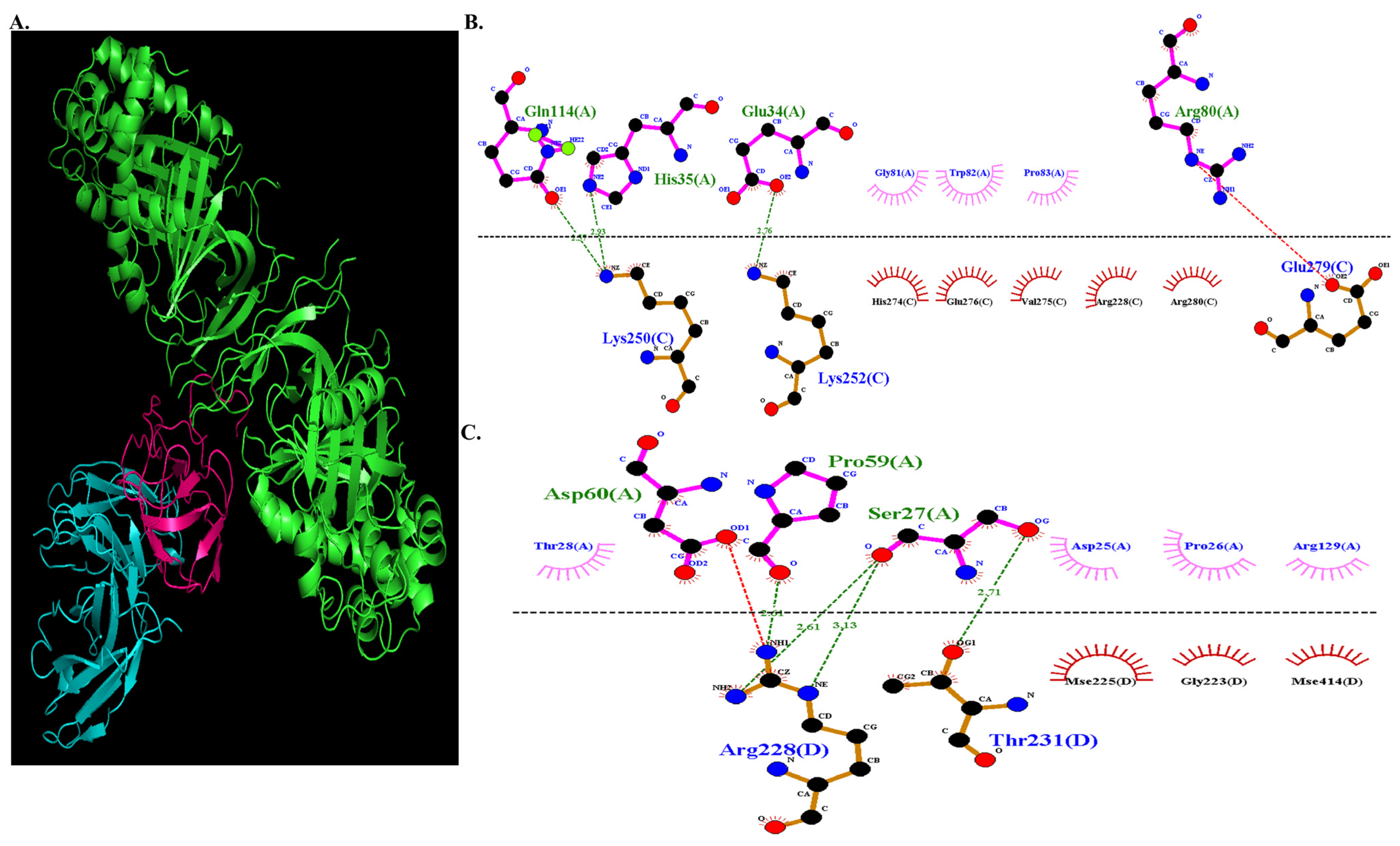
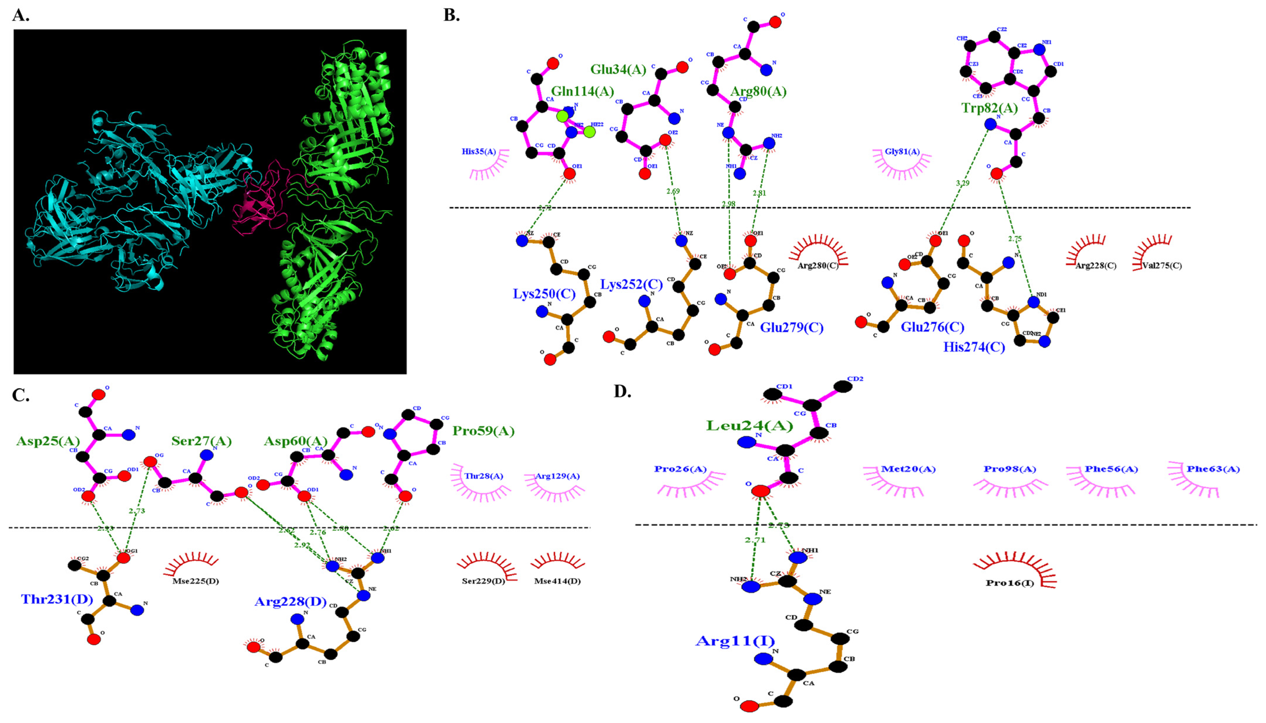
3.3.7. Molecular Docking of COL7A1 with TANGO–Drug Complexes

The TANGO1–drug complexes were then docked with COL7A1 and compared with actual COL7A1-TANGO1 interaction to visualize any alteration in the binding energy. It was revealed that the selected RA drugs hindered the interaction of COL7A1 and TANGO1, as demonstrated by docking analysis of the three drug–TANGO1 complexes, Adalimumab–TANGO1, Golimumab–TANGO1, and Infliximab–TANGO1 with COL7A1. The three drug complexes interacted with −674.7, −679.5, and −670.6 energy-weighted scores, respectively, which is less negative than the TANGO1-COL7A1 interaction score (−1013.4 weighted score), depicting that the drugs are hindering the binding of the two proteins. Infliximab has shown the maximum efficiency in hindering the COL7A1 and TANGO1 interaction, as depicted by energy scores, followed by Adalimumab and Golimumab. These showed that COL7A1 and TANGO1 binding is an important RA-causing factor, which is also targeted by the commonly used RA drugs to resist the disease progression.
The binding residues between COL7A1 and TANGO1 also became altered, and the number of bond interactions also got reduced as the drugs bound with TANGO1, indicating that the drugs hampered the interaction of COL7A1 and TANGO1. The new number of bonds between COL7A1 and TANGO1 after drug interactions were 10, 7, and 14 bonds, whereas COL7A1 and TANGO1 bind with 15 bonds as derived from our earlier docking analysis. The interaction between the Adalimumab–TANGO1 complex are as follows: COL7A1 (Figure 5A) involved 10 bond interactions (A.Gln.114:C.Lys.250; A.Pro.59:D.Arg.228; A.Asp.60:D.Arg.228; A.Asp.60:D.Arg.228; A.Ser.27:D.Arg.228; A.Ser.27:D.Arg.228; A.Ser.27:D.Thr.231; A.Asp.25:D.Thr.231; A.Asp.25:I.Arg.11; A.Leu.24:I.Arg.11) (Figure 5B–D). The interaction between the Golimumab–TANGO1 complex were as follows: COL7A1interaction (Figure 6A) involved 7 bond formation (A.Gln.114:C.Lys.250; A.His.35:C.Lys.250; A.Glu.34:C.Lys.252; A.Pro.59:D.Arg.228; A.Ser.27:D.Arg.228; A.Ser.27:D.Arg.228; A.Ser.27:D.Thr.231) (Figure 6B,C). Likewise, the interaction between the Infliximab–TANGO1 complex: were as follows: COL7A1 interaction (Figure 7A) involved 14 bond formation (A.Gln.114:C.Lys.250; A.Glu.34:C.Lys.252; A.Trp.82:C.His.274; A.Trp.82:C.Glu.276; A.Arg.80:C.Glu.279; A.Arg.80:C.Glu.279; A.Ser.27:D.Arg.228; A.Ser.27:D.Thr.231; A.Asp.60:D.Arg.228; A.Asp.60:D.Arg.228; A.Pro.59:D.Arg.228; A.Asp.25:D.Thr.231; A.Leu.24:I.Arg.11; A.Leu.24:I.Arg.11) (Figure 7B–D, Table 2).

4. Discussion

RA is a chronic inflammatory disease that affects the synovial joints, leading to damage in the cartilage matrix [2]. This matrix is majorly formed by different collagens (type II, IX, and XI), hydroxyapatite, and proteoglycans [33]. The integration of the extracellular matrix and its regulation is a major factor that determines the RA pathogenesis [34]. While Collagen type Ⅱ has been identified as a major RA biomarker, further exploration is essential to identify additional proteins specific for RA pathogenesis. As RA progresses, cartilage degradation becomes a major concern, underscoring the importance of the investigation of different collagens to comprehend the disease mechanism [34].
Numerous in vitro, in vivo, and clinical studies have highlighted the involvement of various collagen peptides and collagen hydrolysates in arthritis development, immune response management, amino acid balance, and joint health [1]. One such collagen, COL7A1, remains the least explored in RA pathophysiology despite its significant role in anchoring fibrils. This protein is the major component of anchoring fibrils, involved in maintaining skin integrity, and its dysregulation causes serious skin disorders such as Dystrophic Epidermolysis Bullosa (DEB) [5,35]. Previous reports have reported the association of the emergence of cardiovascular defects in RA patients with COL7A1 [8]. Hence, exploring COL7A1 may unveil RA pathogenesis and suggest the development of therapeutic targets. A comprehensive analysis of collagens, integral components of the cell–matrix, is essential to elucidate RA pathogenesis and identify novel therapeutic targets.
Our study involved the expression analysis of COL7A1 in RA patient plasma samples, revealing its significant upregulation during RA conditions (Figure 1A,B). Further investigations showed higher COL7A1 expression in RA patient PBMCs and RA-induced synovial cells, marking it as a prominent factor dysregulated during RA (Figure 1E). Since synovium is the prominent site of inflammation, we mimic the RA condition in synovial secondary cell line SW982 via TNF-α treatment to induce inflammation and observed a positive correlation of COL7A1 expression with an increase in inflammation (Figure 2A).
Further, to explore the mechanistic action of COL7A1, our next target was to find its interacting proteins. TANGO1 protein, a transporter protein facilitating collagen trafficking from ER exit sites, was reported as a major protein to transport COL7A1 [15]. Therefore, an increase in COL7A1 expression might be linked with the regulation of TANGO1. Given their roles in maintaining cell integrity, we analyzed the link between COL7A1 and TANGO1, examining their association with inflammatory conditions during RA pathogenesis. We therefore conducted a knockdown experiment of COL7A1 in RA-induced synovial cells to analyze its effect on TANGO1 and RA-induced inflammation. It was observed that knocking down COL7A1 led to decreased expression of TANGO1 and other pro-inflammatory cytokines, including NF-κB p65, IL-6, and TNF-α (Figure 2B), indicating COL7A1’s pivotal role in RA-mediated inflammation.
Further, we dig deep to explore the mechanistic implication of COL7A1 and TANGO1 in mediating RA pathogenesis. Hence, an in silico analysis of these two proteins was attempted and deduced 20 common interacting proteins of COL7A1 and TANGO1 (Figure 3A). After matching the proteins with RA-linked genes, we have retrieved four proteins, COL1A1, MIA3, SERPINH1, and GORASP1, which were found to be directly linked with RA, thereby indicating that the two proteins have a prominent impact on the proteins that are associated directly with RA progression. Additionally, in order to find out the association between COL7A1 and TANGO1 with the RA-linked pathways, the GO and pathway enrichment analysis further highlighted the involvement of COL7A1, TANGO1, and their interacting proteins in adaptive immune system pathways, antigen presentation, class I MHC, MHC class II antigen presentation, and ER to Golgi Anterograde Transport, which are the major regulators of RA pathogenesis. This indicated that COL7A1 and its transporter protein are a major factor modulating the crucial signaling pathways involved in RA pathogenesis.
The illustration of the pathogenic role of COL7A1 and TANGO1 raises our interest in finding whether COL7A1 and TANGO1 can be utilized as prominent therapeutic molecules and whether targeting their functioning can lead to RA remission. Hence, we analyzed the action of Disease-modifying antirheumatic drugs (DMARDs), commonly used drugs in RA, through molecular docking studies to see whether they impact these two proteins and their regulation. Molecular docking analysis revealed that the interaction of the drugs with TANGO1 hindered the binding of COL7A1 and TANGO1, thereby hampering the transport of COL7A1 from ER exit sites via TANGO1. This was analyzed by observing the binding energy of COL7A1 and TANGO1 and the change in their interacting sites after the binding of the drugs. Additionally, the number of bonds between the two proteins also decreased upon binding with DMARDs. Thereby, these results demonstrated that the drugs are efficient in hindering the COL7A1 and TANGO1 pathogenic regulation, suggesting that these proteins can act as potential therapeutic targets to abrogate RA progression. Therefore, we believe that COL7A1 has the potential to be a prominent disease marker, and utilizing these proteins as a therapeutic target can be very effective in ameliorating RA progression. In the future, therapeutic strategies and medications can be developed by considering these proteins and their regulation as the prime focus to target.

5. Conclusions

It has been discovered that the major collagen COL7A1 plays a vital role in regulating the pathogenesis of Rheumatoid arthritis (RA). During the progression of RA, the transporter protein TANGO1 becomes activated to transport COL7A1. This process is hindered by the suppression of COL7A1 expression, which ultimately leads to the downregulation of TANGO1 and inflammatory proteins. RA drugs are also beneficial in hampering the interaction of the two proteins, which indicates that COL7A1 and TANGO1 can be potent therapeutic targets to diminish RA. In the future, the protein targets and their functioning can be targeted to develop innovative drugs for RA.

Supplementary Materials

The following supporting information can be downloaded at https://www.mdpi.com/article/10.3390/immuno4040029/s1, Figure S1: Western Blot analysis- The expression level of COL7A1 to check the effective concentration of Si-COL7A1, SW982 cells were treated with different concentrations of Si-COL7A1 (1–50 nM) for 48 h, COL7A1 was significantly knockdown (p = 0.0080) at 50 nM. NF-κB p65 was also downregulated (p = 0.0283) at 50nM concentration; Figure S2: Interaction of drugs with TANGO1 by molecular docking; Table S1: The clinical demography characteristics of patients with RA and HC; Table S2: The amplification primer sequences of the human-specific gene.

Author Contributions

Conceptualization, D.C. and P.A.; Methodology, D.C., P.A., L.J., M.S. and S.M.; Validation, D.C., L.J. and M.S.; Formal analysis, D.C., P.A., L.J. and M.S.; Resources, U.K. and S.B.; Data curation, L.J.; Writing—original draft, D.C., P.A., L.J., M.S. and S.M.; Writing—review & editing, D.C., P.A., L.J., M.S., S.M. and S.B.; Visualization, P.A., L.J., M.S. and S.M.; Supervision, S.B.; Project administration, S.B.; Funding acquisition, S.B. All authors have read and agreed to the published version of the manuscript.

Funding

This research was supported by the Council of Scientific and Industrial Research (CSIR) New Delhi, Project code OLP 2305 Government of India, for financial support.

Institutional Review Board Statement

The study protocol was ethically approved by All India Institute of Medical Sciences (AIIMS), New Delhi, India (Rseg No IEC-37/07.02.2020, RP-15/2020) and the study protocols complied with the Declaration of Helsinki.

Informed Consent Statement

Written informed consent has been obtained from the patient(s) to publish this paper.

Data Availability Statement

The original contributions presented in the study are included in the article/Supplementary Materials; further inquiries can be directed to the corresponding author.

Acknowledgments

Pankaj Yadav was responsible for transporting biological samples from the hospital to the lab. We thank Council of Scientific & Industrial Research–Institute of Genomics and Integrative Biology (CSIR–IGIB), Delhi, India, for providing the research platform, AcSIR, for academic support, and the Department of Rheumatology, All India Institute of Medical Sciences (AIIMS), New Delhi, India, for providing the patient samples.

Conflicts of Interest

The authors declare no conflicts of interest.

Abbreviations

Rheumatoid arthritis (RA), Collagen type VIIA1(COL7A1), recessive dystrophic epidermolysis bullosa (RDEB), matrix metalloproteinase (MMPs), endoplasmic reticulum (ER), Transport And Golgi Organization 1 (TANGO1), ER Golgi Intermediate Compartment (ERGIC), healthy controls (HCs), Western blot (WB), Enzyme-linked immunosorbent assay (ELISA), Sodium dodecyl sulfate–polyacrylamide gel electrophoresis (SDS-PAGE), Peripheral blood mononuclear cells (PBMCs), Quantitative Real-Time Polymerase Chain Reaction (qRT-PCR), Tumor necrosis factor-alpha (TNF-α), Radioimmunoprecipitation assay buffer (RIPA), Bicinchoninic Acid Protein Assay (BCA), Nuclear factor kappa-light-chain-enhancer of activated B cells (NF-κB) p65, Interleukin 6 (IL-6), Disease-modifying antirheumatic drugs (DMARDs).

References

  1. Elango, J.; Zamora-Ledezma, C.; Ge, B.; Hou, C.; Pan, Z.; Bao, B.; Pérez Albacete Martínez, C.; Granero Marín, J.M.; Maté Sánchez de Val, J.E.; Wu, W. Paradoxical Duel Role of Collagen in Rheumatoid Arthritis: Cause of Inflammation and Treatment. Bioengineering 2022, 9, 321. [Google Scholar] [CrossRef] [PubMed]
  2. Rannou, F.; François, M.; Corvol, M.-T.; Berenbaum, F. Cartilage breakdown in rheumatoid arthritis. Jt. Bone Spine 2006, 73, 29–36. [Google Scholar] [CrossRef] [PubMed]
  3. Stuart, J.M.; Huffstutter, E.H.; Townes, A.S.; Kang, A.H. Incidence and specificity of antibodies to types I, II, III, IV, and V collagen in rheumatoid arthritis and other rheumatic diseases as measured by 125I-radioimmunoassay. Arthritis Rheum. 1983, 26, 832–840. [Google Scholar] [CrossRef] [PubMed]
  4. Gelse, K.; Pöschl, E.; Aigner, T. Collagens—Structure, function, and biosynthesis. Adv. Drug Deliv. Rev. 2003, 55, 1531–1546. [Google Scholar] [CrossRef] [PubMed]
  5. Chung, H.J.; Uitto, J. Type VII collagen: The anchoring fibril protein at fault in dystrophic epidermolysis bullosa. Dermatol. Clin. 2010, 28, 93–105. [Google Scholar] [CrossRef]
  6. Wei, Q.; Zhu, X.; Wang, L.; Zhang, W.; Yang, X.; Wei, W. Extracellular matrix in synovium development, homeostasis and arthritis disease. Int. Immunopharmacol. 2023, 121, 110453. [Google Scholar] [CrossRef]
  7. Ptashekas, R.S.; Vaĭtkene, D.I. Formation of collagen fibrils in the synovial membrane in rheumatoid arthritis (electron microscope study). Arkhiv Anat. Gistol. I Embriol. 1983, 84, 81–89. [Google Scholar]
  8. García-Bermúdez, M.; López-Mejías, R.; González-Juanatey, C.; Corrales, A.; Castañeda, S.; Miranda-Filloy, J.A.; González-Gay, M.A. Association Study of MIA3 rs17465637 Polymorphism with Cardiovascular Disease in Rheumatoid Arthritis Patients. DNA Cell Biol. 2012, 31, 1412–1417. [Google Scholar] [CrossRef]
  9. König, A.; Bruckner-Tuderman, L. Transforming growth factor-beta stimulates collagen VII expression by cutaneous cells in vitro. J. Cell Biol. 1992, 117, 679–685. [Google Scholar] [CrossRef] [PubMed]
  10. Sidwell, R.U.; Yates, R.; Atherton, D. Dilated cardiomyopathy in dystrophic epidermolysis bullosa. Arch. Dis. Child. 2000, 83, 59–63. [Google Scholar] [CrossRef]
  11. Küttner, V.; Mack, C.; Gretzmeier, C.; Bruckner-Tuderman, L.; Dengjel, J. Loss of collagen VII is associated with reduced transglutaminase 2 abundance and activity. J. Investig. Dermatol. 2014, 134, 2381–2389. [Google Scholar] [CrossRef] [PubMed]
  12. Nyström, A.; Velati, D.; Mittapalli, V.R.; Fritsch, A.; Kern, J.S.; Bruckner-Tuderman, L. Collagen VII plays a dual role in wound healing. J. Clin. Investig. 2013, 123, 3498–3509. [Google Scholar] [CrossRef] [PubMed]
  13. Küttner, V.; Mack, C.; Rigbolt, K.T.G.; Kern, J.S.; Schilling, O.; Busch, H.; Bruckner-Tuderman, L.; Dengjel, J. Global remodelling of cellular microenvironment due to loss of collagen VII. Mol. Syst. Biol. 2013, 9, 657. [Google Scholar] [CrossRef]
  14. Saito, K.; Maeda, M. Not just a cargo receptor for large cargoes; an emerging role of TANGO1 as an organizer of ER exit sites. J. Biochem. 2019, 166, 115–119. [Google Scholar] [CrossRef] [PubMed]
  15. Saito, K.; Chen, M.; Bard, F.; Chen, S.; Zhou, H.; Woodley, D.; Polischuk, R.; Schekman, R.; Malhotra, V. TANGO1 Facilitates Cargo Loading at Endoplasmic Reticulum Exit Sites. Cell 2009, 136, 891–902. [Google Scholar] [CrossRef] [PubMed]
  16. Wilson, D.G.; Phamluong, K.; Li, L.; Sun, M.; Cao, T.C.; Liu, P.S.; Modrusan, Z.; Sandoval, W.N.; Rangell, L.; Solloway, M.J. Global defects in collagen secretion in a Mia3/TANGO1 knockout mouse. J. Cell Biol. 2011, 193, 935–951. [Google Scholar] [CrossRef]
  17. Guillemyn, B.; Nampoothiri, S.; Syx, D.; Malfait, F.; Symoens, S. Loss of TANGO1 Leads to Absence of Bone Mineralization. JBMR Plus 2021, 5, e10451. [Google Scholar] [CrossRef] [PubMed]
  18. Sasahira, T.; Kirita, T.; Yamamoto, K.; Ueda, N.; Kurihara, M.; Matsushima, S.; Bhawal, U.K.; Bosserhoff, A.K.; Kuniyasu, H. Transport and Golgi organisation protein 1 is a novel tumour progressive factor in oral squamous cell carcinoma. Eur. J. Cancer 2014, 50, 2142–2151. [Google Scholar] [CrossRef]
  19. Biswas, S.; Sharma, S.; Saroha, A.; Bhakuni, D.S.; Malhotra, R.; Zahur, M.; Oellerich, M.; Das, H.R.; Asif, A.R. Identification of novel autoantigen in the synovial fluid of rheumatoid arthritis patients using an immunoproteomics approach. PLoS ONE 2013, 8, e56246. [Google Scholar] [CrossRef]
  20. Monu Agnihotri, P.; Saquib, M.; Sarkar, A.; Chakraborty, D.; Kumar, U.; Biswas, S. Transthyretin and Receptor for Advanced Glycation End Product’s Differential Levels Associated with the Pathogenesis of Rheumatoid Arthritis. J. Inflamm. Res. 2021, 14, 5581–5596. [Google Scholar] [CrossRef]
  21. Sarkar, A.; Chakraborty, D.; Kumar, V.; Malhotra, R.; Biswas, S. Upregulation of leucine-rich alpha-2 glycoprotein: A key regulator of inflammation and joint fibrosis in patients with severe knee osteoarthritis. Front. Immunol. 2022, 13, 1028994. [Google Scholar] [CrossRef] [PubMed]
  22. Venn, J.I. On the diagrammatic and mechanical representation of propositions and reasonings. Lond. Edinb. Dublin Philos. Mag. J. Sci. 1880, 10, 1–18. [Google Scholar] [CrossRef]
  23. Aihaiti, Y.; Tuerhong, X.; Ye, J.-T.; Ren, X.-Y.; Xu, P. Identification of pivotal genes and pathways in the synovial tissue of patients with rheumatoid arthritis and osteoarthritis through integrated bioinformatic analysis. Mol. Med. Rep. 2020, 22, 3513–3524. [Google Scholar] [CrossRef] [PubMed]
  24. Shannon, P.; Markiel, A.; Ozier, O.; Baliga, N.S.; Wang, J.T.; Ramage, D.; Amin, N.; Schwikowski, B.; Malik, S.; Ideker, T.; et al. Cytoscape: A software environment for integrated models of biomolecular interaction networks. Genome Res. 2003, 13, 2498–2504. [Google Scholar] [CrossRef] [PubMed]
  25. Chakraborty, D.; Sarkar, A.; Mann, S.; Monu Agnihotri, P.; Saquib, M.; Biswas, S. Estrogen-mediated differential protein regulation and signal transduction in rheumatoid arthritis. J. Mol. Endocrinol. 2022, 69, R25–R43. [Google Scholar] [CrossRef] [PubMed]
  26. Chakraborty, D.; Gupta, K.; Biswas, S. Potential role of Bavachin in Rheumatoid arthritis: Informatics approach for rational based selection of phytoestrogen. J. Herb. Med. 2023, 38, 100640. [Google Scholar] [CrossRef]
  27. Yuan, S.; Chan, H.C.S.; Hu, Z. Using PyMOL as a platform for computational drug design. Wiley Interdiscip. Rev. Comput. Mol. Sci. 2017, 7, e1298. [Google Scholar] [CrossRef]
  28. Kaloni, D.; Chakraborty, D.; Tiwari, A.; Biswas, S. In silico studies on the phytochemical components of Murraya koenigii targeting TNF-α in rheumatoid arthritis. J. Herb. Med. 2020, 24, 100396. [Google Scholar] [CrossRef]
  29. Tian, W.; Chen, C.; Lei, X.; Zhao, J.; Liang, J. CASTp 3.0: Computed atlas of surface topography of proteins. Nucleic Acids Res. 2018, 46, W363–W367. [Google Scholar] [CrossRef]
  30. Kozakov, D.; Hall, D.R.; Xia, B.; Porter, K.A.; Padhorny, D.; Yueh, C.; Beglov, D.; Vajda, S. The ClusPro web server for protein–protein docking. Nat. Protoc. 2017, 12, 255–278. [Google Scholar] [CrossRef]
  31. Alekseenko, A.; Ignatov, M.; Jones, G.; Sabitova, M.; Kozakov, D. Protein–protein and protein–peptide docking with ClusPro server. Protein Struct. Predict. 2020, 157–174. [Google Scholar]
  32. Laskowski, R.A.; Swindells, M.B. LigPlot+: Multiple Ligand–Protein Interaction Diagrams for Drug Discovery. J. Chem. Inf. Model. 2011, 51, 2778–2786. [Google Scholar] [CrossRef] [PubMed]
  33. Alcaide-Ruggiero, L.; Molina-Hernández, V.; Granados, M.M.; Domínguez, J.M. Main and Minor Types of Collagens in the Articular Cartilage: The Role of Collagens in Repair Tissue Evaluation in Chondral Defects. Int. J. Mol. Sci. 2021, 22, 13329. [Google Scholar] [CrossRef] [PubMed]
  34. Elhaj Mahmoud, D.; Kaabachi, W.; Sassi, N.; Mokhtar, A.; Ben Ammar, L.; Rekik, S.; Tarhouni, L.; Kallel-Sellami, M.; Cheour, E.; Laadhar, L.; et al. Expression of extracellular matrix components and cytokine receptors in human fibrocytes during rheumatoid arthritis. Connect. Tissue Res. 2021, 62, 720–731. [Google Scholar] [CrossRef]
  35. Dang, N.; Murrell, D.F. Mutation analysis and characterization of COL7A1 mutations in dystrophic epidermolysis bullosa. Exp. Dermatol. 2008, 17, 553–568. [Google Scholar] [CrossRef]
Figure 1. (A) Western blot analysis of COL7A1 in RA representing the level of COL7A1 in RA (n = 30) pooled plasma compared to healthy controls (n = 30) (p =0.0037) with 3-fold upregulated expression. (B) Quantitative ELISA of COL7A1 in RA Plasma (n = 30) and control (n = 30) indicated a significantly up level with a 1.47-fold (p =0.0015) in RA compared to HC. (C) Correlation graph of COL7A1 and ACCPA level in RA patients. (D) Correlation graph of COL7A1 and DAS-28 ESR level in RA patients. (E) Upregulated significant gene expression of COL7A1 (p = 0.0056) and (F) TANGO1 (p = 0.0001) in RA PBMCs compared to HC (n = 6 each) by qRT-PCR. The statistical significance was determined by Student’s t-test, ** p < 0.01 and **** p < 0.0001.
Immuno 04 00029 g001
Figure 2. (A) Western blot analysis. The expression level of COL7A1 (1.2-fold) (p = 0.0304), TANGO1 (2.5-fold) (p = 0.0144), and NF-κB p65(2-fold) (p = 0.0001), were significantly downregulated at 50nM of si-COL7A1 treatment in TNF-α (10 ng/mL) induced SW982 cells for 24 h. Vinculin was used as a loading control. (B) The mRNA expression of COL7A1 (p = 0.0012), (C) TANGO1 (p = 0.0001), (D) IL-6 (p = 0.0077), (E) TNF-α (p = 0.0008), (F) NF-κB p65 (p = 0.0001) significantly downregulated in SW982 cells transfected with si-COL7A1 at 50nM for 48 h and induction of TNF-α (10 ng/mL) for 24 h by qRT-PCR. The statistical significance was determined by Student’s t-test, * p < 0.05, ** p < 0.01, *** p < 0.001 and **** p < 0.0001.
Immuno 04 00029 g002
Figure 3. (A) Venn diagram representing matched COL7A1- and TANGO1-interacting proteins and commonly matched RA genes. (B) PPI of common COL7A1- and TANGO1-interacting proteins on the basis of degree of contribution. (C) PPI of common COL7A1- and TANGO1-interacting proteins on the basis of RA-associated pathways. (D) Gene Ontology of common COL7A1- and TANGO1-interacting proteins. (E) Reactome and KEGG pathway enrichment of common COL7A1- and TANGO1-interacting proteins.
Immuno 04 00029 g003aImmuno 04 00029 g003b
Figure 4. (A) Three-dimensional PyMOL image of Hsp47–collagen complex, i.e., collagen (Green) docked with TANGO1 protein (Blue). (B) Two-dimensional LigPlot image of interacting amino acid residues of A chain of TANGO1 protein and C chain of collagen complex.
Immuno 04 00029 g004
Figure 5. (A) Three-dimensional PyMOL image of Adalimumab (Blue) Tango1 (Pink) docked with collagen complex (Green). (B) Two-dimensional LigPlot Image interacting amino acid residues of A chain of TANGO1 protein and C chain of Collagen VII when the DMARD drug Adalimumab is attached between these two proteins. (C) Two-dimensional LigPlot Image interacting amino acid residues of A chain of TANGO1 protein and D chain of Collagen VII when the DMARD drug Adalimumab is attached between these two proteins. (D) Two-dimensional LigPlot Image interacting amino acid residues of A chain of TANGO1 protein and I chain of Collagen VII when the DMARD drug Adalimumab is attached between these two proteins.
Immuno 04 00029 g005
Figure 6. (A) Three-dimensional PyMOL image of Golimumab (Blue) Tango1 (Pink) docked with collagen complex (Green). (B) Two-dimensional LigPlot Image interacting amino acid residues of A chain of TANGO1 protein and C chain of Collagen VII when the DMARD drug Golimumab is attached between these two proteins. (C) Two-dimensional LigPlot Image interacting amino acid residues of A chain of TANGO1 protein and D chain of Collagen VII when the DMARD drug Golimumab is attached between these two proteins.
Immuno 04 00029 g006
Figure 7. (A) Three-dimensional PyMOL image of Infliximab (Blue) Tango1 (Pink) docked with collagen complex (Green). (B) Two-dimensional LigPlot Image interacting amino acid residues of A chain of TANGO1 protein and C chain of Collagen VII when the DMARD drug Infliximab is attached between these two proteins. (C) Two-dimensional LigPlot Image interacting amino acid residues of A chain of TANGO1 protein and D chain of Collagen VII when the DMARD drug Infliximab is attached between these two proteins. (D) Two-dimensional LigPlot Image interacting amino acid residues of A chain of TANGO1 protein and I chain of Collagen VII when the DMARD drug Infliximab is attached between these two proteins.
Immuno 04 00029 g007
Table 1. Protein–protein interaction: interacting amino acid residues of A chain of TANGO1 protein and C chain of collagen complex.
ProteinsLowest Binding EnergyTANGO1 (Chain A)COL7A1 (Chain C)
COL7A1-TANGO1−1013.4Asp 118Arg 116
Glu 119Arg 116
Asp 130Ser 119
Gly 127Ser 119
Gly 127Ser 119
Asp 121Asn 125
Val 123Asn 125
Phe 125Thr 127
Phe 125Thr 127
Pro 59His 262
Ser 27His 262
Asp 126Ala 261
Asp 126Ser 265
Arg 92Glu 362
Arg 129Gln 396
Table 2. Protein–protein interaction: interacting amino acid residues of A chain of TANGO1 protein and C, D, and I chain of Collagen VII, when the DMARDs are attached between these two proteins.
DMARDsLowest Binding EnergyTANGO1 (Chain A)COL7A1 (Chain C)
1.
Adalimumab
−674.7Gln 114Lys 250
TANGO1 (Chain A)COL7A1 (Chain D)
Pro 59Arg 228
Asp 60Arg 228
Asp 60Arg 228
Ser 27Arg 228
Ser 27Arg 228
Ser 27Thr 231
Asp 25Thr 231
TANGO1 (Chain A)COL7A1 (Chain I)
Asp 25Arg 11
Leu 24Arg 11
2.
Golimumab
−679.5TANGO1 (Chain A)COL7A1 (Chain C)
Gln 114Lys 250
His 35Lys 250
Glu 34Lys 252
TANGO1 (Chain A)COL7A1 (Chain D)
Pro 59Arg 228
Ser 27Arg 228
Ser 27Arg 228
Ser 27Thr 231
3.
Infliximab
−670.6TANGO1 (Chain A)COL7A1 (Chain C)
Gln 114Lys 250
Glu 34Lys 252
Trp 82His 274
Trp 82Glu 276
Arg 80Glu 279
Arg 80Glu 279
TANGO1 (Chain A)COL7A1 (Chain D)
Ser 27Arg 228
Ser 27Thr 231
Asp 60Arg 228
Asp 60Arg 228
Pro 59Arg 228
Asp 25Thr 231
TANGO1 (Chain A)COL7A1 (Chain I)
Leu 24Arg 11
Leu 24Arg 11
Disclaimer/Publisher’s Note: The statements, opinions and data contained in all publications are solely those of the individual author(s) and contributor(s) and not of MDPI and/or the editor(s). MDPI and/or the editor(s) disclaim responsibility for any injury to people or property resulting from any ideas, methods, instructions or products referred to in the content.

