Article

Anti-Inflammatory Effect of Deep-Sea Mineral Water on LPS-Induced Inflammation in Raw 264.7 Murine Macrophage Cells and Zebrafish Larvae

by
Hitihami M. S. M. Wijerathna
1,2,
Bulumulle P. M. Vileka Jayamali
1,3,
Deok-Soo Moon
4,
Choong-Gon Kim
5,
Sumi Jung
1,3,6,* and
Jehee Lee
1,3,6,*
1
Department of Marine Life Sciences & Center for Genomic Selection in Korean Aquaculture, Jeju National University, Jeju 63243, Republic of Korea
2
Department of Aquaculture and Seafood Technology, Faculty of Fisheries and Ocean Sciences, Ocean University of Sri Lanka, Colombo 01500, Sri Lanka
3
Marine Life Research Institute, Jeju National University, Jeju 63333, Republic of Korea
4
Seawater Energy Plant Research Center, Korea Research Institute of Ships and Ocean Engineering, Goseong 24747, Republic of Korea
5
Marine Ecosystem Research Center, Korea Institute of Ocean Science and Technology, Busan 49111, Republic of Korea
6
Marine Molecular Genetics Lab, Jeju National University, 102 Jejudaehakno, Jeju 63243, Republic of Korea
*
Authors to whom correspondence should be addressed.
Immuno 2024, 4(4), 344-357; https://doi.org/10.3390/immuno4040022
Submission received: 9 September 2024 / Revised: 1 October 2024 / Accepted: 4 October 2024 / Published: 7 October 2024

Abstract

Deep-sea mineral water (DSW) consists of different compositions and properties. The composition can be varied in different seas all around the world. However, previous studies have investigated that DSW is a potential candidate that can be used to prevent different inflammatory diseases. Even though inflammation is an important protective mechanism in an animal, excessive inflammation causes organ failure and ultimate death. Therefore, the present study was carried out to investigate the anti-inflammatory effect of DSW extracted from the South Korean Sea to unveil its potential as an anti-inflammatory drug. To perform this, first, we have compared the cytotoxic effect of DSW on RAW 264.7 murine macrophage cells with NaCl and normal-sea water (NSW). Results reveal that DSW enhances cell survival while other treatments negatively affect cell survival. Furthermore, we have investigated that DSW reduces the LPS-induced cell apoptosis compared to the NaCl- and NSW-treated cells. Moreover, DSW has the ability to suppress the pro-inflammatory cytokine transcription (TNF-α, IL-1β, and IL-6) and NO production upon LPS treatment. In-vivo survival assay in zebrafish larvae shows a more than 50% survival rate in 10, 20, 30, 40, or 50% concentrations of DSW-treated larvae compared to NaCl- or NSW-treated larvae. Further investigations unveiled that DSW can negatively regulate the neutrophil and macrophage recruitment to the inflammatory site, which was induced by fin-fold amputation in zebrafish larvae and pro-inflammatory cytokine (tnf-α, il-1β, and il-6) secretion. Taken together, the present study concluded that DSW may have the ability to act as an anti-inflammatory drug to suppress excessive inflammation and subsequent consequences.

1. Introduction

Inflammation is a complex defensive mechanism of biological systems that is activated by various stimuli such as pathogens, autoimmune responses, chemical agents, and noxious mechanical [1,2]. It is a primary mechanism of the body to protect itself by destroying invading pathogens and healing and restoring the function of damaged tissues [2]. Immune myeloid cells such as macrophages produce proinflammatory mediators, including nitric oxide (NO) and cytokines, for instance, tumor necrosis factor-α (TNF-α), interleukin-6 (IL-6), and interleukin-1β (IL-1β) upon inflammatory signals to initiate and propagate the inflammatory response [3,4]. Inflammation can be divided into acute and chronic inflammation [1]. An acute inflammatory response is the preliminary non-specific response of the body against an infection or trauma, which becomes severe within a short time period and symptoms may exist for a few days only [5]. Chronic inflammation can be long lasting for several months to years and this excessive stimulation leads to organ dysfunction and death [1,5]. Diabetes, cardiovascular diseases, arthritis and joint diseases, allergies, and chronic obstructive pulmonary disease are categorized as chronic inflammatory diseases [1].
Activation of macrophages by lipopolysaccharides (LPS), which is a major element of Gram-negative bacteria cell walls, results in the release of NO- and cytokine-like inflammatory mediators [6,7]. NO plays an essential role in host innate immune responses against pathogen (bacteria, viruses, fungi, and parasites) infections [8]. Moreover, cytokines also play a major role against pathogen infections and are produced by innate immune cells upon pathogen detection or endogenous danger signals via various receptor families such as toll-like receptors (TLRs), C-type lectin receptors, NOD-like receptors, and RIG-I-like receptors [9]. However, excessive production of NO and cytokines leads to various inflammatory diseases [8]. Therefore, attenuation of inflammatory mediators and cytokines is a crucial factor to be considered when developing anti-inflammatory drugs. In this regard, DSW plays a crucial role in avoiding inflammatory diseases [10,11].
The water extracted from a depth of more than 200 m is considered as DSW. DSW is rich in various types of minerals, such as magnesium (Mg), sodium (Na), calcium (Ca), potassium (K), and zinc (Zn), with some other trace minerals [12]. In addition, DSW has high purity, nutrition, stability, and low temperature as well [12]. Nowadays, there is a variety of mineral supplementary pills available in the market to prevent inflammatory diseases [13], and a number of studies have been carried out to investigate the properties of DSW on disease prevention [12,13]. Moreover, deep-sea mineral water (DSW) has been shown to improve cholesterol profiles in both the liver and serum [14,15]. DSW may help alleviate cardiovascular issues and lower blood pressure by enhancing cardiovascular hemodynamics [16,17]. Additionally, a study demonstrated that the anti-obesity properties of DSW contribute to reductions in fat and body weight [18]. Bottled DSW drinking water products, extracted from different locations, are available in the market. However, the properties and composition of DSW extracted from different areas in the ocean can be varied. Therefore, we have carried out experiments to investigate the effect of DSW extracted from the South Korean Sea on LPS-induced inflammatory response in RAW 264.7 and zebrafish larvae as an animal model to unveil its potential as an anti-inflammatory drug.

2. Materials and Methods

2.1. Preparation and Acquisition of Seawater

DSW and NSW were obtained from the Seawater Energy Plant Research Center at the Korea Research Institute of Ships & Ocean Engineering. The DSW sample for the experiment was collected from a depth of 500 m in Goseong, Gangwon-do, South Korea. It was prepared using an electrodialysis device (CJT-200, ASTOM, Tokyo, Japan) to desalinate monovalent ions and concentrate minerals. The electrodialysis device employed a selective ion-exchange membrane (the 110 membrane) for monovalent cations (Na+, K+) and anions (Cl). The mineral components of DSW, including Na+, K+, Mg2+, and Ca2+, were analyzed by ion chromatography (model: ICS-1000, Thermo Scientific, Waltham, MA, USA /Cation Column: IonPac CS12A, Thermo Scientific, Waltham, MA, USA) and salinity was measured by using the METTLER TOLEDO Seven Multi Meter (Columbus, OH, USA). The mineral components of both NSW and DSW are provided in Table 1. The DSW used in this experiment was desalinated to a NaCl concentration of 18 ppt (parts per thousand). Additionally, the NSW was diluted from its original concentration to match the 18 ppt salinity of the DSW for the experiments.

2.2. Cell Maintenance and Media Preparation

Raw 264.7 cells were maintained in Dulbecco’s Modified Eagle’s Medium (DMEM; Sigma-Aldrich, St. Louis, MO, USA) with 10% of heat-inactivated bovine serum (FBS, Gibco-BRL; Life Technologies, Carlsbad, CA, USA) as a supplement and 1% antibiotic-antimycotic (Gibco-BRL; Life Technologies, Carlsbad, CA, USA) at 37 °C in a humidified incubator with 5% CO2. Additionally, a 2× cell culture medium was prepared using DMEM powder with high glucose and pyruvate (Gibco-BRL; Life Technologies, Carlsbad, CA, USA) according to the manufacturer’s protocol. Then, 2× DMEM was supplemented with 20% FBS (Gibco-BRL; Life Technologies, Carlsbad, CA, USA) and 2% antibiotic–antimycotic (Gibco-BRL; Life Technologies, Carlsbad, CA, USA). Desired concentrations of NaCl, NSW, and DSW were prepared as percentage values for the total volume of the cell culture medium using stock solutions of NaCl, NSW, and DSW. Since the salinity of DSW is 18 ppt, the salinity in the stock solutions of NaCl and NSW was also adjusted to 18 ppt by adding third-distilled (3D) water. The calculated amounts, expressed as volume percentages, were then mixed with 2× medium and 3D water to prepare 1× cell culture media with varying volume percentages of NaCl, NSW, and DSW.

2.3. Cells Cytotoxicity Analysis

MTT assay was performed to investigate the cytotoxicity of RAW 264.7 cells upon treatment with increasing concentrations of DSW or NSW. RAW 264.7 cells were seeded into 24 well plates with a concentration of 1 × 10⁵ cells/mL and incubated for 12 h at 37 °C in a humidified incubator with 5% CO2. Then, the cell culture medium was replaced with 0, 10, 20, 30, 40, and 50% of DSW or NSW and incubated. The 0% treatment serves as the control group, consisting of a 1× diluted cell culture medium made from 2× medium and 3D water. After 24 h, cell morphology was observed under the phase-contrast microscope (Leica DMi8; Leica Microsystems, Wetzlar, Germany) and images were captured. Thereafter, MTT assay was performed as per the method described by Kumar et al. [19] with some modifications. Briefly, the cell culture medium was removed and replaced with 180 µL serum-free DMEM media and 20 µL of 5 mg/mL 3-(4,5-dimethylthiazol-2-yl)-2, 5-diphenyltetrazolium bromide (MTT) solution. Then, the plate was covered with aluminum foil and incubated at 37 °C in an incubator for 3 h. After that, serum-free DMEM media were replaced with 200 µL of dimethyl sulfoxide (DMSO) (AMRESCO, LLC, 6681 Cochran Road, Solon, OH 44139, USA), covered with an aluminum foil, and kept on a shaker for 30 min at room temperature (RT) to completely dissolve the dark formazan. Finally, 50 µL of dissolved formazan in DMSO was transferred to a 96-well plate to measure the absorbance at 500 nm using a Multiskan Sky microplate reader (Thermo Fisher Scientific, Waltham, MA, USA). The experiment was conducted in triplicate and the percentage cell viability was determined by considering the control cells (0% concentration) as 100% viable. 25 and 50% concentrations were selected as suitable concentrations for future experiments.
Moreover, the anti-apoptotic effect of DSW on RAW 264.7 murine macrophage cells upon LPS treatment was compared with Control, NaCl, and NSW. To perform this, RAW 264.7 cells were seeded into 24 well plates with a concentration of 1 × 10⁵ cells/mL and incubated for 12 h with recommended conditions as two sets. Then, the culture medium was replaced with 25 and 50% of NaCl, NSW, or DSW. To maintain a consistent concentration of the cell culture medium, 2× medium was used and diluted to 1× with NaCl, NSW, and DSW using 3D water to prepare the required treatment concentrations. Control cells were maintained in 1× cell culture medium diluted with 3D water. After 2 h, cells in one experimental setup were treated with 500 ng/mL lipopolysaccharides (LPS) from Escherichia coli 055: B5 (Sigma-Aldrich), and the other set of cells was left untreated. After 24 h, cell morphology was observed and captured using a phase-contrast microscope (Leica DMi8; Leica Microsystems, Wetzlar, Germany) and MTT assay was performed to investigate the cell viability. Experiments were performed in triplicates and cell viability was calculated as percentage values considering the untreated control cells as 100% viable.

2.4. NO Assay

NO levels in the culture supernatants of LPS-treated and untreated cells were determined using the Nitric Oxide [NO] Plus Detection Kit (iNtRON Biotechnology, Seongnam, Gyeonggi-do, Republic of Korea) according to the manufacturer’s protocol. The experiment was performed in triplicates and illustrated in a graph as percentage values.

2.5. Cytokine Induction Analysis in Raw 264.7 Cells

The effect of DSW on cytokine induction upon LPS treatment was evaluated and compared with NaCl and NSW in Raw 264.7 murine macrophage cells by quantitative reverse transcription PCR (RT-qPCR) analysis. The experimental setup was arranged similarly to the cell cytotoxicity analysis conducted upon LPS treatment. Control, NaCl-, NSW-, or DSW-treated cells were harvested after 24 h from LPS treatment. Total RNA was extracted from the collected cells using the RNAiso Plus kit (Takara Bio Inc., Shiga, Japan). The quantity of the extracted RNA was measured by a μDrop Plate (Thermo Fisher Scientific, Waltham, MA, USA) and Multiskan SkyHigh microplate reader (Thermo Scientific, USA). The quality of the RNA was evaluated by measuring the absorbance values at 260 and 280 nm using a Multiskan SkyHigh microplate reader and agarose gel electroporation. cDNA was synthesized using the PrimerScript II First Strand cDNA Synthesis Kit (Takara Bio Inc., Shiga, Japan) from 2.5 μg of extracted RNA. Finally, the mRNA fold induction levels of TNF-α, IL-1β, and IL-6 were quantified using the qPCR method with TB Green® Premix Ex Taq™ (Tli RNaseH Plus; Takara Bio Inc, Shiga, Japan) in a Thermal Cycler Dice™ Real-Time System III, (Model: TP951; Takara Bio Inc, Shiga, Japan). The primers were designed using the IDT primer Quest Tool (TNF-α sense 5′-CCG ATG GGT TGT ACC TTG TCT ACT CC-3′, anti-sense 5′-TGG TAT GAG ATA GCA AAT CGG CTG ACG-3′; IL-1β sense 5′-TGG AGT TTG AGT CTG CAG AGT TCC C-3′, anti-sense 5′-TCA ATT ATG TCC TGA CCA CTG TTG TTT CCC-3′; IL-6β sense 5′-CAG AAG GAG TGG CTA AGG ACC AAG A-3′, anti-sense 5′-AAC GCA CTA GGT TTG CCG AGT AGA-3′). The transcription levels of cytokines were calculated using the Livak 2−ΔΔCT method [20].

2.6. In Vivo Survival Assay for Zebrafish Eggs and Larvae

Four cell stage zebrafish eggs were cultured in zebrafish embryo medium (E3 water) (5 mM NaCl, 0.17 mM KCl, 0.33 mM CaCl2, 0.33 mM MgSO4 in dH2O) with different concentrations (0, 10, 20, 30, 40, and 50%) of NaCl, NSW, or DSW. To prepare the required concentrations, 2× E3 water was prepared and diluted to 1× by mixing 3D water with the appropriate amounts of NaCl, NSW, or DSW stock solutions. The control treatment was prepared using 2× E3 water diluted with 3D water into 1× E3 water. Fifteen eggs were introduced into each treatment and two replicates were performed. The culture medium was renewed daily, and surviving eggs and larvae were counted until 6 days post fertilization (dpf). Data were analyzed and illustrated in graphs as survival percentage values.

2.7. Analysis of DSW Effect on Macrophage and Neutrophil Recruitment to the Amputated Fin Fold of Zebrafish Larvae

The effect of DSW on macrophage and neutrophil recruitment to the amputated fin fold of zebrafish was investigated by using Tg(mpeg:EGFP) and Tg(mpx:mCherry) zebrafish larvae, respectively [21]. Fin folds of 3dpf zebrafish larvae were amputated and introduced into NaCl, NSW, or DSW in E3 water with 15 and 30% concentrations. To ensure consistent concentrations of E3 water, 2× E3 water was diluted to 1× E3 by mixing it with 3D water and the appropriate amounts of NaCl, NSW, or DSW. The control treatment was prepared by diluting 2× E3 water into 1× E3 water using only 3D water. After incubation of larvae at 28 °C for 12 and 24 h, mpeg:EGFP and mpx:mCherry zebrafish larvae were observed under green and red fluorescence under fluorescence microscope at 400 × magnification (Leica DMi8; Leica Microsystems, Wetzlar, Germany), respectively. Uncut larvae and amputated larvae in E3 water were used as the control groups. Ten fish were used for each treatment and recruited macrophages and neutrophils to the fin fold area were counted and graphed as average values.

2.8. Cytokine Induction Analysis in Zebrafish Larvae

To investigate the effect of DSW on cytokine induction in zebrafish larvae, 3 dpf larvae (15 larvae per treatment) were cultured in 15 and 30% of NaCl, NSW, or DSW in E3 water as described above. Then, larvae were treated with 100 µg of LPS from Escherichia coli 0111:B4 (Sigma-Aldrich) and a similar treatment group was left untreated as the control group. After a 12 and 24 h incubation period at 28 °C, larvae were harvested and immediately frozen in liquid nitrogen and stored at −80 °C, followed by total RNA extraction performed with TRIzol reagent (Thermo Fisher Scientific, Waltham, MA, USA). Then, cDNA synthesis was performed using the PrimeScript II first-strand cDNA synthesis kit (Takara, Japan). The transcription levels of tnf-α, il-1β, and il-6 were analyzed using RT-qPCR with the primers designed using the IDT primer Quest Tool (tnf-α sense 5′-CTC TCC GCT CTT CAG TTG ACC-3′, anti-sense 5′-GTG TGG TTT TGC CGT GGT C-3′; il-1β sense 5′-GAG AGC TGA GCC AAG ATG AAG C-3′, anti-sense 5′-CAC TCC AAT CAG GCT CAC TAT GG-3′; il-6 sense 5′-CTT CAT GAG TCT CGC TGA CCC-3′, anti-sense 5′-CAG ATC TCT GGC CAT CAG ATG C-3′).

2.9. Statistical Analysis

Results are indicated as the means of three replicate determinations ± standard deviation. The statistical differences between groups were analyzed using Student’s t-test with GraphPad Prism Version 8.0.2 software (GraphPad Software, Inc., San Diego, CA, USA). Statistical significance was set at p < 0.05.

3. Results and Discussion

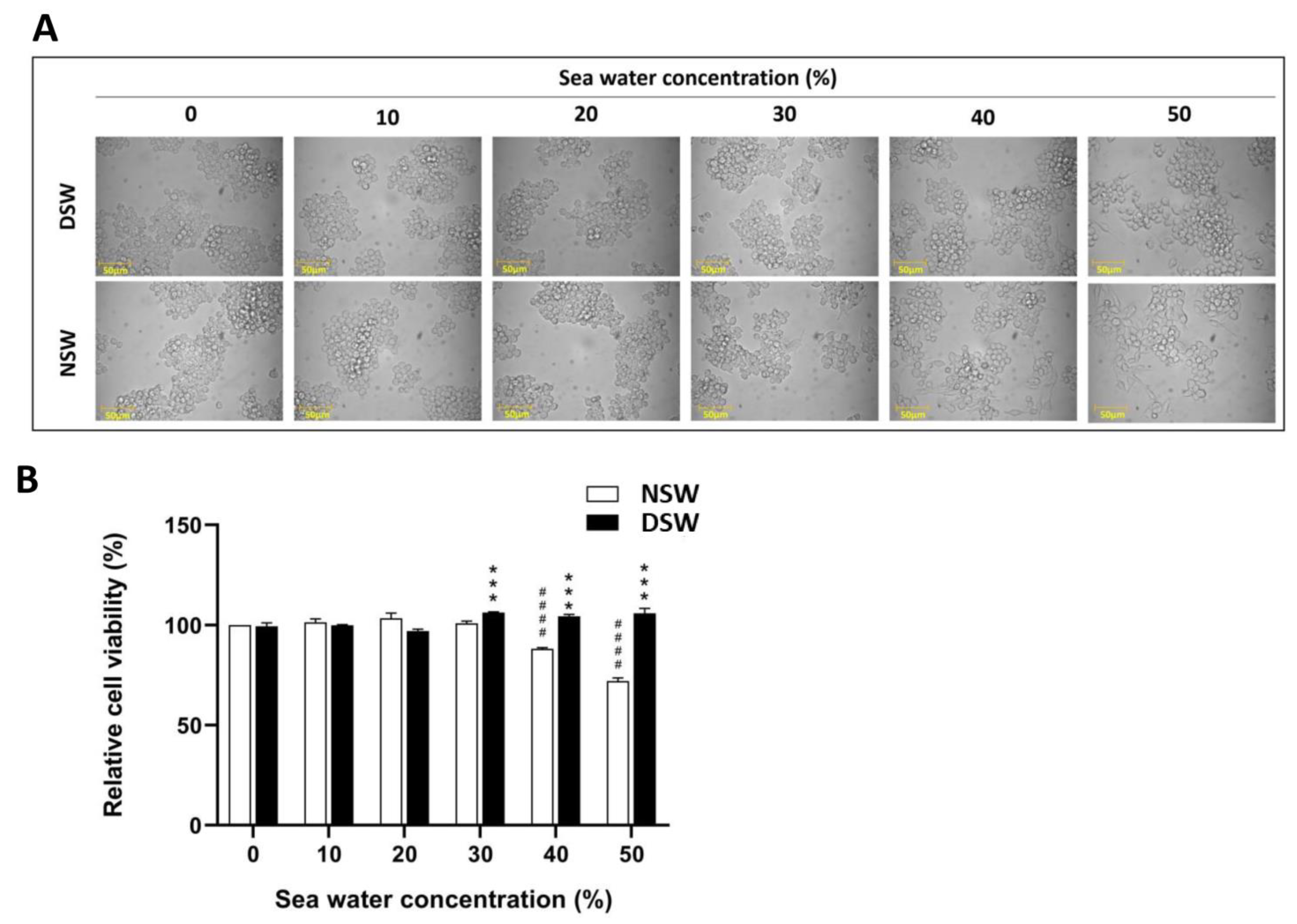
3.1. DSW Enhances the Viability of RAW 264.7 Macrophage Cells

Macrophages are infiltrated or tissue-resident immune cells crucially important in innate immunity for tissue repair after injury upon pathogen infections or other environmental challenges [22]. To determine the cytotoxicity of DSW in vitro, RAW 264.7 cells were cultured in culture media with different concentrations (0, 10, 20, 30, 40, and 50%) of DSW or NSW for 24 h. Thereafter, cell morphology was observed, and viable cells were analyzed using the MTT assay (Figure 1). Cell images show that there is no significant effect of DSW on the morphology of RAW 264.7 murine macrophage cells until 30% concentration and NSW until 20% concentration (Figure 1A). However, 40 and 50% of DSW-treated cells and 30 to 50% of NSW-treated cells show morphological differences with activation. Interestingly, MTT assay results reveal that the increasing concentration of DSW enhances the cell viability, while NSW reduces the cell viability (Figure 1B). Similarly, Maehira et al. show that DSW can enhance the viability of osteoblastic cells [23]. Furthermore, Chun et al. also found that refined DSW has no cytotoxicity effect on RAW 264.7 murine macrophage cells [12]. Taken together, our results indicated that up to 50% concentration, there is no cytotoxic effect of DSW on RAW 264.7 murine macrophage cells, and cell viability is significantly increased. However, increasing the concentration of DSW changes the morphology of RAW 264.7 murine macrophage cells. A similar observation was found in a previous study that macrophages have the capability to proliferate and activate with IL-4 treatment [24]. However, the mechanism of DSW in the above-mentioned scenario must be investigated.

3.2. DSW Reduces LPS-Induced Apoptosis in RAW 264.7 Macrophage Cells

LPS is the major activating mediator of macrophage [12]. LPS triggers the toll-like receptor 4 (TLR4) and activates pro-inflammatory gene transcription [25]. Moreover, NO and TNF are primary mediators released by macrophages, which are crucial in the apoptosis process [26]. To investigate the effect of DSW on LPS-induced apoptosis, RAW 264.7 murine macrophage cells were cultured with 25 and 50% of NaCl, NSW, or DSW in a cell culture medium. Two hours later, cells were treated with 500 ng/mL of LPS for 24 h. Thereafter, cell morphology was observed, and relative cell viability was analyzed using the MTT assay (Figure 2). Results reveal that the LPS-treated cells cultured in 25 or 50% of NaCl and NSW show activation and cell death (Figure 2A). Relative cell viability results elicited that LPS-treated cells cultured in 25 and 50% of DSW-containing culture medium showed significantly high cell viability compared to the control, NaCl-, and NSW-treated cells (Figure 2B). The above analysis indicates that DSW could suppress LPS-induced apoptosis in RAW 264.7 murine macrophage cells.

3.3. DSW Suppresses LPS-Induced NO Production and Pro-Inflammatory Cytokine Induction

Macrophages express indispensable inflammatory mediators such as nitric oxide synthase (iNOS), chemokines, and pro-inflammatory cytokines including TNF-α, IL-1β, IL-6, and IL-10, which are paramount in different chronic and acute inflammatory diseases [27]. In an abnormal situation, iNOS-synthesized NO overproduction is a prominent pro-inflammatory mediator that induces inflammation [28]. LPS is considered a stimulant that induces NO production via MAPKs and NF-kB pathways [27]. Therefore, we stimulated NO production in RAW 264.7 murine macrophage cells that were cultured in a cell culture medium with 25 or 50% of NaCl, NSW, or DSW by treating 500 ng/mL of LPS. After 24 h, we evaluated the effect of DSW on NO production compared to the NaCl, NSW, and control groups. Results elucidated that both 25 and 50% concentrations of DSW show low NO production compared to the NaCl and NSW (Figure 3A). The important observation is the cells stimulated by LPS in 25% DSW-containing cell culture medium show a significant reduction in NO production compared to the control group. However, even though the 50% DSW shows a reduction in NO production, it is significantly higher than the control group. Thus, this reveals that DSW shows negative regulatory effects against NO production in the inflammation process.
Moreover, we have investigated the pro-inflammatory cytokine production by RAW 264.7 murine macrophage cells upon LPS treatment (Figure 3B–D). To investigate that, we performed RT-qPCR analysis to find out the mRNA transcription levels of TNF-α, IL-1β, and IL-6 in the cells with the same experimental conditions as above. Results show that RAW 264.7 murine macrophage cells cultured in both concentrations of DSW reduce TNF-α, IL-1β, and IL-6 significantly compared to the control groups and other treatments. In accordance with our results, Chun et al. also discovered that DSW can abolish the production of TNF-α, IL-1β, IL-6, and IL-10 [12]. Taken together, our investigation reveals that DSW has the potential to impair NO and pro-inflammatory cytokine production in the inflammatory process of macrophage cells.

3.4. DSW Shows Less Lethal Effect on Zebrafish Larvae

Zebrafish is an ideal model for biomedical research compared to other vertebrates due to its fully discovered genome, easy alteration of DNA makeup, external fertilization, high fecundity, high growth rate, and transparent embryo and larval stages [29,30]. Water quality is a pivotal factor to be considered in zebrafish research as it affects every aspect of zebrafish physiology. The most important parameters are temperature, conductivity, pH, hardness, alkalinity, nitrogenous wastes, and dissolved gases [31]. Therefore, before investigating the immune effect of DSW in zebrafish, we analyzed whether there is any lethal effect of DSW on zebrafish embryos and larvae in different concentrations.
To perform this, we cultured four cell stage zebrafish eggs in E3 medium with 10, 20, 30, 40, and 50% of NaCl, NSW, or DSW and counted the dead embryos and larvae until 6 dpf (Figure 4). Thereafter, the survival percentage was calculated and graphed. Results show that culturing of zebrafish embryos and larvae in 10% and 20% NaCl-, NSW-, or DSW-containing E3 medium shows more than 80% survival, indicating that all the treatments show little effect on the mortality of zebrafish embryos and larvae in 10 and 20% concentrations (Figure 4A,B). Moreover, in 30% of treatments, NSW shows less than 30% of survival with high mortality (Figure 4C). However, NaCl or DSW treatments do not show a significant effect on the mortality of fish. Survival percentages of 40% and 50% of NaCl-, NSW-, or DSW-treated zebrafish embryos and larvae show similar patterns. However, NSW treatment shows the highest mortality in both 40 and 50% concentrations with 0% survival at 1 dpf (Figure 4C,D). Also, NaCl treatment shows 0% survival at 6 dpf. However, DSW shows the lowest mortality even in 50% concentration by showing a survival percentage of not less than 50% (Figure 4E). By considering these results, we have decided to use 15% and 30% concentrations of NaCl, NSW, or DSW for future experiments.

3.5. DSW Reduces the Macrophage and Neutrophil Recruitment to the Amputated Fin Fold

Fin fold amputation is a method to create an inflammation in a zebrafish model to study inflammation and regeneration [32]. However, excessive inflammation leads to damage to healthy organs and tissues [1]. Macrophages and neutrophils play a pivotal task in inflammatory diseases [22]. Therefore, in the present study, we have investigated the effect of DSW on macrophage and neutrophil recruitment to the amputated fin fold.
To perform this, 3 dpf Tg(mpeg:EGFP) (macrophage with green fluorescence) and Tg(mpx:mCherry) (neutrophil with red florescence) zebrafish larvae fin folds were amputated and introduced to the E3 medium containing 15 or 30% of NaCl, NSW, or DSW. Post-amputation (hpa) fluorescence images were obtained 12 and 24 h after, and the average number of macrophages and neutrophils were calculated and graphed (Figure 5). Results reveal that both macrophage and neutrophil recruitment to the amputated fin fold of the larvae cultured in E3 medium containing DSW is lower in both concentrations (15 and 30%) compared to the control, NaCl-, or NSW-containing E3 medium. These results demonstrated that DSW may have anti-inflammatory properties by hindering the recruitment of macrophages and neutrophils to the inflammatory site.

3.6. DSW Reduces LPS-Induced Pro-Inflammatory Cytokine Production

LPS-induced inflammation results in cytokine production [33]. LPS is primarily identified by toll-like receptor 4 (TLR-4) followed by downstream signaling stimulating the production of cytokines, such as TNF-α, IL-1β, and IL-6 [33,34]. Therefore, to investigate the effect of DSW on pro-inflammatory cytokine transcription in zebrafish larvae, we cultured 3 dpf zebrafish larvae in 15 and 30% of NaCl, NSW, or DSW in E3 water, and the fish were treated with 100 µg/mL of LPS. Then, fish were harvested at 12 and 24 h post-treatment (hpt) to perform RT-qPCR to analyze the transcription levels of tnf-α, il-1β, and il-6 (Figure 6). Results demonstrated that zebrafish larvae cultured in both 15 and 30% DSW concentrations show a significant reduction in tnf-α transcription level compared to the control fish upon LPS treatment at 12 hpt (Figure 6A). However, 24 hpt results show that only 15% of DSW has the ability to suppress tnf-α transcription compared to the control upon LPS treatment. Moreover, all the treatments (NaCl, NSW, or DSW) suppress the LPS-induced il-1β transcription level of zebrafish larvae in both concentrations at 12 and 24 hpt (Figure 6B). In the case of il-6 transcription in zebrafish cultured in different culture mediums with NaCl, NSW, or DSW, both concentrations show low transcription levels at 12 hpt compared to the control (Figure 6C). However, 24 h later, 30% DSW treatment shows the lowest il-6 expression level upon LPS treatment while other treatments show high or non-significant differences with the control group. Taken together, the above results reveal that DSW has the ability to suppress pro-inflammatory cytokine production upon LPS-induced inflammation.
Currently, there are only drinking water bottles made from deep-sea mineral water (DSW), but several studies have explored its various health benefits [15,16,17,35,36]. The effects of DSW can vary depending on the mineral composition of the water sourced from different locations. Therefore, ongoing research is necessary to uncover the health benefits of DSW from various regions. At present, the industry primarily focuses on producing DSW water bottles as a commercial product. However, given the identified health benefits, scientists could conduct further research to develop direct medical products, such as injections or oral medications, by concentrating the minerals found in DSW.

4. Conclusions

In conclusion, the present study reveals that DSW extracted from the South Korean Sea has the ability to suppress the inflammation in RAW 264.7 murine macrophage cells and the zebrafish model by hindering the excessive activation of pro-inflammatory cytokines and NO production. Furthermore, DSW negatively regulates the recruitment of neutrophils and macrophages to the inflammatory site that was induced by fin fold amputation in zebrafish larvae. Moreover, DSW does not show a cytotoxic effect in concentrations less than 40% in both RAW 264.7 murine macrophage cells and zebrafish larvae. Taken together, our results concluded that the DSW tested in the present study is a potential candidate for use as an anti-inflammatory drug to treat inflammatory diseases.

Author Contributions

H.M.S.M.W.: conceptualization, methodology, investigation, formal analysis, and writing—original draft. B.P.M.V.J.: methodology and investigation. D.-S.M.: resources and methodology. C.-G.K.: resources and methodology. S.J.: methodology, investigation, and writing—review and editing. J.L.: resources, supervision, writing—review and editing, project administration, and funding acquisition. All authors have read and agreed to the published version of the manuscript.

Funding

This research was supported by the Korea Institute of Marine Science & Technology Promotion (KIMST) and funded by the Ministry of Oceans and Fisheries (RS-2022-KS221543).

Institutional Review Board Statement

This study was conducted according to the guidelines set by the Animal Experiment Ethics Committee at Jeju National University (Approval Number: 2019-0014).

Informed Consent Statement

Not applicable.

Data Availability Statement

The data from this study are accessible upon reasonable request from the corresponding author.

Acknowledgments

This research was supported by the Korea Institute of Marine Science & Technology Promotion (KIMST) funded by the Ministry of Oceans and Fisheries (RS-2022-KS221543).

Conflicts of Interest

The authors declare they have no competing financial interests or personal relationships that could have appeared to influence the work reported in this paper.

References

  1. Pahwa, R.; Goyal, A.; Jialal, I. Chronic Inflammation. In StatPearls; StatPearls Publishing: Treasure Island, FL, USA, 2022. [Google Scholar]
  2. Fujiwara, N.; Kobayashi, K. Macrophages in Inflammation. Curr. Drug Targets—Inflamm. Allergy 2005, 4, 281–286. [Google Scholar] [CrossRef]
  3. Karunarathne, W.A.H.M.; Lee, K.T.; Choi, Y.H.; Jin, C.-Y.; Kim, G.-Y. Anthocyanins Isolated from Hibiscus Syriacus L. Attenuate Lipopolysaccharide-Induced Inflammation and Endotoxic Shock by Inhibiting the TLR4/MD2-Mediated NF-κB Signaling Pathway. Phytomedicine 2020, 76, 153237. [Google Scholar] [CrossRef] [PubMed]
  4. Palmieri, E.M.; McGinity, C.; Wink, D.A.; McVicar, D.W. Nitric Oxide in Macrophage Immunometabolism: Hiding in Plain Sight. Metabolites 2020, 10, 429. [Google Scholar] [CrossRef] [PubMed]
  5. Kumar, R.; Clermont, G.; Vodovotz, Y.; Chow, C.C. The Dynamics of Acute Inflammation. J. Theor. Biol. 2004, 230, 145–155. [Google Scholar] [CrossRef] [PubMed]
  6. Ryu, J.-H.; Ahn, H.; Kim, J.Y.; Kim, Y.-K. Inhibitory Activity of Plant Extracts on Nitric Oxide Synthesis in LPS-Activated Macrophages. Phytother. Res. 2003, 17, 485–489. [Google Scholar] [CrossRef] [PubMed]
  7. Liu, X.; Yin, S.; Chen, Y.; Wu, Y.; Zheng, W.; Dong, H.; Bai, Y.; Qin, Y.; Li, J.; Feng, S.; et al. LPS-induced Proinflammatory Cytokine Expression in Human Airway Epithelial Cells and Macrophages via NF-κB, STAT3 or AP-1 Activation. Mol. Med. Rep. 2018, 17, 5484–5491. [Google Scholar] [CrossRef]
  8. Yoon, S.-B.; Lee, Y.-J.; Park, S.K.; Kim, H.-C.; Bae, H.; Kim, H.M.; Ko, S.-G.; Choi, H.Y.; Oh, M.S.; Park, W. Anti-Inflammatory Effects of Scutellaria Baicalensis Water Extract on LPS-Activated RAW 264.7 Macrophages. J. Ethnopharmacol. 2009, 125, 286–290. [Google Scholar] [CrossRef]
  9. Vogelpoel, L.T.C.; Baeten, D.L.P.; de Jong, E.C.; den Dunnen, J. Control of Cytokine Production by Human Fc Gamma Receptors: Implications for Pathogen Defense and Autoimmunity. Front. Immunol. 2015, 6, 79. [Google Scholar] [CrossRef]
  10. Matz, H.; Orion, E.; Wolf, R. Balneotherapy in Dermatology. Dermatol. Ther. 2003, 16, 132–140. [Google Scholar] [CrossRef]
  11. Lee, K.-S.; Chun, S.-Y.; Lee, M.-G.; Kim, S.; Jang, T.-J.; Nam, K.-S. The Prevention of TNF-α/IFN-γ Mixture-Induced Inflammation in Human Keratinocyte and Atopic Dermatitis-like Skin Lesions in Nc/Nga Mice by Mineral-Balanced Deep Sea Water. Biomed. Pharmacother. 2018, 97, 1331–1340. [Google Scholar] [CrossRef]
  12. Chun, S.-Y.; Lee, K.-S.; Nam, K.-S. Refined Deep-Sea Water Suppresses Inflammatory Responses via the MAPK/AP-1 and NF-κB Signaling Pathway in LPS-Treated RAW 264.7 Macrophage Cells. Int. J. Mol. Sci. 2017, 18, 2282. [Google Scholar] [CrossRef] [PubMed]
  13. Nam, J.; Kim, K.J.; Park, G.; Kim, B.G.; Jeong, G.-H.; Jeon, J.; Hurh, B.S.; Kim, J.Y. Anti-Inflammatory Properties of Mineral-Balanced Deep Sea Water in In-Vitro and In-Vivo Models of Inflamed Intestinal Epithelium. Appl. Sci. 2020, 10, 5183. [Google Scholar] [CrossRef]
  14. Sheu, M.-J.; Chou, P.-Y.; Lin, W.-H.; Pan, C.-H.; Chien, Y.-C.; Chung, Y.-L.; Liu, F.-C.; Wu, C.-H. Deep Sea Water Modulates Blood Pressure and Exhibits Hypolipidemic Effects via the AMPK-ACC Pathway: An in vivo Study. Mar. Drugs 2013, 11, 2183–2202. [Google Scholar] [CrossRef]
  15. He, S.; Hao, J.; Peng, W.; Qiu, P.; Li, C.; Guan, H. Modulation of Lipid Metabolism by Deep-Sea Water in Cultured Human Liver (HepG2) Cells. Mar. Biotechnol. 2014, 16, 219–229. [Google Scholar] [CrossRef]
  16. Shen, J.-L.; Hsu, T.-C.; Chen, Y.-C.; Hsu, J.-D.; Yang, L.-C.; Tsai, F.-J.; Li, C.-C.; Cheng, Y.-W.; Huang, C.-Y.; Tzang, B.-S. Effects of Deep-Sea Water on Cardiac Abnormality in High-Cholesterol Dietary Mice: Cardiac Protective Effects of Deep-Sea Water. J. Food Biochem. 2012, 36, 1–11. [Google Scholar] [CrossRef]
  17. Katsuda, S.; Yasukawa, T.; Nakagawa, K.; Miyake, M.; Yamasaki, M.; Katahira, K.; Mohri, M.; Shimizu, T.; Hazama, A. Deep-Sea Water Improves Cardiovascular Hemodynamics in Kurosawa and Kusanagi-Hypercholesterolemic (KHC) Rabbits. Biol. Pharm. Bull. 2008, 31, 38–44. [Google Scholar] [CrossRef]
  18. Ha, B.G.; Park, J.; Shin, E.J.; Shon, Y.H. Effects of Balanced Deep-sea Water on Adipocyte Hypertrophy and Liver Steatosis in High-fat, Diet-induced Obese Mice. Obesity 2014, 22, 1669–1678. [Google Scholar] [CrossRef] [PubMed]
  19. Kumar, P.; Nagarajan, A.; Uchil, P.D. Analysis of Cell Viability by the MTT Assay. Cold Spring Harb. Protoc. 2018, 2018, pdb-rot095505. [Google Scholar] [CrossRef]
  20. Livak, K.J.; Schmittgen, T.D. Analysis of Relative Gene Expression Data Using Real-Time Quantitative PCR and the 2−ΔΔCT Method. Methods 2001, 25, 402–408. [Google Scholar] [CrossRef]
  21. Shanaka, K.A.S.N.; Jung, S.; Madushani, K.P.; Wijerathna, H.M.S.M.; Neranjan Tharuka, M.D.; Kim, M.-J.; Lee, J. Generation of Viperin-Knockout Zebrafish by CRISPR/Cas9-Mediated Genome Engineering and the Effect of This Mutation under VHSV Infection. Fish Shellfish Immunol. 2022, 131, 672–681. [Google Scholar] [CrossRef]
  22. Watanabe, S.; Alexander, M.; Misharin, A.V.; Budinger, G.R.S. The Role of Macrophages in the Resolution of Inflammation. J. Clin. Investig. 2019, 129, 2619–2628. [Google Scholar] [CrossRef] [PubMed]
  23. Maehira, F.; Iinuma, Y.; Eguchi, Y.; Miyagi, I.; Teruya, S. Effects of Soluble Silicon Compound and Deep-Sea Water on Biochemical and Mechanical Properties of Bone and the Related Gene Expression in Mice. J. Bone Miner. Metab. 2008, 26, 446–455. [Google Scholar] [CrossRef]
  24. Jenkins, S.J.; Ruckerl, D.; Cook, P.C.; Jones, L.H.; Finkelman, F.D.; van Rooijen, N.; MacDonald, A.S.; Allen, J.E. Local Macrophage Proliferation, Rather than Recruitment from the Blood, Is a Signature of T H 2 Inflammation. Science 2011, 332, 1284–1288. [Google Scholar] [CrossRef]
  25. Valledor, A.F.; Comalada, M.; Santamaría-Babi, L.F.; Lloberas, J.; Celada, A. Macrophage Proinflammatory Activation and Deactivation: A Question of Balance. Adv. Immunol. 2010, 108, 1–20. [Google Scholar] [CrossRef] [PubMed]
  26. Xaus, J.; Comalada, M.; Valledor, A.F.; Lloberas, J.; López-Soriano, F.; Argilés, J.M.; Bogdan, C.; Celada, A. LPS Induces Apoptosis in Macrophages Mostly through the Autocrine Production of TNF-α. Blood 2000, 95, 3823–3831. [Google Scholar] [CrossRef]
  27. Yoo, M.-S.; Shin, J.-S.; Choi, H.-E.; Cho, Y.-W.; Bang, M.-H.; Baek, N.-I.; Lee, K.-T. Fucosterol Isolated from Undaria Pinnatifida Inhibits Lipopolysaccharide-Induced Production of Nitric Oxide and pro-Inflammatory Cytokines via the Inactivation of Nuclear Factor-κB and P38 Mitogen-Activated Protein Kinase in RAW264.7 Macrophages. Food Chem. 2012, 135, 967–975. [Google Scholar] [CrossRef]
  28. Sharma, J.N.; Al-Omran, A.; Parvathy, S.S. Role of Nitric Oxide in Inflammatory Diseases. Inflammopharmacol 2007, 15, 252–259. [Google Scholar] [CrossRef]
  29. Teame, T.; Zhang, Z.; Ran, C.; Zhang, H.; Yang, Y.; Ding, Q.; Xie, M.; Gao, C.; Ye, Y.; Duan, M.; et al. The Use of Zebrafish (Danio Rerio) as Biomedical Models. Anim. Front. 2019, 9, 68–77. [Google Scholar] [CrossRef] [PubMed]
  30. Choi, T.-Y.; Choi, T.-I.; Lee, Y.-R.; Choe, S.-K.; Kim, C.-H. Zebrafish as an Animal Model for Biomedical Research. Exp. Mol. Med. 2021, 53, 310–317. [Google Scholar] [CrossRef]
  31. Hammer, H.S. Water Quality For Zebrafish Culture. In The Zebrafish in Biomedical Research; Elsevier: Amsterdam, The Netherlands, 2020; pp. 321–335. ISBN 978-0-12-812431-4. [Google Scholar]
  32. Xie, Y.; Meijer, A.H.; Schaaf, M.J.M. Modeling Inflammation in Zebrafish for the Development of Anti-Inflammatory Drugs. Front. Cell Dev. Biol. 2021, 8, 620984. [Google Scholar] [CrossRef]
  33. Martins, G.R.; Gelaleti, G.B.; Moschetta, M.G.; Maschio-Signorini, L.B.; Zuccari, D.A.P.D.C. Proinflammatory and Anti-Inflammatory Cytokines Mediated by NF- κ B Factor as Prognostic Markers in Mammary Tumors. Mediat. Inflamm. 2016, 2016, 1–10. [Google Scholar] [CrossRef] [PubMed]
  34. Zhao, J.; Bi, W.; Xiao, S.; Lan, X.; Cheng, X.; Zhang, J.; Lu, D.; Wei, W.; Wang, Y.; Li, H.; et al. Neuroinflammation Induced by Lipopolysaccharide Causes Cognitive Impairment in Mice. Sci. Rep. 2019, 9, 5790. [Google Scholar] [CrossRef] [PubMed]
  35. Hwang, H.S.; Kim, H.A.; Lee, S.H.; Yun, J.W. Anti-Obesity and Antidiabetic Effects of Deep Sea Water on Ob/Ob Mice. Mar. Biotechnol. 2009, 11, 531–539. [Google Scholar] [CrossRef] [PubMed]
  36. Lewicki, S.; Zdanowski, R.; Krzyżowska, M.; Lewicka, A.; Dębski, B.; Niemcewicz, M.; Goniewicz, M. The Role of Chromium III in the Organism and Its Possible Use in Diabetes and Obesity Treatment. Ann. Agric. Environ. Med. 2014, 21, 331–335. [Google Scholar] [CrossRef]
Figure 1. DSW increases the RAW 264.7 cell viability in high concentrations compared to the NSW. RAW 264.7 cells were treated with different concentrations of DSW or NSW for 24 h. (A) cellular morphology was captured by using phase-contrast microscope and (B) relative cell viability was determined by using MTT assay. Data are indicated as mean ± SD (n = 2). Statistical significances (p < 0.05) between control (0%, normal-sea water) and each treatment were calculated using Student’s t-test. ( ***, p ≤ 0.001; ####, p ≤ 0.0001).
Immuno 04 00022 g001
Figure 2. DSW reduces LPS-induced apoptosis in RAW 264.7 cells. RAW 264.7 cells were cultured with 25 or 50% of NaCl, NSW, or DSW with cell culture medium and treated with 500 ng/mL LPS for 24 h. (A) Cellular morphology was captured by using a phase-contrast microscope and (B) relative cell viability was determined by using MTT assay. Data are indicated as mean ± SD (n = 2). Statistical significances (p < 0.05) between control (oh, normal-sea water) and each treatment were calculated using Student’s t-test. (ns, p > 0.05; *, p ≤ 0.05; **, p ≤ 0.01; ***, p ≤ 0.001; ****, p ≤ 0.0001).
Immuno 04 00022 g002
Figure 3. DSW suppresses LPS-induced NO production and TNF-α, IL-1β, and IL-6 mRNA expression levels in RAW 264.7 cells. RAW 264.7 cells were cultured with 25 or 50% of NaCl, NSW, or DSW in cell culture medium and treated with 500 ng/mL LPS for 24 h. Another similar group was kept untreated. (A) NO production percentage and (B) TNF-α, (C) IL-1β, and (D) IL-6 relative mRNA expression levels were analyzed by RT-qPCR. Data are indicated as mean ± SD (n = 2). Statistical significances (p < 0.05) between control and each treatment were calculated using Student’s t-test. (ns, p > 0.05; *, p ≤ 0.05; **, p ≤ 0.01; ***, p ≤ 0.001; ****, p ≤ 0.0001).
Immuno 04 00022 g003
Figure 4. Survival percentage of zebrafish eggs and larvae cultured in various concentrations of NaCl, NSW, or DSW until 6dpf. Four cell stage zebrafish eggs were cultured in 0, 10, 20, 30, 40, and 50% of NaCl, NSW, or DSW in E3 water, and surviving eggs and larvae were counted daily until 6 dpf. Survival percentage was calculated in (A) 10, (B) 20, (C) 30, (D) 40, and (E) 50% treatment groups. Data are indicated as mean values (n = 15).
Immuno 04 00022 g004
Figure 5. DSW hinders the macrophage and neutrophil recruitment to the amputated fin fold of Tg(mpeg:EGFP) and Tg(mpx:mCherry) zebrafish larvae. Fin fold of 3 dpf zebrafish larvae was amputated and cultured in 15 and 30% of NaCl, NSW, or DSW in E3 water, and (A) green (macrophages) and (B) red (neutrophils) fluorescence images were obtained at 12 and 24 h. Average number of (C) macrophages and (D) neutrophils recruited to the area within the 100 µm range from the edge of the amputated fin of Tg(mpeg:EGFP) and Tg(mpx:mCherry) larvae were calculated. Data are indicated as mean ± SD (n = 10). Statistical significances (p < 0.05) between control and each treatment were calculated using Student’s t-test. (ns, p > 0.05; *, p ≤ 0.05; **, p ≤ 0.01; ***, p ≤ 0.001; ****, p ≤ 0.0001).
Immuno 04 00022 g005
Figure 6. DSW suppressed the cytokine production in zebrafish larvae upon LPS treatment. The 3 dpf zebrafish larvae were cultured in 15 and 30% of NaCl, NSW, or DSW in E3 water and the fish were treated with LPS (100 µg/mL); at the same time, fish in the control group were cultured in only E3 water. Similar groups were kept untreated and (A) tnf-α, (B) il-1β, and (C) il-6 mRNA expression levels were analyzed by RT-qPCR after 12 and 24 h post-treatment. Data are indicated as mean ± SD (n = 3). Statistical significances (p < 0.05) between control and each treatment were calculated using Student’s t-test. (ns, p > 0.05; *, p ≤ 0.05; **, p ≤ 0.01; ***, p ≤ 0.001; ****, p ≤ 0.0001).
Immuno 04 00022 g006
Table 1. Seawater chemical parameters.
Type of SeawaterNa+ (mg/L)Mg2+ (mg/L)Ca2+ (mg/L)K+ (mg/L)Salinity (ppt)
Deep-sea mineral water (Desalinated concentrate, DSW)70.61175.2448.019.418
Normal seawater (NSW) (Salinity adjusted to 18 ppt)5484.4582.6175.2164.918
Disclaimer/Publisher’s Note: The statements, opinions and data contained in all publications are solely those of the individual author(s) and contributor(s) and not of MDPI and/or the editor(s). MDPI and/or the editor(s) disclaim responsibility for any injury to people or property resulting from any ideas, methods, instructions or products referred to in the content.

