Article

Unlocking the Puzzle: Investigating the Role of Interleukin 17 Genetic Polymorphisms, Circulating Lymphocytes, and Serum Levels in Venezuelan Women with Recurrent Pregnancy Loss

by
Jenny Valentina Garmendia
1,2,*,
Isaac Blanca
1,
María Johanna Peña
1,
Claudia Valentina De Sanctis
2 and
Juan Bautista De Sanctis
1,2,*
1
Institute of Immunology, Faculty of Medicine, Universidad Central de Venezuela, Caracas 1053, Venezuela
2
Institute of Molecular and Translational Medicine, Faculty of Medicine and Dentistry, Palacky University, 77900 Olomouc, Czech Republic
*
Authors to whom correspondence should be addressed.
Immuno 2024, 4(4), 301-311; https://doi.org/10.3390/immuno4040019
Submission received: 2 August 2024 / Revised: 24 September 2024 / Accepted: 24 September 2024 / Published: 26 September 2024
(This article belongs to the Section Reproductive Immunology)

Abstract

In recurrent pregnancy loss (RPL), peripheral and local immune cells are activated, decreasing the leukocyte tolerogenic response in the uterus and decidua. The aim was to examine the role of IL-17 in RPL critically. The study included genetic polymorphism, the analysis of the number of circulating IL-17 lymphocyte populations, before and after cell priming, serum cytokine quantification, and the assessment of T-reg cells in a group of 50 RPL and 50 normal women from the admixed Venezuelan population. The study found no differences in the genetic polymorphisms rs2275913 and rs763780. However, when IL-17+ cell populations of controls and RPL patients were compared, a significant increase was observed in the cell populations CD3+ and CD4+ (p < 0.001), while the contrary was recorded in CD8+ and CD56+ cells. Upon cell priming, all IL-17+ populations were significantly decreased (p < 0.001) in RPL patients compared to controls. The increase in IL-17A in the serum of RPL patients may be due to the CD4+ population, while cell exhaustion after activation could be responsible for decreased CD8+ cell population. The number of CD4CD25 FoxP3+ cells was significantly reduced (p < 0.001), and the number of activated HLADR+ cells was significantly increased (p < 0.001) in RPL patients. The absence of differences in the genetic polymorphism compared to controls suggests that biological factors influence IL-17 levels in RPL patients. This finding has significant implications for the understanding and potential treatment of RPL.

1. Introduction

Recurrent pregnancy loss (RPL) is defined explicitly as the occurrence of two or more miscarriages before reaching 20 weeks of gestation [1]. The global incidence of RPL is estimated at 2% [1,2,3]. The causes include genetic, anatomical, endocrine, infectious factors, and immune abnormalities such as increased levels of antibodies and increased circulating NK cell cytotoxicity [1,2,3,4]. Compared to normal pregnancies, high Interleukin-17 (IL-17) circulating levels were found in RPL patients [5,6,7]. The cytokine is involved in the endometrium’s extravasation and accumulation of neutrophils. It worsens local inflammation and damage and thereby increases the risk of miscarriage [5,6,7]. This suggests that IL-17 may play a crucial role in the occurrence and progression of RPL [7]. The cell types usually involved in IL-17 production are CD3CD4 cells. A subpopulation of CD3CD4 T helper (Th) cells that produce IL17 is defined as Th17. On the other hand, other cells also transcribe and secrete IL-17. The cytokine is produced by CD8 cells, natural killer (NK) cells, defined as CD3−CD56+CD16+ cells, natural killer T (NKT) cells, CD3+CD56+CD16−, and T γδ cells, and CD3 cells with γδ T cell receptor instead of the classical αβ [8,9]. NK, NKT, and Tγδ cells are mainly involved in innate immune and adaptative immune responses [7,8]. There are mixed reports on NKT cells, although this cell subpopulation could also be involved in RPL [9].
Over the past decade, many studies have explored the connection between IL-17 gene polymorphism and RPL [10,11,12,13,14,15,16,17]. Variations in the IL-17A and IL-17F genes have been associated with the development of various human diseases [18]. Specifically, polymorphic loci known as rs2275913 and rs763780 in the coding regions of IL-17A and IL-17F are closely linked to IL-17 secretion [19]. However, these findings have been conflicting and have sparked controversy based on the possible impact of ethnic and population differences on genetic polymorphisms and backgrounds [18,19,20].
There are no specific data on Venezuela concerning the risk of RPL; however, there is a general consensus that the rates may be similar to those of other populations. Part of the Venezuelan population is genetically admixed, with a prevalence of Caucasian genes but mixed with African and Amerindian groups [21,22,23,24]. This admixed population differs from other Latin American countries, probably due to gene segregation among Eastern African and Amerindian populations [21,22,23,24].
Most reports regarding RPL and IL-17 refer to the local production of the cytokine by stimulated cells [12,13], and only a few have dealt with the circulating number of Th17 cells [25]. Moreover, no reports have addressed the relationship between genetic IL-17 SNP, circulating lymphocytes, and the response to cell activation.
The present report aims to analyze the relationship between genetic polymorphism, circulating IL-17 cells, the effect of stimulation on intracellular IL-17, and the circulating levels of IL-17A using two groups with identical genetically admixed conditions: controls and patients with RPL.

2. Materials and Methods

2.1. Human Samples

The study was a case–control study involving patients who met the criteria for RPL: patients reported spontaneous miscarriages of 2–3 pregnancies without medically defined reasons. The patients with RPL underwent a complete clinical examination by internists, clinical immunologists, and gynecologists. A complete laboratory screening was performed, including immunological assays (autoimmunity panel plus assessment of cell populations by flow cytometry), hormonal analysis, and infectious screening and genetic analysis. Patients with abnormal results were excluded, as well as patients with cancer, severe endometriosis, and vaginal or HPV infection. A total of 50 women with RPL were selected and were not undergoing medical treatment at the time of sample collection.
The study included 50 healthy women with normal pregnancies and without medical conditions such as viral diseases, hypertension, diabetes, metabolic syndrome, or hormonal imbalances. Samples from controls were taken when patients were attending medical checkups by a gynecologist that included routine vaginal and endometrial screening. Only women with normal conditions were included in the control group.
Patients and controls were screened for a panel of autoimmunity markers that included erythrocyte sedimentation rate, C-reactive protein, anti-nuclear antibodies, anti-DNA, anti-citrullinated antibodies, cryoglobulins, circulating immune complex and complement components C3 and C4. All the values recorded were within the normal range in both groups.
Written consent was obtained from all individuals interested in participating in the study. The genetic admixture of both the patients and controls was previously verified [21,22]. The study received approval from the Ethical Committee of the Institute of Immunology, Faculty of Medicine, Caracas, Venezuela (approval number 20052308).
Twelve ml of blood was taken from each individual with a syringe and divided into three parts. One tube with heparin (4 mL) was used for hematological counts and cell stimulation. One tube with EDTA (4 mL) was used for a buffy coat and cDNA extraction, and one EDTA tube (4 mL) was used to obtain plasma samples for the ELISA assay. From the first tube, the sample was divided into two aliquots, one for blood cell counting and assessment of baseline parameters by flow cytometry and one for cell stimulation.

2.2. Antibodies and Reagents

Anti-human IL-17A/F monoclonal mouse IgG1 Clone # 41802 with phycoerythrin (PE) was obtained from R&D Systems (Minneapolis, MN, USA). The following antibodies were from BioLegend (San Diego, CA, USA): anti-CD3 (clone UCHT1), CD4 (clone OKT4), CD8 (clone Leu2), CD25 (clone M-A251), CD56 (QA17A16), CD45RA (clone HI100), CD45RO (clone UCHL1), HLA-DR (clone L243), and FoxP3 (clone 206D). Becton Dickinson (BD, San Jose, CA, USA) Pharm Lyse™ (catalog 555899) kit and the BD Cytofix/Cytoperm™ Plus Fixation/Permeabilization Solution Kit with BD GolgiPlug™ (catalog 555028) were purchased from Becton Dickinson. Cell Activation Cocktail (with Brefeldin A) ™ (catalog # 423304) was purchased from BioLegend.

2.3. Genetic Polymorphism Analysis

On the same day, the cDNA was obtained from the buffy coat of the tube containing EDTA. Genomic DNA isolation was performed using the AxyPrep Blood Genomic DNA Miniprep Kit from Axygen Biosciences, Union City, CA, USA. This specific kit can purify up to 12 µg of genomic DNA in 250 µL of non-coagulated blood. DNA isolation protocol was performed as described by the manufacturer with minor modifications described previously [26]. The samples’ pure DNA (260/280 ratio ≥ 1.8) was expressed in µg/mL for the assays.
The primer sequences were obtained from the literature for (rs2275913) [27] and (rs763780) [28].
primers for IL-17A (rs2275913)
Froward 5′-TCTCCATCTCCATCACCTTTG-3′
Reverse 5′-GTCCAAATCAGCAAGAGCATC-3′,
primers for IL-17F (rs763780),
Forward 5′-CACTGGTGCTCTGATGAGGA-3′
Reverse 5′-CATTGTGCTTTGGCTTGCT-3′
The experimental procedure was slightly modified from previously published protocols [26,27]. Twenty nanograms of DNA was combined with a 40 µL reaction mixture containing 1.5 mM MgCl2, 100 ng of each primer, 500 µM deoxyribonucleoside triphosphate, and 0.6 IU of Taq DNA polymerase (Promega®). The amplification of DNA was achieved through polymerase chain reaction (PCR) using a Mini cycler™ thermal cycler (MJ Research, Waltham, MA, USA). The process involved an initial single cycle of 10 min at 95 °C, followed by 35 cycles of 30 s at 94 °C, 45 s at 60 °C, and 30 s at 72 °C.
Subsequently, all PCR products were incubated with endonucleases XmnI (for polymorphism rs2275913) and NIaIII (for polymorphism rs763780). The PCR products were incubated at 37 °C with the enzyme and visualized using ethidium bromide staining after separation by electrophoresis on a 3.0% agarose gel at 80 V for 100 min. The sequencing service at the Venezuelan Institute for Scientific Research (IVIC) sequenced the amplification products. All the results of the RFLP were confirmed.

2.4. Analysis of Intracellular IL-17 in Different Cell Subpopulations

BD Pharm Lyse™ was used to lyse erythrocytes on fresh samples or after culture. The remaining lymphocytes were washed and adjusted to 250,000 cells/tube. Anti-human IL-17A/F-PE was used for intracellular detection of IL-17. The detection of IL-17 was performed by flow cytometry according to the protocol suggested by the supplier. Then, the cells were labeled extracellularly with anti-CD4, anti-CD8, or anti-CD56 from BioLegend. The analysis was performed using Beckman Coulter’s (Hialeah, FL, USA) EPICS XL apparatus.
Intracellular analysis of FoxP3 was also performed in non-stimulated cells only. After intracellular labeling of FoxP3, the cells were washed and labeled with CD4 and CD25 to determine the cell subpopulation.
To ascertain the effect of cell stimulation, the cells with 1 mL of blood were incubated with the Cell Activation Cocktail for 6 h according to the manufacturer’s protocol. The cells were treated for 12 h with the optimal concentration of the cocktail and then fixed with cytoplasm and permeabilized. Anti-IL17 PE was then added to the permeabilized cells. The cells were then washed and labeled with CD4, CD8, or CD56 to analyze the different subpopulations.

2.5. Determination of Il-17A from Serum Samples and Supernatants

The Quantikine HS ELISA Kit from R&D Systems (Minneapolis, MN, USA) was used to analyze IL-17A in the plasma of controls and RPL patients. The analysis was performed according to the manufacturer’s instructions.

2.6. Statistical Analysis

The minimum sample size was determined to be 45. The calculation was carried out using an online calculator (accessible at www.calculator.net, accessed on 2 April 2024) based on the SNP data showing a 3% frequency in the general population for rs1051740 (referenced at https://opensnp.org, accessed on 2 April 2024).
The calculations were performed using GraphPad Prism version 10. Subsequently, the results were analyzed using Chi-square with Yates’ correction. Both absolute and relative frequencies were employed, with statistical significance attributed to a p-value of less than 0.05. The crude data are in Supplementary Materials.

3. Results

Table 1 shows the population characteristics. The only significant (p < 0.0001) differences were in the number of miscarriages registered in the study. The majority of the controls had two pregnancies.
Table 2 shows the analysis of the SNPs rs2275913, which corresponds to IL-17A, and re763780, which corresponds to IL-17F. There were no statistical differences between controls and RPL patients. The odds ratios (OR) were the same for both groups.
Table 3 illustrates the difference in total leukocytes, different lymphocyte populations, and subpopulations. The number of total leukocytes did not differ between the two groups; however, the number of lymphocytes was increased in the RPL group.
Most analyzed cell populations differed significantly between the groups except for the number of NKT cells. The percentage of CD3 cells in the RPL group had lower significance than that of total lymphocytes. The amount of NK cells in RPL patients was considerably higher; however, the index T cells/NK cells was significantly lower in RPL patients compared to controls. Even though the percentages of CD3CD45RO and CD3CD45RA cells were similar and nonsignificant in both groups, the absolute number was higher for RPL patients. The number of activated lymphocytes, particularly T cells, was significantly higher in RPL patients than in controls.
Figure 1 illustrates the total number of IL-17 cells, T cells, NK cells, and T cell subpopulations before and after stimulation with PMA/ionomycin. Significant differences were recorded in basal CD3 cells before (p < 0.005) and after stimulation (p < 0.0001) in RPL patients. For NK cells, the number of cells in RPL patients was significantly low before and after stimulation (p < 0.0001). However, the increase after priming was 8-fold higher in RPL patients than in controls. The number of basal CD4IL17 cells in RPL patients was higher than in the control group in basal conditions; however, after stimulation, it was significantly lower than in the control group. The controls showed a 2.5-fold increase upon stimulation compared to 1.5-fold in RPL patients. Finally, in both basal and stimulated CD8 cells, the number of IL-17 positive cells in RPL patients was significantly lower with a similar induction upon stimulation, 2.9-fold in controls, and 3.4-fold in RPL patients.
A significant increase in IL17A was observed in the plasma samples of RPL patients compared to controls (Figure 2). This increase did not correlate with the number of cells IL-17A/F detected independently of the population or subpopulation studies (r = 0.18 for total, 0.1 for CD3, 0.15 for CD4, 0.05 for CD8). There was also no significant correlation with the amount of total activated HLADR+ cells; however, a mild significant correlation (r = 0.3, p < 0.05) was observed between the number of CD3/HLADR+ cells and IL-17A levels in RPL patients, but not in controls (r = 0.05, p = 0.6). No correlation was observed between the number of IL17 cells and Treg cells. No other significant correlations were observed with the different markers.
Figure 3 shows the number of CD4 Treg cells. There was a significant reduction in the number of positive cells. This decrease was not correlated with increased CD3 or CD4 (r = 0.2) in patients or controls.

4. Discussion

RPL is a complex medical entity, and it involves two types: primary (no successful pregnancy) and secondary, a successful pregnancy followed by recurrent miscarriage. There is no medical explanation in most cases; the karyotype of the fetus is normal, and there are no accountable medical alterations to explain the miscarriages.
In the endometrium, tolerogenic NK, macrophages, dendritic cells, and T cells are essential to maintain pregnancy [1,2,3,4,5]. However, local exposure to pathogens or vaginal or endometrial bacterial, fungal, or viral infections induces the migration of cells, which compromise the tolerogenic milieu and consequently induce pregnancy termination [1,2,3,4]. IL-17 is a marker of the inflammatory process for cell migration of neutrophils and other subpopulations [5,6,7]; Th17 cells are involved in the rejection of the fetus or affect the implantation of the zygote [5,6,7,8]. There are, however, questions referring to the role of circulating proinflammatory cell populations in the bloodstream of RPL patients. The migration of these cells in the endometrial compartment has not been well analyzed due to the ethical complexity involved. The only available information refers to pregnancies terminated in normal conditions and compares them to pathological ones. A one-time point may not be sufficient for a critical assessment of events.
Lee SK and coworkers [29] showed in the peripheral blood of RPL patients an increased amount of IL17+ cells as compared to CD4 + FoxP3 + cells. Also, the authors showed a positive correlation between those IL-17-producing cells with IFN γ and TNFα positive cells and a decreased number of IL-10-producing cells. The authors monitored IL-17 in total lymphocytes and not in specific subpopulations. The present report indicates that CD3IL-17A levels are increased in patients with RPL, but this increase is not comparable to the amount of circulating IL-17A/F-positive cells. Genetic polymorphism could not explain this phenomenon. Also, it is unclear why there is a difference between the amount of CD4-positive IL17 cells and the CD8IL17 and NKIL17 cell populations. It can be proposed that the decrease in these specific cell populations is due to previous activation. On the other hand, it cannot be discarded that tissue lymphocytes may be responsible for the increase in circulating levels of the cytokine. Further studies are underway to characterize the circulating Th17 in RPL patients.
The effect of cell stimulation is a complex phenomenon since PMA/ionomycin primes cells directly in an antigen-independent manner, promoting the transcription and secretion of cytokines, including IFNγ and IL-17 [30,31,32,33] and stimulating cells aimed to ascertain probable cell activation mechanisms involved with IL-17 in different cell populations. A valid hypothesis is that the circulating cells were already activated and probably exhausted prior to stimulation, particularly CD8 cells. The remarkable increase in IL-17 positiveness in NK cells from RPL patients led us to suggest that IL-17 transcription and secretion are less affected. A possible switch from NK peripheral tolerogenic cells to inflammatory cytotoxic NK cells upon PMA/ionomycin stimulation is probable, along with IL-2 [34].
Since we used total blood for stimulation to recreate the physiological conditions and not in purified cell types, we cannot determine if the secretion of other protein or non-protein intermediates secreted by different cell types are responsible for the decrease in IL-17 production in RPL patients. Based on the data recorded on HLA-DR expression, we propose that PMA/ionomycin may have affected the stimulated cell subpopulations, which were responsible for producing intermediates that suppress the production of IL-17A/F. The amount of IL-17A in the plasma is not accountable for modulating the number of IL-17-positive cells, as we could not reproduce the effects.
It is unclear but probable that the unbalanced cytokines reported in RPL unmask a possible autoimmune disease. However, in our cohort, patients were screened for a panel of autoimmunity markers described in Material and Methods; each patient’s values and those of controls were within the normal range. Patients were not undergoing medical treatment at the time of sample collection, which is critical in these types of studies.
The association of the two genetic polymorphisms IL-17A rs2275913 and IL-17F rs763780 with RPL is debatable [10,11,14,15,16,17]. Essentially, the association reports are contradictory; some suggest that rs763780 is associated with a decreased risk of RPL in the Iranian population, while others suggest that rs2275913 is protective in the same population [10,11,16,20]. Also, contradictory reports have been observed in the Chinese population [15,16,17,18,19]. However, in our admixed population, there was no difference; similar frequencies were observed in both groups. New, well-defined genetic studies are required to determine if there is a relationship between these two polymorphisms and RPL. Moreover, based on the data reported in this manuscript, it is highly improbable that significant changes recorded in the cell populations are due to genetic polymorphisms. The conclusion is based on the dichotomy between the number of circulating cells positive for IL-17 and the values recorded in plasma. The CD4/CD17 subpopulation was similar in both groups, suggesting that the sizes of their Th17 cell subpopulations may be comparable. The decrease in IL-17 in stimulated cells requires attention and should be further studied.
In the present manuscript, our group tried combining genetic analysis with data on cell subpopulation, cell activation, and cytokine detection in plasma patients with RPL. Indeed, several questions remain to be solved. However, this manuscript is one of the few that have reported a complete analysis of genetic polymorphism, cell subpopulations expressing IL-17, circulating IL-17 levels, and the importance of cell stimulation on IL-17. This information is crucial for the admixed, well-defined genetic population that we studied. Even though we are unaware of the possible activation of cells in the endometrial cavity or even in the early stages of implantation or decidua, we can assume peripheral inflammation in RPL, cell activation markers, and IL-17A plasma levels. Eventually, peripheral inflammation affects local inflammation. It can be proposed that the assessment of inflammation markers can be critical in RPL and may serve as an initial focus for future studies in evaluating the role of IL-17 in RPL and the response of those patients to therapy.

5. Conclusions

  • There is no association between the two polymorphisms analyzed, IL-17A rs2275913 and IL-17F rs763780, with RPL. The values recorded from plasma of RPL patients were independent of the genetic polymorphisms.
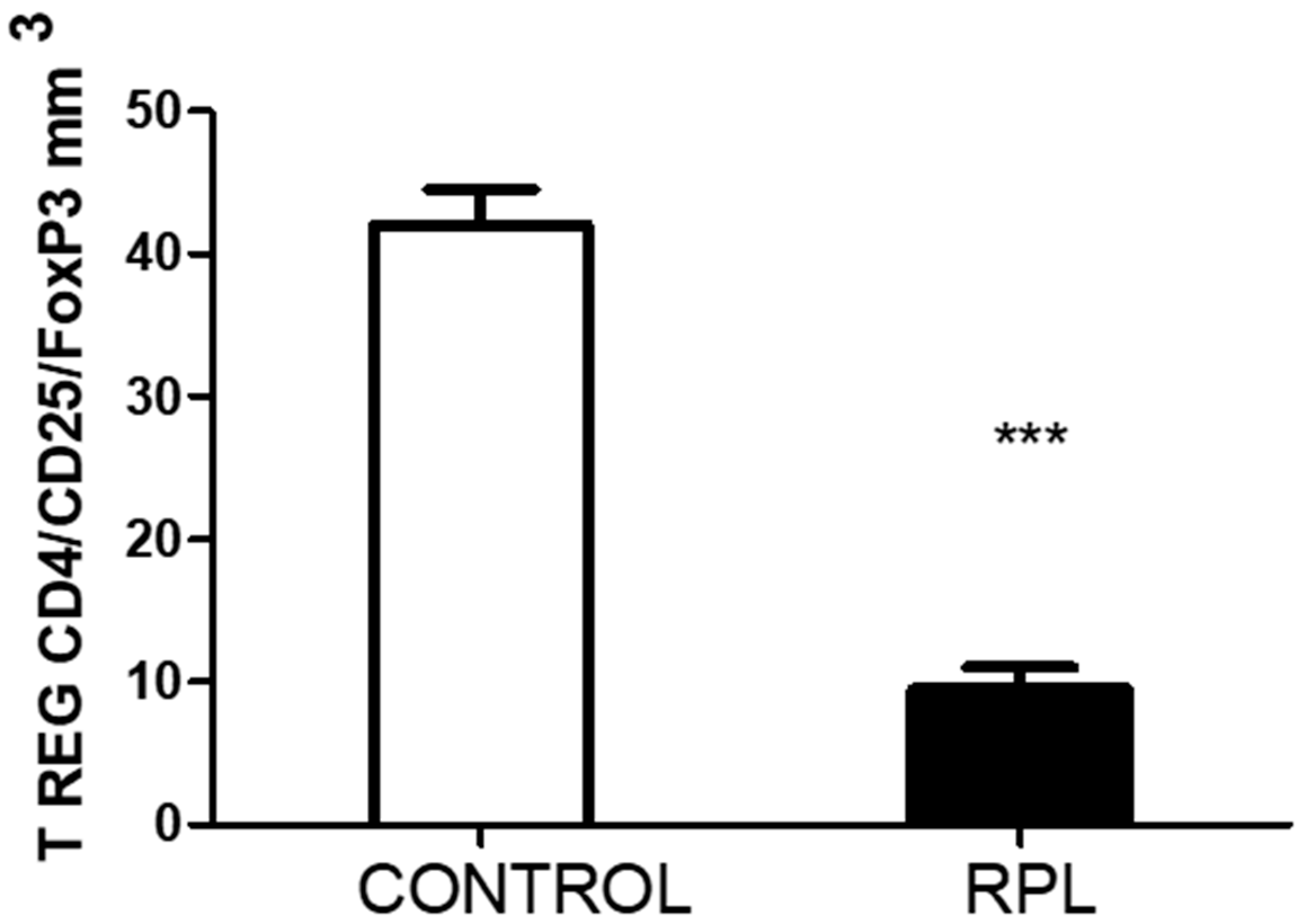
  • The peripheral lymphocytes of RPL patients were activated based on the percentage of HLA-DR expression. In addition, the number of T regulatory cells decreased.
  • The number of IL-17-positive cells, CD8 and CD56, was significantly lower in RPL patients than in controls. However, IL-17 positiveness in the CD3CD4 subpopulation was higher in RPL patients than in controls.
  • In stimulated cells, the response of all different cell populations and subpopulations was lower in RPL patients than in the controls. The effect of PMA/ionomycin stimulation on whole blood may be responsible for this effect.
  • NK cells of RPL patients responded significantly more strongly upon PMA ionomycin stimulation than those of controls.

6. Limitations of the Study

There are three critical limitations in the present study.
  • Other circulating cytokines were not measured in the present study. An analysis of inflammatory and anti-inflammatory cytokines may have helped us identify which Th17 cell subpopulation was circulating in these patients, as Navarron-Compán et al. [30] proposed in chronic diseases.
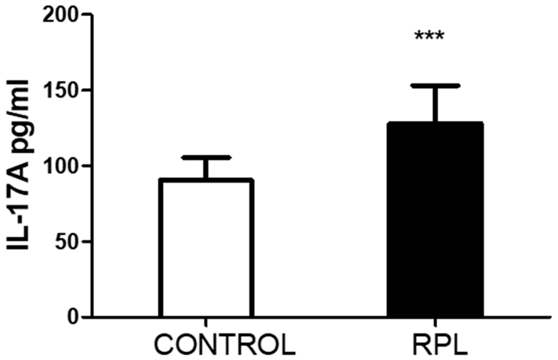
  • Unfortunately, we could not quantify the number of CD4/CD25/FoxP3 cells in the stimulated samples, which could have been an essential point for comparing with IL-17. The significant differences recorded in the basal levels suggest that upon higher circulating levels of IL-17, the number of T regulatory cells decreases.
  • Some of these RPL patients did not continue visits to the fertility clinic for other procedures due to the costs, so we could not perform a follow-up to ascertain if they had any successful pregnancies.

Supplementary Materials

The following supporting information can be downloaded at: https://www.mdpi.com/article/10.3390/immuno4040019/s1. The document contains raw data about the patients.

Author Contributions

Conceptualization, J.V.G. and I.B.; methodology, J.V.G., I.B., M.J.P. and J.B.D.S.; validation, J.B.D.S., C.V.D.S. and J.V.G.; formal analysis, C.V.D.S. and J.B.D.S.; investigation, M.J.P., J.V.G. and I.B.; resources, J.V.G.; data curation, J.B.D.S.; writing—original draft preparation, J.V.G. and C.V.D.S.; writing—review and editing, J.B.D.S.; visualization, J.B.D.S.; supervision, J.V.G. and J.B.D.S.; project administration, J.V.G.; funding acquisition, J.V.G. and J.B.D.S. All authors have read and agreed to the published version of the manuscript.

Funding

This research was funded by the Universidad Central de Venezuela’s Counsel for Scientific and Human Development (CDCH-UCV), grant number PG 09-6599-2006/1.

Institutional Review Board Statement

The study was conducted following the Declaration of Helsinki and approved by the Institutional Review Board (or Ethics Committee) of the Institute of Immunology, Faculty of Medicine, Universidad Central de Venezuela (protocol code 20052308, date of approval 15 September 2005).

Informed Consent Statement

Informed consent was obtained from all subjects involved in the study.

Data Availability Statement

The crude data are available from the authors.

Acknowledgments

The authors thank the patients and controls involved in the study and Dariana Parra, Irene Rodríguez, and Perla Chirinos for their valuable help in sample processing.

Conflicts of Interest

The authors declare no conflicts of interest.

References

  1. Pillarisetty, L.S.; Mahdy, H. Recurrent Pregnancy Loss; StatPearls Publishing: Treasure Island, FL, USA, 2024. Available online: https://www.ncbi.nlm.nih.gov/books/NBK554460/ (accessed on 4 June 2024).
  2. Stephenson, M.D. Frequency of factors associated with habitual abortion in 197 couples. Fertil. Steril. 1996, 66, 24–29. [Google Scholar] [PubMed]
  3. Ford, H.B.; Schust, D.J. Recurrent pregnancy loss: Etiology, diagnosis, and therapy. Rev. Obstet. Gynecol. 2009, 2, 76–83. [Google Scholar] [PubMed]
  4. Cuadrado-Torroglosa, I.; García-Velasco, J.A.; Alecsandru, D. Maternal-Fetal Compatibility in Recurrent Pregnancy Loss. J. Clin. Med. 2024, 13, 2379. [Google Scholar] [CrossRef] [PubMed]
  5. Saito, S.; Nakashima, A.; Ito, M.; Shima, T. Clinical implication of recent advances in our understanding of IL-17 and reproductive immunology. Expert. Rev. Clin. Immunol. 2011, 7, 649–657. [Google Scholar] [CrossRef]
  6. Fu, B.; Tian, Z.; Wei, H. TH17 cells in human recurrent pregnancy loss and pre-eclampsia. Cell. Mol. Immunol. 2014, 11, 564–570. [Google Scholar] [CrossRef] [PubMed]
  7. Dai, M.; Xu, Y.; Gong, G.; Zhang, Y. Roles of immune microenvironment in the female reproductive maintenance and regulation: Novel insights into the crosstalk of immune cells. Front. Immunol. 2023, 14, 1109122. [Google Scholar] [CrossRef]
  8. Cui, H.; Wang, N.; Li, H.; Bian, Y.; Wen, W.; Kong, X.; Wang, F. The dynamic shifts of IL-10-producing Th17 and IL-17-producing Treg in health and disease: Crosstalk between ancient "Yin-Yang" theory and modern immunology. Cell Commun. Signal. 2024, 22, 99. [Google Scholar] [CrossRef]
  9. Moura, G.A.; Rocha, Y.M.; Moura, F.L.D.; Freitas, J.O.; Rodrigues, J.P.V.; Gonçalves, V.P.; Nicolete, R. Immune system cells modulation in patients with reproductive issues: A systematic review approach. JBRA Assis. Reprod. 2024, 28, 78–89. [Google Scholar] [CrossRef]
  10. Najafi, S.; Hadinedoushan, H.; Eslami, G.; Aflatoonian, A. Association of IL-17A and IL-17 F gene polymorphisms with recurrent pregnancy loss in Iranian women. J. Assist. Reprod. Genet. 2014, 31, 1491–1496. [Google Scholar] [CrossRef]
  11. Zhang, M.; Xu, J.; Bao, X.; Niu, W.; Wang, L.; Du, L.; Zhang, N.; Sun, Y. Association between Genetic Polymorphisms in Interleukin Genes and Recurrent Pregnancy Loss—A Systematic Review and Meta-Analysis. PLoS ONE 2017, 12, e0169891. [Google Scholar] [CrossRef]
  12. Vahid, S.A.; Ghaebi, M.; Ahmadi, M.; Nouri, M.; Danaei, S.; Aghebati-Maleki, L.; Ardehaie, R.M.; Yousefi, B.; Hakimi, P.; Hojjat-Farsangi, M.; et al. Altered T-cell subpopulations in recurrent pregnancy loss patients with cellular immune abnormalities. J. Cell. Physiol. 2019, 234, 4924–4933. [Google Scholar] [CrossRef]
  13. Farshchi, M.; Abdollahi, E.; Saghafi, N.; Hosseini, A.; Fallahi, S.; Rostami, S.; Rostami, P.; Rafatpanah, H.; Habibagahi, M. Evaluation of Th17 and Treg cytokines in patients with unexplained recurrent pregnancy loss. J. Clin. Transl. Res. 2022, 8, 256–265. [Google Scholar]
  14. Ali, S.; Majid, S.; Ali, N.; Banday, M.Z.; Taing, S.; Wani, S.; Almuqbil, M.; Alshehri, S.; Shamim, K.; Rehman, M.U. Immunogenetic Role of IL17A Polymorphism in the Pathogenesis of Recurrent Miscarriage. J. Clin. Med. 2022, 11, 7448. [Google Scholar] [CrossRef]
  15. Ma, Y.; Ma, M.; Ye, S.; Liu, Y.; Zhao, X.; Wang, Y. Association of IL-17 and IL-27 polymorphisms with susceptibility to recurrent pregnancy loss and pre-eclampsia: A systematic review and meta-analysis. Immun. Inflamm. Dis. 2023, 11, e1057. [Google Scholar] [CrossRef]
  16. Keshavarz Motamed, A.; Zarei, Z.H.; Mirfakhraee, H.; Shariatinia, F.; Akbari, M.; Ziagham, S.; Igder, S.; Zarei, N. Association of Interleukin-17A rs2275913 Polymorphism with Recurrent Miscarriage: A Systematic Review and Meta-Analysis Study. Int. J. Fertil. Steril. 2023, 18, 7–11. [Google Scholar]
  17. Li, D.; Uskenbayeva, N.; Fang, L.; Xu, Y.; Yan, H.; Zhang, K.; Wang, J. Genetic polymorphism of IL-17 influences susceptibility to recurrent pregnancy loss in a Chinese population. Medicine 2024, 103, e38333. [Google Scholar] [CrossRef]
  18. Gao, J.F.; Zhang, H.; Lv, J.; Wang, L.; Fan, Y.Y. Associations of the IL-17A rs2275913 and IL-17F rs763780 polymorphisms with the risk of digestive system neoplasms: A meta-analysis. Int. Immunopharmacol. 2019, 67, 248–259. [Google Scholar] [CrossRef]
  19. Li, J.; Tian, H.; Jiang, H.J.; Han, B. Interleukin-17 SNPs and serum levels increase ulcerative colitis risk: A meta-analysis. World J. Gastroenterol. 2014, 20, 15899–15909. [Google Scholar] [CrossRef]
  20. Stavros, S.; Panagopoulos, P.; Machairiotis, N.; Potiris, A.; Mavrogianni, D.; Sfakianakis, A.; Drakaki, E.; Christodoulaki, C.; Panagiotopoulos, D.; Sioutis, D.; et al. Association between cytokine polymorphisms and recurrent pregnancy loss: A review of current evidence. Int. J. Gynecol. Obstet. 2024, 167, 45–57. [Google Scholar] [CrossRef]
  21. Conesa, A.; Fernández-Mestre, M.; Padrón, D.; Toro, F.; Silva, N.; Tassinari, P.; Blanca, I.; Martin, M.P.; Carrington, M.; Layrisse, Z. Distribution of killer cell immunoglobulin-like receptor genes in the mestizo population from Venezuela. Tissue Antigens 2010, 75, 724–729. [Google Scholar] [CrossRef]
  22. del Fortes, P.M.; Gill, G.; Paredes, M.E.; Gamez, L.E.; Palacios, M.; Blanca, I.; Tassinari, P. Allele and haplotype frequencies at human leukocyte antigen class I and II genes in Venezuela’s population. Ann. Biol. Clin. 2012, 70, 175–181. [Google Scholar]
  23. Bryc, K.; Velez, C.; Karafet, T.; Moreno-Estrada, A.; Reynolds, A.; Auton, A.; Hammer, M.; Bustamante, C.D.; Ostrer, H. Colloquium paper: Genome-wide patterns of population structure and admixture among Hispanic/Latino populations. Proc. Natl. Acad. Sci. USA 2010, 107 (Suppl. S2), 8954–8961. [Google Scholar] [CrossRef]
  24. De Oliveira, T.C.; Secolin, R.; Lopes-Cendes, I. A review of ancestrality and admixture in Latin America and the Caribbean focusing on native American and African descendant populations. Front. Genet. 2023, 14, 1091269. [Google Scholar] [CrossRef]
  25. Niafar, M.; Samaie, V.; Soltani-Zangbar, M.S.; Motavalli, R.; Dolati, S.; Danaii, S.; Mehdizadeh, A.; Yousefi, M. The association of Treg and Th17 cells development factors and anti-TPO autoantibodies in patients with recurrent pregnancy loss. BMC Res. Notes 2023, 16, 302. [Google Scholar] [CrossRef]
  26. Peña, M.J.; De Sanctis, C.V.; De Sanctis, J.B.; Garmendia, J.V. Frequency of Gene Polymorphisms in Admixed Venezuelan Women with Recurrent Pregnancy Loss: Microsomal Epoxy Hydroxylase (rs1051740) and Enos (rs1799983). Curr. Issues Mol. Biol. 2024, 46, 3460–3469. [Google Scholar] [CrossRef]
  27. Xie, Z.; Ding, X.; Wang, Y.; Zhang, M. The rs2275913 polymorphism of the interleukin-17A gene is associated with the risk of ovarian endometriosis. J. Obstet. Gynaecol. 2023, 43, 2199852. [Google Scholar] [CrossRef]
  28. Li, H.; Zhou, Z.; Tai, W.; Feng, W.; Zhang, D.; Gu, X.; Yang, R. Decreased Frequency of IL-17F rs763780 Site Allele G is Associated With Genetic Susceptibility to Immune Thrombocytopenia in a Chinese Population. Clin. Appl. Thromb. Hemost. 2017, 23, 466–471. [Google Scholar] [CrossRef]
  29. Lee, S.K.; Kim, J.Y.; Hur, S.E.; Kim, C.J.; Na, B.J.; Lee, M.; Gilman-Sachs, A.; Kwak-Kim, J. An imbalance in interleukin-17-producing T and Foxp3⁺ regulatory T cells in women with idiopathic recurrent pregnancy loss. Hum. Reprod. 2011, 26, 2964–2971. [Google Scholar] [CrossRef]
  30. Navarro-Compán, V.; Puig, L.; Vidal, S.; Ramírez, J.; Llamas-Velasco, M.; Fernández-Carballido, C.; Almodóvar, R.; Pinto, J.A.; Galíndez-Aguirregoikoa, E.; Zarco, P.; et al. The paradigm of IL-23-independent production of IL-17F and IL-17A and their role in chronic inflammatory diseases. Frontier. Immunol. 2023, 14, 1191782. [Google Scholar] [CrossRef]
  31. Mills, K.H. Induction, function and regulation of IL-17-producing T cells. Eur. J. Immunol. 2008, 38, 2636–2649. [Google Scholar] [CrossRef]
  32. Purvis, H.A.; Stoop, J.N.; Mann, J.; Woods, S.; Kozijn, A.E.; Hambleton, S.; Robinson, J.H.; Isaacs, J.D.; Anderson, A.E.; Hilkens, C.M. Low-strength T-cell activation promotes Th17 responses. Blood 2010, 116, 4829–4837. [Google Scholar] [CrossRef] [PubMed]
  33. Hou, H.; Zhou, Y.; Yu, J.; Mao, L.; Bosco, M.J.; Wang, J.; Lu, Y.; Mao, L.; Wu, X.; Wang, F.; et al. Establishment of the Reference Intervals of Lymphocyte Function in Healthy Adults Based on IFN-γ Secretion Assay upon Phorbol-12-Myristate-13-Acetate/Ionomycin Stimulation. Front. Immunol. 2018, 9, 172. [Google Scholar] [CrossRef] [PubMed]
  34. Pandya, A.D.; Al-Jaderi, Z.; Høglund, R.A.; Holmøy, T.; Harbo, H.F.; Norgauer, J.; Maghazachi, A.A. Identification of human NK17/NK1 cells. PLoS ONE 2011, 6, e26780. [Google Scholar] [CrossRef]
Figure 1. The results of flow cytometry assessment of total CD3 cells, CD4, CD8, and NK cells before and after stimulation with PMA/ionomycin are illustrated. Significant differences were reported. ** p < 0.005, *** p < 0.0001, n = 50.
Immuno 04 00019 g001
Figure 2. The amount of IL-17A detected in the plasma of controls and RPL patients. There was a statistical significance. *** p < 0.0001, n = 50.
Immuno 04 00019 g002
Figure 3. Difference between the number of T-reg cells in control and RPL patients. There was a statistical significance. *** p < 0.0001, n = 50.
Immuno 04 00019 g003
Table 1. Demographic data from patients with RPL and controls.
ControlsRPL
n5050
Age (years)34.3 ± 6.534.1 ± 4.5
# Pregnancies (%)1 (10%)2 (50%)
2 (60%)3 (35%)
3 (30%)>3 (15%)
# Miscarriages (%)02 (40%)
>2 (60%)
Duration of pregnancy (weeks)37.3 ± 2.28.1 ± 2.5
The table represents the number of women involved in the study and the percentage of individuals who were pregnant or had a miscarriage.
Table 2. IL-17 gene polymorphisms studied.
PolymorphismControlRPLpOR
rs2275913
Genotype
GG40410.91.0
GA880.9
AA21
G48500.81.0
A1090.8
rs763780
Genotype
AA47470.91.0
GA120.9
GG21
A48500.91.0
G330.9
The results refer to the number of individuals analyzed in each case. p refers to the statistical analysis of both groups and the odds ratio (OR). No significant differences were encountered between the groups.
Table 3. The absolute number of cell lymphocyte populations and subpopulations in mm3.
ControlRPLp
Total leukocytes6700 ± 8146766 ± 7270.7
Total lymphocytes2105 ± 2412359 ± 239>0.001
CD3 T lymphocytes1530 ± 2081614 ± 1860.03
NK cells (CD56/CD16)186 ± 37261 ± 36>0.001
NKT cells (CD3/CD56)34 ± 1135 ± 140.7
Index T cell/NK cell8.5 ± 1.66.2 ± 0.8>0.001
CD3/CD45RA839 ± 1341266 ± 165>0.001
CD3/CD45RO928 ± 1231076 ± 151>0.001
HLA DR+ cells153 ± 43247 ± 49>0.001
CD3/HLA DR+22 ± 882 ± 52>0.001
The table represents the number of cells for each cell population. The number of cells in cell populations was calculated using the percentage of expression assessed by flow cytometry and adjusted to the total number of lymphocytes reported. Since the number of lymphocytes was higher in RPL patients, the number of cells increased.
Disclaimer/Publisher’s Note: The statements, opinions and data contained in all publications are solely those of the individual author(s) and contributor(s) and not of MDPI and/or the editor(s). MDPI and/or the editor(s) disclaim responsibility for any injury to people or property resulting from any ideas, methods, instructions or products referred to in the content.

