Review

Cervical Cancer Outcome and Tumor-Associated Macrophages: Research Evidence

1
Molecular Oncology and Viral Pathology GRP, Research Center of IPO Porto (CI-IPOP)/Pathology and Laboratory Medicine Department/Clinical Pathology SV, Portuguese Institute of Oncology of Porto (IPO Porto), 4200-072 Porto, Portugal
2
ICBAS—Instituto de Ciências Biomédicas Abel Salazar, Universidade do Porto, 4050-313 Porto, Portugal
3
FP-I3ID, FP-BHS, CEBIMED and Faculty of Health Sciences, University Fernando Pessoa, 4200-150 Porto, Portugal
*
Author to whom correspondence should be addressed.
Immuno 2022, 2(3), 460-468; https://doi.org/10.3390/immuno2030028
Submission received: 21 May 2022 / Revised: 12 July 2022 / Accepted: 20 July 2022 / Published: 27 July 2022

Abstract

:
Inflammation is a key factor in cancer promotion. Tumor-associated macrophages (TAMs), as part of the tumor microenvironment, are often associated with the progression of tumors and a worse prognosis in many cancers, namely on cervical cancer. This work exhaustively summarizes the conclusions of the different studies published concerning TAMs function in cervical cancer, from in vitro studies using cancer cell lines to the clinical perspective (histological samples-based studies). Most studies have led to the conclusion that TAMs increased density is directly related to increased severity of a malignant cervical lesion. Additionally, TAMs are normally polarized into an M2 phenotype, benefiting and promoting tumor progression, resulting in a worse disease outcome. The tumor microenvironment is also a highly critical contributor that not only influences tumor natural history but also modulates the specific immune response.

1. Introduction

Cervical cancer is one of the most commonly diagnosed types of tumor in women in the world [1], being the most relevant cause for the development of this specific type of cancer is the persistent infection carried out by the Human Papilloma Virus (HPV) [1,2]. If the immune system is unable to prevent a persistent infection, HPV can transform normal cells into malignant cells, promoting, also, chronic inflammation and the development of advanced precursor lesions [3].
Evidence suggests that inflammation is a central key in cancer initiation and promotion [4]. It is responsible for tumor development, invasion [5], and angiogenesis [6]. The fact is that solid tumors are infiltrated by cells of the immune system indicates that a significant percentage of cancer cases are associated with chronic infection or inflammation [7,8].
Macrophages are immune cells whose function is to carry phagocytosis and immune surveillance [9]. They are frequently present in the tumor microenvironment, being referred to as tumor-associated macrophages (TAMs) and are known for their antitumor activity [10]. Plasticity is a characteristic of TAMs, and therefore they can be influenced and polarized in response to an environmental stimulus [11]. Its role in cancer is not yet fully established [12], but convincing evidence shows that TAMs can promote and sustain cancer malignancy [13].
Most of the published studies resembling the presence of TAMs on the tumor microenvironment report its association with the progression of the tumor and a worse disease outcome [11,14]. The same seems to happen in cervical cancer [15]. However, different deductions arise based on the macrophage phenotypes [16] or lesions [17].
Many publications have explored and suggested a crucial role for TAMs in the progression of many types of cancer. Recently, a study focused on the macrophage’s role in gynecological cancers but briefly approached its role in cervical cancer [18]. Additionally, more works reviewed the role of TAMs and inflammation in cervical cancer development [19,20].
The main goal of this review paper is to summarize the overall remarks of several published studies concerning TAMs implications for cervical cancer, either by analyzing their role in tumor promotion or disease prognosis. For this purpose, studies were collected and exhaustively analyzed from a clinical and research perspective, including both histological samples and in vitro assays on cell lines, respectively, to clarify a relationship between TAMs and the occurrence and development of cervical cancer.

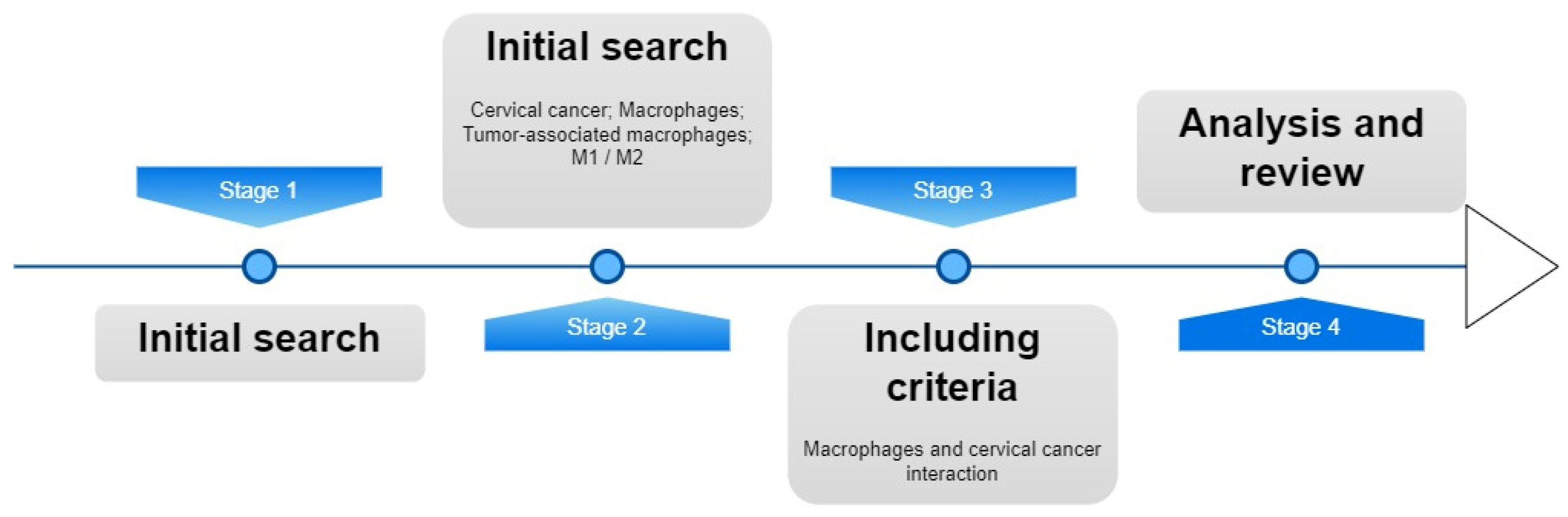
2. Materials and Methods

The literature search was performed in PubMed, Elsevier, Science Direct, and Google Scholar for all studies using terms related to cervical cancer (e.g., cervical cancer, cervix, etc.) and tumor-associated macrophages (e.g., tumor-associated macrophages, TAMs, M1, M2, etc.). Preliminary reading and analysis allowed the selection of 26 studies, which were subsequently exhaustively analyzed. Among the ones referred to before the scientific articles which focus on macrophages and their relation with cervical cancer and that have results obtained using in vitro studies with macrophages and cervical cancer cell lines were used in the revision (Figure 1).

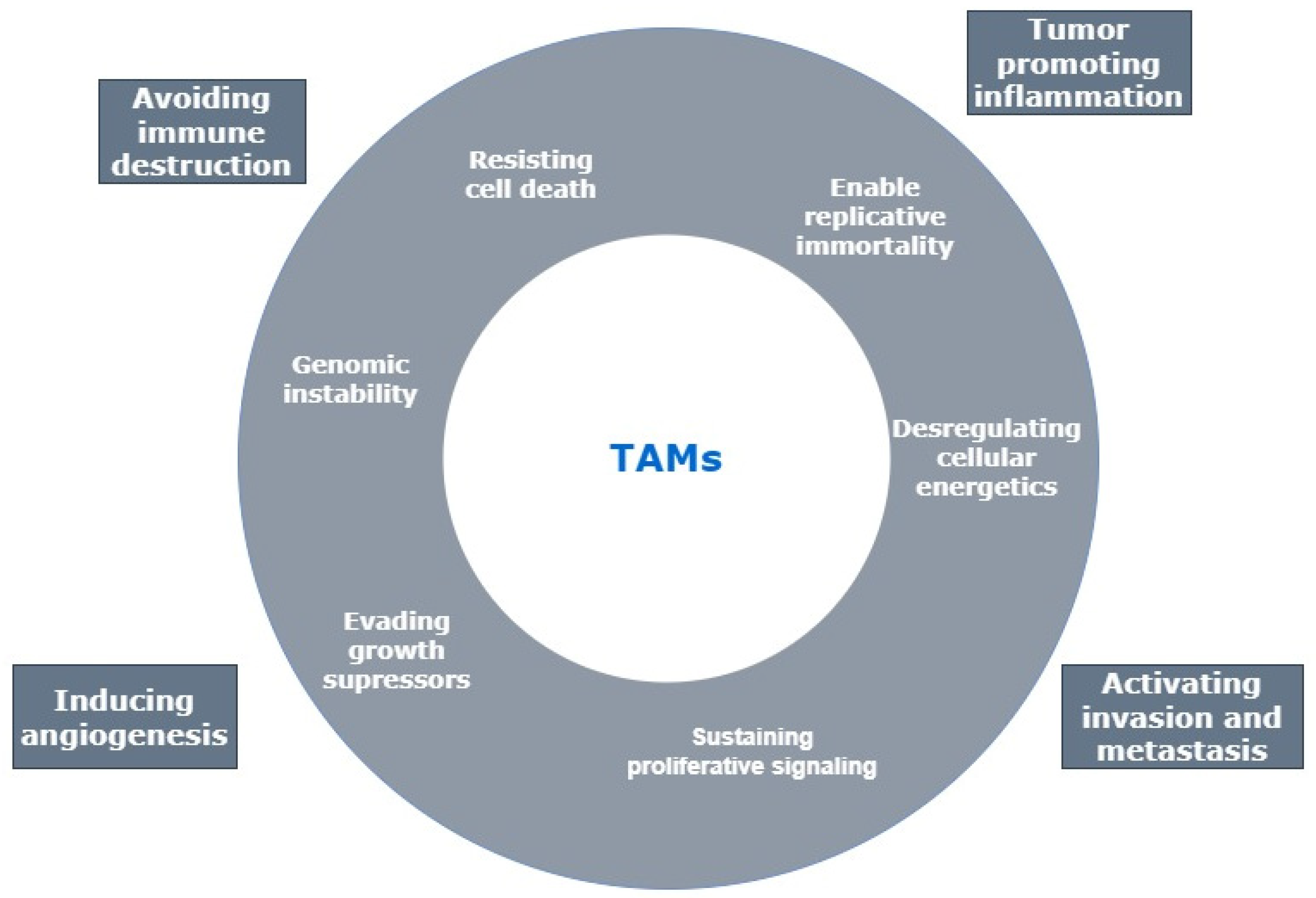
3. Tumor-Associated Macrophages

Macrophages differentiate from bone marrow monocytes and can adopt different phenotypes that differentially express specific markers [21,22].
As previously stated, macrophages can be induced to adopt different polarization states, known as inflammatory states, with different cytokines expression profiles [23]. Classically activated macrophages, known as M1, have antitumor activity in established tumors [24]. However, an M1 phenotype dysfunction in a chronic inflammation can result in cancer promotion, due to the release of oxidation products [25]. Alternatively, activated macrophages, also known as M2, have tissue repair and remodeling properties and can enhance angiogenesis [26], tumor invasion [27], and development [28]. The cluster of differentiation 68 (CD68) is a generic macrophage marker, and CD163, a member of the scavenger receptor cysteine-rich (SRCR), is mainly expressed in the M2 phenotype and associated with tumor enhancement [16,28]. So, being Macrophages recognized for their cytotoxic and antitumor activity; they can act as tumor promoters by secreting factors that will enhance tumor invasion and metastasization [29].
Macrophages are characterized by their high plasticity and TAMs can be recruited and influenced by tumors and end up aiding and promoting tumor proliferation [30]. The tumor microenvironment, with its complex network of infiltrating cells, blood vessels, and secreted factors, leads to the polarization of TAMs towards phenotypes expressing a characteristic cytokine profile [12]. TAMs contribute to inflammation and tumorigenesis, taking part in the hallmarks of cancer (Figure 2). The direct interaction with T cells or the release of immunosuppressive cytokines and proteases facilitate immunosuppression. Thus, the tumor avoids immune destruction. Activation of invasion and metastasis occurs by secretion of metalloproteinases, proteases, and cathepsins that take part in cell adhesion, and the release of angiogenic factors, such as vascular endothelial growth factor (VEGF), induces angiogenesis [30,31,32].
TAMs presence has been associated with a poor disease outcome as well as a poor prognosis and a shorter survival for several types of cancer [15,33,34] being the high density of TAMs related to a bad prognosis in 80 % of published studies [35].
Cytokines produced by TMAs, such as TNF-α, can be associated with a higher risk of developing invasive cervical cancer [36]. Although studies usually focus on the effect of macrophages and tumor progression, some demonstrate that some cytokines polymorphisms are related to an increased risk of developing cancer [37,38]. In addition, polymorphisms in endothelial cell-specific forms of nitric oxide synthases have been strongly associated with advanced prostate cancer, as well as the development of prostate bone metastasis [38]. However, TAMs density can be linked to a better prognosis in other types of cancer [37,39] as colorectal cancer, for which the presence of TAMs might enhance survival in patients [35].
Since macrophages can be present in the microenvironment and have an impact on immunosuppression, recent strategies exploit TAMs characteristics. Blocking monocyte recruitment into tumor tissue, decreasing TAMs density, targeting TAMs activation, or even reprograming into displaying an antitumor phenotype, are examples of clinical trial approaches [40,41,42].

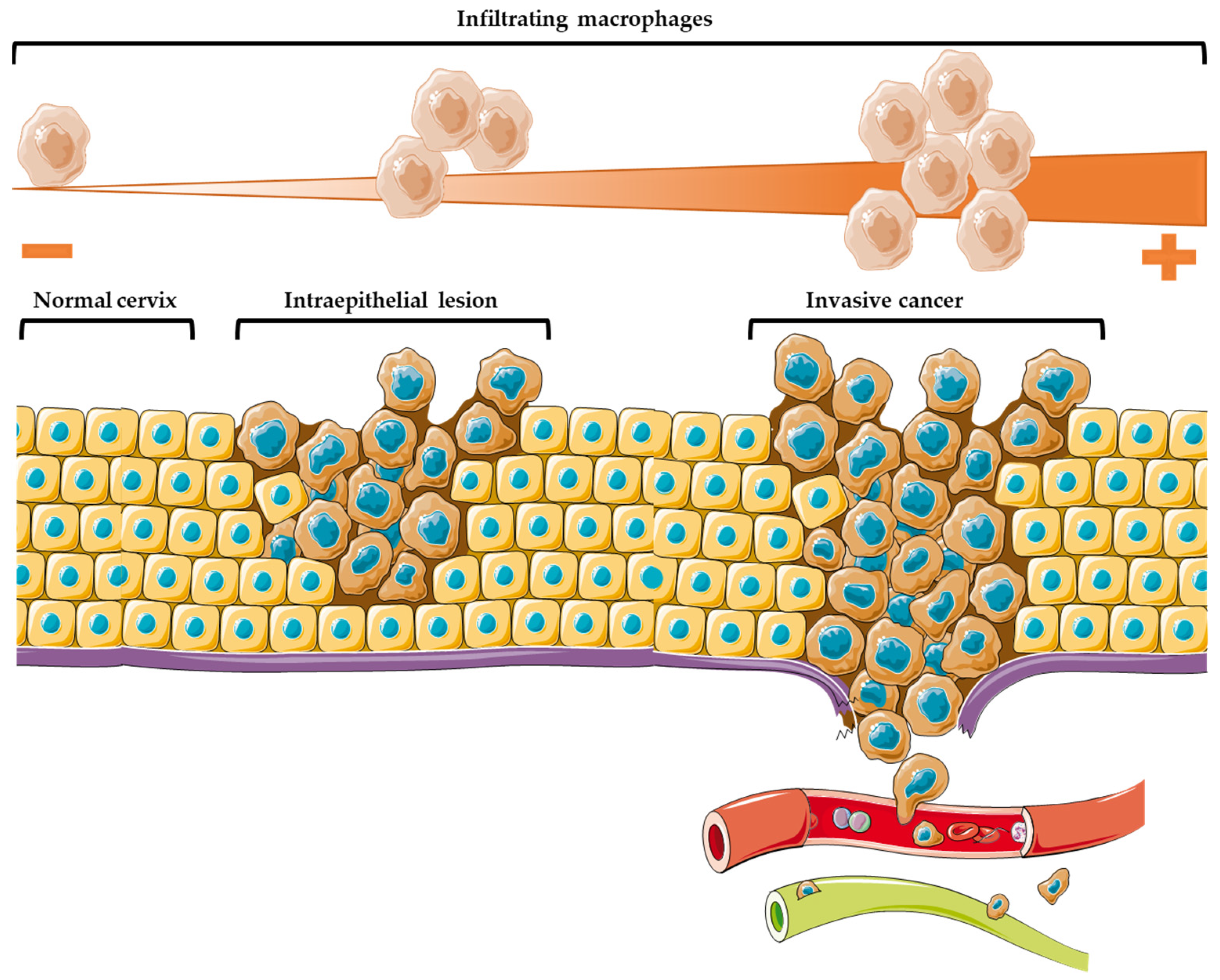
3.1. TAMs and Cervical Cancer: Outcome

Whereas macrophages infiltration has been associated with tumor advanced stages and a worse outcome, opposite conclusions were also observed [43].
Overall, when compared to normal tissues, macrophage density was increased in both precursor lesions and established cervical cancer (Figure 3). A higher density in TAMs has been reported in invasive cervical cancer, which agrees with studies associating the number/density of TAMs with a poor prognosis in cervical cancer cases [34], as well as in other types of cancer, including breast, gastric, or head and neck cancer [44,45,46].
Early on, a positive correlation was found between macrophage counts and disease progression [47] as well as elevated counts of infiltrating macrophages in invasive carcinomas [48]. Later, superior macrophage counts where found in CIN (cervical intraepithelial neoplasia) II, III and SCC (squamous cell cancer) when compared to normal tissue, revealing an association between macrophage infiltration and progression to malignancy [49]. Additional studies have also reported increased stromal macrophages in high-grade CIN and cervical cancer [50].
Nonetheless, studies report exceptions where macrophage counts neither correlate with tumor stage nor with survival [43]. While studying the clinical and functional significance of TAMs, Ding et al. observed enrichment of macrophages in cancer nests compared to less developed lesions and normal tissue but found no significant correlation with the FIGO stage [17].
A higher density of TAMs was also found in carcinoma in situ and established cervical cancer in Jiang et al. and Utrera-Barillas et al. works, respectively. Authors related the interaction between TAMs and tumor cells to synergistically promote angiogenesis [51,52]. Ding et al. also highlighted the possibility that macrophages are actively involved in lymphatic metastasis in the tumor stroma [17].
Contrarily, a relation seems to occur when a specific macrophage phenotype is considered and studied.
M2 TAMs were found to be elevated in neoplastic tissue [53] and a close correlation between lesion severity and an increased density in M2 macrophages was reported [54]. Furthermore, CD163+ macrophages demonstrated a stronger correlation with the advanced FIGO stage and lymphatic metastasis than CD68+ cells [16].
Another work identified a relationship between M2 TAMs and invasion patterns in squamous cervical cancer, noticing a significant increase in M2 macrophage content in tumor tissue arrays with diffuse infiltration patterns [53]. Furthermore, Petrillo et al. have shown that differentiation of macrophages into M2 can lead to resistance to platinum therapies, which is in line with the conclusion of studies for various types of cancer that show evidence of a reduced effect of chemotherapy and radiotherapy [15,55]. Dijkgraaf et al. corroborated their results by showing that chemotherapeutic agents induced the proliferation of M2-type macrophages, which may lead to indirect resistance to anticancer therapy [56].
Heeren et al. investigated the effect of PD-L1 expression on immune cells in either squamous cell carcinoma or adenocarcinoma of the cervix, identifying them as tumor-associated macrophages, reportedly CD163+ and CD14+ representing the M2 phenotype. The presence of these cells was associated with a poor-disease specific survival [57].
Regardless of the type of study, it is clear that an increase in TAMs is an indicator of a worse prognosis [58] and that TAM infiltration correlates with cervical cancer progression [59]. Taken altogether, these findings suggest that TAMs favor the spread of cancer and lymphatic invasion in an interplay with cancer cells [50].
Even so, some studies have the limitation of not discriminating macrophage phenotypes [51,60]. In addition, M2 TAMs might also be causing a worse prognosis [61], revealing tumor-promoting capacity demonstrated by several works [15,42,54]. A reduced M1/M2 ratio, as reported by Petrillo et al., might explain, at least to some extent, the severity [15] and is propitious to tumor progression [25].
To the best of our knowledge, there is no study regarding the effect of the M1 phenotype on cervical cancer. However, this phenotype was associated with a higher expectance of survival in non-small cell lung cancer [62], highlighting the contrasting roles of macrophages and the need to distinguish between their phenotypes because of their distinct effects. A reduced M1/M2 ratio is of great importance to disease outcome and implications [15] to the result, from M1 and M2 balance due to the antagonistic activities of these phenotypes [15,30,54].
The variations observed between the results expressed in the literature, particularly in the association between disease stages, increased TAMs, and prognosis, may reflect differences related to sample size, antibodies used, tumor grade, or stage; even though the same methodology has been applied to assess TAMs density in tumors.

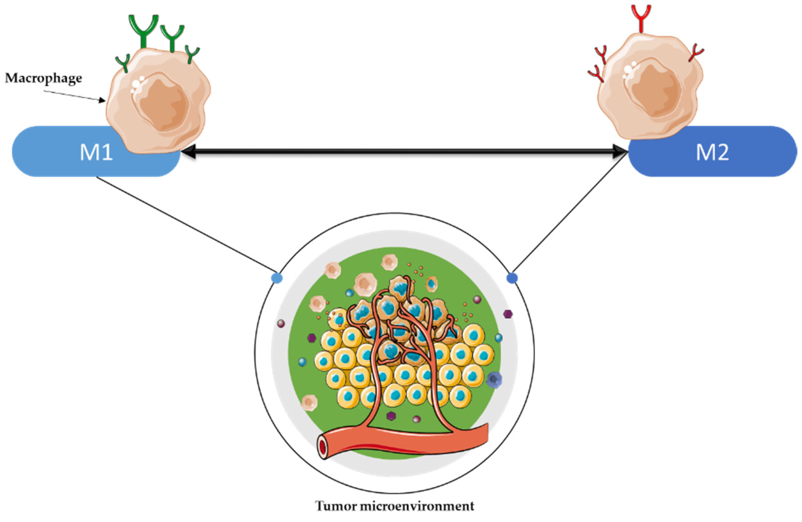
3.2. TAMs Activity on Cervical Cancer Cell Lines

While in some studies analyzed cervical tissue samples [16,43,53], other tested cervical cancer cell lines and studied changes on macrophage phenotype [49,56,63,64].
Pedraza-Brindis and coworkers investigated whether supernatants produced by cervical cancer cell lines could induce an M2 phenotype switch in THP-1 macrophages. Their work revealed that factors secreted by cancer cells induced macrophages to express CD163 (an M2 tag) and a different cytokine profile production. The decrease in pro-inflammatory cytokines resulted in cervical cancer proliferation, angiogenesis, and metastasis [63].
Sánchez-Reyes et al. evaluated the effect of cervical cancer cell lines in U937-derived macrophages. In agreement with the previous study, macrophages under the influence of cancer supernatants expressed CD163 and Interleukin-10 (IL-10), presuming a shift to an M2 phenotype [64].
While investigating the impact of platinum-based chemotherapeutic agents on cervical cancer cell lines and their ability to influence differentiation, Dijkgraaf et al. found that these agents promote the expression of inflammatory factors such as IL-6 and prostaglandin E2 (PGE2), interfering with the normal nuclear factor-kB (NF-kB) signaling pathway. However, the tumor cell lines induced the expansion of IL-10-producing M2 macrophages, reflecting a decrease in the immune potentiating response [56].
Heusinkveld et al. studied the effect of cervical cancer supernatants on monocyte differentiation and function. The authors found that monocytes were changed to an M2-type phenotype by tumor-produced IL-6 and PGE2. However, after interaction with T helper 1 (Th1) cells, M2 macrophages could turn into activated M1 macrophages with pro-inflammatory activity [65].
These studies highlight the importance of the microenvironment in carcinogenesis and tumor development (Figure 4).
Tumor-infiltrating macrophages can be recruited and polarized by factors secreted by the tumor to induce macrophages into a phenotype that benefits its survival and expansion [63], usually associated with an M2 phenotype and the expression of CD136 as the principal marker [28].
Cytokines also play an indispensable role in tumor progression. Tumors can manipulate immune cells, including TAMs, which, in return, produces specific cytokines that enhance tumor growth, development, invasion, and metastasization [8].

4. Conclusions

Considering the different studies (in tumor tissue and cell lines), cervical cancer, like many other cancers, is infiltrated by TAMs, and there is evidence that an increase in the density of TAMs density is related to the severity of a lesion [54], a poor outcome of the disease and poor response to treatment [14,15].
On its whole, the tumor microenvironment might be responsible for the polarization of macrophages into an M2 phenotype, characterized by a low antitumor activity, which facilitates tumor development. Furthermore, experiences with cell lines corroborated the role of M2 macrophages in suppressing the immune response, allowing tumor development.
The analysis of the different studies accessed suggests that the increase in TAMs in the tumor environment is related to the augmentation in the degree of cervical lesions (by the degree of lesions, the comparison between pre-malignant and neoplastic lesions or/and grading/staging of neoplasms should be considered invasive). In addition, TAMs may express an M2 phenotype, characterized by immunosuppression, tissue remodeling, and tumor-promoting activities. This phenotype’s presence may also be a consequence of factors secreted by the tumor and an explanation for the poor prognosis observed in cases of TAM infiltration.

Author Contributions

Conceptualization, B.H., T.P., R.M. and F.C.; writing—original draft preparation, B.H.; writing—review and editing, B.H., R.M. and F.C. All authors have read and agreed to the published version of the manuscript.

Funding

FCT/MCTES—Foundation for Science and Technology I.P. from the Minister of Science, Technology, and Higher Education (PIDDAC) and the European Regional Development Fund (ERDF) by the COMPETE—Programa Operacional Factores de Competitividade (POFC) and the Research Center of the Portuguese Oncology Institute of Porto (project no. PI86-CI-IPOP-66-2019). Fernando Pessoa University, FP-I3ID, FP-BHS, Grupo de Investigação Translacional em Longevidade e Saúde (GIT-LoSa).

Conflicts of Interest

The authors declare no conflict of interest.

References

  1. Ibeanu, O.A. Molecular Pathogenesis of Cervical Cancer. Cancer Biol. Ther. 2011, 11, 295–306. [Google Scholar] [CrossRef] [PubMed] [Green Version]
  2. Sung, H.; Ferlay, J.; Siegel, R.L.; Laversanne, M.; Soerjomataram, I.; Jemal, A.; Bray, F. Global Cancer Statistics 2020: GLOBOCAN Estimates of Incidence and Mortality Worldwide for 36 Cancers in 185 Countries. CA A Cancer J. Clin. 2021, 71, 209–249. [Google Scholar] [CrossRef] [PubMed]
  3. Smola, S. Immunopathogenesis of HPV-Associated Cancers and Prospects for Immunotherapy. Viruses 2017, 9, 254. [Google Scholar] [CrossRef]
  4. Candido, J.; Hagemann, T. Cancer-Related Inflammation. J. Clin. Immunol. 2013, 33, 79–84. [Google Scholar] [CrossRef] [PubMed]
  5. Nakamura, K.; Smyth, M.J. Targeting Cancer-Related Inflammation in the Era of Immunotherapy. Immunol. Cell Biol. 2017, 95, 325–332. [Google Scholar] [CrossRef] [PubMed]
  6. Mukai, K.; Tsai, M.; Saito, H.; Galli, S.J. Mast Cells as Sources of Cytokines, Chemokines, and Growth Factors. Immunol. Rev. 2018, 282, 121–150. [Google Scholar] [CrossRef]
  7. de Marzo, A.M.; Platz, E.A.; Sutcliffe, S.; Xu, J.; Grönberg, H.; Drake, C.G.; Nakai, Y.; Isaacs, W.B.; Nelson, W.G. Inflammation in Prostate Carcinogenesis. Nat. Rev. Cancer 2007, 7, 256–269. [Google Scholar] [CrossRef] [Green Version]
  8. Grivennikov, S.I.; Greten, F.R.; Karin, M. Immunity, Inflammation, and Cancer. Cell 2010, 140, 883–899. [Google Scholar] [CrossRef] [Green Version]
  9. Garg, A.D.; Agostinis, P. Cell Death and Immunity in Cancer: From Danger Signals to Mimicry of Pathogen Defense Responses. Immunol. Rev. 2017, 280, 126–148. [Google Scholar] [CrossRef] [PubMed]
  10. Condeelis, J.; Pollard, J.W. Macrophages: Obligate Partners for Tumor Cell Migration, Invasion, and Metastasis. Cell 2006, 124, 263–266. [Google Scholar] [CrossRef] [PubMed] [Green Version]
  11. Hao, N.-B.; Lü, M.-H.; Fan, Y.-H.; Cao, Y.-L.; Zhang, Z.-R.; Yang, S.-M. Macrophages in Tumor Microenvironments and the Progression of Tumors. Clin. Dev. Immunol. 2012, 2012, 948098. [Google Scholar] [CrossRef] [PubMed] [Green Version]
  12. Wang, M.; Zhao, J.; Zhang, L.; Wei, F.; Lian, Y.; Wu, Y.; Gong, Z.; Zhang, S.; Zhou, J.; Cao, K.; et al. Role of Tumor Microenvironment in Tumorigenesis. J. Cancer 2017, 8, 761–773. [Google Scholar] [CrossRef]
  13. Pollard, J.W. Tumour-Educated Macrophages Promote Tumour Progression and Metastasis. Nat. Rev. Cancer 2004, 4, 71–78. [Google Scholar] [CrossRef]
  14. Sousa, S.; Määttä, J. The Role of Tumour-Associated Macrophages in Bone Metastasis. J. Bone Oncol. 2016, 5, 135–138. [Google Scholar] [CrossRef] [Green Version]
  15. Petrillo, M.; Zannoni, G.F.; Martinelli, E.; Anchora, L.P.; Ferrandina, G.; Tropeano, G.; Fagotti, A.; Scambia, G. Polarisation of Tumor-Associated Macrophages toward M2 Phenotype Correlates with Poor Response to Chemoradiation and Reduced Survival in Patients with Locally Advanced Cervical Cancer. PLoS ONE 2015, 10, e0136654. [Google Scholar] [CrossRef] [PubMed]
  16. Chen, X.-J.; Han, L.-F.; Wu, X.-G.; Wei, W.-F.; Wu, L.-F.; Yi, H.-Y.; Yan, R.-M.; Bai, X.; Zhong, M.; Yu, Y.; et al. Clinical Significance of CD163+ and CD68+ Tumor-Associated Macrophages in High-Risk HPV-Related Cervical Cancer. J. Cancer 2017, 8, 3868–3875. [Google Scholar] [CrossRef] [PubMed] [Green Version]
  17. Ding, H.; Cai, J.; Mao, M.; Fang, Y.; Huang, Z.; Jia, J.; Li, T.; Xu, L.; Wang, J.; Zhou, J.; et al. Tumor-Associated Macrophages Induce Lymphangiogenesis in Cervical Cancer via Interaction with Tumor Cells. APMIS 2014, 122, 1059–1069. [Google Scholar] [CrossRef]
  18. Krishnan, V.; Schaar, B.; Tallapragada, S.; Dorigo, O. Tumor Associated Macrophages in Gynecologic Cancers. Gynecol. Oncol. 2018, 149, 205–213. [Google Scholar] [CrossRef]
  19. Liu, Y.; Li, L.; Li, Y.; Zhao, X. Research Progress on Tumor-Associated Macrophages and Inflammation in Cervical Cancer. BioMed Res. Int. 2020, 2020, 1–6. [Google Scholar] [CrossRef]
  20. Wang, Q.; Steger, A.; Mahner, S.; Jeschke, U.; Heidegger, H. The Formation and Therapeutic Update of Tumor-Associated Macrophages in Cervical Cancer. Int. J. Mol. Sci. 2019, 20, 3310. [Google Scholar] [CrossRef] [Green Version]
  21. Murray, P.J.; Allen, J.E.; Biswas, S.K.; Fisher, E.A.; Gilroy, D.W.; Goerdt, S.; Gordon, S.; Hamilton, J.A.; Ivashkiv, L.B.; Lawrence, T.; et al. Macrophage Activation and Polarization: Nomenclature and Experimental Guidelines. Immunity 2014, 41, 14–20. [Google Scholar] [CrossRef] [PubMed] [Green Version]
  22. Qian, B.-Z.; Pollard, J.W. Macrophage Diversity Enhances Tumor Progression and Metastasis. Cell 2010, 141, 39–51. [Google Scholar] [CrossRef] [PubMed] [Green Version]
  23. Mantovani, A.; Sica, A.; Sozzani, S.; Allavena, P.; Vecchi, A.; Locati, M. The Chemokine System in Diverse Forms of Macrophage Activation and Polarization. Trends Immunol. 2004, 25, 677–686. [Google Scholar] [CrossRef] [PubMed]
  24. Alberts, A.R.; Schoots, I.G.; Bokhorst, L.P.; Drost, F.-J.H.; van Leenders, G.J.; Krestin, G.P.; Dwarkasing, R.S.; Barentsz, J.O.; Schröder, F.H.; Bangma, C.H.; et al. Characteristics of Prostate Cancer Found at Fifth Screening in the European Randomized Study of Screening for Prostate Cancer Rotterdam: Can We Selectively Detect High-Grade Prostate Cancer with Upfront Multivariable Risk Stratification and Magnetic Resonance Imaging? Eur. Urol. 2018, 73, 343–350. [Google Scholar] [CrossRef] [PubMed]
  25. Funes, S.C.; Rios, M.; Escobar-Vera, J.; Kalergis, A.M. Implications of Macrophage Polarization in Autoimmunity. Immunology 2018, 154, 186–195. [Google Scholar] [CrossRef] [PubMed] [Green Version]
  26. Cui, X.; Morales, R.-T.T.; Qian, W.; Wang, H.; Gagner, J.-P.; Dolgalev, I.; Placantonakis, D.; Zagzag, D.; Cimmino, L.; Snuderl, M.; et al. Hacking Macrophage-Associated Immunosuppression for Regulating Glioblastoma Angiogenesis. Biomaterials 2018, 161, 164–178. [Google Scholar] [CrossRef]
  27. Vinnakota, K.; Zhang, Y.; Selvanesan, B.C.; Topi, G.; Salim, T.; Sand-Dejmek, J.; Jönsson, G.; Sjölander, A. M2-like Macrophages Induce Colon Cancer Cell Invasion via Matrix Metalloproteinases. J. Cell. Physiol. 2017, 232, 3468–3480. [Google Scholar] [CrossRef]
  28. Joyce, J.A.; Pollard, J.W. Microenvironmental Regulation of Metastasis. Nat. Rev. Cancer 2009, 9, 239–252. [Google Scholar] [CrossRef]
  29. Netea-Maier, R.T.; Smit, J.W.A.; Netea, M.G. Metabolic Changes in Tumor Cells and Tumor-Associated Macrophages: A Mutual Relationship. Cancer Lett. 2018, 413, 102–109. [Google Scholar] [CrossRef]
  30. Aras, S.; Zaidi, M.R. TAMeless Traitors: Macrophages in Cancer Progression and Metastasis. Br. J. Cancer 2017, 117, 1583–1591. [Google Scholar] [CrossRef] [Green Version]
  31. Hanahan, D.; Weinberg, R.A. Hallmarks of Cancer: The Next Generation. Cell 2011, 144, 646–674. [Google Scholar] [CrossRef] [Green Version]
  32. Hanahan, D. Hallmarks of Cancer: New Dimensions. Cancer Discov. 2022, 12, 31–46. [Google Scholar] [CrossRef] [PubMed]
  33. Cao, J.; Liu, J.; Xu, R.; Zhu, X.; Zhao, X.; Qian, B.-Z. Prognostic Role of Tumour-Associated Macrophages and Macrophage Scavenger Receptor 1 in Prostate Cancer: A Systematic Review and Meta-Analysis. Oncotarget 2017, 8, 83261–83269. [Google Scholar] [CrossRef] [PubMed]
  34. Bingle, L.; Brown, N.J.; Lewis, C.E. The Role of Tumour-Associated Macrophages in Tumour Progression: Implications for New Anticancer Therapies. J. Pathol. 2002, 196, 254–265. [Google Scholar] [CrossRef]
  35. Laoui, D.; van Overmeire, E.; de Baetselier, P.; van Ginderachter, J.A.; Raes, G. Functional Relationship between Tumor-Associated Macrophages and Macrophage Colony-Stimulating Factor as Contributors to Cancer Progression. Front. Immunol. 2014, 5, 489. [Google Scholar] [CrossRef] [Green Version]
  36. Duarte, I.; Santos, A.; Sousa, H.; Catarino, R.; Pinto, D.; Matos, A.; Pereira, D.; Moutinho, J.; Canedo, P.; Machado, J.C.; et al. G-308A TNF-α Polymorphism Is Associated with an Increased Risk of Invasive Cervical Cancer. Biochem. Biophys. Res. Commun. 2005, 334, 588–592. [Google Scholar] [CrossRef]
  37. Lewis, C.E.; Pollard, J.W. Distinct Role of Macrophages in Different Tumor Microenvironments. Cancer Res. 2006, 66, 605–612. [Google Scholar] [CrossRef] [PubMed] [Green Version]
  38. Medeiros, R.M.; Morais, A.; Vasconcelos, A.; Costa, S.; Pinto, D.; Oliveira, J.; Ferreira, P.; Lopes, C. Outcome in Prostate Cancer: Association with Endothelial Nitric Oxide Synthase Glu-Asp298 Polymorphism at Exon 71. Clin. Cancer Res. 2002, 8, 3433–3437. [Google Scholar] [PubMed]
  39. Ong, S.-M.; Tan, Y.-C.; Beretta, O.; Jiang, D.; Yeap, W.-H.; Tai, J.J.Y.; Wong, W.-C.; Yang, H.; Schwarz, H.; Lim, K.-H.; et al. Macrophages in Human Colorectal Cancer Are Pro-Inflammatory and Prime T Cells towards an Anti-Tumour Type-1 Inflammatory Response. Eur. J. Immunol. 2012, 42, 89–100. [Google Scholar] [CrossRef]
  40. Sawa-Wejksza, K.; Kandefer-Szerszeń, M. Tumor-Associated Macrophages as Target for Antitumor Therapy. Arch. Immunol. Ther. Exp. 2018, 66, 97–111. [Google Scholar] [CrossRef] [Green Version]
  41. Mantovani, A.; Marchesi, F.; Malesci, A.; Laghi, L.; Allavena, P. Tumour-Associated Macrophages as Treatment Targets in Oncology. Nat. Rev. Clin. Oncol. 2017, 14, 399–416. [Google Scholar] [CrossRef] [PubMed]
  42. Yang, L.; Zhang, Y. Tumor-Associated Macrophages, Potential Targets for Cancer Treatment. Biomark. Res. 2017, 5, 25. [Google Scholar] [CrossRef] [PubMed] [Green Version]
  43. Davidson, B.; Goldberg, I.; Gotlieb, W.H.; Lerner-Geva, L.; Ben-Baruch, G.; Agulansky, L.; Novikov, I.; Kopolovic, J. Macrophage Infiltration and Angiogenesis in Cervical Squamous Cell Carcinomaclinicopathologic Correlation. Acta Obstet. Gynecol. Scand. 1999, 78, 240–244. [Google Scholar] [CrossRef]
  44. Zhang, J.; Yan, Y.; Yang, Y.; Wang, L.; Li, M.; Wang, J.; Liu, X.; Duan, X.; Wang, J. High Infiltration of Tumor-Associated Macrophages Influences Poor Prognosis in Human Gastric Cancer Patients, Associates with the Phenomenon of EMT. Medicine 2016, 95, e2636. [Google Scholar] [CrossRef]
  45. Zhang, Q.W.; Liu, L.; Gong, C.Y.; Shi, H.S.; Zeng, Y.H.; Wang, X.Z.; Zhao, Y.W.; Wei, Y.Q. Prognostic Significance of Tumor-Associated Macrophages in Solid Tumor: A Meta-Analysis of the Literature. PLoS ONE 2012, 7, 1–14. [Google Scholar] [CrossRef] [Green Version]
  46. Zhao, X.; Qu, J.; Sun, Y.; Wang, J.; Liu, X.; Wang, F.; Zhang, H.; Wang, W.; Ma, X.; Gao, X.; et al. Prognostic Significance of Tumor-Associated Macrophages in Breast Cancer: A Meta-Analysis of the Literature. Oncotarget 2017, 8, 30576–30586. [Google Scholar] [CrossRef] [Green Version]
  47. Hammes, L.S.; Tekmal, R.R.; Naud, P.; Edelweiss, M.I.; Kirma, N.; Valente, P.T.; Syrjänen, K.J.; Cunha-Filho, J.S. Macrophages, Inflammation and Risk of Cervical Intraepithelial Neoplasia (CIN) Progression—Clinicopathological Correlation. Gynecol. Oncol. 2007, 105, 157–165. [Google Scholar] [CrossRef]
  48. Davidson, B.; Goldberg, I.; Kopolovic, J. Inflammatory Response in Cervicallntraepithelial Neoplasia and Squamous Cell Carcinoma of the Uterine Cervix. Pathol. Res. Pract. 1997, 193, 491–495. [Google Scholar] [CrossRef]
  49. Mazibrada, J.; Rittà, M.; Mondini, M.; de Andrea, M.; Azzimonti, B.; Borgogna, C.; Ciotti, M.; Orlando, A.; Surico, N.; Chiusa, L.; et al. Interaction between Inflammation and Angiogenesis during Different Stages of Cervical Carcinogenesis. Gynecol. Oncol. 2008, 108, 112–120. [Google Scholar] [CrossRef] [PubMed]
  50. Kobayashi, A.; Weinberg, V.; Darragh, T.; Smith-McCune, K. Evolving Immunosuppressive Microenvironment during Human Cervical Carcinogenesis. Mucosal Immunol. 2008, 1, 412–420. [Google Scholar] [CrossRef]
  51. Jiang, S.; Yang, Y.; Fang, M.; Li, X.; Yuan, X.; Yuan, J. Co-Evolution of Tumor-Associated Macrophages and Tumor Neo-Vessels during Cervical Cancer Invasion. Oncol. Lett. 2016, 12, 2625–2631. [Google Scholar] [CrossRef] [PubMed] [Green Version]
  52. Utrera-Barillas, D.; Castro-Manrreza, M.; Castellanos, E.; Gutiérrez-Rodríguez, M.; Arciniega-Ruíz de Esparza, O.; García-Cebada, J.; Velazquez, J.R.; Flores-Reséndiz, D.; Hernández-Hernández, D.; Benítez-Bribiesca, L. The Role of Macrophages and Mast Cells in Lymphangiogenesis and Angiogenesis in Cervical Carcinogenesis. Exp. Mol. Pathol. 2010, 89, 190–196. [Google Scholar] [CrossRef] [PubMed]
  53. Li, Y.; Huang, G.; Zhang, S. Associations between Intratumoral and Peritumoral M2 Macrophage Counts and Cervical Squamous Cell Carcinoma Invasion Patterns. Int. J. Gynecol. Obstet. 2017, 139, 346–351. [Google Scholar] [CrossRef] [PubMed]
  54. Swangphon, P.; Pientong, C.; Sunthamala, N.; Bumrungthai, S.; Azuma, M.; Kleebkaow, P.; Tangsiriwatthana, T.; Sangkomkamhang, U.; Kongyingyoes, B.; Ekalaksananan, T. Correlation of Circulating CD64+/CD163+ Monocyte Ratio and Stroma/Peri-Tumoral CD163+ Monocyte Density with Human Papillomavirus Infected Cervical Lesion Severity. Cancer Microenviron. 2017, 10, 77–85. [Google Scholar] [CrossRef] [PubMed]
  55. Guo, Q.; Jin, Z.; Yuan, Y.; Liu, R.; Xu, T.; Wei, H.; Xu, X.; He, S.; Chen, S.; Shi, Z.; et al. New Mechanisms of Tumor-Associated Macrophages on Promoting Tumor Progression: Recent Research Advances and Potential Targets for Tumor Immunotherapy. J. Immunol. Res. 2016, 2016, 9720912. [Google Scholar] [CrossRef] [PubMed] [Green Version]
  56. Dijkgraaf, E.M.; Heusinkveld, M.; Tummers, B.; Vogelpoel, L.T.C.; Goedemans, R.; Jha, V.; Nortier, J.W.R.; Welters, M.J.P.; Kroep, J.R.; van der Burg, S.H. Chemotherapy Alters Monocyte Differentiation to Favor Generation of Cancer-Supporting M2 Macrophages in the Tumor Microenvironment. Cancer Res. 2013, 73, 2480–2492. [Google Scholar] [CrossRef] [Green Version]
  57. Heeren, A.M.; Punt, S.; Bleeker, M.C.G.; Gaarenstroom, K.N.; van der Velden, J.; Kenter, G.G.; de Gruijl, T.D.; Jordanova, E.S. Prognostic Effect of Different PD-L1 Expression Patterns in Squamous Cell Carcinoma and Adenocarcinoma of the Cervix. Mod. Pathol. 2016, 29, 753–763. [Google Scholar] [CrossRef] [PubMed] [Green Version]
  58. Singh, S.; Mehta, N.; Lilan, J.; Budhthoki, M.B.; Chao, F.; Yong, L. Initiative Action of Tumor-Associated Macrophage during Tumor Metastasis. Biochim. Open 2017, 4, 8–18. [Google Scholar] [CrossRef]
  59. Guo, F.; Kong, W.; Zhao, G.; Cheng, Z.; Ai, L.; Lv, J.; Feng, Y.; Ma, X. The Correlation between Tumor-Associated Macrophage Infiltration and Progression in Cervical Carcinoma. Biosci. Rep. 2021, 2016, 9720912. [Google Scholar] [CrossRef] [PubMed]
  60. Ring, K.L.; Yemelyanova, A.V.; Soliman, P.T.; Frumovitz, M.M.; Jazaeri, A.A. Potential Immunotherapy Targets in Recurrent Cervical Cancer. Gynecol. Oncol. 2017, 145, 462–468. [Google Scholar] [CrossRef]
  61. Sica, A.; Schioppa, T.; Mantovani, A.; Allavena, P. Tumour-Associated Macrophages Are a Distinct M2 Polarised Population Promoting Tumour Progression: Potential Targets of Anti-Cancer Therapy. Eur. J. Cancer 2006, 42, 717–727. [Google Scholar] [CrossRef]
  62. Ma, J.; Liu, L.; Che, G.; Yu, N.; Dai, F.; You, Z. The M1 Form of Tumor-Associated Macrophages in Non-Small Cell Lung Cancer Is Positively Associated with Survival Time. BMC Cancer 2010, 10, 112. [Google Scholar] [CrossRef] [PubMed] [Green Version]
  63. Pedraza-Brindis, E.J.; Sánchez-Reyes, K.; Hernández-Flores, G.; Bravo-Cuellar, A.; Jave-Suárez, L.F.; Aguilar-Lemarroy, A.; Gómez-Lomelí, P.; López-López, B.A.; Ortiz-Lazareno, P.C. Culture Supernatants of Cervical Cancer Cells Induce an M2 Phenotypic Profile in THP-1 Macrophages. Cell. Immunol. 2016, 310, 42–52. [Google Scholar] [CrossRef] [PubMed]
  64. Sánchez-Reyes, K.; Bravo-Cuellar, A.; Hernández-Flores, G.; Lerma-Díaz, J.M.; Jave-Suárez, L.F.; Gómez-Lomelí, P.; de Celis, R.; Aguilar-Lemarroy, A.; Domínguez-Rodríguez, J.R.; Ortiz-Lazareno, P.C. Cervical Cancer Cell Supernatants Induce a Phenotypic Switch from U937-Derived Macrophage-Activated M1 State into M2-Like Suppressor Phenotype with Change in Toll-Like Receptor Profile. BioMed Res. Int. 2014, 2014, 683068. [Google Scholar] [CrossRef]
  65. Heusinkveld, M.; de Vos van Steenwijk, P.J.; Goedemans, R.; Ramwadhdoebe, T.H.; Gorter, A.; Welters, M.J.P.; van Hall, T.; van der Burg, S.H. M2 Macrophages Induced by Prostaglandin E 2 and IL-6 from Cervical Carcinoma Are Switched to Activated M1 Macrophages by CD4 + Th1 Cells. J. Immunol. 2011, 187, 1157–1165. [Google Scholar] [CrossRef] [PubMed] [Green Version]
Figure 1. Study development step-by-step. Stage 1 and 2 consisted in an initial search using cervical cancer related keywords. In stage 3 occurred the selection of studies relating macrophages and cervical cancer. Stage 4 comprised the analysis of such studies and the compiling of information.
Immuno 02 00028 g001
Figure 2. Major role of tumor associated macrophages (TAMs) in the hallmarks of cancer. Adapted [30] from Aras, S. & Zaidi, M. R. TAMeless traitors: macrophages in cancer progression and metastasis. British Journal of Cancer 117, 1583, [2017], and [31] Hanahan, D. & Weinberg, Robert A. Hallmarks of Cancer: The Next Generation. Cell 144, 646–674, [2011].
Immuno 02 00028 g002
Figure 3. An increase in infiltrating macrophages might be related to the progression of cancer. Figure composed by using and modifying pictures Servier Medical Art. Servier Medical Art by Servier is licensed under a Creative Commons Attribution 3.0 Unported License (https://creativecommons.org/licenses/by/3.0/ (accessed on 1 July 2022)).
Immuno 02 00028 g003
Figure 4. Interactions within the tumor microenvironment (TME) can switch macrophages profile. This figure was modified from pictures Servier Medical Art. Servier Medical Art by Servier is licensed under a Creative Commons Attribution 3.0 Unported License (https://creativecommons.org/licenses/by/3.0/ (accessed on 1 July 2022)).
Immuno 02 00028 g004
Publisher’s Note: MDPI stays neutral with regard to jurisdictional claims in published maps and institutional affiliations.

