Review

Overview of Memory NK Cells in Viral Infections: Possible Role in SARS-CoV-2 Infection

by
Juan Bautista De Sanctis
*,
Jenny Valentina Garmendia
and
Marián Hajdúch
Institute of Molecular and Translational Medicine, Faculty of Medicine and Dentistry, Czech Advanced Technology and Research Institute (Catrin), Palacky University, 77111 Olomouc, Czech Republic
*
Author to whom correspondence should be addressed.
Immuno 2022, 2(1), 52-67; https://doi.org/10.3390/immuno2010005
Submission received: 26 November 2021 / Revised: 31 December 2021 / Accepted: 31 December 2021 / Published: 5 January 2022
(This article belongs to the Special Issue Natural Killer Cells: From Bench to Bedside)

Abstract

:
NK cells have usually been defined as cells of the innate immune system, although they are also involved in adaptative responses. These cells belong to the innate lymphocyte cells (ILC) family. They remove unwanted cells, tumoral cells and pathogens. NK cells are essential for viral infection clearance and are involved in tolerogenic responses depending on the dynamic balance of the repertoire of activating and inhibitory receptors. NK plasticity is crucial for tissue function and vigilant immune responses. They directly eliminate virus-infected cells by recognising viral protein antigens using a non-MHC dependent mechanism, recognising viral glycan structures and antigens by NCR family receptors, inducing apoptosis by Fas-Fas ligand interaction, and killing cells by antibody-dependent cell cytotoxicity via the FcγIII receptor. Activating receptors are responsible for the clearance of virally infected cells, while inhibitory KIR receptor activation impairs NK responses and facilitates virus escape. Effective NK memory cells have been described and characterised by a low NKG2A and high NKG2C or NKG2D expression. NK cells have also been used in cell therapy. In SARS-CoV-2 infection, several contradicting reports about the role of NK cells have been published. A careful analysis of the current data and possible implications will be discussed.

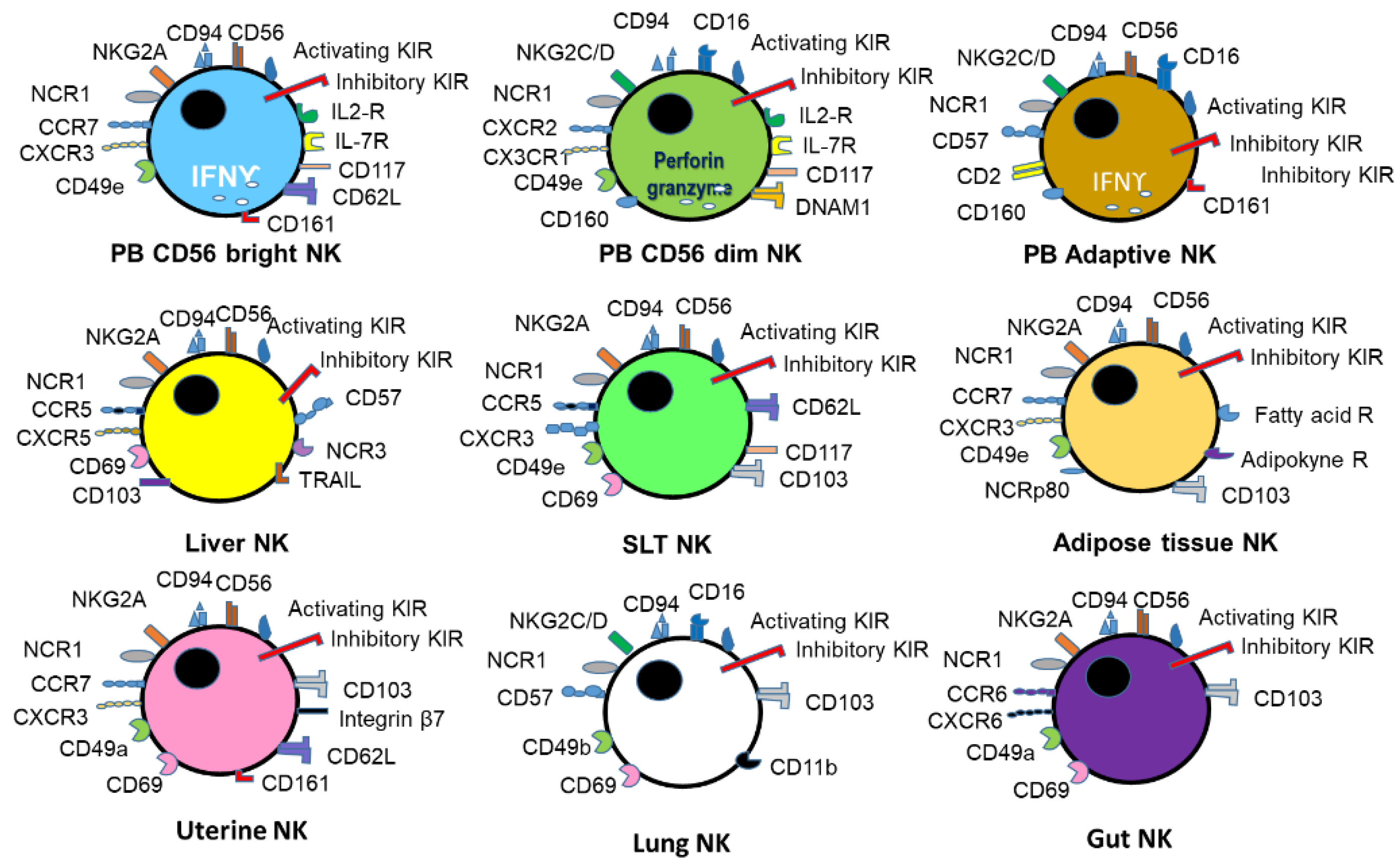
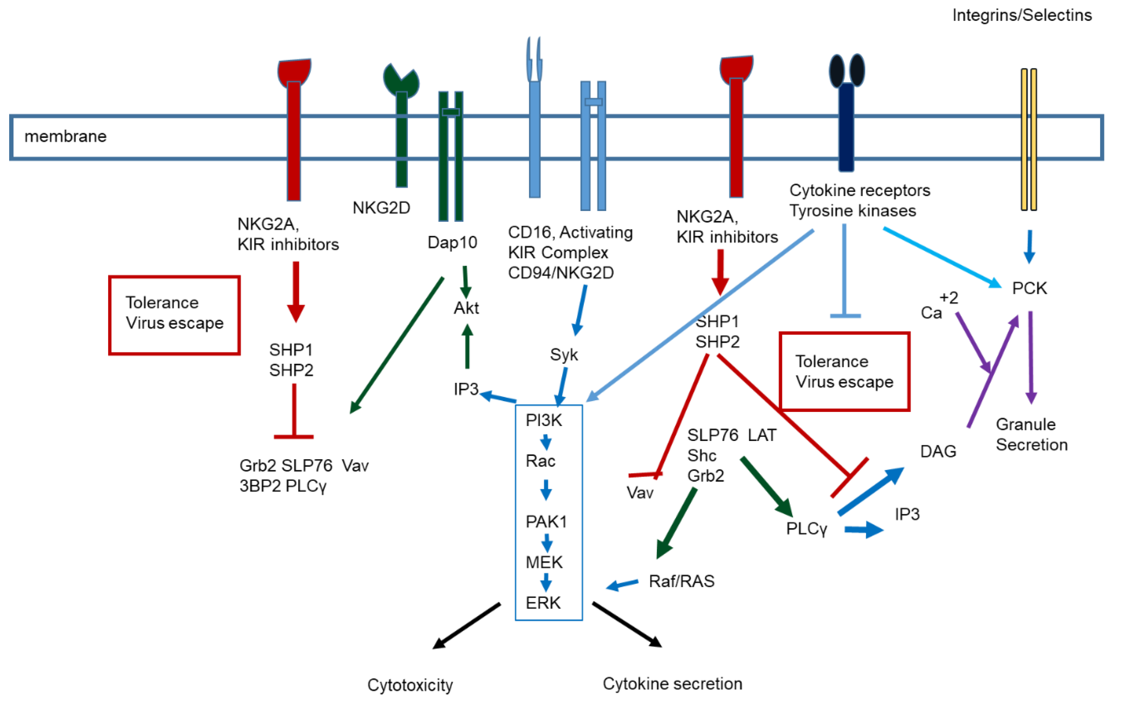
1. NK Biology and Function

NK cells are usually defined as immune cells that belong to the innate immune response [1,2,3,4]. They were described as cells capable of killing several tumour cell lines without previous antigen presentation [1,2,3,4]. It was shown then that NK cells lack the TCR and BCR receptors capable of binding specific antigens, and consequently, it was assumed that these cells lack antigen recognition [1,2,3,4]. This definition has changed in the latest years due to the increasing evidence of memory responses of NK cells in animal models and humans [5].
In humans, NK cells are divided into two main subpopulations CD56bright/CD16dim and CD56dim/CD16bright [1,2,3,4]. The first is composed of cooperative and tolerogenic cells, and the second is more cytotoxic against tumours or virus-infected cells [1,2,3,4]. However, NK cell functionality depends on signal transduction generated by killing receptors (NCR), killer immunoglobulin-like receptors (KIR), integrin and selectin expression, and cytotoxic receptor expression [1,2,3,4,5]. There are several populations of tissue-resident NK cells: uterine, decidual, adipose, lung, liver, spleen, tonsil, gut [1,2,3,4,5,6]. The antigens of these cells depend on tissue; for example, CD56dim/CD16bright NK cells are more prevalent in the lungs than the CD56bright/CD16dim positive cells and the CD56bright, CD16low, NKG2A/CD94, CD49a+, NCR1+, integrin β7+, CD117+, DX5−, which are located in the uterus [1,2,3,4,5,6,7,8,9].
There is a consensus in mouse models that NK cells are part of the innate lymphoid cell populations (ILC1, ILC2 and ILC3), and these cells are essential in many immune cell responses [3,4,5,6,7,8,9,10]. These cells are important for immune response against pathogens [3,4,5,6,7,8,9,10].
NK cells’ role in compensating cell responses were observed in the immunodeficient RAG ng/neg mice with impaired T and B lymphocyte responses [3,4,5,6,7,8,9,10,11,12,13]. The recognition of several viruses and viral antigens in this mice strain has led to the conclusion that NK cells respond to antigen and are activated by recalled antigens. It can be concluded that the response to an antigen is not dependent on the strick immunoglobulin domain type of receptors, suggesting that NK binding to antigen does not follow the same pattern. The KIR receptor family members and CD94 are crucial in tumour antigen binding [3,4,5,6,7,8,9,10,11,12,13]. In humans, the analysis of ILC and NK cells is still under scrutiny primarily due to NK cell plasticity and tissue involvement [14].
In patients who have undergone marrow transplants, NK cells are the first lymphocyte cell population to be replenished. In antibody production, NK cells may cooperate with B cells, which enhances cell immune response in several events, from pathogen elimination to cancer immune vigilant responses [1,2,3,4,5,6,7,8,9,10,11,12,13]. NK cells contribute to controlling viral infections and cooperate or compensate for CD8 antiviral activity [8,13,14,15]. In the present review, the role of NK cells in several viral infections will be addressed, as well as the particular part of NK cells in SARS-CoV-2 infection.
Table 1 illustrates the different receptors involved in NK cell activation and inhibition involved in pathogen elimination in mouse and human cells. Several factors modulate the receptor expressions depending on the type of NK cell subpopulation, the bystander cells or tissue cells and or the presence of cytokines [4,15,16,17]. The difference between mouse and human NK cells is relevant; however, the CD94/NK2G complex is similar [4,15,16,17]. The importance of non-MHC is dependent on recognition of antigens and the LY/KIR receptors [15,16,17]. Recognition of viral antigens and glycan-binding inter-actions is also crucial in NK cell function since glycan-binding may prime NK cells similarly to NKT cells [15,16,17]. However, the exact structure of glycans involved in different responses is still a matter of discussion.
Figure 1 represents the general expression of receptors in human peripheral blood NK cells and some tissue NK cells. A general overview of signal transduction of human NK cells is observed in Figure 2.

2. Viral Infection and NK Cell Response

There are several specific and nonspecific mechanisms by which NK cells control viral infection [1,2,3,4,5,6,7,8,9,13,14,15]. Specific viral proteins can activate NK cells, recognised by NCR receptors, KIR receptors, FasL-Fas receptors, or Fc receptor activation (CD16) directly or through the immune complex [1,2,3,4,5,6,7,8,9,13,14,15]. The release of IFN-γ, IL-12, or IL-18 can activate the bystander cells, influencing virus-infected cell elimination. IFN α and β produced by the viral exposed cells can also activate them [1,2,3,4,5,6,7,8,9,13,14,15]. It has been shown that viral infections induce antigen-dependent NK cell memory [8,15,16,17,18]. Memory cells are actively primed upon recalled antigen [8,15,16,17,18]. Their activation is related to an enhanced specific cytotoxic response against target cells [8,15,16,17,18]. Memory cells lack NKG2A expression but have an increased expression of NKG2C. Both receptors bind HLA E molecules; however, it is binding to NKG2A receptors that induces NK cell inhibition and NKG2C activation [8,15,16,17,18]. Figure 2 illustrates the different signal transduction pathways involved in NK cell activation and their role in virus escape. In essence, combined activation may prevent the inhibition generated by inhibitory receptors and CD161 [8,15,16,17,18].
In mice, the Ly49H+ NK cell population increases after viral infection [13,14,15,16,17]. The viral response depends on DNAM-1 NKG2D cell activation [13,14,15,16,17]. In addition, antigen priming depends on the expression NKG2D [11,12,13,14]. ILC plays a critical role in eliminating viruses [12,13,14,15,16,17,18]. However, questions arise about the role of tissue-resident ILC in some viral infections. There is also doubt if the antiviral response depends on a sequence of events that will generate a particular lymphocyte subpopulation responsible for the antiviral response [18].
The interaction of viral antigens, cell antigens expressed in virally infected cells, and NK receptors are illustrated in Table 2. The table mainly focuses on well-described interactions and summarised critical antigens and receptors. However, studies on different antigens and receptors are still evolving based upon responses analysed in virally infected populations in which age and gender may be relevant [1,2,3,4,5,6,7,8,9,10,11,12,13,14,15,16,17,18,19].
It has been described that NK cells from CMV seropositive patients with good cellular response against the virus have high CD2 expression and low expression of NKG2A, Siglec-7, NCR3 and FcεR1γ [5,9,19,20,21]. Similarly, the higher expression of NKG2C is directly related to NK cell activation, a higher cytotoxic response, and CD57 expression [22]. These cell antigens have been associated with NK cell memory [15,17,18]. When NKG2C deficient individuals are infected with HCMV, a generation of memory response assessed post bone transplant suggests that NKG2C positive cells are just a subpopulation of NK memory cells [23]. Cytokine-induced memory-like NK cells have also been observed in several diseases and may be crucial for adaptive immune cell reconstitution [19,21,22,23,24,25,26]. In lung infections, memory-like KIR+/NKG2C+/CD57+/CD49a+/CD56dim/CD16- NK cells have been identified [27]. These cells lack CD16 and have low expression of CD56; however, they can be avidly activated through NKG2C [24,25,26,27].
In T cells, CMV downregulates the expression of CD28. Consequently, the signal transduction generated by it is impaired [28,29], along with an increased amount of KIR inhibitory receptors specific for MHC class I. T and NK cell responses against CMV seem to depend on the sum of effective ligand binding [9,28,29,30]. The effect of aging was also shown for NK cells [29,30]
Herpesviruses have been shown to target T cell function by interacting with TCR and HLA-I molecules [30]. Several viral products have been shown to interfere with host TAP proteins and consequently HLA-I expression, leading to an impaired CTL-mediated recognition [30,31]. On the other hand, the decrease in HLA-I expression makes infected cells susceptible to NK-cell killing, and the expression of inhibitory KIRs facilitates viral escape by impairing the cytotoxic response [9,17,29,30]. NKG2D, NCR1, NCR2 and NCR3 are essential in viral antigen recognition inducing cell priming even before MHC-Class I expression decreases [9,17,29,30,31]. In pseudorabies infections, involvement of the glycoprotein D with CD113 is vital in viral recognition; however, the interaction of viral proteins with CD300, an inhibitory receptor, seem to be important in viral escape [32].
Epstein Barr virus (EBV), a member of the gamma herpes virus family, is a double stranded DNA virus that infects preferentially epithelial submucosal cells [33,34]. It is the cause of infectious mononucleosis. It has been related as a risk for multiple sclerosis patients; however, the virus may induce malignant transformation of B cells and NK or T cell lymphomas [33,34]. NK cells are essential in controlling EBV infection by eliminating EBV transformed cells and limiting EBV viral load [30,35]. EBV causes the expansion of CD56 bright cells in the tonsils. In this tissue, the NK cell subpopulation CD56 bright/NKG2A+/CD94+/CD54+/CD62L–/NCR44 produces a high amount of IFNγ, protecting the tissue from tumour generation and B cell infection [36]. It has been unclear why this NK cell population, which differs in NKG2A expression compared to other memory NK cells, is highly responsive upon EBV [30,35]. In CMV infected patients, EBV induces NKG2A+/CD56dim NK cell subset, compared with the protective NKG2C+/CD56dim subset involved in chronic viral infection [35]. It is unclear if a chronic viral infection impairs NK response and increases the risk of another viral infection or if the antiviral CD8 response is the predominant CMV infection [35,36]. In a recent review on γ herpesvirus, Münz [37] analysed the importance of the NK subpopulation CD56−CD16+NKG2A−KIR+CD39+CXCR6+. This subpopulation differs from the ones observed in other viral infections. In addition, this subpopulation may also be influenced by CMV infection. Even though a genetic connection among viral antigens and NK receptors was shown, more research is required.
The immune response to the first dengue viral infection is generally appropriate [38]. On the other hand, the increased number of antibodies generating immune complexes can be deleterious in a second exposition [38]. Virus activated NK cells are recruited in the skin during dengue infection and may play a role in viral cutaneous manifestations [39]. The response seems dependent on IL-18 [40]. On the other hand, viral proteins induce HLA expression in infected cells, which, after binding to KIR receptors, are involved in NK tolerogenic responses [40]. Quintino de Carvalho and coworkers [41] showed an essential difference in NK production of IFNγ and cytotoxic responses in patients with the sight of probably severe dengue infection compared to those of mild disease. The interaction between megakaryocytes and NK cells has been essentially studied in cancer, but not in viral related disorders. This response may enhance patient reCOVery.
In West Nile virus infection, age is an essential factor related to NK cell response. A nonfunctional NK response is observed in elders [42,43]. The effect has been associated with IFN Υ production and NKG2D expression, suggesting that a decreased cell response may be related to signal transduction associated with the cytokine [42,43]. It is also unclear if comorbidities may influence the response of NK cells or whether coinfection with another pathogen is responsible for the lack of response.
The response to influenza A depends on NCR receptors 1–3 [44]. However, the most relevant of the three seems to be NCR46 binding to viral hemaglutinin. As in West Nile virus infection, the response against the virus decreases with age [45]. In the recent SARS-CoV-2 pandemic, it is unclear how the immune response against the two viruses is related [46,47]. Achdout and coworkers [46] have shown that the immune response against influenza decreases the death rate of coinfection in case of combined infection. The immune response against SARS-CoV-2 was not protective. Also, it has been suggested that obesity hampers immune response against both viruses [47]. Thus, the clinical recommended protection in individuals susceptible to both infections is to vaccinate against both viruses [48]. There are also assumptions that the BCG vaccine may enhance a protective immune response against viral infection [48]. The results of an extensive clinical trial (BRACE) may provide the needed support for the previous argument [48].
Hemaglutinin (HA) and neuraminidase (NA) are key ligands recognised by NK cells. Parainfluenza, respiratory syncytial virus, Sendai virus and Newcastle viral infections NK responses are mainly related to HA and NA recognition dependent on NCR receptors [17,36,49,50]. The signal transduction induced by NCR activation induces the expression of activating receptors involved in killing infected cells. However, viral escape still occurs when IFN signaling is impaired and may affect NK responses [9,17,36,49,50]. Reactions to vaccines may provide the basis for effective NK responses in viral infections.
Wagstaffe and coworkers [50] have shown that the Ebola virus glycoprotein induces the transcription and secretion of IL18 related to NK cell activation encountered in this infection [50,51]. Vaccination against the Ebola virus activates NK cells responsible and T cells to generate good protection against the infection [50]. Interestingly, the involvement of NK cells provides further support on the importance of these cells in vaccine effectiveness. Even though there are still questions concerning the participation of tolerogenic NK responses in vaccine effectiveness [52], the role of NK cells should not be questioned.
Many years ago, our group was able to show an impaired NK cytotoxic response in untreated HCV patients [53]. The lack of markers for NK cells did not allow us to ascertain the importance of different NK cell populations; however, it was clear that the presence of immune complexes and leukocyte infection could hamper NK responses [53,54,55]. The treatment scheme for those patients was IFNα, and some patients did not respond well to the treatment; nowadays, the treatment is predominantly based on antiviral drugs, which enhances IFN repose and, consequently, NK responses [56]. The critical points in the interaction among the virus and NK cells were the expression of CD81 and scavenger receptors and E2 and NS3 protein. Impaired signal transduction may be responsible for NK cells’ lack of cytotoxic effect against virus-infected cells [57]. In a recent report, Doyle and coworkers [58] postulated that hepatic CD56 Bright/CD16 negative NK cells might be critical for an excellent local immune response and consequently maintain liver homeostasis [58]. Questions arise, however, as to if the tissue-resident NK cells can control only local infection.
Another interesting viral disease in which NK cells play an essential role is in HIV infection. When NK cells from uninfected individuals are exposed to HIV, these cells produce increased amounts of IFN-γ, TNF-α and chemokines CCL3, CCL4, and CCL5, ligands of CCR5, suggesting that these cells may play an essential role in protecting T from infection by reducing the probability of virus binding to the CCR5 and CD4 [59]. Interleukin 22 also seems to be involved in the decreased expression of CCR5 with increased phosphorylation [59]. This cytokine may also be involved in antigen presentation and DC activation, diminishing the possibility of viral escape with eventual accumulation in reservoirs or niches [59].
There are three central proposals of how NK cells may control HIV infection: (1) control of dendritic cell (DC) maturation, (2) increased expression of PAMP receptors, and (3) proinflammatory cytokine secretion. NK cells lyse immature DC through NCR3 and induce antigen-dependent presentation by upregulating HLA expression through IFN-γ in the mature infected DC cells [9,30]. The increase in PAMP receptors, mainly TLR3, TLR7, TLR8 and TLR9, are essential for viral recognition and nucleotide sensing and NK activation of DC. HIV infection induces an expansion of NK cells, particularly the KIR3DS1 + KIR3DL1 + NK cell subset [30,59,60]. In-vitro studies showed that the HLA-B Bw480I+ decreases NK cell cytotoxic activity against infected cells by stimulating the inhibitory receptor KIR3DL1 and related to the disease’s progression [30,59,60]. In vivo, the presence of this subpopulation of KIR3DS1 + NK cells seems to protect infected individuals from virus burden by HLA-F [60,61]. HIV-infected cells expressing HLA-F activate KIR3DS1+ NK cells and consequently induce anti-HIV activity; the HLA-F/KIR3DS1 interaction is sufficient to activate NK cell functions [60]. It follows then that the control of viral infection does not follow the strict pattern of viral antigen-dependent response only. However, it is unclear if NK memory cells may be able to eliminate HIV niches if they can eradicate the virus by maintaining adaptive immune responses. Perhaps, as pointed out by Van de Wijer and coworkers [61], the use of long term antiviral treatment, the presence of reservoir and the increased susceptibility of cytomegalovirus plus other infections may decrease in the long term the effect of NK memory cells. Subclinical cytomegalovirus infection may affect CD8 T cell cytotoxicity [62] and NK cells [59,62,63,64].
Adenoviruses have been used in different schemes, from vaccines to the generation of oncolytic viruses [65]. The engagement of E3/19K viral protein with NKG2D may induce NK cell activation; however, these proteins are involved in HLA sequestration in the infected cells [66]. Immune response against adenoviruses has not been very active in recent years, but has been useful in molecular biology.

3. SARS-CoV-2 Infection and NK Cells

3.1. Virus Infection and Immune Response

Innate and adaptive immune responses are essential to overcome SARS-CoV-2 infection [67,68]. An impaired innate immune response leads to an exacerbated cytokine inflammatory response (cytokine storm) that induce organ dysfunction and cell death, jeopardising the host response against the virus [67,68]. The hypoxia caused by the viral infection induces hypoxia-inducing factor (HIF), magnifying the inflammatory response [9,67]. NK cells may contribute to the cytokine storm generated by a viral infection and may also aid in decreasing the effective adaptative response [9].
One of the reported events in COVID-19 infection is the decrease in circulating lymphocyte populations and several viral diseases [67,68]. The reduction in lymphocyte number in the peripheral blood is due to the increased traffic to lung tissue [67,68]. The number of NK cells decreases in peripheral blood, and the number of cells increases in adaptive-like tissue residue in the lung [67,68]. NK cells seem essential in the first stages of virus infection to the lungs since NK cells migrate along with macrophages and neutrophils into the lung due to the secretion of chemokines and IL-6 [67,68]. The production of IFNγ by NK cells is crucial to decrease viral load; however, the activation of the cells by FcR binding of IgG1 and IgG3, secreted by B cells, of NK cells and neutrophils induces more cell death and complement consumption [67,68]. Moreover, neutrophil FcR activation causes NETosis increasing inflammation and tissue damage [67,68,69]. Thus, danger signals are crucial in lung immunopathology.

3.2. NK Cells in SARS-CoV-2 Infection

Several reports have been on NK cell numbers and peripheral blood subpopulations in SARS-CoV-2 infection. A recent review discussed that non-conventional T cell responses were not thoroughly analysed in other coronavirus infections, including SARS-CoV-1 [67]. The role of NKT and TΥδ subpopulations in memory responses was partially studied [67]. Animal models may aid in assessing markers and cell populations; however, the mouse model is partly helpful due to the lack of expression of ACE2 receptor.
NKG2D expression and high perforin and granzyme B levels are observed in CD56 bright NK cells with COVID severity as lowered in SARS-CoV-2 severe patients [70]. This subpopulation, however, can be suppressed by IL-10 and TGF-β secreted by immune cells or tissue cells [70,71,72,73]. Blocking TGF β could restore NK cell activity. T regulatory cells are involved in NK cell activation by decreasing NKG2D expression, which may affect the response of these cells in viral infections [70,71,72,73]. In addition, TGF β induces tolerogenic responses on NK cells. IFN is critical to the antiviral response against the virus, especially for NK cells [74]. It would be interesting to assess if the production of type I IFN would decrease the inhibitory effect of TGFβ as proposed by Bastard and coworkers [75] with the increased incidence of anti-IFN antibodies in elderly individuals. The antibodies against IFN-α2 were also detected in convalescent plasma [76].
Another interesting hypothesis has been discussed in which the glycoprotein content of the S protein and heparan sulfate binding in tissue could induce an increased expression of KIR inhibitory receptors [77,78]. Heparan sulfate and heparanase have been involved in NK cytotoxic responses [79,80]. Heparanase activity, degrading membrane heparin sulfate, is critical in cell activation, cytotoxicity, migration (probably due to CD44 receptor) and cytokine production [79]. It can be suggested that Coronavirus S protein binding to heparin sulfate may impair NK cell activation and consequently decrease the antiviral response.
In Ebola infection, it has been shown that the glycosylated Ebola glycoprotein can bind to homing receptors and in dendritic cells to DC-SIGN [51,81]. The interaction of these glycoproteins may affect cell tissue migration and NK cell response [79,80,81] and is crucial for viral clearance [9,17,73,79,80,81,82].
Several reports in the literature suggest that CD8 cells cytotoxic responses against SARS-CoV-2 can protect against viral infection or severe disease [67,82]. These CD8 responses may be related to alpha coronavirus infections or other pathogens [67,82]. However, the researchers’ main question refers to MHC class I restriction; however, other non HLA class I mediated responses could be essential to generate memory response [82,83]. Moreover, one can envision that low viral load infection can develop an effective memory response, that high viral load infection may induce cytokine storm, and, consequently, an impaired response.
If the individual is exposed to a high viral load, the probability of infecting target cells and the quick progression of the infection may induce an exacerbated immune response (cytokine storm) [67]. The excessive immune response could also be observed in individuals with no viral protective CD8, T and NK cell responses [67,68,69,70,71,72,73,74,75,76,77,78,79,80,81,82,83]. The marked production of cytokines may induce tolerogenic CD4 T cells to inhibit the effective antiviral immune response [84].

3.3. NK Cell Therapy

NK therapy clinical trials have recently been designed to treat SARS-CoV-2 infection [85,86,87,88]. The therapy is based in vitro, stimulating NK cells obtained from (1) peripheral blood mononuclear cells, (2) NK cells generated from stem cell precursors, (3) genetically modified NK cell lines [85,86,87,88]. Herrera et al. [88]. Recently described the presence of memory NK cells of convalescent donors after adoptive therapy. The first, FDA-approved cell therapy, is based on an allogeneic cryopreserved NK cell therapy. Cell immunotherapy is based on NK cells stimulation used for cancer immunotherapy. The cells are derived from human placental CD34+ cells and expanded and stimulated in vitro (CYNK-001). Safety and efficacy clinical with CYNK-001 are underway for patients with moderate SARS-CoV-2 infection (NCT04365101). The other clinical trial involves chimaeric antigen receptor (CAR)-NK cell therapy on SARS-CoV-2 infected patients at an early stage (up to 14 days symptoms). In-vitro studies have shown that the effectiveness of CAR NK cells is directed against the Spike protein presented by infected cell lines [89]. However, it would be naïve to think that NK cell therapy responses would be in the clinic due to the high cost of the therapy.
Another treatment refers to NK cells derived from the umbilical cord and is genetically modified to express NKG2D-ACE2 CARs (NCT04324996). These cells secrete an IL-15 antagonist and a soluble cFV that neutralises GM-CSF. GM-CSF was shown to be a crucial cytokine associated with the physiopathology of the disease, and it also modulates CD4+ Th1 cells. Another approach to activate the cells is by managing checkpoints [90]. Maybe specific antibodies against inhibitory receptors aside from PD1 and CTLA4 would be helpful. Biological checkpoint inhibitor treatment may increase NK cytotoxicity and decrease the probable tolerogenic response.
Antibodies against the SARS-CoV-2 virus generate immune complexes [91]. These immune complexes may aid viral immunopathogenesis since the complex enhance neutrophil activation and tissue damage [91,92,93]. It could be suggested that IgG immune complexes are involved in chronicity and severity since they can suppress effective NK cell responses, as shown in cancer therapy [94]. However, the induction of adaptative responses through virus exposure of vaccine treatment could modify the response of NK cells [19,52]. Even though hepatic manifestations of SARS-CoV-2 are not very common in humans, there are descriptions of hepatitis following coronavirus infection in animals. The tissue-specific immune response differs in different models and may be challenging to ascertain the importance of NK cells.
During pregnancy, conventional NK cells protect pathogens and protect decidual NK cells and bystander cells [6]. The tissue, however, expresses ACE2 receptors, which are involved in SARS-CoV-2 infection [6,67,95]. Therefore, uterine infection of the virus affects local NK cells and may hamper fetal growth and survival [6,95,96]. On the other hand, vaccines have protected pregnant women, and neonatal protective antibodies have been detected [97].
Exhaustion markers on NK cells, expression of PD-1 has been reported in SARS-CoV-2 patients [98]. The CD56 bright subpopulation decreases while the CD56 dim subpopulation increases. Varchetta and coworkers [68] showed that this ratio in SARS-CoV-2 infected patients that died. In the survivors, there is a decrease in the expression of CD69, TIM-3 and PD1 at convalescence [68]. There is a parallel decrease in secretory IL6, IL-8 and IL-1β [68]. One may envision that the memory responses of NK cells are affected in the patients that did not survive. However, this decrease in memory response could be due to another infection or comorbidities responsible for this impairment [99,100,101,102]. As an example, Couturier and Lewis [63] were able to show that, in HIV infection, the involvement of adipose tissue and CD4 and NK and NKT cells is crucial for virus latency. One may envision that the penetrance of antiretroviral therapies in adipose tissue may predispose that this organ is a reservoir for the virus. In addition, essential changes in adipokines have been described upon antiretroviral treatment with the migration of adipose tissue. Migration could be a secondary event related to viral infection and persistence. CD8 response may be essential to understand the protective response to coronavirus in these patients.
Probably the metabolism of NK cells is also affected by the viral infection. In a murine model of Friend retrovirus, Littwitz–Salomon and coworkers [64] showed that iron was crucial for NK cell functions and affected the viral infection. Electrolyte impairment, potassium, chloride and sodium, have been described in SARS-CoV-2 infection [67]. Ferritin is a known predictor of SARS-CoV-2 severity [103,104]. The deleterious effects of iron can probably be observed by the increased cell death due, at least in part, to ferroptosis [105]. Iron deficiency could be partially involved in an impaired NK response in severe patients. IFN γ produced by NK cells can also induce NETosis, enhancing cell death [105]. However, abnormal potassium transport is related to hepatitis B virus-associated acute-on-chronic liver failure in mice with experimental fulminant hepatitis [106]. More research is required on this topic since potassium channels are essential in NK cytotoxic response since Kctd9-deficient mice exhibited an impaired NK maturation. These cells produce insufficient IFN-γ and granzyme B upon stimulation and, therefore, a decreased cytotoxic response against tumour or virus-infected cells [106].
Table 3 and Table 4 provide a general outlook of the involvement of NK cells, NK cells receptors in viral infections, and different mechanisms of NK cell memory induced not only by the viral infection itself but also by cytokines and vaccines. The induction of vaccines’ T, B and NK memory responses should be considered a priority since the protection may be efficient and long-lasting. The response of NK cells to SARS-CoV-2 vaccines is still ongoing research dependent upon cellular immune assays [107]. Most memory immune assays in SARS-CoV-2 have validated CD4 memory responses and have been less effective in assessing CD8 and NK memory responses. However, several groups may provide the required tools to unravel the effective SARS-CoV-2 immune response puzzle.
The exciting issue to further investigate is NK memory cells and cell plasticity-based upon the epigenetic modulations [108]. The main problem to induce effective memory NK responses is ageing.
Recently, the probability of combined viral infection, primarily in the elder population, has prompted the sanitary authorities to vaccinate against influenza and coronavirus. However, Achdout and coworkers [46] pointed out that a combined protective response can be observed with influenza vaccine rather than SARS-CoV-2 vaccine. It could be envisioned that a combined protein vaccine for SARS-CoV-2 may be necessary to maintain memory responses for more extended periods in the risk populations.

4. Conclusions

Memory responses against pathogens are crucial for the survival of the individual. Most of the work has been focused on T and B lymphocytes; however, NK cell memory also plays an essential role in eliminating virally infected cells or contributing to the cytotoxic and helper T cell and B cell responses. There are still important points to address in this topic that requires attention. In particular, in SARS-CoV-2 infection, NK cells play a role, a matter of research. Hopefully, new vaccines against the virus may enhance memory responses and aid in the resolution of the SARS-CoV-2 pandemic.

Author Contributions

Conceptualisation; investigation; writing J.V.G. and J.B.D.S.; funding acquisition, J.B.D.S. and M.H. All authors have read and agreed to the published version of the manuscript.

Funding

This research was funded by a grant from the Ministry of Education, Youth and Sport, Czech Republic: Molecular and Cellular Clinical Approach to Healthy Ageing, ENOCH (European Regional Development Fund project No. CZ.02.1.01/0.0/0.0/16_019/0000868, IMTM #869/V19).

Acknowledgments

The authors would like to thank Dolores Moreno, Isaac Blanca and Alexis Garcia for helpful discussions.

Conflicts of Interest

The authors declare no conflict of interest.

References

  1. Vivier, E.; Tomasello, E.; Baratin, M.; Walzer, T.; Ugolini, S. Functions of natural killer cells. Nat. Immunol. 2008, 9, 503–510. [Google Scholar] [CrossRef] [PubMed]
  2. Caligiuri, M.A. Human natural killer cells. Blood 2008, 112, 461–469. [Google Scholar] [CrossRef]
  3. Freud, A.G.; Mundy-Bosse, B.L.; Yu, J.; Caligiuri, M.A. The Broad Spectrum of Human Natural Killer Cell Diversity. Immunity 2017, 47, 820–833. [Google Scholar] [CrossRef] [PubMed] [Green Version]
  4. Chen, Y.; Lu, D.; Churov, A.; Fu, R. Research Progress on NK Cell Receptors and Their Signaling Pathways. Mediat. Inflamm. 2020, 2020, 6437057. [Google Scholar] [CrossRef] [PubMed]
  5. Schwane, V.; Huynh-Tran, V.H.; Vollmers, S.; Yakup, V.M.; Sauter, J.; Schmidt, A.H.; Peine, S.; Altfeld, M.; Richert, L.; Körner, C. Distinct Signatures in the Receptor Repertoire Discriminate CD56bright and CD56dim Natural Killer Cells. Front. Immunol. 2020, 11, 568927. [Google Scholar] [CrossRef]
  6. Garmendia, J.V.; De Sanctis, J.B. A Brief Analysis of Tissue-Resident NK Cells in Pregnancy and Endometrial Diseases: The Importance of Pharmacologic Modulation. Immuno 2021, 1, 174–193. [Google Scholar] [CrossRef]
  7. Goodier, M.R.; Riley, E.M. Regulation of the human NK cell compartment by pathogens and vaccines. Clin. Transl. Immunol. 2021, 10, e1244. [Google Scholar] [CrossRef] [PubMed]
  8. Beaulieu, A.M. Memory responses by Natural Killer cells. J. Leukoc. Biol. 2018, 104, 1087–1096. [Google Scholar] [CrossRef]
  9. Mancini, M.; Vidal, S.M. Mechanisms of Natural Killer Cell Evasion through Viral Adaptation. Annu. Rev. Immunol. 2020, 38, 511–539. [Google Scholar] [CrossRef]
  10. Huntington, N.D.; Cursons, J.; Rautela, J. The cancer-natural killer cell immunity cycle. Nat. Rev. Cancer 2020, 20, 437–454. [Google Scholar] [CrossRef]
  11. Sojka, D.K. Uterine Natural Killer Cell Heterogeneity: Lessons from Mouse Models. Front. Immunol. 2020, 11, 290. [Google Scholar] [CrossRef] [Green Version]
  12. Zhou, J.; Tian, Z.; Peng, H. Tissue-resident NK cells and other innate lymphoid cells. Adv. Immunol. 2020, 145, 37–53. [Google Scholar]
  13. Hashemi, E.; Malarkannan, S. Tissue-Resident NK Cells: Development, Maturation, and Clinical Relevance. Cancers 2020, 12, 1553. [Google Scholar] [CrossRef]
  14. Seillet, C.; Brossay, L.; Vivier, E. Natural killers or ILC1s? That is the question. Curr. Opin. Immunol. 2021, 68, 48–53. [Google Scholar] [CrossRef]
  15. Karo, J.M.; Sun, J.C. Novel molecular mechanism for generating NK-cell fitness and memory. Eur. J. Immunol. 2015, 45, 1906–1915. [Google Scholar] [CrossRef] [Green Version]
  16. Diaz-Salazar, C.; Sun, J.C. Coordinated Viral Control by Cytotoxic Lymphocytes Ensures Optimal Adaptive NK Cell Responses. Cell Rep. 2020, 32, 108186. [Google Scholar] [CrossRef]
  17. Björkström, N.K.; Strunz, B.; Ljunggren, H.G. Natural killer cells in antiviral immunity. Nat. Rev. Immunol. 2021, 1–12. [Google Scholar] [CrossRef]
  18. Ma, L.; Li, Q.; Cai, S.; Peng, H.; Huyan, T.; Yang, H. The role of NK cells in fighting the virus infection and sepsis. Int. J. Med. Sci. 2021, 18, 3236–3248. [Google Scholar] [CrossRef] [PubMed]
  19. Dębska-Zielkowska, J.; Moszkowska, G.; Zieliński, M.; Zielińska, H.; Dukat-Mazurek, A.; Trzonkowski, P.; Stefańska, K. KIR Receptors as Key Regulators of NK Cells Activity in Health and Disease. Cells 2021, 10, 1777. [Google Scholar] [CrossRef] [PubMed]
  20. Galitska, G.; Coscia, A.; Forni, D.; Steinbrueck, L.; De Meo, S.; Biolatti, M.; De Andrea, M.; Cagliani, R.; Leone, A.; Bertino, E.; et al. Genetic Variability of Human Cytomegalovirus Clinical Isolates Correlates with Altered Expression of Natural Killer Cell-Activating Ligands and IFN-γ. Front. Immunol. 2021, 12, 532484. [Google Scholar] [CrossRef] [PubMed]
  21. Aguilar, E.G.; Dunai, C.; Judge, S.J.; Zamora, A.E.; Khuat, L.T.; Vick, L.V.; Collins, C.P.; Stoffel, K.M.; Alvarez, M.; Barao, I.; et al. Activation status dictates the function of unlicensed natural killer cells in mice and humans. Blood Adv. 2021, 5, 4219–4232. [Google Scholar] [CrossRef]
  22. Kared, H.; Martelli, S.; Tan, S.W.; Simoni, Y.; Chong, M.L.; Yap, S.H.; Newell, E.W.; Pender, S.L.F.; Kamarulzaman, A.; Rajasuriar, R.; et al. Adaptive NKG2C+CD57+ Natural Killer Cell and Tim-3 Expression During Viral Infections. Front. Immunol. 2018, 9, 686. [Google Scholar] [CrossRef]
  23. Foley, B.; Cooley, S.; Verneris, M.R.; Curtsinger, J.; Luo, X.; Waller, E.K.; Anasetti, C.; Weisdorf, D.; Miller, J.S. Human cytomegalovirus (CMV)-induced memory-like NKG2C(+) NK cells are transplantable and expand in vivo in response to recipient CMV antigen. J. Immunol. 2012, 189, 5082–5088. [Google Scholar] [CrossRef] [Green Version]
  24. Kobyzeva, P.A.; Streltsova, M.A.; Erokhina, S.A.; Kanevskiy, L.M.; Telford, W.G.; Sapozhnikov, A.M.; Kovalenko, E.I. CD56dim CD57- NKG2C+ NK cells retaining proliferative potential are possible precursors of CD57+ NKG2C+ memory-like NK cells. J. Leukoc. Biol. 2020, 108, 1379–1395. [Google Scholar] [CrossRef]
  25. Abou-Daya, K.I.; Oberbarnscheidt, M.H. Innate allorecognition in transplantation. J. Heart Lung Transplant. 2021, 40, 557–561. [Google Scholar] [CrossRef]
  26. Della Chiesa, M.; Sivori, S.; Carlomagno, S.; Moretta, L.; Moretta, A. Activating KIRs and NKG2C in Viral Infections: Toward NK Cell Memory? Front. Immunol. 2015, 6, 573. [Google Scholar] [CrossRef] [PubMed] [Green Version]
  27. Marquardt, N.; Kekäläinen, E.; Chen, P.; Lourda, M.; Wilson, J.N.; Scharenberg, M.; Bergman, P.; Al-Ameri, M.; Hård, J.; Mold, J.E.; et al. Unique transcriptional and protein-expression signature in human lung tissue-resident NK cells. Nat. Commun. 2019, 10, 3841. [Google Scholar] [CrossRef] [Green Version]
  28. Campos, C.; Pera, A.; Sanchez-Correa, B.; Alonso, C.; Lopez-Fernandez, I.; Morgado, S.; Tarazona, R.; Solana, R. Effect of age and CMV on NK cell subpopulations. Exp. Gerontol. 2014, 54, 130–137. [Google Scholar] [CrossRef]
  29. López-Botet, M.; Muntasell, A.; Martínez-Rodríguez, J.E.; López-Montañés, M.; Costa-García, M.; Pupuleku, A. Development of the adaptive NK cell response to human cytomegalovirus in the context of aging. Mech. Ageing Dev. 2016, 158, 23–26. [Google Scholar] [CrossRef] [PubMed] [Green Version]
  30. Zuo, W.; Zhao, X. Natural killer cells play an important role in virus infection control: Antiviral mechanism, subset expansion and clinical application. Clin. Immunol. 2021, 227, 108727. [Google Scholar] [CrossRef] [PubMed]
  31. Verweij, M.C.; Horst, D.; Griffin, B.D.; Luteijn, R.D.; Davison, A.J.; Ressing, M.E.; Wiertz, E.J. Viral inhibition of the transporter associated with antigen processing (TAP): A striking example of functional convergent evolution. PLoS Pathog. 2015, 11, e1004743. [Google Scholar] [CrossRef] [PubMed] [Green Version]
  32. Grauwet, K.; Vitale, M.; De Pelsmaeker, S.; Jacob, T.; Laval, K.; Moretta, L.; Parodi, M.; Parolini, S.; Cantoni, C.; Favoreel, H.W. Pseudorabies Virus US3 Protein Kinase Protects Infected Cells from NK Cell-Mediated Lysis via Increased Binding of the Inhibitory NK Cell Receptor CD300a. J. Virol. 2015, 90, 1522–1533. [Google Scholar] [CrossRef] [PubMed] [Green Version]
  33. Levin, L.I.; Munger, K.L.; O’Reilly, E.J.; Falk, K.I.; Ascherio, A. Primary infection with the Epstein-Barr virus and risk of multiple sclerosis. Ann. Neurol. 2010, 67, 824–830. [Google Scholar] [CrossRef] [PubMed] [Green Version]
  34. Tangye, S.G. Genetic susceptibility to EBV infection: Insights from inborn errors of immunity. Hum. Genet. 2020, 139, 885–901. [Google Scholar] [CrossRef] [PubMed]
  35. Png, Y.T.; Yang, A.Z.Y.; Lee, M.Y.; Chua, M.J.M.; Lim, C.M. The Role of NK Cells in EBV Infection and EBV-Associated NPC. Viruses 2021, 13, 300. [Google Scholar] [CrossRef]
  36. Jud, A.; Kotur, M.; Berger, C.; Gysin, C.; Nadal, D.; Lünemann, A. Tonsillar CD56brightNKG2A+ NK cells restrict primary Epstein-Barr virus infection in B cells via IFN-γ. Oncotarget 2017, 8, 6130–6141. [Google Scholar] [CrossRef] [Green Version]
  37. Münz, C. Natural Killer Cell Responses during Human γ-Herpesvirus Infections. Vaccines 2021, 9, 655. [Google Scholar] [CrossRef]
  38. Shabrish, S.; Karnik, N.; Gupta, V.; Bhate, P.; Madkaikar, M. Impaired NK cell activation during acute dengue virus infection: A contributing factor to disease severity. Heliyon 2020, 6, e04320. [Google Scholar] [CrossRef]
  39. Zimmer, C.L.; Cornillet, M.; Solà-Riera, C.; Cheung, K.W.; Ivarsson, M.A.; Lim, M.Q.; Marquardt, N.; Leo, Y.S.; Lye, D.C.; Klingström, J.; et al. NK cells are activated and primed for skin-homing during acute dengue virus infection in humans. Nat. Commun. 2019, 10, 3897. [Google Scholar] [CrossRef] [Green Version]
  40. McKechnie, J.L.; Beltrán, D.; Pitti, A.; Saenz, L.; Araúz, A.B.; Vergara, R.; Harris, E.; Lanier, L.L.; Blish, C.A.; López-Vergès, S. HLA Upregulation During Dengue Virus Infection Suppresses the Natural Killer Cell Response. Front. Cell. Infect. Microbiol. 2019, 9, 268. [Google Scholar] [CrossRef] [Green Version]
  41. Quintino-de-Carvalho, I.L.; Gonçalves-Pereira, M.H.; Ramos, M.F.; de Aguiar Milhim, B.H.G.; Da Costa, Ú.L.; Santos, É.G.; Nogueira, M.L.; Da Costa Santiago, H. ILC1 and NK cells are sources of IFN-γ and other inflammatory cytokines associated to distinct clinical presentation in early dengue infection. J. Infect. Dis. 2021, jiab312. [Google Scholar] [CrossRef] [PubMed]
  42. Montgomery, R.R. Age-related alterations in immune responses to West Nile virus infection. Clin. Exp. Immunol. 2017, 187, 26–34. [Google Scholar] [CrossRef] [PubMed] [Green Version]
  43. Yao, Y.; Strauss-Albee, D.M.; Zhou, J.Q.; Malawista, A.; Garcia, M.N.; Murray, K.O.; Blish, C.A.; Montgomery, R.R. The natural killer cell response to West Nile virus in young and old individuals with or without a prior history of infection. PLoS ONE 2017, 12, e0172625. [Google Scholar]
  44. Luczo, J.M.; Ronzulli, S.L.; Tompkins, S.M. Influenza A Virus Hemagglutinin and Other Pathogen Glycoprotein Interactions with NK Cell Natural Cytot.oxicity Receptors NKp46, NKp44, and NKp30. Viruses 2021, 13, 156. [Google Scholar] [CrossRef]
  45. Prigge, A.D.; Ma, R.; Coates, B.M.; Singer, B.D.; Ridge, K.M. Age-Dependent Differences in T-Cell Responses to Influenza A Virus. Am. J. Respir. Cell Mol. Biol. 2020, 63, 415–423. [Google Scholar] [CrossRef] [PubMed]
  46. Achdout, H.; Vitner, E.B.; Politi, B.; Melamed, S.; Yahalom-Ronen, Y.; Tamir, H.; Erez, N.; Avraham, R.; Weiss, S.; Cherry, L.; et al. Increased lethality in influenza and SARS-CoV-2 coinfection is prevented by influenza immunity but not SARS-CoV-2 immunity. Nat. Commun. 2021, 12, 5819. [Google Scholar] [CrossRef]
  47. Hulme, K.D.; Noye, E.C.; Short, K.R.; Labzin, L.I. Dysregulated Inflammation during Obesity: Driving Disease Severity in Influenza Virus and SARS-CoV-2 Infections. Front. Immunol. 2021, 12, 770066. [Google Scholar] [CrossRef]
  48. Pittet, L.F.; Messina, N.L.; Gardiner, K.; Orsini, F.; Abruzzo, V.; Bannister, S.; Bonten, M.; Campbell, J.L.; Croda, J.; Dalcolmo, M.; et al. BCG vaccination to reduce the impact of COVID-19 in healthcare workers: Protocol for a randomised controlled trial (BRACE trial). BMJ Open 2021, 11, e052101. [Google Scholar] [CrossRef]
  49. Zheng, J.; Wen, L.; Yen, H.L.; Liu, M.; Liu, Y.; Teng, O.; Wu, W.F.; Ni, K.; Lam, K.T.; Huang, C.; et al. Phenotypic and Functional Characteristics of a Novel Influenza Virus Hemagglutinin-Specific Memory NK Cell. J. Virol. 2021, 95, e00165-21. [Google Scholar] [CrossRef]
  50. Wagstaffe, H.R.; Clutterbuck, E.A.; Bockstal, V.; Stoop, J.N.; Luhn, K.; Douoguih, M.; Shukarev, G.; Snape, M.D.; Pollard, A.J.; Riley, E.M.; et al. Ebola virus glycoprotein stimulates IL-18-dependent natural killer cell responses. J. Clin. Investig. 2020, 130, 3936–3946. [Google Scholar] [CrossRef] [Green Version]
  51. Jarahian, M.; Marstaller, K.; Banna, N.; Ahani, R.; Etemadzadeh, M.H.; Boller, L.K.; Azadmanesh, K.; Cid-Arregui, A.; Khezri, A.; Berger, M.R.; et al. Activating Natural Killer Cell Receptors, Selectins, and Inhibitory Siglecs Recognize Ebolavirus Glycoprotein. J. Innate Immun. 2021, 1–13. [Google Scholar] [CrossRef]
  52. Cox, A.; Cevik, H.; Feldman, H.A.; Canaday, L.M.; Lakes, N.; Waggoner, S.N. Targeting natural killer cells to enhance vaccine responses. Trends Pharmacol. Sci. 2021, 42, 789–801. [Google Scholar] [CrossRef]
  53. Corado, J.; Toro, F.; Rivera, H.; Bianco, N.E.; Deibis, L.; De Sanctis, J.B. Impairment of natural killer (NK) cytotoxic activity in hepatitis C virus (HCV) infection. Clin. Exp. Immunol. 1997, 109, 451–457. [Google Scholar] [CrossRef]
  54. Toro, F.; Conesa, A.; Garcia, A.; Bianco, N.E.; De Sanctis, J.B. Increased peroxide production by polymorphonuclear cells of chronic hepatitis C virus-infected patients. Clin. Immunol. Immunopathol. 1998, 88, 169–175. [Google Scholar] [CrossRef] [PubMed]
  55. Marino, R.; Deibis, L.; De Sanctis, J.B.; Bianco, N.E.; Toro, F. Interaction of immune complexes isolated from hepatitis C virus-infected individuals with human cell lines. Med. Microbiol. Immunol. 2005, 194, 73–80. [Google Scholar] [CrossRef] [PubMed]
  56. Adenugba, A.; Hornung, M.; Weigand, K.; Peschel, G.; Junger, H.; Kupke, P.; Lang, H.; Marquardt, J.U.; Zimmermann, T.; Geissler, E.K.; et al. Ribavirin Improves NK Cell IFNγ Response During Sofosbuvir-based DAA Therapy in HCV-infected Liver Transplant Recipients. Transplantation 2021, 105, 2226–2238. [Google Scholar] [CrossRef] [PubMed]
  57. Velásquez, D.; Mijares, M.; Toro, F.; Blanca, I.; De Sanctis, J.B. Efect of E2 Protein of Hepatitis C virus on NK cell activation. Rev. Fac. Med. UCV 2012, 35, 40–45. [Google Scholar]
  58. Doyle, E.H.; Aloman, C.; El-Shamy, A.; Eng, F.; Rahman, A.; Klepper, A.L.; Haydel, B.; Florman, S.S.; Fiel, M.I.; Schiano, T.; et al. A subset of liver resident natural killer cells is expanded in hepatitis C-infected patients with better liver function. Sci. Rep. 2021, 11, 1551. [Google Scholar] [CrossRef] [PubMed]
  59. Flórez-Álvarez, L.; Hernandez, J.C.; Zapata, W. NK Cells in HIV-1 Infection: From Basic Science to Vaccine Strategies. Front. Immunol. 2018, 9, 2290. [Google Scholar] [CrossRef]
  60. Kiani, Z.; Bruneau, J.; Geraghty, D.E.; Bernard, N.F. HLA-F on Autologous HIV-Infected Cells Activates Primary NK Cells Expressing the Activating Killer Immunoglobulin-Like Receptor KIR3DS1. J. Virol. 2019, 93, e00933-19. [Google Scholar] [CrossRef] [Green Version]
  61. Van de Wijer, L.; van der Heijden, W.A.; Ter Horst, R.; Jaeger, M.; Trypsteen, W.; Rutsaert, S.; van Cranenbroek, B.; van Rijssen, E.; Joosten, I.; Joosten, L.; et al. The Architecture of Circulating Immune Cells Is Dysregulated in People Living With HIV on Long Term Antiretroviral Treatment and Relates With Markers of the HIV-1 Reservoir, Cytomegalovirus, and Microbial Translocation. Front. Immunol. 2021, 12, 661990. [Google Scholar] [CrossRef]
  62. Christensen-Quick, A.; Massanella, M.; Frick, A.; Rawlings, S.A.; Spina, C.; Vargas-Meneses, M.; Schrier, R.; Nakazawa, M.; Anderson, C.; Gianella, S. Subclinical Cytomegalovirus DNA Is Associated with CD4 T Cell Activation and Impaired CD8 T Cell CD107a Expression in People Living with HIV despite Early Antiretroviral Therapy. J. Virol. 2019, 93, e00179-19. [Google Scholar] [CrossRef] [PubMed] [Green Version]
  63. Couturier, J.; Lewis, D.E. HIV Persistence in Adipose Tissue Reservoirs. Curr. HIV/AIDS Rep. 2018, 15, 60–71. [Google Scholar] [CrossRef]
  64. Littwitz-Salomon, E.; Moreira, D.; Frost, J.N.; Choi, C.; Liou, K.T.; Ahern, D.K.; O’Shaughnessy, S.; Wagner, B.; Biron, C.A.; Drakesmith, H.; et al. Metabolic requirements of NK cells during the acute response against retroviral infection. Nat. Commun. 2021, 12, 5376. [Google Scholar] [CrossRef]
  65. Lamarche, B.J.; Orazio, N.I.; Goben, B.; Meisenhelder, J.; You, Z.; Weitzman, M.D.; Hunter, T. Repair of protein-linked DNA double strand breaks: Using the adenovirus genome as a model substrate in cell-based assays. DNA Repair 2019, 74, 80–90. [Google Scholar] [CrossRef]
  66. Sester, M.; Ruszics, Z.; Mackley, E.; Burgert, H.G. The transmembrane domain of the adenovirus E3/19K protein acts as an endoplasmic reticulum retention signal and contributes to intracellular sequestration of major histocompatibility complex class I molecules. J. Virol. 2013, 87, 6104–6117. [Google Scholar] [CrossRef] [PubMed] [Green Version]
  67. De Sanctis, J.B.; García, A.H.; Moreno, D.; Hajduch, M. Coronavirus infection: An immunologists’ perspective. Scand. J. Immunol. 2021, 93, e13043. [Google Scholar] [CrossRef] [PubMed]
  68. Varchetta, S.; Mele, D.; Oliviero, B.; Mantovani, S.; Ludovisi, S.; Cerino, A.; Bruno, R.; Castelli, A.; Mosconi, M.; Vecchia, M.; et al. Unique immunological profile in patients with COVID-19. Cell. Mol. Immunol. 2021, 18, 604–612. [Google Scholar] [CrossRef]
  69. Becker, C.; Reinhardt, C. Unexpected role of natural killer cell-derived interferon-γ as a driver of NETosis and DVT. J. Thromb. Haemost. 2021, 17, 400–402. [Google Scholar] [CrossRef] [Green Version]
  70. Maucourant, C.; Filipovic, I.; Ponzetta, A.; Aleman, S.; Cornillet, M.; Hertwig, L.; Strunz, B.; Lentini, A.; Reinius, B.; Brownlie, D.; et al. Natural killer cell immunotypes related to COVID-19 disease severity. Sci. Immunol. 2020, 5, eabd6832. [Google Scholar] [CrossRef]
  71. Kim, H.; Byun, J.E.; Yoon, S.R.; Koohy, H.; Jung, H.; Choi, I. SARS-CoV-2 peptides bind to NKG2D and increase NK cell activity. Cell. Immunol. 2021, 371, 104454. [Google Scholar] [CrossRef] [PubMed]
  72. Mukund, K.; Nayak, P.; Ashokkumar, C.; Rao, S.; Almeda, J.; Betancourt-Garcia, M.M.; Sindhi, R.; Subramaniam, S. Immune Response in Severe and Non-Severe Coronavirus Disease 2019 (COVID-19) Infection: A Mechanistic Landscape. Front. Immunol. 2021, 12, 738073. [Google Scholar] [CrossRef] [PubMed]
  73. Hsieh, W.C.; Lai, E.Y.; Liu, Y.T.; Wang, Y.F.; Tzeng, Y.S.; Cui, L.; Lai, Y.J.; Huang, H.C.; Huang, J.H.; Ni, H.C.; et al. NK cell receptor and ligand composition influences the clearance of SARS-CoV-2. J. Clin. Investig. 2021, 131, e146408. [Google Scholar] [CrossRef]
  74. Krämer, B.; Knoll, R.; Bonaguro, L.; ToVinh, M.; Raabe, J.; Astaburuaga-García, R.; Schulte-Schrepping, J.; Kaiser, K.M.; Rieke, G.J.; Bischoff, J.; et al. Early IFN-α signatures and persistent dysfunction are distinguishing features of NK cells in severe COVID-19. Immunity 2021, 54, 2650–2669.e14. [Google Scholar] [CrossRef]
  75. Bastard, P.; Orlova, E.; Sozaeva, L.; Lévy, R.; James, A.; Schmitt, M.M.; Ochoa, S.; Kareva, M.; Rodina, Y.; Gervais, A.; et al. Preexisting autoantibodies to type I IFNs underlie critical COVID-19 pneumonia in patients with APS-1. J. Exp. Med. 2021, 218, e20210554. [Google Scholar] [CrossRef]
  76. Raadsen, M.P.; Gharbharan, A.; Jordans, C.C.E.; Mykytyn, A.Z.; Lamers, M.M.; van den Doel, P.B.; Endeman, H.; van den Akker, J.P.C.; Geurtsvan Kessel, C.H.; Koopmans, M.P.G.; et al. Interferon-α2 Auto-antibodies in Convalescent Plasma Therapy for COVID-19. J. Clin. Immunol. 2021. [Google Scholar] [CrossRef]
  77. Brusilovsky, M.; Cordoba, M.; Rosental, B.; Hershkovitz, O.; Andrake, M.D.; Pecherskaya, A.; Einarson, M.B.; Zhou, Y.; Braiman, A.; Campbell, K.S.; et al. Genome-wide siRNA screen reveals a new cellular partner of NK cell receptor KIR2DL4: Heparan sulfate directly modulates KIR2DL4-mediated responses. J. Immunol. 2013, 191, 5256–5267. [Google Scholar] [CrossRef] [Green Version]
  78. Chittum, J.E.; Sankaranarayanan, N.V.; O’Hara, C.P.; Desai, U.R. On the Selectivity of Heparan Sulfate Recognition by SARS-CoV-2 Spike Glycoprotein. ACS Med. Chem. Lett. 2021, 12, 1710–1717. [Google Scholar] [CrossRef] [PubMed]
  79. Mayfosh, A.J.; Goodall, K.J.; Nguyen, T.; Baschuk, N.; Hulett, M.D. Heparanase is a regulator of natural killer cell activation and cytotoxicity. J. Leukoc. Biol. 2021. [Google Scholar] [CrossRef]
  80. De Sanctis, J.B.; Arciniegas, E.; Bianco, N.E. Lipoprotein lipase protects bovine endothelial cells from human NK cytotoxic activity. Cell. Immunol. 2004, 227, 59–69. [Google Scholar] [CrossRef]
  81. Wawina-Bokalanga, T.; Vanmechelen, B.; Lhermitte, V.; Martí-Carreras, J.; Vergote, V.; Koundouno, F.R.; Akoi-Boré, J.; Thom, R.; Tipton, T.; Steeds, K.; et al. Human Diversity of Killer Cell Immunoglobulin-Like Receptors and Human Leukocyte Antigen Class I Alleles and Ebola Virus Disease Outcomes. Emerg. Infect. Dis. 2021, 27, 76–84. [Google Scholar] [CrossRef]
  82. Rangchaikul, P.; Venketaraman, V. SARS-CoV-2 and the Immune Response in Pregnancy with Delta Variant Considerations. Infect. Dis. Rep. 2021, 13, 993–1008. [Google Scholar] [CrossRef]
  83. Niessl, J.; Sekine, T.; Buggert, M. T cell immunity to SARS-CoV-2. Semin. Immunol. 2021, 55, 101505. [Google Scholar] [CrossRef]
  84. Geng, X.; Li, M.; Cui, B.; Lu, C.; Liu, X.; Zhang, P.; Liu, B.; Ma, C.; Shen, Y.; Lu, Z. CD4+CD25+Foxp3+ regulatory T cells suppress NKG2D-mediated NK cell cytotoxicity in peripheral blood. Medicine 2019, 98, e15722. [Google Scholar] [CrossRef]
  85. Rajaram, S.; Canaday, L.M.; Ochayon, D.E.; Rangel, K.M.; Ali, A.; Gyurova, I.E.; Krishnamurthy, D.; Fletcher, J.S.; Reighard, S.D.; Cox, A.; et al. The Promise and Peril of Natural Killer Cell Therapies in Pulmonary Infection. Immunity 2020, 52, 887–889. [Google Scholar] [CrossRef] [PubMed]
  86. Jassem, J.; Marek-Trzonkowska, N.M.; Smiatacz, T.; Arcimowicz, Ł.; Papak, I.; Jassem, E.; Zaucha, J.M. Successful Treatment of Persistent SARS-CoV-2 Infection in a B-Cell Depleted Patient with Activated Cytotoxic T and NK Cells: A Case Report. Int. J. Mol. Sci. 2021, 22, 10934. [Google Scholar] [CrossRef]
  87. García-García, I.; Guerra-García, P.; Ferreras, C.; Borobia, A.M.; Carcas, A.J.; Queiruga-Parada, J.; Vicario, J.L.; Mirones, I.; Solano, C.; Eguizabal, C.; et al. A phase I/II dose-escalation multi-center study to evaluate the safety of infusion of natural killer cells or memory T cells as adoptive therapy in coronavirus pneumonia and/or lymphopenia: RELEASE study protocol. Trials 2021, 22, 674. [Google Scholar] [CrossRef] [PubMed]
  88. Herrera, L.; Martin-Inaraja, M.; Santos, S.; Inglés-Ferrándiz, M.; Azkarate, A.; Perez-Vaquero, M.A.; Vesga, M.A.; Vicario, J.L.; Soria, B.; Solano, C.; et al. Identifying SARS-CoV-2 “memory” NK cells from COVID-19 convalescent donors for adoptive cell therapy. Immunology 2021. [Google Scholar] [CrossRef] [PubMed]
  89. Ma, M.T.; Badeti, S.; Chen, C.H.; Kim, J.; Choudhary, A.; Honnen, B.; Reichman, C.; Calianese, D.; Pinter, A.; Jiang, Q.; et al. CAR-NK Cells Effectively Target SARS-CoV-2-Spike-Expressing Cell Lines In Vitro. Front. Immunol. 2021, 12, 652223. [Google Scholar] [CrossRef]
  90. Wang, C.; Cui, A.; Bukenya, M.; Aung, A.; Pradhan, D.; Whittaker, C.A.; Agarwal, Y.; Thomas, A.; Liang, S.; Amlashi, P.; et al. Reprogramming NK cells and macrophages via combined antibody and cytokine therapy primes tumors for elimination by checkpoint blockade. Cell Rep. 2021, 37, 110021. [Google Scholar] [CrossRef]
  91. Mazzitelli, I.; Bleichmar, L.; Ludueña, M.G.; Pisarevsky, A.; Labato, M.; Chiaradia, V.; Finocchieto, P.; Paulin, F.; Hormanstorfer, M.; Baretto, M.C.; et al. Immunoglobulin G Immune Complexes May Contribute to Neutrophil Activation in the Course of Severe Coronavirus Disease 2019. J. Infect. Dis. 2021, 224, 575–585. [Google Scholar] [CrossRef] [PubMed]
  92. Chen, X.; Rostad, C.A.; Anderson, L.J.; Sun, H.Y.; Lapp, S.A.; Stephens, K.; Hussaini, L.; Gibson, T.; Rouphael, N.; Anderson, E.J. The development and kinetics of functional antibody-dependent cell-mediated cytotoxicity (ADCC) to SARS-CoV-2 spike protein. Virology 2021, 559, 1–9. [Google Scholar] [CrossRef]
  93. Chakraborty, S.; Gonzalez, J.; Edwards, K.; Mallajosyula, V.; Buzzanco, A.S.; Sherwood, R.; Buffone, C.; Kathale, N.; Providenza, S.; Xie, M.M.; et al. Proinflammatory IgG Fc structures in patients with severe COVID-19. Nat. Immunol. 2021, 22, 67–73. [Google Scholar] [CrossRef]
  94. Goodier, M.R.; Lusa, C.; Sherratt, S.; Rodriguez-Galan, A.; Behrens, R.; Riley, E.M. Sustained Immune Complex-Mediated Reduction in CD16 Expression after Vaccination Regulates NK Cell Function. Front. Immunol. 2016, 7, 384. [Google Scholar] [CrossRef] [Green Version]
  95. Chadchan, S.B.; Popli, P.; Maurya, V.K.; Kommagani, R. The SARS-CoV-2 receptor, angiotensin-converting enzyme 2, is required for human endometrial stromal cell decidualization. Biol. Reprod. 2021, 104, 336–343. [Google Scholar] [CrossRef]
  96. Shmeleva, E.V.; Colucci, F. Maternal natural killer cells at the intersection between reproduction and mucosal immunity. Mucosal Immunol. 2021, 14, 991–1005. [Google Scholar] [CrossRef]
  97. Jorgensen, S.C.; Burry, L.; Tabbara, N. The role of maternal COVID-19 vaccination in providing immunological protection to the newborn. Pharmacotherapy 2021. [Google Scholar] [CrossRef]
  98. Li, M.; Guo, W.; Dong, Y.; Wang, X.; Dai, D.; Liu, X.; Wu, Y.; Li, M.; Zhang, W.; Zhou, H.; et al. Elevated Exhaustion Levels of NK and CD8+ T Cells as Indicators for Progression and Prognosis of COVID-19 Disease. Front. Immunol. 2020, 11, 580237. [Google Scholar] [CrossRef]
  99. Saitoh, S.; Van Wijk, K.; Nakajima, O. Crosstalk between Metabolic Disorders and Immune Cells. Int. J. Mol. Sci. 2021, 22, 10017. [Google Scholar] [CrossRef]
  100. Hsu, A.T.; Gottschalk, T.A.; Tsantikos, E.; Hibbs, M.L. The Role of Innate Lymphoid Cells in Chronic Respiratory Diseases. Front. Immunol. 2021, 12, 733324. [Google Scholar] [CrossRef] [PubMed]
  101. Delaney, J.A.C.; Olson, N.C.; Sitlani, C.M.; Fohner, A.E.; Huber, S.A.; Landay, A.L.; Heckbert, S.R.; Tracy, R.P.; Psaty, B.M.; Feinstein, M.; et al. Natural killer cells, gamma delta T cells and classical monocytes are associated with systolic blood pressure in the multi-ethnic study of atherosclerosis (MESA). BMC Cardiovasc. Disord. 2021, 21, 45. [Google Scholar] [CrossRef]
  102. Gardner, G.; Fraker, C.A. Natural Killer Cells as Key Mediators in Type I Diabetes Immunopathology. Front Immunol. 2021, 12, 722979. [Google Scholar] [CrossRef] [PubMed]
  103. Habib, H.M.; Ibrahim, S.; Zaim, A.; Ibrahim, W.H. The role of iron in the pathogenesis of COVID-19 and possible treatment with lactoferrin and other iron chelators. Biomed. Pharmacother. 2021, 136, 111228. [Google Scholar] [CrossRef] [PubMed]
  104. Muhoberac, B.B. What Can Cellular Redox, Iron, and Reactive Oxygen Species Suggest About the Mechanisms and Potential Therapy of COVID-19? Front. Cell. Infect. Microbiol. 2020, 10, 569709. [Google Scholar] [CrossRef] [PubMed]
  105. Chen, Z.; Jiang, J.; Fu, N.; Chen, L. Targetting ferroptosis for blood cell-related diseases. J. Drug Target. 2021, 1–15. [Google Scholar] [CrossRef]
  106. Lange, C.; Csernok, E.; Moosig, F.; Holle, J.U. Immune stimulatory effects of neutrophil extracellular traps in granulomatosis with polyangiitis. Clin. Exp. Rheumatol. 2017, 35 (Suppl. 103), 33–39. [Google Scholar]
  107. Melgaço, J.G.; Azamor, T.; Silva, A.M.V.; Linhares, J.H.R.; Dos Santos, T.P.; Mendes, Y.S.; de Lima, S.M.B.; Fernandes, C.B.; da Silva, J.; de So, A.F.; et al. Two-Step In Vitro Model to Evaluate the Cellular Immune Response to SARS-CoV-2. Cells 2021, 10, 2206. [Google Scholar] [CrossRef]
  108. Lau, C.M.; Wiedemann, G.M.; Sun, J.C. Epigenetic regulation of natural killer cell memory. Immunol. Rev. 2021. [Google Scholar] [CrossRef] [PubMed]
Figure 1. Different NK cell populations. The figure represents some NK cell populations. Peripheral blood (PB) circulating NK cells are defined by CD56 or CD16 antigen. The adaptative NK cell can present different antigens depending on the stimulating factors, CD16 or CD56. However, they may express NKG2C/D preferentially over NKG2A. For the rest of the subpopulations, the figure includes the most relevant. The expression of receptors is dependent upon the tissue. The term SLT refers to NK cells present in small lymphatic tissue.
Immuno 02 00005 g001
Figure 2. General signal transduction responses in NK cells. The figure represents the general involved in Nk cell responses. In essence, the difference in NK responses corresponds to the array of activating and inhibiting receptors. The blue and green lines represent activating signal pathways, and the red represents inhibitory signals. The green and the blue ways are similar; however, they depend on the activating receptors, NKG2D, CD160 or NCR. Activating cytokines receptors may play an important role in priming or enhancing the response of other receptors and the decrease in inhibitory receptors. Integrins and selecting activate PKC and enhance NK mobility and secretion.
Immuno 02 00005 g002
Table 1. NK receptors are involved in NK function and pathogen elimination.
SpeciesNK Cell Receptor Natural Cellular LigandEffect on NK Function
MouseNCR46Vimentin, viral antigenActivating
GAG
NKG2DRAE 1a, b, d, g
H60a-c, MULT1
DNAM1CD112, CD155
CD94/NKG2CQa-1
CD94/NKG2EQa-1
CD16IgG
LY49DH2Dd
LY49HCMV glycoprotein
Ly49PCMV glycoprotein
CD94/NKG2AQa-1Inhibiting
KLRG1Cadherins (E, N, and R)
LY49AH2Dd, H2Dk
LY49IH2Dk
NK1.1Lectin
CD244CD48Activating/Inhibiting
HumanCD94/NKG2C CD94/NKG2D DAP-12/HLA Activating
MICA A/B, ULPB1-6
NCR30 B7-H6, BAT 3, GAG
NCR44 Heparan sulfate, heparin. GAG
NCR46 Vimentin, Viral antigens, GAG
IgG
CD16HLA-C2
KIR2DS1
KIR2DS2HLA-C
KIR2DS4HLA-C
KIR2DS5HLA-C
KIR3DS1HLA-B, HLA-F
KIR2DL4HLA-G
DNAM1CD112, CD155
NTBANTB-A, viral antigens
CD94/NKG2AHLA-EInhibiting
KIR3DL1/2HLA-C
KIR2DL2/3HLA-B/HLA-C
KIR2DL1HLA-C
KLRG1Cadherins (E, N, and R)
ILT2HLA-E
CD244CD48Activating/Inhibiting
The table represents the most ligand and significant biological effects; however, the expression of the receptor may differ depending on genetic polymorphisms and/or multiple antigen-binding events. GAG corresponds to glycosaminoglycans, and NTB-A corresponds to NK-T-B-antigen.
Table 2. Viral antigens and NK receptors in different viral infections.
SpeciesVirusVirus Viral Ligand or Virus-Cellular LigandsNk Cell Receptor
MouseMCMVm157, m154Ly49H/DAP12
(C57BL/6), Ly49I (129)
H-2Dk with m04Ly49P/DAP12
Virus-induced cell stress ligands: Rae1, H60a-c, Mult1bNKG2D
m154 reduces CD48cCD244
Ectromelia vQa-1bCD94/NKG2E
Influenza A vHANKp46
Human HCMVpp65, HLA-E. Cell stress ligands: MICA/B, ULBP1-4b, CD94/NKG2C NKG2D
RL-11, UL118 LILRB1. UL40, UL18NKp30, FcΥR,
HSVgE, gD, gD FcγRKIR2DS4, KIR2DS2,
Pseudorabies vgD, CD300aKIR3DS1
EBVvIL10, HLA-C, HANKp30, NKp40, NKp46,
KSVv/MIP-II, K5,NKG2D
Vaccinia vHACD112 (DNAM-1 L).
Dengue vEnvelope-proteinKIR2DS1, NKG2D
West NileEnvelope-protein, Hemagglutinin (HA)CXCR3/CCR3 receptors,
Influenza A vHA-neuraminidaseNKG2D
ParainfluenzaHA-neuraminidaseNKp30, NKp46
Sendai vHA, HA-neuraminidaseNKp44
Newcastle vHA, HA-neuraminidaseNKp44,
Ebola vViral GlycoproteinNKp46
HCVE-2 protein, Scavenger receptor, NS3, CD81NKp46
HIVHLA-BNKp44, NKp46
Vpu reducesNKp44, NKp46
NTB-ANKp30
Adenovirus 5E3/19KNKp30, NKp46,
Papilloma VE6, E7CD94/NKG2C
KIR2DL1/KIR2DL3
KIR3DS1, NKp44, NKp46
NTB-A
NKG2D
KIR2DL1, KIR2DL2,
KIR2DL3
The table represents the different ligands HA is hemagglutinin. The other viral proteins have been identified independently.
Table 3. Analysis between virus-induced memory NK cells and peripheral NK and tissue-resident NK.
Virus Infection-Induced
Memory
NK Cell ReceptorNK PeripheralTissue-Resident NK Cells
CMVCD94/NKG2C+ CD57+, KIR2DS4, KIR2DS2,
KIR3DS1.
Increased peripheral NK cells in elderly individuals. Induction of CD57+ from CD56dim CD57-cellsImpairment of tissue to peripheral ILC cells
Dengue virusCD94/NKG2C+. Inhibition of memory through KIR3DL1 by NS1 viral protein.It increased peripheral CD56 bright cells. Skin homing CLA+ NK cell phenotype
Ebola virusCD94/NKG2C+ CD57+Increase in CD56 neg CD16pos supopulationsActive liver NK cells
HIVCD94/NKG2C+ CD57+Increased frequencies of CD16pos CD56 ng NK cellsLymph node, liver, placenta activated by infected cells
Hepatitis CCD57+ KLRG1+ Increased NCR46, CD56 brightActive liver NK cells
Influenza virusCD16+CD49a+CXCR3+ Reduced CD56 brightIncrease of lung NK cells
SARS-CoV-2CD56di, NKG2C, Ksp37+Increase of CD56dim CD57+ cellsActive lung NK cells, Decidual, liver
Table 4. NK receptors involved in different types of memory after viral infection.
MemoryNK Cell Receptor Notes
Induced by virusCD94/NKG2C+ CD57+ HCMV induced memory. Present in young individuals, less probable on elders. Virus-induced mature NK cells undergo homeostatic cell division. They enhanced cytotoxic response and ADCC.
InfectionKIR2DS4, KIR2DS2,
HumanKIR3DS1
MouseLy49H+/DAP12 NK cells that quickly respond to virus challenge. Other virus-binding receptors may be involved.
DNAM-1
CXCR6+
Cytokine-induced memoryIL-12R, IL-15R, IL-18RStimulation with IL-12, IL-15, IL-18 cytokines induces a pool of long-lived NK cells with enhanced cytokine reactivity and can kill virus cells. Increase ADCC.
Mouse and Human
Vaccine-inducedNKG2D + CXCR6+Influenza, BcG, Ebola, SARS-CoV-2
Memory
Mouse and human
Publisher’s Note: MDPI stays neutral with regard to jurisdictional claims in published maps and institutional affiliations.

