Review

Recent Advances in Understanding the Role of TIGIT+ Follicular Helper T Cells in IgG4-Related Disease

Division of Rheumatology, Department of Internal Medicine, Keio University School of Medicine, Tokyo 160-8582, Japan
*
Author to whom correspondence should be addressed.
Immuno 2021, 1(4), 380-390; https://doi.org/10.3390/immuno1040026
Submission received: 30 September 2021 / Revised: 21 October 2021 / Accepted: 22 October 2021 / Published: 26 October 2021
(This article belongs to the Special Issue Is IgG4-Related Disease a Systemic Autoimmune Disease?)

Abstract

:
IgG4-related disease (IgG4-RD) is a fibro-inflammatory disease characterized by elevated serum IgG4 levels and massive infiltration of IgG4+plasma cells. Although storiform fibrosis, obliterative phlebitis and IgG4+plasma cell infiltration are well described pathological features in this disease, the excessive formation of tertiary lymphoid organs (TLOs), particularly in the early phase of the disease lesions, has gained much attention. TLOs of IgG4-RD are orchestrated by specific immune cell subsets including follicular helper T cells (Tfh), CD20+ B cells, and CD21+ follicular dendritic cells (FDCs). Tfh is the key player of this disease because recent studies have suggested the pathological role of this immune cell subset in formation of TLOs, helping IgG4+plasma cell differentiation, inducing storiform fibrosis by secreting interleukin-4, and activating cytotoxic T cells by secreting interleukin-21. We have recently identified a new Tfh subset which expresses T cell immunoreceptor with immunoglobulin and ITIM domain (TIGIT). TIGIT+Tfh efficiently produces interleukin-21 through OX40 signal, and the increase in peripheral TIGIT+Tfh cells reflects disease activity in IgG4-RD. TIGIT is important to mediate the retention and positioning of TIGIT+Tfh within TLOs through interaction with CD155 expressed on CD21+ FDCs. In this review, we summarize and discuss recent progress in understanding the pathogenesis of IgG4-RD, focusing on TIGIT+Tfh.

1. Introduction

IgG4-related disease (IgG4-RD) is a fibro-inflammatory disease, which was initially first reported from Japan [1,2]. Since the establishment of the disease concept [3,4], it has been revealed that this disease can affect any organ of the body [5,6,7,8,9], and the prevalence of this disease has increased rapidly in recent years. High infiltration of IgG4+plasma cells in affected organs and marked fibrosis can cause swelling, hypertrophy, and dysfunction of systemic organs, but the underlying pathophysiology remains unknown. Considering that various other inflammatory diseases such as ANCA-associated vasculitis and idiopathic multicentric Castleman’s disease can mimic IgG4-RD [10,11,12,13,14,15,16,17,18], the elucidation of pathophysiological features to distinguish between IgG4-RD and its mimickers is essential. Glucocorticoids are still the mainstream of treatment in this disease, and the initial response to treatment is usually good [19,20], however, the disease relapses at a high rate during glucocorticoid tapering or discontinuation, and side effects caused by long-term glucocorticoid use are a big clinical problem in IgG4-RD [21,22]. Therefore, identification of potential target molecules for treatment is an urgent issue. In this review, we summarize and discuss recent progress in understanding the pathogenesis of IgG4-RD, focusing on T cell immunoreceptor with immunoglobulin and ITIM domain (TIGIT) + follicular helper T (Tfh) cells, a newly identified high interleukin (IL)-21 producing a Tfh subset.

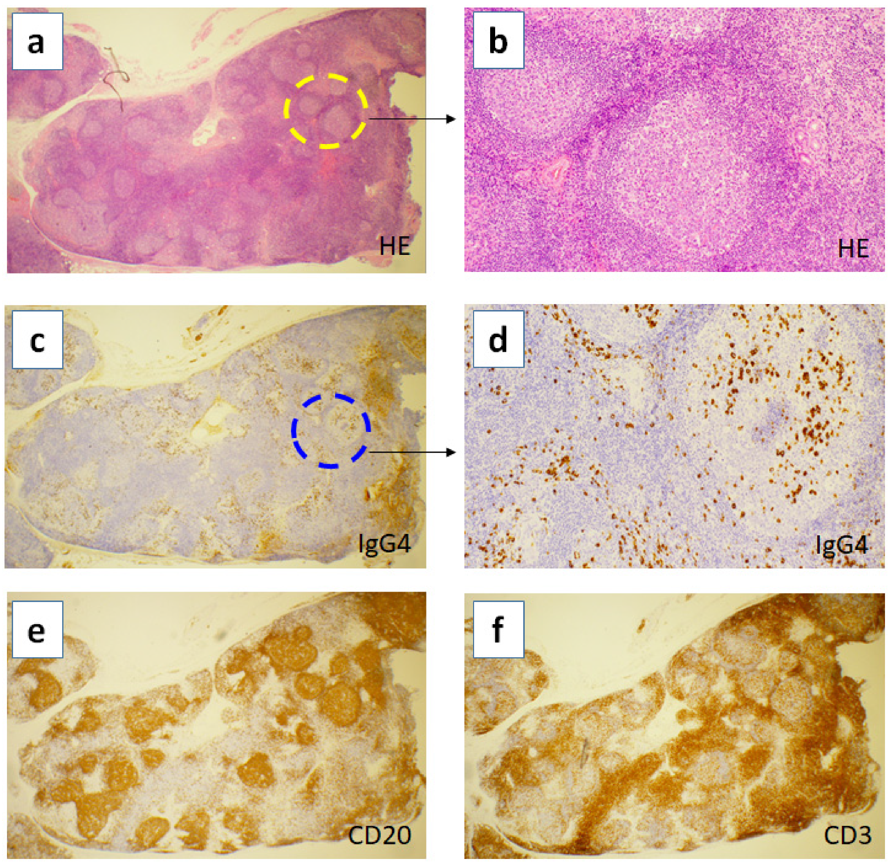
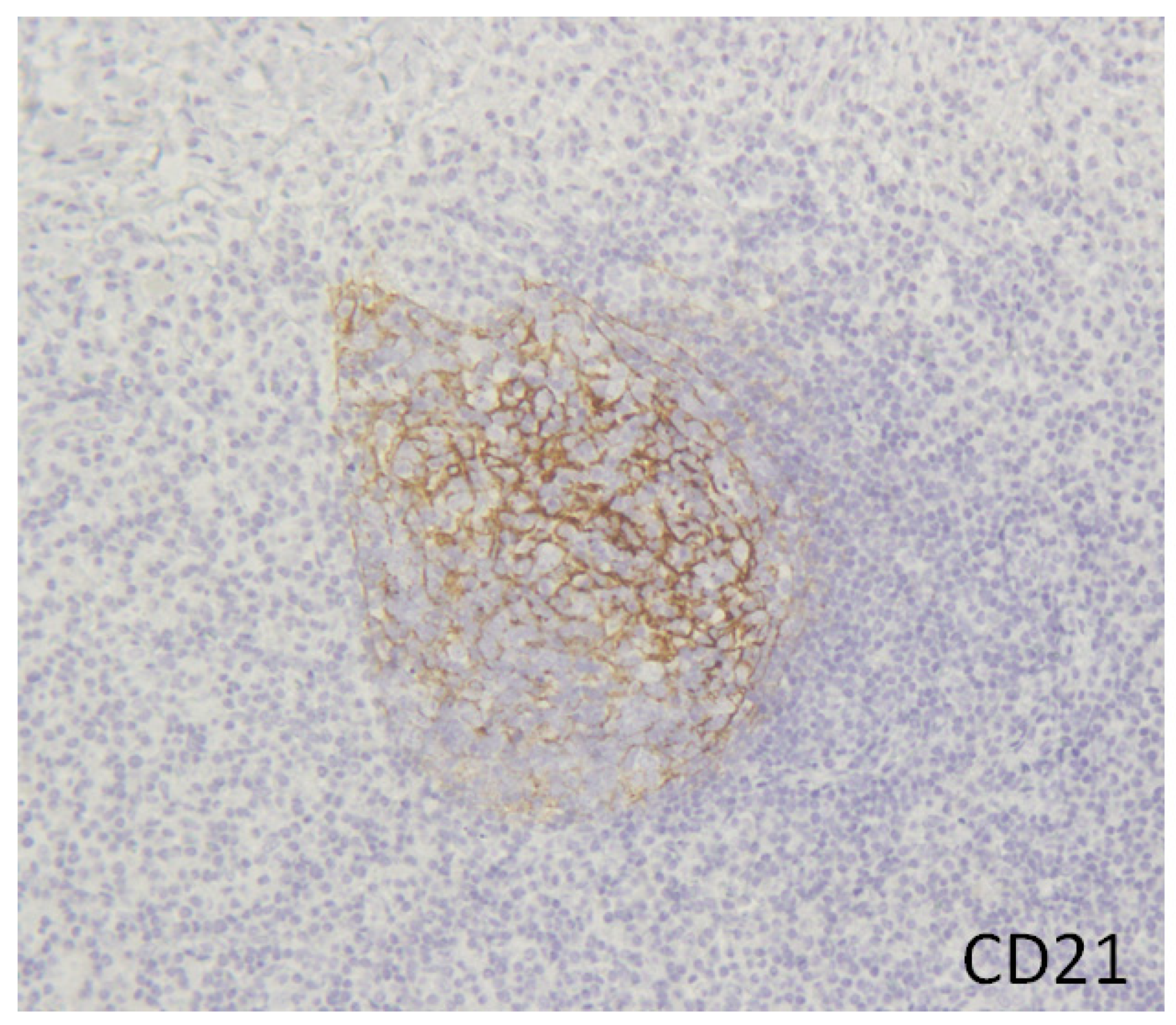
2. Hyper Formation of Tertiary Lymphoid Organs in the Inflamed Tissues of IgG4-RD

Although storiform fibrosis, obliterative phlebitis and IgG4+plasma cell infiltration are well recognized pathological findings in IgG4-RD, the excessive formation of tertiary lymphoid organs (TLOs) has gained much attention. TLOs are ectopic germinal centre formations that develop in non-lymphoid tissues at sites of chronic inflammation such as tumours, infections, allograft rejections, and autoimmune diseases [23,24,25]. TLOs mainly consist of aggregates of T cells, B cells, and follicular dendritic cells (FDCs) networks. TLOs can display the activities of germinal centres, contributing to the local adaptive immune responses at chronic inflammatory lesions. Hyperformation of TLOs can frequently be found In IgG4-RD inflamed tissues (Figure 1a,b).
In IgG4-RD, head and neck lesions such as lacrimal and salivary glands, and retroperitoneal fibrosis more frequently exhibit TLOs formation than pancreas and kidneys [26,27,28,29,30]. The difference of the frequencies of TLOs formation according to the affected organs of IgG4-RD might result from the difference in timing of obtaining tissue samples. A recent study has reported that kidney tissue affected by IgG4-RD demonstrated TLOs formation without fibrosis in the renal parenchyma and around medium-sized vessels when the biopsy was conducted in the very early stages of the disease [29]. Therefore, TLOs formation is the early event of the affected lesions, while fibrosis formation occurs in the later stage of the disease. Interestingly, in the other fibro-inflammatory diseases such as rheumatoid arthritis-associated interstitial lung disease, TLOs formation in lungs emerges first, followed by the formation of fibrotic areas around them [31,32,33,34].
One of the important observations in the TLOs of IgG4-RD is that IgG4 class switching and production develop locally inside the TLOs (Figure 1c,d). Once IgG4-producing B cells differentiate into plasmablasts and plasma cells, they go out of the TLOs and infiltrate into the interfollicular area. This means that a germinal centre reaction similar to that of lymph nodes is occurring at the TLOs of tissues inflamed by IgG4-RD. Although this disease has been initially recognized as the massive infiltration of B cells and plasma cells in the affected lesions, recent studies have emphasized that not only B cells but also T cells dominate the tissue infiltrates (Figure 1e,f). Recent advances in this field have further clarified the importance of the specific T helper subset, Tfh, at the TLOs in inducing IgG4 class switching as well as plasma cell differentiation and maturation in the pathogenesis of IgG4-RD [35,36].
More recently, we and others have identified that CD21+ follicular dendritic cells (FDCs) reside inside the TLOs of IgG4-RD (Figure 2) [37,38]. FDCs secrete CXCL13 (the ligand of CXCR5) and recruit CXCR5+ Tfh and B cells into the target tissues to form TLOs. In fact, CXCL13 were detected at a high level in the affected tissues [39], and our recent study using serum proteomic analysis identified that CXCL13 was one of the most significantly elevated proteins in patients with IgG4-RD [40]. FDCs are crucial not only for recruiting Tfh and B cells to form TLOs but also for intimate interaction with those immune cells.
FDCs are shaped like a “meshwork” inside the TLOs of IgG4-RD to contact intimately with Tfh and B cells and directly activate Tfh to promote germinal centre reaction. Thus, we believe that FDCs are one of the culprit immune cells which play a crucial role in the upstream events of the IgG4-RD disease process.

3. Tfh Is Essential for TLOs Formation

It has been known since the 1960s that helper T cells are important for inducing differentiation of naive B cells into plasmablasts and plasma cells and antigen-specific antibody production. IL-4 was identified as a B cell regulator in the 1980s, and the importance of co-stimulatory molecules such as CD40-CD40 ligand was reported in the 1990s [41]. As noted above, ectopic germinal centres in TLOs are essential for antibody production in the local inflammatory sites. Importantly, deficiency of the gene involved in Th2 cells, which was thought to control humoral immunity, did not impair the formation of germinal centres, suggesting the existence of other T helper subsets that control humoral immunity [42]. Then, CXCR5+ Tfh, which are present in lymphoid tissues such as tonsils, lymph nodes, and TLOs, were discovered in 2000 [43,44,45], and IL-21, which assists B cell differentiation and maturation and immunoglobulin class switching, was found to be produced from Tfh [42]. In 2009, the transcription factor Bcl6 was reported as a master regulator of Tfh, and Tfh was established as an independent T helper subset [46,47,48]. Of note, deficiency of Bcl6 impaired germinal centre formation [46,47]. Co-culture of Tfh and naïve B cells actually induced differentiation into plasmablasts/plasma cells and assisted antibody production [49]. B cells have antibodies that have a higher affinity for antigens by causing somatic mutations in the genes in the immunoglobulin variable region during proliferation and maturation by the help of Tfh in the germinal centres. Furthermore, in recent years, Tfh in human peripheral blood was classified into three subsets, Tfh1, Tfh2, and Tfh17, and it has been revealed that each subset has different cytokine secretion and B cell assisting ability [49]. It was now clarified that Tfh is a specific and independent T helper subset that exerts functions specialized in humoral immunity such as formation of germinal centres, B cell differentiation, affinity maturation, and antibody production.

4. Tfh in IgG4-RD

We previously reported a characteristic increase in circulating plasmablasts in IgG4-RD [50]. High-throughput sequencing study proved that the plasmablasts of this disease are oligoclonal and the immunoglobulin gene has a high degree of somatic mutation [51]. These findings suggest that B cells are differentiated and matured into plasmablasts in a Tfh-dependent manner in this disease. Thus, we further examined Tfh and their subsets in patients with IgG4-RD and found that Tfh, particularly Tfh2 subset was characteristically increased in peripheral blood [50,52]. Tfh2 count showed a strong correlation with serum IgG4 level, IgG4/IgG ratio, and plasmablast count in treatment-naïve patients with IgG4-RD [50,52]. The magnitude of IgG4+plasma cell infiltration at the lesion site was also positively correlated with circulating Tfh2 count [53]. The analysis of biopsy samples from patients with IgG4-RD further demonstrated that about 70% of tissue-infiltrating CD4+ T cells were Tfh; Tfh2 was most dominant among Tfh subsets [54,55]. Our in vitro functional analysis revealed that patient-derived Tfh2 actually induced naive B cells to differentiate into IgG4-producing plasmablasts/plasma cells at a high rate [52]. Therefore, Tfh2 was established as the central immune cell subset which induces plasmablast differentiation and IgG4 production in IgG4-RD.
In the analysis of serum cytokines in IgG4-RD, the increase in Tfh2 counts was significantly correlated with serum IL-4 and IgG4 levels [50]. In addition, Tfh2 counts and IgG4 levels tended to be high in cases with high IL-21 levels [50]. Functional analysis also demonstrated that IL-4 induced IgG4 class switching, while IL-21 promoted plasmablast/plasma cell differentiation in IgG4-RD [56]. In addition, IL-21 was proved to be important for hyperplastic TLOs formation in IgG4-RD [26,57]. IL-4 also plays a role in inducing CCL18-producing M2 macrophages, which contribute to the generation of fibrosis observed in IgG4-RD [58,59,60]. The study of M2 macrophages in the inflamed submandibular glands of IgG4-RD reported that fibrosis scores positively correlated with the frequency of M2 macrophages [59]. Furthermore, CCL18 expression was co-localized with M2 macrophages in the IgG4-related submandibular glands [59]. Not only the inflamed submandibular glands but also the mediastinal fibrosis tissue of IgG4-RD demonstrated the localization of many M2 macrophages in the lesions of storiform fibrosis [8]. These findings suggest that Tfh2 is a source of IL-4 and IL-21, which are involved in IgG4 production, induction of plasmablast/plasma cell differentiation, and fibrosis process.
We examined the serial changes in the number of Tfh2, plasmablasts, serum IgG4, and IL-4 before and after glucocorticoid treatment; plasmablast counts, IgG4, and IL-4 levels were decreased, whereas the increase of Tfh2 counts was not corrected [50]. A recent study also reported that Tfh2 counts in IgG4-RD remained unchanged, even after clinical improvement by rituximab treatment [61]. Furthermore, we also found the re-activation of Tfh2 at recurrence of the disease [52]. These results indicate that glucocorticoid or rituximab is not sufficient to suppress the number of Tfh2 and/or their pathogenic function, resulting in the future relapse of the disease.
Collectively, Tfh2 is associated with the upstream pathogenic process of the disease mechanisms such as TLOs formation, IgG4 class switching, and fibrosis. Identification of new target molecules involved in Tfh2 differentiation, proliferation and function is desired for the development of new, more effective and less side-effect therapies.

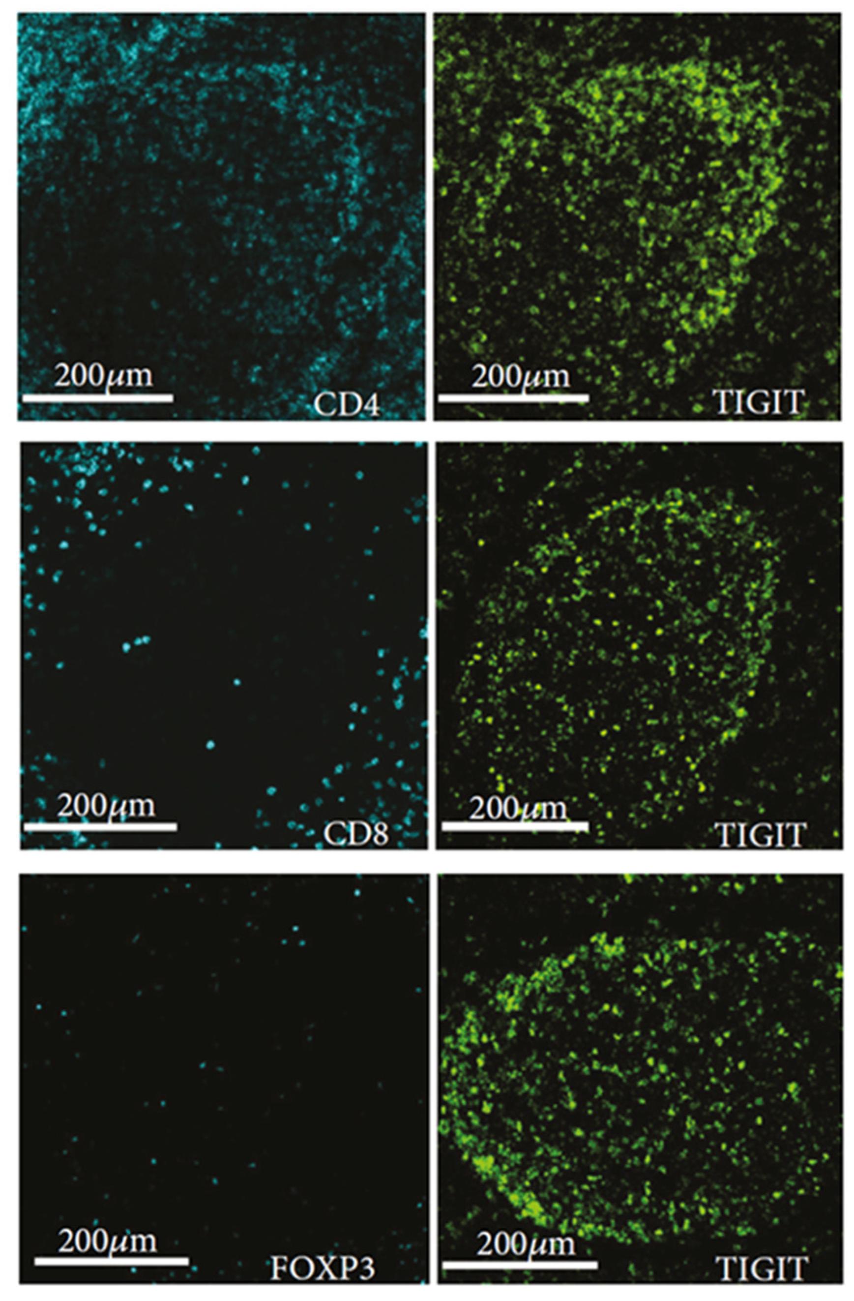
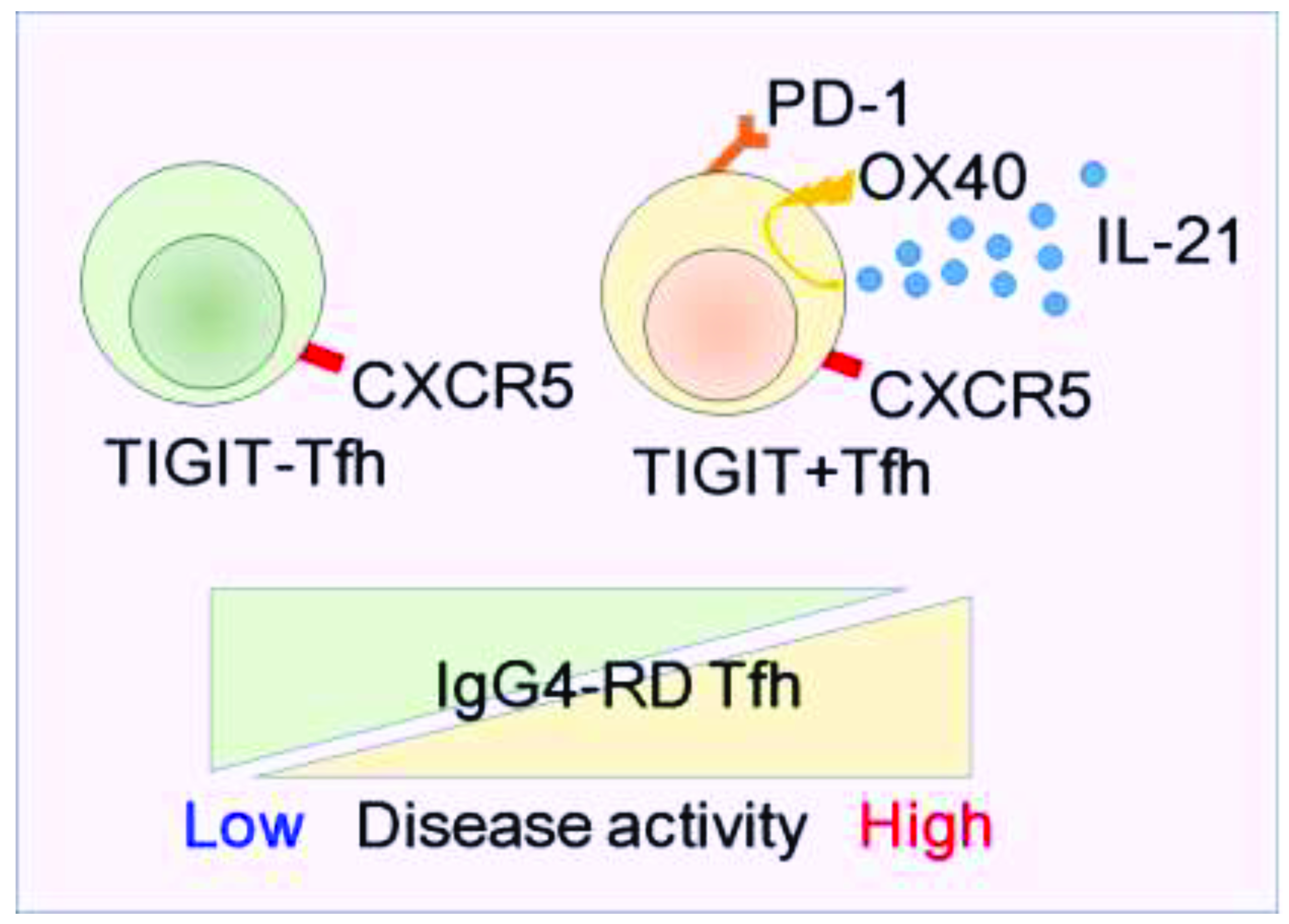
5. Newly Identified Tfh Subset, “TIGIT+ Tfh”

IL-21 is a pleiotropic cytokine vital for the generation and function of Tfh, formation and maintenance of germinal centres, differentiation and maturation of plasmablasts, and activation of cytotoxic T cells [62,63]. Thus, identification of the molecular marker for detecting high IL-21 producing Tfh population is essentially important to assess and understand functionally active Tfh responses. We previously reported the increase of activated PD-1+ CXCR5+ Tfh in peripheral blood in patients with IgG4-RD, correlating with their disease activity [52]. On the other hand, among CXCR5- T helper cells, PD-1+ cell fractions have been reported as T peripheral helper (Tph) cells that produce IL-21 and have the ability to assist the differentiation of memory B cells into plasmablasts [64]. Therefore, PD-1 expression is no longer specific to Tfh. Furthermore, when we consider PD-1, which is an immune checkpoint receptor, as a therapeutic target for autoimmune diseases, it may suppress both Tfh and Tph in a non-specific manner.
It would be ideal to target Tfh-specific molecules as therapeutics for diseases with Tfh-dominant pathology, while targeting Tph-specific molecules with Tph-dominant pathological conditions. Hence, we have discovered that TIGIT, another immune checkpoint receptor, is a novel surface molecule preferentially expressed on Tfh but not other T helper cells [65]. In human tonsils, TIGIT expression was limited to Tfh residing within the germinal centre (Figure 3 and Figure 4), while TIGIT was not expressed in dendritic cells, macrophages, and B cells [66]. Of interest, TIGIT+Tfh highly produced IL-21, suggesting that TIGIT+Tfh population is a functionally active Tfh [65]. Mechanistically, we have identified OX40 as the responsible signal involved in the high production of IL-21 in TIGIT+Tfh [65]. On the other hand, the expression of CD155 (PVR), a ligand of TIGIT, was limited to FDCs closely contacting with TIGIT+Tfh within germinal centres [67]. Considering the capacity of TIGIT to mediate strong adhesion to CD155, TIGIT may facilitate retention of Tfh within germinal centres. Additionally, FDCs express OX40 ligand to induce high IL-21 production from OX40+ TIGIT+Tfh [68,69]. The involvement of TIGIT in Tfh-dependent B-cell responses is also supported by a study showing a defect in IgG production in CD155-deficient mice [70]. Together, these findings suggest that TIGIT-CD155 interactions may be crucial in regulating function and positioning of Tfh within germinal centres, contributing to Tfh-dependent B-cell responses. Therefore, when TIGIT is considered a therapeutic target, Tfh-targeted treatment can be achieved.
We have demonstrated that patients with IgG4-RD, a representative Tfh-associated disease, exhibited the significant increase of TIGIT+Tfh in peripheral blood, correlating with disease activity, the number of affected organs, serum IgG4 level, and Tfh-derived IL-21 production (Figure 5) [65]. Thus, the increase of TIGIT+Tfh reflects active, ongoing Tfh responses. TIGIT+Tfh and associated molecules such as CD155, OX40 ligand, and OX40 would be the potential therapeutic targets in this disease.

6. Conclusions

Recent advances in our understanding of the pathogenesis of IgG4-RD have revealed the underlying mechanisms of IgG4+plasma cell differentiation and fibrosis formation (Figure 6). In early stages of the disease, chronic inflammation might induce the accumulation of antigen-presenting FDCs, which further recruit CXCR5+Tfh and B cells by secreting CXCL13. Then, TLOs emerge in the inflamed tissues, orchestrating Tfh cells, B cells, and FDCs networks. TIGIT might play a role in the retention and positioning of Tfh through interaction with CD155 on FDCs. Those TIGIT+Tfh express OX40, and OX40 ligand from FDCs stimulates TIGIT+Tfh to let them produce high amounts of IL-21. IL-21 is important for B cell maturation and differentiation and maintenance of TLOs formation. IL-4 secreted from Tfh specifically induces IgG4 class switching, and in later stages of the disease IL-4 also plays a role in inducing M2 macrophages, which are involved in the process of fibrosis formation by secreting CCL18. Thus, organ swelling and damage is caused in this disease. Further research in the future to determine whether these disease-associated molecules can replace glucocorticoids as therapeutic targets would be desirable.

Funding

This review received no external funding.

Data Availability Statement

The data underlying this article are available in the article.

Acknowledgments

M.A. is supported by JSPS KAKENHI Grant Number 21K16292.

Conflicts of Interest

M.A. has received speaker fees from Pfizer Japan Inc. and Asahi Kasei Pharma Corp. Y.K. has received grants or speaker fees from AbbVie, Astellas, Ayumi, Asahi Kasei Pharma Corp, Bristol-Myers Squibb, Chugai, Eisai, Eli Lilly, Hisamitsu, Jansen, Kissei, Pfizer, Sanofi, Takeda, Tanabe-Mitsubishi and UCB.

References

  1. Hamano, H.; Kawa, S.; Horiuchi, A.; Unno, H.; Furuya, N.; Akamatsu, T.; Fukushima, M.; Nikaido, T.; Nakayama, K.; Usuda, N.; et al. High Serum IgG4 Concentrations in Patients with Sclerosing Pancreatitis. N. Engl. J. Med. 2001, 344, 732–738. [Google Scholar] [CrossRef]
  2. Kamisawa, T.; Funata, N.; Hayashi, Y.; Eishi, Y.; Koike, M.; Tsuruta, K.; Okamoto, A.; Egawa, N.; Nakajima, H. A new clinicopathological entity of IgG4-related autoimmune disease. J. Gastroenterol. 2003, 38, 982–984. [Google Scholar] [CrossRef]
  3. Umehara, H.; Okazaki, K.; Masaki, Y.; Kawano, M.; Yamamoto, M.; Saeki, T.; Matsui, S.; Sumiada, T.; Mimori, T.; Tanaka, Y.; et al. A novel clinical entity, IgG4-related disease (IgG4RD): General concept and details. Mod. Rheumatol. 2012, 22, 1–14. [Google Scholar] [CrossRef]
  4. Stone, J.H.; Zen, Y.; Deshpande, V. IgG4-related disease. N. Engl. J. Med. 2012, 366, 539–551. [Google Scholar] [CrossRef]
  5. Akiyama, M.; Kaneko, Y.; Takeuchi, T. Etiology of IgG4-Related Pulmonary Hypertension. Cardiology 2020, 145, 263–266. [Google Scholar] [CrossRef]
  6. Takanashi, S.; Akiyama, M.; Nishina, N.; Kaneko, Y.; Takeuchi, T. Characteristics and prognosis of IgG4-related skin disease: A case report and systematic literature review. Autoimmun. Rev. 2021, 20, 102805. [Google Scholar] [CrossRef] [PubMed]
  7. Akiyama, M.; Kaneko, Y.; Takeuchi, T. Characteristics and prognosis of IgG4-related periaortitis/periarteritis: A systematic literature review. Autoimmun. Rev. 2019, 18, 102354. [Google Scholar] [CrossRef]
  8. Takanashi, S.; Akiyama, M.; Suzuki, K.; Otomo, K.; Takeuchi, T. IgG4-related fibrosing mediastinitis diagnosed with computed tomography-guided percutaneous needle biopsy: Two case reports and a review of the literature. Medicine 2018, 97, e10935. [Google Scholar] [CrossRef]
  9. Akiyama, M.; Kaneko, Y.; Hayashi, Y.; Takeuchi, T. IgG4-related disease involving vital organs diagnosed with lip biopsy: A case report and literature review. Medicine 2016, 95, e3970. [Google Scholar] [CrossRef] [PubMed]
  10. Akiyama, M.; Kaneko, Y.; Takeuchi, T. Eosinophilic granulomatosis with polyangiitis can manifest lacrimal and salivary glands swelling by granulomatous inflammation: A potential mimicker of IgG4-related disease. Ann. Rheum. Dis. 2020. Online ahead of print. [Google Scholar] [CrossRef] [PubMed]
  11. Akiyama, M.; Kaneko, Y.; Takeuchi, T. Characteristics and prognosis of ANCA-positive retroperitoneal fibrosis: A systematic literature review. Autoimmun. Rev. 2020, 19, 102642. [Google Scholar] [CrossRef] [PubMed]
  12. Akiyama, M.; Takanashi, S.; Takeuchi, T.; Kaneko, Y. Salivary gland involvement in ANCA-associated vasculitis: A systematic literature review. Autoimmun. Rev. 2021, 20, 102940. [Google Scholar] [CrossRef]
  13. Akiyama, M.; Zeisbrich, M.; Ibrahim, N.; Ohtsuki, S.; Berry, G.J.; Hwang, P.H.; Goronzy, J.J.; Weyand, C.M. Neutrophil Extracellular Traps Induce Tissue-Invasive Monocytes in Granulomatosis with Polyangiitis. Front. Immunol. 2019, 10, 2617. [Google Scholar] [CrossRef]
  14. Matsumoto, K.; Akiyama, M.; Kajio, N.; Otomo, K.; Suzuki, K.; Nishina, N.; Kasuya, K.; Oishi, N.; Kameyama, K.; Takeuchi, T. Adolescent PR3-ANCA-positive hypertrophic pachymeningitis: A case report and review of the literature. Medicine 2018, 97, e0521. [Google Scholar] [CrossRef] [PubMed]
  15. Sasaki, T.; Akiyama, M.; Kaneko, Y.; Takeuchi, T. IgG4-related disease and idiopathic multicentric Castleman’s disease: Confusable immune-mediated disorders. Rheumatology 2021, keab634, Online ahead of print. [Google Scholar] [CrossRef]
  16. Sasaki, T.; Akiyama, M.; Kaneko, Y.; Mori, T.; Yasuoka, H.; Suzuki, K.; Yamaoka, K.; Okamoto, S.; Takeuchi, T. Distinct features distinguishing IgG4-related disease from multicentric Castleman’s disease. RMD Open 2017, 3, e000432. [Google Scholar] [CrossRef] [PubMed]
  17. Akiyama, M.; Yasuoka, H.; Takeuchi, T. Interleukin-6 in idiopathic multicentric Castleman’s disease after long-term tocilizumab. Ann. Hematol. 2017, 96, 2117–2119. [Google Scholar] [CrossRef] [PubMed]
  18. Anan, R.; Akiyama, M.; Kaneko, Y.; Kikuchi, J.; Suzuki, K.; Matsubara, S.; Takeuchi, T. Polymyositis with elevated serum IgG4 levels and abundant IgG4+plasma cell infiltration: A case report and literature review. Medicine 2017, 96, e8710. [Google Scholar] [CrossRef]
  19. Akiyama, M.; Takeuchi, T. IgG4-Related Disease: Beyond Glucocorticoids. Drugs Aging 2018, 35, 275–287. [Google Scholar] [CrossRef]
  20. Khosroshahi, A.; Wallace, Z.S.; Crowe, J.L.; Akamizu, T.; Azumi, A.; Carruthers, M.N.; Chari, S.T.; Della-Torre, E.; Frulloni, L.; Goto, H.; et al. International Consensus Guidance Statement on the Management and Treatment of IgG4-Related Disease. Arthritis Rheumatol. 2015, 67, 1688–1699. [Google Scholar] [CrossRef]
  21. Sasaki, T.; Akiyama, M.; Kaneko, Y.; Yasuoka, H.; Suzuki, K.; Yamaoka, K.; Takeuchi, T. Risk factors of relapse following glucocorticoid tapering in IgG4-related disease. Clin. Exp. Rheumatol. 2018, 36, 186–189. [Google Scholar]
  22. Masamune, A.; Nishimori, I.; Kikuta, K.; Tsuji, I.; Mizuno, N.; Iiyama, T.; Kanno, A.; Tachibana, Y.; Ito, T.; Kamisawa, T.; et al. Randomised controlled trial of long-term maintenance corticosteroid therapy in patients with autoimmune pancreatitis. Gut 2017, 66, 487–494. [Google Scholar] [CrossRef]
  23. Luo, S.; Zhu, R.; Yu, T.; Fan, H.; Hu, Y.; Mohanta, S.K.; Hu, D. Chronic Inflammation: A Common Promoter in Tertiary Lymphoid Organ Neogenesis. Front. Immunol. 2019, 10, 2938. [Google Scholar] [CrossRef] [Green Version]
  24. Jones, G.W.; Hill, D.G.; Jones, S.A. Understanding Immune Cells in Tertiary Lymphoid Organ Development: It Is All Starting to Come Together. Front. Immunol. 2016, 7, 401. [Google Scholar] [CrossRef] [PubMed] [Green Version]
  25. Domblides, C.; Rochefort, J.; Riffard, C.; Panouillot, M.; Lescaille, G.; Teillaud, J.L.; Mateo, V.; Dieu-Nosjean, M.C. Tumor-Associated Tertiary Lymphoid Structures: From Basic and Clinical Knowledge to Therapeutic Manipulation. Front. Immunol. 2021, 12, 698604. [Google Scholar] [CrossRef]
  26. Maehara, T.; Moriyama, M.; Nakashima, H.; Miyake, K.; Hayashida, J.N.; Tanaka, A.; Shinozaki, S.; Kubo, Y.; Nakamura, S. Interleukin-21 contributes to germinal centre formation and immunoglobulin G4 production in IgG4-related dacryoadenitis and sialoadenitis, so-called Mikulicz’s disease. Ann. Rheum. Dis. 2012, 71, 2011–2019. [Google Scholar] [CrossRef] [PubMed] [Green Version]
  27. Kasashima, S.; Zen, Y.; Kawashima, A.; Konishi, K.; Sasaki, H.; Endo, M.; Matsumoto, Y.; Kawakami, K.; Kasashima, F.; Moriya, M.; et al. Inflammatory abdominal aortic aneurysm: Close relationship to IgG4-related periaortitis. Am. J. Surg. Pathol. 2008, 32, 197–204. [Google Scholar] [CrossRef]
  28. Zen, Y.; Nakanuma, Y. IgG4-related disease: A cross-sectional study of 114 cases. Am. J. Surg. Pathol. 2010, 34, 1812–1819. [Google Scholar] [CrossRef]
  29. Miyanaga, T.; Mizuguchi, K.; Hara, S.; Zoshima, T.; Inoue, D.; Nishioka, R.; Mizushima, I.; Ito, K.; Fuji, H.; Yamada, K.; et al. Tertiary lymphoid tissue in early-stage IgG4-related tubulointerstitial nephritis incidentally detected with a tumor lesion of the ureteropelvic junction: A case report. BMC Nephrol. 2021, 22, 34. [Google Scholar] [CrossRef]
  30. Cargill, T.; Makuch, M.; Sadler, R.; Lighaam, L.C.; Peters, R.; van Ham, M.; Klenerman, P.; Bateman, A.; Rispens, T.; Barnes, E.; et al. Activated T-Follicular Helper 2 Cells Are Associated with Disease Activity in IgG4-Related Sclerosing Cholangitis and Pancreatitis. Clin. Transl. Gastroenterol. 2019, 10, e00020. [Google Scholar] [CrossRef] [PubMed]
  31. Rangel-Moreno, J.; Hartson, L.; Navarro, C.; Gaxiola, M.; Selman, M.; Randall, T.D. Inducible bronchus-associated lymphoid tissue (iBALT) in patients with pulmonary complications of rheumatoid arthritis. J. Clin. Investig. 2006, 116, 3183–3194. [Google Scholar] [CrossRef] [Green Version]
  32. Turesson, C.; Matteson, E.L.; Colby, T.V.; Vuk-Pavlovic, Z.; Vassallo, R.; Weyand, C.M.; Tazellaar, H.D.; Limper, A.H. Increased CD4+ T cell infiltrates in rheumatoid arthritis-associated interstitial pneumonitis compared with idiopathic interstitial pneumonitis. Arthritis Rheum. 2005, 52, 73–79. [Google Scholar] [CrossRef]
  33. Atkins, S.R.; Turesson, C.; Myers, J.L.; Tazelaar, H.D.; Ryu, J.H.; Matteson, E.L.; Bongartz, T. Morphologic and quantitative assessment of CD20+ B cell infiltrates in rheumatoid arthritis-associated nonspecific interstitial pneumonia and usual interstitial pneumonia. Arthritis Rheum. 2006, 54, 635–641. [Google Scholar] [CrossRef] [PubMed]
  34. Akiyama, M.; Kaneko, Y.; Yamaoka, K.; Kondo, H.; Takeuchi, T. Association of disease activity with acute exacerbation of interstitial lung disease during tocilizumab treatment in patients with rheumatoid arthritis: A retrospective, case-control study. Rheumatol. Int. 2016, 36, 881–889. [Google Scholar] [CrossRef]
  35. Akiyama, M.; Suzuki, K.; Yasuoka, H.; Kaneko, Y.; Yamaoka, K.; Takeuchi, T. Follicular helper T cells in the pathogenesis of IgG4-related disease. Rheumatology 2018, 57, 236–245. [Google Scholar] [CrossRef] [PubMed] [Green Version]
  36. Akiyama, M.; Suzuki, K.; Kaneko, Y.; Takeuchi, T. IgG4-Related Disease: A Growing Appreciation of Follicular Helper T Cell Expansion. Clin. Transl. Gastroenterol. 2019, 10, e00076. [Google Scholar] [CrossRef]
  37. Akiyama, M.; Kaneko, Y. Comment on: Persistence of circulating T-follicular helper cells after rituximab is associated with relapse of IgG4-related disease. Rheumatology 2021, keab687, Online ahead of print. [Google Scholar] [CrossRef] [PubMed]
  38. Satoh-Nakamura, T.; Kurose, N.; Kawanami, T.; Nakamura, T.; Iwao-Kawanami, H.; Nakajima, A.; Miki, M.; Sakai, T.; Fujita, Y.; Tanaka, M.; et al. CD14+ follicular dendritic cells in lymphoid follicles may play a role in the pathogenesis of IgG4-related disease. Biomed. Res. 2015, 36, 143–153. [Google Scholar] [CrossRef] [Green Version]
  39. Ohta, M.; Moriyama, M.; Maehara, T.; Gion, Y.; Furukawa, S.; Tanaka, A.; Hayashida, J.N.; Yamauchi, M.; Ishiguro, N.; Mikami, Y.; et al. DNA Microarray Analysis of Submandibular Glands in IgG4-Related Disease Indicates a Role for MARCO and Other Innate Immune-Related Proteins. Medicine 2016, 95, e2853. [Google Scholar] [CrossRef] [Green Version]
  40. Takanashi, S.; Kikuchi, J.; Sasaki, T.; Akiyama, M.; Yasuoka, H.; Yoshimoto, K.; Seki, N.; Sugahara, K.; Chiba, K.; Kaneko, Y.; et al. Lymphadenopathy in IgG4-related disease: A phenotype of severe activity and poor prognosis, with eotaxin-3 as a new biomarker. Rheumatology 2021, 60, 967–975. [Google Scholar] [CrossRef]
  41. Crotty, S. A brief history of T cell help to B cells. Nat. Rev. Immunol. 2015, 15, 185–189. [Google Scholar] [CrossRef] [PubMed]
  42. Kopf, M.; Le Gros, G.; Coyle, A.J.; Kosco-Vilbois, M.; Brombacher, F. Immune responses of IL-4, IL-5, IL-6 deficient mice. Immunol. Rev. 1995, 148, 45–69. [Google Scholar] [CrossRef] [PubMed]
  43. Schaerli, P.; Willimann, K.; Lang, A.B.; Lipp, M.; Loetscher, P.; Moser, B. CXC chemokine receptor 5 expression defines follicular homing T cells with B cell helper function. J. Exp. Med. 2000, 192, 1553–1562. [Google Scholar] [CrossRef] [PubMed] [Green Version]
  44. Kim, C.H.; Rott, L.S.; Clark-Lewis, I.; Campbell, D.J.; Wu, L.; Butcher, E.C. Subspecialization of CXCR5+ T cells: B helper activity is focused in a germinal center-localized subset of CXCR5+ T cells. J. Exp. Med. 2001, 193, 1373–1381. [Google Scholar] [CrossRef] [Green Version]
  45. Breitfeld, D.; Ohl, L.; Kremmer, E.; Ellwart, J.; Sallusto, F.; Lipp, M.; Försteret, R. Follicular B helper T cells express CXC chemokine receptor 5, localize to B cell follicles, and support immunoglobulin production. J. Exp. Med. 2000, 192, 1545–1552. [Google Scholar] [CrossRef]
  46. Nurieva, R.I.; Chung, Y.; Martinez, G.J.; Yang, X.O.; Tanaka, S.; Matskevitch, T.D.; Wang, Y.H.; Dong, C. Bcl6 mediates the development of T follicular helper cells. Science 2009, 325, 1001–1005. [Google Scholar] [CrossRef] [Green Version]
  47. Yu, D.; Rao, S.; Tsai, L.M.; Lee, S.K.; He, Y.; Sutcliffe, E.L.; Srivastava, M.; Linterman, M.; Zheng, L.; Simpson, N.; et al. The transcriptional repressor Bcl-6 directs T follicular helper cell lineage commitment. Immunity 2009, 31, 457–468. [Google Scholar] [CrossRef] [PubMed]
  48. Johnston, R.J.; Poholek, A.C.; DiToro, D.; Yusuf, I.; Eto, D.; Barnett, B.; Dent, A.L.; Craft, J.; Crotty, S. Bcl6 and Blimp-1 are reciprocal and antagonistic regulators of T follicular helper cell differentiation. Science 2009, 325, 1006–1010. [Google Scholar] [CrossRef] [Green Version]
  49. Morita, R.; Schmitt, N.; Bentebibel, S.E.; Ranganathan, R.; Bourdery, L.; Zurawski, G.; Foucat, E.; Dullaers, M.; Oh, S.H.; Sabzghabaei, N.; et al. Human blood CXCR5(+)CD4(+) T cells are counterparts of T follicular cells and contain specific subsets that differentially support antibody secretion. Immunity 2011, 34, 108–121. [Google Scholar] [CrossRef] [Green Version]
  50. Akiyama, M.; Suzuki, K.; Yamaoka, K.; Yasuoka, H.; Takeshita, M.; Kaneko, Y.; Kondo, H.; Kassai, Y.; Miyazaki, T.; Morita, R.; et al. Number of Circulating Follicular Helper 2 T Cells Correlates with IgG4 and Interleukin-4 Levels and Plasmablast Numbers in IgG4-Related Disease. Arthritis Rheumatol. 2015, 67, 2476–2481. [Google Scholar] [CrossRef] [Green Version]
  51. Mattoo, H.; Mahajan, V.S.; Della-Torre, E.; Sekigami, Y.; Carruthers, M.; Wallace, Z.S.; Deshpande, V.; Stone, J.H.; Pillai, S. De novo oligoclonal expansions of circulating plasmablasts in active and relapsing IgG4-related disease. J. Allergy Clin. Immunol. 2014, 134, 679–687. [Google Scholar] [CrossRef] [PubMed] [Green Version]
  52. Akiyama, M.; Yasuoka, H.; Yamaoka, K.; Suzuki, K.; Kaneko, Y.; Kondo, H.; Kassai, Y.; Koga, K.; Miyazaki, T.; Morita, R.; et al. Enhanced IgG4 production by follicular helper 2 T cells and the involvement of follicular helper 1 T cells in the pathogenesis of IgG4-related disease. Arthritis Res. Ther. 2016, 18, 167. [Google Scholar] [CrossRef] [Green Version]
  53. Akiyama, M.; Kaneko, Y.; Yamaoka, K.; Hayashi, Y.; Yasuoka, H.; Suzuki, K.; Takeuchi, T. Subclinical labial salivary gland involvement in IgG4-related disease affected with vital organs. Clin. Exp. Rheumatol. 2015, 33, 949–950. [Google Scholar] [PubMed]
  54. Kamekura, R.; Takano, K.; Yamamoto, M.; Kawata, K.; Shigehara, K.; Jitsukawa, S.; Nagaya, T.; Ito, F.; Sato, A.; Ogasawara, S.; et al. Cutting Edge: A Critical Role of Lesional T Follicular Helper Cells in the Pathogenesis of IgG4-Related Disease. J. Immunol. 2017, 199, 2624–2629. [Google Scholar] [CrossRef]
  55. Chen, Y.; Lin, W.; Yang, H.; Wang, M.; Zhang, P.; Feng, R.; Chen, H.; Peng, L.; Zhang, X.; Zhao, Y.; et al. Aberrant Expansion and Function of Follicular Helper T Cell Subsets in IgG4-Related Disease. Arthritis Rheumatol. 2018, 70, 1853–1865. [Google Scholar] [CrossRef] [PubMed]
  56. Akiyama, M.; Yasuoka, H.; Yoshimoto, K.; Takeuchi, T. Interleukin-4 contributes to the shift of balance of IgG subclasses toward IgG4 in IgG4-related disease. Cytokine 2018, 110, 416–419. [Google Scholar] [CrossRef]
  57. Yajima, H.; Yamamoto, M.; Shimizu, Y.; Sakurai, N.; Suzuki, C.; Naishiro, Y.; Imai, K.; Shinomura, Y.; Takahashi, H. Loss of interleukin-21 leads to atrophic germinal centers in multicentric Castleman’s disease. Ann. Hematol. 2016, 95, 35–40. [Google Scholar] [CrossRef] [PubMed]
  58. Akiyama, M.; Yasuoka, H.; Yoshimoto, K.; Takeuchi, T. CC-chemokine ligand 18 is a useful biomarker associated with disease activity in IgG4-related disease. Ann. Rheum. Dis. 2018, 77, 1386–1387. [Google Scholar] [CrossRef] [Green Version]
  59. Furukawa, S.; Moriyama, M.; Tanaka, A.; Maehara, T.; Tsuboi, H.; Iizuka, M.; Hayashida, J.N.; Ohta, M.; Saeki, T.; Notohara, K.; et al. Preferential M2 macrophages contribute to fibrosis in IgG4-related dacryoadenitis and sialoadenitis, so-called Mikulicz’s disease. Clin. Immunol. 2015, 156, 9–18. [Google Scholar] [CrossRef] [Green Version]
  60. Tsuboi, H.; Iizuka-Koga, M.; Asashima, H.; Takahashi, H.; Kudo, H.; Ono, Y.; Honda, F.; Iizuka, A.; Segawa, S.; Abe, S.; et al. Upregulation and pathogenic roles of CCL18-CCR8 axis in IgG4-related disease. Mod. Rheumatol. 2020, 30, 729–737. [Google Scholar] [CrossRef]
  61. Mancuso, G.; Jofra, T.; Lanzillotta, M.; Aiuti, A.; Cicalese, M.P.; di Colo, G.; Dagna, L.; Fousteri, G.; Della-Torre, E. Persistence of circulating T-follicular helper cells after rituximab is associated with relapse of IgG4-related disease. Rheumatology 2021, 60, 3947–3949. [Google Scholar] [CrossRef]
  62. Spolski, R.; Leonard, W.J. Interleukin-21: A double-edged sword with therapeutic potential. Nat. Rev. Drug Dis. 2014, 13, 379–395. [Google Scholar] [CrossRef]
  63. Long, D.; Chen, Y.; Wu, H.; Zhao, M.; Lu, Q. Clinical significance and immunobiology of IL-21 in autoimmunity. J. Autoimmun. 2019, 99, 1–14. [Google Scholar] [CrossRef]
  64. Rao, D.A.; Gurish, M.F.; Marshall, J.L.; Slowikowski, K.; Fonseka, C.Y.; Liu, Y.; Donlin, L.T.; Henderson, L.A.; Wei, K.; Mizoguchi, F.; et al. Pathologically expanded peripheral T helper cell subset drives B cells in rheumatoid arthritis. Nature 2017, 542, 110–114. [Google Scholar] [CrossRef] [PubMed]
  65. Akiyama, M.; Suzuki, K.; Yoshimoto, K.; Yasuoka, H.; Kaneko, Y.; Takeuchi, T. Peripheral TIGIT+ T Follicular Helper Cells That Produce High Levels of Interleukin-21 via OX40 Represent Disease Activity in IgG4-Related Disease. Front. Immunol. 2021, 12, 651357. [Google Scholar] [CrossRef] [PubMed]
  66. Blessin, N.C.; Simon, R.; Kluth, M.; Fischer, K.; Hube-Magg, C.; Li, W.; Makrypidi-Fraune, G.; Wellge, B.; Mandelkow, T.; Debatin, N.F.; et al. Patterns of TIGIT Expression in Lymphatic Tissue, Inflammation, and Cancer. Dis. Markers 2019, 2019, 5160565. [Google Scholar] [CrossRef]
  67. Boles, K.S.; Vermi, W.; Facchetti, F.; Fuchs, A.; Wilson, T.J.; Diacovo, T.G.; Cella, M.; Colonna, M. A novel molecular interaction for the adhesion of follicular CD4 T cells to follicular DC. Eur. J. Immunol. 2009, 39, 695–703. [Google Scholar] [CrossRef] [PubMed] [Green Version]
  68. Tahiliani, V.; Hutchinson, T.E.; Abboud, G.; Croft, M.; Salek-Ardakani, S. OX40 Cooperates with ICOS to Amplify Follicular Th Cell Development and Germinal Center Reactions during Infection. J. Immunol. 2017, 198, 218–228. [Google Scholar] [CrossRef]
  69. Fu, N.; Xie, F.; Sun, Z.; Wang, Q. The OX40/OX40L Axis Regulates T Follicular Helper Cell Differentiation: Implications for Autoimmune Diseases. Front. Immunol. 2021, 12, 670637. [Google Scholar] [CrossRef] [PubMed]
  70. Seth, S.; Ravens, I.; Kremmer, E.; Maier, M.K.; Hadis, U.; Hardtke, S.; Forster, R.; Bernhardt, G. Abundance of follicular helper T cells in Peyer’s patches is modulated by CD155. Eur. J. Immunol. 2009, 39, 3160–3170. [Google Scholar] [CrossRef]
Figure 1. Pathological findings of IgG4-RD. The inflamed tissue of lacrimal gland from IgG4-RD shows many formations of tertiary lymphoid organs by hematoxylin-eosin (HE) staining at low power field (a). Tertiary lymphoid organs within yellow circle (a) are further displayed at high power field (b). IgG4 staining (brown) demonstrates active IgG4-producing B cell differentiation within tertiary lymphoid organs at low power field (c). IgG4-producing B cells within tertiary lymphoid organs inside the blue circle (c) are also shown at high power field (d). CD20 staining (brown, low power field) demonstrates that CD20+ B cells mainly reside within tertiary lymphoid organs (e). CD3 staining (brown, low power field) shows that CD3+ T cells infiltrate inside and outside of tertiary lymphoid organs (f).
Immuno 01 00026 g001
Figure 2. CD21+ follicular dendritic cells in IgG4-RD. CD21 staining (brown, high power field) demonstrates that CD21+ follicular dendritic cells shape like “meshwork” and reside within tertiary lymphoid organs at the affected lacrimal gland of IgG4-RD.
Immuno 01 00026 g002
Figure 3. Human Tfh residing within the germinal centre highly express TIGIT. In human tonsils, TIGIT expression is most frequently and strongly detected in Tfh located at the germinal centres. The pictures presented in this figure were reused and adapted from Ref [66].
Immuno 01 00026 g003
Figure 4. TIGIT is most highly expressed in Tfh among T lymphocyte subsets. In human tonsils, TIGIT expression is predominantly expressed in Tfh located in the germinal centre than CD8+ T cells and FOXP3+ T regulatory cells. The pictures presented in this figure were reused and adapted from Ref [66].
Immuno 01 00026 g004
Figure 5. The differences between TIGIT+ and TIGIT- Tfh. TIGIT+Tfh express surface OX40, and OX40 signal induces high IL-21 production in TIGIT+Tfh. The increase in peripheral blood TIGIT+Tfh reflects disease activity and active Tfh responses in IgG4-RD.
Immuno 01 00026 g005
Figure 6. Scheme of the pathogenesis of IgG4-RD. In early stages of the disease, antigen-presenting follicular dendritic cells (FDCs) recruit CXCR5+ Tfh and B cells by secreting CXCL13. Then, tertiary lymphoid organs (TLOs) develop in the affected tissues, consisting of Tfh cells, B cells, and FDCs networks. TIGIT is important for retention and positioning of Tfh via interaction with CD155 on FDCs. TIGIT+Tfh exhibit OX40, and OX40 ligand from FDCs activates TIGIT+Tfh to let them produce high amounts of IL-21. IL-21 plays a crucial role in B cell maturation and differentiation and maintenance of TLOs formation. Tfh-derived IL-4 induces IgG4 class switching. In later stages of the disease, IL-4 induces M2 macrophages differentiation. M2 macrophages are involved in the process of fibrosis formation by producing CCL18 and ultimately the organ swelling and damage associated with the disease develop.
Immuno 01 00026 g006
Publisher’s Note: MDPI stays neutral with regard to jurisdictional claims in published maps and institutional affiliations.

