Article

An Altered Gut Microbiota–Brain Axis in Fragile X Syndrome May Explain Autistic Traits in Some Patients

by
Yolanda de Diego-Otero
1,2,*,
Ana Bodoque-García
3,
Carolina Quintero-Navarro
2,4,
Rocío Calvo-Medina
2,5 and
José María Salgado-Cacho
2,6,*
1
Cellular Biology, Physiology and Immunology Department, University of Córdoba, 14071 Cordoba, Spain
2
ASPAINEA Association, 29003 Malaga, Spain
3
Faculty of Science, University of Málaga, 29010 Malaga, Spain
4
Faculty of Psychology, University of Málaga, 29010 Malaga, Spain
5
Paediatric Department, Regional University Maternal and Child Hospital, 29011 Malaga, Spain
6
Health Science Faculty, Universidad Isabel I, 09003 Burgos, Spain
*
Authors to whom correspondence should be addressed.
Psychiatry Int. 2025, 6(3), 107; https://doi.org/10.3390/psychiatryint6030107
Submission received: 9 May 2025 / Revised: 24 July 2025 / Accepted: 27 August 2025 / Published: 4 September 2025

Abstract

The gut microbiota plays an essential role in human health, influencing gut–brain communication. Imbalances in this microbial ecosystem, termed dysbiosis, have been associated with increased gut permeability and gastrointestinal symptoms commonly reported in autism spectrum disorder (ASD), without implying a direct causal role in ASD itself. This study aimed to determine whether alterations in gut microbiota exist in individuals with Fragile X Syndrome (FXS), with or without ASD, compared to ASD patients and neurotypical controls, and to identify microbiota biomarkers associated with these disorders. Stool samples from Caucasian individuals aged 3–18 years belonging to four groups (ASD, FXS, FXS + ASD, and controls) were analysed by amplifying the V3–V4 region of the bacterial 16S rRNA gene to characterize microbiota composition. Significant differences were found among patient groups compared to neurotypical controls, with notable similarities between the ASD and FXS + ASD groups. Additionally, specific microbiota biomarkers were identified for each patient group. These findings suggest that distinct microbiota alterations are associated with FXS and ASD, which may contribute to a more accurate characterization of symptoms in these disorders and could serve as potential biomarkers for assessing neurodevelopmental risk.

1. Introduction

1.1. Fragile X Syndrome, Autism Spectrum Disorder, and Their Relationship with Gut Microbiota

1.1.1. Fragile X Syndrome

Fragile X Syndrome (FXS) is a rare genetic disorder caused by a mutation in the FMR1 gene, and it is considered the most common inherited genetic cause of intellectual disability. It has been frequently associated with autism, being recognized as the most frequent genetic cause of autism [1]. FXS primarily leads to behavioural, learning, and language impairments, as well as affective disorders, social anxiety, attention deficit hyperactivity disorder (ADHD), epilepsy, autism traits, and, in many cases, intellectual disability, which tends to become more pronounced with age [2].
FXS has a prevalence of approximately 1 in 2500–4000 males and 1 in 7000–8000 females. However, the likelihood of being a carrier is significantly higher, especially among females, with an estimated prevalence of 1 in every 250 women and 1 in every 800 men [3]. This disorder follows an X-linked dominant inheritance pattern with incomplete penetrance, meaning that some male carriers may be asymptomatic or exhibit no clinical manifestations.
At the molecular level, FXS results from the expansion of CGG trinucleotide repeats in the 5′ untranslated region of the FMR1 gene, located at Xq27.3 on the X chromosome. In healthy individuals, this gene typically contains 5 to 40 CGG repeats. However, when the number of repeats expands to 55–200, it is classified as a premutation, whilst expansions exceeding 200 repeats result in a full mutation [4,5].
The expansion of CGG repeats leads to hypermethylation of the FMR1 gene promoter, causing gene silencing and, consequently, a significant reduction or complete absence of the Fragile X Mental Retardation Protein (FMRP) [6].
FMRP plays a crucial role in the nervous system, and it is involved in cognitive development, as it binds to mRNA molecules, regulating their transport, translocation, and stabilization, which is essential for maintaining synaptic connections [7,8]. Additionally, FMRP interacts with ion channel-forming proteins, modulating their activity [9]. It is also involved in the redox system, enhancing NADPH oxidase activity due to RAC1 activation, and oxidative stress has been detected in the brain of the Fmr1-KO mouse model [10,11].
FXS is the most commonly known cause of autism, with approximately 30% of children with FXS also meeting the diagnostic criteria for autism spectrum disorder (ASD) [6]. Studies indicate that FXS patients with ASD tend to have lower IQ scores than those with FXS alone, along with more severe behavioural disturbances [12]. It is hypothesized that autism in FXS may be influenced by additional genetic factors interacting with the FXS mutation [13].
Moreover, 2% to 6% of children diagnosed with ASD also have FXS [12]. In ASD individuals with FXS, lower levels of FMRP have been observed in the brain, compared to those with FXS alone [14]. Additionally, these patients often exhibit additional chromosomal or genetic abnormalities, or environmental factors distinct from those found in individuals with ASD alone [15].
The role of the gut microbiome in FXS remains largely unexplored. Several studies about gut microbiome analysis have used FXS mouse and rat models [16,17], but human studies still need to be performed.

1.1.2. Microbiome Alterations in Autism Spectrum Disorders

ASD encompasses a group of neurodevelopmental conditions that primarily affect social interactions and communication. Individuals with ASD typically exhibit restricted interests, repetitive behaviours, and difficulties in social adaptation [18]. Additional behaviours associated with ASD include aggressive conduct, rigid or unusual eating habits, and heightened anxiety [19].
Because of the wide variability in symptoms and severity, the term ‘spectrum’ is used to describe these disorders, which include Asperger’s syndrome and pervasive developmental disorders [18,19].
Recent studies indicate a rising prevalence of ASD in young children. The incidence rate among four-year-olds increased from 13.4 per 1000 children in 2010 to 17.2 per 1000 in 2014 [20], with boys being more frequently affected than girls. Recent published data about the prevalence of ASD indicated 1.70 and 1.85% in U.S children aged 4 and 8 years, respectively, while prevalence in Europe ranged between 0.38 and 1.55% [21].
The aetiology of ASD involves multiple factors, including environmental influences such as nutritional deficiencies, immune system dysfunction, and viral infections [18,22]. However, a combination of genetic and environmental factors is widely considered the most plausible explanation [23].
A subset of children with ASD exhibit gut microbiota dysbiosis and gastrointestinal issues [23,24]. Evidence suggests that dysbiosis may exacerbate gastrointestinal and behavioural symptoms in ASD, while microbiota-targeted interventions aim to alleviate these symptoms rather than ASD itself [25]. Studies have shown that children with more severe gastrointestinal issues tend to exhibit higher levels of aggression and significant sleep disturbances compared to those with ASD, but without notable gut problems, highlighting the potential role of gut microbiota in exacerbating ASD symptomatology [19].
Further research has revealed that some children with ASD experience metabolic imbalances and impaired intestinal disaccharide absorption, likely due to reduced expression of genes encoding disaccharide transporters [26].
Additionally, these patients exhibit increased intestinal permeability in comparison to healthy controls. Interestingly, some healthy siblings of ASD individuals also present increased gut permeability, suggesting that this characteristic is not necessarily a consequence of ASD but may contribute to associated symptoms [27,28]. Other perinatal factors associated with higher ASD likelihood include preterm birth, caesarean delivery, low birth weight, and lack of breastfeeding [29,30].

1.2. Human Gut Microbiota: Factors Influencing Its Composition

Newborns have an unstable microbiota with low diversity and an immature immune system that relies on immunogenic stimuli from the developing microbiota. Immediately after birth, immune cells remain unstimulated and recognize all surrounding antigens as self, thereby blocking inflammatory responses against them [31].
Most bacteria colonizing the neonate originate from the maternal vaginal, intestinal, and epithelial microbiota, in addition to environmental bacteria present at birth [32]. The immune cells primarily responsible for recognizing these immunogenic stimuli are macrophages and dendritic cells, which detect microorganism-specific molecules through Toll-like receptors (TLRs). Immune tolerance is essential for preventing immune responses against commensal microorganisms and dietary components [31].
Until recently, the placenta was considered a sterile environment, with the assumption that the neonate’s first exposure to microorganisms occurred during delivery. However, recent studies have revealed that the placenta harbours a unique microbiome, which shares some similarities with the oral microbiome [33].
As a result, it is now believed that the foetus is first exposed to bacteria in utero through the placenta, and this microbial contact may vary with gestational age [34]. The placental microbiome consists of non-pathogenic commensal microbiota, identified through 16S rRNA gene sequencing analyses of prokaryotic communities. Mode of delivery significantly influences neonatal microbial colonization, with minimal exposure occurring in caesarean deliveries [31]. Breastfeeding also plays a crucial role in shaping the infant gut microbiota. The establishment of the gut microbiota during infancy is crucial for long-term health. Until the age of two, its composition remains highly heterogeneous and unstable. However, after weaning, the colonic microbiota becomes more complex, and by ages two to three, it undergoes maturation, achieving a stable composition that persists into adulthood. Despite this stability, factors such as diet and lifestyle can still influence the microbiota. Thus, the period from conception to two or three years of age represents a critical window for gut microbiota development. Its composition is determined not only by the previously mentioned factors but also by environmental interactions and lifestyle choices [35].
As ageing progresses, physiological changes lead to modifications in the gut microbiota composition, resulting in a decrease in microbial diversity. Another significant factor affecting gut microbiota composition is antibiotic use, as it disrupts microbial community balance by interfering with host–microbe interactions. The administration of antibiotics during early life reduces microbiota diversity, potentially leading to long-term health consequences [30]. Additionally, both pathogenic and commensal microorganisms compete for nutrients in the intestine. However, commensal bacteria can modify the microenvironment by altering pH levels and producing bacteriocins, which restrict the survival of pathogenic microorganisms. Nevertheless, many pathogens have developed immune evasion strategies, allowing them to proliferate within the host [31].

1.3. Relationship Between Gut Microbiota and the Nervous System: The Gut–Brain Axis

In recent years, there has been increasing interest in studying human gut microbiota, as growing evidence suggests that it influences host metabolic pathways, energy acquisition, and nutrient absorption and is linked to diseases such as diabetes and obesity. Additionally, research has shown that gut microbiota alterations are associated with neurological disorders, including depression and autism spectrum disorder (ASD), particularly through mechanisms involving the gut–brain axis [36,37,38].
The gut–brain axis is a bidirectional communication system that connects the central nervous system (CNS) with the gastrointestinal tract. This communication involves several pathways, including the CNS, the autonomic nervous system (ANS), the hypothalamic–pituitary–adrenal (HPA) axis, the enteric nervous system (ENS), the gut microbiota and its metabolites (e.g., neurotransmitters, cytokines, short-chain fatty acids), and the vagus nerve [24].
Several studies have demonstrated that commensal bacteria play a critical role in maintaining CNS function, and disruptions in gut–brain communication have been associated with auto-immune and neurological disorders [25,39].
The ENS contains a dense network of neurons lining the intestines, and it has been observed that gut microbiota interacts not only with ENS neurons and intestinal cells but also directly influences the CNS. Microbiota-derived metabolites interact with specific receptors on enteroendocrine L-cells or translocate directly through the intestinal epithelium into the peripheral circulation [40].

1.4. Gut Microbiota Influence on Brain Development and Function

Gut microbiota may influence brain function, whilst the brain, through the pathways mentioned earlier (neuroimmune, neuroendocrine, and nervous system signalling), exerts physiological effects on the gut, such as regulation of motility, secretion, and immune function. In turn, microbiota-generated metabolites can modulate brain function and influence mood states [41,42]. Studies in germ-free mice have shown that animals raised in sterile environments exhibit behavioural alterations compared to those with normal gut microbiota [43].
Moreover, it has been demonstrated that gut microbiota plays a role in the early programming of the hypothalamic–pituitary–adrenal axis, thereby modulating stress responses throughout life. Several studies suggest that newborns have an immature stress response system, coinciding with the initial colonization of the gut. However, as they develop, this response matures [40].
Furthermore, individuals experiencing chronic stress exhibit increased intestinal permeability, allowing bacteria to cross the mucosal barrier and directly interact with the immune and neuronal cells of the ENS. Stress also modulates immune responses, and neuroimmune mechanisms triggered by stress may contribute to stress-related psychopathologies [40,41,42]. It has also been noted that gut microbiota not only plays a role in digestion, metabolism, and immune function but can also modulate mental states and induce mood changes. Recent findings suggest that gut microbiota may also regulate sleep. This is thought to occur because certain microbial metabolites may induce the transcription of circadian genes. Additionally, depression has been linked to circadian rhythm disturbances, as many patients experiencing insomnia also suffer from depression [44].

1.5. Influence of Gut Microbiota on Hippocampal Neurogenesis and the Blood–Brain Barrier

Experimental evidence from animal models suggests that gut microbiota may modulate hippocampal neurogenesis and that impaired neurogenesis can be partially influenced by administering probiotics containing specific bacterial strains [45].
Additionally, the blood–brain barrier (BBB), which is critical in regulating the transfer of substances between the bloodstream and the brain, is influenced by certain molecules that either enhance or restrict permeability [42]. Thus, gut microbiota can regulate the secretion, turnover, and expression of neurotransmitters, contributing to the maintenance of tight junction integrity and the intestinal barrier. Alterations in the activity of the enteric nervous system (ENS) had deleterious effects on the brain through microbiota-derived metabolites. Similarly, the central nervous system (CNS) can regulate the gut microbiota composition through the release of peptides which induce bacterial lysis [23].
Furthermore, the brain controls intestinal functions, including the secretion of substances essential for maintaining mucosal biofilms and the regulation of intestinal permeability [23]. When the intestinal barrier becomes more permeable due to microbiota imbalances, the tight junctions between enterocytes weaken, leading to the development of inflammatory diseases in the gastrointestinal tract. If bacterial metabolites enter the bloodstream and cross the blood–brain barrier, they may trigger neurological alterations potentially implicated in various mental disorders, including autism, depression, and schizophrenia [42].
To investigate the possible role of gut microbiota dysbiosis in the autistic symptoms observed in FXS patients, we hypothesize that individuals with FXS and autism symptoms, compared to those with ASD and neurotypical controls, exhibit specific alterations in their gut microbiota composition. These alterations may either be unique to each disorder or specifically associated with autistic symptomatology, regardless of genetic aetiology. To test this hypothesis, we propose determining the gut microbiota composition in faecal samples, conducting a comparative analysis between four groups: FXS patients, FXS patients with ASD, ASD patients, neurotypical controls. Additionally, we aim to identify specific gut microbiota biomarkers for each patient group.

2. Materials and Methods

2.1. Study Setting and Population

The study was conducted at the research laboratory of the Biomedical Research Institute of Málaga (IBIMA), located at Hospital Civil de Málaga. To achieve the previously stated objectives, the parents of the participating patients were contacted and informed about the details of the study. The patients and healthy controls included in this study were recruited through a project funded by the Alicia Koplowitz Foundation, with approval from the Ethics Committee of the Regional University Hospital of Málaga. A total of 31 children and adolescents of Caucasian ethnicity, aged 3 to 18 years and residing in Málaga, were included in the study (Table 1). Of these, 6 participants were diagnosed with Fragile X Syndrome (FXS), 9 participants had autism spectrum disorder (ASD), 7 had FXS + ASD, and 9 were healthy neurotypical controls. A total of 27 males and 4 females participated. None of them had recent gastrointestinal infections or diarrhoea, and none had been treated with antibiotics or probiotics for at least two months prior to sample collection. Two participants in the FXS and FXS + ASD groups were 18 years old. Given the relative stability of gut microbiota composition after 2–3 years of age, as reported in previous studies [46,47,48], these participants were retained in the analysis. This aspect is acknowledged as a potential limitation.

2.1.1. Eligibility Criteria

FXS group: Required to provide a molecular genetic diagnosis of Fmr1 full mutation.
ASD group: Required to present a medical report indicating an ASD diagnosis established by a specialist physician.
FXS + ASD: Required to provide a molecular genetic diagnosis of Fmr1 full mutation and to present a medical report indicating an ASD diagnosis established by a specialist physician.
Control group: Healthy siblings of the included individuals who had been clinically evaluated and confirmed as neurotypical.

2.1.2. Age Range Considerations

The age range of participants was defined to minimize variability-related errors across groups. Since the composition of gut microbiota changes with age, controlling for age was essential to avoid highly divergent results within the same group.

2.1.3. Ethical Considerations

Signed informed consent was obtained from the parents of the participants, as all were minors. Parents were informed about the study objectives, as well as the potential risks and benefits of participation. Each participant was evaluated to ensure they met the inclusion and exclusion criteria and were able to cooperate with sample collection.

2.2. Collection, Transport, and Processing of Biological Samples

Each participating family was provided with a specific stool collection kit, which contained a pair of gloves, a sterile stool collection container, a wooden spatula, a smaller sterile stool container for biobank freezing, and instructions for use.
Stool samples were collected at the participant’s home by their parents, following strict sterility conditions. After collection, samples were stored in the home freezer until they were retrieved and transported to the laboratory to prevent microbiota composition alterations.
Frozen samples were collected within seven days of their initial collection. Upon arrival at the IBIMA research laboratory, samples were stored at −20 °C in a dedicated freezer. Samples were transported to the laboratory under frozen conditions, using a portable cooler containing a frozen cooling plate to maintain optimal sample conditions. All samples were processed within one hour of arrival at the laboratory.
A 200 mg fraction of the thawed sample was used for immediate processing. The remaining stool sample was stored at −20 °C for future use.
Due to logistical constraints, it was not possible to store the stool samples at −80 °C immediately after collection, as the families collected the samples at home. All samples were frozen at −20 °C promptly and remained frozen during transportation and laboratory processing. Although −80 °C is considered the optimal temperature for long-term preservation, previous studies have shown that storage at −20 °C for short periods does not significantly affect microbiota profiling results [48].

2.3. DNA Extraction

DNA extraction was performed using the Stool DNA Isolation Kit (Norgen BIOTEK Corp., Thorold, ON, Canada), following the manufacturer’s instructions.

2.4. 16S rRNA Gene-Based Sequencing

The following workflow (Figure 1) was followed by Novogene, the company responsible for sequencing the microbiota of the analysed samples.

Sequencing Steps

(A) Upon receiving the genomic DNA, its concentration and purity were assessed using 1% agarose gel electrophoresis. Each sample was diluted to 1 ng/µL with sterile water. (B) PCR amplification of the 16SV3-V4 region of the 16S rRNA gene was performed using specific primers designed to target V3 and V4 regions. (C) The quality of amplified PCR products was determined via 2% agarose gel electrophoresis, and 400–450 bp fragments were selected. (D) The PCR products were pooled in equal proportions before undergoing purification. (E) Libraries were prepared and then (F) sequenced using the Illumina HiSeq platform, generating 250 bp paired-end reads.

2.5. Read Assembly and Quality Control

The sequencing reads were assembled using the FLASH tool (Fast Length Adjustment of Short Reads to Improve Genome Assemblies, v1.2.7, http://ccb.jhu.edu/software/FLASH/ (accessed on 20 March 2020)). Raw sequences were subjected to specific quality filtering conditions to obtain high-quality clean sequences, following the QIIME (v1.7.0) quality control process (http://qiime.org/scripts/split_libraries_fastq.html (accessed on 20 March 2020)). The sequences were compared against the Gold database (http://drive5.com/uchime/uchime_download.html (accessed on 20 March 2020)) using the UCHIME algorithm (http://www.drive5.com/usearch/manual/uchime_algo.html (accessed on 20 March 2020)) to detect and eliminate chimeric sequences, thereby obtaining effective sequences (http://www.drive5.com/usearch/manual/chimera_formation.html (accessed on 20 March 2020)).

2.6. Taxonomic Annotation

Sequence analysis was performed using Uparse software (Uparse v7.0.1001, http://drive5.com/uparse/ (accessed on 20 March 2020)).

2.6.1. Operational Taxonomic Unit (OTU) Classification

Sequences with ≥97% similarity were assigned to the same OTU. A representative sequence was selected for each OTU for further analysis.

2.6.2. Taxonomic Identification

Each representative sequence was classified taxonomically using the RDP classifier algorithm (Ramer–Douglas–Peucker, v2.2, http://sourceforge.net/projects/rdp-classifier/ (accessed on 20 March 2020)). The taxonomic classification was performed by comparing sequences against GreenGenes’ database (https://greengenes.lbl.gov/Download/Sequence_Data/Arb_databases/ (accessed on 20 March 2020)).

2.6.3. Phylogenetic and Abundance Analysis

The phylogenetic relationships between different OTUs and the differences in dominant species across samples or study groups were analysed through multiple sequence alignment using the MUSCLE program (Version 3.8.31, http://www.drive5.com/muscle/ (accessed on 20 March 2020)).
OTU abundance data were normalized based on the sample with the lowest number of sequences. Alpha and beta diversity analyses were conducted using normalized data.

2.7. Assessment of Read Coverage and Alpha and Beta Diversity Calculation

Good’s coverage index was used to assess the representativeness of the sample (i.e., the sequencing depth in relation to the total microbial population).

2.7.1. Alpha Diversity

Alpha diversity measures the complexity of microbial diversity within a sample, including:
Species Richness
Diversity indices such as the Shannon, Simpson, and Chao1 indices.
The Shannon Index (H’) measures the overall diversity within a microbial community [49]. The Simpson Index (D) measures species dominance within a community [50]. The Chao1 Index estimates species richness, based on the number of singletons (species represented by one individual) and doubletons (species represented by two individuals) in a sample [51]. All indices were calculated using QIIME (v1.7.0) and visualized using R software (v2.15.3).
Statistical Analysis of Alpha Diversity
When normality and homogeneity of variance assumptions were not met, the non-parametric Wilcoxon signed-rank test was applied to compare mean ranks between two related samples and determine statistically significant differences [52].
Venn diagrams were used to illustrate the number of shared and unique OTUs across different sample groups. Overlapping circles represent logical relationships between sets of OTUs.

2.7.2. Beta Diversity

Beta diversity assesses differences in microbiota composition between study groups. Unweighted UniFrac distances between paired samples were visualized as a heatmap, using QIIME (v1.7.0).
Principal Component Analysis (PCA) was conducted to reduce dimensionality and visualize sample clustering based on microbial community structure, using FactoMineR and ggplot2 in R (v2.15.3). ANOSIM (Analysis of Similarities), a non-parametric multivariate test, was applied to detect differences in microbial community composition across groups.
Taxonomic Differential Abundance Analysis
To identify taxa showing significant differences between groups, Student’s t-test was applied. The t-test was used to compare mean values between the two groups, assuming that all dependent variables followed a normal distribution [53,54].
LEfSe Analysis (Linear Discriminant Analysis Effect Size)
LEfSe identifies the taxa most likely to explain differences between groups. It first applies a Kruskal–Wallis non-parametric test to detect statistically significant differences in abundance. A Wilcoxon test is then used to assess biological consistency. Finally, Linear Discriminant Analysis (LDA) estimates the effect size of taxon abundance differences [55]. In all cases, statistical analyses were performed using R software.

3. Results

3.1. Alpha Diversity

Table 2 presents the mean alpha diversity index values estimated for each of the four study groups: ASD, FXS, FXS + ASD, and Neurotypical–Control.
Good’s coverage index reached 99.9% for each of the four groups, indicating that the number of valid sequencing reads (71,783) was sufficient to reliably characterize the composition of the microbiota. This high coverage supports the validity and robustness of the sequencing data.
To assess significant differences in Shannon and Simpson alpha diversity indices among the study groups, the Wilcoxon rank-sum test was used instead of Student’s t-test, since normality assumptions were not met.
The results showed that the ASD and FXS + ASD groups had significantly higher Shannon Index and Simpson Index values (p < 0.05) compared to the control group. However, no significant differences were found in the number of OTUs or the Chao1 Index.
A qualitative analysis was performed, excluding abundance differences, to evaluate the distribution of OTUs across study groups. Figure 2 presents the number of shared and unique OTUs across the microbiota of each group. The total number of OTUs per group was control (C): 1307 OTUs; FXS (A): 951 OTUs; ASD (B): 1396 OTUs; FXS + ASD (F): 1305 OTUs. A total of 607 OTUs were shared among all four groups.
The control group had the highest number of unique OTUs (266 OTUs), whereas the FXS group had the lowest number of unique OTUs (74 OTUs). The ASD and FXS + ASD groups shared the highest number of OTUs.

3.2. Gut Microbiota Composition

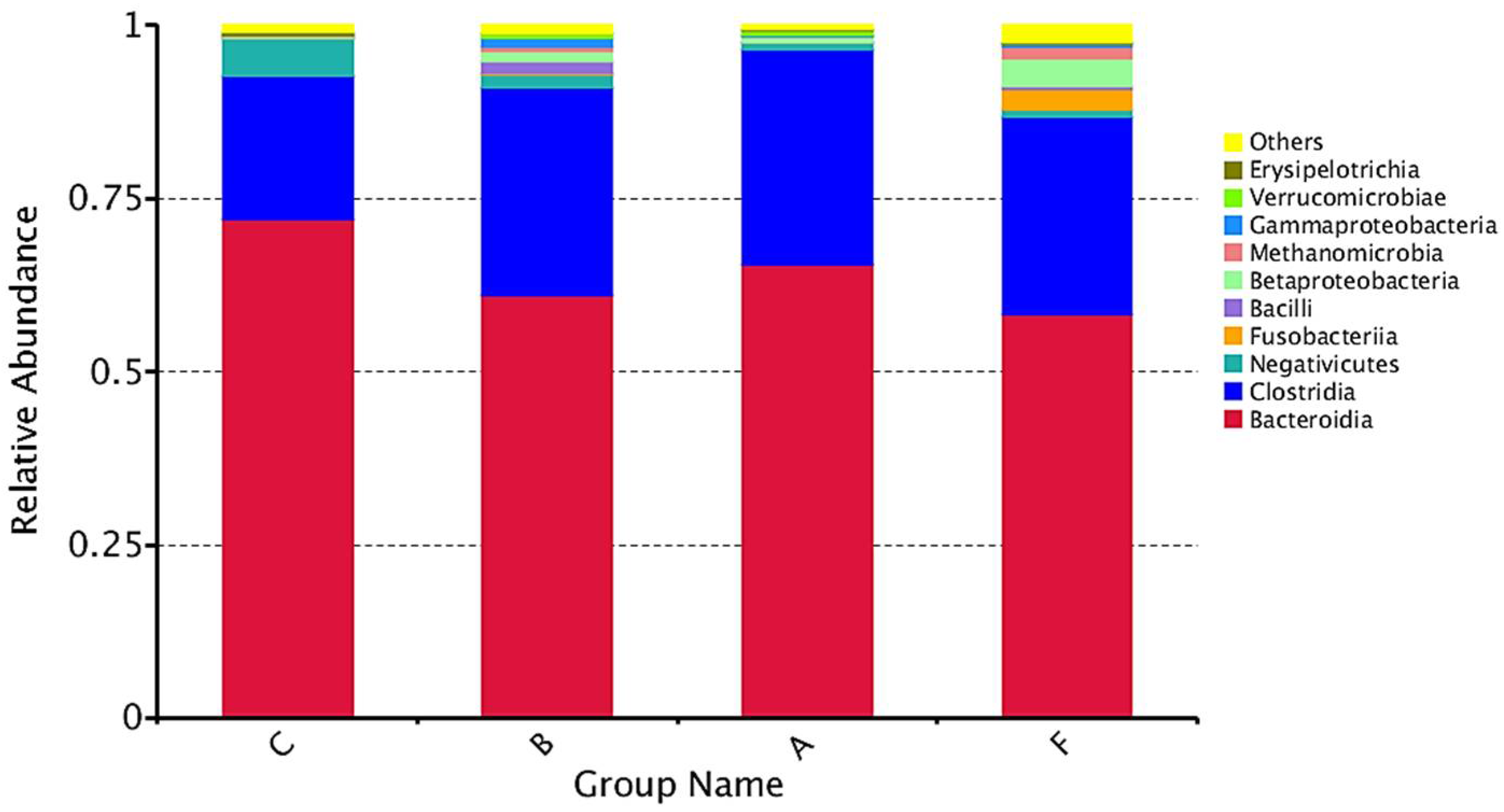
To visually represent the mean gut microbiota composition per group, bar plots were generated to display the 10 most abundant phyla (Figure 3) and the 10 most abundant classes (Figure 4). Phyla or classes with a representation below 1% were grouped under “Others” in both figures.

3.2.1. Phylum-Level Analysis

Figure 3 shows that the most abundant phylum among all study groups was Bacteroidetes, followed by Firmicutes. Although quantitative and qualitative differences were not pronounced between groups, the ASD and FXS + ASD groups exhibited similar abundances of Bacteroidetes and Proteobacteria. The FXS + ASD group showed higher levels of Fusobacteria. The control group showed a greater relative abundance of Bacteroidetes and lower Firmicutes abundance compared to the patient groups.

3.2.2. Class-Level Analysis

Figure 4 displays class-level microbiota distribution. The classes Bacteroidia and Clostridia were primarily responsible for the abundance of Bacteroidetes and Firmicutes, respectively. The FXS group showed greater similarity to the ASD and FXS + ASD groups than to the control group regarding Bacteroidia and Clostridia abundance. Among patient groups, FXS + ASD exhibited the most distinct microbiota’s composition compared to the control group.

3.2.3. Heatmap Analysis

Figure 5 visualizes the abundance of different taxa across the study groups. Colours represent the relative abundance of a given taxon in a specific group. The FXS + ASD group exhibited the highest number of taxa with increased abundance compared to other groups.

3.3. Beta Diversity

To determine the dissimilarity among the microbiota of the different study groups, a Principal Component Analysis (PCA) was performed.
Figure 6 shows the percentage of variance explained by the first two principal components. Together, these two components account for 27.49% of the variability among the study groups. It can be observed that there is considerable overlap between the four groups. Additionally, the FXS + ASD group exhibits high variability among its samples, as indicated by points located far from each other, resulting in an enlarged ellipsoid for this group. Conversely, the FXS group shows little variability among its samples, as all points are located closely together.
To verify if the variation between groups was significantly greater than within-group variation, an ANOSIM analysis was conducted. No significant differences were found between the analysed groups, with a p-value > 0.05.
Taxa showing significant differences (p < 0.05) in abundance when comparing groups pairwise using Student’s t-test are shown in Table 3. Significant taxonomic differences were observed between the following groups: ASD vs. FXS, FXS vs. Control, ASD vs. Control, FXS vs. FXS + ASD, and ASD vs. FXS + ASD. The microbiota of the ASD group exhibits significant differences in a greater number of taxa compared to the control group.
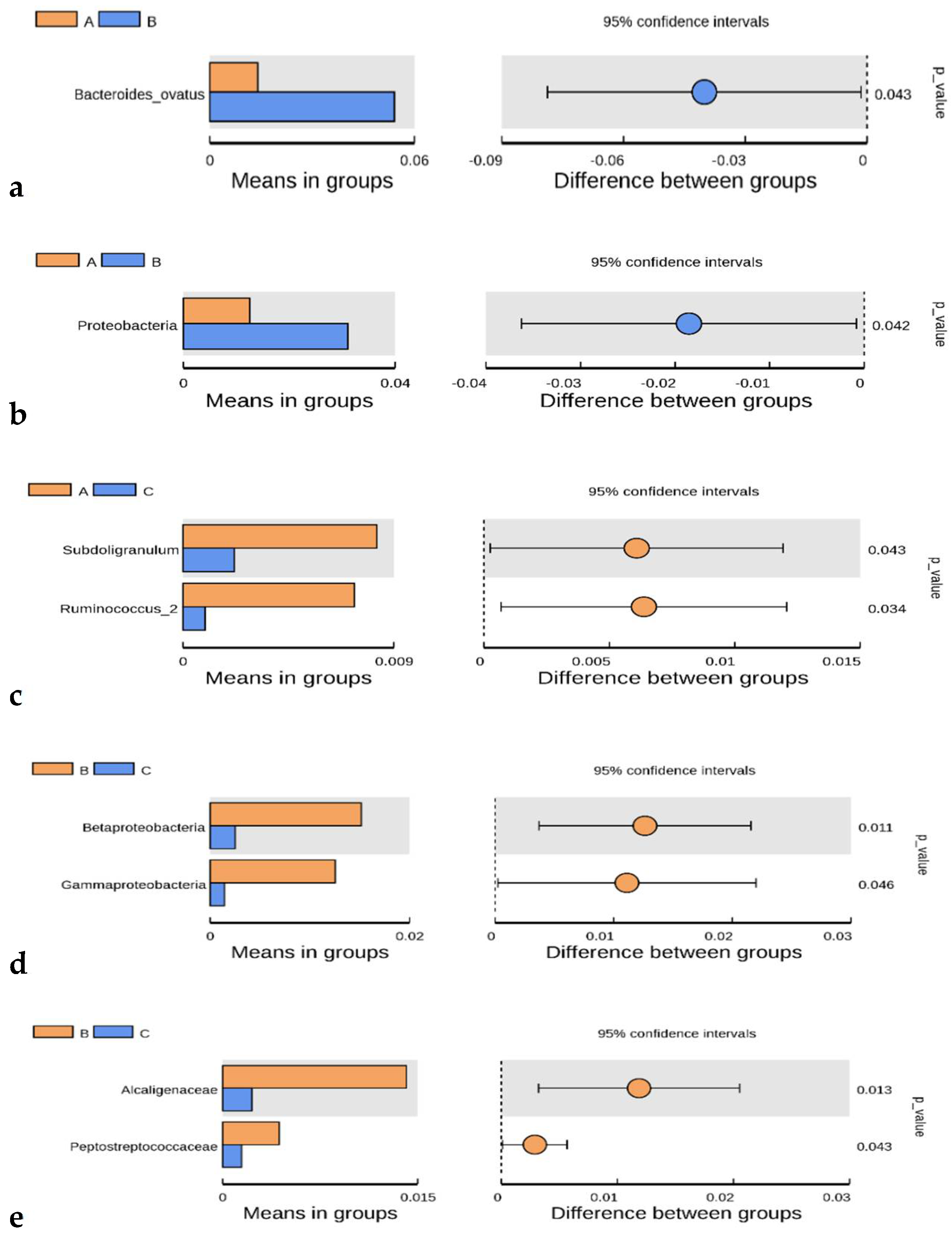
Figure 7 illustrates the taxa with significant differences in average relative abundance between the two study groups. An asterisk (*) indicates the groups with a higher average value for each particular taxon, assessing biomarkers with statistical differences between groups.
The left panel of each graph in Figure 7 shows the abundance of species with significant differences between groups. Each bar represents the mean abundance of each group for the species exhibiting significant differences. The right panel shows the confidence interval of variation between groups. The leftmost side of each circle represents the lower limit of the 95% confidence interval, while the rightmost side represents the upper limit. The centre of the circle represents the difference in mean value. The circle colour corresponds to the group with a higher mean value. The value indicated on the right is the p-value for the comparison between groups, indicating which taxon or taxa are differential and could represent significant biomarkers for classifying populations, even proposing interventions with probiotics/prebiotics to check if microbiota and the characteristic symptomatology of the pathologies could be modified.

3.4. Biomarker Analysis Through Linear Discriminant Analysis (LDA)

Figure 8 presents LDA score histograms obtained from the LEfSe analysis, which were used to evaluate biomarkers with statistically significant differences between groups. Each graph in Figure 8 displays biomarkers or taxa that contribute to the greatest differences in microbiota composition between groups (LDA > 4). These taxa characterize one group compared to another specific group. The FXS microbiota was characterized by a higher abundance of Roseburia inulinivorans (Figure 8a). Both the ASD (B) and FXS + ASD (F) groups shared the same biomarker (Bacteroides ovatus) when compared to the FXS group (A) (Figure 8b,d).

4. Discussion

Advances in sequencing technologies and bioinformatics tools have enabled a detailed characterization of gut microbiota composition and its metabolic functions in humans, providing insights into its crucial role in health. Several factors influence human gut microbiota composition, including diet, physical activity, and antibiotic use [56,57]. A healthy gut microbiota is maintained by a balance favouring commensal microorganisms over pathogens; however, when this balance is disrupted, pathological conditions may arise [31]. It has been demonstrated that the gut microbiota interacts with the brain via the gut–brain axis, and alterations in this axis can contribute to various nervous system disorders, including ASD [42].

4.1. Alpha Diversity

In our alpha diversity analysis, significant differences were observed in diversity and dominance indices between patients (FXS, ASD, and FXS + ASD) and the control group, with ASD patients showing the highest diversity and dominance values compared to controls. These findings contrast with those of a previous study [58], where no significant alpha diversity differences were found between ASD individuals and neurotypical controls.
Regarding the number of OTUs, the highest value was observed in the FXS + ASD group, although with high standard deviation. This result contrasts with [59], who reported a reduced OTU count in ASD microbiota compared to controls. It is essential to consider other variables, such as diet and population differences, that could also influence gut microbiota composition.
The Venn diagram analysis showed that ASD and FXS + ASD groups share the highest number of OTUs. However, the total OTU count in ASD microbiota was the highest, whereas FXS had the lowest number of OTUs. This could be due to variability in OTU counts within each group, which was highest in ASD and lowest in FXS, despite its smaller sample size (6 individuals). The FXS + ASD group, consisting of seven individuals, had a total OTU count similar to that of the control group (nine individuals).

4.2. Microbiota Composition

Across all four groups, Bacteroidetes and Firmicutes were the dominant phyla, consistent with previous studies on human gut microbiota [60]. Bacteroidetes were primarily represented by the class Bacteroidia, whilst Firmicutes were dominated by the class Clostridia. Interestingly, ASD and FXS + ASD microbiota were notably similar in terms of phyla abundance. The control group exhibited a higher Bacteroidetes/Firmicutes ratio than the ASD group [61]. This imbalance has been linked to increased intestinal permeability and a higher prevalence of food intolerances in ASD individuals compared to neurotypical controls [62].
At the class level, FXS microbiota showed greater similarity to ASD and FXS + ASD microbiota than to controls. Some studies have associated increased Firmicutes colonization, particularly the class Clostridia, with autism symptom severity [63].

4.3. Heatmap and Beta Diversity

The heatmap analysis revealed that FXS microbiota (A) was most similar to the control group, whereas FXS + ASD (F) was the most distinct. Despite previous findings that link increased Clostridia abundance to ASD symptomatology, our heatmap showed that FXS individuals had an even higher Clostridia abundance than ASD patients, suggesting its potential role in FXS pathology. Clostridia are known to produce p-cresol in the gut microbiota, a metabolite associated with repetitive behaviours [62].
The FXS + ASD group also showed elevated Clostridia levels, in agreement with previous studies indicating that the presence of ASD exacerbates intestinal dysbiosis in patients with FXS + ASD [63]. Notably, FXS + ASD microbiota had a higher abundance of Epsilonproteobacteria, particularly Campylobacter, which is associated with acute bacterial enteritis [64].
Although FXS and control microbiota showed only minor differences, previous studies have reported gastrointestinal issues such as constipation in FXS individuals, which could influence gut microbiota composition [65]. Additionally, ASD microbiota was characterized by high Bacilli abundance, a class known to produce toxins involved in various diseases [66].
In beta diversity analysis, FXS samples exhibited the least variability, whereas FXS + ASD showed the highest intra-group variability, suggesting that individuals in this group had highly heterogeneous microbiota profiles. The substantial overlap between the four groups explains why ANOSIM analysis did not reveal significant differences in overall microbiota composition between groups.

4.4. Differential Taxa and Biomarkers

Despite no global differences in beta diversity, specific taxa exhibited significant differences between groups, with ASD microbiota showing the most distinct taxonomic profile compared to controls. Notably, ASD microbiota had a higher abundance of Proteobacteria, particularly class Betaproteobacteria, which has also been found in type II diabetes patients and may influence sugar metabolism [67]. This could help explain the poor disaccharide absorption observed in some ASD children [25].
ASD microbiota also had a higher abundance of Proteobacteria than FXS microbiota. Additionally, Subdoligranulum and Ruminococcus were significantly more abundant in FXS than in controls. Subdoligranulum is typically reduced in irritable bowel syndrome patients [68], whereas Ruminococcus has been linked to Crohn’s disease and autism due to its production of a complex polysaccharide that triggers intestinal inflammation [69,70].
Furthermore, Fusicatenibacter abundance was lower in FXS + ASD group compared to ASD and FXS groups, with the most significant difference observed between FXS + ASD and FXS groups. Fusicatenibacter plays a key role in short-chain fatty acid synthesis from carbohydrates [71], suggesting a potential involvement of this altered microbial profile in the symptomatology of FXS + ASD.

4.5. LEfSe Biomarker Analysis

The LEfSe analysis identified specific taxa that were significantly enriched in certain groups, distinguishing them from others.
FXS microbiota was characterized by a higher abundance of Roseburia inulinivorans, a commensal bacterium-producing butyrate, which has a role in energy metabolism and glucose homeostasis [72]. Although Roseburia has been implicated in neurological disorders, its exact role remains unclear [73].
ASD microbiota was distinguished by Proteobacteria (phylum) and Bacteroides ovatus (species), which also differentiated FXS + ASD from FXS. This suggests that B. ovatus is more abundant in ASD and FXS + ASD, potentially contributing to shared symptomatology between these groups.
Other distinctive biomarkers included Bacteroidales S24-7 (FXS + ASD vs. ASD) and Veillonellaceae (higher in controls vs. FXS + ASD). The high Sutterella abundance in FXS + ASD, previously linked to gastrointestinal infections, has also been reported in ASD microbiota [70].

4.6. Future Directions

Given the small sample size, these results should be considered preliminary, requiring confirmation in larger cohorts. If validated in future studies, these findings could lead to probiotic/prebiotic interventions aimed at modulating gut microbiota and alleviating symptoms in affected patients.
Although two participants were 18 years old, we performed a sensitivity analysis excluding them and confirmed that the main results and conclusions remained consistent. The inclusion of these participants, given the rarity of FXS and FXS + ASD, is justified, but we acknowledge this as a limitation regarding age homogeneity.
Another limitation is that stool samples were stored at −20 °C rather than −80 °C immediately after collection, due to home sampling by families. However, this approach is supported by evidence indicating that short-term storage at −20 °C does not significantly impact microbiota analysis results [48].

5. Conclusions

  • The gut microbiota of patients with ASD, FXS, and FXS + ASD exhibits significant differences compared to that of neurotypical control individuals within the studied population.
  • The microbiota of ASD patients is associated with an increased proportion of Firmicutes phylum members compared to the control group, a characteristic that, according to some authors, is linked to alterations in intestinal permeability.
  • An increased abundance of the class Clostridia was observed in all three patient groups compared to controls, with this increase being most pronounced in FXS patients.
  • Specific biomarkers were identified, distinguishing gut microbiota profiles of the patient groups from one another and from the control group.
  • There are notable similarities in bacterial composition and abundance between the ASD and FXS + ASD groups, particularly regarding the species Bacteroides ovatus, which may be related to shared symptomatology.

Author Contributions

All authors had full access to all the data in the study and take responsibility for the integrity of the data and the accuracy of the data analysis. Study concept and design: Y.d.D.-O.; acquisition of data, analysis and interpretation of data: Y.d.D.-O., A.B.-G., R.C.-M., C.Q.-N. and J.M.S.-C.; drafting of the manuscript: Y.d.D.-O., J.M.S.-C. and R.C.-M.; statistical analysis: Y.d.D.-O.; obtained funding: Y.d.D.-O.; technical tasks: A.B.-G.; study supervision: Y.d.D.-O. and C.Q.-N. All authors have read and agreed to the published version of the manuscript.

Funding

This paper was funded by the Andalusian Regional Ministry of Economy and Knowledge [grant no. CTS546 and no. P10-CTS-05704]. This paper was also partially funded by FEDER “Fondos Europeos de Desarrollo Regional”, Fundación Alicia Koplowitz (Madrid).

Institutional Review Board Statement

The study was approved by the local ethics committee of Málaga Regional University Hospital and conduced in accordance with European Union recommendations (protocol code 2010/63/EU and date of approval: 26 July 2018).

Informed Consent Statement

Informed consent was obtained from all subjects involved in the study.

Data Availability Statement

All data will be shared upon request.

Acknowledgments

The authors thank D.W.E. Ramsden for the revising the English-language content of this paper.

Conflicts of Interest

The authors declare no conflicts of interest.

Abbreviations

The following abbreviations are used in this manuscript:
ASDAutism spectrum disorder
FXSFragile X Syndrome
DNADeoxyribonucleic acid
RRNARibosomal ribonucleic acid
MetaHITHuman Intestinal Tract Metagenomics
HMPHuman Microbiome Project
PCRPolymerase Chain Reaction
CNSCentral nervous system
ANSAutonomous Nervous System
HPAHypothalamic–Pituitary–Adrenal Axis
ENSEnteric nervous system
OTUOperative Taxonomic Unit
FLASHQuick adjustment of the length of short reads to improve genome assemblies
ACPPrincipal Component Analysis
ANOSIMAnalysis of Similarities
LDALinear Discriminant Analysis
LEfSeLinear Discriminant Analysis Effect Size

References

  1. Salcedo-Arellano, M.J.; Cabal-Herrera, A.M.; Punatar, R.H.; Clark, C.J.; Romney, C.A.; Hagerman, R.J. Overlapping molecular pathways leading to autism spectrum disorders, Fragile X syndrome, and targeted treatments. Neurotherapeutics 2021, 18, 265–283. [Google Scholar] [CrossRef]
  2. Riley, C.; Wheeler, A. Assessing the Fragile X Syndrome Newborn Screening Landscape. Pediatrics 2017, 139 (Suppl. S3), S207–S215. [Google Scholar] [CrossRef]
  3. Harris, S.W.; Hessl, D.; Goodlin-Jones, B.; Ferranti, J.; Bacalman, S.; Barbato, I.; Tassone, F.; Hagerman, P.J.; Herman, K.; Hagerman, R.J. Autism profiles of males with fragile X syndrome. Am. J. Ment. Retard. 2008, 113, 427–438. [Google Scholar] [CrossRef] [PubMed]
  4. Kraan, C.M.; Godler, D.E.; Amor, D.J. Epigenetics of fragile X syndrome and fragile X-related disorders. Dev. Med. Child Neurol. 2019, 61, 121–127. [Google Scholar] [CrossRef] [PubMed]
  5. Ribate Molina, M.P.; Pié Juste, J.; Ramos Fuentes, F.J. Síndrome de X Frágil. Protoc. Diagn. Ter. Pediatr. 2010, 1, 85–90. [Google Scholar]
  6. Kim, K.; Hessl, D.; Randol, J.L.; Espinal, G.M.; Schneider, A.; Protic, D.; Aydin, E.Y.; Hagerman, R.J.; Hagerman, P.J. Association between IQ and FMR1 protein (FMRP) across the spectrum of CGG repeat expansions. PLoS ONE 2019, 14, e0226811. [Google Scholar] [CrossRef]
  7. Suhl, J.A.; Chopra, P.; Anderson, B.R.; Bassell, G.J.; Warren, S.T. Analysis of FMRP mRNA target datasets reveals highly associated mRNAs mediated by G-quadruplex structures formed via clustered WGGA sequences. Hum. Mol. Genet. 2014, 23, 5479–5491. [Google Scholar] [CrossRef]
  8. Darnell, J.C.; Van Driesche, S.J.; Zhang, C.; Hung, K.Y.S.; Mele, A.; Fraser, C.E.; Stone, E.F.; Chen, C.; Fak, J.J.; Chi, S.W.; et al. FMRP stalls ribosomal translocation on mRNAs linked to synaptic function and autism. Cell 2011, 146, 247–261. [Google Scholar] [CrossRef] [PubMed]
  9. Castagnola, S.; Delhaye, S.; Folci, A.; Paquet, A.; Brau, F.; Duprat, F.; Jarjat, M.; Grossi, M.; Béal, M.; Martin, S.; et al. New insights into the role of Cav2 protein family in calcium flux deregulation in Fmr1-KO neurons. Front. Mol. Neurosci. 2018, 11, 342. [Google Scholar] [CrossRef]
  10. El Bekay, R.; Romero-Zerbo, Y.; Decara, J.; Sánchez-Salido, L.; Del Arco-Herrera, I.; Rodríguez-de Fonseca, F.; De Diego-Otero, Y. Enhanced markers of oxidative stress, altered antioxidants and NADPH-oxidase activation in brains from Fragile X Mental Retardation 1-deficient mice, a pathological model for Fragile X syndrome. Eur. J. Neurosci. 2007, 26, 3169–3180. [Google Scholar] [CrossRef]
  11. De Diego-Otero, Y.; El Bekay, R.; García-Guirado, F.; Sánchez-Salido, L.; Giráldez-Pérez, R.M. Apocynin, a selective NADPH oxidase (Nox2) inhibitor, ameliorates behavioural and learning deficits in the Fragile X syndrome mouse model. Biomedicines 2024, 12, 2887. [Google Scholar] [CrossRef]
  12. Kau, A.S.M.; Tierney, E.; Bukelis, I.; Stump, M.H.; Kates, W.R.; Trescher, W.H.; Kaufmann, W.E. Social behavior profile in young males with fragile X syndrome: Characteristics and specificity. Am. J. Med. Genet. A 2004, 126, 9–17. [Google Scholar] [CrossRef]
  13. Aishworiya, R.; Chi, M.-H.; Zafarullah, M.; Mendoza, G.; Ponzini, M.D.; Kim, K.; Biag, H.M.B.; Thurman, A.J.; Abbeduto, L.; Hessl, D.; et al. Intercorrelation of molecular biomarkers and clinical phenotype measures in Fragile X syndrome. Cells 2023, 12, 1920. [Google Scholar] [CrossRef] [PubMed]
  14. Fatemi, S.H.; Folsom, T.D. The role of Fragile X Mental Retardation Protein in major mental disorders. Neuropharmacology 2011, 60, 1221–1226. [Google Scholar] [CrossRef] [PubMed]
  15. Reddy, K.S. Cytogenetic abnormalities and fragile-X syndrome in autism spectrum disorder. BMC Med. Genet. 2005, 6, 3. [Google Scholar] [CrossRef]
  16. Altimiras, F.; Garcia, J.A.; Palacios-García, I.; Hurley, M.J.; Deacon, R.; González, B.; Cogram, P. Altered gut microbiota in a Fragile X syndrome mouse model. Front. Neurosci. 2021, 15, 653120. [Google Scholar] [CrossRef] [PubMed]
  17. Salmerón, A.M.; Pérez-Fernández, C.; Abreu, A.C.; Fernández, S.; Tristán, A.I.; Ruiz-Sobremazas, D.; Cabré, M.; Guardia-Escote, L.; Fernández, I.; Sánchez-Santed, F. Exploring microbiota-gut-brain axis biomarkers linked to autism spectrum disorder in prenatally chlorpyrifos-exposed Fmr1 knock-out and wild-type male rats. Toxicology 2024, 506, 153871. [Google Scholar] [CrossRef]
  18. Fattorusso, A.; Di Genova, L.; Dell’Isola, G.B.; Mencaroni, E.; Esposito, S. Autism spectrum disorders and the gut microbiota. Nutrients 2019, 11, 521. [Google Scholar] [CrossRef]
  19. Srikantha, P.; Mohajeri, M.H. The possible role of the microbiota-gut-brain axis in autism spectrum disorder. Int. J. Mol. Sci. 2019, 20, 2115. [Google Scholar] [CrossRef]
  20. Bougeard, C.; Picarel-Blanchot, F.; Schmid, R.; Campbell, R.; Buitelaar, J. Prevalence of autism spectrum disorder and co-morbidities in children and adolescents: A systematic literature review. Front. Psychiatry 2021, 12, 744709. [Google Scholar] [CrossRef]
  21. Shaw, K.A.; Williams, S.; Patrick, M.E.; Valencia-Prado, M.; Durkin, M.S.; Howerton, E.M.; Ladd-Acosta, C.M.; Pas, E.T.; Bakian, A.V.; Bartholomew, P.; et al. Prevalence and Early Identification of Autism Spectrum Disorder Among Children Aged 4 and 8 Years—Autism and Developmental Disabilities Monitoring Network, 16 Sites, United States, 2022. MMWR Surveill. Summ. 2025, 74, 1–22. [Google Scholar] [CrossRef]
  22. Theoharides, T.C.; Kavalioti, M.; Martinotti, R. Factors adversely influencing neurodevelopment. J. Biol. Regul. Homeost. Agents 2019, 33, 1663–1667. [Google Scholar] [CrossRef]
  23. Yang, Y.; Tian, J.; Yang, B. Targeting gut microbiome: A novel and potential therapy for autism. Life Sci. 2018, 194, 111–119. [Google Scholar] [CrossRef]
  24. Li, J.; Jia, H.; Cai, X.; Zhong, H.; Feng, Q.; Sunagawa, S.; Arumugam, M.; Kultima, J.R.; Prifti, E.; Nielsen, T.; et al. An integrated catalog of reference genes in the human gut microbiome. Nat. Biotechnol. 2014, 32, 834–841. [Google Scholar] [CrossRef]
  25. Kang, D.-W.; Adams, J.B.; Coleman, D.M.; Pollard, E.L.; Maldonado, J.; McDonough-Means, S.; Caporaso, J.G.; Krajmalnik-Brown, R. Long-term benefit of microbiota transfer therapy on autism symptoms and gut microbiota. Sci. Rep. 2019, 9, 5821. [Google Scholar] [CrossRef] [PubMed]
  26. Naviaux, R.K. Metabolic features of the cell danger response. Mitochondrion 2014, 16, 7–17. [Google Scholar] [CrossRef]
  27. van De Sande, M.M.H.; van Buul, V.J.; Brouns, F.J.P.H. Autism and nutrition: The role of the gut–brain axis. Nutr. Res. Rev. 2014, 27, 199–214. [Google Scholar] [CrossRef] [PubMed]
  28. Fond, G.; Chevalier, G.; Eberl, G.; Leboyer, M. The potential role of microbiota in major psychiatric disorders: Mechanisms, preclinical data, gastro-intestinal comorbidities and therapeutic options. Presse Med. 2016, 45, 7–19. [Google Scholar] [CrossRef]
  29. Groer, M.W.; Gregory, K.E.; Louis-Jacques, A.; Thibeau, S.; Walker, W.A. The very low birth weight infant microbiome and childhood health. Birth Defects Res. Part C Embryo Today Rev. 2015, 105, 252–264. [Google Scholar] [CrossRef] [PubMed]
  30. Knight, R.; Callewaert, C.; Marotz, C.; Hyde, E.R.; Debelius, J.W.; McDonald, D.; Sogin, M.L. The microbiome and human biology. Annu. Rev. Genom. Hum. Genet. 2017, 18, 65–86. [Google Scholar] [CrossRef]
  31. Adamczak, A.M.; Werblińska, A.; Jamka, M.; Walkowiak, J. Maternal-Foetal/Infant Interactions-Gut Microbiota and Immune Health. Biomedicines 2024, 12, 490. [Google Scholar] [CrossRef]
  32. Thum, C.; Cookson, A.L.; Otter, D.E.; McNabb, W.C.; Hodgkinson, A.J.; Dyer, J.; Roy, N.C. Can nutritional modulation of maternal intestinal microbiota influence the development of the infant gastrointestinal tract? J. Nutr. 2012, 142, 1921–1928. [Google Scholar] [CrossRef]
  33. Theis, K.R.; Romero, R.; Winters, A.D.; Greenberg, J.M.; Gomez-Lopez, N.; Alhousseini, A.; Bieda, J.; Maymon, E.; Pacora, P.; Fettweis, J.M.; et al. Does the human placenta delivered at term have a microbiota? Results of cultivation, quantitative real-time PCR, 16S rRNA gene sequencing, and metagenomics. Am. J. Obstet. Gynecol. 2019, 220, 267.e1–267.e39. [Google Scholar] [CrossRef]
  34. Aagaard, K.; Ma, J.; Antony, K.M.; Ganu, R.; Petrosino, J.; Versalovic, J. The placenta harbors a unique microbiome. Sci. Transl. Med. 2014, 6, 237ra65. [Google Scholar] [CrossRef] [PubMed]
  35. Pantazi, A.C.; Balasa, A.L.; Mihai, C.M.; Chisnoiu, T.; Lupu, V.V.; Kassim, M.A.K.; Mihai, L.; Frecus, C.E.; Chirila, S.I.; Lupu, A.; et al. Development of Gut Microbiota in the First 1000 Days after Birth and Potential Interventions. Nutrients 2023, 15, 3647. [Google Scholar] [CrossRef]
  36. Sharon, G.; Cruz, N.J.; Kang, D.-W.; Gandal, M.J.; Wang, B.; Kim, Y.-M.; Zink, E.M.; Casey, C.P.; Taylor, B.C.; Lane, C.J.; et al. Human gut microbiota from autism spectrum disorder promote behavioural symptoms in mice. Cell 2019, 177, 1600–1618.e17. [Google Scholar] [CrossRef] [PubMed]
  37. Cryan, J.F.; O’Riordan, K.J.; Cowan, C.S.M.; Sandhu, K.V.; Bastiaanssen, T.F.S.; Boehme, M.; Codagnone, M.G.; Cussotto, S.; Fulling, C.; Golubeva, A.V.; et al. The microbiota–gut–brain axis. Lancet Neurol. 2020, 19, 179–194. [Google Scholar] [CrossRef] [PubMed]
  38. Peralta-Marzal, L.N.; Rojas-Velazquez, D.; Rigters, D.; Prince, N.; Garssen, J.; Kraneveld, A.D.; Perez-Pardo, P.; Lopez-Rincon, A. A robust microbiome signature for autism spectrum disorder across different studies using machine learning. Sci. Rep. 2024, 14, 814. [Google Scholar] [CrossRef]
  39. Robles Alonso, V.; Guarner, F. Linking the gut microbiota to human health. Br. J. Nutr. 2013, 109, S21–S26. [Google Scholar] [CrossRef]
  40. Foster, J.A.; McVey Neufeld, K.A. Gut–brain axis: How the microbiome influences anxiety and depression. Trends Neurosci. 2013, 36, 305–312. [Google Scholar] [CrossRef]
  41. Roubalová, R.; Procházková, P.; Papežová, H.; Smitka, K.; Bilej, M.; Tlaskalová-Hogenová, H. Anorexia nervosa: Gut microbiota–immune–brain interactions. Clin. Nutr. 2019, 39, 676–684. [Google Scholar] [CrossRef] [PubMed]
  42. Huang, T.T.; Lai, J.B.; Du, Y.L.; Xu, Y.; Ruan, L.M.; Hu, S.H. Current understanding of gut microbiota in mood disorders: An update of human studies. Front. Genet. 2019, 10, 98. [Google Scholar] [CrossRef] [PubMed]
  43. Ma, Q.; Xing, C.; Long, W.; Wang, H.Y.; Liu, Q.; Wang, R.F. Impact of microbiota on central nervous system and neurological diseases: The gut-brain axis. J. Neuroinflamm. 2019, 16, 53. [Google Scholar] [CrossRef]
  44. Li, Y.; Hao, Y.; Fan, F.; Zhang, B. The role of microbiome in insomnia, circadian disturbance and depression. Front. Psychiatry 2018, 9, 669. [Google Scholar] [CrossRef]
  45. Möhle, L.; Mattei, D.; Heimesaat, M.M.; Bereswill, S.; Fischer, A.; Alutis, M.; French, T.; Hambardzumyan, D.; Matzinger, P.; Dunay, I.R.; et al. Ly6Chi monocytes provide a link between antibiotic-induced changes in gut microbiota and adult hippocampal neurogenesis. Cell Rep. 2016, 15, 1945–1956. [Google Scholar] [CrossRef]
  46. Hill, C.J.; Lynch, D.B.; Murphy, K.; Ulaszewska, M.; Jeffery, I.B.; O’Shea, C.A.; Watkins, C.; Dempsey, E.; Mattivi, F.; Tuohy, K.; et al. Evolution of gut microbiota composition from birth to 24 weeks in the INFANTMET Cohort. Microbiome 2017, 5, 4. [Google Scholar] [CrossRef]
  47. Loh, J.S.; Mak, W.Q.; Tan, L.K.S.; Ng, C.X.; Chan, H.H.; Yeow, S.H.; Foo, J.B.; Ong, Y.S.; How, C.W.; Khaw, K.Y. Microbiota–gut–brain axis and its therapeutic applications in neurodegenerative diseases. Signal Transduct. Target. Ther. 2024, 9, 37. [Google Scholar] [CrossRef]
  48. Stewart, C.J.; Ajami, N.J.; O’Brien, J.L.; Hutchinson, D.S.; Smith, D.P.; Wong, M.C.; Ross, M.C.; Lloyd, R.E.; Doddapaneni, H.; Metcalf, G.A.; et al. Temporal development of the gut microbiome in early childhood from the TEDDY study. Nature 2018, 562, 583–588. [Google Scholar] [CrossRef]
  49. Chao, A.; Shen, T.J. Nonparametric estimation of Shannon’s index of diversity when there are unseen species in sample. Environ. Ecol. Stat. 2003, 10, 429–443. [Google Scholar] [CrossRef]
  50. Simpson, E.H. Measurement of diversity. Nature 1949, 163, 688. [Google Scholar] [CrossRef]
  51. Chao, A.; Hwang, W.-H.; Chen, Y.-C.; Kuo, C.-Y. Estimating the number of shared species in two communities. Stat. Sin. 2000, 10, 227–246. [Google Scholar]
  52. Wilcoxon, F. Individual comparisons of grouped data by ranking methods. J. Econ. Entomol. 1946, 39, 269–270. [Google Scholar] [CrossRef]
  53. Odamaki, T.; Kato, K.; Sugahara, H.; Hashikura, N.; Takahashi, S.; Xiao, J.Z.; Abe, F.; Osawa, R. Age-related changes in gut microbiota composition from newborn to centenarian: A cross-sectional study. BMC Microbiol. 2016, 16, 90. [Google Scholar] [CrossRef]
  54. Fagerland, M.W. T-tests, non-parametric tests, and large studies—A paradox of statistical practice? BMC Med. Res. Methodol. 2012, 12, 78. [Google Scholar] [CrossRef]
  55. Segata, N.; Izard, J.; Waldron, L.; Gevers, D.; Miropolsky, L.; Garrett, W.S.; Huttenhower, C. Metagenomic biomarker discovery and explanation. Genome Biol. 2011, 12, R60. [Google Scholar] [CrossRef]
  56. De Lucas Moreno, B.; González Soltero, R.; Bressa, C.; Bailén, M.; Larrosa, M. Lifestyle modulation of gut microbiota. Nutr. Hosp. 2019, 36, 35–39. [Google Scholar] [CrossRef]
  57. Strati, F.; Cavalieri, D.; Albanese, D.; De Felice, C.; Donati, C.; Hayek, J.; Jousson, O.; Leoncini, S.; Renzi, D.; Calabrò, A.; et al. New evidences on the altered gut microbiota in autism spectrum disorders. Microbiome 2017, 5, 24. [Google Scholar] [CrossRef] [PubMed]
  58. Hughes, H.K.; Rose, D.; Ashwood, P. The gut microbiota and dysbiosis in autism spectrum disorders. Curr. Neurol. Neurosci. Rep. 2018, 18, 81. [Google Scholar] [CrossRef]
  59. Tomova, A.; Husarova, V.; Lakatosova, S.; Bakos, J.; Vlkova, B.; Babinska, K.; Ostatnikova, D. Gastrointestinal microbiota in children with autism in Slovakia. Physiol. Behav. 2015, 138, 179–187. [Google Scholar] [CrossRef] [PubMed]
  60. Narzisi, A.; Masi, G.; Grossi, E. Nutrition and Autism Spectrum Disorder: Between False Myths and Real Research-Based Opportunities. Nutrients 2021, 13, 2068. [Google Scholar] [CrossRef]
  61. Argou-Cardozo, I.; Zeidán-Chuliá, F. Clostridium bacteria and autism spectrum conditions: A systematic review and hypothetical contribution of environmental glyphosate levels. Med. Sci. 2018, 6, 29. [Google Scholar] [CrossRef]
  62. Gabriele, S.; Sacco, R.; Cerullo, S.; Neri, C.; Urbani, A.; Tripi, G.; Malvy, J.; Barthelemy, C.; Bonnet-Brihault, F.; Persico, A.M. Urinary p-cresol is elevated in young French children with autism spectrum disorder: A replication study. Biomarkers 2014, 19, 463–470. [Google Scholar] [CrossRef]
  63. Protic, D.D.; Aishworiya, R.; Salcedo-Arellano, M.J.; Tang, S.J.; Milisavljevic, J.; Mitrovic, F.; Hagerman, R.J.; Budimirovic, D.B. Fragile X syndrome: From molecular aspect to clinical treatment. Int. J. Mol. Sci. 2022, 23, 1935. [Google Scholar] [CrossRef] [PubMed]
  64. Yarza, P.; Yilmaz, P.; Pruesse, E.; Glöckner, F.O.; Ludwig, W.; Schleifer, K.H.; Whitman, W.B.; Euzéby, J.; Amann, R.; Rosselló-Móra, R. Uniting the classification of cultured and uncultured bacteria and archaea using 16S rRNA gene sequences. Nat. Rev. Microbiol. 2014, 12, 635–645. [Google Scholar] [CrossRef] [PubMed]
  65. Barnhill, K.; Devlin, M.; Moreno, H.T.; Potts, A.; Richardson, W.; Schutte, C.; Hewitson, L. Brief report: Implementation of a specific carbohydrate diet for a child with autism spectrum disorder and Fragile X syndrome. J. Autism Dev. Disord. 2020, 50, 1800–1808. [Google Scholar] [CrossRef]
  66. Mallozzi, M.; Viswanathan, V.K.; Vedantam, G. Spore-forming bacilli and clostridia in human disease. Future Microbiol. 2010, 5, 1109–1123. [Google Scholar] [CrossRef]
  67. Larsen, N.; Vogensen, F.K.; van den Berg, F.W.J.; Nielsen, D.S.; Andreasen, A.S.; Pedersen, B.K.; Al-Soud, W.A.; Sørensen, S.J.; Hansen, L.H.; Jakobsen, M. Gut microbiota in human adults with type 2 diabetes differs from non-diabetic adults. PLoS ONE 2010, 5, e9085. [Google Scholar] [CrossRef] [PubMed]
  68. Valdovinos-Díaz, M.A. Intestinal microbiota in digestive disorders: Probiotics, prebiotics and symbiotics. Rev. Gastroenterol. México 2013, 78, 25–27. [Google Scholar] [CrossRef]
  69. Henke, M.T.; Kenny, D.J.; Cassilly, C.D.; Vlamakis, H.; Xavier, R.J.; Clardy, J. Ruminococcus gnavus, a member of the human gut microbiome associated with Crohn’s disease, produces an inflammatory polysaccharide. Proc. Natl. Acad. Sci. USA 2019, 116, 12672–12677. [Google Scholar] [CrossRef]
  70. Wang, L.; Christophersen, C.T.; Sorich, M.J.; Gerber, J.P.; Angley, M.T.; Conlon, M.A. Increased abundance of Sutterella spp. and Ruminococcus torques in feces of children with autism spectrum disorder. Mol. Autism 2013, 4, 42. [Google Scholar] [CrossRef]
  71. Kashtanova, D.A.; Tkacheva, O.N.; Doudinskaya, E.N.; Strazhesko, I.D.; Kotovskaya, Y.V.; Popenko, A.S.; Tyakht, A.V.; Alexeev, D.G. Gut microbiota in patients with different metabolic statuses: Moscow study. Microorganisms 2018, 6, 98. [Google Scholar] [CrossRef] [PubMed]
  72. Wijdeveld, M.; Nieuwdorp, M.; Ijzerman, R.G. The interaction between microbiome and host central nervous system: The gut-brain axis as a potential new therapeutic target in the treatment of obesity and cardiometabolic disease. Expert Opin. Ther. Targets 2020, 24, 639–653. [Google Scholar] [CrossRef] [PubMed]
  73. Tamanai-Shacoori, Z.; Smida, I.; Bousarghin, L.; Loreal, O.; Meuric, V.; Fong, S.B.; Bonnaure-Mallet, M.; Jolivet-Gougeon, A. Roseburia spp.: A marker of health? Future Microbiol. 2017, 12, 157–170. [Google Scholar] [CrossRef] [PubMed]
Figure 1. Workflow implemented by Novogene upon receiving the previously processed DNA samples from the IBIMA laboratory for 16S rRNA gene sequencing. Each step includes quality control checks of both the sample and the generated data.
Psychiatryint 06 00107 g001
Figure 2. Venn diagram of OTU distribution. It illustrates the number of OTUs present in each study group, the unique OTUs per group, and the OTUs shared between two or more groups. Control individuals (C) are in purple. FXS individuals (A) are in blue. ASD individuals (B) are in green. FXS + ASD individuals (F) are in Magenta.
Psychiatryint 06 00107 g002
Figure 3. Relative abundance of the 10 most representative phyla. Compared groups: Control (C), FXS (A), ASD (B), and FXS + ASD (F).
Psychiatryint 06 00107 g003
Figure 4. Relative abundance of the 10 most representative classes. Compared groups: Control (C), FXS (A), ASD (B), and FXS + ASD (F).
Psychiatryint 06 00107 g004
Figure 5. Heatmap of representative gut microbiota classes. Compared groups: Control (C), FXS (A), ASD (B), and FXS + ASD (F).
Psychiatryint 06 00107 g005
Figure 6. Principal Component Analysis (PCA). Each point represents a sample. Each component comprises a set of variables independent from each other. The study groups are Control (C, red), FXS (A, blue), ASD (B, green), and FXS + ASD (F, turquoise).
Psychiatryint 06 00107 g006
Figure 7. Representation of abundance values indicating species with differential values. Differential results are shown by comparing sample groups. The group comparisons are as follows: (a,b) FXS vs. ASD (A vs. B); (c) Control vs. FXS (C vs. A); (dh) ASD vs. Control (B vs. C); (i) FXS + ASD vs. FXS (F vs. A); and (j,k) FXS + ASD vs. ASD (F vs. B).
Psychiatryint 06 00107 g007aPsychiatryint 06 00107 g007b
Figure 8. LDA score histograms from LEfSe analysis. The LDA score histogram represents taxa whose abundance shows significant differences between groups. The letter preceding the taxon name indicates its taxonomic rank, where p = phylum, c = class, o = order, f = family, g = genus, and s = species. The selection criterion is an LDA score above the established threshold (LDA > 4, default value). The LDA score represents effect size, indicating the extent to which a biomarker differentiates between groups. The comparison groups for LEfSe analysis are as follows: (a) FXS vs. Control (A vs. C); (b) ASD vs. FXS (B vs. A); (c) ASD vs. Control (B vs. C); (d) FXS + ASD vs. FXS (F vs. A); (e) FXS + ASD vs. ASD (F vs. B); (f) Control vs. FXS + ASD (C vs. F).
Psychiatryint 06 00107 g008
Table 1. Information on study participants.
CodeGroupAge (Years)GenderPhenotype
BTSXF20CC10MaleControl
BTC23C8FemaleControl
BTSXF16CC10MaleControl
BTC004IC4MaleControl
BTC07C10MaleControl
BTC10C5MaleControl
BTC02I2C8FemaleControl
BTSXF02CC3MaleControl
BTF3G02ContC8MaleControl
BTSXF19A6MaleFXS
BTSXF17A6MaleFXS
BTSXF15A6MaleFXS
BTSXF13A18MaleFXS
BTSXF01A3MaleFXS
BTSXF12A6FemaleFXS
BT023IntB6MaleASD
BT021IntB6MaleASD
BT047B3MaleASD
BT027IntB6MaleASD
BTH02Int2B8MaleASD
BT046B4MaleASD
BT061B5MaleASD
BT004IB4MaleASD
BT012IB3MaleASD
BTSXF18F8MaleFXS + ASD
TRA152INT2F18MaleFXS + ASD
BTSXF05IF4MaleFXS + ASD
BTSXF04F3MaleFXS + ASD
BTSXF9F3FemaleFXS + ASD
BTSXF10F5MaleFXS + ASD
BTSXF11F3MaleFXS + ASD
Table 2. Alpha diversity of gut microbiota in control individuals and patients with ASD, FXS, and FXS + ASD.s.
GroupOTU #ShannonSimpsonCHAO1Good’s Coverage (%)
Control418.89 ± 120.043.85 ± 0.880.81 ± 0.14478.39 ± 140.3399.99 ± 0.00
FXS424.00 ± 88.764.12 ± 0.860.90 ± 0.06 *473.25 ± 106.4199.99 ± 0.00
ASD460.70 ± 172.374.98 ± 0.57 **0.92 ± 0.03 **507.22 ± 184.3499.99 ± 0.00
FXS + ASD468.71 ± 168.964.70 ± 1.29 *0.89 ± 0.11 **524.28 ± 162.4099.99 ± 0.00
(# Number of very similar sequences/diversity index. * values represent mean ± SD. Asterisks indicate significant differences compared to the control group: p < 0.05; ** p < 0.01).
Table 3. Taxa showing significant differences in average relative abundance between two study groups, analysed by Student’s t-test (p < 0.05). An asterisk (*) indicates the groups with a significantly higher mean value for each taxon.
GroupsTaxonp-Value
ASD * vs. FXSBacteroides ovatus (Species)0.043
ASD * vs. FXSProteobacteria (Phylum)0.042
FXS * vs. ControlSubdoligranulum (Genus)0.043
FXS * vs. ControlRuminococcus (Genus)0.034
ASD * vs. ControlBetaproteobacteria (Class)0.011
ASD * vs. ControlGammaproteobacteria (Class)0.046
ASD * vs. ControlAlcaligenaceae (Family)0.013
ASD * vs. ControlPeptostreptococcaceae (Family)0.043
ASD * vs. ControlSubdoligranulum (Genus)0.049
ASD * vs. ControlIntestinibacter (Genus)0.038
ASD * vs. ControlBurkholderiales (Order)0.011
ASD * vs. ControlProteobacteria (Phylum)0.020
FXS * vs. FXS + ASDFusicatenibacter (Genus)0.010
ASD * vs. FXS + ASDPeptostreptococcaceae (Family)0.046
ASD * vs. FXS + ASDFusicatenibacter (Genus)0.046
Disclaimer/Publisher’s Note: The statements, opinions and data contained in all publications are solely those of the individual author(s) and contributor(s) and not of MDPI and/or the editor(s). MDPI and/or the editor(s) disclaim responsibility for any injury to people or property resulting from any ideas, methods, instructions or products referred to in the content.